thrombin-induced contraction in alveolar epithelial cells probed by traction microscopy
TRANSCRIPT

doi: 10.1152/japplphysiol.00185.2006101:512-520, 2006. First published 4 May 2006;J Appl Physiol
Daniel NavajasNúria Gavara, Raimon Sunyer, Pere Roca-Cusachs, Ramon Farré, Mar Rotger andprobed by traction microscopyThrombin-induced contraction in alveolar epithelial cells
You might find this additional info useful...
33 articles, 21 of which you can access for free at: This article citeshttp://jap.physiology.org/content/101/2/512.full#ref-list-1
2 other HighWire-hosted articles: This article has been cited by http://jap.physiology.org/content/101/2/512#cited-by
including high resolution figures, can be found at: Updated information and serviceshttp://jap.physiology.org/content/101/2/512.full
can be found at: Journal of Applied Physiology about Additional material and informationhttp://www.the-aps.org/publications/jappl
This information is current as of October 24, 2012.
http://www.the-aps.org/. © 2006 the American Physiological Society. ISSN: 8750-7587, ESSN: 1522-1601. Visit our website at year (monthly) by the American Physiological Society, 9650 Rockville Pike, Bethesda MD 20814-3991. Copyrightphysiology, especially those papers emphasizing adaptive and integrative mechanisms. It is published 12 times a
publishes original papers that deal with diverse area of research in appliedJournal of Applied Physiology
at DF
G on O
ctober 24, 2012http://jap.physiology.org/
Dow
nloaded from
Thrombin-induced contraction in alveolar epithelial cells probedby traction microscopy
Nuria Gavara, Raimon Sunyer, Pere Roca-Cusachs, Ramon Farre, Mar Rotger, and Daniel NavajasUnitat de Biofısica i Bioenginyeria, Facultat de Medicina, Universitat de Barcelona-Institut d’Investigacions Biomediques August Pi Sunyer, Barcelona, Spain
Submitted 13 February 2006; accepted in final form 24 April 2006
Gavara, Nuria, Raimon Sunyer, Pere Roca-Cusachs, RamonFarre, Mar Rotger, and Daniel Navajas. Thrombin-induced con-traction in alveolar epithelial cells probed by traction microscopy.J Appl Physiol 101: 512–520, 2006. First published May 4, 2006;doi:10.1152/japplphysiol.00185.2006.—Contractile tension of alveo-lar epithelial cells plays a major role in the force balance that regulatesthe structural integrity of the alveolar barrier. The aim of this workwas to study thrombin-induced contractile forces of alveolar epithelialcells. A549 alveolar epithelial cells were challenged with thrombin,and time course of contractile forces was measured by tractionmicroscopy. The cells exhibited basal contraction with total forcemagnitude 55.0 � 12.0 nN (mean � SE, n � 12). Traction forceswere exerted predominantly at the cell periphery and pointed to thecell center. Thrombin (1 U/ml) induced a fast and sustained 2.5-foldincrease in traction forces, which maintained peripheral and centrip-etal distribution. Actin fluorescent staining revealed F-actin polymer-ization and enhancement of peripheral actin rim. Disruption of actincytoskeleton with cytochalasin D (5 �M, 30 min) and inhibition ofmyosin light chain kinase with ML-7 (10 �M, 30 min) and Rho kinasewith Y-27632 (10 �M, 30 min) markedly depressed basal contractiletone and abolished thrombin-induced cell contraction. Therefore, thecontractile response of alveolar epithelial cells to the inflammatoryagonist thrombin was mediated by actin cytoskeleton remodeling andactomyosin activation through myosin light chain kinase and Rhokinase signaling pathways. Thrombin-induced contractile tensionmight further impair alveolar epithelial barrier integrity in the injuredlung.
cell mechanics; contractile tension; alveolar barrier; lung injury
THE ALVEOLAR EPITHELIUM FORMS a semipermeable barrier be-tween the alveolar airspace and the lung interstitium. Theepithelial cell monolayer enables gas exchange but restrictsmovement of liquid, macromolecules, and cells into the alve-oli. A key feature of acute lung injury is alveolar flooding andinfiltration of leukocytes into the alveolar compartment (22,36). Enhanced permeability of the alveolar barrier in acute lunginjury has been associated with disruption of the cell mono-layer by formation of gaps between adjacent cells (22, 36).Therefore, preservation of the physical integrity of the cellmonolayer is a critical requirement for maintenance of epithe-lial barrier function.
Alveolar epithelial cells are mechanically attached to eachother and to the extracellular matrix by means of anchoringjunctions. The physical integrity of the cell monolayer isgoverned by a dynamic force balance at the cell-cell andcell-matrix attachments between centripetal cell mechanicaltension and centrifugal adhesive forces (12, 24). Mechanical
tension arises from both active contraction generated by theactomyosin machinery and passive elastic recoil caused bycyclic stretching due to breathing or mechanical ventilation.Maintenance of the cell monolayer requires tethering adhesiveforces to withstand active and passive tension.
Thrombin is a serine protease that plays a key role in thecoagulation cascade (21). In addition, thrombin has been as-sociated with endothelial barrier dysfunction (5, 12). Culturedendothelial cell monolayers show increased permeability afterthrombin addition, which is indicative of monolayer disruptionwith leakage of liquid and macromolecules through paracellu-lar gaps (2). The detachment between adjacent cells has beenattributed to a rise in contractile activation and loss of cell-celladhesion (5, 12, 23). Endothelial barrier disruption may allowthe passage of thrombin into the interstitial space, stimulatingalveolar epithelial cells at the sites of lung inflammation.Nevertheless, the effect of thrombin in epithelial barrier func-tion remains unclear. Kawkitinarong and coworkers (16) re-cently found increased transepithelial resistance (TER) of al-veolar epithelial cell monolayers in response to thrombin.These authors suggested a barrier protective effect of thrombinin alveolar epithelial cells, which contrasts with the disruptiveresponse found in endothelial cells. Enhancement of epithelialbarrier function reflects changes in the cell force balance. Wehave recently observed that thrombin stiffens alveolar epithe-lial cells (31), which could result in increased internal elastictension. In addition, thrombin has been shown to enhancemyosin light chain (MLC) phosphorylation in alveolar epithe-lial cells (16), which could lead to a further increase in thecentripetal tension applied to the cell attachments. The barrierprotective effect of thrombin reported in alveolar epithelialcells could be explained by a strengthening of cell adhesionthat counterbalances an increase in centripetal forces (16).However, thrombin induces a cortical ring with formation ofactin bundles in alveolar epithelial cells (16, 31). This cytoskel-eton remodeling could result in a more tangential direction ofcontractile peripheral tension, thereby reducing the net disrup-tive force imposed on the cell adhesions. Therefore, directmeasurements of the magnitude and direction of the contractileforces applied by the cell to the external attachments arenecessary to better define the effect of thrombin in the forcebalance that regulates alveolar barrier permeability in lunginflammation. Traction microscopy (TM) is a recently devel-oped technique to probe cell contraction (7, 11). TM allows usto measure the regional distribution of contraction forces andthe time course of the contractile response to pharmacologicalstimuli at the single-cell level.
Address for reprint requests and other correspondence: D. Navajas, Unitatde Biofısica i Bioenginyeria, Facultat de Medicina-Universitat de Barcelona,Casanova 143, 08036 Barcelona, Spain (e-mail: [email protected]).
The costs of publication of this article were defrayed in part by the paymentof page charges. The article must therefore be hereby marked “advertisement”in accordance with 18 U.S.C. Section 1734 solely to indicate this fact.
J Appl Physiol 101: 512–520, 2006.First published May 4, 2006; doi:10.1152/japplphysiol.00185.2006.
8750-7587/06 $8.00 Copyright © 2006 the American Physiological Society http://www. jap.org512
at DF
G on O
ctober 24, 2012http://jap.physiology.org/
Dow
nloaded from

The aim of this work was to study thrombin-induced con-tractile forces of alveolar epithelial cells by TM. Single alve-olar epithelial cells (A549) were challenged with thrombin, andthe time course of traction forces exerted on the substrate wasmeasured by TM. Contribution of actin polymerization tothrombin-induced contraction was assessed by pretreating thecells with cytochalasin D. The role of MLC kinase (MLCK)and Rho kinase pathways of MLC phosphorylation in cellcontraction was evaluated by inhibiting these signaling pathwayswith ML-7 and Y-27632, respectively. F-actin and G-actin stain-ing were used to assess structural changes in actin cytoskeleton.
MATERIALS AND METHODS
Materials
Tissue culture medium RPMI 1640, L-glutamine, penicillin, andstreptomycin were obtained from GIBCO (Gaithersburg, MD), fetalcalf serum from Biological Industries (Kibbutz Beit Haemek, Israel),and collagen I from Upstate (Lake Placid, NY). Unless otherwisespecified, reagents were purchased from Sigma Chemical (St. Louis,MO). Acrylamide, bis-acrylamide, and ammonium persulfate wereobtained from Bio-Rad (Hercules, CA). Fluorescent latex beads andAlexa Fluor 488 DNase I conjugate were supplied by MolecularProbes (Eugene, OR).
Cell Culture
The study was carried out on human alveolar epithelial cells A549(culture line CCL-185 ATCC, Manassas, VA). Cells were cultured inHEPES buffered RPMI 1640 medium supplemented with 10% inac-tivated fetal calf serum, 1 mM L-glutamine, 100 U/ml penicillin, 100mg/ml streptomycin, and 2 �g/ml amphotericin B. Two days beforeTM experiments, cells were detached by means of a brief exposure totrypsin EDTA and plated sparsely on polyacrylamide gel disks (2,500cells/disk), which had previously been coated with 400 �g/ml rat tailcollagen I. The culture medium was replaced by serum-free medium24 h after plating. For actin staining, cells were plated on 12-mm-diameter coverslips 24 h before experiments (15,000 cells/coverslip).
TM
Polyacrylamide gels. Preparation of thin collagen-coated poly-acrylamide gel disks was carried out as described by Pelham andWang (25). Green fluorescent latex beads 0.2 �m in diameter weremixed with 2% acrylamide and 0.3% bis-acrylamide solution (1:125vol/vol bead solution volume of acrylamide mixture). Gel disks �70�m thick and 8 mm in diameter attached to a glass coverslip wereprepared with 5.5 �l of this solution and subsequently coated with 3�g/cm2 collagen I.
Measurement of Young’s modulus of the gels. Young’s modulus (E)of polyacrylamide gels was measured with atomic force microscopyusing a triangular cantilever with a pyramidal tip (Mikromasch,Tallin, Estonia) as previously described (1, 27). The spring constant ofthe atomic force microscopy cantilever was calibrated by the thermalfluctuations method in water (6). Force-displacement curves (1-�mindentation at 1Hz) were recorded at four distant points on the surfaceof four gel samples. E was computed from the force-displacementcurves (4) using nonlinear least squares regression. For each gelsample, the value of E was taken as the average of the four measure-ments done at different surface points. The coefficient of variation(SD/mean) of E within samples was on average 23%. E of the gelsamples was E � 365 � 114 Pa (mean � SD). Variability betweensamples (coefficient of variation � 31%) was comparable with intra-sample variability. The average E measured in the gels was used forcomputing cell traction forces from bead displacement measurements.
Microscopy. Coverslips containing cell-cultured polyacrylamidedisks were mounted on the stage of an inverted fluorescence micro-
scope (Eclipse TE2000, Nikon) placed on a vibration isolation table(Isostation, Newport, Irvine, CA). Bright-field and fluorescence im-ages were acquired with a 12-bit-resolution cooled-charge-coupleddevice camera (Orca, Hamamatsu Photonics). The apparent pixel sizeafter magnification (�40) was 0.16 �m with a resulting field of viewof 161 � 161 �m2.
Actin Staining
Cells were washed twice with phosphate-buffered saline (PBS) andfixed in a 3.7% formaldehyde-PBS solution for 10 min at roomtemperature. After two additional washes with PBS, cells were per-meabilized with a solution of 0.1% Triton X-100 in PBS for 3–5 minand washed again with PBS. Phalloidin-tetramethylrhodamine iso-thiocyanate (0.2 �g/ml) and Alexa Fluor 488 DNase I conjugate (9�g/ml) were used to localize F-actin and G-actin, respectively, asdescribed by Cramer and coworkers (9). Fluorescent dyes werediluted on blocking solution (1% BSA and 0.025% saponin in PBS)and added to coverslips for 40 min at room temperature. After threewashes with PBS, coverslips were mounted on a microscopy slidewith mounting media (mowiol; Calbiochem, La Jolla, CA). F-actin-to-G-actin fluorescence ratio was quantified using fields containing�30 cells imaged with an inverted fluorescence microscope (EclipseTE2000, Nikon) and a 12-bit-resolution cooled-charge-coupled devicecamera (Orca, Hamamatsu Photonics) at �10 magnification. Time ofimage acquisition and image intensity gain were optimally adjustedand kept constant for all experiments. As a positive control of thistechnique in A549 cells, we obtained a 14-fold decrease in F-actin-to-G-actin fluorescence ratio after latrunculin treatment (2 �m) (9).F-actin cytoskeleton imaging was performed with a confocal laserscanning microscope (TCS-NT; Leica Microsystems, Heidelberg,Germany) at �63 magnification.
Measurements
A gel disk with cultured A549 cells was placed in the microscopeand imaged with bright-field illumination. A bright field image of anisolated cell was captured to determine its boundary. Subsequently,the apical surface of the gel was focused, and fluorescence images ofthe microbeads embedded near the surface of the gel were acquired at1 image/min. After 5 min of baseline recording, thrombin (finalconcentration 1 U/ml) or vehicle (control) was added, and fluorescentimages were acquired for an additional 10 min. At the end of therecording, a bright-field image was captured and the cells wereremoved from the gel by exposure to trypsin. Finally, an additionalfluorescent image was recorded to determine the position of the beadsin the unstrained gel (reference image). Measurements were taken inn � 12 cells from different cell-gel samples for thrombin and vehicleexperiments. Twelve percent of isolated cells showed partial detach-ment from the substrate after thrombin addition. Thrombin detachedcells were discarded for TM measurements.
The role of the actin cytoskeleton in the thrombin-induced contrac-tion was studied by pretreating the cell culture with cytochalasin D (5�M) for 30 min before thrombin treatment. The time course of theresponse to thrombin was measured (n � 12) as described above. Therole of MLCK and Rho kinase signaling pathways of MLC phosphor-ylation was studied by pretreating the cell culture with ML-7 (10 �M,n � 12) or Y-27632 (10 �M, n � 12) 30 min before thrombinchallenge.
Polymerization and rearrangement of the actin cytoskeleton wasassessed by staining of F- and G-actin of cells cultured on glasscoverslips. Staining was carried out 10 min after adding thrombin (1U/ml) or vehicle. Images were taken in n � 15 coverslips for eachtreatment. The actin cytoskeleton was also studied in cells treated (10min) with thrombin (1 U/ml) or vehicle after pretreatment for 30 minwith cytochalasin D (5 �M, n � 15), ML-7 (10 �M, n � 15), orY-27632 (10 �M, n � 15).
513THROMBIN-INDUCED CELL CONTRACTION
J Appl Physiol • VOL 101 • AUGUST 2006 • www.jap.org
at DF
G on O
ctober 24, 2012http://jap.physiology.org/
Dow
nloaded from

Data Processing
Cell boundary was determined using a Sobel edge detector algo-rithm (8) implemented in LabView (National Instruments, Austin,TX). The projected area of the cell (A) was computed as the areaenclosed by the cell boundary. The centroid was computed as thecenter of mass of the projected area of the cell. Cell speed wascomputed as the displacement of the centroid between the initial andfinal bright-field images divided by the elapsed time. To computetraction forces (T) exerted by the cell on the substrate, the displace-ment field of the gel substrate was first determined from the storedfluorescent bead images. The displacement field between each fluo-rescence image and the reference image was computed using theImage Correlation Method (30). Images were iteratively divided intosmaller windows, and the displacement field between a pair of imageswas obtained by identifying the coordinates of the peak of thecross-correlation function between each pair of windows. The tractionfield [T(x,y)] was computed from the gel E and the displacement fieldusing constrained Fourier transform traction cytometry (7). In tractionfield computations, the cell boundary estimated with the edge detectoralgorithm was enlarged by 3 �m to ensure that the computed contourencompassed the entire cell edge. Cells with unclear boundary ordisplacement fields incongruent with cell shape were rejected. Thespatial resolution of the displacement and traction maps was 1.3 �m.
For each traction field, the total force magnitude (F) was computedby integrating the magnitude of T(x,y) over the projected area of thecell (14). Although the net vectorial force over the contact area is zero,the integral of the modulus provides a useful index of the cellcontractile strength. The average traction of the cell was computed asF/A. Orientation of the traction force at each point was assessed as theangle between the traction vector and the vector pointing toward thecentroid of the projected area of the cell (see Fig. 4). The spatialdistribution of traction forces was assessed by dividing the projectedarea of the cell into five adjacent regions (1.26 �m thick) containingpoints progressively distant from the cell edge. We computed theaverage traction magnitude in each of these five adjacent bands(distance: 0, 1.26, 2.52, 3.78, 5.04 �m from the cell edge) and in theremaining central region (distance: 6.3 �m) (see Fig. 3). The netcontractile moment (M) was computed as defined by Butler andcoworkers (7). M is a measure of the cell contractile strength that canbe used as an index of cytoskeleton tensile stress (prestress) (35). Thepolarity of cell contraction was defined as Mxx/M, where Mxx is thecontractile moment along the principal axis of contraction (7).
For F-actin-to-G-actin fluorescence ratio quantification, back-ground intensity for each image was calculated and subtracted. Thesum of pixel intensities was computed for each F-actin and G-actinimage, and the resultant values were used to calculate the ratio offluorescent intensities (F/G actin) for each view (15). Five pairs ofimages were acquired for each coverslip and averaged for a singledata point.
Statistics
Unless stated otherwise, data are reported as means � SE. Fortime-course experiments, baseline data were taken as the average ofthe last three values measured before adding thrombin or vehicle.Posttreatment data were taken as the average of the values measuredbetween 8 and 10 min after treatment. Comparisons between twogroups were carried out by paired or unpaired Student’s t-test fordependent or independent samples, respectively. Statistical signifi-cance was assumed at P � 0.05.
RESULTS
Mapping of Cell Contraction
Displacement and traction fields of an A549 cell before andafter thrombin addition are shown in Fig. 1. Under baseline
conditions, the cell exhibited a modest contractile tone withweak traction forces mainly located along the cell periphery,heterogeneously distributed and pointed toward the nucleus.Little traction was observed beneath the central region of thecell. Thrombin induced a marked increase in cell contraction.Force distribution and direction remained similar to baselinetone with the highest force increase at cell edges.
Effect of Thrombin on Cell Contraction and theActin Cytoskeleton
Total force magnitude of A549 cells under baseline condi-tions was 55.0 � 12.0 nN with maximal local traction of157.0 � 17.3 Pa. The area of cells was 993 � 57 �m2
corresponding to an average traction of 37.0 � 5.0 Pa. Cellspeed of control cells was 42.4 � 8.3 nm/min. Thrombincaused a fast and sustained 2.5-fold increase in F (P � 0.001)and M (P � 0.001) following a parallel time course (Fig. 2). Asimilar rise was found in average traction (F/A) (P � 0.001).Thrombin addition resulted in a maximal local traction of280.2 � 26.6 Pa (P � 0.001). No significant change in F andM was observed when vehicle was added to cells (Fig. 2).Baseline traction at the cell periphery was 45.2 � 5.7 Pa anddecreased by 50% in the central region of the cell (Fig. 3).After thrombin addition, a similar rise in traction (�2.5-fold)was observed in the periphery and the center of the cell. Thedistribution of traction orientation was very well fitted with aGaussian fit with a small additional constant term accountingfor the traction field noise (Fig. 4). Under baseline conditions,the Gaussian distribution was relatively narrow (SD � 37.3°)and centered around zero (mean � �1.3°), indicating a pre-dominant centripetal orientation of the contractile tone of thecells. Thrombin resulted in a sharper Gaussian distribution(SD � 23.3°) centered around zero (mean � �1.7°), indicatingenhanced centripetal orientation of cell contraction. Underbaseline conditions, the contractile moment along the principalaxis of contraction was approximately three-fourths of the netcontractile moment (Mxx/M � 0.78 � 0.03). Thrombin did notcause significant changes in cell contraction polarity.
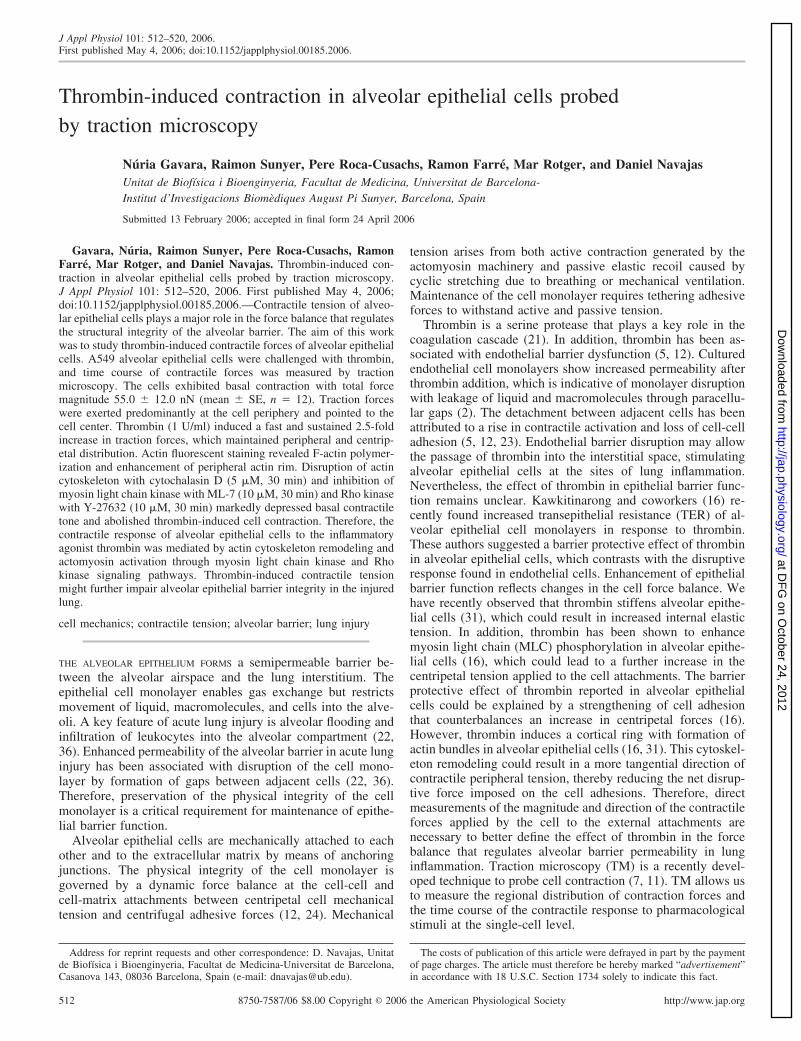
F-actin staining of vehicle-treated cells showed a diffusepattern (Fig. 5). Thrombin caused rearrangement of the F-actincytoskeleton with formation of a marked peripheral rim (Fig.5). F-actin staining of single cells showed actin organizationsimilar to that of confluent cells (Fig. 5, insets). F-actin-to-G-actin fluorescence ratio was 4.0 � 0.4 in vehicle-treated cells.The ratio increased by 27% (P � 0.05) in thrombin-challengedcells (Fig. 6).
Role of the Actin Cytoskeleton in Cell Contraction
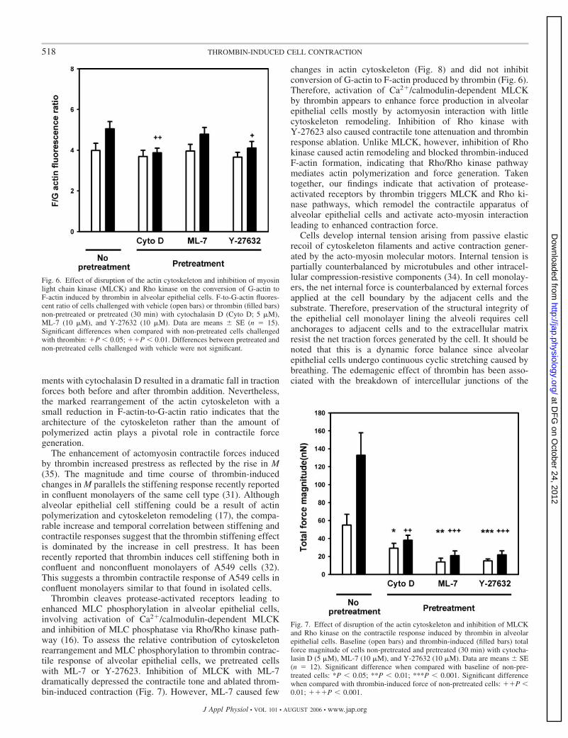
Disruption of the actin cytoskeleton with cytochalasin Ddecreased the baseline contractile force by 47% (P � 0.01)(Fig. 7). Traction force induced by thrombin in cytochalasin Dpretreated cells was 3.5-fold lower than in nonpretreated cells(P � 0.01). No significant changes in F-actin-to-G-actin fluo-rescence ratio were found in cells pretreated with cytochalasinD and subsequently treated with vehicle or thrombin (Fig. 6).Disruption of the actin cytoskeleton was evident in F-actindistribution (Fig. 8). Disappearance of actin structures wasassociated with irregularly polymerized actin aggregatesthroughout the cytoplasm. Thrombin addition did not modifythis pattern.
514 THROMBIN-INDUCED CELL CONTRACTION
J Appl Physiol • VOL 101 • AUGUST 2006 • www.jap.org
at DF
G on O
ctober 24, 2012http://jap.physiology.org/
Dow
nloaded from

Role of Inhibition of MLCK and Rho Kinase inCell Contraction
Inhibition of MLCK with ML-7 decreased baseline tractionforce of alveolar epithelial cells by 76% (P � 0.01) (Fig. 7). Asimilar relaxation of baseline force (74%, P � 0.01) wasobtained when Rho kinase was inhibited with Y-27632. Trac-tion force after thrombin challenge was sixfold lower (P �0.001) when cells were pretreated with either ML-7 orY-27632.
No changes in F-actin-to-G-actin fluorescence ratio wereobserved when MLCK was inhibited with ML-7 (Fig. 6).Thrombin caused a G-actin to F-actin conversion (21%, P �0.05) similar to that observed in cells without MLCK inhibi-tion. Pretreatment with ML-7 did not modify F-actin distribu-tion (Fig. 8). Similarly, inhibition of MLCK did not reduce thethrombin-induced formation of a peripheral rim of actin bun-dles.
Inhibition of Rho kinase did not significantly change F-actin-to-G-actin fluorescence ratio (Fig. 6). Thrombin-inducedpolymerization of F-actin was lower on cells pretreated withY-27632 (12%) but did not attain significance (P � 0.29).Inhibition of Rho kinase slightly modified F-actin cytoskele-ton, resulting in a more diffuse actin distribution (Fig. 8).Thrombin did not induce actin bundle formation in cellspretreated with Y-27632.
DISCUSSION
This study demonstrates that thrombin induces fast andmarked contraction of A549 alveolar epithelial cells. The
distribution of traction forces before and after thrombin chal-lenge was mainly located along the cell periphery and pointedtoward the cell center. Thrombin caused actin polymerizationand enhancement of the peripheral actin rim. Disruption of theactin cytoskeleton and reduction of MLC phosphorylation byinhibition of MLCK or Rho kinase activities attenuated boththe basal contractile state of the cell and the thrombin-inducedcontractile response.
Cell contraction can be readily probed by plating cells on topof a soft elastic gel or casting them inside the gel (13, 18, 23,33). Contractile cell response to pharmacological stimulationcan be assessed with force transducers attached to the gel (18,24) or by measuring the reduction in the gel area (13, 33).These approaches enable assessment of relative changes inaverage contraction strength of the cell culture. However,measurement of force or deformation of the bulk gel does notallow accurate determination of the baseline contractile tone orthe regional distribution and absolute magnitude of contractileforces at the single-cell level. By contrast, TM maps local geldeformations by tracking the displacement of small fluorescentbeads embedded in the gel. By removing the cells attached tothe surface of the gel at the end of the experiment, the relaxedposition of the beads in unstrained gel is easily determined.Moreover, in contrast with microfabricated pillar-array tech-niques (28), the stiffness of the substrate and the spatial forceresolution can be easily tuned to each particular cell type. ThusTM enabled measurement of the time course of the absolutemagnitude and regional distribution of forces exerted by singlealveolar epithelial cells with micrometric spatial resolution.
Fig. 1. Mapping of alveolar epithelial cell contraction in-duced by thrombin. Bright-field image of an A549 cellbefore thrombin stimulation (top). Displacement field (mid-dle) and corresponding traction field (bottom) of the cellbefore (left) and 10 min after thrombin addition (1 U/ml)(right). Cell boundary drawn from the bright-field image isshown as a black line. Color scales indicate the magnitude ofdisplacement (middle) and traction force (bottom). Arrowsdepict the direction and relative magnitude of displacementand traction force. Displacements and tractions were calcu-lated with 1.3-�m spatial resolution. For clarity, arrows aredisplayed with 3.9-�m spacing. Scale bar is 10 �m.
515THROMBIN-INDUCED CELL CONTRACTION
J Appl Physiol • VOL 101 • AUGUST 2006 • www.jap.org
at DF
G on O
ctober 24, 2012http://jap.physiology.org/
Dow
nloaded from

TM requires cell contractile forces to be balanced solely by thegel through cell-substrate attachments. Therefore, TM mea-surements were made on isolated adherent cells that lackcell-cell attachments. In confluent cell monolayers, contractileforces are in part offset by cell-cell tethering forces, thusreducing the contribution of cell-matrix adhesions.
The A549 is a cell line derived from human bronchoalveolarcell carcinoma. These cells are widely used as a model ofalveolar epithelial cells in vitro because they are readily cul-tured and retain important features of type II alveolar cells(20). In particular, it has been recently shown that this cell typeforms a tight monolayer when grown to confluence and that thebarrier permeability is modulated by thrombin (16). Moreover,A549 cells displayed ability similar to that of primary rat typeII cells to contract collagen gels (33). Cell speed of the isolatedA549 cells was 25-fold smaller than that reported for migrating
cell types (10). Moreover, immunofluorescence images ofisolated A549 cells did not display front-rear polarity of theF-actin cytoskeleton (Fig. 5, inset). These results indicate thatthe A549 cells studied in TM experiments adopted a nonmi-grating behavior. The low motility exhibited by the A549 cellscould be attributed to the high concentration of type I collagenused to coat the polyacrylamide gels (26). Therefore, despitethe limitations of transformed cell lines, we considered A549cells to be a suitable model to study contractile properties ofnonmigrating alveolar epithelial cells.
Our TM measurements demonstrate that cultured alveolarepithelial cells exhibit contractile tone under basal conditions.Contraction was stronger at the cell periphery, which is con-sistent with the regional distribution of F-actin cytoskeletonshowing a weak rim. The F-actin pattern we found agrees withprevious reports of alveolar epithelial cells (16, 31). We quan-titatively assessed contraction orientation by computing at eachpoint the direction of the traction vector relative to the centroidof the cell. The histogram of traction angles was very wellfitted by a Gaussian function with the addition of a smallconstant term. The Gaussian distribution was centered aroundzero with a relatively narrow dispersion, indicating that celltraction forces pull inward. The constant term accounts for aweak field of forces randomly orientated, which could partiallyreflect background noise. It should be pointed out that TMmeasures the traction forces exerted on the substrate. There-fore, a centripetal force at the cell edge could be generated byradial stress fibers or by a contractile curved rim whose tensionresults in a net radial component. Alveolar epithelial cellsexhibited marked contractile polarity with Mxx/M � 3/4, re-flecting a principal traction axis with a contractile momentthreefold stronger than that of the perpendicular direction. Thecontractile polarity of alveolar epithelial cells was comparableto that reported on smooth muscle cells (30). Interestingly,isolated alveolar epithelial cells revealed contractile polarityunder symmetrical environmental conditions. Nevertheless, in
Fig. 3. Spatial distribution of traction forces. Average traction within adjacentbands (1.26 �m thick) parallel to the cell boundary and within the centralregion containing points whose distance is �6.3 �m from the cell boundary atbaseline (E) and after thrombin addition (F). Data are means � SE. Inset:adjacent bands computed in a representative cell.
Fig. 2. Time course of contractile response of alveolar epithelial cells chal-lenged with thrombin (F) or vehicle (E). Top: total force magnitude (F).Bottom: net contractile moment (M). Arrows indicate addition of thrombin(final concentration 1 U/ml; n � 12) or vehicle (n � 12). Data are means �SE.
516 THROMBIN-INDUCED CELL CONTRACTION
J Appl Physiol • VOL 101 • AUGUST 2006 • www.jap.org
at DF
G on O
ctober 24, 2012http://jap.physiology.org/
Dow
nloaded from

the alveoli, cell contraction polarity might be regulated by cellsignaling or structural inhomogeneities. Traction exerted byalveolar epithelial cells was in the range of that reported inendothelial cells (3), suggesting comparable actomyosin motoractivity. On the other hand, forces reported on migratingfibroblasts were �10-fold higher (11). The propulsive forcesproduced during cell locomotion might explain the strongertractions exerted on the substrate by migrating cells.
Challenge of alveolar epithelial cells with thrombin pro-duced a rapid and marked increase in the cell contractile state,as measured by F, M, and maximal local force (Figs. 1 and 2).The increase in contraction did not modify the regional distri-bution of traction forces (Figs. 1 and 3) or the contractilepolarity. Thrombin exposure resulted in a sharper and narrowerdistribution of traction angles, indicative of reorientation oftraction forces to a more centripetal direction. In agreementwith other studies in alveolar epithelial cells (16, 31), thrombin
enhanced the peripheral F-actin rim. In addition, we showedthat thrombin induces F-actin polymerization (Fig. 6). Thiscytoskeleton remodeling is consistent with the changes in thetraction force field exhibited by the cells. Enhancement oftraction forces and maintenance of their spatial distribution(Figs. 1 and 3) were associated with the formation of peripheralF-actin bundles (Fig. 5). This association suggests that cy-toskeleton remodeling also contributes to the contractile re-sponse induced by thrombin. Disrupting F-actin microfila-
Fig. 5. Actin cytoskeleton remodeling induced by thrombin on alveolarepithelial cells. F-actin staining with phalloidin-tetramethylrhodamine isothio-cyanate (TRITC) 10 min after adding vehicle (top) or thrombin (1 U/ml)(bottom). Insets: F-actin staining of single cells.
Fig. 4. Histograms of centripetal orientation of contractile forces at baseline(top) and after thrombin addition (bottom). Plots are pooled traction data ofn � 12 cells. Thick solid lines are fit of a Gaussian distribution with anadditional constant term. Inset: sketch of angle definition. T and r represent thetraction vector and the vector pointing toward the centroid (C) of the cell,respectively.
517THROMBIN-INDUCED CELL CONTRACTION
J Appl Physiol • VOL 101 • AUGUST 2006 • www.jap.org
at DF
G on O
ctober 24, 2012http://jap.physiology.org/
Dow
nloaded from
ments with cytochalasin D resulted in a dramatic fall in tractionforces both before and after thrombin addition. Nevertheless,the marked rearrangement of the actin cytoskeleton with asmall reduction in F-actin-to-G-actin ratio indicates that thearchitecture of the cytoskeleton rather than the amount ofpolymerized actin plays a pivotal role in contractile forcegeneration.
The enhancement of actomyosin contractile forces inducedby thrombin increased prestress as reflected by the rise in M(35). The magnitude and time course of thrombin-inducedchanges in M parallels the stiffening response recently reportedin confluent monolayers of the same cell type (31). Althoughalveolar epithelial cell stiffening could be a result of actinpolymerization and cytoskeleton remodeling (17), the compa-rable increase and temporal correlation between stiffening andcontractile responses suggest that the thrombin stiffening effectis dominated by the increase in cell prestress. It has beenrecently reported that thrombin induces cell stiffening both inconfluent and nonconfluent monolayers of A549 cells (32).This suggests a thrombin contractile response of A549 cells inconfluent monolayers similar to that found in isolated cells.
Thrombin cleaves protease-activated receptors leading toenhanced MLC phosphorylation in alveolar epithelial cells,involving activation of Ca2/calmodulin-dependent MLCKand inhibition of MLC phosphatase via Rho/Rho kinase path-way (16). To assess the relative contribution of cytoskeletonrearrangement and MLC phosphorylation to thrombin contrac-tile response of alveolar epithelial cells, we pretreated cellswith ML-7 or Y-27623. Inhibition of MLCK with ML-7dramatically depressed the contractile tone and ablated throm-bin-induced contraction (Fig. 7). However, ML-7 caused few
changes in actin cytoskeleton (Fig. 8) and did not inhibitconversion of G-actin to F-actin produced by thrombin (Fig. 6).Therefore, activation of Ca2/calmodulin-dependent MLCKby thrombin appears to enhance force production in alveolarepithelial cells mostly by actomyosin interaction with littlecytoskeleton remodeling. Inhibition of Rho kinase withY-27623 also caused contractile tone attenuation and thrombinresponse ablation. Unlike MLCK, however, inhibition of Rhokinase caused actin remodeling and blocked thrombin-inducedF-actin formation, indicating that Rho/Rho kinase pathwaymediates actin polymerization and force generation. Takentogether, our findings indicate that activation of protease-activated receptors by thrombin triggers MLCK and Rho ki-nase pathways, which remodel the contractile apparatus ofalveolar epithelial cells and activate acto-myosin interactionleading to enhanced contraction force.
Cells develop internal tension arising from passive elasticrecoil of cytoskeleton filaments and active contraction gener-ated by the acto-myosin molecular motors. Internal tension ispartially counterbalanced by microtubules and other intracel-lular compression-resistive components (34). In cell monolay-ers, the net internal force is counterbalanced by external forcesapplied at the cell boundary by the adjacent cells and thesubstrate. Therefore, preservation of the structural integrity ofthe epithelial cell monolayer lining the alveoli requires cellanchorages to adjacent cells and to the extracellular matrixresist the net traction forces generated by the cell. It should benoted that this is a dynamic force balance since alveolarepithelial cells undergo continuous cyclic stretching caused bybreathing. The edemagenic effect of thrombin has been asso-ciated with the breakdown of intercellular junctions of the
Fig. 7. Effect of disruption of the actin cytoskeleton and inhibition of MLCKand Rho kinase on the contractile response induced by thrombin in alveolarepithelial cells. Baseline (open bars) and thrombin-induced (filled bars) totalforce magnitude of cells non-pretreated and pretreated (30 min) with cytocha-lasin D (5 �M), ML-7 (10 �M), and Y-27632 (10 �M). Data are means � SE(n � 12). Significant difference when compared with baseline of non-pre-treated cells: *P � 0.05; **P � 0.01; ***P � 0.001. Significant differencewhen compared with thrombin-induced force of non-pretreated cells: P �0.01; P � 0.001.
Fig. 6. Effect of disruption of the actin cytoskeleton and inhibition of myosinlight chain kinase (MLCK) and Rho kinase on the conversion of G-actin toF-actin induced by thrombin in alveolar epithelial cells. F-to-G-actin fluores-cent ratio of cells challenged with vehicle (open bars) or thrombin (filled bars)non-pretreated or pretreated (30 min) with cytochalasin D (Cyto D; 5 �M),ML-7 (10 �M), and Y-27632 (10 �M). Data are means � SE (n � 15).Significant differences when compared with non-pretreated cells challengedwith thrombin: P � 0.05; P � 0.01. Differences between pretreated andnon-pretreated cells challenged with vehicle were not significant.
518 THROMBIN-INDUCED CELL CONTRACTION
J Appl Physiol • VOL 101 • AUGUST 2006 • www.jap.org
at DF
G on O
ctober 24, 2012http://jap.physiology.org/
Dow
nloaded from

microvascular endothelium caused by the enhancement ofactive force generation that cell attachments cannot resist (12,23). Intercellular gap formation facilitates extravasation offluid and macromolecules to the interstitial compartment ex-posing epithelial cells to thrombin. Consistently, elevated lev-els of thrombin have been found in bronchoalveolar lavagefluid obtained from patients with asthma and acute respiratorydistress syndrome (19, 29).
In contrast to the fall in TER found in endothelial cells,increased TER has been recently reported in alveolar epithelialcells exposed to thrombin, indicative of improved sealing ofthe cell monolayer (16). This suggests that thrombin has twocompeting effects in the alveolar-capillary barrier function.The thrombin-induced disruption of the endothelial cell barrierhas been attributed to a loss of cell-adhesion and to an increasein centripetal contractile forces (5, 12, 16, 23). On the otherhand, the barrier protective effect of thrombin observed inepithelial cell monolayers could be a result of a decrease incentripetal forces or an increase in tethering forces (16). Ourstudy shows that thrombin increases active centripetal contrac-tile forces generated at the cell periphery in alveolar epithelialcells. In addition, we previously found thrombin-induced stiff-ening in the same cell type (31). Cell stiffening results inincreased passive elastic recoil when the cell is subjected to thebreathing stretch, which might be locally large in nonhomog-enous lung deformation in mechanically ventilated patientswith acute lung injury. Thus thrombin increases both activeand passive components of centripetal forces, which favorsbarrier disruption. Maintenance of the cell monolayer requirescell anchorages to withstand centripetal tension. In our TMexperiments, 12% of isolated cells lacking cell-cell attach-
ments exhibited partial detachment from the gel after thrombinchallenge. This shows that cell-matrix anchorages alone werenot able to resist thrombin-induced contractile forces. In cellmonolayers, cell-cell attachments provide additional tetheringforces to withstand centripetal tension. Accordingly, the throm-bin-induced increase in TER (16) reported in A459 confluentmonolayers indicates that the rise in centripetal tension may becompensated by cell adhesion enhancement, protecting barrierintegrity. In this connection, enhanced cortical cytoskeletonand translocation of ZO-1 tight junction protein from thecytosolic compartment to the cell membrane contact sites werealso reported in alveolar epithelial cells exposed to thrombin(16). However, no changes in -catenin adherens junctionprotein were found (16). We have recently reported cell de-tachment of thrombin-treated A549 cells in confluent mono-layers when subjected to stretch (32). Cell detachment wassubstantially impaired in subconfluent monolayers where an-chorages to adjacent cells are reduced. Taken together, thesefindings suggest that cell-cell attachments play a key role inregulating the structural integrity of the alveolar epithelialbarrier. Direct measurements of cell-cell and cell-matrix teth-ering forces are needed to elucidate the effect of thrombin inthe mechanical strength of cell attachments. This will requirenovel methodological approaches to directly probe in intactcell monolayers the adhesion forces imposed by the cells on thematrix and the adjacent cells. It should be noted that reportedTER measurements were carried out in confluent cell mono-layers under static conditions. However, the rise in elasticrecoil caused by cyclic stretch imposes a higher mechanicalload onto cell junctions, which might compromise monolayerintegrity (32). Importantly, in the damaged alveoli, epithelial
Fig. 8. Effect of disruption of the actin cytoskeleton and inhibition of MLCK and Rho kinase on the actin remodeling induced by thrombin (Thr) in alveolarepithelial cells. F-actin staining with phalloidin-TRITC 10 min after adding vehicle (top) or thrombin (1 U/ml) (bottom). Cells were pretreated with cytochalasinD (5 �M; left), ML-7 (10 �M; middle) and Y-27632 (10 �M; right) for 30 min before thrombin challenge.
519THROMBIN-INDUCED CELL CONTRACTION
J Appl Physiol • VOL 101 • AUGUST 2006 • www.jap.org
at DF
G on O
ctober 24, 2012http://jap.physiology.org/
Dow
nloaded from

cells lose cell-cell contacts, reducing the ability of tetheringforces to withstand the increased centripetal tension induced bythrombin. Thus, in the injured lung, thrombin might have anadverse effect on alveolar epithelial monolayer repair.
In conclusion, we have shown that alveolar epithelial cellsexhibit basal contractile tone, exerting centripetal tractionforces predominantly at the cell periphery. The inflammatoryagonist thrombin enhances contraction of alveolar epithelialcells maintaining peripheral and centripetal force distribution.Cell contraction increase is associated with F-actin polymer-ization and enhancement of the peripheral actin cortex. Con-tractile response to thrombin is mediated by actin cytoskeletonremodeling and actomyosin activation through Ca2/calmod-ulin-dependent MLCK and Rho/Rho kinase signaling path-ways. In the intact alveolar epithelium, the increased contrac-tile centripetal tension may be compensated by cell adhesiontethering forces. However, in the injured lung, thrombin-induced contractile tension might further impair alveolar epi-thelial barrier integrity.
ACKNOWLEDGMENTS
The authors thank N. Wang for help in the gel preparation protocol, X.Trepat for comments, and M. Rodriguez for technical assistance.
GRANTS
This work was supported in part by grants from Ministerio de Ciencia yTecnologia (SAF2005-00110, SAF2003-01334, and NAN2004-09348-C04-04) and Ministerio de Sanidad y Consumo (Red GIRA-G03/063, RedRESPIRA-C03/11, and FIS-PI040929).
REFERENCES
1. Alcaraz J, Buscemi L, Grabulosa M, Trepat X, Fabry B, Farre R, andNavajas D. Microrheology of human lung epithelial cells measured byatomic force microscopy. Biophys J 84: 2071–2079, 2003.
2. Amerongen GPV, van Delft S, Vermeer MA, Collard JG, and vanHinsbergh VWM. Activation of RhoA by thrombin in endothelial hyper-permeability: role of Rho kinase and protein tyrosine kinases. Circ Res 87:335–340, 2000.
3. An SS, Pennella CM, Gonnabathula A, Chen J, Wang N, Gaestel M,Hassoun PM, Fredberg JJ, and Kayyali US. Hypoxia alters biophysicalproperties of endothelial cells via p38 MAPK- and Rho kinase-dependentpathways. Am J Physiol Cell Physiol 289: C521–C530, 2005.
4. Bilodeau GG. Regular pyramid punch problem. J Appl Mech 59: 519–523, 1992.
5. Bogatcheva NV, Garcia JGN, and Verin AD. Molecular mechanisms ofthrombin-induced endothelial cell permeability. Biochemistry (Mosc) 67:75–84, 2002.
6. Burnham NA, Chen X, Hodges CS, Matei GA, Thoreson EJ, RobertsCJ, Davis MC, and Tendler SJB. Comparison of calibration methods foratomic-force microscopy cantilevers. Nanotechnology 14: 1–6, 2003.
7. Butler JP, Tolic-Nørrelykke IM, Fabry B, and Fredberg JJ. Tractionfields, moments, and strain energy that cells exert on their surroundings.Am J Physiol Cell Physiol 282: C595–C605, 2002.
8. Castleman KR. Digital Image Processing. Englewood Cliffs, NJ: Pren-tice-Hall, 1996.
9. Cramer LP, Briggs LJ, and Dawe HR. Use of fluorescently labeleddeoxyribonuclease I to spatially measure G-actin levels in migrating andnon-migrating cells. Cell Motil Cytoskeleton 51: 27–38, 2002.
10. Dallon JC and Othmer HG. How cellular movement determines thecollective force generated by the dictyostelium discoideum slug. J TheorBiol 231: 203–222, 2004.
11. Dembo M and Wang YL. Stresses at the cell-to-substrate interface duringlocomotion of fibroblasts. Biophys J 76: 2307–2316, 1999.
12. Dudek SM and Garcia JGN. Cytoskeletal regulation of pulmonaryvascular permeability. J Appl Physiol 91: 1487–1500, 2001.
13. Fang Q, Liu X, Abe S, Kobayashi T, Wang XQ, Kohyama T, Hashi-moto M, Wyatt T, and Rennard SI. Thormbin induces collagen con-
traction partially through PAR1 activation and PKC-�. Eur Respir J 24:918–924, 2004.
14. Gaudet C, Marganski WA, Kim S, Brown CT, Gunderia V, Dembo M,and Wong JY. Influence of type I collagen surface density on fibroblastspreading, motility, and contractility. Biophys J 85: 3329–3335, 2003.
15. Hirshman CA, Togashi H, Shao D, and Emala CW. G�i-2 is required forcarbachol-induced stress fiber formation in human airway smooth musclecells. Am J Physiol Lung Cell Mol Physiol 275: L911–L916, 1998.
16. Kawkitinarong K, Linz-McGillem L, Birukov KG, and Garcia JGN.Differential regulation of human lung epithelial and endothelial barrierfunction by thrombin. Am J Respir Cell Mol Biol 31: 517–527, 2004.
17. Kole TP, Tseng Y, Huang L, Katz JL, and Wirtz D. Rho kinaseregulates the intracellular micromechanical response of adherent cells torho activation. Mol Biol Cell 15: 3475–3484, 2004.
18. Kolodney MS and Wysolmerski RB. Isometric contraction by fibroblastsand endothelial cells in tissue culture: a quantitative study. J Cell Biol 117:73–82, 1992.
19. Levi M, Schultz MJ, Rijneveld AW, and Van de Poll T. Bronchoal-veolar coagulation and fibrinolysis in endotoxemia and pneumonia. CritCare Med 31: S238–S242, 2003.
20. Lieber M, Smith B, Szakal A, Nelson RW, and Todaro GA. Acontinuous tumor-cell line from a human lung carcionoma with propertiesof type II alveolar epithelial cells. Int J Cancer 17: 62–70, 1976.
21. Macfarlane SR, Seatter MJ, Kanke T, Hunter GD, and Plevin R.Proteinase-activated receptors. Pharmacol Rev 53: 245–282, 2001.
22. Matthay MA and Zimmerman GA. Acute lung injury and the acuterespiratory distress syndrome. Am J Respir Cell Mol Biol 33: 319–327,2005.
23. Moy AB, Blackwell K, and Kamath A. Differential effects of histamineand thrombin on the endothelial barrier function through actin-myosintension. Am J Physiol Heart Circ Physiol 282: H21–H29, 2002.
24. Moy AB, Van Engelenhoven J, Bodmer J, Kamath J, Keese C,Giaever I, Shasby S, and Shasby DM. Histamine and thrombin modulateendothelial focal adhesion through centripetal and centrifugal forces.J Clin Invest 97: 1020–1027, 1996.
25. Pelham RJ Jr and Wang Y. Cell locomotion and focal adhesions areregulated by substrate flexibility. Proc Natl Acad Sci USA 94: 13661–13665, 1997.
26. Planus E, Galiacy S, Matthay M, Laurent V, Gavrilovic J, Murphy G,Clerici C, Isabey D, Lafuma C, and d’Ortho MP. Role of collagenasein mediating in vitro alveolar epithelial wound repair. J Cell Sci 112:243–252, 1999.
27. Rico F, Roca-Cusachs P, Gavara N, Farre R, Rotger M, and NavajasD. Probing mechanical properties of living cells by atomic force micros-copy with blunted pyramidal cantilever tips. Physiol Rev E Stat NonlinSoft Matter Phys 72: 021914, 2005.
28. Tan JL, Tien J, Pirone DM, Gray DS, Bhadriraju K, and Chen CS.Cells lying on a bed of microneedles: an approach to isolate mechanicalforce. Proc Natl Acad Sci USA 104: 1484–1489, 2003.
29. Terada M, Kelly EAB, and Jarjour NN. Increased thrombin activityafter allergen challenge: a potential link to airway remodeling? Am JRespir Crit Care Med 169: 373–377, 2004.
30. Tolic-Nørrelykke IM, Butler JP, Chen J, and Wang N. Spatial andtemporal traction response in human airway smooth muscle cells. Am JPhysiol Cell Physiol 283: C1254–C1266, 2002.
31. Trepat X, Grabulosa M, Buscemi L, Rico F, Farre R, and Navajas D.Thrombin and histamine induce stiffening of alveolar epithelial cells.J Appl Physiol 98: 1567–1574, 2005.
32. Trepat X, Puig F, Gavara N, Fredberg JJ, Farre R, and NavajasD. Effect of stretch on the structural integrity and micromechanicsof human alveolar epithelial cell monolayers exposed to thrombin.Am J Physiol Lung Cell Mol Physiol 290: L1104 –L1110, 2006;doi:10.1152/ajplung.00436.2005.
33. Umino T, Wang H, Zhu Y, Liu X, Manouilova LS, Spurzem JR,Leuschen MP, and Rennard SI. Modification of type I collagenous gelsby alveolar epithelial cells. Am J Respir Cell Mol Biol 22: 702–707, 2000.
34. Wang N, Butler JP, and Ingber DE. Mechanotransduction across thecell-surface and through the cytoskeleton. Science 260: 1124–1127, 1993.
35. Wang N, Tolic-Nørrelykke IM, Chen J, Mijailovich SM, Butler JP,Fredberg JJ, and Stamenovic D. Cell prestress. I. Stiffness and prestressare closely associated in adherent contractile cells. Am J Physiol CellPhysiol 282: C606–C616, 2002.
36. Ware LB and Matthay MA. The acute respiratory distress syndrome.N Engl J Med 342: 1334–1349, 2000.
520 THROMBIN-INDUCED CELL CONTRACTION
J Appl Physiol • VOL 101 • AUGUST 2006 • www.jap.org
at DF
G on O
ctober 24, 2012http://jap.physiology.org/
Dow
nloaded from