platelets and thrombin generation
TRANSCRIPT
Platelets and Thrombin GenerationDougald M. Monroe, Maureane Hoffman, Harold R. Roberts
Abstract—This review examines the evidence that platelets play a major role in localizing and controlling the burst ofthrombin generation leading to fibrin clot formation. From the first functional description of platelets, it has beenrecognized that platelets supply factors that support the activation of prothrombin. Studies have demonstrated that onactivation, the amount of one specific lipid, phosphatidylserine, is significantly increased on the outer leaflet of plateletmembranes. When it was found that phosphatidylserine containing lipid extracts could be substituted for platelets inclotting assays, this suggested the possibility that changes in platelet lipid composition were necessary and sufficient toaccount for platelet surface thrombin generation. Because a growing body of data suggest that platelet-binding proteinsprovide much of the specificity for platelet thrombin generation, we review in this report data suggesting that changesin lipid composition are necessary but not sufficient to account for platelet surface regulation of thrombin generation.Also, we review data suggesting that platelets from different individuals differ in their capacity to generate thrombin,whereas platelets from a single subject support thrombin generation in a reproducible manner. Individual differences inplatelet thrombin generation might be accounted for by differences in platelet-binding proteins. (Arterioscler ThrombVasc Biol. 2002;22:1381-1389.)
Key Words: platelets � thrombin � lipids � phosphatidylserine � coagulation
In the 1880s, Bizzozero1 and Hayem,2 working indepen-dently, wrote about a blood particle, previously observed
by others as a colorless corpuscle smaller than red or whitecells, that they called a hematoblast or platelet. Bizzozerowrote that accumulation of this cell might account for the1875 observation of Zahn3 that bleeding from an injury to ablood vessel was initially blocked by a white thrombus. Thesestudies showed that fibrin was associated with these cells andled them to conclude that platelets supplied a factor that wasrequired for coagulation.1,2 Subsequently, a number of inves-tigators demonstrated that the rate of clotting and prothrom-bin conversion to thrombin was decreased in platelet-poorplasma and increased as a function of platelet number.4–6
See CoverThe initial studies of clotting assays relied on patient
platelets in the platelet-rich plasma as the surface for throm-bin generation. This led to considerable variability in theresults from patient to patient.6 To provide an assay reagentthat gave more reproducible results, platelet-poor plasma wasclotted with phospholipid.7 Assay reproducibility was alsoenhanced by the addition of suspensions of diatomaceousearth to activate clotting.8 This modified assay gave repro-ducible clotting values and was valuable for the diagnosis ofbleeding disorders as well as for establishing the relationshipsbetween the coagulation proteins. But as a result, the use oflipids as a substitute for platelets became widespread.
In studies involving how lipids influence coagulationassays, it was observed that there was a correlation betweenthe amount of a specific lipid, phosphatidylserine, and theability of a lipid surface to promote thrombin generation.9
Other studies looking at platelet activation showed thatcirculating unactivated platelets had low levels of phosphati-dylserine on the outer leaflet of their membranes.10 However,when platelets were activated, the amount of phosphatidyl-serine on the outer leaflets increased dramatically from �2%of the phospholipid content to as much as 12%.11,12 Restingplatelets do not significantly promote factor X or prothrombinactivation.13 Even on activated platelets, procoagulant activ-ity is lost on treatment with phospholipases that cleave thehead groups from phosphatidylserine.14 Because there was anobvious correlation between the observation that activatedplatelets express phosphatidylserine and the observation thatphosphatidylserine in purified lipids is required for thrombingeneration, it was concluded by many that phosphatidylserineexposure provided the primary mechanism for regulatingcoagulation reactions and thrombin generation.10
Cascade Model of CoagulationCoagulation, as shown in Figure 1, has often been representedas 2 somewhat independent pathways that converge to acommon pathway, with thrombin generation as the end pointof the reactions. This model represents an evolution of theinitial models of coagulation based on a cascade or waterfall
Received April 25, 2002; revision accepted July 17, 2002.From the Center for Thrombosis and Hemostasis (D.M.M., H.R.R.), University of North Carolina, Chapel Hill, and the Department of Pathology
(M.H.), Duke University, Durham, NC.Correspondence to Dr Dougald M. Monroe, University of North Carolina at Chapel Hill, Hematology/Oncology, 932 Mary Ellen Jones Bldg, CB 7035,
Chapel Hill, NC 27599-7035. E-mail [email protected]© 2002 American Heart Association, Inc.
Arterioscler Thromb Vasc Biol. is available at http://www.atvbaha.org DOI: 10.1161/01.ATV.0000031340.68494.34
1381 by guest on July 6, 2015http://atvb.ahajournals.org/Downloaded from

hypothesis.15,16 These models give a good representation ofthe processes observed in clinical coagulation laboratorytests. The prothrombin time measures the factors of theso-called extrinsic pathway, and activated partial thrombo-plastin time measures factors in the intrinsic pathway. Al-though this model reflects some of the interactions of theproteins, it has inadequacies as a model of the in vivohemostatic processes. For example, deficiencies of factor XII,high molecular weight kininogen (HK), or prekallikrein donot cause clinical bleeding. Furthermore, others have shownthat under normal circumstances, hemostasis is initiated bytissue factor (TF). In addition, if the intrinsic and extrinsicpathways are essentially separate, then activation of factor Xby the extrinsic pathway should compensate for a lack offactor VIII or IX. However, factors VIII and IX are clearlyessential for hemostasis, because their absence results in thebleeding seen in hemophilia, and this bleeding is not com-pensated by the intact extrinsic system. Interestingly, factorXI deficiency results in a bleeding disorder that has signifi-cant variability between patients and is significantly milderthan bleeding in patients with hemophilia A or B lackingfactors VIII or IX, respectively.17–19
It has long been established that coagulation reactions donot occur physiologically in solution but are localized to asurface.6,20–22 The critical advantage to evolving such amechanism is that the reactions are confined to a specific siteof injury rather than being completely disseminated. Implicitin the view of coagulation shown in Figure 1 is the assump-tion that the level and amount of coagulation factors drive andregulate coagulation and that the surfaces are essentiallypassive agents. Also, implicit in the view of coagulationshown in Figure 1 is that the role of cells is primarily toprovide a lipid-rich surface for coagulation complex assem-bly and that all surfaces with similar lipid composition areessentially identical. This supposition is contradicted by a
number of studies, including those showing that endothelialcells, which are not considered procoagulant, nonethelessexpress significant levels of phosphatidylserine on theirsurface.23 This phosphatidylserine is important for maintain-ing the anticoagulant activity of endothelium by enhancingthrombomodulin activity.23
A direct comparison of platelets and phospholipids hasshown that platelets have complex coagulant activities thatare not completely mimicked by phospholipids.24,25 Also, acareful analysis of the kinetics of platelet activity was notconsistent with the kinetics seen on lipid surfaces.26 This andother recent evidence suggests that platelet protein compo-nents provide virtually all of the specificity and controlmechanisms that determine the expression of procoagulantactivity and subsequent thrombin generation.
Cell-Based Model of CoagulationA useful model to describe coagulation is shown in Figure 2.This model is derived in part from experiments that use cellssuch as monocytes or fibroblasts as a source of TF (ratherthan relipidated TF) and activated platelets as a surface forthrombin generation.27,28 In this model, coagulation occurs in3 overlapping phases: initiation, priming, and propaga-tion.27–31 During the process of hemostasis, a break in thevessel wall brings plasma into contact with TF-bearing cells.This TF may be derived entirely from extravascular sources,such as fibroblasts, or may in part be derived from encryptedsources in blood through a CD62 (P-selectin)/CD15 (P-selectin glycoprotein ligand 1)–mediated mechanism.32–34
Factor VII binds to TF and is rapidly activated35 by coagu-lation proteases36 and by noncoagulation proteases, depend-ing on the cellular location of the TF.37 The factor VIIa/TFcomplex activates factor X and factor IX (Figure 2). Theactivated forms of these 2 proteins, even though activated atthe same site, play very different and distinct roles insubsequent coagulation reactions. Factor Xa can activateplasma factor V on the TF cell38 (as can other cellularproteases39). If factor Xa diffuses from the protected environ-ment of the cell surface from which it was activated, it can berapidly inhibited by the TF pathway inhibitor or antithrombin.However, the factor Xa that remains on the TF cell surfacecan combine with factor Va to produce small amounts ofthrombin.30,40 This thrombin, although not sufficient to cleavefibrinogen throughout a wound, nonetheless plays a criticalrole in amplifying the initial thrombin signal, as shown inFigure 2.30 The initial factor VIIa/TF complex is subsequentlyinhibited by the action of the TF pathway inhibitor incomplex with factor Xa.41,42
In the priming phase (Figure 2), the small amount of initialthrombin binds to platelets that have adhered to extravascularmatrix components at the site of injury mediated in part by thebinding of von Willebrand factor to collagen.43 The processof binding to matrix proteins, especially collagen, partiallyactivates platelets44 and also localizes them near a site of TFexposure. Thrombin enhances platelet activation via protease-activated receptor (PAR) mechanisms. There appears to be asynergy between the collagen activation of platelets and thethrombin activation of platelets, such that dual stimulation bythose 2 agonists results in platelet activity higher than that
Figure 1. Cascade model of coagulation. This scheme is amodification of previously proposed models15,16 and reflects thecomponents tested for in the prothrombin time assay (extrinsicpathway) and the activated partial thromboplastin time assay(intrinsic pathway). The intrinsic pathway consists of HK, factorXII, prekallikrein (PK), factor XI, factor IX, factor VIII, factor X,factor V, and prothrombin (II), which is converted to thrombin(IIa). The activated form of these factors is indicated by addingthe letter “a” as a suffix (eg, IXa). The extrinsic pathway consistsof TF, factor VII, factor X, factor V, and II. Reactions requiring aphospholipid surface are indicated by lipid.
1382 Arterioscler Thromb Vasc Biol. September 2002
by guest on July 6, 2015http://atvb.ahajournals.org/Downloaded from

seen with stimulation by either agonist alone.45 Thrombinactivation of platelets results in degranulation that releasespartially active factor V from platelet � granules.45,46 Co-stimulation of the platelet collagen receptor results in a subsetof the platelet population expressing high levels of factor V.45
Thrombin cleaves the partially activated factor V to a fullyactive form.47 Thrombin also cleaves factor VIII, releasing it
from von Willebrand factor.47 In addition, thrombin activatesfactor XI bound to the platelet surface.48,49 The result of thisstage is a primed activated platelet that rapidly binds thecofactors Va and VIIIa as well as factor XIa.29
In the propagation phase (Figure 2C), the factor IXa/VIIIacomplex assembles when factor IXa reaches the plateletsurface. The initial factor IXa formed by the factor VIIa/TFcomplex can diffuse to the platelet surface, because factorIXa is not rapidly inhibited by antithrombin or other plasmaprotease inhibitors. Additionally, platelet surface factor XIacan then provide additional factor IXa directly on the plateletsurface.48 Factor X is recruited to the activated plateletsurface and is activated by the factor IXa/VIIIa complex. Thisallows factor Xa to move directly into a protected complexwith factor Va,50 where (in the presence of prothrombin)factor Xa is protected from the TF pathway inhibitor51 andantithrombin, even in the presence of heparin.52 Plateletsurface factor Xa/Va complexes generate a burst of thrombinsufficient to form a stable hemostatic fibrin clot.
This model of coagulation provides a rational explanationfor the bleeding tendency in hemophilia. The TF pathway isintact in hemophilic patients so that the initiation and ampli-fication steps occur. This leads to normal platelet activationand may account for the tendency of hemophiliacs to stopbleeding as a result of initial platelet plug formation.53–55 Thisalso accounts for the normal bleeding time characteristic ofhemophilia. However, severe delayed bleeding occurs inhemophilia because there is deficient platelet-dependent fac-tor Xa and thrombin generation throughout the plateletplug.54,55 Factor Xa generated by the factor VIIa/TF complexcannot diffuse to the platelet surface without being inhibited.The only factor Xa that can be incorporated efficiently intoprothrombinase complexes is that formed on the plateletsurface by the factor IXa/VIIIa complex in proximity tofactor Va.50,56 Thus, in hemophilia, there is a failure ofplatelet-surface factor X activation, leading to a decrease inplatelet-surface thrombin generation and ineffective clotformation.
Role of Platelet LipidsExposure of phosphatidylserine as platelets become activatedis necessary for platelet procoagulant activity and is regulatedby active transport mechanisms, including a flip-flop mech-anism in which phosphatidylserine from the inner leafletbecomes exposed on the outer leaflet of the activated plateletsurface.14,57–59 Bleeding has been observed in individuals whohave defects in this flip-flop mechanism, as seen in Scottsyndrome.60,61 This defect is also seen in a canine model witha platelet procoagulant defect.62 Although initial work hasindicated that the most significant role of phosphatidylserinein membranes is to bind the �-carboxyglutamic acid (Gla)residues in coagulation proteins in a calcium-dependentfashion,63 recent studies have indicated that the phosphatidyl-serine head group may instead bind factor Xa at a non-Glasite to act as an allosteric regulator that turns on coagulationby enhancing prothrombin cleavage.64–68
Exposure of phosphatidylserine on the outer leaflet ofplatelets is necessary for platelet procoagulant activity, anddata support the idea that this exposure is sufficient to
Figure 2. Cell-based model of coagulation. In this scheme,coagulation occurs in 3 phases: initiation, priming, and propaga-tion. In the initiation, factor VIIa bound to TF activates factor IXand also factor X. Factor Xa then activates factor V on theTF-bearing cell, complexes with factor Va, and converts a smallamount of II to IIa.30 In the priming phase, the small amount ofinitial IIa activates platelets, causing release of � granule con-tents including factor V, activates factor V, activates factor XI,and activates factor VIII by cleaving it from von Willebrand fac-tor (vWF). Cofactors bind to the platelet surface before theirrespective enzymes.29 The factor VIIa/TF complex is shut downthrough the action of the TF pathway inhibitor (TFPI) in complexwith factor Xa. In the propagation phase, factor IXa generatedby factor VIIa/TF binds to the activated platelets and subse-quently activates factor X. This factor IXa is supplemented byfactor IXa generated on the platelet surface by factor XIa. FactorXa then moves directly into a protected complex with factor Va,resulting in a burst of thrombin generation.
Monroe et al Platelets and Thrombin Generation 1383
by guest on July 6, 2015http://atvb.ahajournals.org/Downloaded from

account for the procoagulant nature of activated platelets (forreview, see Heemskerk et al59). However, there is increasingevidence that specific binding sites on platelets regulateformation of the coagulation complexes.24–26 This conclusionis based in part on our own studies of differences in factorIXa/VIIIa and factor Xa/Va activity on platelets from differ-ent normal individuals.69 These studies examined plateletsfrom different subjects and showed differences betweennormal individuals in phosphatidylserine expression evenwhen their platelets were activated in the same way. Althoughthere were differences in the generation of factor Xa andthrombin between individuals, there was no correlation be-tween levels of phosphatidylserine expression and generationof factor Xa or thrombin, as shown in Figure 3. These datasupport the concept that factors other than phosphatidylserineare necessary for platelet-dependent thrombin generation.
Platelet-Binding Proteins Important for thePriming Step
Although the clotting factors bind to platelets, not all thisbinding transduces a signal to the platelet. Therefore, some ofthe high-affinity binding sites for coagulation factors onplatelets do not fit the classic definition of receptors and inthis review will be referred to as binding proteins (Figure 4).
The small amount of thrombin generated in the initiationstep is critical in the development of the procoagulantresponse.45,47 In the absence of a protected environment,thrombin is inhibited by plasma levels of antithrombin with ahalf-life of �1 minute. Unactivated platelets appear tocontain at least 3 binding proteins for thrombin. One is a siteon the glycoprotein (GP) Ib-IX-V complex that binds tothrombin through the heparin-binding site (thrombin anion–binding exosite 2).70 The second site is one of the PARs,PAR1.71 PAR1 binds to thrombin through the extendedsubstrate-binding site and anion-binding exosite 1. After this7-transmembrane-domain protein is cleaved by thrombin, thenew PAR amino terminus is a tethered ligand that binds to
another site on the same or neighboring PAR and triggers asignaling cascade.71 Some signaling also occurs throughPAR4, but this signal appears to be mainly associated withplatelet aggregation rather than the development of plateletprocoagulant activity.72 The binding of thrombin to the GPIb-IX-V complex significantly enhances the ability of throm-bin to cleave PAR1 and catalyze platelet activation.73 Thebinding of thrombin to the GP Ib-IX-V complex may alsosignal platelet activation through a fibrin-dependent mecha-nism.74 Also, one group has tentatively identified a thrombin-binding site that is thought to be independent of the 2 sitesmentioned above.75
Unactivated factor VIII circulates in association with vonWillebrand factor and can bind to platelets through the vonWillebrand factor–binding site on the GP Ib-IX-V complex.76
Previous studies have shown that platelets potentiate throm-bin activation of factor VIII.77 This potentiation is maximalwhen von Willebrand factor is present.78 We speculate thatbinding to the GP Ib-IX-V complex brings factor VIII/vonWillebrand factor into proximity with thrombin and allowsfor efficient activation of factor VIII and release of the factorVIIIa from von Willebrand factor to the platelet surface.
Factor XI shows saturable, specific, reversible binding toplatelets with a Kd of �10 nmol/L.79 This binding is mediatedthrough a cluster of residues on the third apple domain offactor XI80,81 and occurs even in the absence of HK.82 Bagliaand Walsh82 have shown that HK or prothrombin can enhancethe binding of factor XI to platelets. Factor XI bound to theplatelet surface is efficiently activated by thrombin,48,82
which appears to be the physiological activator.49 One recent
Figure 3. Phosphatidylserine exposure does not correlate withfactor Xa or thrombin (IIa) generation on platelets. Data for thisfigure were taken from Sumner et al,69 and detailed methodscan be found in that study. Platelets from 14 normal individualswere activated with thrombin. Phosphatidylserine expressionwas measured by flow cytometry as calcium-dependent bindingof fluorescein-labeled annexin V. Phosphatidylserine expressionwas characterized as low (1.4 to 1.7), medium (1.7 to 2.0), orhigh (2.1 to 2.4) on the basis of fluorescence units. Factor Xageneration was measured with factor IXa, factor VIIIa, and factorX. IIa generation was measured with factor Xa, and prothrombin(II) was measured with factor Va supplied by the platelets. Notethat the y-axis scale for IIa generation does not go to zero.
Figure 4. Platelet binding proteins important in coagulation.Known platelet proteins are labeled below the protein. Proteinsthat have not yet been identified but that are suspected areindicated by question marks. Binding proteins thought to beimportant in the priming phase are shown in panel A. Bindingproteins thought to be important in the propagation phase areshown in panel B. Activity associated with proteins in the propa-gation phase is not found on unactivated platelets, and activa-tion appears to be required either for physical appearance ofthe protein on the outer leaflet of platelets or for activation ofthese proteins as in the case of GP IIb-IIIa. f.XI indicates factorXI; f.X(a), either factor X or factor Xa; f.Va, factor Va; f.IX(a),either factor IX or factor IXa; and f.VIIIa, factor VIIIa.
1384 Arterioscler Thromb Vasc Biol. September 2002
by guest on July 6, 2015http://atvb.ahajournals.org/Downloaded from

study suggests that the binding site on platelets is on the GPIb subunit of the GP Ib-IX-V complex.83
Platelet-Binding Proteins Important for thePropagation Phase
Factor Va can bind tightly to lipids, and lipid-binding mayaccount for most or all of the procoagulant activity of factorVa on platelets.84,85 Other studies suggest that there may be afactor Va–binding protein on platelets. These studies showthe following: (1) the kinetics of prothrombin activation aredifferent on platelets and lipids26; (2) factor Va can displacefactor V from platelets, whereas factor V cannot completelydisplace factor Va86; (3) platelets from 1 patient appear to bedeficient in factor Va binding87; and (4) a monoclonalantibody can block factor Va binding to platelets but does notblock binding to lipid vesicles.88 Studies of factor Va bindingto platelets are complicated by the presence of factor V(a) inplatelet � granules so that, at present, the data are notcompelling for a platelet-binding protein for factor Va.
Studies by a number of workers have shown that factor Vaacts as a binding protein for factor Xa on platelets.25,88–90
There are also studies suggesting that effector cell proteasereceptor-1 (EPR1) may act as a platelet-binding protein tocoordinate factor Xa interaction with factor Va on platelets.Bouchard et al91 have shown that EPR1 is present on plateletsand that an anti-EPR1 antibody inhibited the formation of thefactor Xa/Va complex in a dose-dependent and plateletdonor–dependent fashion.91 Furthermore, they showed thatformation of the factor Xa/Va complex blocked the bindingof this anti-EPR1 antibody. However, these data are con-trasted by work that either failed to show the presence ofEPR1 mRNA92 or that showed that EPR1 antigen was presenton platelets but did not contribute to the assembly of theprothrombinase complex.90 Combined, these studies suggestthat further work is required to elucidate the role of EPR1 orother binding proteins in factor Xa activity on platelets.
Factor IXa, in the absence of factor VIIIa, binds toactivated platelets with a dissociation constant of 2 to 3nmol/L.93,94 This binding is in contrast to the binding of factorIXa to lipids, which has a Kd that depends on the phosphati-dylserine content of the lipids but is �500 nmol/L at 12%phosphatidylserine (the composition of activated platelets).95
When the Gla domain of factor IXa is removed by proteolyticcleavage, the resulting des-Gla factor IXa binds to plateletswith a Kd similar to that seen for normal factor IXa.96 Bycontrast, des-Gla factor IXa binding to lipids has a Kd of�10 000 nmol/L.95 Also, the homologous protein factor VIIabinds to activated platelets with a Kd �100 nmol/L, consistentwith lipid surface binding.97 Factor IXa binds to �500 siteson activated platelets.93 In the presence of factor VIIIa, thedissociation constant decreases to �0.6 nmol/L.93 Unacti-vated factor IX can bind to �250 of these sites with the sameaffinity as factor IXa.93 Although factor IXa binding toplatelets is enhanced by factor VIIIa, the affinity of zymogenfactor IX for platelets is not altered by factor VIIIa.93 Thissaturable, specific, reversible binding is consistent with aplatelet-binding protein for factor IXa.
Factor VIIIa binds rapidly (within 90 seconds) tothrombin- or collagen-stimulated platelets and platelet-
derived microparticles.29,98 Factor VIII binding to micropar-ticles occurred with on and off rates similar to those for thebinding of factor VIII to lipid vesicles containing phosphati-dylserine and was competed by lipid vesicles containingphosphatidylserine.98 Factor Va, up to 20 nmol/L, enhancedfactor VIII binding to microparticles but inhibited binding athigher concentrations.98 By contrast, 2 other groups lookingat factor VIIIa have shown that it binds to activated but notunactivated platelets, with �500 sites that have a dissociationconstant of �3 nmol/L.26,99,100 The binding occurs on plate-lets activated by thrombin or the PAR1 agonist peptide butnot on platelets activated by ADP.100 This binding is blockedby annexin but not by factor V.99,100 In these studies, the lackof competition by factor V argues for a protein-mediatedbinding as opposed to a purely phosphatidylserine-dependentmechanism,99,100 although one group has suggested that aspecific moiety of phosphatidylserine has sufficient specific-ity to distinguish between factor Va and factor VIIIa.101,102
Fibrinogen binding to platelets is mediated by the integrinGP IIb-IIIa (�IIB�3).103 On unactivated platelets, GP IIb-IIIahas little affinity for soluble fibrinogen.104 Platelet activationleads to activation of GP IIb-IIIa through inside-out signaling,resulting in a significant increase in the affinity for fibrino-gen.104 Platelet fibrinogen binding may be stabilized bycovalent attachment of serotonin to fibrinogen.105 Fibrinogenbinding promotes platelet aggregation, presumably by afibrinogen molecule acting as a bridge between GP IIb-IIIamolecules on different platelets.104,106 There also appears tobe outside-in signaling by the GP IIb-IIIa complex oncefibrinogen is bound.107 This signaling may alter the proco-agulant ability of platelets, because blocking fibrinogenbinding with abciximab, a monoclonal antibody directedagainst GP IIb-IIIa, decreased thrombin generation by 40% to70%.108–111 One group has suggested that prothrombin bind-ing to GP IIb-IIIa may provide a pool of prothrombin foractivation by the factor Xa/Va complex and that abciximabmay decrease thrombin generation by blocking prothrombinbinding.112
Figure 5. Individual variability in platelet procoagulant function.Figure reprinted from Sumner et al,69 with permission fromElsevier Science. Thrombin generation (IIase activity) and factorXa generation (Xase activity) were measured as described in thelegend for Figure 3. There was no significant correlationbetween thrombin generation and factor Xa generation. Notethat neither the x-axis nor the y-axis goes to zero.
Monroe et al Platelets and Thrombin Generation 1385
by guest on July 6, 2015http://atvb.ahajournals.org/Downloaded from
Variability of Platelet Procoagulant Responsein Individuals
As early as 1949, Buckwalter et al6 noted wide variations inthe ability of platelets from different individuals to convertprothrombin to thrombin, even when the platelet number washeld constant. Subsequently, other workers have noted indi-vidual differences in (1) platelet procoagulant activity,91,108
(2) thrombin generation in platelet-rich plasma even whenclotting factors were relatively constant,113 and (3) plateletactivation triggered by thrombin receptor–activating pep-tide.114 Of particular interest is the variability seen in patientswith factor XI deficiency.19 Some patients with modest levelsof factor XI antigen have severe bleeding symptoms,17
whereas other patients with no detectable antigen lack signif-icant bleeding symptoms.18
Studies looking at the contribution of individual differ-ences in platelet procoagulant function have at least 3difficulties. One is that the assays are dependent on plateletnumber.4,6,115 Another difficulty is that all the plateletsfrom an individual may not have an identical function,possibly as a result of differences in platelet age. Forexample, some workers have seen platelets from a singleindividual display distinct subpopulations with respect tofactor X binding116 or factor V binding,45 although otherinvestigators have not seen these subpopulations.29 And athird difficulty in studying individual differences in plate-let procoagulant function is that the results are verydependent on the levels of coagulation factors.117,118 Withan awareness of these difficulties, we have investigated theability of platelets from different individuals to support theconversion of factor X to factor Xa and of prothrombin tothrombin.69 Using platelets isolated from 17 differentindividuals, we have observed differences in factor IXa/factor VIIIa activation of factor X of up to 3-fold (Figure5). We have also observed differences in factor Xa/factorVa activation of prothrombin of up to 5-fold (Figure 5).Platelets from the same individuals gave similar results inrepeated assays even though there was wide variationbetween individuals. A number of factors were examinedin an attempt to determine a mechanism that wouldaccount for this variability. Unexpectedly, as shown inFigure 5, the differences in factor X or prothrombinactivation did not correlate with each other; ie, high levels
of factor Xa generation did not correlate with high levelsof thrombin generation.69 The differences also did notcorrelate with age, sex, platelet factor Va levels, binding ofplasma factor Va, platelet microparticle production, orphosphatidylserine levels (see Figure 3).69
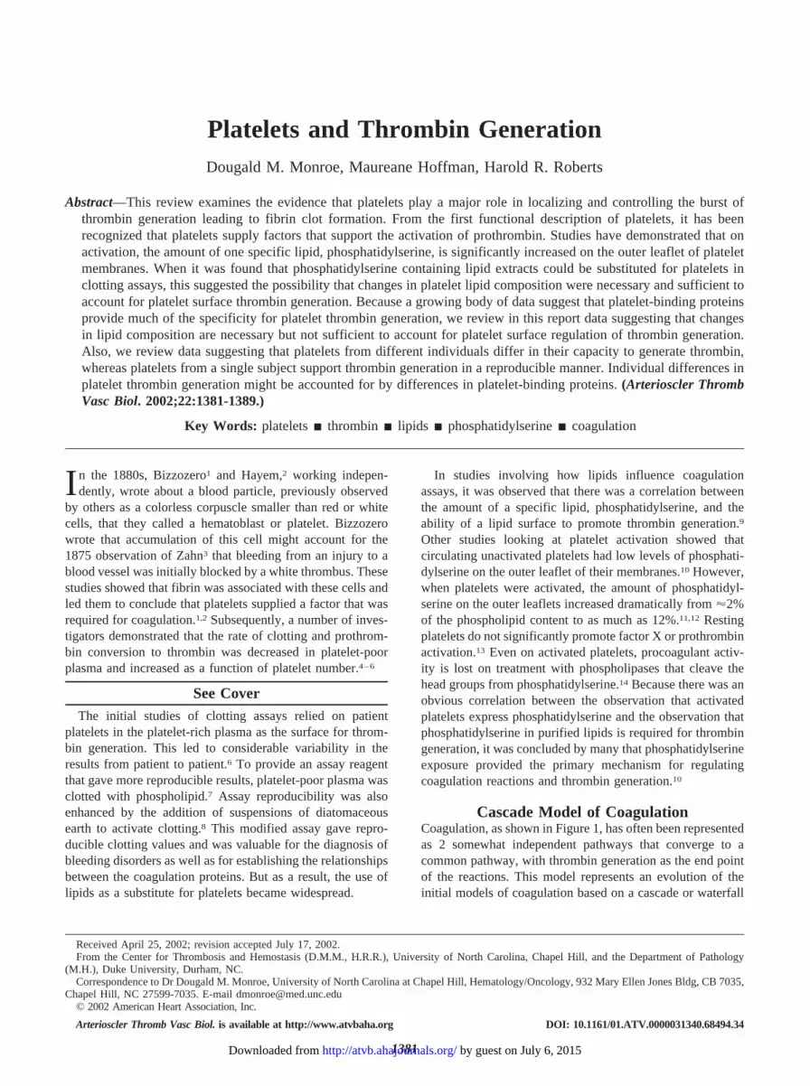
Overall, in a model system of coagulation consisting ofpurified platelets with a highly controlled concentrationsof procoagulant proteins and inhibitors at plasma concen-trations, the differences in thrombin generation betweennormal individuals were significant (Figure 6A). Theexperiments were designed such that the proteins weretightly controlled, and the platelet number was normalizedso that the source of the platelets would be the onlyvariable. The peak level of thrombin for individual 2 isalmost half the peak level in individual 1 (Figure 6A).These data, representative of results on a larger population,demonstrate that thrombin generation in this system isdependent on the source of platelets.
By use of this same cell-based model system, thecontribution of factor XI to thrombin generation wasmeasured by using platelets from different individuals.Platelets from some normal individuals show a largeincrease in thrombin generation on adding factor XI(individual 1 of Figure 6), whereas platelets from othernormal individuals show very little response to factor XI(individual 2 of Figure 6). These differences reflect realdifferences between individuals, inasmuch as the resultshave been shown to be reproducible in repeated experi-ments over a period of years.48
In conclusion, previous and recent studies show that theplatelet surface plays a central role in the promotion andregulation of thrombin generation. This regulation extendsbeyond the expression of phosphatidylserine on the outerleaflet of the platelets and requires binding proteins andreceptors that contribute to promoting and controlling theextent of thrombin generation. Cell-based models of coag-ulation provide the starting point for examining the mech-anisms by which platelets can produce a burst of thrombinin a regulated fashion.
References1. Bizzozero J. Ueber einen neuen formbestandtheil des blutes und dessen
rolle bei der thrombose und der blutgerinnung. Virchows Arch PatholAnat Physiol Klin Med. 1882;90:261–332.
Figure 6. Individual variability in thrombin gener-ation. Platelets from 2 different individuals wereisolated and counted as described previously.94
Thrombin generation was measured in a highlycontrolled setting in which platelets were incu-bated with plasma levels of proteins (prothrom-bin; factors XI, IX, X, VIII, and V; antithrombin III;and TF pathway inhibitor) and a cellular sourceof TF as described previously.29 Individuals 1and 2 are the same in panels A and B. Panel Ashows thrombin generation as a function of timeby using platelets from individual 1 (circles) andindividual 2 (triangles). Panel B shows plateletsfrom individual 1 and individual 2 in the com-plete model (filled symbol) or under identical
conditions, except that factor XI was not included in the protein mixture (open symbol). These results are representative of therange of differences seen in studies of a larger number of individuals.48
1386 Arterioscler Thromb Vasc Biol. September 2002
by guest on July 6, 2015http://atvb.ahajournals.org/Downloaded from

2. Hayem G. Sur le mécanisme de l’arrêt des hémorrhagies. C R AcadSci. 1882;95:18 –21.
3. Zahn FW. Untersuchungen über thrombose: bildung der thromben.Virchows Arch Pathol Anat Physiol Klin Med. 1875;62:81–124.
4. Eagle H. Studies on blood coagulation, IV: the nature of the clottingdeficiency in hemophilia. J Gen Physiol. 1935;18:813– 819.
5. Quick AJ, Stanley-Brown M, Bancroft FW. A study of the coagu-lation defect of hemophilia and in jaundice. Am J Med Sci. 1935;190:501–511.
6. Buckwalter JA, Blythe WB, Brinkhous KM. Effect of blood plateletson prothrombin utilization of dog and human plasmas. Am J Physiol.1949;159:322–331.
7. Bell WN, Alton HG. A brain extract as a substitute for plateletsuspensions in the thromboplastin generation test. Nature. 1954;174:880 – 881.
8. Proctor RR, Rapaport SI. The partial thromboplastin time withkaolin. Am J Clin Pathol. 1961;36:212–219.
9. Lindhout T, Govers-Riemslag JW, van de Waart P, Hemker HC,Rosing J. Factor Va-factor Xa interaction: effects of phospholipidvesicles of varying composition. Biochemistry. 1982;21:5494 –5502.
10. Bevers EM, Rosing J, Zwaal RF. Membrane phospholipids are themajor determinant of the binding site for factor X activating- andprothrombinase complexes at the surface of human platelets. AgentsActions Suppl. 1986;20:69 –75.
11. Comfurius P, Williamson P, Smeets EF, Schlegel RA, Bevers EM,Zwaal RF. Reconstitution of phospholipid scramblase activity fromhuman blood platelets. Biochemistry. 1996;35:7631–7634.
12. Solum NO. Procoagulant expression in platelets and defects leadingto clinical disorders. Arterioscler Thromb Vasc Biol. 1999;19:2841–2846.
13. Bevers EM, Rosing J, Zwaal RF. Development of procoagulantbinding sites on the platelet surface. Adv Exp Med Biol. 1985;192:359 –371.
14. Bevers EM, Comfurius P, Zwaal RF. The nature of the binding forprothrombinase at the platelet surface as revealed by lipolyticenzymes. Eur J Biochem. 1982;122:81– 85.
15. Davie EW, Ratnoff OD. Waterfall sequence for intrinsic bloodclotting. Science. 1964;145:1310 –1312.
16. MacFarlane RG. An enzyme cascade in the blood clottingmechanism, and its function as a biological amplifier. Nature. 1964;202:498 – 499.
17. Leiba H, Ramot B, Many A. Heredity and coagulation studies in tenfamilies with factor XI (plasma thromboplastin antecedent). Br JHaematol. 1965;11:654 – 665.
18. Edson JR, White JG, Krivit W. The enigma of severe factor XIdeficiency without haemorrhagic symptoms. Thromb DiathHaemorrh. 1967;18:324 –348.
19. Bolton-Maggs PH. Factor XI deficiency and its management. Hae-mophilia. 2000;6:100 –109.
20. Mertens K, Bertina RM. The contribution of Ca2� and phospholipidsto the activation of human blood-coagulation factor X by activatedfactor IX. Biochem J. 1984;223:607– 615.
21. Krishnaswamy S, Jones KC, Mann KG. Prothrombinase complexassembly: kinetic mechanism of enzyme assembly on phospholipidvesicles. J Biol Chem. 1988;263:3823–3834.
22. Bom VJ, Bertina RM. The contributions of Ca2�, phospholipids andtissue-factor apoprotein to the activation of human blood-coagulationfactor X by activated factor VII. Biochem J. 1990;265:327–336.
23. Ravanat C, Archipoff G, Beretz A, Freund G, Cazenave JP, Frey-ssinet JM. Use of annexin-V to demonstrate the role of phosphati-dylserine exposure in the maintenance of haemostatic balance byendothelial cells. Biochem J. 1992;282:7–13.
24. Walsh PN, Lipscomb MS. Comparison of the coagulant activities ofplatelets and phospholipids. Br J Haematol. 1976;33:9 –18.
25. Miletich JP, Jackson CM, Majerus PW. Properties of the factor Xabinding site on human platelets. J Biol Chem. 1978;253:6908 – 6916.
26. Nesheim ME, Furmaniak-Kazmierczak E, Henin C, Côtè G. On theexistence of platelet receptors for factor V(a) and factor VIII(a).Thromb Haemost. 1993;70:80 – 86.
27. Kjalke M, Monroe DM, Hoffman M, Oliver JA, Ezban M, RobertsHR. Active site-inactivated factors VIIa, Xa, and IXa inhibit indi-vidual steps in a cell-based model of tissue factor-initiated coagu-lation. Thromb Haemost. 1998;80:578 –584.
28. Hoffman M, Monroe DM. A cell based model of hemostasis. ThrombHaemost. 2001;85:958 –965.
29. Monroe DM, Roberts HR, Hoffman M. Platelet procoagulantcomplex assembly in a tissue factor-initiated system. Br J Haematol.1994;88:364 –371.
30. Monroe DM, Hoffman M, Roberts HR. Transmission of a proco-agulant signal from tissue factor-bearing cell to platelets. BloodCoagul Fibrinolysis. 1996;7:459 – 464.
31. Butenas S, Brummel KE, Branda RF, Paradis SG, Mann KG.Mechanism of factor VIIa-dependent coagulation in hemophiliablood. Blood. 2002;99:923–930.
32. Rauch U, Bonderman D, Bohrmann B, Badimon JJ, Himber J,Riederer MA, Nemerson Y. Transfer of tissue factor from leukocytesto platelets is mediated by CD15 and tissue factor. Blood. 2000;96:170 –175.
33. Furie B, Furie BC, Flaumenhaft R. A journey with platelet P-se-lectin: the molecular basis of granule secretion, signalling and celladhesion. Thromb Haemost. 2001;86:214 –221.
34. Bouchard BA, Tracy PB. Platelets, leukocytes, and coagulation. CurrOpin Hematol. 2001;8:263–269.
35. Nemerson Y, Repke D. Tissue factor accelerates the activation ofcoagulation factor VII: the role of a bifunctional coagulationcofactor. Thromb Res. 1985;40:351–358.
36. Wildgoose P, Kisiel W. Activation of human factor VII by factorsIXa and Xa on human bladder carcinoma cells. Blood. 1989;73:1888 –1895.
37. Kazama Y, Hamamoto T, Foster DC, Kisiel W. Hepsin, a putativemembrane-associated serine protease, activates human factor VII andinitiates a pathway of blood coagulation on the cell surface leadingto thrombin formation. J Biol Chem. 1995;270:66 –72.
38. Monkovic DD, Tracy PB. Activation of human factor V by factor Xaand thrombin. Biochemistry. 1990;29:1118 –1128.
39. Allen DH, Tracy PB. Human coagulation factor V is activated to thefunctional cofactor by elastase and cathepsin G expressed at themonocyte surface. J Biol Chem. 1995;270:1408 –1415.
40. Tracy PB, Rohrbach MS, Mann KG. Functional prothrombinasecomplex assembly on isolated monocytes and lymphocytes. J BiolChem. 1983;258:7264 –7267.
41. Warn-Cramer BJ, Maki SL, Zivelin A, Rapaport SI. Partial purifi-cation and characterization of extrinsic pathway inhibitor (the factorXa-dependent plasma inhibitor of factor VIIa/tissue factor). ThrombRes. 1987;48:11–22.
42. Broze GJ, Warren LA, Novotny WF, Higuchi DA, Girard JJ,Miletich JP. The lipoprotein-associated coagulation inhibitor thatinhibits the factor VII-tissue factor complex also inhibits factor Xa:insight into its possible mechanism of action. Blood. 1988;71:335–343.
43. Andrews RK, Shen Y, Gardiner EE, Berndt MC. Platelet adhesionreceptors and (patho)physiological thrombus formation. Histol His-topathol. 2001;16:969 –980.
44. Baumgartner HR. Platelet interaction with collagen fibrils in flowingblood, I: reaction of human platelets with alpha chymotrypsin-digested subendothelium. Thromb Haemost. 1977;37:1–16.
45. Alberio L, Safa O, Clemetson KJ, Esmon CT, Dale GL. Surfaceexpression and functional characterization of alpha-granule factor Vin human platelets: effects of ionophore A23187, thrombin, collagen,and convulxin. Blood. 2000;95:1694 –1702.
46. Viskup RW, Tracy PB, Mann KG. The isolation of human plateletfactor V. Blood. 1987;69:1188 –1195.
47. Pieters J, Lindhout T, Hemker HC. In situ-generated thrombin is theonly enzyme that effectively activates factor VIII and factor V inthromboplastin-activated plasma. Blood. 1989;74:1021–1024.
48. Oliver JA, Monroe DM, Roberts HR, Hoffman M. Thrombin acti-vates factor XI on activated platelets in the absence of factor XII.Arterioscler Thromb Vasc Biol. 1999;19:170 –177.
49. Baglia FA, Walsh PN. Thrombin-mediated feedback activation offactor XI on the activated platelet surface is preferred over contactactivation by factor XIIa or factor XIa. J Biol Chem. 2000;275:20514 –20519.
50. Scandura JM, Walsh PN. Factor X bound to the surface of activatedhuman platelets is preferentially activated by platelet-bound factorIXa. Biochemistry. 1996;35:8903– 8913.
51. Franssen J, Salemink I, Willems GM, Wun TC, Hemker HC,Lindhout T. Prothrombinase is protected from inactivation by tissuefactor pathway inhibitor: competition between prothrombin and in-hibitor. Biochem J. 1997;323:33–37.
Monroe et al Platelets and Thrombin Generation 1387
by guest on July 6, 2015http://atvb.ahajournals.org/Downloaded from

52. Rezaie AR. Prothrombin protects factor Xa in the prothrombinasecomplex from inhibition by the heparin-antithrombin complex.Blood. 2001;97:2308 –2313.
53. Wester J, Sixma JJ, Geuze JJ, Heijnen HF. Morphology of thehemostatic plug in human skin wounds: transformation of the plug.Lab Invest. 1979;41:182–192.
54. Sixma JJ, van den Berg A. The haemostatic plug in haemophilia A:a morphological study of haemostatic plug formation in bleedingtime skin wounds of patients with severe haemophilia A. Br JHaematol. 1984;58:741–753.
55. Vander Velden P, Giles AR. A detailed morphological evaluation ofthe evolution of the haemostatic plug in normal, factor VII and factorVIII deficient dogs. Br J Haematol. 1988;70:345–355.
56. Hoffman M, Monroe DM, Oliver JA, Roberts HR. Factors IXa andXa play distinct roles in tissue factor-dependent initiation of coag-ulation. Blood. 1995;86:1794 –1801.
57. Sune A, Bette-Bobillo P, Bienvenue A, Fellmann P, Devaux PF.Selective outside-inside translocation of aminophospholipids inhuman platelets. Biochemistry. 1987;26:2972–2978.
58. Bevers EM, Tilly RH, Senden JM, Comfurius P, Zwaal RF. Exposureof endogenous phosphatidylserine at the outer surface of stimulatedplatelets is reversed by restoration of aminophospholipid translocaseactivity. Biochemistry. 1989;28:2382–2387.
59. Heemskerk JWM, Bevers EM, Lindhout T. Platelet activation andblood coagulation. Thromb Haemost. 2002;88:186 –194.
60. Weiss HJ, Vicic WJ, Lages BA, Rogers J. Isolated deficiency ofplatelet procoagulant activity. Am J Med. 1979;67:206 –213.
61. Rosing J, Bevers EM, Comfurius P, Hemker HC, van Dieijen G,Weiss HJ, Zwaal RF. Impaired factor X and prothrombin activationassociated with decreased phospholipid exposure in platelets from apatient with a bleeding disorder. Blood. 1985;65:1557–1561.
62. Brooks MB, Catalfamo JL, Brown HA, Ivanova P, Lovaglio J. Ahereditary bleeding disorder of dogs caused by a lack of plateletprocoagulant activity. Blood. 2002;99:2434 –2441.
63. Furie BC, Blumenstein M, Furie B. Metal binding sites of a gamma-carboxyglutamic acid-rich fragment of bovine prothrombin. J BiolChem. 1979;254:12521–12530.
64. Koppaka V, Wang J, Banerjee M, Lentz BR. Soluble phospholipidsenhance factor Xa-catalyzed prothrombin activation in solution. Bio-chemistry. 1996;35:7482–7491.
65. Srivastava A, Quinn-Allen MA, Kim SW, Kane WH, Lentz BR.Soluble phosphatidylserine binds to a single identified site in the C2domain of human factor Va. Biochemistry. 2001;40:8246 – 8255.
66. Banerjee M, Majumder R, Weinreb G, Wang J, Lentz BR. Role ofprocoagulant lipids in human prothrombin activation, 2: solublephosphatidylserine upregulates and directs factor X(a) to appropriatepeptide bonds in prothrombin. Biochemistry. 2002;41:950 –957.
67. Srivastava A, Wang J, Majumder R, Rezaie AR, Stenflo J, EsmonCT, Lentz BR. Localization of phosphatidylserine binding sites tostructural domains of factor Xa. J Biol Chem. 2002;277:1855–1863.
68. Wu JR, Zhou C, Majumder R, Powers DD, Weinreb G, Lentz BR.Role of procoagulant lipids in human prothrombin activation, 1:prothrombin activation by factor X(a) in the absence of factor V(a)and in the absence and presence of membranes. Biochemistry. 2002;41:935–949.
69. Sumner WT, Monroe DM, Hoffman M. Variability in platelet pro-coagulant activity in healthy volunteers. Thromb Res. 1996;81:533–543.
70. De Cristofaro R, De Candia E, Landolfi R, Rutella S, Hall SW.Structural and functional mapping of the thrombin domain involvedin the binding to the platelet glycoprotein Ib. Biochemistry. 2001;40:13268 –13273.
71. Vu TK, Hung DT, Wheaton VI, Coughlin SR. Molecular cloning ofa functional thrombin receptor reveals a novel proteolyticmechanism of receptor activation. Cell. 1991;64:1057–1068.
72. Andersen H, Greenberg DL, Fujikawa K, Xu W, Chung DW, DavieEW. Protease-activated receptor 1 is the primary mediator ofthrombin-stimulated platelet procoagulant activity. Proc Natl AcadSci U S A. 1999;96:11189 –11193.
73. De Candia E, Hall SW, Rutella S, Landolfi R, Andrews RK, DeCristofaro R. Binding of thrombin to glycoprotein Ib accelerates the hydrolysisof Par-1 on intact platelets. J Biol Chem. 2001;276:4692–4698.
74. Soslau G, Class R, Morgan DA, Foster C, Lord ST, Marchese P,Ruggeri ZM. Unique pathway of thrombin-induced platelet aggre-
gation mediated by glycoprotein Ib. J Biol Chem. 2001;276:21173–21183.
75. Hayes KL, Tracy PB. The platelet high affinity binding site forthrombin mimics hirudin, modulates thrombin-induced platelet acti-vation, and is distinct from the glycoprotein Ib-IX-V complex. J BiolChem. 1999;274:972–980.
76. Suzuki H, Shima M, Kamisue S, Nakai H, Nogami K, Shibata M,Morichika S, Tanaka I, Giddings JC, Yoshioka A. The role of plateletvon Willebrand factor in the binding of factor VIII to activatedplatelets. Thromb Res. 1998;90:207–214.
77. Hultin MB. Modulation of thrombin-mediated activation of factor VIII:C bycalcium ions, phospholipid, and platelets. Blood. 1985;66:53–58.
78. Beguin S, Kumar R, Keularts I, Seligsohn U, Coller BS, Hemker HC.Fibrin-dependent platelet procoagulant activity requires GPIbreceptors and von Willebrand factor. Blood. 1999;93:564 –570.
79. Greengard JS, Heeb MJ, Ersdal E, Walsh PN, Griffin JH. Binding ofcoagulation factor XI to washed human platelets. Biochemistry.1986;25:3884 –3890.
80. Baglia FA, Jameson BA, Walsh PN. Identification and character-ization of a binding site for platelets in the Apple 3 domain ofcoagulation factor XI. J Biol Chem. 1995;270:6734 – 6740.
81. Ho DH, Baglia FA, Walsh PN. Factor XI binding to activatedplatelets is mediated by residues R(250), K(255), F(260), and Q(263)within the apple 3 domain. Biochemistry. 2000;39:316 –323.
82. Baglia FA, Walsh PN. Prothrombin is a cofactor for the binding offactor XI to the platelet surface and for platelet-mediated factor XIactivation by thrombin. Biochemistry. 1998;37:2271–2281.
83. Baglia FA, Badellino KO, Li CQ, Lopez JA, Walsh PN. Factor XIbinding to the platelet glycoprotein Ib-IX-V complex promotesfactor XI activation by thrombin. J Biol Chem. 2002;277:1662–1668.
84. Rosing J, Tans G, Govers-Riemslag JW, Zwaal RF, Hemker HC. Therole of phospholipids and factor Va in the prothrombinase complex.J Biol Chem. 1980;255:274 –283.
85. Sims PJ, Wiedmer T, Esmon CT, Weiss HJ, Shattil SJ. Assembly ofthe platelet prothrombinase complex is linked to vesiculation of theplatelet plasma membrane: studies in Scott syndrome: an isolateddefect in platelet procoagulant activity. J Biol Chem. 1989;264:17049 –17057.
86. Tracy PB, Peterson JM, Nesheim ME, McDuffie FC, Mann KG.Interaction of coagulation factor V and factor Va with platelets.J Biol Chem. 1979;254:10354 –10361.
87. Miletich JP, Kane WH, Hofmann SL, Stanford N, Majerus PW.Deficiency of factor Xa-factor Va binding sites on the platelets of apatient with a bleeding disorder. Blood. 1979;54:1015–1022.
88. Tracy PB, Nesheim ME, Mann KG. Platelet factor Xa receptor.Methods Enzymol. 1992;215:326 –360.
89. Nesheim ME, Taswell JB, Mann KG. The contribution of bovinefactor V and factor Va to the activity of prothrombinase. J BiolChem. 1979;254:10952–10962.
90. Briedé JJ, Heemskerk JW, van’t Veer C, Hemker HC, Lindhout T.Contribution of platelet-derived factor Va to thrombin generation onimmobilized collagen- and fibrinogen-adherent platelets. ThrombHaemost. 2001;85:509 –513.
91. Bouchard BA, Catcher CS, Thrash BR, Adida C, Tracy PB. Effectorcell protease receptor-1, a platelet activation-dependent membraneprotein, regulates prothrombinase-catalyzed thrombin generation.J Biol Chem. 1997;272:9244 –9251.
92. Zaman GJ, Conway EM. The elusive factor Xa receptor: failure todetect transcripts that correspond to the published sequence ofEPR-1. Blood. 2000;96:145–148.
93. Ahmad SS, Rawala-Sheikh R, Walsh PN. Comparative interactionsof factor IX and factor IXa with human platelets. J Biol Chem.1989;264:3244 –3251.
94. Hoffman M, Monroe DM, Roberts HR. Coagulation factor IXabinding to activated platelets and platelet-derived microparticles: aflow cytometric study. Thromb Haemost. 1992;68:74 –78.
95. Jones ME, Griffith MJ, Monroe DM, Roberts HR, Lentz BR. Com-parison of lipid binding and kinetic properties of normal, variant, andgamma-carboxyglutamic acid modified human factor IX and factorIXa. Biochemistry. 1985;24:8064 – 8069.
96. Rawala-Sheikh R, Ahmad SS, Monroe DM, Roberts HR, Walsh PN.Role of gamma-carboxyglutamic acid residues in the binding offactor IXa to platelets and in factor-X activation. Blood. 1992;79:398 – 405.
1388 Arterioscler Thromb Vasc Biol. September 2002
by guest on July 6, 2015http://atvb.ahajournals.org/Downloaded from

97. Monroe DM, Hoffman M, Oliver JA, Roberts HR. Platelet activity ofhigh-dose factor VIIa is independent of tissue factor. Br J Haematol.1997;99:542–547.
98. Gilbert GE, Sims PJ, Wiedmer T, Furie B, Furie BC, Shattil SJ.Platelet-derived microparticles express high affinity receptors forfactor VIII. J Biol Chem. 1991;266:17261–17268.
99. Nesheim ME, Pittman DD, Wang JH, Slonosky D, Giles AR,Kaufman RJ. The binding of 35S-labeled recombinant factor VIII toactivated and unactivated human platelets. J Biol Chem. 1988;263:16467–16470.
100. Ahmad SS, Scandura JM, Walsh PN. Structural and functional char-acterization of platelet receptor-mediated factor VIII binding. J BiolChem. 2000;275:13071–13081.
101. Gilbert GE, Drinkwater D, Barter S, Clouse SB. Specificity ofphosphatidylserine-containing membrane binding sites for factorVIII: studies with model membranes supported by glass micro-spheres (lipospheres). J Biol Chem. 1992;267:15861–15868.
102. Gilbert GE, Drinkwater D. Specific membrane binding of factor VIIIis mediated by O-phospho-L-serine, a moiety of phosphatidylserine.Biochemistry. 1993;32:9577–9585.
103. Parise LV, Phillips DR. Reconstitution of the purified plateletfibrinogen receptor: fibrinogen binding properties of the glycopro-tein IIb-IIIa complex. J Biol Chem. 1985;260:10698 –10707.
104. Marguerie GA, Plow EF, Edgington TS. Human platelets possess aninducible and saturable receptor specific for fibrinogen. J Biol Chem.1979;254:5357–5363.
105. Dale GL, Friese P, Batar P, Hamilton SF, Reed GL, Jackson KW,Clemetson KJ, Alberio L. Stimulated platelets use serotonin toenhance their retention of procoagulant proteins on the cell surface.Nature. 2002;415:175–179.
106. Bennett JS, Vilaire G. Exposure of platelet fibrinogen receptors byADP and epinephrine. J Clin Invest. 1979;64:1393–1401.
107. Chen YP, O’Toole TE, Ylanne J, Rosa JP, Ginsberg MH. A pointmutation in the integrin beta 3 cytoplasmic domain (S7523P)
impairs bidirectional signaling through alpha IIb beta 3 (plateletglycoprotein IIb-IIIa). Blood. 1994;84:1857–1865.
108. Kumar R, Beguin S, Hemker HC. The effect of fibrin clots andclot-bound thrombin on the development of platelet procoagulantactivity. Thromb Haemost. 1995;74:962–968.
109. Reverter JC, Beguin S, Kessels H, Kumar R, Hemker HC, Coller BS.Inhibition of platelet-mediated, tissue factor-induced thrombin gen-eration by the mouse/human chimeric 7E3 antibody: potential impli-cations for the effect of c7E3 Fab treatment on acute thrombosis and“clinical restenosis.” J Clin Invest. 1996;98:863– 874.
110. Li Y, Spencer FA, Ball S, Becker RC. Inhibition of platelet-dependentprothrombinase activity and thrombin generation by glycoprotein IIb/IIIareceptor-directed antagonists: potential contributing mechanism of benefitin acute coronary syndromes. J Thromb Thrombolysis. 2000;10:69–76.
111. Butenas S, Cawthern KM, van’t Veer C, DiLorenzo ME, Lock JB,Mann KG. Antiplatelet agents in tissue factor-induced blood coag-ulation. Blood. 2001;97:2314 –2322.
112. Byzova TV, Plow EF. Networking in the hemostatic system: integrinalphaiibbeta3 binds prothrombin and influences its activation. J BiolChem. 1997;272:27183–27188.
113. Brummel KE, Paradis SG, Branda RF, Mann KG. Oral anticoagu-lation thresholds. Circulation. 2001;104:2311–2317.
114. Lasne D, Krenn M, Pingault V, Arnaud E, Fiessinger JN, Aiach M, RenduF. Interdonor variability of platelet response to thrombin receptor activation:influence of PlA2 polymorphism. Br J Haematol. 1997;99:801–807.
115. Peyrou V, Lormeau JC, Herault JP, Gaich C, Pfliegger AM, HerbertJM. Contribution of erythrocytes to thrombin generation in wholeblood. Thromb Haemost. 1999;81:400 – 406.
116. Bouchard BA, Tracy PB. Platelet regulation of thrombin generationin cardiovascular disease. Ital Heart J. 2001;2:819 – 823.
117. Andrew M, Schmidt B, Mitchell L, Paes B, Ofosu F. Thrombingeneration in newborn plasma is critically dependent on the concen-tration of prothrombin. Thromb Haemost. 1990;63:27–30.
118. Butenas S, van’t Veer C, Mann KG. “Normal” thrombin generation.Blood. 1999;94:2169 –2178.
Monroe et al Platelets and Thrombin Generation 1389
by guest on July 6, 2015http://atvb.ahajournals.org/Downloaded from

Dougald M. Monroe, Maureane Hoffman and Harold R. RobertsPlatelets and Thrombin Generation
Print ISSN: 1079-5642. Online ISSN: 1524-4636 Copyright © 2002 American Heart Association, Inc. All rights reserved.
Greenville Avenue, Dallas, TX 75231is published by the American Heart Association, 7272Arteriosclerosis, Thrombosis, and Vascular Biology
doi: 10.1161/01.ATV.0000031340.68494.342002;22:1381-1389; originally published online August 1, 2002;Arterioscler Thromb Vasc Biol.
http://atvb.ahajournals.org/content/22/9/1381World Wide Web at:
The online version of this article, along with updated information and services, is located on the
http://atvb.ahajournals.org//subscriptions/
at: is onlineArteriosclerosis, Thrombosis, and Vascular Biology Information about subscribing to Subscriptions:
http://www.lww.com/reprints
Information about reprints can be found online at: Reprints:
document. AnswerPermissions and Rights Question andunder Services. Further information about this process is available in the
permission is being requested is located, click Request Permissions in the middle column of the Web page whichCopyright Clearance Center, not the Editorial Office. Once the online version of the published article for
can be obtained via RightsLink, a service of theArteriosclerosis, Thrombosis, and Vascular Biologyin Requests for permissions to reproduce figures, tables, or portions of articles originally publishedPermissions:
by guest on July 6, 2015http://atvb.ahajournals.org/Downloaded from