the tyrosine kinase network regulating mast cell activation
TRANSCRIPT
The tyrosine kinase network regulating mast cell activation
Alasdair M. Gilfillan1 and Juan Rivera2
1Laboratory of Allergic Diseases, National Institute of Allergy and Infectious Diseases, NationalInstitutes of Health, Bethesda, MD, USA2Laboratory of Immune Cell Signaling, National Institute of Arthritis and Musculoskeletal and SkinDiseases, National Institutes of Health, Bethesda, MD, USA
SummaryMast cell mediator release represents a pivotal event in the initiation of inflammatory reactionsassociated with allergic disorders. These responses follow antigen-mediated aggregation ofimmunoglobulin E (IgE)-occupied high affinity receptors for IgE (FcεRI) on the mast cellsurface,a response which can be further enhanced following stem cell factor-induced ligation ofthe mast cell growth factor receptor KIT. Activation of tyrosine kinases is central to the ability ofboth FcεRI and KIT to transmit downstream signaling events required for the regulation of mastcell activation. Whereas KIT possesses inherent tyrosine kinase activity, FcεRI requires therecruitment of Src family tyrosine kinases and Syk to control the early receptor-proximal signalingevents. The signaling pathways propagated by these tyrosine kinases can be further upregulated bythe Tec kinase Bruton's tyrosine kinase (Btk) and downregulated by the actions of the tyrosine Srchomology 2 domain-containing phosphatase 1 (SHP1) and SHP2. In this review, we discuss theregulation and role of specific members of this tyrosine kinase network in KIT and FcεRI-mediated mast cell activation.
Keywordsmast cells; FcεRI; KIT; Btk; Lyn; Fyn
Introduction: mast cell activationMast cells are tissue-resident cells of hematopoietic lineage which are derived fromCD13+CD34+KIT(CD117)+ bone marrow progenitors (1). Although mast cells play animportant role in the innate immune response elicited as part of the body's host defensereaction to invading pathogens (2,3), these cells are also responsible for the detrimentalexaggerated reactions to antigen observed in anaphylaxis, atopy, and rhinitis (4). Theseconditions are induced following the release of potent inflammatory mediators as aconsequence of receptor-mediated mast cell activation (5). Such mediators include thefollowing: (i) granule-associated mediators, including histamine, serotonin (5-hydroxytryptamine), and a variety of proteases and peptidases which are pre-synthesizedand released following fusion of the secretory granules with the cytosolic membrane; (ii)eicosanoids such as prostaglandin D2 (PGD2) and leukotriene C4 (LTC4), which aregenerated and released following activation of cytosolic phospholipase A2 (PLA2), whichcatalyzes the hydrolysis of arachidonyl-containing phospholipids to yield the eicosanoidprecursor, arachidonic acid; and (iii) cytokines including interleukin-2 (IL-2), IL-3, IL-4,
Address for correspondence: Juan Rivera Laboratory of Immune Cell Signaling, NIAMS, NIH Building 10, Room 13C103 MSC193010 Center Drive Bethesda, MD 20892-1930 USA Phone: 301 496 7592 Fax: 301 480 1580 Email: [email protected].
NIH Public AccessAuthor ManuscriptImmunol Rev. Author manuscript; available in PMC 2010 March 1.
Published in final edited form as:Immunol Rev. 2009 March ; 228(1): 149–169. doi:10.1111/j.1600-065X.2008.00742.x.
NIH
-PA Author Manuscript
NIH
-PA Author Manuscript
NIH
-PA Author Manuscript
IL-5, IL-6, IL-10, IL-13, granulocyte-macrophage colony-stimulating factor (GM-CSF), andtumor necrosis factor α (TNFα), and chemokines including CCL-2, CCL-3, CCL-5, andCXCL8, which are generated following transcriptional activation (6-9).
Although multiple receptors are capable of mediating or modifying mast cell activation(10,11), the major receptor responsible for the clinical manifestations of this response isFcεRI, the high affinity receptor for immunoglobulin E (IgE)(6,12,13). The FcεRI expressedon mast cells is a complex of four receptor subunits (Fig. 1): a single transmembrane-spanning α subunit that contains the IgE-binding site within its extracellular domain; the βchain that has four membrane spans and an immunoreceptor tyrosine-based activation motif(ITAM) within the C-terminal cytosolic domain; and two single transmembrane spanning γchains that exist as a homodimer (6). As with the β chain, the γ chain COOH-terminalcytosolic domain also contains an ITAM.
Phosphorylation of these ITAMs at their canonical tyrosine residues creates novel sites forthe binding of a variety of other signaling proteins, through the recognition of thismodification by phospho-tyrosine binding regions (domains), such as the Src homology 2(SH2) domain, found in many proteins (6,11,14,15). However, the β and γ ITAMs arefunctionally distinct. The γ ITAM provides the ability to initiate downstream signals,whereas the β ITAM functions to amplify these signals (12,16,17). This functionaldivergence is defined, at least in part, by the unique subsets of proteins that interact with therespective ITAMs (6,11,14,18,19). Thus, this multimeric receptor is capable of initiating andregulating IgE-dependent mast cell activation in a manner that elicits potent but controlledcellular responses.
In addition to the FcεRI, a number of other receptors that regulate or modify mast cellfunction are expressed on mast cells. The most critical of these receptors is KIT (Fig. 1), thereceptor for stem cell factor (SCF), also termed steel factor or KIT ligand, which isresponsible for mast cell development and homeostasis (9,20-24). As with specific Gprotein-coupled receptor (GPCR) ligands such as adenosine, PGE2 and C3a (7,10), SCF canalso substantially potentiate antigen-mediated mast cell activation (25). Both KIT- andFcεRI-mediated signaling are intimately controlled by the highly regulated balance betweenthe phosphorylated and non-phosphorylated states of component signaling proteins (Fig. 1).Whereas latent or intermediary signaling processes are primarily controlled by serine/threonine or lipid phosphorylation, immediate receptor-proximal events are primarilycontrolled by tyrosine phosphorylation as regulated by specific tyrosine kinases. Theseevents modify critical signaling proteins, which either results in enhanced activation orallows inducible binding to other interacting signaling molecules. Hence, tyrosine kinasescan be considered as the central driving force for critical downstream processes (Fig. 1)leading to mast cell activation. In this review, we thus discuss the receptor-mediatedactivation of specific classes of tyrosine kinases in mast cells and the roles that these kinasesplay in mast cell biology. However, multiple other downstream signaling processes are alsoinvolved in receptor-mediated mast cell activation. Readers are referred to a number ofrecent review articles for further discussion of these events (6,7,10,11,13,26,27).
Mast cell tyrosine kinasesRole of KIT in mast cell activation
KIT (CD117) is a protein of approximately 145 kDa whose expression is largely but notexclusively restricted to cells of hematopoietic lineage and melanocytes (28,29) (Fig. 2). It isa type III tyrosine kinase and member of the transmembrane receptors with tyrosine kinaseactivity superfamily (28,30-34). Dimerization of KIT, following ligation with SCF, inducesactivation of the inherent catalytic activity associated with the split tyrosine kinase domain
Gilfillan and Rivera Page 2
Immunol Rev. Author manuscript; available in PMC 2010 March 1.
NIH
-PA Author Manuscript
NIH
-PA Author Manuscript
NIH
-PA Author Manuscript
contained within the cytosolic COOH-terminal region (28,33). The major substrates foractivated KIT are specific tyrosine residues contained within the cytosolic domain of KIT(Fig. 2). In human KIT, these include tyrosine (Y)568 and Y570 in the juxtamembrane region,Y703, Y721, Y730, and Y747 in the linker region between split catalytic domains, Y823 andY900 in the COOH-terminal catalytic domain, and Y936 in the COOH-terminus (28).However, as discussed below, other signaling proteins are also directly phosphorylated byKIT (28,30,32). The phosphorylated residues on KIT provide docking sites for associatingsignaling molecules required for the biological responses elicited by KIT (Figs 1 and 2).These include phospholipase Cγ1 (PLCγ1)[phospho (p)Y936], the p85 subunit ofphosphoinositide 3-kinase (PI3K) (pY721), the Src family tyrosine kinases Lyn and Fyn(pY568 / pY570), the cytosolic adapter molecules Grb2 (pY703 and pY936) and SHC (py568 /pY570), and the tyrosine SH2 domain-containing phosphatase 1 (SHP1) (pY568 and pY570)(28,30) (Fig. 2). As with the FcεRI, early events associated with activated KIT likely occurin specialized gycolipid-enriched membrane microdomains conceptually termed lipid rafts(35). Thus, the binding of critical signaling molecules to phosphorylated KIT, followingauto-/trans-phosphorylation, allows recruitment of these proteins to the membrane-associated receptor signaling complex resident within these domains.
At least in human mast cells, auto/transphosphorylation of KIT is a very rapid but transientevent following SCF challenge, with peak phosphorylation being observed within 1-2 minand then substantially but not completely decreasing by 5-10 min (36). KIT-dependenttyrosine phosphorylation, both directly and indirectly, of downstream signaling moleculessuch as linker for activated T cells 2 (LAT2), Bruton's tyrosine kinase (Btk), and PLCγ1(Fig. 1) display similar kinetics (36,37). However, secondary signaling events, followingKIT activation, including the phosphorylation of AKT, components of the molecular targetof rapamycin (mTOR) cascade, and the mitogen-associated protein kinases (MAPKs)extracellular signal-regulated kinases 1 and 2 (ERK1/2), p38, and c-Jun N-terminal kinase(JNK), are delayed with maximal phosphorylation being observed 5-10 min after SCFchallenge (37-39). As with KIT phosphorylation, these responses are rapidly downregulated(37-39); thus, they likely become refractory to further SCF challenge. However, downstreamsignaling events (Fig. 1), especially those regulating transcription, must persist to preventthe mast cells from undergoing apoptosis. Certainly, mast cells deprived of SCF will displaymanifestations of apoptosis and start to die within 24-48 h (40). Thus mice defective in SCF(Sl/Sld) (41) or functional KIT (W/Wv and W/Wsh) (42,43) are deficient in mast cells. Thisobservation illustrates that SCF-induced KIT activation is an essential process for theexpansion of mast cells from their progenitor cells and their subsequent maturation andsurvival in their resident tissues (24). In addition to its role in mast cell development andhomeostasis, SCF-induced KIT activation may contribute to the homing of mast cells totheir sites of residence in vivo, as suggested by its potent chemotactic properties for bothmouse and human mast cells (38,44) and its ability to promote adhesion of mast cells toextracellular matix proteins (38,45).
A number of activating mutations have been described to be present in KIT that result inconstitutively enhanced kinase activity associated with specific myeloproliferative disorders(9,31). In particular, an activating aspartic acid to valine substitution at amino acid position816 within the second catalytic domain of human KIT is linked to systemic mastocytosis(9,22), whereas a valine to glycine substitution at amino acid position 560, which enhancesthe catalytic activity by preventing binding of and downregulation of KIT activity by SHP1,is associated with gastrointestinal stomal cell tumors (GIST).
SCF, at least under experimental conditions, can markedly enhance mast cell activation,although on its own, at physiological concentrations, SCF has little effect on mast celldegranulation or cytokine production (36). Of note is the ability of SCF to dramatically
Gilfillan and Rivera Page 3
Immunol Rev. Author manuscript; available in PMC 2010 March 1.
NIH
-PA Author Manuscript
NIH
-PA Author Manuscript
NIH
-PA Author Manuscript
enhance degranulation at concentrations of antigen that produce little detectabledegranulation in the absence of SCF (46). Should such a relationship exist in vivo, then it isquite conceivable that SCF may be as equally important as antigen in disorders such asasthma, anaphylaxis, and rhinitis resulting from exaggerated activation of mast cells.Although there is little evidence to support such a concept in vivo, the role of the SCF-KITinteraction in mast cell homeostasis implies that antigen-mediated mast cell activation invivo would occur with ongoing KIT activation. Of particular interest is the reportedincidence of anaphylaxis in approximately 50% of mastocytosis patients (47) and thecorresponding observation that a subset of patients diagnosed with idiopathic anaphylaxisdisplay markers of mastocytosis, including the D816V mutation in KIT (48). Theseobservations would imply that the aberrant KIT activity associated with mastocytosisproduces a hyperactive mast cell phenotype.
Our studies and those conducted in collaboration with the laboratory of Dr. Michael Beaven(NHLBI/NIH) have sought to determine the mechanisms regulating the synergisticenhancement of antigen-mediated mast cell degranulation and cytokine production by SCF.Initial studies conducted in human mast cells revealed that the inability of SCF to inducedegranulation in the absence of SCF may be related to its similar inability to promote theactivation of protein kinase C (PKC) (36), an essential signaling component for theaforementioned response (7). Conversely, the enhanced degranulation and potentiallyenhanced cytokine generation observed following concurrent challenge with SCF andantigen appeared to be linked to a similar enhancement of PLCγ1 activation leading to anexaggerated calcium signal (36). These studies were extended to investigate which receptor-proximal events regulated this response. By examining the early tyrosine phosphorylationevents elicited by both receptors, we determined that both antigen and SCF induced therapid phosphorylation of the transmembrane adapter molecule LAT2 (formerly termed non-T cell activation linker or linker for activated B cells) (Fig. 1), in both mouse and humanmast cells (46). Furthermore, concurrent addition of antigen and SCF elicited a greaterresponse than that induced by either agent alone (36,46). In contrast to the antigen-mediatedresponse, however, LAT2 phosphorylation in response to SCF was observed in the absenceof LAT1 phosphorylation (46).
Although the FcεRI required the cytosolic tyrosine kinases Lyn and spleen tyrosine kinase(Syk) for antigen-induced LAT2 phosphorylation, SCF/KIT-induced LAT2 phosphorylationrequired neither Lyn or Syk nor other tyrosine kinases including Fyn and Btk (46,49). Thesedata, which suggested that KIT could directly phosphorylate LAT2, were confirmed instudies utilizing KIT and LAT2 constructs transduced in 293T cells (49). Further studiesconducted in 293T cells, using mutant LAT2 constructs, surprisingly revealed that the suiteof tyrosine residues phosphorylated by KIT, Lyn, and Syk were dramatically different (49).Whereas Syk primarily phosphorylated tyrosines (Y136, Y193 and Y233 of human LAT2)contained within putative binding sites (-YXN-) for the cytosolic adapter molecule Grb2,Lyn and KIT phosphorylated tyrosines (Y95, Y118 and Y136 for Lyn and Y118 for KIT)within the putative Grb2-binding sites and tyrosines (Y110 and Y119 for Lyn and Y110 forKIT) outside of the putative Grb2-binding sites (49). In pull down studies, we observed thatPLCγ1 could bind to the two terminal tyrosines (Y193 and Y233) phosphorylated by Syk.However, as these are not recognized binding sites for PLCγ1, these interactions likely areindirect via Grb2 or similar cytosolic adapter molecule (49). Although we could notdetermine the signaling molecules recruited to LAT2 following its phosphorylation by Lynor KIT, the flanking sequence around Y110, which is phosphorylated by Lyn and KIT, ishighly homologous to the sequence containing Y568 and Y570, which is phosphorylated inKIT following its activation. This sequence in KIT has been proposed to bind SHP1, Lyn,and Fyn. Thus, it is possible that these molecules are also recruited to LAT2 in a similarKIT- or Lyn-dependent manner.
Gilfillan and Rivera Page 4
Immunol Rev. Author manuscript; available in PMC 2010 March 1.
NIH
-PA Author Manuscript
NIH
-PA Author Manuscript
NIH
-PA Author Manuscript
While the identity of the molecules recruited to LAT2 following phosphorylation by KIThave yet to be elucidated, gene knockdown studies have provided evidence to support a rolefor KIT-mediated LAT2 phosphorylation and indeed FcεRI-dependent LAT2phosphorylation in mast cell activation. In this respect, human mast cells pre-incubated for64 h with small interfering RNA (siRNA) oligonucleotides targeting LAT2 or human mastcells stably transfected with LAT2-targeted short hairpin (shRNA) display significantlyreduced antigen-mediated and KIT-enhanced degranulation (46,49). In addition, althoughLAT1 was not observed to be phosphorylated by KIT, knockdown of LAT1 alsosubstantially reduced these responses (46). These and other observations led us to proposethat whereas LAT1 may be the principle coordinator of signaling pathways regulatingantigen-mediated degranulation through the eventual activation of PLCγ1, LAT2 may serveto coordinate an amplification pathway to further enhance or maintain PLCγ1 activity (7). Byinteracting with this latter pathway, assuming that LAT1 is concurrently phosphorylatedfollowing antigen challenge, KIT provides the necessary phosphorylation events required forthe enhancement of PLCγ1 and subsequently elevation of the intracellular calciumconcentration. As further discussed in the section describing the roles of Tec kinases in mastcell activation, this latter response may be mediated via a PI3K-Btk regulated pathway,downstream of LAT2 phosphorylation.
Although KIT possesses inherent tyrosine kinase activity, studies revealed that specific KIT-mediated mast cell responses are also dependent of the activation of the Src family tyrosinekinases Lyn and Fyn. Activated KIT both binds, as discussed above, and activates Lyn andFyn. Mutation of the pY567 (mouse sequence) Src kinase-binding site in KIT abrogates SCF-induced calcium flux and chemotaxis (50). Similarly SCF-mediated mast cell proliferationand chemotaxis is substantially reduced in lyn-/- bone marrow-derived mast cells (BMMCs)(51-53), and SCF-mediated chemotaxis is reduced in fyn-/- BMMCs (51,54). In lyn-/-
BMMCs, this defect was associated with decreased KIT phosphorylation (pY823, pY703,pY719, pY730 and pY567/570, mouse sequence) in addition to a decrease in activation of JNKand decrease in STAT3 phosphorylation (51). In contrast to these observations, the defectivechemotactic response observed in fyn-/- BMMCs was not associated with defective KITphosphorylation but was associated with a decrease in KIT-mediated SHP-2 and p38phosphorylation (51). Although it is unclear whether Fyn plays a role in the ability of SCF toenhance either antigen-mediated degranulation or cytokine production, as discussed later,Lyn is essential for this response. The roles of Lyn and Fyn in the context of FcεRI-mediated mast cell signaling are discussed in greater detail below.
Src family kinases in mast cell activationThe role of Src family protein tyrosine kinases (Src PTKs) in the FcεRI-mediated activationand function of mast cells is complex. The structure of these kinases is well defined (Fig. 2),and their mechanism of activation has been previously reviewed (55) (Fig. 3). These kinasesare used by a variety of cell surface receptors on the mast cell (56), and accumulatedevidence suggests that they act in both a positive and negative manner to control theactivation and subsequent responses of the mast cell. In this section, we primarily discussthe recent advances in how the FcεRI utilizes these Src PTKs to initiate and regulate cellularsignals and responses. The findings demonstrate an increasing complexity in how Src PTKsfunction and shed new light on their key regulatory role in controlling mast cell activation.
Coupling of Src kinase activity to FcεRIThe lack of intrinsic kinase activity of the multimeric FcεRI makes its association with akinase a prerequisite for transducing signals that elicit mast cell responses (Fig. 3). Theactivity of the Src PTK Lyn is essential for phosphorylation of the tyrosine residues in theITAMs through transphosphorylation (57). However, interaction of FcεRI with Lyn must
Gilfillan and Rivera Page 5
Immunol Rev. Author manuscript; available in PMC 2010 March 1.
NIH
-PA Author Manuscript
NIH
-PA Author Manuscript
NIH
-PA Author Manuscript
precede the former's phosphorylation. Such interactions have been postulated to occur viathe residence of Lyn in lipid rafts and the recruitment of FcεRI into these domains upon itsaggregation by cell-bound IgE antibodies that encounter a specific antigen (58-60) (Fig. 3).Lipid rafts are not only enriched in cholesterol but also in sphingolipids and other saturatedphospholipids. They also contain a variety of signaling proteins including Lyn kinase. Thus,aggregation of FcεRI may cause coalesence of receptors with Lyn based on the biophysicalproperties of the surrounding lipid milleu around both the FcεRI and Lyn. However, somestudies argue that protein-protein interactions are required for FcεRI phosphorylation, basedon the findings that a small fraction of Lyn can be found to weakly interact with the FcεRIβsubunit prior to engagement of this receptor and that exclusion of Lyn from lipid rafts onlymodestly affects FcεRI phosphorylation (61-63). It is known that the interaction of Lyn withFcεRI is greatly enhanced by stimulation of this receptor. This increased interaction ismediated by Lyn SH2 domain binding to the phosphorylated Y219 in the FcεRIβ ITAM(18,19). Mutation of Y219 of the FcεRIβ ITAM causes a marked reduction in FcεRIphosphorylation, suggesting that this interaction is key to maintain receptor phosphorylation.Obviously, FcεRI-Lyn interactions are likely to be greatly increased when both of theseproteins are concentrated in lipid rafts. Thus, while there is still substantial uncertainty as towhether lipid rafts are necessary for initiating FcεRI phosphorylation, their contribution tothe maintenance of receptor phosphorylation appears more certain (59,62,64).
The aforementioned requirements of protein-protein versus protein-lipid interactions,however, should not be viewed as mutually exclusive. Two-photon fluorescence lifetimeimaging microscopy and fluorescence polarization anisotropy imaging studies have recentlyprovided support for a mutually inclusive model. Studies in which the cholesterol-richmembrane domains are labeled with the lipid analog dil-C18 and FcεRI is labeled with Alexa488-IgE demonstrated that aggregation of FcεRI causes the coalesence of dil-C18-labeledmembrane domains and redistribution to membrane patches containing FcεRI (65). Thisresults in an increased fluorescence lifetime of both dil-C18 and Alexa 488-IgE; an increasein dil-C18 fluoresence lifetime was previously shown to reflect increased lipid order.Furthermore, flouresence resonance energy transfer (FRET) occurs between the Alexa 488-labeled receptor and dil-C18-labeled domains (66) following FcεRI engagement, suggestingthe reordering and close proximity of the cholesterol-enriched membrane and the aggregatedFcεRI. These studies are consistent with prior work using high resolution electronmicroscopy (67,68). Here it was found that compartmentation of FcεRI and Lyn occured insmall electron-dense microdomains prior to FcεRI engagement. Consistent with the abovestudies, aggregation of FcεRI enhanced the size of these microdomains increasing thenumbers of receptors and Lyn within.
Collectively, these studies define the cholesterol-rich membrane microdomains as a lipidenvironment that is dynamic, small, and likely to contain limited numbers of proteins priorto cell stimulation. A unifying hypothesis for the differing views on how Lyn couples toFcεRI phosphorylation emerges. One might postulate that a small fraction of FcεRI is found,at any given time, within these dynamic cholesterol-rich microdomains, an environment thatmay also contain Lyn kinase. In this model, aggregation of FcεRI would inducetransphosphorylation with a neighboring receptor as it brings together receptors with orwithout Lyn kinase. The coalescence of the lipid domains would stabilize and/or increaseFcεRI phosphorylation and cause assembly of a stable signaling complex. This model is alsoin agreement with the finding that FcεRI aggregation induces membrane changes that allowcoalesence of proteins and lipids required for efficient propagation of signals (65).
The Src PTK Fyn (Fig. 2) has also been shown to be important for activation of mast cellsupon FcεRI aggregation and to co-immunoprecipitate with this receptor (69). Functionally,the loss of Fyn in mast cells impairs mast cell degranulation and cytokine production
Gilfillan and Rivera Page 6
Immunol Rev. Author manuscript; available in PMC 2010 March 1.
NIH
-PA Author Manuscript
NIH
-PA Author Manuscript
NIH
-PA Author Manuscript
(69,70). How Fyn might couple to FcεRI was until recently unknown. In unpublished work(Nora Fierro and Juan Rivera), Fyn was found to associate with the FcεRIβ ITAM andappears to be recruited, similarly to Lyn, via an SH2 domain-phosphotyrosine interaction(Fig. 3). However, unlike Lyn, Fyn does not appear to participate in the phosphorylation ofFcεRI (70). Two striking features are revealed in the recent work (Nora Fierro and JuanRivera, unpublished observations). First, chemical cross-linking studies, whereby Fyn orLyn are covalently bound to the FcεRI, demonstrated that distinct populations of receptorsassociate with Fyn or Lyn. Second, FRET experiments show that Lyn can be found toassociate with FcεRI in resting conditions, whereas Fyn association requires FcεRIaggregation. Moreover, Lyn association with FcεRI appears to be more transient than that ofFyn. These findings suggest that these interactions may be taking place in differentcompartments, a postulate that is currently under investigation. Nonetheless, the findings todate argue that the interaction of FcεRI with Fyn is likely to be spatiotemporally distinctfrom that with Lyn but is key in promoting the downstream signaling required for mast celleffector responses.
Src kinases: beyond FcεRI proximal interactionsFyn kinase
Beyond its coupling to FcεRI, Fyn primarily functions to positively regulate mast cellresponsiveness. Previous studies have demonstrated a role for this Src PTK in regulating theactivation of PI3K (69,71) and thus its product phosphatidylinositol-3,4,5-triphosphate(PIP3)(Fig. 1). Fyn-deficient mast cells show a substantial reduction in PIP3, whichcorrelates with the reduced degranulation response of these cells. Moreover, considerableevidence has been accumulated for an essential role of PIP3 in mast cell responses. This hasbeen demonstrated through varied approaches that encompass the use of PI3K inhibitors andthrough genetic manipulation of PI3K itself (72). PIP3 is a potent lipid second messengerthat functions to assemble signalosomes at the cell membrane and coordinates the activity ofmultiple signaling proteins with calcium responses, the cytoskeleton, and the nucleus (73).
In an early study of Fyn function in mast cells, a more transient calcium response was notedrelative to wildtype cells (69). Using more sensitive fluorometric methods, we found that theloss of Fyn showed little effect on the mobilization of calcium from intracellular stores butinstead impaired calcium influx (Ryo Suzuki and Juan Rivera, unpublished observation).The mechanism for this effect is unclear; however, several possibilities exist. One possiblemechanism is through Fyn-dependent phosphorylation of plasma membrane calciumchannels. Members of the transient receptor potential channel (TRPC) family are knownsubstrates for Src family kinase, like Fyn (74). Moreover, TRPC channels have been shownto contribute to calcium influx in a mast cell line (RBL-2H3) (75). A second scenario is thatFyn could contribute to calcium influx through its role in the activation of sphingosinekinase 2 (SphK2) and the generation of sphingosine-1-phosphate (S1P) (Fig. 1). S1P is ahighly biologically active molecule that functions as an intracellular lipid second messengeras well as a ligand for a family of GPCRs expressed on the cell surface of many cell types.Fyn kinase is required for the activation of SphK2 (76). Much like the defective calciuminflux that is seen upon loss of Fyn expression, the genetic deletion of SphK2 also caused aloss in calcium influx (77). However, whether Fyn's control of calcium influx is entirelymediated via its role in the production of S1P or is combined with its ability tophosphorylate calcium channels is not known. Obviously, the intracellular targets of Fynand S1P must be identified to appropriately address this issue with certainty.
Various other functions of Fyn have been described in mast cells. Fyn has been shown tophosphorylate PLD2 in the RBL mast cell line (Fig. 1) (78), and it is thought that this maycontribute to its role in promoting degranulation, as PLD2 activity is required for this event
Gilfillan and Rivera Page 7
Immunol Rev. Author manuscript; available in PMC 2010 March 1.
NIH
-PA Author Manuscript
NIH
-PA Author Manuscript
NIH
-PA Author Manuscript
(79). Fyn has also been implicated in the organization of the cell cytoskeleton, in vesicletrafficking, and chemotaxis. Regulation of microtubule formation and its nucleation from themembrane in activated mast cells was shown to be dependent on complexes of γ-tubulinwith Fyn and Syk kinases (80,81). Microtubule function and the ability to translocatepreformed mast cell granules to the plasma membrane also appears to depend on Fyn and isindependent of calcium (82). Moreover, association of Fyn with cytoskeletal proteins likevimentin has been shown in mast cells, and such proteins are likely targets of Fyn activity(83). Thus, beyond its role in FcεRI proximal signaling, it appears that Fyn functions inmany of the cellular processes required for mast cell activation and effector function.
Lyn kinaseAs mentioned above, Lyn kinase plays a positive role in activating mast cells through itsphosphorylation of FcεRI (Figs 1 and 3). In addition, Lyn phosphorylation of othersubstrates such as Syk, Btk, the adapter LAT2, and the cytokine regulator Tom1L1(reviewed in 6,84), seemingly has a positive effect on mast cell functions. Unlike Fyn,however, Lyn also plays an important role as a negative regulator of mast cell effectorresponses (85-87). Lyn-deficient mice develop atopic-like allergic disease, and mast cellsderived from these mice are hyperresponsive when stimulated via FcεRI (85). We now knowthat Lyn is required for phosphorylation of the lipid raft-localized C-terminal Src kinase(Csk)-binding protein (Cbp) and, thus, for membrane targeting of the regulatory kinase Csk,a kinase that negatively regulates Src PTKs through phosphorylation of a negativeregulatory tyrosine in their COOH-termini (Figs 4 and 5). This causes Src PTKs to assume a`closed' conformation through intramolecular interaction of the phosphorylated C-terminaltyrosine with the kinases' own SH2 domain. This regulatory step is mediated by the Lynkinase that is localized in lipid rafts, where Cbp is also found, and is required todownregulate Fyn kinase activity (63) (Fig. 4).
Another dominant mechanism by which Lyn exerts negative control on mast cell effectorresponses is through the SH2 domain-containing inositol phosphatase-1 (SHIP-1) andSHIP-2. In the absence of Lyn, SHIP-1 activity is increased, causing an increase in theconcentration of intracellular PIP3 (86). As mentioned above, PIP3 is important for mast celldegranulation and other responses. Unlike Fyn-deficient mast cells, Lyn-deficient mast cellshave increased levels of intracellular PIP3, which is associated with their enhanceddegranulation (85). Both SHIP-1- and SHIP-2-deficient mast cells also show an enhanceddegranulation and cytokine response (88,89). Furthermore, constitutive increases inintracellular PIP3 through downregulation of another inositol phosphatase, phosphatase andtensin homolog (PTEN), also results in enhanced degranulation and cytokine production(90). Thus, Lyn plays a key negative regulatory role in controlling the levels of PIP3 andthus suppressing the extent of a mast cells response.
One proposed mechanism by which Lyn kinase may play both a positive and negative rolein mast cell activation has been revealed by studies of the effect of low or high strengthstimulation on responses in wildtype and Lyn-deficient mast cells (87). This work showsthat Lyn activity is required for mast cell degranulation and cytokine production whenencountering a low strength stimulus. In contrast when encountering a high strengthstimulus, Lyn activity is dispensable for the observed enhanced degranulation and cytokineproduction (87). These studies also conclude that negative regulation by Lyn is mediatedthrough its interaction with the FcεRIβ. Taken together with the finding that Lyn's negativerole is predominantly mediated by the pool of Lyn in lipid rafts (63), one might concludethat the interaction of Lyn with FcεRIβ is likely to occur upon coalesence of these domains.Therefore, this fits well with the concept that a strong stimulus, which leads to extensivecoalesence of microdomains (65,66), would serve to promote the negative role of Lyn kinasein order to control the extent of the inflammatory response.
Gilfillan and Rivera Page 8
Immunol Rev. Author manuscript; available in PMC 2010 March 1.
NIH
-PA Author Manuscript
NIH
-PA Author Manuscript
NIH
-PA Author Manuscript
Fgr, Hck, and Src kinasesThe Src PTKs, Fgr, Hck, and Src are abundantly expressed in mast cells (13). However,much less is known about their role in mast cell activation and function. Early studiesshowed that Src co-immunoprecipitated with FcεRI (91), but more recent work suggests thatthese observations may have been due to the failure to solubilize lipid rafts with thedetergent conditions used in these studies. In contrast, Src has been found to associate withPKCδ (92), a kinase that phosphorylates the FcεRIγ on threonine residues (93), but the roleof Src in regulating this event is not known. Fgr, like Fyn, is able to phosphorylate PLD2 inmast cells (78). Thus, it also likely contributes to the activation of this lipase and to its rolein promoting mast cell degranulation. In contrast, Fgr (and Lyn and Hck as well) does notappear to be required for mast cell spreading and lamellipodia formation in mast cells (54).Moreover, our own unpublished observations (Yasuko Furumoto, Santa Eglite, and JuanRivera) rule out an appreciable direct role for Fgr, Hck, or Src in mast cell degranulation.Nonetheless, Hck appears to have an indirect role in promoting mast cell degranulation. Thisview is supported by studies on Hck-deficient mast cells (94), where the findings point toHck-mediated negative regulatory control on Lyn activation. Thus, in the absence of Hck,Lyn activity is substantially increased suppressing degranulation and cytokine productionwhen cells encounter a strong stimulus. The mechanism for this effect is unknown, but itdoes not appear to be mediated through loss of Cbp phosphorylation and, thus, the loss ofCsk-mediated control of Lyn activity, since Cbp phosphorylation was enhanced. Moreover,Fyn activity was normal in the absence of Hck, suggesting normal Cbp/Csk-mediatedregulation of Fyn (Fig. 4). One possible explanation comes from studies demonstrating thatthe lipid raft environment is essentially devoid of phosphatases that dephosphorylate theactive site tyrosine of Lyn (58) as well as studies showing that the loss of some lipid raftproteins like LAT1 causes the increased presence of similar proteins, like LAT2, in thesedomains (95,96). Furthermore, we have found that the loss of Lyn expression causes moreFyn to localize in lipid rafts (Nora Fierro and Juan Rivera, unpublished observations). Thus,if the loss of Hck expression results in the increased localization of Lyn in lipid rafts, thiswould be consistent with increased Lyn activity and with the finding that the negativeregulatory role of Lyn is localized to lipid raft-residing Lyn kinase. While this activityremains to be tested, it is clear that Hck appears to dampen the negative function of Lynkinase. Collectively, the few studies to date on Fgr, Hck, and Src suggest that these kinaseshave contributory but not dominant roles in mast cell effector functions.
Syk kinase in mast cell activation and functionSyk and its closely related family member ζ-associated protein of 70 kDa (ZAP-70) havelong been recognized as key components of signaling pathways in B cells and T cells,respectively (97,98). The structure of Syk (Fig. 2) is well defined and is a topic that has beenextensively reviewed (99-101). Beyond its catalytic function, key elements for Syk functioninclude its NH2-terminal tandem SH2 domains, and the multiple phosphorylated tyrosineresidues that form novel binding sites for interacting proteins of which the majority arefound in the interdomain A and B regions of this kinase (Fig. 2). Syk activation occursprimarily through its binding of phosphorylated ITAMs on receptors that utilize its activityfor eliciting cellular effector functions (reviewed in 99,101). However, recent reportssuggest alternate methods of activation, such as by PKCδ's phosphorylation of Syk (102).An alternatively spliced form of Syk (SykB) has also been identified that differs from Sykby lacking 23 amino acids in the interdomain B region (reviewed in 99,101). While thecatalytic activity of SykB is comparable to that of Syk, it seems less efficient in binding tophosphorylated ITAMs.
Syk is essential for the propagation of signals in mast cells (6), following binding to thephosphorylated ITAMs of FcεRIγ (103,104) (Fig. 1). Syk binding to the phosphorylated γ
Gilfillan and Rivera Page 9
Immunol Rev. Author manuscript; available in PMC 2010 March 1.
NIH
-PA Author Manuscript
NIH
-PA Author Manuscript
NIH
-PA Author Manuscript
chain ITAM is a necessary step for its activation in response to FcεRI stimulation. Whileweak interaction of Syk with FcεRIβ phospho-ITAM peptides can be observed in vitro, thereis little evidence of this interaction in vivo. Once activated, Syk is essential in theamplification of mast cell signaling and in driving normal mast cell effector responses(105,106). Syk function in mast cells and other immune cells has been well reviewed(99,101,107), so it will be covered very briefly herein. However, it is important to note thatSyk deficiency results in a non-responsive mast cell that shows no calcium response,degranulation, or cytokine production following FcεRI stimulation (105,108). Thus, Syk iscritical for calcium responses. Key to its amplification function and its ability to regulatecalcium responses is its capacity to interact with multiple signaling proteins and tophosphorylate the key substrates required to assemble signaling networks, namely adaptermolecules.
There are 10 tyrosine residues that can be modified by phosphorylation when Syk isactivated. These phospho-Ys appear to mediate distinct functions. Y130 is localized ininterdomain A and is thought to play a role in both mediating the activation of Syk and itsrelease from interacting with an antigen receptor (109). Somewhat consistent with theseresults, in B cells the deletion of interdomain A resulted in the loss of interaction of Sykwith the B-cell antigen receptor (BCR) (110). However, in this case, Syk becamephosphorylated independent of BCR engagement and failed to induce calcium responses.There are five Y residues in interdomain B (Fig. 2), and Y290 lies within the 23 amino acidinsert that is deleted in the alternatively spliced SykB isoform. As indicated above, SykB isless effective in interacting with ITAMs. However, upon FcεRI stimulation, the mutation ofY290 does not appear to affect the function of Syk (111). In contrast, Y317, Y342, and Y346
all appear to be important for Syk function. Mutation of Y317 has been shown to result in again of function in mast cells (112). The corresponding Y residue in ZAP-70 was shown tobind the negative regulator c-Cbl (113), which has been described to regulate thedegradation of active Syk (114). Phosphorylation of Y342 of Syk is thought to be importantfor interaction and phosphorylation of Vav1 (115) (Fig. 1). However, in mast cells, mutationof Y342 did not alter the phosphorylation of Vav1 but instead showed decreased PLCγphosphorylation and a decreased ability to interact with phosphorylated ITAMs (116).Importantly, this mutant form of Syk failed to reconstitute degranulation when expressed inSyk-deficient mast cells. Based on its analogous Y residue in ZAP-70, mutation of Y346 ofSyk in mast cells might have been predicted to cause a loss of PLCγ interaction and functionthat would manifest in poor mast cell degranulation (reviewed in 99). However, mutation ofthis Y346 showed minimal effect on mast cell degranulation and probing of this site with aphospho-Y346-specific antibody suggests that this site is not heavily phosphorylated uponFcεRI stimulation of mast cells (116). At the moment there is no information on the functionof Y358 of Syk. Y519 and Y520 lie in the activation loop of Syk. Mutation of either or both ofthese Y residues showed no loss in Syk kinase activity (106). Surprisingly, however, uponexpression of these Syk mutants, at levels comparable to endogenous Syk in mast cells,there was a failure to transduce signals with no observable protein tyrosine phosphorylationor mast cell degranulation. Overexpression of these mutants could partially restore somesignaling. Thus, these Y residues appear to be important in the propagation of signals by Sykthrough an as yet to be defined mechanism. This view is consistent with the finding thattheir phosphorylation mirrors the downstream propagation of signals required for mast celldegranulation (117). The function of the COOH-terminal cluster of Y624 and Y625 in mastcells is not known. However, in T cells, expression of a Syk chimera with mutations at thesesites has demonstrated enhanced T-cell antigen receptor signaling, thus implying a negativeregulatory role for these tyrosines (reviewed in 99).
Syk activation is central for mast cell effector function and the allergic response. Thus, itsactivity should be tightly regulated to avoid the injurious effects of spurious or sustained
Gilfillan and Rivera Page 10
Immunol Rev. Author manuscript; available in PMC 2010 March 1.
NIH
-PA Author Manuscript
NIH
-PA Author Manuscript
NIH
-PA Author Manuscript
activation. Regulation of Syk activity occurs at multiple levels. Syk is maintained in aninactive state through intramolecular interactions of its SH2 domains with phosphorylated Yresidues (Y130 and Y317 have been implicated in this binding) in its interdomains A and B(100,118). What causes the loss of this intramolecular binding so that Syk can achieve theappropriate conformation to be active is not known, since phosphatase-mediateddephosphorylation of these sites has not been demonstrated. However, activation mightsimply be mediated through a shift in the equilibrium where the abundance ofphosphorylated ITAMs upon FcεRI stimulation may provide a higher affinity binding sitethat stabilizes Syk in the open conformation leading to its increased activity. This view issupported by the finding that Syk can be activated by incubation with phosphorylated ITAMpeptides (104,119). This also suggests a potential negative regulatory step in its activationmight be through dephosphorylation of the ITAMs (120), a topic further discussed in afollowing section. Once activated, Syk activity can be controlled by multiple mechanisms.In B cells, Syk binds to Cbl and is ubiquitylated and degraded (114). In mast cells, the lossof Cbl-b expression (but not that of c-Cbl) caused enhanced Syk phosphorylation andenhanced mast cell effector responses (121).
Tec kinases in mast cell activationBtk
Tec kinases are a group of structurally conserved cytosolic tyrosine kinases which comprisefive family members: Btk, Etk (BMX), Itk, Rlk, and Tec (122,123). Although mast cellsexpress four of these members, Btk, Itk, Rlk, and Tec, only Btk has been documented todirectly play a major role in mast cell responses. A lack of Btk is linked toimmunodeficiency in both the mouse [X-linked immunodeficiency (XID)] and human (X-linked agammaglobulinemia) as a consequence of defective B-cell signaling (124). As withother members of the Tec kinase family, Btk contains an N-terminal pleckstin homology(PH) domain, a Tec homology (TH) domain, an SH2 domain, an SH3 domain, and a C-terminal catalytic domain (122) (Fig. 2). Btk is activated following phosphorylation ofcritical tyrosines as a consequence of receptor aggregation. The initial Src kinase-dependentphosphorylation of Y551 within the activation loop (125) results in an increase in catalyticactivity leading to autophosphorylation at Y223 within the SH3 domain (126), therebyfurther enhancing its activation status. As assessed by immunoprecipitation studies, in vitrokinase assays, and by the use of phospho-specific antibodies (recognizing pY223 and pY551),Btk is rapidly activated upon tyrosine phosphorylation in mast cells following challengewith either antigen or SCF, where maximal phosphorylation is observed within 60 s of cellactivation (37). Although not so apparent at early time points (< I min), at later time points(>5 min) a synergistic enhancement of Btk phosphorylation is observed when both agentsare added concurrently. These events are largely dependent on Lyn, as evidenced by themarkedly attenuated Btk phosphorylation at position Y233 in lyn-/- BMMCs challenged witheither SCF or antigen (37). However, as Btk phosphorylation has been reported to also bedependent on Syk (127), it is possible that such control is indirect via Syk activation.
In addition to being phosphorylated, Btk is also translocated from the cytosol to themembrane in response to antigen (125). This is necessary for Btk to interact with thereceptor-signaling complex (Fig. 1) and to access its substrate(s). As in B cells, thisinteraction likely involves binding of the PH domain of Btk to membrane-associated PIP3following its generation as a consequence of the activation of PI3K (128,129). Althoughadditional molecular interactions for Btk and other Tec family members have beendocumented in other cell types including B and T cells (122,123), these interactions in mastcells are relatively unknown. Certainly, Btk does not appear to bind to the receptor subunits(130). Nevertheless, in addition to PIP3, under experimental conditions, the PH domain ofBtk can interact with PKC family members including PKCα, PKCβ,I and PKCβII, PKCε,
Gilfillan and Rivera Page 11
Immunol Rev. Author manuscript; available in PMC 2010 March 1.
NIH
-PA Author Manuscript
NIH
-PA Author Manuscript
NIH
-PA Author Manuscript
and PKCδ (127,131). In activated mast cells however, Btk primarily associates with PKCβI(131). PKC phosphorylates Btk at serine (S)223, thereby inhibiting its catalytic activity(127). Conversely, the phosphorylation of PKCβ1 and its translocation to the membrane is,in part, Btk dependent (Fig. 1), implying that PKCβ1 may function as a regulator of aninhibitory feedback loop for the regulation of Btk activity (132).
Evidence for a role for Btk in mast cell function has primarily come from studies conductedin mast cells derived from the bone marrow of XID and btk-/- mice and supported by studiesutilizing gene knockdown approaches and phamacological inhibitors of Btk activity. In bothXID (133-135) and btk-/- BMMCs (37,133-136), antigen-mediated degranulation issignificantly reduced, although this defect is more marked in btk-/- BMMCs (~50%reduction) compared to XID BMMCs (~25%) (133). Similarly, XID mice, primed with anti-DNP-IgE then triggered with antigen, displayed a slightly reduced early phase and markedlyreduced late phase anaphylactic response (135). Inhibition of antigen-mediateddegranulation has also been observed in both human and mouse mast cells treated withpharmacological compounds, including terreic acid (137), desatinib (138,139), andhypothemycin (140), that block Btk activity, and in RBL 3H3 cells treated with Btk-targetedsiRNA oligonucleotides (141). The release of cytokines including TNF-α, IL-6, IL-13 frombtk-/- BMMCs (37,127,135,136,140) and IL-2 is also reduced to various extents in culturesof btk-/- BMMCs with the release of IL-2 being almost completely attenuated (136,142). Theresidual degranulation and cytokine production observed in response to antigen are virtuallyabolished in btk-/-lyn-/- BMMCs (136). As Btk activation is dependent on Lyn (37), thisimplies that Btk and Lyn may be components of both linear and parallel pathways regulatingmast cell activation.
In contrast to the partial inhibition of antigen-mediated degranulation observed in the btk-/-
BMMCs, we have observed that the ability of SCF to enhance the residual antigen-mediateddegranulation is essentially absent in the btk-/- BMMCs and indeed in the lyn-/- BMMCs(37), suggesting that the SCF-mediated response is more dependent on the Lyn-dependentactivation of Btk than is the antigen-mediated response. We previously demonstrated thatability of SCF to enhance antigen-mediated degranulation was similarly dependent on PI3K(72). However, we have not consistently observed synergistic enhancement of Akt (alsoknown as PKB) phosphorylation, a surrogate marker for PI3K activation, following antigenand SCF challenge in mast cells. Indeed, if anything, the phosphorylation of Akt, at least inmouse BMMCs, appears to be lower when the cells are triggered with SCF in the presenceof antigen than with SCF alone (37). This latter response is not observed in lyn-/- BMMCs,suggesting that Lyn may regulate a negative regulatory pathway for PI3K activation in mastcells. Furthermore, these data suggest that although PI3K is a critical component of thesignaling pathway leading to the enhancement of antigen-mediated degranulation by SCF, itis not responsible for the synergy per se. An explanation for this apparent dichotomy may bethat PI3K is required to recruit Btk to the membrane, whereas the synergistic activation ismediated by enhanced Btk activation.
The attenuated antigen-mediated degranulation observed in the btk-/- BMMCs can beexplained by a defect in regulatory pathways leading to calcium mobilization in these cells.Both PLCγ1 and PLCγ2 phosphorylation is significantly reduced in btk-/- BMMCs andvirtually abolished in btk-/-lyn-/- cells (37,136). By extension from studies on Tec kinasesconducted in T cells and B cells (122,143), it is likely that PLCγ1 and PLCγ2 are directsubstrates of Btk. Downstream inositol-1,4,5,-triphosphate (IP3) generation and theconsequential increase in intracellular calcium concentrations observed in response toantigen are decreased in a similar manner in btk-/- BMMCs (136). Furthermore, the SCF-dependent enhancement of antigen-induced PLCγ1 phosphorylation and calcium flux isalmost entirely abrogated in the btk-/- BMMCs (37). These observations thus support our
Gilfillan and Rivera Page 12
Immunol Rev. Author manuscript; available in PMC 2010 March 1.
NIH
-PA Author Manuscript
NIH
-PA Author Manuscript
NIH
-PA Author Manuscript
conclusion that the PI3K-dependent, Lyn-mediated activation of Btk may be part of asignaling pathway, downstream of LAT2, which controls the amplification and/ormaintenance of the PLCγ1-dependent calcium signal initially dependent upon interactions ofPLCγ1 with LAT and SLP-76.
The defective calcium signal observed in btk-/- BMMCs may in part explain the reducedcytokine production observed in these cells. In this respect, we observed that the activation-state phosphorylation of S54 on the calcium-dependent transcription factor NFAT, inresponses to both antigen and SCF, was diminished in btk-/- BMMCs (37). It is likely,however, that other transcriptional pathways also contribute to the Btk-regulated cytokinegeneration in mast cells. For example, we have observed that phophorylation of thetranscription factor NFκB in response to both antigen and SCF is also decreased in btk-/-
BMMCs (37). Antigen-dependent phosphorylation of the MAPKs p38 and JNK but notERK is also reduced in btk-/- BMMCs, a defect that is more marked in btk-/-lyn-/- BMMCs(37,136). However, these responses appear to more obvious at earlier (10 min) than later (30min) time points. Nevertheless, these and other data (136,142,144) have provided evidencethat at least IL-2 generation may be controlled by JNK-dependent AP1 transcriptionalregulation downstream of Btk.
In addition to mast cell degranulation, Btk may also be involved in other mast cellresponses. For example, it has been observed that btk-/- BMMCs grown in the presence ofIL-3 show greater expansion in culture than do wildtype BMMCs grown under the sameconditions (145). This may be explained by reduced apoptosis due to inhibition of the JNKpro-apoptotic signal rather than enhanced division. Furthermore, studies using the btk-/-
BMMCs suggest that Btk contributes to the signaling pathays mediating mast cell spreadingin response to highly cytokinergic IgE (146) but not in response to SCF (133).
ItkItk (EMT) has also been proposed to play a role in mast cell activation, although theevidence to support this role is conflicting. As with Btk, Itk activity is regulated by the Srckinase-dependent phosphorylation of a critical tyrosine (Y511) in the activation loopfollowed by autophosphorylation of a regulatory tyrosine (y180) in the SH3 domain (147).Itk is tyrosine phosphorylated in mouse BMMCs following antigen challenge (148),suggesting a role for this response in mast cell activation. Indeed, it has been reported thatantigen-induced degranulation of airway and cultured itk-/- mast cells in response tocompound 48/80 is reduced (149). In vivo, whereas there were limited changes in mast cell-dependent allergic responses in itk-/- mice, there was a dramatic attenuation of both acute(plasma extravasation) and late phase inflammatory allergic responses (infiltration ofeosinophils and lymphocytes and T-helper 2 cytokine production in the lungs). In this study,no differences were observed in levels of circulating levels of IgE or IgG. Similarly, micelacking Itk were reported to have dramatically reduced lung inflammation and mucousproduction and a reduced T-cell influx into the lung (150). However, in this study, it wasreported that circulating levels of IgE were elevated in the itk-/- mice. Furthermore, itk-/-
mice have reported reduced airway responses to methacholine and diminishedhyperresponsiveness to antigen accompanied by reduced T-helper 2 cytokine production(151).
In contrast to these reports, a recent study has reported that mouse BMMCs lacking itk haveno discernable defects in antigen-mediated degranulation or in signaling events regulatingthis response (152). However, the release of cytokines, including IL-13 and TNF-α, inresponse to antigen was increased, and this increase appeared to be linked to elevated levelsof nuclear NFAT. Furthermore, in this study, the reduced airway responsiveness todinitrophenol-human serum albumin (DNP-HSA) following passive sensitization with anti-
Gilfillan and Rivera Page 13
Immunol Rev. Author manuscript; available in PMC 2010 March 1.
NIH
-PA Author Manuscript
NIH
-PA Author Manuscript
NIH
-PA Author Manuscript
DNP IgE in vivo was associated with elevated circulating IgE levels. In addition, transfer ofitk-/- BMMCs to W/Wv mast cell-deficient mice fully rescued histamine release in vivo,suggesting that the defective allergic responses observed in itk-/- was not due to lack ofexpression of the kinase in mast cells (152). Thus, the authors concluded that the observeddefects in the allergic responses the itk-/- mice in vivo was due to the increased circulatingIgE levels competing for the anti-DNP IgE rather than a defect in the mast cell signalingprocess (152).
Regulation of the tyrosine kinase network by dephosphorylationA number of regulatory steps that control the activity of the tyrosine kinases expressed inmast cells were discussed in the preceding sections. Here, we focus on the converse keyregulatory step that controls the tyrosine phosphorylation status of signaling proteins,namely dephosphorylation. Tyrosine dephosphorylation is mediated by protein tyrosinephosphatases (PTPs), which catalyze the removal of the phosphate group from Y residues ina selective manner. Mast cells express a variety of PTPs (153), and, while some progress hasbeen made in understanding their function, our knowledge of their role is still quite limited.Here we will focus on those PTPs where there is evidence for a role in regulating mast cellsignaling or function.
A role for CD45 in mast cell activationThe PTP CD45 increases the activity of Src PTKs by dephosphorylation of the COOH-terminal tyrosine that inactivates these kinases (Figs 3 and 5). As briefly mentioned above,phosphorylation of the Y residue at the COOH-terminus of the Src PTK promotes anintramolecular interaction with its own SH2 domain and suppresses catalytic activity byinducing a closed conformation of the kinase (6,107). Dephosphorylation of the COOH-terminal Y residue by CD45 causes an open conformation of the kinase allowingaccessibility to the ATP-binding site in the catalytic domain and promoting thephosphorylation of an activation loop Y residue, which increases Src PTK activity.
A role for CD45 in IgE-mediated degranulation was first reported in basophils with thefinding that a monoclonal antibody directed to CD45 inhibited histamine release fromhuman basophils (154). Later studies on the role of CD45 in RBL variants expressing highor low levels of CD45 demonstrated a relationship between the levels of CD45 expressionand the degranulation response (155). Low levels of CD45 caused poor degranulation, mostnotably when IgE-loaded cells were stimulated with suboptimal doses of antigen. FcεRI-associated PTK activity was also reduced in cells expressing low levels of CD45 as was thephosphorylation of this receptors β and γ subunits, suggesting a loss of Lyn activity. Incontrast, high expression of CD45 resulted in strong phosphorylation of the FcεRI β and γsubunits and facilitated mast cell degranulation even at low doses of antigen. Co-immunoprecipitation of CD45 with FcεRI was also found, suggesting that CD45 is a keycomponent in FcεRI-mediated mast cell activation. Beyond its role in FcεRI-mediated mastcell activation, CD45 was also demonstrated to play a role in IL-3-mediated mast cellactivation and responses. BMMCs from cd45-/- mice stimulated with IL-3 showed increasedactivation of the Janus kinase 2 (JAK2) and enhanced proliferative responses (156). Thiseffect appeared to be independent of Src PTKs and was attributed to negative regulation ofJAK activity by CD45, since in vitro experiments demonstrated the interaction of CD45with JAKs and the dephosphorylation of the latter.
CD45 (Fig. 5) thus appears to negatively regulate mast cell activation and function.However, many questions remain to be answered. For example, the mechanism by whichCD45 mediates its activating role in mast cell activation is not known. Moreover, whetherLyn is the target (or the sole target) of CD45 activity is not completely clear. Given the
Gilfillan and Rivera Page 14
Immunol Rev. Author manuscript; available in PMC 2010 March 1.
NIH
-PA Author Manuscript
NIH
-PA Author Manuscript
NIH
-PA Author Manuscript
recent evidence that multiple Src PTKs play a role in mast cell activation and function,revisiting what targets of CD45 activity may be key to mast cell activation seems warranted.Regardless, it is of particular interest to determine if CD45 plays a role in the sensitivity ofthe response of an allergic individual upon encountering an allergen.
SH2 domain-containing nonreceptor PTPs: SHP-1 and SHP-2SHP-1 is expressed mostly in hematopoietic cells, whereas SHP-2 is more ubiquitouslyexpressed. Both SHP-1 and SHP-2 contain two adjacent NH2-terminal SH2 domains, andseveral alternatively spliced isoforms of SHP-1 exclude one of these SH2 domains (Fig. 5).SHP-2 also contains a proline-rich sequence at its COOH-terminus that appears suited tointeract with proteins containing SH3 domains. Deficiency in either SHP-1 or SHP-2manifests in severe disease, a topic that has been recently reviewed (157).
These phosphatases function to negatively regulate the activation of mast cells. While theirmode of action is not completely defined, these PTPs appear to function upon FcεRIengagement or regulate FcεRI signals through inhibitory receptors that recruit these PTPsand downregulate FcεRI-mediated mast cell activation. This latter topic of immunoreceptortyrosine-based inhibitory motif (ITIM)-containing receptors and their function has also beenwell reviewed (158) and will not be covered herein. Instead, herein we focus on how thesePTPs may regulate FcεRI-mediated mast cell signaling and function.
Co-immunoprecipitation of SHP-1 and SHP-2 with FcεRI has been shown (103,104). SHP-1is found to be constitutively associated with FcεRI, whereas SHP-2 is recruited upon antigenstimulation of this receptor. In vitro phospho-ITAM peptide pulldown experiments revealedan association of SHP-2 with the FcεRIβ ITAM but no association of SHP-1 with either theFcεRIβ or γ ITAM was observed, suggesting that the observed association in co-immunoprecipitation experiments was likely to be indirect (104,159). SHP-1 appears toexert opposing roles in FcεRI-mediated mast cell signaling. Overexpression experiments inthe RBL mast cell line demonstrated that SHP-1 caused the decreased phosphorylation ofFcεRI and Syk but, in contrast, an enhanced phosphorylation of JNK and increased theproduction of TNF was observed (160). This finding suggests that while SHP-1 may play anegative role proximal to FcεRI, its downstream function may be required to activatepathways needed for gene transcription or translation. Curiously, these experiments foundno role for SHP-1 in degranulation (as measured by histamine release), even though Sykphosphorylation was considerably reduced. Beyond its ability to bind to multiple signalingproteins like Grb2, Dos/Gab2 (71), and FcεRI (161), surprisingly little is known aboutSHP-2 function in regulating mast cell activation. Thus, while SHP-1 and SHP-2 appear tobe strong candidates in the regulation of FcεRI phosphorylation, more direct evidence of thisrole is required.
A recent study (120) on the regulation of Syk activation in mast cells, however, suggest akey role for PTPs in regulating Syk activation and demonstrates the potential role of SHP-1and SHP-2 in the dephosphorylation of the FcεRIγ ITAM Y residues. Using massspectrometry, it was noted that phosphorylation of Y58 (C-terminal ITAM Y) of the FcεRIγappears to be less abundant than that at Y47 (N-terminal ITAM Y). In addition, expressionof a mutant FcεRIγ (Y47F) in mast cells derived from FcεRIγ-null mice revealed that, uponFcεRI stimulation, there was minimal phosphorylation at the Y58 site. Since these Y residuesare known targets of Lyn kinase, the ability of Lyn to phosphorylate both sites was testedand shown to be similar. Moreover, the inhibition of PTP activity with pervanadatedemonstrated that both Y residues could be efficiently phosphorylated in cells. Mutation ofeither Y residue inhibited mast cell responses. However, the meager phosphorylation at Y58
alone was able to induce modest Syk activation and weak calcium responses. Furthermore,Syk was found to bind to a mutant FcεRIγ where only Y58 could be phosphorylated but not
Gilfillan and Rivera Page 15
Immunol Rev. Author manuscript; available in PMC 2010 March 1.
NIH
-PA Author Manuscript
NIH
-PA Author Manuscript
NIH
-PA Author Manuscript
to one where only Y47 could be phosphorylated. Collectively, the findings suggested that,once phosphorylated, the Y58 site FcεRIγ is likely a target of preferential dephosphorylation.A test of this postulate revealed that Y58 is more susceptible to dephosphorylation by SHP-1and SHP-2 than Y47 (120). Thus, one might surmise that control of Y58 phosphorylation isessential in regulating the activation of Syk and the extent of mast cell response.
Other PTPs in mast cellsMultiple other PTPs appear to be expressed in mast cells (reviewed in 153). These includeHePTP, PTP20, PRL1, PRL2, PTP-MEG1, and PTP-MEG2. However, beyond establishingtheir presence in primarily the RBL mast cell line, there is little known on their role in mastcell activation and function. HePTP is preferentially expressed in hematopoietic cells, andthis PTP is tyrosine phosphorylated upon FcεRI stimulation of RBL cells (162). However,this phosphatase does not co-immunoprecipitate with FcεRI, suggesting that it is notassociated with this receptor and may function in controlling downstream signaling.Consistent with this view was the finding that tyrosine phosphorylation of HePTP can bestimulated by the calcium ionophore A23187 and that the absence of calcium in theextracellular medium causes a dramatic reduction in its phosphorylation when cells arestimulated via FcεRI. As the influx of calcium is also required for mast cell degranulationand calcium ionophores bypass the early signaling steps in FcεRI-mediated mast cellactivation, it is suggested that HePTP is likely to function at a distal step to early signalingevents in mast cells. However, it is not known what role tyrosine phosphorylation of HePTPplays in its activity in mast cells. Thus, much work is left to do in evaluating the role of thisPTP in mast cell function.
PTP-MEG2 also appears to have a role in mast cell function. This PTP is expressed in RBLcells and is localized on secretory vesicles (163). Interestingly, the overexpression of thisPTP caused a dramatic enlargement of secretory vesicles suggestive of fusion. This responsemay reflect a role for this PTP in controlling late steps in the secretory apparatus of mastcells that may be key to the degranulation process, since fusion of granules takes placeduring this process. Obviously, the target(s) of PTPMEG2 activity may be of considerabletherapeutic interest.
As might be garnered from this section, there is still much to be learned about the role ofPTPs in controlling the tyrosine kinase network involved in mast cell activation. Nonethless,it is clear that PTPs also play both positive and negative roles in mast cell activation andfunction. Moreover, the PTP network is equally complex and equally critical to the tyrosinekinase network in regulating mast cell function. The coming efforts are likely to providediscoveries that challenge the existing paradigm of PTPs as regulators rather than initiatorsof signaling pathways.
Conclusions and perspectivesIt is clear from the discussions presented in this review that the tyrosine phosphorylationstatus of key regulatory proteins is critical for the ability of antigen to induce mast cellactivation and for SCF to modify this response. Protein tyrosine phosphorylation is a highlyregulated process depending on the equilibrium of phosphorylation, induced by tyrosinekinases, and dephosphorylation induced by tyrosine phosphatases.
Because of its central role in controlling calcium and in propagating signals required forFcεRI-mediated mast cell degranulation, Syk is considered a potential therapeutic target inallergic disease (164). This topic has been extensively reviewed (165,166) and is beyondfocus of this review. However, strategies that target the catalytic function of Syk (167) aswell as those that target its interactions (168,169) have shown efficacy in inhibiting mast cell
Gilfillan and Rivera Page 16
Immunol Rev. Author manuscript; available in PMC 2010 March 1.
NIH
-PA Author Manuscript
NIH
-PA Author Manuscript
NIH
-PA Author Manuscript
effector functions. These studies reflect the central role of Syk, as a kinase whose activityand interactions are important in driving mast cell signaling and function. Likewise, Srckinases play a central role in FcεRI-mediated mast cell activation and the ability of KIT toenhance this response. However, the recognition that Lyn may negatively regulate mast cellactivation and that the absence of Lyn in mice can lead to an increased anaphylacticresponse questions the utility of agents targeting this kinase activity in the treatment ofanaphylaxis and atopic disease. At the moment, our knowledge of whether Fyn kinase mightserve as a valuable therapeutic target is limited. However, given that it is expressed in manycells, it is unlikely that the generalized approach of inhibiting its kinase activity would notresult in some undesirable effects. Nonetheless, as we gain knowledge of its role in mast cellsignaling and function, new avenues with therapeutic potential may be identified.
The Tec kinase Btk appears to be a component of an amplification pathway contributing toFcεRI-mediated degranulation and cytokine production and regulating the KIT-mediatedenhancement of this response. Although compounds inhibiting Btk activity have proved toeffectively inhibit mast cell-mediated responses in vivo, as with Src kinase and Sykinhibitors, a potential detrimental issue for the treatment of allergic disease in humans wouldbe immunodeficiency, due to targeting of these critical kinases in B cells. The ability of KITto enhance FcεRI-mediated mast cell activation has led to the suggestion that targeting KITkinase activity may be a possible adjuvant approach for the treatment of mast cell disorders(170,171). Certainly, they would appear to have potential utility for the treatment ofmastocytosis (170,171). A compound co-targeting KIT and FcεRI-mediated signaling wasalso demonstrated to effectively inhibit an anaphylactic response in the mouse (140).
The approach of targeting both regulatory and initiating receptors has also shown efficacywhen co-crosslinking inhibitory and activatory receptors on mast cells (172,173). In somecases, this is mediated by the targeting of a tyrosine phosphatase associated with theinhibitory receptor (like paired Ig-like receptor-B) to cause dephosphorylation ofcomponents normally phosphorylated by engagement of the activating receptor, like FcεRI(174). This strategy holds promise for therapeutic intervention, as it increases targetspecificity, since FcεRI is expressed primarily on mast cells and basophils. Regardless, it isclear that increasing our knowledge on how tyrosine kinase networks function in mast cellactivation will likely inform us on the suitability of potential therapeutic targets and continueto define new areas of therapeutic interest.
AcknowledgmentsResearch in the authors' laboratory is supported by the NIAMS (J.R.) NIAID (A.M.G.) Intramural Programs withinthe National Institutes of Health, USA.
References1. Kirshenbaum AS, et al. Demonstration that human mast cells arise from a progenitor cell population
that is CD34+, c-kit+, and expresses aminopeptidase N (CD13). Blood. 1999; 94:2333–2342.[PubMed: 10498605]
2. Marshall JS. Mast-cell responses to pathogens. Nat Rev Immunol. 2004; 4:787–799. [PubMed:15459670]
3. Galli SJ, Nakae S, Tsai M. Mast cells in the development of adaptive immune responses. NatImmunol. 2005; 6:135–142. [PubMed: 15662442]
4. Galli SJ, Tsai M, Piliponsky AM. The development of allergic inflammation. Nature. 2008;454:445–454. [PubMed: 18650915]
5. Metcalfe DD, Baram D, Mekori YA. Mast cells. Physiol Rev. 1997; 77:1033–1079. [PubMed:9354811]
Gilfillan and Rivera Page 17
Immunol Rev. Author manuscript; available in PMC 2010 March 1.
NIH
-PA Author Manuscript
NIH
-PA Author Manuscript
NIH
-PA Author Manuscript
6. Rivera J, Gilfillan AM. Molecular regulation of mast cell activation. J Allergy Clin Immunol. 2006;117:1214–1225. quiz 1226. [PubMed: 16750977]
7. Gilfillan AM, Tkaczyk C. Integrated signalling pathways for mast-cell activation. Nat RevImmunol. 2006; 6:2318–2330.
8. Tkaczyk C, Jensen BM, Iwaki S, Gilfillan AM. Adaptive and innate immune reactions regulatingmast cell activation: from receptor-mediated signaling to responses. Immunol Allergy Clin NorthAm. 2006; 26:427–450. [PubMed: 16931287]
9. Metcalfe DD. Mast cells and mastocytosis. Blood. 2008; 112:946–956. [PubMed: 18684881]10. Kuehn HS, Gilfillan AM. G protein-coupled receptors and the modification of FcεRI-mediated
mast cell activation. Immunol Lett. 2007; 113:59–69. [PubMed: 17919738]11. Kraft S, Kinet JP. New developments in FcεRI regulation, function and inhibition. Nat Rev
Immunol. 2007; 7:365–378. [PubMed: 17438574]12. Nadler MJ, Matthews SA, Turner H, Kinet JP. Signal transduction by the high-affinity
immunoglobulin E receptor FcεRI: coupling form to function. Adv Immunol. 2000; 76:325–355.[PubMed: 11079101]
13. Blank U, Rivera J. The Ins and Outs of IgE-dependent mast cell exocytosis. Trends Immunol.2004; 25:266–273. [PubMed: 15099567]
14. Furumoto Y, et al. Rethinking the role of Src family protein tyrosine kinases in the allergicresponse: New insights on the functional coupling of the high affinity IgE receptor. Immunol Res.2004; 30:241–254. [PubMed: 15477664]
15. Rivera J. Molecular adapters in FcεRI signaling and the allergic response. Curr Opin Immunol.2002; 14:688–693. [PubMed: 12413516]
16. Dombrowicz D, et al. Allergy-associated FcRβ is a molecular amplifier of IgE- and IgG-mediatedin vivo responses. Immunity. 1998; 8:517–529. [PubMed: 9586641]
17. Kraft S, Rana S, Jouvin MH, Kinet JP. The role of the FcεRI β-chain in allergic diseases. Int ArchAllergy Immunol. 2004; 135:62–72. [PubMed: 15316148]
18. On M, Billingsley JM, Jouvin MH, Kinet JP. Molecular dissection of the FcRβ signaling amplifier.J Biol Chem. 2004; 279:45782–45790. [PubMed: 15339926]
19. Furumoto Y, Nunomura S, Terada T, Rivera J, Ra C. The FcεRIβ immunoreceptor tyrosine-basedactivation motif exerts inhibitory control on MAPK and IκB kinase phosphorylation and mast cellcytokine production. J Biol Chem. 2004; 279:49177–49187. [PubMed: 15355979]
20. Tsai M, et al. Induction of mast cell proliferation, maturation, and heparin synthesis by the rat c-kitligand, stem cell factor. Proc Natl Acad Sci USA. 1991; 88:6382–6386. [PubMed: 1712491]
21. Kitamura Y, Oboki K, Ito A. Molecular mechanisms of mast cell development. Immunol AllergyClin North Am. 2006; 26:387–405. [PubMed: 16931285]
22. Kitamura Y, Tsujimura T, Jippo T, Kasugai T, Kanakura Y. Regulation of development, survivaland neoplastic growth of mast cells through the c-kit receptor. Int Arch Allergy Immunol. 1995;107:54–56. [PubMed: 7542102]
23. Broudy VC. Stem cell factor and hematopoiesis. Blood. 1997; 90:1345–1364. [PubMed: 9269751]24. Okayama Y, Kawakami T. Development, migration, and survival of mast cells. Immunol Res.
2006; 34:97–115. [PubMed: 16760571]25. Bischoff SC, Dahinden CA. c-kit ligand: a unique potentiator of mediator release by human lung
mast cells. J Exp Med. 1992; 175:237–244. [PubMed: 1370529]26. Kim MS, Radinger M, Gilfillan AM. The multiple roles of phosphoinositide 3-kinase in mast cell
biology. Trends Immunol. 2008; 29:493–501. [PubMed: 18775670]27. Rivera J, Fierro NA, Olivera A, Suzuki R. Chapter 3. New insights on mast cell activation via the
high affinity receptor for IgE. Adv Immunol. 2008; 98:85–120. [PubMed: 18772004]28. Ronnstrand L. Signal transduction via the stem cell factor receptor/c-Kit. Cell Mol Life Sci. 2004;
61:2535–2548. [PubMed: 15526160]29. Broudy VC, et al. Human umbilical vein endothelial cells display high-affinity c-kit receptors and
produce a soluble form of the c-kit receptor. Blood. 1994; 83:2145–2152. [PubMed: 7512842]30. Linnekin D. Early signaling pathways activated by c-Kit in hematopoietic cells. Int J Biochem Cell
Biol. 1999; 31:1053–1074. [PubMed: 10582339]
Gilfillan and Rivera Page 18
Immunol Rev. Author manuscript; available in PMC 2010 March 1.
NIH
-PA Author Manuscript
NIH
-PA Author Manuscript
NIH
-PA Author Manuscript
31. Lennartsson J, Jelacic T, Linnekin D, Shivakrupa R. Normal and oncogenic forms of the receptortyrosine kinase kit. Stem Cells. 2005; 23:16–43. [PubMed: 15625120]
32. Roskoski R Jr. Signaling by Kit protein-tyrosine kinase--the stem cell factor receptor. BiochemBiophys Res Commun. 2005; 337:1–13. [PubMed: 16129412]
33. Roskoski R Jr. Structure and regulation of Kit protein-tyrosine kinase--the stem cell factorreceptor. Biochem Biophys Res Commun. 2005; 338:1307–1315. [PubMed: 16226710]
34. Mol CD, et al. Structure of a c-kit product complex reveals the basis for kinase transactivation. JBiol Chem. 2003; 278:31461–31464. [PubMed: 12824176]
35. Jahn T, Leifheit E, Gooch S, Sindhu S, Weinberg K. Lipid rafts are required for Kit survival andproliferation signals. Blood. 2007; 110:1739–1747. [PubMed: 17554062]
36. Hundley TR, et al. Kit and FcεRI mediate unique and convergent signals for release ofinflammatory mediators from human mast cells. Blood. 2004; 104:2410–2417. [PubMed:15217825]
37. Iwaki S, et al. Btk plays a crucial role in the amplification of FcεRI-mediated mast cell activationby kit. J Biol Chem. 2005; 280:40261–40270. [PubMed: 16176929]
38. Kim MS, Kuehn HS, Metcalfe DD, Gilfillan AM. Activation and function of the mTORC1pathway in mast cells. J Immunol. 2008; 180:4586–4595. [PubMed: 18354181]
39. Tkaczyk C, Metcalfe DD, Gilfillan AM. Determination of protein phosphorylation in FcεRI-activated human mast cells by immunoblot analysis requires protein extraction under denaturingconditions. J Immunol Methods. 2002; 268:239–243. [PubMed: 12215392]
40. Metcalfe DD, Mekori JA, Rottem M. Mast cell ontogeny and apoptosis. Exp Dermatol. 1995;4:227–230. [PubMed: 8528594]
41. Kitamura Y, Go S. Decreased production of mast cells in S1/S1d anemic mice. Blood. 1979;53:492–497. [PubMed: 367470]
42. Tono T, et al. c-kit Gene was not transcribed in cultured mast cells of mast cell-deficient Wsh/Wshmice that have a normal number of erythrocytes and a normal ckit coding region. Blood. 1992;80:1448–1453. [PubMed: 1381627]
43. Kitamura Y, Go S, Hatanaka K. Decrease of mast cells in W/Wv mice and their increase by bonemarrow transplantation. Blood. 1978; 52:447–452. [PubMed: 352443]
44. Nilsson G, Butterfield JH, Nilsson K, Siegbahn A. Stem cell factor is a chemotactic factor forhuman mast cells. J Immunol. 1994; 153:3717–3723. [PubMed: 7523504]
45. Lorentz A, Schuppan D, Gebert A, Manns MP, Bischoff SC. Regulatory effects of stem cell factorand interleukin-4 on adhesion of human mast cells to extracellular matrix proteins. Blood. 2002;99:966–972. [PubMed: 11807000]
46. Tkaczyk C, et al. NTAL phosphorylation is a pivotal link between the signaling cascades leadingto human mast cell degranulation following KIT activation and FcεRI aggregation. Blood. 2004;104:207–214. [PubMed: 15010370]
47. Brockow K, Jofer C, Behrendt H, Ring J. Anaphylaxis in patients with mastocytosis: a study onhistory, clinical features and risk factors in 120 patients. Allergy. 2008; 63:226–232. [PubMed:18186813]
48. Akin C, et al. Demonstration of an aberrant mast-cell population with clonal markers in a subset ofpatients with “idiopathic” anaphylaxis. Blood. 2007; 110:2331–2333. [PubMed: 17638853]
49. Iwaki S, et al. Kit- and FcεRI-induced differential phosphorylation of the transmembrane adaptormolecule NTAL/LAB/LAT2 allows flexibility in its scaffolding function in mast cells. Cell Signal.2008; 20:195–205. [PubMed: 17993265]
50. Ueda S, et al. Critical roles of c-Kit tyrosine residues 567 and 719 in stem cell factor-inducedchemotaxis: contribution of src family kinase and PI3-kinase on calcium mobilization and cellmigration. Blood. 2002; 99:3342–3349. [PubMed: 11964302]
51. Samayawardhena LA, Hu J, Stein PL, Craig AW. Fyn kinase acts upstream of Shp2 and p38mitogen-activated protein kinase to promote chemotaxis of mast cells towards stem cell factor.Cell Signal. 2006; 18:1447–1454. [PubMed: 16442778]
52. Shivakrupa R, Linnekin D. Lyn contributes to regulation of multiple Kit-dependent signalingpathways in murine bone marrow mast cells. Cell Signal. 2005; 17:103–109. [PubMed: 15451030]
Gilfillan and Rivera Page 19
Immunol Rev. Author manuscript; available in PMC 2010 March 1.
NIH
-PA Author Manuscript
NIH
-PA Author Manuscript
NIH
-PA Author Manuscript
53. O'Laughlin-Bunner B, et al. Lyn is required for normal stem cell factor-induced proliferation andchemotaxis of primary hematopoietic cells. Blood. 2001; 98:343–350. [PubMed: 11435302]
54. Samayawardhena LA, Kapur R, Craig AW. Involvement of Fyn kinase in Kit and integrin-mediated Rac activation, cytoskeletal reorganization, and chemotaxis of mast cells. Blood. 2007;109:3679–3686. [PubMed: 17213284]
55. Hunter T. Protein kinases and phosphatases: the yin and yang of protein phosphorylation andsignaling. Cell. 1995; 80:225–236. [PubMed: 7834742]
56. Rivera J, Olivera A. Src family kinases and lipid mediators in control of allergic inflammation.Immunol Rev. 2007; 217:255–268. [PubMed: 17498064]
57. Pribluda VS, Pribluda C, Metzger H. Transphosphorylation as the mechanism by which the high-affinity receptor for IgE is phosphorylated upon aggregation. Proc Natl Acad Sci USA. 1994;91:11246–11250. [PubMed: 7526393]
58. Young RM, Holowka D, Baird B. A lipid raft environment enhances Lyn kinase activity byprotecting the active site tyrosine from dephosphorylation. J Biol Chem. 2003; 278:20746–20752.[PubMed: 12670955]
59. Sheets ED, Holowka D, Baird B. Critical role for cholesterol in Lyn-mediated tyrosinephosphorylation of FcεRI and their association with detergent-resistant membranes. J Cell Biol.1999; 145:877–887. [PubMed: 10330413]
60. Field KA, Holowka D, Baird B. Compartmentalized activation of the high affinity immunoglobulinE receptor within membrane domains. J Biol Chem. 1997; 272:4276–4280. [PubMed: 9020144]
61. Vonakis BM, Haleem-Smith H, Benjamin P, Metzger H. Interaction between the unphosphorylatedreceptor with high affinity for IgE and Lyn kinase. J Biol Chem. 2001; 276:1041–1050. [PubMed:11010962]
62. Kovarova M, et al. Structure-function analysis of Lyn kinase association with lipid rafts andinitiation of early signaling events after Fcε receptor I aggregation. Mol Cell Biol. 2001; 21:8318–8328. [PubMed: 11713268]
63. Kovarova M, et al. Cholesterol-deficiency in a murine model of Smith-Lemli-Opitz Syndromereveals increased mast cell responsiveness. J Exp Med. 2006; 203:1161–1171. [PubMed:16618793]
64. Rivera J, Gonzalez-Espinosa C, Kovarova M, Parravicini V. The architecture of IgE-dependentmast cell signaling. A complex story. Allergy Clin Immunol Int. 2002; 14:25–36.
65. Davey AM, Walvick RP, Liu Y, Heikal AA, Sheets ED. Membrane order and molecular dynamicsassociated with IgE receptor cross-linking in mast cells. Biophys J. 2007; 92:343–355. [PubMed:17040981]
66. Davey AM, Krise KM, Sheets ED, Heikal AA. Molecular perspective of antigen-mediated mastcell signaling. J Biol Chem. 2008; 283:7117–7127. [PubMed: 18093971]
67. Wilson BS, Pfeiffer JR, Surviladze Z, Gaudet EA, Oliver JM. High resolution mapping of mast cellmembranes reveals primary and secondary domains of FcεRI and LAT. J Cell Biol. 2001;154:645–658. [PubMed: 11489921]
68. Wilson BS, Pfeiffer JR, Oliver JM. Observing FcεRI signaling from the inside of the mast cellmembrane. J Cell Biol. 2000; 149:1131–1142. [PubMed: 10831616]
69. Parravicini V, et al. Fyn kinase initiates complementary signals required for IgE-dependent mastcell degranulation. Nat Immunol. 2002; 3:741–748. [PubMed: 12089510]
70. Gomez G, et al. Impaired FcεRI-dependent gene expression and defective eicosanoid and cytokineproduction as a consequence of Fyn-deficiency in mast cells. J Immunol. 2005; 175:7602–7610.[PubMed: 16301670]
71. Gu H, et al. Essential role for Gab2 in the allergic response. Nature. 2001; 412:186–190. [PubMed:11449275]
72. Ali K, et al. Essential role for the p110δ phosphoinositide 3-kinase in the allergic response. Nature.2004; 431:1007–1011. [PubMed: 15496927]
73. Cantley LC. The phosphoinositide 3-kinase pathway. Science. 2002; 296:1655–1657. [PubMed:12040186]
74. Hisatsune C, et al. Regulation of TRPC6 channel activity by tyrosine phosphorylation. J BiolChem. 2004; 279:18887–18894. [PubMed: 14761972]
Gilfillan and Rivera Page 20
Immunol Rev. Author manuscript; available in PMC 2010 March 1.
NIH
-PA Author Manuscript
NIH
-PA Author Manuscript
NIH
-PA Author Manuscript
75. Ma HT, et al. Canonical transient receptor potential 5 channel in conjunction with Orai1 andSTIM1 allows Sr2+ entry, optimal influx of Ca2+, and degranulation in a rat mast cell line. JImmunol. 2008; 180:2233–2239. [PubMed: 18250430]
76. Olivera A, et al. IgE-dependent activation of sphingosine kinases 1 and 2 and secretion ofsphingosine 1-phosphate requires Fyn kinase and contributes to mast cell responses. J Biol Chem.2006; 281:2515–2525. [PubMed: 16316995]
77. Olivera A, et al. The sphingosine kinase-sphingosine-1-phosphate axis is a determinant of mast cellfunction and anaphylaxis. Immunity. 2007; 26:287–297. [PubMed: 17346996]
78. Choi WS, et al. Activation of RBL-2H3 mast cells is dependent on tyrosine phosphorylation ofphospholipase D by Fyn and Fgr. Mol Cell Biol. 2004; 24:6980–6992. [PubMed: 15282299]
79. Choi WS, Kim YM, Combs C, Frohman MA, Beaven MA. Phospholipases D1 and D2 regulatedifferent phases of exocytosis in mast cells. J Immunol. 2002; 168:5682–5689. [PubMed:12023367]
80. Macurek L, et al. Regulation of microtubule nucleation from membranes by complexes ofmembrane-bound gamma-tubulin with Fyn kinase and phosphoinositide 3-kinase. Biochem J.2008 doi:10.1042/BJ20080909.
81. Sulimenko V, et al. Regulation of microtubule formation in activated mast cells by complexes ofgamma-tubulin with Fyn and Syk kinases. J Immunol. 2006; 176:7243–7253. [PubMed:16751367]
82. Nishida K, et al. FcεRI-mediated mast cell degranulation requires calcium-independentmicrotubule-dependent translocation of granules to the plasma membrane. J Cell Biol. 2005;170:115–126. [PubMed: 15998803]
83. Nahm DH, et al. Identification of Fyn-binding proteins in MC/9 mast cells using massspectrometry. Biochem Biophys Res Commun. 2003; 310:202–208. [PubMed: 14511671]
84. Zhang J, Suzuki K, Hitomi T, Siraganian RP. TOM1L1 is a Lyn substrate involved in FcεRIsignaling in mast cells. J Biol Chem. 2007; 282:37669–37677. [PubMed: 17977829]
85. Odom S, et al. Negative regulation of immunoglobulin E-dependent allergic responses by Lynkinase. J Exp Med. 2004; 199:1491–1502. [PubMed: 15173205]
86. Hernandez-Hansen V, et al. Dysregulated FcεRI signaling and altered Fyn and SHIP activities inLyn-deficient mast cells. J Immunol. 2004; 173:100–112. [PubMed: 15210764]
87. Xiao W, et al. Positive and negative regulation of mast cell activation by Lyn via the Fc∊RI. JImmunol. 2005; 175:6885–6892. [PubMed: 16272347]
88. Huber M, et al. The src homology 2-containing inositol phosphatase (SHIP) is the gatekeeper ofmast cell degranulation. Proc Natl Acad Sci USA. 1998; 95:11330–11335. [PubMed: 9736736]
89. Leung WH, Bolland S. The inositol 5'-phosphatase SHIP-2 negatively regulates IgE-induced mastcell degranulation and cytokine production. J Immunol. 2007; 179:95–102. [PubMed: 17579026]
90. Furumoto Y, et al. Cutting edge: lentiviral shRNA silencing of PTEN in human mast cells revealsconstitutive signals that promote cytokine secretion and cell survival. J Immunol. 2006; 176:5167–5171. [PubMed: 16621980]
91. Eiseman E, Bolen JB. Engagement of the high-affinity IgE receptor activates src protein-relatedtyrosine kinases. Nature. 1992; 355:78–80. [PubMed: 1370575]
92. Song JS, et al. Tyrosine phosphorylation-dependent and -independent associations of proteinkinase C-δ with Src family kinases in the RBL-2H3 mast cell line: regulation of Src family kinaseactivity by protein kinase C-δ. Oncogene. 1998; 16:3357–3368. [PubMed: 9692543]
93. Germano P, Gomez J, Kazanietz MG, Blumberg PM, Rivera J. Phosphorylation of the γ chain ofthe high affinity receptor for immunoglobulin E by receptor-associated protein kinase C-δ. J BiolChem. 1994; 269:23102–23107. [PubMed: 8083212]
94. Hong H, et al. The Src family kinase Hck regulates mast cell activation by suppressing aninhibitory Src family kinase Lyn. Blood. 2007; 110:2511–2519. [PubMed: 17513616]
95. Zhu M, Liu Y, Koonpaew S, Ganillo O, Zhang W. Positive and negative regulation of Fcε-mediated signaling by the adaptor protein LAB/NTAL. J Exp Med. 2004; 200:991–1000.[PubMed: 15477350]
96. Rivera J. NTAL/LAB and LAT: A balancing act in mast cell activation and function. TrendsImmunol. 2005; 26:119–122. [PubMed: 15745852]
Gilfillan and Rivera Page 21
Immunol Rev. Author manuscript; available in PMC 2010 March 1.
NIH
-PA Author Manuscript
NIH
-PA Author Manuscript
NIH
-PA Author Manuscript
97. Zoller KE, MacNeil IA, Brugge JS. Protein tyrosine kinases Syk and ZAP-70 display distinctrequirements for Src family kinases in immune response receptor signal transduction. J Immunol.1997; 158:1650–1659. [PubMed: 9029101]
98. Bu JY, Shaw AS, Chan AC. Analysis of the interaction of ZAP-70 and syk protein-tyrosine kinaseswith the T-cell antigen receptor by plasmon resonance. Proc Natl Acad Sci USA. 1995; 92:5106–5110. [PubMed: 7761456]
99. Sada K, Takano Y, Yanagi S, Yamamura H. Structure and function of Syk protein-tyrosine kinase.J Biochem. 2001; 130:177–186. [PubMed: 11481033]
100. Arias-Palomo E, Recuero-Checa MA, Bustelo XR, Llorca O. 3D structure of Syk kinasedetermined by single-particle electron microscopy. Biochem Biophys Acta. 2007; 1774:1493–1499. [PubMed: 18021750]
101. Siraganian RP, Zhang J, Suzuki K, Sada K. Protein tyrosine kinase Syk in mast cell signaling.Mol Immunol. 2002; 38:1229–1233. [PubMed: 12217388]
102. Bijli KM, Fazal F, Minhajuddin M, Rahman A. Activation of Syk by protein kinase C-δ regulatesthrombin-induced intercellular adhesion molecule-1 expression in endothelial cells via tyrosinephosphorylation of RelA/p65. J Biol Chem. 2008; 283:14674–14684. [PubMed: 18362147]
103. Benhamou M, Ryba NJP, Kihara H, Nishikata H, Siraganian RP. Protein-tyrosine kinase p72syk
in high affinity receptor signaling. Identification as a component of pp72 and associaition withthe receptor γ chain after receptor aggregation. J Biol Chem. 1993; 268:23318–23324. [PubMed:7693687]
104. Kihara H, Siraganian RP. Src homology 2 domains of Syk and Lyn bind totyrosinephosphorylated subunits of the high affinity IgE receptor. J Biol Chem. 1994;269:22427–22432. [PubMed: 8071371]
105. Costello PS, et al. Critical role for the tyrosine kinase Syk in signalling through the high affinityIgE receptor of mast cells. Oncogene. 1996; 13:2595–2605. [PubMed: 9000133]
106. Zhang J, Kimura T, Siraganian RP. Mutations in the activation loop tyrosines of protein tyrosinekinase Syk abrogate intracellular signaling but not kinase activity. J Immunol. 1998; 161:4366–4374. [PubMed: 9780214]
107. Berton G, Mocsai A, Lowell CA. Src and Syk kinases: key regulators of phagocytic cellactivation. Trends Immunol. 2005; 26:208–214. [PubMed: 15797511]
108. Zhang J, Berenstein EH, Evans RL, Siraganian RP. Transfection of Syk protein tyrosine kinasereconstitutes high affinity IgE receptor-mediated degranulation in a Syk-negative variant of ratbasophilic leukemia RBL-2H3 cells. J Exp Med. 1996; 184:71–79. [PubMed: 8691151]
109. Keshvara LM, Isaacson C, Harrison ML, Geahlen RL. Syk activation and dissociation from theB-cell antigen receptor is mediated by phosphorylation of tyrosine 130. J Biol Chem. 1997;272:10377–10381. [PubMed: 9099676]
110. Adachi T, Wienands J, Tsubata T, Kurosaki T. Interdomain A is crucial for ITAM-dependent and-independent regulation of Syk. Biochem Biophys Res Comm. 2007; 364:111–117. [PubMed:17936247]
111. Latour S, Zhang J, Siraganian RP, Veillette A. A unique insert in the linker domain of Syk isnecessary for its function in immunoreceptor signalling. EMBO J. 1998; 17:2584–2595.[PubMed: 9564041]
112. Sada K, Zhang J, Siraganian RP. Point mutation of a tyrosine in the linker region of Syk results ina gain of function. J Immunol. 2000; 164:338–344. [PubMed: 10605028]
113. Meng W, Sawasdikosol S, Burakoff SJ, Eck MJ. Structure of the amino-terminal domain of Cblcomplexed to its binding site on ZAP-70 kinase. Nature. 1999; 398:84–90. [PubMed: 10078535]
114. Rao N, et al. The non-receptor tyrosine kinase Syk is a target of Cbl-mediated ubiquitylation uponB-cell receptor stimulation. EMBO J. 2001; 20:7085–7095. [PubMed: 11742985]
115. Deckert M, Tartare-Deckert S, Couture C, Mustelin T, Altman A. Functional and physicalinteractions of Syk family kinases with the Vav proto-oncogene product. Immunity. 1996; 5:591–604. [PubMed: 8986718]
116. Zhang J, Berenstein E, Siraganian RP. Phosphorylation of Tyr342 in the linker region of Syk iscritical for FcεRI signaling in mast cells. Mol Cell Biol. 2002; 22:8144–8154. [PubMed:12417718]
Gilfillan and Rivera Page 22
Immunol Rev. Author manuscript; available in PMC 2010 March 1.
NIH
-PA Author Manuscript
NIH
-PA Author Manuscript
NIH
-PA Author Manuscript
117. Zhang J, Billingsley ML, Kincaid RL, Siraganian RP. Phosphorylation of Syk activation looptyrosines is essential for Syk function. An in vivo study using a specific anti-Syk activation loopphosphotyrosine antibody. J Biol Chem. 2000; 275:35442–35447. [PubMed: 10931839]
118. Sada K, Zhang J, Siraganian RP. SH2 domain-mediated targeting, but not localization, of Syk inthe plasma membrane is critical for FcεRI signaling. Blood. 2001; 97:1352–1359. [PubMed:11222380]
119. Minoguchi K, et al. Activation of protein tyrosine kinase p72syk by FcεRI aggregation in ratbasophilic leukemia cells. p72syk is a minor component but the major protein tyrosine kinase ofpp72. J Biol Chem. 1994; 269:16902–16908. [PubMed: 7515887]
120. Yamashita T, et al. Differential dephosphorylation of the FcRγ ITAM tyrosines with dissimilarpotential for activating Syk. J Biol Chem. 2008 doi/10.1074/jbc.M802679200.
121. Zhang J, Chiang YJ, Hodes RJ, Siraganian RP. Inactivation of c-Cbl or Cbl-b differentiallyaffects signaling from the high affinity IgE receptor. J Immunol. 2004; 173:1811–1818.[PubMed: 15265912]
122. Schwartzberg PL, Finkelstein LD, Readinger JA. TEC-family kinases: regulators of T-helper-celldifferentiation. Nat Rev Immunol. 2005; 5:284–295. [PubMed: 15803148]
123. Felices M, Falk M, Kosaka Y, Berg LJ. Tec kinases in T cell and mast cell signaling. AdvImmunol. 2007; 93:145–184. [PubMed: 17383541]
124. Lindvall JM, et al. Bruton's tyrosine kinase: cell biology, sequence conservation, mutationspectrum, siRNA modifications, and expression profiling. Immunol Rev. 2005; 203:200–215.[PubMed: 15661031]
125. Rawlings DJ, et al. Activation of BTK by a phosphorylation mechanism initiated by Src familykinases. Science. 1996; 271:822–825. [PubMed: 8629002]
126. Park H, et al. Regulation of Btk function by a major autophosphorylation site within the SH3domain. Immunity. 1996; 4:515–525. [PubMed: 8630736]
127. Kawakami Y, et al. Regulation of protein kinase CβI by two protein-tyrosine kinases, Btk andSyk. Proc Natl Acad Sci USA. 2000; 97:7423–7428. [PubMed: 10852954]
128. Kawakami Y, Yao LB, Han W, Kawakami T. Tec family protein-tyrosine kinases and pleckstrinhomology domains in mast cells. Immunol Lett. 1996; 54:113–117. [PubMed: 9052864]
129. Saito K, Scharenberg AM, Kinet JP. Interaction between the Btk PH domain andphosphatidylinositol-3,4,5-trisphosphate directly regulates Btk. J Biol Chem. 2001; 276:16201–16206. [PubMed: 11279148]
130. Kawakami Y, et al. Tyrosine phosphorylation and activation of bruton tyrosine kinase upon FcεRIcross-linking. Mol Cell Biol. 1994; 14:5108–5113. [PubMed: 7518558]
131. Yao L, Kawakami Y, Kawakami T. The pleckstrin homology domain of Bruton tyrosine kinaseinteracts with protein kinase C. Proc Natl Acad Sci USA. 1994; 91:9175–9179. [PubMed:7522330]
132. Kang SW, et al. PKCβ modulates antigen receptor signaling via regulation of Btk membranelocalization. EMBO J. 2001; 20:5692–5702. [PubMed: 11598012]
133. Setoguchi R, Kinashi T, Sagara H, Hirosawa K, Takatsu K. Defective degranulation and calciummobilization of bone-marrow derived mast cells from Xid and Btk-deficient mice. Immunol Lett.1998; 64:109–118. [PubMed: 9870661]
134. Kawakami Y, Kitaura J, Hata D, Yao L, Kawakami T. Functions of Bruton's tyrosine kinase inmast and B cells. J Leukoc Biol. 1999; 65:286–290. [PubMed: 10080529]
135. Hata D, et al. Involvement of Bruton's tyrosine kinase in FcεRI-dependent mast cell degranulationand cytokine production. J Exp Med. 1998; 187:1235–1247. [PubMed: 9547335]
136. Kawakami Y, et al. Redundant and opposing functions of two tyrosine kinases, Btk and Lyn, inmast cell activation. J Immunol. 2000; 165:1210–1219. [PubMed: 10903718]
137. Kawakami Y, et al. Terreic acid, a quinone epoxide inhibitor of Bruton's tyrosine kinase. ProcNatl Acad Sci USA. 1999; 96:2227–2232. [PubMed: 10051623]
138. Hantschel O, et al. The Btk tyrosine kinase is a major target of the Bcr-Abl inhibitor dasatinib.Proc Natl Acad Sci USA. 2007; 104:13283–13288. [PubMed: 17684099]
Gilfillan and Rivera Page 23
Immunol Rev. Author manuscript; available in PMC 2010 March 1.
NIH
-PA Author Manuscript
NIH
-PA Author Manuscript
NIH
-PA Author Manuscript
139. Kneidinger M, et al. The effects of dasatinib on IgE receptor-dependent activation and histaminerelease in human basophils. Blood. 2008; 111:3097–3107. [PubMed: 18180381]
140. Jensen BM, Beaven MA, Iwaki S, Metcalfe DD, Gilfillan AM. Concurrent inhibition of kit- andFcεRI-mediated signaling: coordinated suppression of mast cell activation. J Pharmacol ExpTher. 2008; 324:128–138. [PubMed: 17925481]
141. Heinonen JE, Smith CI, Nore BF. Silencing of Bruton's tyrosine kinase (Btk) using shortinterfering RNA duplexes (siRNA). FEBS Lett. 2002; 527:274–278. [PubMed: 12220673]
142. Hata D, et al. Bruton's tyrosine kinase-mediated interleukin-2 gene activation in mast cells.Dependence on the c-Jun N-terminal kinase activation pathway. J Biol Chem. 1998; 273:10979–10987. [PubMed: 9556577]
143. Fluckiger AC, et al. Btk/Tec kinases regulate sustained increases in intracellular Ca2+ followingB-cell receptor activation. EMBO J. 1998; 17:1973–1985. [PubMed: 9524120]
144. Kawakami Y, Hartman SE, Holland PM, Cooper JA, Kawakami T. Multiple signaling pathwaysfor the activation of JNK in mast cells: involvement of Bruton's tyrosine kinase, protein kinase C,and JNK kinases, SEK1 and MKK7. J Immunol. 1998; 161:1795–1802. [PubMed: 9712046]
145. Kawakami Y, et al. Bruton's tyrosine kinase regulates apoptosis and JNK/SAPK kinase activity.Proc Natl Acad Sci USA. 1997; 94:3938–3942. [PubMed: 9108083]
146. Kitaura J, et al. Regulation of highly cytokinergic IgE-induced mast cell adhesion by Src, Syk,Tec, and protein kinase C family kinases. J Immunol. 2005; 174:4495–4504. [PubMed:15814670]
147. Wilcox HM, Berg LJ. Itk phosphorylation sites are required for functional activity in primary Tcells. J Biol Chem. 2003; 278:37112–37121. [PubMed: 12842872]
148. Kawakami Y, et al. Activation and interaction with protein kinase C of a cytoplasmic tyrosinekinase, Itk/Tsk/Emt, on FcεRI cross-linking on mast cells. J Immunol. 1995; 155:3556–3562.[PubMed: 7561053]
149. Forssell J, et al. Interleukin-2-inducible T cell kinase regulates mast cell degranulation and acuteallergic responses. Am J Respir Cell Mol Biol. 2005; 32:511–520. [PubMed: 15778496]
150. Mueller C, August A. Attenuation of immunological symptoms of allergic asthma in mice lackingthe tyrosine kinase ITK. J Immunol. 2003; 170:5056–5063. [PubMed: 12734350]
151. Ferrara TJ, Mueller C, Sahu N, Ben-Jebria A, August A. Reduced airway hyperresponsivenessand tracheal responses during allergic asthma in mice lacking tyrosine kinase inducible T-cellkinase. J Allergy Clin Immunol. 2006; 117:780–786. [PubMed: 16630934]
152. Iyer AS, August A. The Tec family kinase, IL-2-inducible T cell kinase, differentially controlsmast cell responses. J Immunol. 2008; 180:7869–7877. [PubMed: 18523250]
153. Heneberg P, Draber P. Nonreceptor protein tyrosine and lipid phosphatases in type I Fcε receptor-mediated activation of mast cells and basophils. Int Arch Allergy Immunol. 2002; 128:253–263.[PubMed: 12218363]
154. Hook WA, Berenstein EH, Zinsser FU, Fischler C, Siraganian RP. Monoclonal antibodies to theleukocyte common antigen (CD45) inhibit IgE-mediated histamine release from humanbasophils. J Immunol. 1991; 147:2670–2676. [PubMed: 1717571]
155. Murakami K, Sato S, Nagasawa S, Yamashita T. Regulation of mast cell signaling through high-affinity IgE receptor by CD45 protein tyrosine phosphatase. Int Immunol. 2000; 12:169–176.[PubMed: 10653852]
156. Irie-Sasaki J, et al. CD45 is a JAK phosphatase and negatively regulates cytokine receptorsignalling. Nature. 2001; 409:349–354. [PubMed: 11201744]
157. Chong ZZ, Maiese K. The Src homology 2 domain tyrosine phosphatases SHP-1 and SHP-2:diversified control of cell growth, inflammation, and injury. Histol Histopathol. 2007; 22:1251–1267. [PubMed: 17647198]
158. Daeron M, Jaeger S, Du Pasquier L, Vivier E. Immunoreceptor tyrosine-based inhibition motifs: aquest in the past and future. Immunol Rev. 2008; 224:11–43. [PubMed: 18759918]
159. Kimura T, et al. Syk-independent tyrosine phosphorylation and association of the protein tyrosinephosphatases SHP-1 and SHP-2 with the high affinity IgE receptor. J Immunol. 1997; 159:4426–4434. [PubMed: 9379041]
Gilfillan and Rivera Page 24
Immunol Rev. Author manuscript; available in PMC 2010 March 1.
NIH
-PA Author Manuscript
NIH
-PA Author Manuscript
NIH
-PA Author Manuscript
160. Xie ZH, Zhang J, Siraganian RP. Positive regulation of c-Jun N-terminal kinase and TNF-αproduction but not histamine release by SHP-1 in RBL-2H3 mast cells. J Immunol. 2000;164:1521–1528. [PubMed: 10640770]
161. Kimura T, et al. Downstream signaling molecules bind to different phosphorylatedimmunoreceptor tyrosine-based activation motif (ITAM) peptides of the high affinity IgEreceptor. J Biol Chem. 1996; 271:27962–27968. [PubMed: 8910399]
162. Swieter M, Berenstein EH, Swaim WD, Siraganian RP. Aggregation of IgE receptors in ratbasophilic leukemia 2H3 cells induces tyrosine phosphorylation of the cytosolic protein-tyrosinephosphatase HePTP. J Biol Chem. 1995; 270:21902–21906. [PubMed: 7545170]
163. Wang X, et al. Enlargement of secretory vesicles by protein tyrosine phosphatase PTP-MEG2 inrat basophilic leukemia mast cells and Jurkat T cells. J Immunol. 2002; 168:4612–4619.[PubMed: 11971009]
164. Kovarova M, Rivera J. A molecular understanding of mast cell activation and the promise of anti-allergic therapeutics. Curr Med Chem. 2004; 11:2083–2091. [PubMed: 15279568]
165. Wong BR, Grossbard EB, Payan DG, Masuda ES. Targeting Syk as a treatment for allergic andautoimmune disorders. Expert Opin Investig Drugs. 2004; 13:743–762.
166. Masuda ES, Schmitz J. Syk inhibitors as treatment for allergic rhinitis. Pulm Pharmacol Ther.2008; 21:461–467. [PubMed: 17669674]
167. Braselmann S, et al. R406, an orally available spleen tyrosine kinase inhibitor blocks Fc receptorsignaling and reduces immune complex-mediated inflammation. J Pharmacol Exp Ther. 2006;319:998–1008. [PubMed: 16946104]
168. Moriya K, et al. ER-27319, a novel acridone-related compound, inhibits release of antigen-induced allergic mediators from mast cells by selective inhibition of FcεRI-mediated activationof Syk. Proc Natl Acad Sci USA. 1997; 94:12539–12544. [PubMed: 9356485]
169. Mazuc E, et al. A novel druglike spleen tyrosine kinase binder prevents anaphylactic shock whenadministered orally. J Allergy Clin Immunol. 2008; 122:188–194. [PubMed: 18539317]
170. Jensen BM, Akin C, Gilfillan AM. Pharmacological targeting of the KIT growth factor receptor: atherapeutic consideration for mast cell disorders. Br J Pharmacol. 2008; 154:1572–1582.[PubMed: 18500355]
171. Jensen BM, Metcalfe DD, Gilfillan AM. Targeting kit activation: a potential therapeutic approachin the treatment of allergic inflammation. Inflamm Allergy Drug Targets. 2007; 6:57–62.[PubMed: 17352689]
172. Bruhns P, Fremont S, Daeron M. Regulation of allergy by Fc receptors. Curr Opin Immunol.2005; 17:662–669. [PubMed: 16214316]
173. Saxon A, Kepley C, Zhang K. “Accentuate the negative, eliminate the positive”: engineeringallergy therapeutics to block allergic reactivity through negative signaling. J Allergy ClinImmunol. 2008; 121:320–325. [PubMed: 18086492]
174. Chen CC, Kong DW, Cooper MD, Kubagawa H. Mast cell regulation via pairedimmunoglobulin-like receptor PIR-B. Immunol Res. 2002; 26:191–197. [PubMed: 12403357]
Gilfillan and Rivera Page 25
Immunol Rev. Author manuscript; available in PMC 2010 March 1.
NIH
-PA Author Manuscript
NIH
-PA Author Manuscript
NIH
-PA Author Manuscript
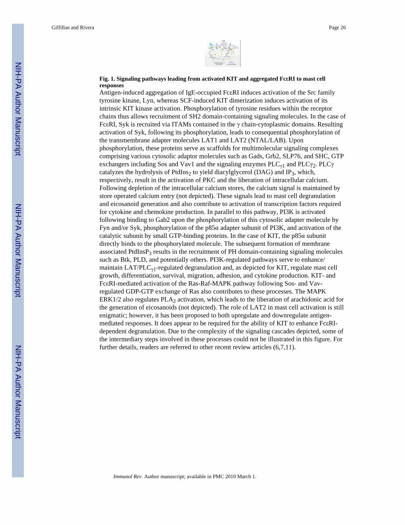
Fig. 1. Signaling pathways leading from activated KIT and aggregated FcεRI to mast cellresponsesAntigen-induced aggregation of IgE-occupied FcεRI induces activation of the Src familytyrosine kinase, Lyn, whereas SCF-induced KIT dimerization induces activation of itsintrinsic KIT kinase activation. Phosphorylation of tyrosine residues within the receptorchains thus allows recruitment of SH2 domain-containing signaling molecules. In the case ofFcεRI, Syk is recruited via ITAMs contained in the γ chain-cytoplasmic domains. Resultingactivation of Syk, following its phosphorylation, leads to consequential phosphorylation ofthe transmembrane adapter molecules LAT1 and LAT2 (NTAL/LAB). Uponphosphorylation, these proteins serve as scaffolds for multimolecular signaling complexescomprising various cytosolic adaptor molecules such as Gads, Grb2, SLP76, and SHC, GTPexchangers including Sos and Vav1 and the signaling enzymes PLCγ1 and PLCγ2. PLCγcatalyzes the hydrolysis of PtdIns2 to yield diacylglycerol (DAG) and IP3, which,respectively, result in the activation of PKC and the liberation of intracellular calcium.Following depletion of the intracellular calcium stores, the calcium signal is maintained bystore operated calcium entry (not depicted). These signals lead to mast cell degranulationand eicosanoid generation and also contribute to activation of transcription factors requiredfor cytokine and chemokine production. In parallel to this pathway, PI3K is activatedfollowing binding to Gab2 upon the phosphorylation of this cytosolic adapter molecule byFyn and/or Syk, phosphorylation of the p85α adapter subunit of PI3K, and activation of thecatalytic subunit by small GTP-binding proteins. In the case of KIT, the p85α subunitdirectly binds to the phosphorylated molecule. The subsequent formation of membraneassociated PtdInsP3 results in the recruitment of PH domain-containing signaling moleculessuch as Btk, PLD, and potentially others. PI3K-regulated pathways serve to enhance/maintain LAT/PLCγ1-regulated degranulation and, as depicted for KIT, regulate mast cellgrowth, differentiation, survival, migration, adhesion, and cytokine production. KIT- andFcεRI-mediated activation of the Ras-Raf-MAPK pathway following Sos- and Vav-regulated GDP-GTP exchange of Ras also contributes to these processes. The MAPKERK1/2 also regulates PLA2 activation, which leads to the liberation of arachidonic acid forthe generation of eicosanoids (not depicted). The role of LAT2 in mast cell activation is stillenigmatic; however, it has been proposed to both upregulate and downregulate antigen-mediated responses. It does appear to be required for the ability of KIT to enhance FcεRI-dependent degranulation. Due to the complexity of the signaling cascades depicted, some ofthe intermediary steps involved in these processes could not be illustrated in this figure. Forfurther details, readers are referred to other recent review articles (6,7,11).
Gilfillan and Rivera Page 26
Immunol Rev. Author manuscript; available in PMC 2010 March 1.
NIH
-PA Author Manuscript
NIH
-PA Author Manuscript
NIH
-PA Author Manuscript
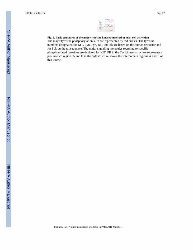
Fig. 2. Basic structures of the major tyrosine kinases involved in mast cell activationThe major tyrosine phosphorylation sites are represented by red circles. The tyrosinenumbers designated for KIT, Lyn, Fyn, Btk, and Itk are based on the human sequence andfor Syk on the rat sequence. The major signaling molecules recruited to specificphosphorylated tyrosines are depicted for KIT. PR in the Tec kinases structure represents aproline-rich region. A and B in the Syk structure shows the interdomain regions A and B ofthis kinase.
Gilfillan and Rivera Page 27
Immunol Rev. Author manuscript; available in PMC 2010 March 1.
NIH
-PA Author Manuscript
NIH
-PA Author Manuscript
NIH
-PA Author Manuscript
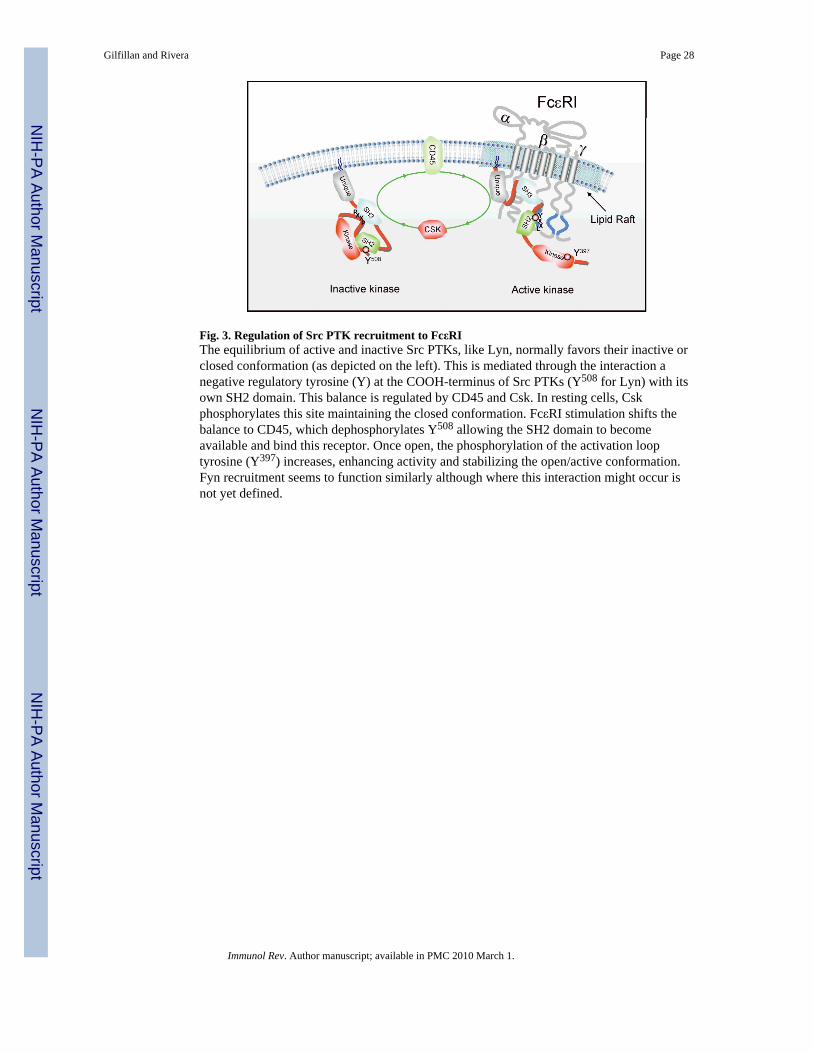
Fig. 3. Regulation of Src PTK recruitment to FcεRIThe equilibrium of active and inactive Src PTKs, like Lyn, normally favors their inactive orclosed conformation (as depicted on the left). This is mediated through the interaction anegative regulatory tyrosine (Y) at the COOH-terminus of Src PTKs (Y508 for Lyn) with itsown SH2 domain. This balance is regulated by CD45 and Csk. In resting cells, Cskphosphorylates this site maintaining the closed conformation. FcεRI stimulation shifts thebalance to CD45, which dephosphorylates Y508 allowing the SH2 domain to becomeavailable and bind this receptor. Once open, the phosphorylation of the activation looptyrosine (Y397) increases, enhancing activity and stabilizing the open/active conformation.Fyn recruitment seems to function similarly although where this interaction might occur isnot yet defined.
Gilfillan and Rivera Page 28
Immunol Rev. Author manuscript; available in PMC 2010 March 1.
NIH
-PA Author Manuscript
NIH
-PA Author Manuscript
NIH
-PA Author Manuscript

Fig. 4. Cbp/CSK-mediated regulation of Fyn activityIn resting mast cells, active Lyn kinase phosphorylates the adapter Cbp at Y314 at low levels.Phosphorylation of this site causes the binding and membrane localization of CSK, which isnormally localized in the cytoplasm. CSK can then target the COOH-terminal negativeregulatory tyrosine (Y) residue found in Src PTKs. In Fyn, Y525 is a target for CSK activityleading to Fyn inactivation by inducing a closed conformation via intramolecular interactionwith its own SH2 domain. The low level of Cbp phosphorylation maintains the equilibriumbetween active and inactive Src PTKs in resting cells. Upon FcεRI stimulation, the balanceshifts towards increased activation of Src PTKs, likely through CD45. This new equilibriumis governed by a Lyn-dependent increase in Cbp phosphorylation and membrane CSKrecruitment, which controls the activity of Fyn by inactivation as described above. Thus, theabsence of Lyn leads to a loss in control of Fyn activity and mast cells are hyperresponsive(63,85).
Gilfillan and Rivera Page 29
Immunol Rev. Author manuscript; available in PMC 2010 March 1.
NIH
-PA Author Manuscript
NIH
-PA Author Manuscript
NIH
-PA Author Manuscript
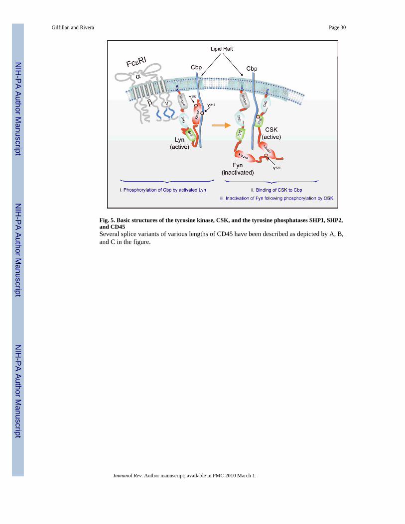
Fig. 5. Basic structures of the tyrosine kinase, CSK, and the tyrosine phosphatases SHP1, SHP2,and CD45Several splice variants of various lengths of CD45 have been described as depicted by A, B,and C in the figure.
Gilfillan and Rivera Page 30
Immunol Rev. Author manuscript; available in PMC 2010 March 1.
NIH
-PA Author Manuscript
NIH
-PA Author Manuscript
NIH
-PA Author Manuscript