the tvlegu-1, a legumain-like cysteine proteinase, plays a key role in trichomonas vaginalis...
TRANSCRIPT

1
rossana arroyo
Asunto: RV: Your article has been published
‐‐‐‐‐Mensaje original‐‐‐‐‐ De: Heba Allam [mailto:[email protected]] Enviado el: miércoles, 02 de enero de 2013 6:18 Para: [email protected] Asunto: Your article has been published Dear Prof. Arroyo: I am pleased to let you know that your article has been published in its final form in the "BioMed Research International:" Rossana Arroyo, "The TvLEGU‐1, a Legumain‐Like Cysteine Proteinase, Plays a Key Role in Trichomonas vaginalis Cytoadherence," BioMed Research International, vol. 2013, Article ID 561979, 18 pages, 2013. doi:10.1155/2013/561979. You may access this article from the Table of Contents of Volume 2013, which is located at the following link: http://www.hindawi.com/journals/bmri/contents/ Alternatively, you may directly access your article at the following location: http://www.hindawi.com/journals/bmri/2013/561979/ "BioMed Research International" is an open access journal, meaning that the full‐text of all published articles is made freely available on the journal’s website with no subscription or registration barriers. Best regards, Heba Allam BioMed Research International Hindawi Publishing Corporation http://www.hindawi.com/


Hindawi Publishing CorporationJournal of Biomedicine and BiotechnologyVolume 2012, Article ID 561979, 18 pagesdoi:10.1155/2012/561979
Research Article
The TvLEGU-1, a Legumain-Like Cysteine Proteinase,Plays a Key Role in Trichomonas vaginalis Cytoadherence
Francisco Javier Rendon-Gandarilla,1 Lucero de los Angeles Ramon-Luing,1
Jaime Ortega-Lopez,2 Ivone Rosa de Andrade,3 Marlene Benchimol,3 and Rossana Arroyo1
1 Departamento de Infectomica y Patogenesis Molecular, Centro de Investigacion y de Estudios Avanzados del Instituto PolitecnicoNacional, Avenida IPN No. 2508, Col. San Pedro Zacatenco, 07360 Mexico City, DF, Mexico
2 Departamento de Biotecnologıa y Bioingenierıa, Centro de Investigacion y de Estudios Avanzados del Instituto Politecnico Nacional,Avenida IPN No. 2508, Col. San Pedro Zacatenco, 07360 Mexico City, DF, Mexico
3 Laboratorio de Ultraestrutura Celular, Universidade Santa Ursula, Rua Jornalista Orlando Dantas 36, Botafogo,22231-010 Rio de Janeiro, RJ, Brazil
Correspondence should be addressed to Rossana Arroyo, [email protected]
Received 31 July 2012; Revised 21 September 2012; Accepted 28 September 2012
Academic Editor: Luis I. Terrazas
Copyright © 2012 Francisco Javier Rendon-Gandarilla et al. This is an open access article distributed under the CreativeCommons Attribution License, which permits unrestricted use, distribution, and reproduction in any medium, provided theoriginal work is properly cited.
The goal of this paper was to characterize a Trichomonas vaginalis cysteine proteinase (CP) legumain-1 (TvLEGU-1) and determineits potential role as a virulence factor during T. vaginalis infection. A 30-kDa band, which migrates in three protein spots (pI∼6.3,∼6.5, and ∼6.7) with a different type and level of phosphorylation, was identified as TvLEGU-1 by one- and two-dimensionalWestern blot (WB) assays, using a protease-rich trichomonad extract and polyclonal antibodies produced against the recombinantTvLEGU-1 (anti-TvLEGU-1r). Its identification was confirmed by mass spectrometry. Immunofluorescence, cell binding, andWB assays showed that TvLEGU-1 is upregulated by iron at the protein level, localized on the trichomonad surface and inlysosomes and Golgi complex, bound to the surface of HeLa cells, and was found in vaginal secretions. Additionally, the IgGand Fab fractions of the anti-TvLEGU-1r antibody inhibited trichomonal cytoadherence up to 45%. Moreover, the Aza-PeptidylMichael Acceptor that inhibited legumain proteolytic activity in live parasites also reduced levels of trichomonal cytoadherenceup to 80%. In conclusion, our data show that the proteolytic activity of TvLEGU-1 is necessary for trichomonal adherence. Thus,TvLEGU-1 is a novel virulence factor upregulated by iron. This is the first report that a legumain-like CP plays a role in a pathogencytoadherence.
1. Introduction
Trichomoniasis is one of the most common sexually trans-mitted infections worldwide caused by Trichomonas vaginalis[1]. Trichomonal adherence to host cells is a multifactorialprocess where adhesins and proteinases play importantroles [2–8]. Proteinases are abundant in T. vaginalis, beingreported more than 400 distinct proteinase genes in thedraft of its genome. Up to 220 correspond to the cysteinetype (CP) [9], but only 23 CPs have been detected by two-dimensional (2D) substrate gel electrophoresis [10], less wereidentified by recent proteomic studies [11–15], and only fewCP genes have been cloned and characterized [15–22]. These
gene products show homology to cathepsin L-like peptidases,which belong to the papain-like CP family of clan CA and tothe legumain-like CP family of clan CD [23, 24].
The thiol proteinases of this parasitic protozoan havebeen implicated in a variety of biological events includingnutrient acquisition [25], immune evasion [26, 27], andvirulence [1, 4, 5, 7, 8, 22, 28–35]. The expression of someof these CPs is regulated by environmental factors suchas pH and the redox state [1], polyamines [31], and iron[18, 20, 21, 30, 32, 35].
Iron is an essential nutrient for growth, metabolism, andvirulence of T. vaginalis [36]. The environment of the humanvagina, especially its nutrients and the iron concentration,

2 Journal of Biomedicine and Biotechnology
is constantly changing throughout the menstrual cycle.T. vaginalis may respond to varying iron concentrationsby differential gene expression through poorly understoodmechanisms [20, 21, 37] in order to survive, grow, andcolonize the vaginal hostile environment.
We previously reported that some of the CPs of the 30-kDa region are involved in cytoadherence [4, 5, 7]. Thisregion is formed by at least six spots with proteolytic activitythat correspond to two distinct CP families: the papain-like family of clan CA, represented by four spots with pIbetween 4.5 and 5.5, and the legumain-like family of clanCD, represented by two spots with pI 6.3 and 6.5 [38] thatare differentially regulated by iron at the transcript andproteolytic activity levels [21].
Among the ten legumain-like CP genes reported inthe draft of the T. vaginalis genome [9], we have clonedand sequenced two cDNAs coding for the TvLEGU-1 andTvLEGU-2 precursor proteinases of 42.8- and 47.2-kDa.These CPs were classified within the asparaginyl endopep-tidase (AE) subfamily of the family C13, belonging tothe clan CD [38]. The family C13 of peptidases includestwo distinct subfamilies with different functions, the gly-cosylphosphatidylinositol (GPI): protein transamidase andthe asparaginyl endopeptidase. Interestingly, TvLEGU-1 andTvLEGU-2 share ∼30% amino acid identity with the AEsubfamily and ∼26% with the GPI: protein transami-dase subfamily [38]. We also showed that the amountof TvLEGU-1 transcript is positively regulated by iron,whereas the TvLEGU-2 mRNA is not affected by it [21].Additionally, TvLEGU-1 is one of the most immuno-genic proteinases detected by trichomoniasis patient sera[15].
Thus, the main goal of this work was to identify,characterize, and determine the function of TvLEGU-1. Ourdata show that TvLEGU-1 is a surface proteinase upregulatedby iron, with affinity to the surface of HeLa cells that plays amajor role in trichomonal cytoadherence. Hence, TvLEGU-1is a novel virulence factor of T. vaginalis that is also releasedin vaginal secretions during infection.
2. Materials and Methods
2.1. Parasites and HeLa Cell Cultures. The fresh clinicalT. vaginalis isolate CNCD 147 [7, 15, 29] was used inthis study. Parasites were kept in culture at 37◦C up totwo weeks by daily passage in trypticase-yeast extract-maltose (TYM) medium [39] supplemented with 10% heat-inactivated horse serum (HIHS) (TYM-HIHS), containing∼20 μM iron [36]. Parasites in the logarithmic phase weregrown either in iron-rich or in iron-depleted medium bythe addition into the culture medium of 250 μM ferrousammonium sulfate or 150 μM 2-2 dipyridyl (Sigma Co., StLouis, MO, USA) an iron-chelator, respectively, as previouslyreported [30]. HeLa cells were grown in Dulbecco’s ModifiedEagle Medium (DMEM) (Gibco Laboratories, Grand Island,NY) supplemented with 10% HIHS at 37◦C for 48 h in a5% CO2 atmosphere to obtain confluent cell monolayers[6].
2.2. Generation of Antiserum against Recombinant TvLEGU-1.Rabbits were subcutaneously inoculated four times at two-week intervals with 0.3 mg of the affinity-purified TvLEGU-1r protein [15] in the presence of Freund’s complete adjuvant(Gibco) for the first immunization. Booster injections weregiven in Freund’s incomplete adjuvant (Gibco). The immuneserum (anti-TvLEGU-1r) was obtained seven days afterthe last immunization [40]. This antiserum was used inwestern blot (WB) analysis, indirect immunofluorescence,and cytoadherence inhibition assays. Preimmune (PI) serumwas obtained before the immunization schedule began andwas used as a negative control in all the experiments withantibodies.
2.3. Papain Fragmentation of IgG to Fab. To obtain the Fabfragment, 0.5 mg/mL purified IgGs from the anti-TvLEGU-1r or PI serum [40, 41] in PBS pH 8.0 were digestedwith 0.2 mg/mL papain in digestion buffer (PBS pH 8.0containing 0.02 M cysteine and 0.02 M EDTA) at 37◦C for6 h. The reaction was stopped with 0.3 M iodoacetamidein PBS pH 8.0. After digestion, samples were dialyzed inPBS pH 7.0 for 18 h at 4◦C and incubated with protein Aagarose during 2 h to eliminate the Fc fraction and recoverthe unbound Fab fragment [40].
2.4. Two-Dimensional Gel Electrophoresis (2DE). The 2DEfor protease-rich extracts was performed as recentlydescribed [22]. Briefly, for the first dimension, supernatantfrom lysed parasites (6 × 10⁷ cells/mL equivalent to 500 μgprotein) in rehydration solution (Bio-Rad) was loaded onto a7 cm Ready immobilized pH gradient (IPG) strips (linear pHgradient 4–7; Bio-Rad). IPG strips were actively rehydratedfor 16 h at 4◦C. Isoelectric focusing (IEF) of proteins wasperformed in three steps: 250 V for 20 min, 4 000 V for3 h, and a gradual increase up to 10 000 V-h. For reductionand alkylation, strips were equilibrated in buffer I and II(Bio-Rad) for 10 min at room temperature each. Proteinswere resolved by SDS-PAGE using 12% polyacrylamidegels, silver-stained, or transferred onto nitrocellulose (NC)membrane for WB detection. Gels and NC membraneswere documented using the ChemiDoc-XRS (Bio-Rad)and analyzed using the Quantity One software (Bio-Rad).A tridimensional analysis using the PD Quest (Bio-Rad)and Melanie software was also performed for differentiallyexpressed proteins. Three independent protein preparationswere done, each obtained from an independent parasiteculture, and similar results were observed.
2.5. Proteinase Identification. Identification of protein spotswas performed at the Protein Unit of the Columbia Uni-versity (NY, USA) as before [15]. Protein spots of interestwere manually excised from silver-stained gels, distained,and prepared for in-gel digestion with trypsin. Resultingpeptides were analyzed by MALDI-TOF mass spectrometry(MS) peptide mass mapping method on a Voyager DE pro-mass spectrometer in the linear mode (Applied Biosystems).Peptide masses were searched against the National Center for

Journal of Biomedicine and Biotechnology 3
Table 1: Peptides identified by MALDI-TOF-MS analysis of the three TvLEGU-1 protein spots of Trichomonas vaginalis detected by theanti-TvLEGU-1r antibody.
Peptide numbera Positionb (aa) Number in the sequencec m/z (av)d Spot 1e Spot 2e Spot 3e Amino acid sequencef
1 13–27 I 1749.9186 − + + FAVLIAGSNDFYNYR
2 28–41 II 1734.9719 + + + HQADIFNMYQQLVK
3 41–66 III 2774.0025 − + + GFDDQHITMMAYDDIALSSENPFR
4 42–66 IV 2930.1882 − + + RGFDDQHITMMAYDDIALSSENPFR
5 75–84 V 1101.2126 − + + HVNIYPGSSK
6 85–105 VI 2399.6528 − + + INYAHNSVTADQFYTVLTTLK
7 144–151 VII 911.4059 + + − AFDTMEAK
8 157–174 VIII 1994.1426 − + + LFFGIEACYSGSVAAVFR
9 157–176 IX 2193.5226 + + + LFFGIEACYSGSVAAVFRAK
10 232–248 X 1931.1107 − + + AQTTGSHVCYYGDVNMKaConsecutive number assigned to the identified peptides. bPosition in amino acids (aa) residues of the identified peptides (start-end) in the aa sequence of the
T. vaginalis TvLEGU-1 [38]. cArbitrary nomenclature used to describe the ten identified peptides in T. vaginalis TvLEGU-1 (see Supplementary Figure 1S inSupplementary Material available online at doi:10.1155/2012/561979). dPeptide mass average m/z (av) identified by MALDI-TOF-MS after tryptic digestionof the three protein spots obtained from 2DE of protease-rich extracts from T. vaginalis grown in normal iron conditions (Figure 1). ePresence (+) or absence(−) of the peptides identified by MALDI-TOF-MS in the three TvLEGU-1 protein spots analyzed. fAmino acid sequence of the peptides obtained from atheoretical tryptic digestion of the deduced aa sequence of TvLEGU-1 [38] with identical masses to the experimental one (Supplementary Figure 1S).
Table 2: Densitometric analysis of the three TvLEGU-1 protein spots observed in silver-stained gels and WB NC membranes from parasitesgrown in high and low iron concentrations.
Spot numberPixel intensity of silver stained spots Pixel intensity of WB spots
High irona Low ironb High ironc Low irond
1 292685.95 83473.87 93367.94 29862.21
2 637882.72 416358.36 296207.75 245016.26
3 1781948.40 1171066.01 842341.15 794637.06aDensitometry values of the spots detected in the silver stained gel in high iron condition (H) of Figure 2(A). bDensitometry values of the spots detected in
the silver stained gel in low iron condition (L) of Figure 2(A). cDensitometry values of the spots detected in the WB NC membranes in high iron condition(H) of Figure 2(A). dDensitometry values of the spots detected in the WB NC membranes in low iron condition (L) of Figure 2(A).
Biotechnology nonredundant database (NCBInr) using theMASCOT program (http://matrixscience.com/).
2.6. Western Blot Analysis (WB). Total trichomonad pro-teins, proteinase-rich extracts, proteins obtained after cell-binding assays from (2 × 10⁷) parasites grown in iron-rich medium, obtained as before [7], and TCA-precipitatedproteins present in vaginal washes (VWs) (100 μL) frompatients with vaginitis (Table 2) were separated by SDS-PAGE using 10% polyacrylamide gels. Duplicated gels weretransferred onto NC membranes for WB. The TvLEGU-1proteinase was immunodetected with the anti-TvLEGU-1rrabbit serum (at 1 : 1000 to 1 : 40000 dilutions). As a quantitycontrol, a monoclonal antibody against α-tubulin (ZymedLaboratories, South San Francisco, CA) (at 1 : 100 dilution)was used. WB was developed by chemiluminescence usingthe ECL-Plus kit (Amersham Co., Arlington Heights, IL,USA) and SuperSignal Femto Maximum Sensitivity Sub-strate kit (Pierce) and documented using the ChemiDoc-XRS(Bio-Rad).
To determine the presence of phosphorylations onTvLEGU-1, anti-phospho-Ser, -Tyr, and -Thr monoclonalantibodies at 1 : 500 dilution (Zymed) were used in 2DE-WBassays over NC membranes containing protease-rich extracts
and developed by chemiluminescence. These experimentswere performed at least three times with similar results.
2.7. In Vitro Secretion Kinetic Assay. The in vitro secretionassay was performed as previously described [22, 33]. Briefly,after 18 h of growth in iron-rich conditions, parasites wereharvested, washed three times with PBS pH 7.0, and sus-pended in PBS-0.5% maltose at 1 × 10⁶ cells/mL parasitedensity. Parasites were incubated for 15, 30, 60, and 90 min at37◦C, collected by centrifugation at 700 g, and supernatantswere analyzed directly by substrate-gel electrophoresis and byWB after TCA-precipitation. The viability of trichomonadswas assessed by trypan blue exclusion throughout the assay.
2.8. Indirect Immunofluorescence Assay. For confocal micros-copy, parasites grown in iron-rich conditions were fixedwith 4% paraformaldehyde for 1 h at 37◦C, washed withPBS, and half of them were treated with 50 mM NH4Cl/PBSpH 7.0 for 10 min, washed with PBS, and with 1 N HCLfor 1 h and permeabilized with 0.2% Triton X-100 for10 min. The other half was used as nonpermeabilizedparasites. Permeabilized and nonpermeabilized parasiteswere blocked with 1% fetal bovine serum for 15 min and

4 Journal of Biomedicine and Biotechnology
Table 3: The average number of T. vaginalis parasites attached to HeLa cell monolayers per coverslip with or without treatment usingdifferent proteinase inhibitors (Figure 8(B)).
Treatment Parasites attached Inhibition (%)
Nonea 1450 0.0± 13.9176
MA (5 μM)b 742 46.33± 7.7764
MA (10 μM) 556 60.78± 5.6311
MA (50 μM) 301 77.23± 5.2907
TLCK (1 mM) 622 57.05± 6.6416
Leupeptin (0.2 mM) 898 36.79± 6.1359
E-64 (0.18 mM) 662 50.16± 8.1047aNone corresponds to the control parasites without treatment with CP inhibitors. The number of parasites attached to the HeLa cell monolayer was taken
as 100% adherence for comparative purpose. bMA corresponds to the Aza-Peptidyl Michael Acceptor, a legumain-specific inhibitor. These differences werestatistically significant with a P < 0.001 (Figure 8(B)).
with 0.2 M glycine for 1 h at room temperature. Then,trichomonads were incubated for 18 h at 4◦C, with theanti-TvLEGU-1r or PI serum used as a negative control,both at 1 : 1 000 dilution. Parasites were incubated with thesecondary antibody, fluorescein isothiocyanate-conjugatedanti-rabbit immunoglobulins (Pierce) at 1 : 200 dilution for1 h at 37◦C, washed, mounted with Vectashield mountingsolution (Vector Laboratories), and visualized by confocalmicroscopy with a Leica LSM-SPC-5 Mo inverted confocalmicroscope fitted with HCXPLapo lambda blue 63 × 1.4 oilimmersion lens. Time series were captured and processedusing the confocal LAS AF software (Leica). Also, live HeLacells were incubated with 10 μg/mL TvLEGU-1r or super-natant from an in vitro secretion assay for 30 min at 37◦C,washed with PBS, fixed, blocked, and treated with antibodiesas the parasites described above for immunofluorescenceassays.
For lysosomal colocalization assays, the acidic compart-ments of T. vaginalis were stained with 1 μM LysoTrackerRED DND-99 (Invitrogen) for 12 h at 37◦C in TYM mediumsupplemented with 10% heat-inactivated horse serum. Afterthat, parasites were processed for indirect immunofluores-cence with the anti-TvLEGU-1r antibody as described in theprevious paragraph.
For immunogold labeling assays, parasites were fixedovernight at room temperature in 0.5% glutaraldehyde, 4%formaldehyde in 0.1 M cacodylate buffer. Afterwards, cellswere dehydrated in ethanol and embedded in Unicryl. Ultra-thin sections were harvested on 300 mesh nickel grids. Thesamples were washed and incubated with 50 mM ammoniumchloride for 30 min in order to quench free aldehyde groups.The sections were incubated in a series of blocking solutions(PBS containing 1% bovine albumin (BSA), 3% PBS/BSA,and 0.2% Tween-20, pH 8.0) for 10 min on each step. Cellswere incubated with the anti-TvLEGU-1r antibody at 1 : 50dilution, overnight. After several washes in 1% PBS/BSA, thesections were incubated with 10 nm gold-labeled goat anti-rabbit IgG (BB International, UK). As control some sampleswere incubated only with the secondary antibody. Finally,sections were stained with 5% uranyl acetate and 1% leadcitrate and then observed with a JEOL 1210 transmissionelectron microscope.
2.9. Cell-Binding Assay for Proteinases. To detect the affinityof the native and recombinant TvLEGU-1 proteins to thesurface of host cells, we performed cell-binding assays aspreviously described [7]. Briefly, a clarified detergent extractfrom 2× 10⁷ parasites or 25 μg of TvLEGU-1r was incubatedfor 18 h at 4◦C with 1 × 10⁶ glutaraldehyde-fixed HeLa cells.The native and recombinant TvLEGU-1 proteins bound tothe surface of fixed-HeLa cells were eluted with Laemmlisample buffer for 20 min at 37◦C. The released proteinswere analyzed by SDS-PAGE and blotted onto NC for WBdetection with the anti-TvLEGU-1r antibody.
2.10. Cytoadherence Inhibition Assay. Cytoadherence inhibi-tion assays were performed over confluent HeLa cell mono-layers on 96-well microtiter plates as previously described[7, 42]. Briefly, [3H]-thymidine-labeled parasites were incu-bated for 30 min at 4◦C with 0, 50, and 100 μg/mL IgG orFab fraction from the anti-TvLEGU-1r or PI serum beforeinteraction with HeLa cell monolayers.
The cytoadherence inhibition assays with proteinaseinhibitors were performed over confluent live HeLa cellmonolayers on 12 mm coverslips as recently described [41].Briefly, cell monolayers (5 × 10⁵ cells/coverslip) wereincubated with live parasites (1 × 10⁶ cells/well) previouslylabeled with 25 mM CellTracker Blue CMAC (MolecularProbes) in serum-free DMEM-TYM (2 : 1) medium andincubated at 37◦C for 30 min and 5% CO2. For inhibitionexperiments before interaction with HeLa cell monolayerslabeled parasites were incubated for 20 min at 4◦C withdifferent CP inhibitors (1 mM TLCK, 0.2 mM leupeptin,or 0.18 mM E-64; all purchased from Sigma) used ascontrols. The Aza-Peptidyl Michael Acceptor (Mu-Ala-Ala-AAsn-CH=CH–CON, kindly donated by Dr. James Powers),a specific inhibitor for legumains [43], was also used at 5,10, and 50 μM. After interaction with CP inhibitors parasiteswere washed and added to HeLa cell monolayers. After theinteraction, the coverslips were washed with warm PBS, fixedwith 4% paraformaldehyde, and mounted on slides. Eachcondition was performed in triplicate, and ten fields with a40x magnification were analyzed per coverslip. Fluorescentparasites adhered to host cells (in blue) were countedusing an Eclipse 80i epifluorescence microscope (Nikon)and the NIS-Elements BR 2.1 software (Nikon) (Table 3).
Journal of Biomedicine and Biotechnology 5
The experiment was repeated at least two independent timeswith similar results.
2.11. Measurement of the Proteolytic Activity of Live Parasites.To detect the proteolytic activity of live trichomonads, 2.5 ×10⁵ parasites were incubated for 20 min at 4◦C with 1 mMTLCK, 0.2 mM leupeptin, 0.18 mM E-64, or 50 μM Aza-Peptidyl Michael Acceptor [43]. The activity was measuredwith two CPs substrates (Z-Phe-Arg-AMC for papains;and Cbz-Ala-Ala-AAsn-AMC for legumains). Release offree 7-amino-4-methylcoumarin (AMC) was measured byemission at excitation wavelengths of 355 and 460 nm,respectively, in a luminometer (BioTek) using a Gen5 2.0Data Analysis Software. The linear regression of the substratehydrolysis curves was used to calculate initial velocities.The experiment was repeated at least three independenttimes with similar results. The viability of trichomonads wasassessed by trypan blue exclusion throughout the assay.
2.12. Statistical Analysis. The statistically significant dif-ference between means was determined by analysis ofvariance (ANOVA) using GraphPad Prism 5.0. The data wereanalyzed by one-way ANOVA using the Bonferroni methodcomparing all pairs of columns (P < 0.001) for Figures 7(A),7(B), 8(B), 8(C), and 8(D). The scores showing statisticalsignificance are indicated in the figures with asterisks. Thecorresponding P values are indicated in the figure legends.
3. Results
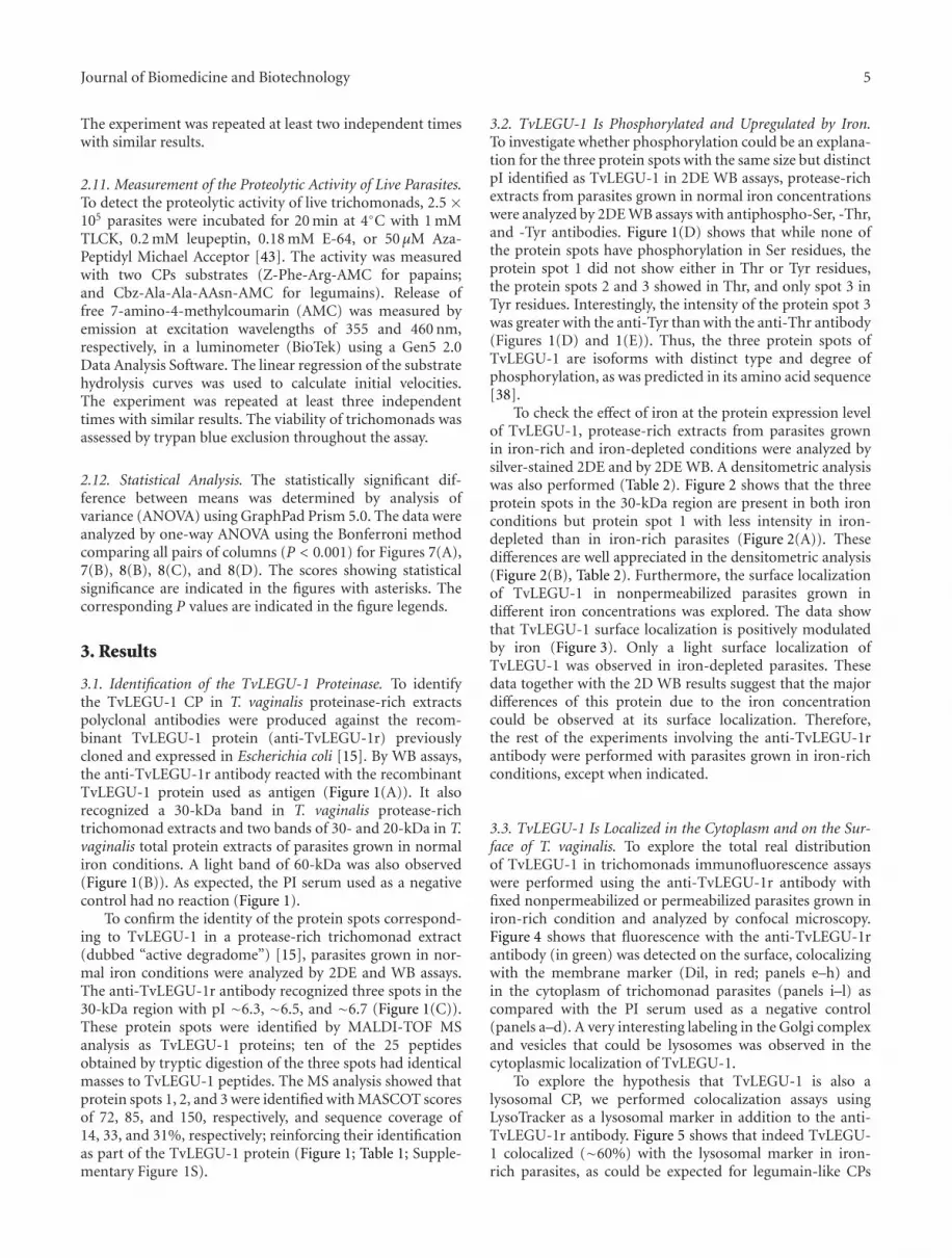
3.1. Identification of the TvLEGU-1 Proteinase. To identifythe TvLEGU-1 CP in T. vaginalis proteinase-rich extractspolyclonal antibodies were produced against the recom-binant TvLEGU-1 protein (anti-TvLEGU-1r) previouslycloned and expressed in Escherichia coli [15]. By WB assays,the anti-TvLEGU-1r antibody reacted with the recombinantTvLEGU-1 protein used as antigen (Figure 1(A)). It alsorecognized a 30-kDa band in T. vaginalis protease-richtrichomonad extracts and two bands of 30- and 20-kDa in T.vaginalis total protein extracts of parasites grown in normaliron conditions. A light band of 60-kDa was also observed(Figure 1(B)). As expected, the PI serum used as a negativecontrol had no reaction (Figure 1).
To confirm the identity of the protein spots correspond-ing to TvLEGU-1 in a protease-rich trichomonad extract(dubbed “active degradome”) [15], parasites grown in nor-mal iron conditions were analyzed by 2DE and WB assays.The anti-TvLEGU-1r antibody recognized three spots in the30-kDa region with pI ∼6.3, ∼6.5, and ∼6.7 (Figure 1(C)).These protein spots were identified by MALDI-TOF MSanalysis as TvLEGU-1 proteins; ten of the 25 peptidesobtained by tryptic digestion of the three spots had identicalmasses to TvLEGU-1 peptides. The MS analysis showed thatprotein spots 1, 2, and 3 were identified with MASCOT scoresof 72, 85, and 150, respectively, and sequence coverage of14, 33, and 31%, respectively; reinforcing their identificationas part of the TvLEGU-1 protein (Figure 1; Table 1; Supple-mentary Figure 1S).
3.2. TvLEGU-1 Is Phosphorylated and Upregulated by Iron.To investigate whether phosphorylation could be an explana-tion for the three protein spots with the same size but distinctpI identified as TvLEGU-1 in 2DE WB assays, protease-richextracts from parasites grown in normal iron concentrationswere analyzed by 2DE WB assays with antiphospho-Ser, -Thr,and -Tyr antibodies. Figure 1(D) shows that while none ofthe protein spots have phosphorylation in Ser residues, theprotein spot 1 did not show either in Thr or Tyr residues,the protein spots 2 and 3 showed in Thr, and only spot 3 inTyr residues. Interestingly, the intensity of the protein spot 3was greater with the anti-Tyr than with the anti-Thr antibody(Figures 1(D) and 1(E)). Thus, the three protein spots ofTvLEGU-1 are isoforms with distinct type and degree ofphosphorylation, as was predicted in its amino acid sequence[38].
To check the effect of iron at the protein expression levelof TvLEGU-1, protease-rich extracts from parasites grownin iron-rich and iron-depleted conditions were analyzed bysilver-stained 2DE and by 2DE WB. A densitometric analysiswas also performed (Table 2). Figure 2 shows that the threeprotein spots in the 30-kDa region are present in both ironconditions but protein spot 1 with less intensity in iron-depleted than in iron-rich parasites (Figure 2(A)). Thesedifferences are well appreciated in the densitometric analysis(Figure 2(B), Table 2). Furthermore, the surface localizationof TvLEGU-1 in nonpermeabilized parasites grown indifferent iron concentrations was explored. The data showthat TvLEGU-1 surface localization is positively modulatedby iron (Figure 3). Only a light surface localization ofTvLEGU-1 was observed in iron-depleted parasites. Thesedata together with the 2D WB results suggest that the majordifferences of this protein due to the iron concentrationcould be observed at its surface localization. Therefore,the rest of the experiments involving the anti-TvLEGU-1rantibody were performed with parasites grown in iron-richconditions, except when indicated.
3.3. TvLEGU-1 Is Localized in the Cytoplasm and on the Sur-face of T. vaginalis. To explore the total real distributionof TvLEGU-1 in trichomonads immunofluorescence assayswere performed using the anti-TvLEGU-1r antibody withfixed nonpermeabilized or permeabilized parasites grown iniron-rich condition and analyzed by confocal microscopy.Figure 4 shows that fluorescence with the anti-TvLEGU-1rantibody (in green) was detected on the surface, colocalizingwith the membrane marker (Dil, in red; panels e–h) andin the cytoplasm of trichomonad parasites (panels i–l) ascompared with the PI serum used as a negative control(panels a–d). A very interesting labeling in the Golgi complexand vesicles that could be lysosomes was observed in thecytoplasmic localization of TvLEGU-1.
To explore the hypothesis that TvLEGU-1 is also alysosomal CP, we performed colocalization assays usingLysoTracker as a lysosomal marker in addition to the anti-TvLEGU-1r antibody. Figure 5 shows that indeed TvLEGU-1 colocalized (∼60%) with the lysosomal marker in iron-rich parasites, as could be expected for legumain-like CPs

6 Journal of Biomedicine and Biotechnology
31
45
66.2
(kD
a)
97.4116.5
200
TvLEGU-1r
CB
B
21.5
1 2
(A)
(C)
(D) (E)
(B)3
PI
α-T
vLE
GU
-1r
31
45
66.2
(kD
a)
97.4116.5
200
4 7Si
lver
21.5
31
45
66.2
(kD
a)
97.4116.5
200
21.5
31
45
66.2
(kD
a)
97.4116.5
200
21.5
pI 4 7pI 4 7pI
PI
α-T
vLE
GU
-1r
4 7
Silver
α-PSer
α-PThr
α-PTyr
pI
31
45
66.2
(kD
a)
(kD
a)
97.4116.5
200
21.5
Tv extracts
CB
B
CB
B
30
1 2 3 4 5
20
∗
PI
α-T
vLE
GU
-1r
α-T
vLE
GU
-1r
1
2
2
3
3 3
Figure 1: Recognition of TvLEGU-1 by the anti-TvLEGU-1r antibody in total protein extracts and protease-rich extracts of T. vaginalis.(A) Coomassie brilliant blue-stained purified recombinant TvLEGU-1r protein (CBB; lane 1). WB assays of TvLEGU-1r incubated withthe anti-TvLEGU-1r antibody (lane 2) or with the PI serum (lane 3) used as a negative control, both at 1 : 40000 dilution. Arrowheadpoints to the recombinant protein band TvLEGU-1r (∼46-kDa). kDa, molecular weight markers in kilodaltons (Bio-Rad). (B) Coomassiebrilliant blue-stained protein patterns of trichomonad protease-rich (lane 1) or total protein extracts (lane 3); WB assays of duplicated gelstransferred onto NC membranes containing trichomonad protease-rich (lane 2) or total protein extracts (lane 4) incubated with the anti-TvLEGU-1r antibody, or with the PI serum (lane 5) used as a negative control, both at 1 : 20000 dilution. Arrowheads show the position ofthe native TvLEGU-1 (∼30- and ∼20-kDa) proteins. Asterisk shows a light protein band of ∼60-kDa also detected by the anti-TvLEGU-1rantibody. kDa, molecular weight markers in kilodaltons (Bio-Rad). (C) Silver-stained 2DE protease-rich extracts from parasites grown inregular medium (panel a). WB of duplicate gels transferred onto NC membranes incubated with the anti-TvLEGU-1r antibody (panel b)or with the PI (panel c) serum both at 1 : 5000 dilution. Numbers 1–3 show the position of the TvLEGU-1 protein spots. (D) Differentialphosphorylation of TvLEGU-1. 2DE WB assays from duplicate protease-rich extracts from part (C) separated by SDS-PAGE were (a) silver-stained or transferred onto NC membranes and incubated with the (b) antiphosphoserine (α-PSer), (c) antiphosphothreonine (α-PThr),and (d) antiphosphotyrosine (α-PTyr) monoclonal antibodies. pI, direction of IEF using IPG Ready-strips (linear gradient of pH 4–7; Bio-Rad). Numbers show the position of the TvLEGU-1 spots. (E) Landscape representation of the densitometric analysis (PDQuest and Melaniesoftware) of the protein spots corresponding to TvLEGU-1. Numbers 1–3 show the position of the TvLEGU-1 spots.
(Figure 5(A)). Moreover, immunogold localization assaysconfirmed the cytoplasmic localization of TvLEGU-1 invacuoles/lysosomes containing degrading material and inthe Golgi complex (Figure 5(B)), suggesting that this is
an excreted/secreted proteinase. These data suggest thatTvLEGU-1 could have multiple functions that will dependon its cellular localization possible modulated by the ironconcentrations.

Journal of Biomedicine and Biotechnology 7
4 7
Silv
er
H
L
H
L
(A) (B)
WB
[Fe2+]pI
Figure 2: Iron effect on the protein expression of TvLEGU-1. (A) Silver-stained 2DE pattern of protease-rich extracts, corresponding tothe 30-kDa region from (a) parasites grown in iron-rich (H) or (b) iron-depleted (L) conditions. (c) and (d) 2DE WB assays of duplicatedgels (a) and (b) transferred onto NC membranes and incubated with the anti-TvLEGU-1r antibody (1 : 5000 dilution). pI, direction of IEFusing IPG Ready-strips (linear gradient of pH 4–7; Bio-Rad). Numbers 1–3 show the position of the distinct TvLEGU-1 protein spots. (B)Landscape representation of the densitometric analysis of the three TvLEGU-1 protein spots of (a) and (b) silver-stained gels and (c) and(d) WB shown in (A) was carried out with the Melanie and PDQuest (Bio-Rad) programs. Numbers 1–3 correspond to the protein spots ofTvLEGU-1.
3.4. TvLEGU-1 Binds to the Surface of HeLa Cells. To deter-mine whether TvLEGU-1 binds to the surface of HeLa cells,cell-binding and WB assays were performed with protease-rich extracts from parasites grown in iron-rich medium andfixed HeLa cells. WB assays showed that the anti-TvLEGU-1r antibody reacted with the trichomonad 30-kDa bandthat bound to fixed HeLa cells (Figure 6(A)), suggestingthe presence of TvLEGU-1. This was confirmed in a cell-binding assay using the recombinant TvLEGU-1 protein thatalso bound to the surface of fixed HeLa cells (Figure 6(B)),whereas the bovine serum albumin (BSA) used as a negativecontrol did not bind as expected (Figure 6(B)). Additionally,TvLEGU-1r was recognized by the antinative CP30 antibody[7] in WB assays (Figure 6(C)). Together, these data showthat TvLEGU-1 is one of the CP30 proteinases that interactwith the surface of HeLa cells [7].
Furthermore, to confirm it, immunofluorescence assaysusing fixed and live HeLa cells incubated with the TvLEGU-1r protein and the anti-TvLEGU-1r antibody were per-formed. Confocal microscopy images showed that indeedTvLEGU-1r bound to the surface of fixed and live HeLacells, whereas HeLa cells directly incubated with the anti-TvLEGU-1r antibody used as a negative control had noreaction as expected (Figures 6(D) and 6(E)).
3.5. TvLEGU-1 Participates in T. vaginalis Cytoadherence. Tostudy the role of TvLEGU-1 in trichomonal adherence, weperformed adherence inhibition assays over HeLa cell mono-layers by preincubating [3H]-thymidine-labeled iron-richparasites with varied concentrations of the anti-TvLEGU-1rIgG or Fab fractions. Figure 7 shows that the anti-TvLEGU-1r antibody inhibited the levels of T. vaginalis adherence toHeLa cell monolayers in a concentration-dependent manner.A maximum inhibition of ∼45%, using 100 μg/mL of IgGsor Fab fractions, was observed. IgGs or Fab fractions from PIserum used as a negative control did not affect trichomonalcytoadherence. These results illustrate that TvLEGU-1 is avirulence factor that plays a role in cellular attachment as oneof the 30-kDa CPs required for trichomonal adherence [7].
3.6. TvLEGU-1 Proteolytic Activity Is Necessary for T vaginalisCytoadherence. To determine whether TvLEGU-1 prote-olytic activity was required for cellular attachment, livenonradioactive-labeled parasites [41] were treated withdistinct CP inhibitors (TLCK, leupeptin, or E-64) or withincreasing concentrations (0, 5, 10, and 50 μg/mL) of a spe-cific legumain inhibitor the Aza-Peptidyl Michael Acceptor(Mu-Ala-Ala-AAsn-CH=CH–CON) [43] before interactionwith live HeLa cell monolayers. Figures 8(A) and 8(B)

8 Journal of Biomedicine and Biotechnology
H N L
FIT
CD
ILD
AP
IM
erge
Figure 3: The TvLEGU-1 surface localization on T. vaginalis is affected by iron. Parasites grown in iron-rich (H; a, b, c, and d), normal (N;e, f, g, and h), and iron-depleted (L; i, j, k, and l) conditions, fixed and nonpermeabilized were incubated with the anti-TvLEGU-1r antibody(1 : 100 dilution). Anti-rabbit IgG-FITC (in green) was used as a secondary antibody (1 : 100 dilution) (a, e, and i). Parasite membranes werelabeled with DIL (in red; b, f, and j). Nuclei were labeled with DAPI (in blue; c, g, and k). Merge (d, h, and l) in yellow indicates colocalization.Bars: 15 μm (d, h, and l).
show that the specific legumain inhibitor decreased thelevels of T. vaginalis adherence to HeLa cell monolayers ina concentration-dependent manner up to ∼80%, whereasTLCK, leupeptin, and E-64 inhibited ∼60, ∼40, and ∼50%,respectively. The average number of parasites without treat-ment attached to HeLa cells per coverslip was higher thanthe number of parasites treated with inhibitors (Table 3),
and these differences were statistically significant P < 0.001(Figure 8(B)). These results suggest that both legumainand papain-like CP proteolytic activities are necessary fortrichomonal cytoadherence, especially the legumain-likeactivity. These data are consistent with previous reports [4].
To check the effect of these inhibitors over the CPproteolytic activity of live parasites fluorescent substrates

Journal of Biomedicine and Biotechnology 9
PI
PNP
FIT
CD
ILD
AP
IM
erge
α-TvLEGU-1r
Figure 4: The TvLEGU-1 protein is localized on the surface and cytoplasm of T. vaginalis. Parasites grown in iron-rich conditions, fixednonpermeabilized (NP) were incubated with the PI serum, (1 : 100 dilution) as a negative control (a, b, c, and d). Nonpermeabilized (NP;e, f, g, and h) and permeabilized (P; i, j, k, and l) parasites were incubated with the anti-TvLEGU-1r antibody (1 : 100 dilution). Anti-rabbitIgG-FITC (in green) was used as a secondary antibody (1 : 100 dilution) (a, e, and i). Parasite membranes were labeled with DIL (in red; b,f, and j). Nuclei were labeled with DAPI (in blue; c, g, and k). Merge (d, h, and l) in yellow indicates colocalization. Bars: 15 μm (d, h, and l).
for papain-like (Z-Phe-Arg-AMC) and legumain-like (Cbz-Ala-Ala-AAsn-AMC) CPs were used. Live untreated parasitesused as control showed proteolytic activity for both sub-strates, which were taken as 100% activity. Parasites treatedwith the specific legumain inhibitor (Aza-Peptidyl Michael
Acceptor) abolished the legumain-like proteolytic activity(Figure 8(C)) and has no effect on the papain-like proteolyticactivity (Figure 8(D)). TLCK, a potent inhibitor of papain-like and legumain-like CPs, greatly reduced both proteolyticactivities (∼80% and ∼95%, resp.) of treated parasites as

10 Journal of Biomedicine and Biotechnology
LysoT FITC
DAPI Merge(A)
(B)
Figure 5: TvLEGU-1 is also localized in lysosomes and Golgi complex of T. vaginalis. (A) Parasites grown in iron-rich and labeled withLysoTracker were fixed, blocked, and incubated with the anti-TvLEGU-1r antibody (1 : 100 dilution) and a secondary antibody coupled toFITC stained in green; lysosomes were stained in red with LysoTracker, LysoT; and nuclei in blue with DAPI. The samples were analyzed byconfocal microscopy. (B) Immunogold labeling of thin cryosections of parasites using the anti-TvLEGU-1r antibody at 1 : 100 dilution. Thesamples were analyzed by transmission electron microscopy. The labeling is observed on (a) Golgi complex (G) and (b) vesicles similar tolysosomes (L). Bars: 200 nm and 300 nm, respectively.
expected (Figures 8(C) and 8(D)). E-64 and leupeptin,potent inhibitors of papain-like CPs, greatly reduced thepapain-like proteolytic activity (between ∼80 to ∼90%) oftreated parasites (Figure 8(D)) and had a minimal or noeffect on the legumain-like proteolytic activity (Figure 8(C))of live parasites. Therefore, both types of CP proteolyticactivity are present in live parasites and are necessary fortrichomonal adherence to host cells (Figure 8).
3.7. The TvLEGU-1 Proteinase Is Expressed during Infec-tion and Is Present in Vaginal Secretions of Patients withTrichomoniasis. To investigate the relevance of TvLEGU-1during trichomonal infection, we analyzed vaginal washesfrom vaginitis patients with [Tv (+)] or without [Tv (−)]T. vaginalis (Table 4) for the presence of TvLEGU-1 byTCA-precipitation and WB assays using the anti-TvLEGU-1rantibody. Figure 9 shows that the anti-TvLEGU-1r antibody

Journal of Biomedicine and Biotechnology 11
31
45 45
66.2(k
Da)
(kD
a) (kD
a)
97.4116.5
TvExHeLa
TvLEGU-1rBSAHeLa
21.5
31 31
45
66.297.4
116.5
66.2
97.4116.5
21.5 21.5
1 2 3 1 2 3 4 5 1 2 3 4
TvLEGU-1r TvLEGU-1r
CB
B
PI
Fixe
d H
eLa
Live
HeL
a
FITCDAPI Merge
FITCDIL Merge
BSA
(A) (B)
(D)
(E)
(C)
α-T
vLE
GU
-1r
α-T
vLE
GU
-1r
− − −− − −
−−−
−+ + + +
++ +
+ ++ +
30 kDa
α-C
P30
α-T
vLE
GU
-1r
Live
HeL
a+
TvL
EG
U-1
rFi
xed
HeL
a+
TvL
EG
U-1
r
Figure 6: TvLEGU-1 binds to the surface of fixed and live HeLa cells. (A) Coomassie brilliant blue-stained protease-rich extracts fromparasites grown in iron-rich conditions (lane 1). WB assay of eluted proteinases after cell-binding assay of protease-rich extracts with fixedHeLa cells (lane 2) or mock HeLa cells (lane 3) incubated with the anti-TvLEGU-1r antibody (1 : 1 000 dilution). (B) The TvLEGU-1rprotein interacts with fixed HeLa cells. Coomassie brilliant blue-stained TvLEGU-1r protein (lane 1) and TvLEGU-1r protein eluted aftercell-binding assays with fixed HeLa cells (lane 2). Coomassie brilliant blue-stained bovine serum albumin (BSA) (lane 3) and BSA elutedafter cell-binding assays with fixed HeLa cells (lane 4) used as a specificity control; mock experiment with fixed HeLa cells (lane 5). (C)Recognition of TvLEGU-1r by the anti-CP30 antibody. Coomassie brilliant blue-stained TvLEGU-1r (lane 1); WB assay of the TvLEGU-1rprotein incubated with the anti-CP30 (1 : 5 000 dilution) [7] or the anti-TvLEGU-1r antibody (1 : 10000 dilution), or PI serum (1 : 1 0000dilution) used as a negative control (lanes 2, 3, and 4, resp.); kDa, molecular weight markers in kilodaltons. Protein bands were visualized bySDS-PAGE on 10% polyacrylamide gels. Arrowheads show the position of TvLEGU-1 (A), BSA (B), or TvLEGU-1r ((B) and (C)) proteins.(D) Confocal microscopy images after immunofluorescence assays using the anti-TvLEGU-1r antibody with fixed HeLa cells incubated withthe TvLEGU-1r protein (a, b, and c). Fixed HeLa cells directly incubated with the anti-TvLEGU-1r antibody were used as negative controls.Conjugated anti-rabbit IgG-FITC was used as a secondary antibody (1 : 100 dilution) (b and e). Nuclei stained with DAPI (a and d). Mergeand bars: 20 μm (c and f). (E) Confocal microscopy images after immunofluorescence assays using the anti-TvLEGU-1r antibody with liveHeLa cells incubated with the TvLEGU-1r protein (a, b, and c). Live HeLa cells were directly incubated with the anti-TvLEGU-1r antibodyand used as negative controls (d, e, and f). Conjugated anti-rabbit IgG-FITC was used as a secondary antibody (1 : 100 dilution) (b and e).Parasite membranes were labeled with DIL (a and d). Nuclei labeled with DAPI, merge, and bars: 18 μm (c and f).

12 Journal of Biomedicine and Biotechnology
100
80
500 100
60
40
20
0
120
PIα-TvLEGU-1r
∗
Cyt
oadh
eren
ce o
fT.
vag
inal
is(%
)
lgG (μg/mL)
100
80
500 100
60
40
20
0
120
PIα-TvLEGU-1r
∗∗
Cyt
oadh
eren
ce o
fT.
vag
inal
is(%
)
Fab (μg/mL)
(A) (B)
Figure 7: TvLEGU-1 participates in T. vaginalis cytoadherence. For cytoadherence inhibition experiments, [3H]-thymidine-labeled iron-rich parasites (1 × 10⁶ cell/mL) were incubated for 20 min at 4◦C with different concentrations (0, 50, and 100 μg/mL) of the IgG (A) andFab (B) fractions from the anti-TvLEGU-1r or PI serum before interaction with HeLa cell monolayers. (A) Cytoadherence inhibition withIgG fractions from the anti-TvLEGU-1r (black bar) or PI (white bar) serum. (B) Cytoadherence inhibition with Fab fractions from theanti-TvLEGU-1r (black bar) or PI (white bar) serum. Each bar is the mean of the percentage of cytoadherence of triplicate samples; errorbars represent the standard deviations of three experiments in triplicate with similar results. ∗P < 0.05 is the significance of the differencebetween 100 μg/mL IgG fractions of the control PI serum and the anti-TvLEGU-1r serum. ∗P < 0.001 is the significance of the differencebetween 50 or 100 μg/mL Fab fractions of the control PI serum and the anti- TvLEGU-1r serum.
detected the presence of protein bands that ranged from35- to ∼30-kDa in Tv (+) VWs (Figure 9(A)), but nonein those Tv (−) with other vaginitis (Figure 9(B)), used asnegative controls. These data illustrate that the TvLEGU-1 isexpressed and might be released during infection.
To confirm this, in vitro secretion kinetic assays wereperformed (0, 15, 30, 60, and 90 min). Zymograms ofthe supernatants showed a 30-kDa band with proteolyticactivity released through time (Figure 9(C)) from parasitesthat exhibited ∼95 to ∼99% viability measured by trypanblue exclusion assays. WB assays of the TCA-precipitatedsupernatants using the anti-TvLEGU-1r antibody confirmedthe presence of TvLEGU-1 among the released proteins andits amount increased through time. The anti-α-tubulin anti-body used as a negative control gave no reaction as expected,suggesting that no significant parasite lysis occurred.
Moreover, immunofluorescence assays were also per-formed using the anti-TvLEGU-1r antibody with cellsobtained from Tv (+) VWs and with live HeLa cellsincubated with TvLEGU-1-containing parasite supernatantsfrom the in vitro secretion assays (Figure 9(C)). The confocalmicroscopy images showed that endogenous TvLEGU-1decorates the surface of live HeLa cells and cells obtainedfrom Tv (+) VWs (Figure 9(D)). These data show thatTvLEGU-1 is part of the excretion/secretion products fromlive trichomonads that also bound to the surface of cellspresent in vaginal secretions (Figure 9).
4. Discussion
Numerous thiol-proteinases, including cathepsin L- andlegumain-like proteinases, are encoded in the T. vaginalisgenome. However, few have been characterized at either themolecular, biochemical, or functional level. Understandingthe role of CPs, especially those of the legumain-like type inthis parasite, is relevant, as they are known to be involvedin numerous biological processes including the host-parasiteinterplay.
In this study, we identified and characterized one of theten legumain-like proteinases described in the draft of the T.vaginalis genome sequence [9], TvLEGU-1 [38], that showedmultiple localizations, in the lysosomes and Golgi complexwhen in the cytoplasm and also at the parasite surface in thepresence of iron (Figures 3–5). We confirmed its positive ironregulation [21] at the protein level (Figure 2) and surfacelocalization (Figures 3 and 4). Moreover, we demonstratedits role in trichomonal adherence (Figures 7 and 8) and itspresence in vaginal secretions during trichomonal infection(Figure 9).
The three protein spots in the 30-kDa region, withdistinct pI identified by 2DE WB and MS as TvLEGU-1(Figure 1, Table 1), are in agreement with the proteinspots identified as TvLEGU-1 in the trichomonad activedegradome [15]. These represent isoforms with a differenttype and level of phosphorylation (Figure 1(D)) as previ-ously suggested [38]. These findings are consistent with

Journal of Biomedicine and Biotechnology 13
FluorescenceMerge
Con
trol
Aza
-pep
tide
MA
TLC
KLe
up
E-6
4100
80
(C)
(D)(A)
(B)
00
5 10 50 1 0.2
(mM)
0.18
60
40
20
0
120
100
80
Pro
teol
ytic
act
ivit
y (%
)
0 0 50 1 0.2 0.18
60
40
20
0
120
100
80
Pro
teol
ytic
act
ivit
y (%
)
0 0 50 1 0.2 0.18
60
40
20
0
120
(μM)
(mM)(μM)
(mM)(μM)
CAza-Peptide MATLCK
Leup
E-64
∗∗
∗∗
∗∗
∗∗
∗∗∗
Cyt
oadh
eren
ce o
fT.
vag
inal
is(%
)
Figure 8: Effect of distinct CP inhibitors in trichomonal cytoadherence ((A) and (B)) and proteolytic activity of live parasites ((C) and(D)). (A) Fluorescence microscopy of a representative cytoadherence inhibition assay (over live HeLa cell monolayers) of fluorescence-labeled parasites pretreated with different CP inhibitors. Panels a and b show parasites without treatment (100% adherence). Panels c andd correspond to parasites treated with 50 μM legumain inhibitor Aza-Peptide Michael Acceptor (Mu-Ala-Ala-AAsn-CH=CH-CON). Panelse and f show parasites treated with 1 mM TLCK. Panels g and h, parasites treated with 0.2 mM Leupeptin (Leup). Panels i and j, parasitestreated with 0.18 mM E-64. (B) Data from the fluorescent parasites of the cytoadherence inhibition assay show the percentage of T. vaginalisbound to HeLa cell monolayers in the absence (0, used as a control) or presence of distinct concentrations of CP inhibitors described in (A).Each bar is the mean of the percentage of triplicate samples; error bars represent the standard deviations of two experiments in triplicatewith similar results. ∗P < 0.001 is the significance of the difference between the control and the distinct treatments. (C) Proteolytic activityof live trichomonads over legumain substrate (Cbz-Ala-Ala-AAsn-AMC). Live parasites were incubated with the same inhibitors previouslydescribed in (A), and the released fluorescence from the legumain substrate was measured in a fluorometer. Each bar is the mean of thepercentage of triplicate samples; error bars represent the standard deviations of three experiments in triplicate with similar results. ∗P <0.001 is the significance of the difference between the control and the distinct treatments. (D) Proteolytic activity of live trichomonads overpapain substrate (Z-Phe-Arg-AMC). Live parasites were incubated with the same inhibitors previously described in (A), and the releasedfluorescence from the papain substrate was measured in a fluorometer. Each bar is the mean of the percentage of triplicate samples; errorbars represent the standard deviations of three experiments in triplicate with similar results. ∗P < 0.001 is the significance of the differencebetween the control and the distinct treatments.

14 Journal of Biomedicine and Biotechnology
Table 4: Characteristics of the biological samples from patients with vaginitis.
Samplenumbera
Clinicaldiagnosisb
T. vaginalisc
Other microorganismsd
Wet mount InPouchTv
HGM483 + − + Corynebacterium sp., coagulase-negative Staphylococcus sp.
HGM315 + + + Lactobacillus sp., coagulase-negative Staphylococcus sp.
HGM295 + + + +e
HGM225 + + + Coagulase-negative Staphylococcus sp.
HGM124 + + + Corynebacterium sp.
HGM114 + + + +e
HGM9 − − − Candida sp.
HGM39 − − − Corynebacterium sp.
HGM47 − − − Candida sp.
HGM48 − − − Corynebacterium sp.
HGM67 − − − Candida sp., Corynebacterium sp.
HGM331 − − − Coagulase-negative Staphylococcus sp., Gardnerella vaginalis, Lactobacillus sp.aBiological samples obtained from Laboratorio Central del Hospital General de Mexico (HGM). bClinical diagnosis of cervicovaginitis in all patients. cPresence
of T. vaginalis detected by direct wet mount microscopic observation and by in vitro culture with the InPouchTv system. dPresence of bacteria in the wetmount that were identified by a microbiological analysis. ePresence of bacteria in the wet mount that were not identified by a microbiological analysis as therest of the samples.
legumains from other organisms such as the Cs-legumainfrom Clonorchis sinensis with a similar pattern in 2DE gels[44], which is also phosphorylated.
Interestingly, the anti-TvLEGU-1r antibody showed adifferent recognition in the WB assays depending of theprotein preparation. This could be due to the presence offewer amount of protein in the protease-rich extract corre-sponding to the different protein bands that the antibodywas unable to detect them. However, previous data showedthat a 60-kDa protein spot was also observed in the 2DE gelsfrom protease-rich extracts, and it was identified as part ofthe TvLEGU-1 by MS [15]. At this point, we do not have anexplanation to this high molecular size TvLEGU-1 protein.It is something that needs to be studied further. Althoughthe 20-kDa protein was not observed nor identified by MS inthe protease-rich extract 2DE gels [15], we can speculate thatthis protein could represent a processing stage of TvLEGU-1or a degradation product. Additionally, we could not discardthat these proteins are the intracellular forms of TvLEGU-1. It is also something that deserves further investigationto help to understand the way this protein is processed,activated, localized, and showed different functions thatcould be modulated by the iron concentrations in themicroenvironment.
TvLEGU-1 is one of the 30-kDa CPs localized on thesurface of iron-rich trichomonad parasites (Figure 3) thatalso bound to the surface of HeLa cells (Figures 6 and9). These are properties consistent with the proteolyticactivity necessary for trichomonal cytoadherence [4, 5, 7]. Asexpected, the antibodies against the recombinant TvLEGU-1r inhibited trichomonal adherence in a similar range(Figure 7) as the anti-CP30 antibody [7]. Additionally, T.vaginalis cytoadherence reduction by the specific legumaininhibitor supports that the TvLEGU-1 proteolytic activitymay play a major role in the host-parasite interaction duringtrichomonal adherence (Figure 8). Therefore, this finding is
consistent and corroborates our previous observations thatthe CP30 proteolytic activity is necessary for trichomonaladherence [4, 5, 7] and a legumain-like CP, TvLEGU-1, ispart of it. Additionally, TvLEGU-1 is positively regulated byiron at the protein level (Figure 2) and surface localization(Figures 3 and 4), similar to the iron upregulation of thetrichomonad adhesins [6, 41, 42, 45]. This behavior couldbe expected for molecules that participate in the sametrichomonal virulence property, cytoadherence [4, 6, 7, 45].Interestingly, the lack of surface recognition of the anti-TvLEGU-1r antibody in the Triton X-100-permeabilizedparasites could be explained based on previous reports.These demonstrate that nonionic detergents, such as TritonX-100, redistribute and solubilize phospholipid anchoredproteins, even in previously fixed cells. Detergents suchas Triton X-100 also have significant adverse affects onthe immunochemical analysis of gangliosides and GPI-anchored proteins [46, 47]. Consistent with this explanationrecent unpublished data suggest that TvLEGU-1, lackingtransmembrane domains [38], is on the parasite membranethrough a putative phospholipid anchor (work in progress).
The localization of the TvLEGU-1 in different compart-ments, particularly in lysosomes, suggests the typical rolefor TvLEGU-1, participating in the lysosomal degradationof food [48–50] or internal organelles during an autophagyprocess for remodelling of the cellular components [51]or in the parasite metabolism. This is in addition to itsnew role as a virulence factor involved in cytoadherence, ashas been demonstrated in here, supporting previous data[6, 41, 42]. Moreover, the localization of TvLEGU-1 inthe Golgi complex suggests that this CP undergoes partof its processing and maturation steps in this organelle asoccurs with other legumains. Commonly, these proteinasesare translated as a preproform, transferred through the Golgicomplex as the proform of legumain with a molecular massof 56-kDa, and localized in late endocytic compartments as

Journal of Biomedicine and Biotechnology 15
1 2 3 4 5 6
(kD
a)
403530
25
Tv
(+)
1 2 3 4 5 6
(kD
a)
40353025 T
v (−
)
Zymogram
WB
WB
15 30 60 90
1 2 3 4
t (min)
30 kDa
α-TvLEGU-1r
α-Tubulin
(A) (B) (C)
(D)
DAPI
Cel
ls in
VW
FITC Merge
α-TvLEGU-1r
HeL
a+
supe
rnat
ants
Figure 9: Presence of TvLEGU-1 in vaginal secretions and in in vitro secretion assays. (A) WB assays of TCA-precipitated proteins presentin VWs from T. vaginalis positive culture patients [Tv (+)] (lanes 1–6) incubated with the anti-TvLEGU-1r antibody. (B) WB assays ofTCA-precipitated proteins from people with other vaginitis [Tv (−)] used as negative controls (lanes 1–6) incubated with the anti-TvLEGU-1r antibody. (C) Zymogram and WB assays of the proteins present in the in vitro secretion products obtained from metabolically activeparasites (1 × 10⁶ cells/mL) that were incubated in PBS-0.5% maltose at 37◦C for 15, 30, 60, and 90 min (lanes 1–4, resp.). NC membranescontaining the TCA-precipitated in vitro secretion products incubated with the anti-TvLEGU-1r antibody (1 : 10,000) or the anti-α-tubulinantibody (1 : 1000) used as a negative control. (D) Confocal microscopy images of fixed cells obtained from vaginal washes (VWs) and fromlive HeLa cell. (a, b, and c) Cells from VWs of patients with trichomoniasis confirmed by in vitro culture [Tv (+)] directly incubated with theanti-TvLEGU-1r antibody. (d, e, and f) Live HeLa cells incubated with supernatants from the in vitro secretion assays in which TvLEGU-1is present (C) and with the anti-TvLEGU-1r antibody. Conjugated anti-rabbit IgG-FITC was used as a secondary antibody (1 : 100 dilution)(b and e). Nuclei labeled with DAPI (a and d); merge, and bars: 20 μm (c and f).
the mature enzyme with a molecular mass of 46-kDa [52], asoccurs in lysosomal cysteine proteinase [53].
The legumain CPs, which belong to the clan CD, aredistinct from those of all other clans (with regards totheir amino acid sequence, tertiary structure fold, proteinsubstrates, effect of inhibitors, and biological functions).Their discovery has led to a reassessment of the relevance ofroles played by CPs in parasitic protozoa [23, 24]. Moreover,legumains have greater specificity in their functions than theclan CA enzymes. Thus, the fact that legumain-like CPs havea very restricted type of substrates [48], as compared withthe papain-like CPs, which are very promiscuous [23, 24],
suggests that this could be one of the major proteolyticactivities necessary for trichomonal adherence to host cells[4, 5, 7]. We can speculate that this type of CPs such asTvLEGU-1 will unmask the surface of T. vaginalis by degrad-ing the host proteins covering the adhesins, as previouslysuggested [4, 5, 54]. However, it is important to mention thatas shown here the proteolytic activity of CPs of both clans,CA and CD, is necessary for trichomonal cytoadherence(Figure 8). One explanation is that both types of CPs couldbe necessary for directly processing the same protein targetssuch as the host proteins that cover the trichomonad surface[4, 5, 54]. Another possible explanation is through a similar

16 Journal of Biomedicine and Biotechnology
mechanism as the one described for hemoglobinolysis ofparasitic organisms such as apicomplexan and nematodes,where the participation of several proteinases is necessaryin a cascade of proteolytic activation. A clan CD cysteinepeptidase of the legumain type is implicated in the firststep activating proteinases of clan CA directly involved inhemoglobin degradation [55–57].
Therefore, we could speculate that in T. vaginalis anactivation cascade could also be occurring to uncover theparasite surface, in which TvLEGU-1 will participate inthe first step activating the papain-like CPs involved inhost protein degradation. Once trichomonad CPs digest theproteinaceous cover on the T. vaginalis surface, by any ofthe two proposed pathways, the adhesins will then be freeto interact with the host cell receptors for attachment [6,41, 42]. Thus, further studies will be required to determinewhether TvLEGU-1 directly participates in host proteindegradation or in the first step of a putative activationpathway that will degrade particular host proteins as aprerequisite for cytoadherence. It also will be relevant toidentify the substrate proteins for TvLEGU-1. Further workis needed to examine these questions.
It is relevant to mention that the recombinant proteinTvLEGU-1r obtained in this study did not have proteolyticactivity and could not be activated (data not shown) forbiochemical and direct functional assays, using the samereported conditions [58]. This could be due to the factthat the tvlegu-1 gene was expressed in bacteria and lackedthe sequence coding the first 10 aa residues of the N-terminal region, which may be required for its correctfolding and activation. In spite of that, the recombinantTvLEGU-1 interacted with fixed HeLa cell, as the nativeprotein did, suggesting that, in TvLEGU-1, the cell-bindingand catalytic domains are different and could functionseparately. Moreover, identification of TvLEGU-1 in vaginalwashes from women with active trichomoniasis is consistentwith the presence of the CP30 proteolytic activity [7] intrichomoniasis symptomatic patients [7, 59], suggesting that,during infection, T. vaginalis releases several CPs, includingTvLEGU-1, which is highly immunogenic [15]. Interestingly,we observed several bands (35- to ∼30-kDa) specificallyrecognized by the anti-TvLEGU-1r antibody in the Tv (+)VWs analyzed, suggesting the presence of different process-ing states of TvLEGU-1 during infection. Further work isneeded to determine the processing steps of TvLEGU-1.
5. Conclusion
In this work, we have demonstrated that indeed CP pro-teolytic activity is necessary for trichomonal adherence tohost epithelial cells that is consistent and corroborates ourprevious observations. One of these CPs is the TvLEGU-1,a legumain-like CP that is located in lysosomes, Golgicomplex, and at the parasite surface in the presence of ironand shows different levels of phosphorylation. It will beinteresting to identify the particular substrates for this CP, inaddition to determine the phosphorylation or dephospho-rylation effect on the proteolytic activity and its impact on
cytoadherence. This CP was also found in vaginal secretionsof patients with trichomoniasis, supporting its potential asbiomarker. This work is the first paper that shows that alegumain-like CP plays a role in a pathogen cytoadherence.
Acknowledgments
This work was supported by Grants 68949, 58611, 153093,and 162123 (to R. Arroyo) from Consejo Nacional de Cienciay Tecnologıa (CONACYT) Mexico and Grants ICYT-299and ICYT-219 from Instituto de Ciencia y Tecnologıa delDistrito Federal (ICyTDF) Mexico (to R. Arroyo). F. J.Rendon-Gandarilla was scholarship recipient from CONA-CYT, ICyTDF, and the program “CREACION JOVEN”from ICyTDF and Instituto de la Juventud. The authorsare grateful to Patricia A. Cuellar Silva for proofreadingthe paper, to Ivan J. Galvan-Mendoza for his help withthe confocal analysis, and Alfredo Padilla-Barberi and Biol.Rodrigo Moreno-Campos for the art work. Q. F. B. LeticiaAvila-Gonzalez, M. V. Z. Manuel Flores-Cano, and MarthaG. Aguilar-Romero are acknowledged for their technicalassistance. The legumain inhibitor Aza-Peptidyl MichaelAcceptor used in this study was synthesized and kindlydonated by Dr. James Powers.
References
[1] D. Petrin, K. Delgaty, R. Bhatt, and G. Garber, “Clinical andmicrobiological aspects of Trichomonas vaginalis,” ClinicalMicrobiology Reviews, vol. 11, no. 2, pp. 300–317, 1998.
[2] J. F. Alderete and G. E. Garza, “Specific nature of Trichomonasvaginalis parasitism of host cell surfaces,” Infection and Immu-nity, vol. 50, no. 3, pp. 701–708, 1985.
[3] J. F. Alderete and G. E. Garza, “Identification and propertiesof Trichomonas vaginalis proteins involved in cytadherence,”Infection and Immunity, vol. 56, no. 1, pp. 28–33, 1988.
[4] R. Arroyo and J. F. Alderete, “Trichomonas vaginalis surfaceproteinase activity is necessary for parasite adherence toepithelial cells,” Infection and Immunity, vol. 57, no. 10, pp.2991–2997, 1989.
[5] R. Arroyo and J. F. Alderete, “Two Trichomonas vaginalissurface proteinases bind to host epithelial cells and are relatedto levels of cytoadherence and cytotoxicity,” Archives of MedicalResearch, vol. 26, no. 3, pp. 279–285, 1995.
[6] R. Arroyo, J. Engbring, and J. F. Alderete, “Molecular basisof host epithelial cell recognition by Trichomonas vaginalis,”Molecular Microbiology, vol. 6, no. 7, pp. 853–862, 1992.
[7] M. R. Mendoza-Lopez, C. Becerril-Garcia, L. V. Fattel-Facendaet al., “CP30, a cysteine proteinase involved in Trichomonasvaginalis cytoadherence,” Infection and Immunity, vol. 68, no.9, pp. 4907–4912, 2000.
[8] H. Hernandez, I. Sariego, G. Garber, R. Delgado, O. Lopez,and J. Sarracent, “Monoclonal antibodies against a 62 kDaproteinase of Trichomonas vaginalis decrease parasite cytoad-herence to epithelial cells and confer protection in mice,”Parasite Immunology, vol. 26, no. 3, pp. 119–125, 2004.
[9] J. M. Carlton, R. P. Hirt, J. C. Silva et al., “Draft genomesequence of the sexually transmitted pathogen Trichomonasvaginalis,” Science, vol. 315, no. 5809, pp. 207–212, 2007.

Journal of Biomedicine and Biotechnology 17
[10] K. A. Neale and J. F. Alderete, “Analysis of the proteinasesof representative Trichomonas vaginalis isolates,” Infection andImmunity, vol. 58, no. 1, pp. 157–162, 1990.
[11] P. Cuervo, E. Cupolillo, C. Britto et al., “Differential solubleprotein expression between Trichomonas vaginalis isolatesexhibiting low and high virulence phenotypes,” Journal ofProteomics, vol. 71, no. 1, pp. 109–122, 2008.
[12] J. B. De Jesus, P. Cuervo, M. Junqueira et al., “Applicationof two-dimensional electrophoresis and matrix-assisted laserdesorption/ionization time-of-flight mass spectrometry forproteomic analysis of the sexually transmitted parasite Tri-chomonas vaginalis,” Journal of Mass Spectrometry, vol. 42, no.11, pp. 1463–1473, 2007.
[13] J. B. De Jesus, P. Cuervo, C. Britto et al., “Cysteine peptidaseexpression in Trichomonas vaginalis isolates Displaying High-And low-virulence phenotypes,” Journal of Proteome Research,vol. 8, no. 3, pp. 1555–1564, 2009.
[14] K. Y. Huang, K. Y. Chien, Y. C. Lin et al., “A proteome referencemap of Trichomonas vaginalis,” Parasitology Research, vol. 104,no. 4, pp. 927–933, 2009.
[15] L. A. Ramon-Luing, F. J. Rendon-Gandarilla, R. E. Cardenas-Guerra et al., “Immunoproteomics of the active degradometo identify biomarkers for Trichomonas vaginalis,” Proteomics,vol. 10, no. 3, pp. 435–444, 2010.
[16] D. J. Mallinson, B. C. Lockwood, G. H. Coombs, and M. J.North, “Identification and molecular cloning of four cysteineproteinase genes from the pathogenic protozoon Trichomonasvaginalis,” Microbiology, vol. 140, no. 10, pp. 2725–2735, 1994.
[17] C. R. Leon-Sicairos, I. Perez-Martınez, M. E. Alvarez-Sanchez,I. Lopez-Villasenor, and R. Arroyo, “Two Trichomonas vagi-nalis loci encoding for distinct cysteine proteinases showa genomic linkage with putative inositol hexakisphosphatekinase (IP6K2) or an ABC transporter gene,” The Journal ofEukaryotic Microbiology, vol. 50, pp. 702–705, 2003.
[18] C. R. Leon-Sicairos, J. Leon-Felix, and R. Arroyo, “Tvcp12:a novel Trichomonas vaginalis cathepsin L-like cysteineproteinase-encoding gene,” Microbiology, vol. 150, no. 5, pp.1131–1138, 2004.
[19] E. Solano-Gonzalez, M. E. Alvarez-Sanchez, L. Avila-Gonzalez, V. H. Rodrıguez-Vargas, R. Arroyo, and J. Ortega-Lopez, “Location of the cell-binding domain of CP65, a65 kDa cysteine proteinase involved in Trichomonas vaginaliscytotoxicity,” International Journal of Biochemistry and CellBiology, vol. 38, no. 12, pp. 2114–2127, 2006.
[20] E. Solano-Gonzalez, E. Burrola-Barraza, C. Leon-Sicairos etal., “The trichomonad cysteine proteinase TVCP4 transcriptcontains an iron-responsive element,” FEBS Letters, vol. 581,no. 16, pp. 2919–2928, 2007.
[21] J. C. Torres-Romero and R. Arroyo, “Responsiveness ofTrichomonas vaginalis to iron concentrations: evidence fora post-transcriptional iron regulation by an IRE/IRP-likesystem,” Infection, Genetics and Evolution, vol. 9, no. 6, pp.1065–1074, 2009.
[22] L. D. L. A. Ramon-Luing, F. J. Rendon-Gandarilla, J. Puente-Rivera, L. Avila-Gonzalez, and R. Arroyo, “Identification andcharacterization of the immunogenic cytotoxic TvCP39 pro-teinase gene of Trichomonas vaginalis,” International Journal ofBiochemistry and Cell Biology, vol. 43, no. 10, pp. 1500–1511,2011.
[23] A. J. Barrett and N. D. Rawlings, “Evolutionary lines ofcysteine peptidases,” Biological Chemistry, vol. 382, no. 5, pp.727–733, 2001.
[24] J. C. Mottram, M. J. Helms, G. H. Coombs, and M. Sajid,“Clan CD cysteine peptidases of parasitic protozoa,” Trends inParasitology, vol. 19, no. 4, pp. 182–187, 2003.
[25] M. W. Lehker, T. H. Chang, D. C. Dailey, and J. F. Alderete,“Specific erythrocyte binding is an additional nutrient acquisi-tion system for Trichomonas vaginalis,” Journal of ExperimentalMedicine, vol. 171, no. 6, pp. 2165–2170, 1990.
[26] J. F. Alderete, D. Provenzano, and M. W. Lehker, “Ironmediates Trichomonas vaginalis resistance to complementlysis,” Microbial Pathogenesis, vol. 19, no. 2, pp. 93–103, 1995.
[27] D. Provenzano and J. F. Alderete, “Analysis of human immu-noglobulin-degrading cysteine proteinases of Trichomonasvaginalis,” Infection and Immunity, vol. 63, no. 9, pp. 3388–3395, 1995.
[28] D. C. Dailey, T. H. Chang, and J. F. Alderete, “Characterizationof Trichomonas vaginalis haemolysis,” Parasitology, vol. 101,no. 2, pp. 171–175, 1990.
[29] M. E. Alvarez-Sanchez, L. Avila-Gonzalez, C. Becerril-Garcıa,L. V. Fattel-Facenda, J. Ortega-Lopez, and R. Arroyo, “A novelcysteine proteinase (CP65) of Trichomonas vaginalis involvedin cytotoxicity,” Microbial Pathogenesis, vol. 28, no. 4, pp. 193–202, 2000.
[30] M. E. Alvarez-Sanchez, E. Solano-Gonzalez, C. Yanez-Gomez,and R. Arroyo, “Negative iron regulation of the CP65 cysteineproteinase cytotoxicity in Trichomonas vaginalis,” Microbesand Infection, vol. 9, no. 14-15, pp. 1597–1605, 2007.
[31] M. E. Alvarez-Sanchez, B. I. Carvajal-Gamez, E. Solano-Gonzalez et al., “Polyamine depletion down-regulates expres-sion of the Trichomonas vaginalis cytotoxic CP65, a 65-kDacysteine proteinase involved in cellular damage,” InternationalJournal of Biochemistry and Cell Biology, vol. 40, no. 11, pp.2442–2451, 2008.
[32] R. Hernandez-Gutierrez, J. Ortega-Lopez, and R. Arroyo, “A39-kDa Cysteine Proteinase CP39 from Trichomonas vaginalis,Which Is Negatively Affected by Iron May Be Involved inTrichomonal Cytotoxicity,” The Journal of Eukaryotic Micro-biology, vol. 50, supplement 1, pp. 696–698, 2003.
[33] R. Hernandez-Gutierrez, L. Avila-Gonzalez, J. Ortega-Lopez,F. Cruz-Talonia, G. Gomez-Gutierrez, and R. Arroyo, “Tri-chomonas vaginalis: characterization of a 39-kDa cysteineproteinase found in patient vaginal secretions,” ExperimentalParasitology, vol. 107, no. 3-4, pp. 125–135, 2004.
[34] U. Sommer, C. E. Costello, G. R. Hayes et al., “Identification ofTrichomonas vaginalis cysteine proteases that induce apoptosisin human vaginal epithelial cells,” The Journal of BiologicalChemistry, vol. 280, no. 25, pp. 23853–23860, 2005.
[35] S. Kummer, G. R. Hayes, R. O. Gilbert, D. H. Beach, J. J. Lucas,and B. N. Singh, “Induction of human host cell apoptosisby Trichomonas vaginalis cysteine proteases is modulated byparasite exposure to iron,” Microbial Pathogenesis, vol. 44, no.3, pp. 197–203, 2008.
[36] T. E. Gorrell, “Effect of culture medium iron content onthe biochemical composition and metabolism of Trichomonasvaginalis,” Journal of Bacteriology, vol. 161, no. 3, pp. 1228–1230, 1985.
[37] A. Smith and P. Johnson, “Gene expression in the unicellulareukaryote Trichomonas vaginalis,” Research in Microbiology,vol. 162, no. 6, pp. 646–654, 2011.
[38] J. Leon-Felix, J. Ortega-Lopez, R. Orozco-Solıs, and R.Arroyo, “Two novel asparaginyl endopeptidase-like cysteineproteinases from the protist Trichomonas vaginalis: theirevolutionary relationship within the clan CD cysteine pro-teinases,” Gene, vol. 335, no. 1-2, pp. 25–35, 2004.

18 Journal of Biomedicine and Biotechnology
[39] L. S. Diamond, “The establishment of various trichomonadsof animals and man in axenic cultures,” The Journal ofParasitology, vol. 43, no. 4, pp. 488–490, 1957.
[40] D. Harlow and E. Lane, Antibodies: A Laboratory Manual, ColdSpring Harbor Laboratory, New York, NY, USA, 1988.
[41] P. Meza-Cervantez, A. Gonzalez-Robles, R. E. Cardenas-Guerra et al., “Pyruvate: ferredoxin oxidoreductase (PFO)is a surface-associated cell-binding protein in Trichomonasvaginalis and is involved in trichomonal adherence to hostcells,” Microbiology, vol. 157, no. 12, pp. 3469–3482, 2011.
[42] V. Moreno-Brito, C. Yanez-Gomez, P. Meza-Cervantez etal., “A Trichomonas vaginalis 120 kDa protein with identityto hydrogenosome pyruvate:ferredoxin oxidoreductase is asurface adhesin induced by iron,” Cellular Microbiology, vol.7, no. 2, pp. 245–258, 2005.
[43] A. Ovat, F. Muindi, C. Fagan et al., “Aza-peptidyl michaelacceptor and epoxide inhibitors—potent and selectiveinhibitors of Schistosoma mansoni and Ixodes ricinuslegumains (asparaginyl endopeptidases),” Journal of MedicinalChemistry, vol. 52, no. 22, pp. 7192–7210, 2009.
[44] J. W. Ju, H. N. Joo, M. R. Lee et al., “Identification of a sero-diagnostic antigen, legumain, by immunoproteomic analysisof excretory-secretory products of Clonorchis sinensis adultworms,” Proteomics, vol. 9, no. 11, pp. 3066–3078, 2009.
[45] M. W. Lehker, R. Arroyo, and J. F. Alderete, “The regulation byiron of the synthesis of adhesins and cytoadherence levels inthe protozoan Trichomonas vaginalis,” Journal of ExperimentalMedicine, vol. 174, no. 2, pp. 311–318, 1991.
[46] A. Prado, J. L. R. Arrondo, A. Villena, F. M. Goni, and J. M.Macarulla, “Membrane-surfactant interactions The effect oftriton X-100 on sarcoplasmic reticulum vesicles,” Biochimicaet Biophysica Acta, vol. 733, no. 1, pp. 163–171, 1983.
[47] M. Heffer-Lauc, B. Viljetic, K. Vajn, R. L. Schnaar, and G. Lauc,“Effects of detergents on the redistribution of gangliosidesand GPI-anchored proteins in brain tissue sections,” Journal ofHistochemistry and Cytochemistry, vol. 55, no. 8, pp. 805–812,2007.
[48] P. M. Dando, M. Fortunato, L. Smith, C. G. Knight, J. E.McKendrick, and A. J. Barrett, “Pig kidney legumain: anasparaginyl endopeptidase with restricted specificity,” Bio-chemical Journal, vol. 339, no. 3, pp. 743–749, 1999.
[49] E. Dall and H. Brandstetter, “Activation of legumain involvesproteolytic and conformational events, resulting in a context-and substrate-dependent activity profile,” Acta Crystallograph-ica Section F, vol. 68, no. 1, pp. 24–31, 2012.
[50] J. Lee and M. Bogyo, “Synthesis and evaluation of aza-peptidylinhibitors of the lysosomal asparaginyl endopeptidase, legu-main,” Bioorganic and Medicinal Chemistry Letters, vol. 22, no.3, pp. 1340–1343, 2012.
[51] A. Øverbye, F. Sætre, L. K. Hagen, H. T. Johansen, andP. O. Seglen, “Autophagic activity measured in whole rathepatocytes as the accumulation of a novel BHMT fragment(p10), generated in amphisomes by the asparaginyl proteinase,legumain,” Autophagy, vol. 7, no. 9, pp. 1011–1027, 2011.
[52] J. M. Chen, M. Fortunato, and A. J. Barrett, “Activation ofhuman prolegumain by cleavage at a C-terminal asparagineresidue,” Biochemical Journal, vol. 352, no. 2, pp. 327–334,2000.
[53] V. Turk, B. Turk, and D. Turk, “Lysosomal cysteine proteases:facts and opportunities,” The EMBO Journal, vol. 20, no. 17,pp. 4629–4633, 2001.
[54] A. F. Garcia, M. Benchimol, and J. F. Alderete, “Trichomonasvaginalis polyamine metabolism is linked to host cell adher-ence and cytotoxicity,” Infection and Immunity, vol. 73, no. 5,pp. 2602–2610, 2005.
[55] M. Sajid, J. H. McKerrow, E. Hansell et al., “Functional expres-sion and characterization of Schistosoma mansoni cathepsinB and its trans-activation by an endogenous asparaginylendopeptidase,” Molecular and Biochemical Parasitology, vol.131, no. 1, pp. 65–75, 2003.
[56] C. R. Caffrey, J. H. McKerrow, J. P. Salter, and M. Sajid, “Blood“n” guts: an update on schistosome digestive peptidases,”Trends in Parasitology, vol. 20, no. 5, pp. 241–248, 2004.
[57] M. Horn, M. Nussbaumerova, M. Sanda et al., “Hemoglobindigestion in blood-feeding ticks: mapping a multipeptidasepathway by functional proteomics,” Chemistry and Biology,vol. 16, no. 10, pp. 1053–1063, 2009.
[58] M. G. Gotz, K. E. James, E. Hansell et al., “Aza-peptidylMichael acceptors. A new class of potent and selectiveinhibitors of asparaginyl endopeptidases (legumains) fromevolutionarily diverse pathogens,” Journal of Medicinal Chem-istry, vol. 51, no. 9, pp. 2816–2832, 2008.
[59] M. Yadav, M. L. Dubey, I. Gupta, and N. Malla, “Cysteineproteinase 30 (CP30) and antibody response to CP30 inserum and vaginal washes of symptomatic and asymptomaticTrichomonas vaginalis-infected women,” Parasite Immunology,vol. 29, no. 7, pp. 359–365, 2007.