the tau tubulin kinases ttbk1/2 promote accumulation of pathological tdp-43
TRANSCRIPT
The Tau Tubulin Kinases TTBK1/2 Promote Accumulationof Pathological TDP-43Nicole F. Liachko1,2, Pamela J. McMillan3,4., Timothy J. Strovas1., Elaine Loomis1, Lynne Greenup3,
Jill R. Murrell5, Bernardino Ghetti5, Murray A. Raskind2,3, Thomas J. Montine6,7,8, Thomas D. Bird1,2,6,
James B. Leverenz1,3,4,6,7, Brian C. Kraemer1,2,4*
1 Geriatric Research Education and Clinical Center, Veterans Affairs Puget Sound Health Care System, Seattle, Washington, United States of America, 2 Department of
Medicine, University of Washington, Seattle, Washington, United States of America, 3 Mental Illness Research Education and Clinical Center, Veterans Affairs Puget Sound
Health Care System, Seattle, Washington, United States of America, 4 Department of Psychiatry and Behavioral Sciences, University of Washington, Seattle, Washington,
United States of America, 5 Department of Pathology & Laboratory Medicine, Indiana University School of Medicine, Indianapolis, Indiana, United States of America,
6 Department of Neurology, University of Washington, Seattle, Washington, United States of America, 7 Parkinson’s Disease Research Education and Clinical Center,
Veterans Affairs Puget Sound Health Care System, Seattle, Washington, United States of America, 8 Department of Pathology, University of Washington, Seattle,
Washington, United States of America
Abstract
Pathological aggregates of phosphorylated TDP-43 characterize amyotrophic lateral sclerosis (ALS) and frontotemporallobar degeneration (FTLD-TDP), two devastating groups of neurodegenerative disease. Kinase hyperactivity may be aconsistent feature of ALS and FTLD-TDP, as phosphorylated TDP-43 is not observed in the absence of neurodegeneration.By examining changes in TDP-43 phosphorylation state, we have identified kinases controlling TDP-43 phosphorylation in aC. elegans model of ALS. In this kinome-wide survey, we identified homologs of the tau tubulin kinases 1 and 2 (TTBK1 andTTBK2), which were also identified in a prior screen for kinase modifiers of TDP-43 behavioral phenotypes. Using refinedmethodology, we demonstrate TTBK1 and TTBK2 directly phosphorylate TDP-43 in vitro and promote TDP-43phosphorylation in mammalian cultured cells. TTBK1/2 overexpression drives phosphorylation and relocalization of TDP-43 from the nucleus to cytoplasmic inclusions reminiscent of neuropathologic changes in disease states. Furthermore,protein levels of TTBK1 and TTBK2 are increased in frontal cortex of FTLD-TDP patients, and TTBK1 and TTBK2 co-localizewith TDP-43 inclusions in ALS spinal cord. These kinases may represent attractive targets for therapeutic intervention forTDP-43 proteinopathies such as ALS and FTLD-TDP.
Citation: Liachko NF, McMillan PJ, Strovas TJ, Loomis E, Greenup L, et al. (2014) The Tau Tubulin Kinases TTBK1/2 Promote Accumulation of Pathological TDP-43. PLoS Genet 10(12): e1004803. doi:10.1371/journal.pgen.1004803
Editor: George Robert Jackson, Baylor College of Medicine, United States of America
Received April 17, 2014; Accepted October 3, 2014; Published December 4, 2014
This is an open-access article, free of all copyright, and may be freely reproduced, distributed, transmitted, modified, built upon, or otherwise used by anyone forany lawful purpose. The work is made available under the Creative Commons CC0 public domain dedication.
Data Availability: The authors confirm that all data underlying the findings are fully available without restriction. All relevant data are within the paper and itsSupporting Information files.
Funding: This work was supported by grants from the Department of Veterans Affairs [Merit Review Grant #1147891 to BCK and CDA2 Award #I01BX007080 toNFL] and National Institutes of Health [R01NS064131 to BCK, AG 000057-31 to NFL, 2P50AG005136-27 and 5P50NS2062684-02 to JBL]). The funders had no role instudy design, data collection and analysis, decision to publish, or preparation of the manuscript.
Competing Interests: The authors have declared that no competing interests exist.
* Email: [email protected]
. These authors contributed equally to this work.
Introduction
Ubiquitinated, hyperphosphorylated inclusions of the protein
TDP-43 characterize disease-affected neurons in patients with
amyotrophic lateral sclerosis (ALS) and frontotemporal lobar
dementia (FTLD-TDP) [1,2]. Mutations in the human gene
coding for TDP-43, TARDBP, were found to cause ALS in a
subset of affected families, supporting a causal role for TDP-43 in
disease initiation [3–7]. In addition to being the hallmark lesions in
ALS and FTLD-TDP, inclusions containing TDP-43 are varyingly
present in some other neurodegenerative diseases, including
Alzheimer’s disease (AD), Parkinson’s disease, dementia with
Lewy bodies, Huntington’s disease, and chronic traumatic
encephalopathy (CTE) [8–12], where the severity of TDP-43
pathologic change is associated with the rate of cognitive decline in
affected patients [13]. Many model systems including C. elegans,
Drosophila, zebrafish, mice, and rats have demonstrated neuro-
toxicity resulting from mutant TDP-43 [14–19]. Therefore, TDP-
43 pathologic change is not merely a hallmark of disease, but
TDP-43 dysfunction can cause neurodegeneration.
TDP-43 undergoes a number of pathological modifications in
disease-affected neurons including ubiquitination, phosphoryla-
tion, and proteolytic processing. These modifications may promote
aggregation and the formation of detergent-insoluble inclusions.
The precise molecular cause underlying neurotoxicity in most
TDP-43 proteinopathies remains unclear, although the toxicity of
mutant TDP-43 expressed in multiple model systems indicates it
may be acting through a gain-of-function mechanism via aberrant
interactions with proteins and/or nucleic acids [20]. Phosphory-
lation is a robust and consistent hallmark of pathological TDP-43,
and detection of phosphorylation at tandem serines 409 and 410
characterizes virtually all TDP-43 proteinopathy cases [21,22].
PLOS Genetics | www.plosgenetics.org 1 December 2014 | Volume 10 | Issue 12 | e1004803
In order to investigate the causes driving pathological TDP-43
phosphorylation, we have developed a C. elegans model of TDP-
43 proteinopathy exhibiting TDP-43 phosphorylation dependent
neurodegeneration and neurotoxicity; in C. elegans, phosphory-
lation of TDP-43 at serines 409 and 410 suffices to promote TDP-
43 mediated neurotoxicity [14]. Further, we have used the model
to previously identify the kinase CDC7 as a direct modulator of
TDP-43 motor phenotypes [23]. This work also showed multiple
kinases regulate TDP-43 phosphorylation in C. elegans, because
detectable phosphorylated TDP-43 remains in the absence of
CDC7. Inhibition of the kinases CDC7 or CK1 has also been
shown to reduce but not eliminate TDP-43 phosphorylation in
cultured cells [23,24]. Here we utilize the direct detection of
changes in TDP-43 phosphorylation by immunoblot analysis of
TDP-43 phosphorylation state to discover additional TDP-43
kinases in C. elegans. We have identified homologs of the tau
tubulin kinases TTBK1 and TTBK2 and characterized their
function as regulators of TDP-43 phosphorylation. TTBK1/2
may be attractive drug targets for therapeutic interventions in
TDP-43 proteinopathies such as FTLD-TDP and ALS.
Results
RNAi screen for TDP-43 kinases controlling pS409/410TDP-43 levels
To identify TDP-43 kinases, we undertook a comprehensive
survey utilizing kinase-targeting RNAi coupled with direct
immunoblot detection of changes in TDP-43 phosphorylation in
C. elegans. We have assembled an RNAi library targeting 451
predicted kinase genes in C. elegans (95% coverage of the
predicted kinases found in the C. elegans genome, Table S1). This
library has been previously employed to identify kinase modifiers
of TDP-43 dependent behavioral phenotypes, and identified
CDC7 as a direct TDP-43 kinase responsible for promoting
TDP-43 neurotoxicity [23]. However, CDC7 is not solely
responsible for the phosphorylation observed in our C. elegansmodel as detectable phosphorylation at S409/410 is still observed
in a cdc-7(2/2) null mutant background. Thus other kinases play
conserved roles phosphorylating TDP-43, and previous behavior-
based screening may have failed to uncover kinases with multiple
roles in vivo, or kinases whose loss of function could adversely
impact motor function or viability independent of TDP-43. To
identify additional TDP-43 kinases, a direct biochemical assay of
TDP-43 phosphorylation in TDP-43 transgenic C. elegans was
used to screen for alterations in pS409/410 TDP-43 phosphor-
ylation. Populations of transgenic C. elegans expressing ALS-
mutant M337V TDP-43 were grown on bacteria producing
double stranded RNA targeting each kinase, then harvested and
tested by immunoblot for changes in TDP-43 phosphorylation (S1
Figure). Transgenic C. elegans expressing ALS mutant TDP-43
exhibit post-translational modification of TDP-43 including
prominent phosphorylation [14] in addition to altered proteolytic
processing and ubiquitination. Candidate TDP-43 modifying
kinases were selected whose knockdown by RNAi robustly reduced
the observed TDP-43 phosphorylation relative to control treated
animals. Apparent hits were retested by RNAi and immunoblot to
confirm decreased TDP-43 phosphorylation, and the identity of
positive RNAi clones was confirmed by direct DNA sequencing.
Candidate kinases with human homologs acting on serine and/or
threonine residues (S/T) were selected for further analysis. A total
of 7 candidate S/T kinases were identified that consistently
decreased TDP-43 S409/410 phosphorylation following RNAi
treatment (Table 1). Interestingly, two of these kinases, cdc-7 and
mlk-1, were identified previously in behavior-based screening for
TDP-43 kinases [23]. Behavior-based screening also identified
three additional homologs of the mammalian tau tubulin kinases
TTBK1 and TTBK2, in the CK1 group. The CK1 group of
kinases has greatly expanded in C. elegans, from 12 members in
humans to 86 members in C. elegans, including 32 TTBK and
TTBKL (TTBK-like) family members [25]. The dramatic
expansion of the CK1 family of kinases in C. elegans suggests a
diversification of functional roles for the TTBK1/2 like kinases in
the nematode.
RNAi can inactivate multiple genes simultaneously depending
on their sequence similarity, potentially confounding the identi-
fication of any single gene responsible for TDP-43 phosphoryla-
tion. To unambiguously determine the effects of single kinase gene
loss of function on TDP-43 phosphorylation, we generated TDP-
43 transgenic animals with viable deletion mutants eliminating the
kinase active domain of each candidate gene of interest (Table 1).
Each of these kinase mutants was tested for changes in the amount
of phosphorylated TDP-43 by immunoblot. Three of the kinase
loss of function mutations tested, cdc-7(2/2), H05L14.1(2/2),
and dkf-2(2/2), dramatically reduce TDP-43 phosphorylation
with only moderate or no changes in total levels of TDP-43,
consistent with the results from the initial RNAi screen (Fig. 1A–C
and S2 Figure). We observed a slight decrease in levels of a shorter
37 kDa isoform of TDP-43 (Fig. 1A), but the appearance of higher
or lower molecular weight species, including multimers, post-
translationally modified protein species, or translational variants,
appears relatively unchanged (see S2 Figure for the full a-TDP-43
immunoblot), and after quantitation, only dkf-2(2/2) exhibited
significant differences in total TDP-43 levels. cdc-7(2/2) has been
previously characterized as a TDP-43 kinase [23], but we are
including analysis of its mutant phenotypes in Fig. 1 for
comparison with H05L14.1(2/2) and dkf-2(2/2).Changes in TDP-43 transgenic animal locomotion can be used
as a sensitive measure of TDP-43 toxicity to motor neurons. In
fact, we observe that the cdc-7(2/2);TDP-43, H05L14.1(2/2
);TDP-43 or dkf-2(2/2);TDP-43 had a more natural and
vigorous movement profile relative to the TDP-43 transgene
Author Summary
Aggregated proteins are a hallmark of many neurodegen-erative diseases. In ALS and FTLD-TDP, these aggregatescontain abnormal TDP-43 modified by phosphorylation.Protein phosphorylation normally controls protein activity,stability, or location, but in some neurodegenerativediseases the phosphorylated proteins accumulate inexcess. Kinases are the enzymes responsible for proteinphosphorylation. We have identified two TDP-43 kinases,TTBK1 and TTBK2, using a novel approach combiningreverse genetics and biochemical screening to identify thekinases responsible for changes in TDP-43 phosphoryla-tion. We show TTBK1 and TTBK2 directly phosphorylateTDP-43 in vitro, and control TDP-43 phosphorylation incellular and simple animal models of ALS. This hasuncovered a molecular mechanism by which pathologicalphosphorylated TDP-43 can occur in disease. To determinewhether changes in TTBK1/2 protein are contributing toTDP-43 pathology, we examined diseased brain and spinalcord tissue from patients with ALS or FTLD-TDP. Weobserved changes in the abundance of TTBK1 and TTBK2in disease-affected neurons, and the coexistence of TTBK1/2 with phosphorylated TDP-43 aggregates in both FTLD-TDP and ALS. Therefore, increased abundance or activity ofTTBK1 or TTBK2 may contribute to the neurodegenerationobserved in ALS and FTLD-TDP.
TTBK1/2 Phosphorylate TDP-43
PLOS Genetics | www.plosgenetics.org 2 December 2014 | Volume 10 | Issue 12 | e1004803
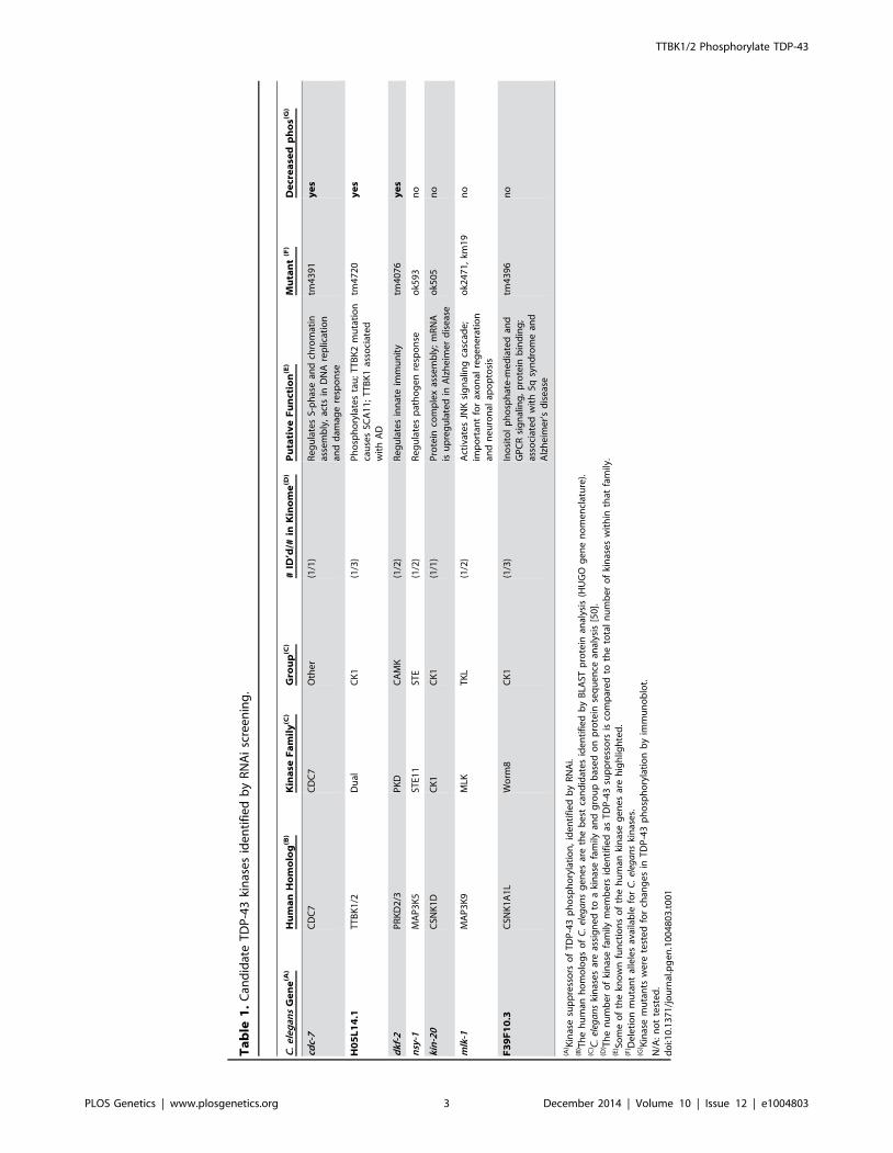
Ta
ble
1.
Can
did
ate
TD
P-4
3ki
nas
es
ide
nti
fie
db
yR
NA
isc
ree
nin
g.
C.
ele
gan
sG
en
e(A
)H
um
an
Ho
mo
log
(B)
Kin
ase
Fa
mil
y(C
)G
rou
p(C
)#
ID’d
/#in
Kin
om
e(D
)P
uta
tiv
eF
un
ctio
n(E
)M
uta
nt
(F)
De
cre
ase
dp
ho
s(G)
cdc-
7C
DC
7C
DC
7O
the
r(1
/1)
Re
gu
late
sS-
ph
ase
and
chro
mat
inas
sem
bly
,ac
tsin
DN
Are
plic
atio
nan
dd
amag
ere
spo
nse
tm4
39
1y
es
H0
5L
14
.1T
TB
K1
/2D
ual
CK
1(1
/3)
Ph
osp
ho
ryla
tes
tau
;T
TB
K2
mu
tati
on
cau
ses
SCA
11
;T
TB
K1
asso
ciat
ed
wit
hA
D
tm4
72
0y
es
dkf
-2P
RK
D2
/3P
KD
CA
MK
(1/2
)R
eg
ula
tes
inn
ate
imm
un
ity
tm4
07
6y
es
nsy
-1M
AP
3K
5ST
E11
STE
(1/2
)R
eg
ula
tes
pat
ho
ge
nre
spo
nse
ok5
93
no
kin
-20
CSN
K1
DC
K1
CK
1(1
/1)
Pro
tein
com
ple
xas
sem
bly
;m
RN
Ais
up
reg
ula
ted
inA
lzh
eim
er
dis
eas
eo
k50
5n
o
mlk
-1M
AP
3K
9M
LKT
KL
(1/2
)A
ctiv
ate
sJN
Ksi
gn
alin
gca
scad
e;
imp
ort
ant
for
axo
nal
reg
en
era
tio
nan
dn
eu
ron
alap
op
tosi
s
ok2
47
1,
km1
9n
o
F3
9F
10
.3C
SNK
1A
1L
Wo
rm8
CK
1(1
/3)
Ino
sito
lp
ho
sph
ate
-me
dia
ted
and
GP
CR
sig
nal
ing
,p
rote
inb
ind
ing
;as
soci
ate
dw
ith
5q
syn
dro
me
and
Alz
he
ime
r’s
dis
eas
e
tm4
39
6n
o
(A) K
inas
esu
pp
ress
ors
of
TD
P-4
3p
ho
sph
ory
lati
on
,id
en
tifi
ed
by
RN
Ai.
(B) T
he
hu
man
ho
mo
log
so
fC
.el
ega
ns
ge
ne
sar
eth
eb
est
can
did
ate
sid
en
tifi
ed
by
BLA
STp
rote
inan
alys
is(H
UG
Og
en
en
om
en
clat
ure
).(C
) C.
eleg
an
ski
nas
es
are
assi
gn
ed
toa
kin
ase
fam
ilyan
dg
rou
pb
ase
do
np
rote
inse
qu
en
cean
alys
is[5
0].
(D) T
he
nu
mb
er
of
kin
ase
fam
ilym
em
be
rsid
en
tifi
ed
asT
DP
-43
sup
pre
sso
rsis
com
par
ed
toth
eto
tal
nu
mb
er
of
kin
ase
sw
ith
inth
atfa
mily
.(E
) Som
eo
fth
ekn
ow
nfu
nct
ion
so
fth
eh
um
anki
nas
eg
en
es
are
hig
hlig
hte
d.
(F) D
ele
tio
nm
uta
nt
alle
les
avai
lab
lefo
rC
.el
ega
ns
kin
ase
s.(G
) Kin
ase
mu
tan
tsw
ere
test
ed
for
chan
ge
sin
TD
P-4
3p
ho
sph
ory
lati
on
by
imm
un
ob
lot.
N/A
:n
ot
test
ed
.d
oi:1
0.1
37
1/j
ou
rnal
.pg
en
.10
04
80
3.t
00
1
TTBK1/2 Phosphorylate TDP-43
PLOS Genetics | www.plosgenetics.org 3 December 2014 | Volume 10 | Issue 12 | e1004803
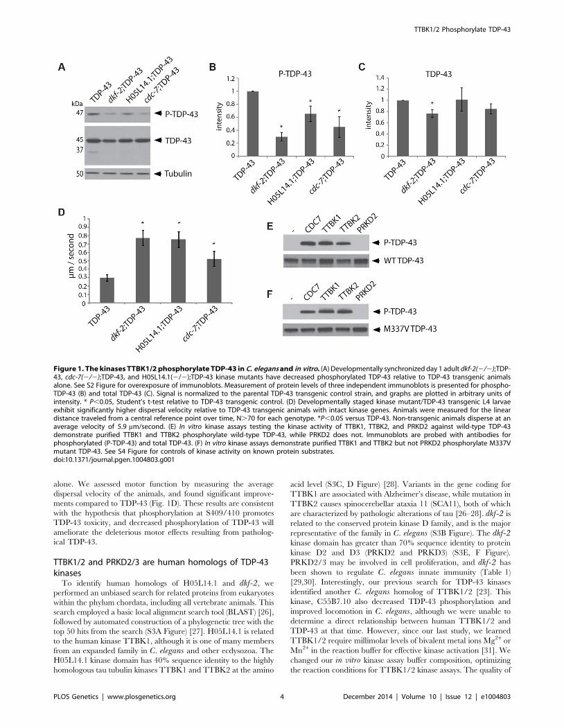
alone. We assessed motor function by measuring the average
dispersal velocity of the animals, and found significant improve-
ments compared to TDP-43 (Fig. 1D). These results are consistent
with the hypothesis that phosphorylation at S409/410 promotes
TDP-43 toxicity, and decreased phosphorylation of TDP-43 will
ameliorate the deleterious motor effects resulting from patholog-
ical TDP-43.
TTBK1/2 and PRKD2/3 are human homologs of TDP-43kinases
To identify human homologs of H05L14.1 and dkf-2, we
performed an unbiased search for related proteins from eukaryotes
within the phylum chordata, including all vertebrate animals. This
search employed a basic local alignment search tool (BLAST) [26],
followed by automated construction of a phylogenetic tree with the
top 50 hits from the search (S3A Figure) [27]. H05L14.1 is related
to the human kinase TTBK1, although it is one of many members
from an expanded family in C. elegans and other ecdysozoa. The
H05L14.1 kinase domain has 40% sequence identity to the highly
homologous tau tubulin kinases TTBK1 and TTBK2 at the amino
acid level (S3C, D Figure) [28]. Variants in the gene coding for
TTBK1 are associated with Alzheimer’s disease, while mutation in
TTBK2 causes spinocerebellar ataxia 11 (SCA11), both of which
are characterized by pathologic alterations of tau [26–28]. dkf-2 is
related to the conserved protein kinase D family, and is the major
representative of the family in C. elegans (S3B Figure). The dkf-2kinase domain has greater than 70% sequence identity to protein
kinase D2 and D3 (PRKD2 and PRKD3) (S3E, F Figure).
PRKD2/3 may be involved in cell proliferation, and dkf-2 has
been shown to regulate C. elegans innate immunity (Table 1)
[29,30]. Interestingly, our previous search for TDP-43 kinases
identified another C. elegans homolog of TTBK1/2 [23]. This
kinase, C55B7.10 also decreased TDP-43 phosphorylation and
improved locomotion in C. elegans, although we were unable to
determine a direct relationship between human TTBK1/2 and
TDP-43 at that time. However, since our last study, we learned
TTBK1/2 require millimolar levels of bivalent metal ions Mg2+ or
Mn2+ in the reaction buffer for effective kinase activation [31]. We
changed our in vitro kinase assay buffer composition, optimizing
the reaction conditions for TTBK1/2 kinase assays. The quality of
Figure 1. The kinases TTBK1/2 phosphorylate TDP-43 in C. elegans and in vitro. (A) Developmentally synchronized day 1 adult dkf-2(2/2);TDP-43, cdc-7(2/2);TDP-43, and H05L14.1(2/2);TDP-43 kinase mutants have decreased phosphorylated TDP-43 relative to TDP-43 transgenic animalsalone. See S2 Figure for overexposure of immunoblots. Measurement of protein levels of three independent immunoblots is presented for phospho-TDP-43 (B) and total TDP-43 (C). Signal is normalized to the parental TDP-43 transgenic control strain, and graphs are plotted in arbitrary units ofintensity. * P,0.05, Student’s t-test relative to TDP-43 transgenic control. (D) Developmentally staged kinase mutant/TDP-43 transgenic L4 larvaeexhibit significantly higher dispersal velocity relative to TDP-43 transgenic animals with intact kinase genes. Animals were measured for the lineardistance traveled from a central reference point over time, N.70 for each genotype. *P,0.05 versus TDP-43. Non-transgenic animals disperse at anaverage velocity of 5.9 mm/second. (E) In vitro kinase assays testing the kinase activity of TTBK1, TTBK2, and PRKD2 against wild-type TDP-43demonstrate purified TTBK1 and TTBK2 phosphorylate wild-type TDP-43, while PRKD2 does not. Immunoblots are probed with antibodies forphosphorylated (P-TDP-43) and total TDP-43. (F) In vitro kinase assays demonstrate purified TTBK1 and TTBK2 but not PRKD2 phosphorylate M337Vmutant TDP-43. See S4 Figure for controls of kinase activity on known protein substrates.doi:10.1371/journal.pgen.1004803.g001
TTBK1/2 Phosphorylate TDP-43
PLOS Genetics | www.plosgenetics.org 4 December 2014 | Volume 10 | Issue 12 | e1004803
purified TTBK1/2 kinases also affects their activity in vitro. We
compared purified TTBK1/2 from different commercial sources
side by side in an in vitro kinase assay against a known target, tau,
and found major differences in kinase activity (S4A Figure). Our
previous characterization of TTBK1/2 as potential TDP-43
kinases used commercially available purified kinase with low
activity against tau. Switching to a more active kinase preparation
and modifying the buffer composition in the assay allowed a re-
assessment of these potential TDP-43 kinases in vitro.
Human TTBK1/2 directly phosphorylate TDP-43TDP-43 kinases may act directly by phosphorylating TDP-43
S409/410 or may act indirectly by regulating the activity of other
direct TDP-43 kinases. The amino acid sequence in the C-
terminus of TDP-43 near S409/410 is consistent with the known
CK1 family kinase consensus sequence S/TpXXS/T [32]. The
PRKD kinase consensus sequence LXRXMSXXSFX [33], does
not conform well with the sequence of human TDP-43. To
empirically determine whether human TTBK1/2 or PRKD2/3
are direct TDP-43 kinases, we tested the ability of purified active
kinase enzymes to phosphorylate TDP-43 at S409/410 and S403/
404 in vitro (Fig. 1E, F, S4B Figure). We found that TTBK1 and 2
can directly phosphorylate both wild-type (WT) and familial ALS
mutant TDP-43 (M337V TDP-43) under optimized reaction
conditions that include magnesium. These conditions support
robust phosphorylation of human tau protein, a known substrate
of TTBK1/2 (S4A Figure, [31]). Although our preparation of
PRKD2 kinase was enzymatically active against a known
phosphorylation substrate, histone H1 [34] (S4C Figure), PRKD2
was unable to phosphorylate TDP-43 under any conditions tested,
indicating its effect on TDP-43 phosphorylation may be indirect
through the activation of other direct TDP-43 kinases or
regulation of other downstream members of a TDP-43 regulatory
pathway. If the kinases CDC7, TTBK1/2, or PRKD2/3 are in a
common regulatory pathway, they may directly phosphorylate one
another. Using an in vitro kinase assay with purified human
kinases, we observed robust auto-phosphorylation by TTBK1 and
modest auto-phosphorylation by TTBK2 and PRKD2, consistent
with known activities of these kinases [28,31,34]. We also tested
pairwise combinations of these kinases to determine any relative
increases in phosphorylation. However, we did not see any
significant increases in phosphorylation on these kinases (S4D
Figure). Therefore, any indirect regulation of TDP-43 phosphor-
ylation by PRKD2 may be through other unknown members of
one or several regulatory pathways controlling TDP-43 phosphor-
ylation.
TTBK1/2 promote TDP-43 phosphorylation in vivoTTBK1/2 kinase hyperactivity may contribute to the patho-
logical phosphorylated TDP-43 observed in both FTLD-TDP and
ALS. To test whether increased cellular levels of TTBK1/2
activity suffice to drive TDP-43 phosphorylation, we transfected
full-length TTBK1 and TTBK2 cDNAs into HEK293 cells.
HEK293 cells have some neuronal characteristics and may be
derived from a subpopulation of neuronal precursor cells in the
embryonic kidney [35]. This cell line is especially useful for
biochemical assays requiring high efficiency transfection rates. In
the absence of other cellular stresses, we observed robust induction
of TDP-43 phosphorylation by immunoblot following transfection
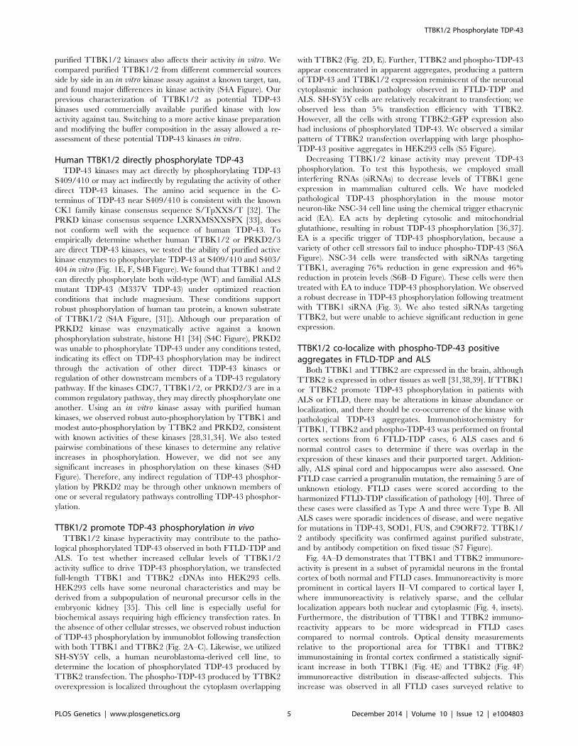
with both TTBK1 and TTBK2 (Fig. 2A–C). Likewise, we utilized
SH-SY5Y cells, a human neuroblastoma-derived cell line, to
determine the location of phosphorylated TDP-43 produced by
TTBK2 transfection. The phospho-TDP-43 produced by TTBK2
overexpression is localized throughout the cytoplasm overlapping
with TTBK2 (Fig. 2D, E). Further, TTBK2 and phospho-TDP-43
appear concentrated in apparent aggregates, producing a pattern
of TDP-43 and TTBK1/2 expression reminiscent of the neuronal
cytoplasmic inclusion pathology observed in FTLD-TDP and
ALS. SH-SY5Y cells are relatively recalcitrant to transfection; we
observed less than 5% transfection efficiency with TTBK2.
However, all the cells with strong TTBK2::GFP expression also
had inclusions of phosphorylated TDP-43. We observed a similar
pattern of TTBK2 transfection overlapping with large phospho-
TDP-43 positive aggregates in HEK293 cells (S5 Figure).
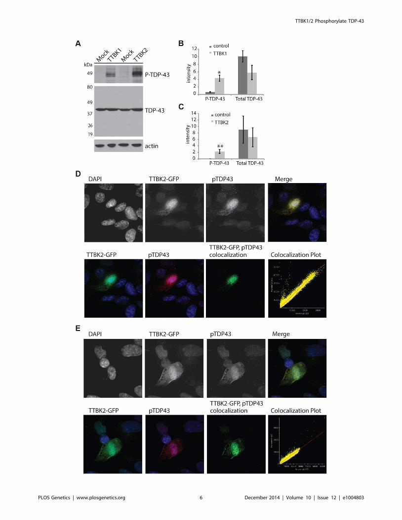
Decreasing TTBK1/2 kinase activity may prevent TDP-43
phosphorylation. To test this hypothesis, we employed small
interfering RNAs (siRNAs) to decrease levels of TTBK1 gene
expression in mammalian cultured cells. We have modeled
pathological TDP-43 phosphorylation in the mouse motor
neuron-like NSC-34 cell line using the chemical trigger ethacrynic
acid (EA). EA acts by depleting cytosolic and mitochondrial
glutathione, resulting in robust TDP-43 phosphorylation [36,37].
EA is a specific trigger of TDP-43 phosphorylation, because a
variety of other cell stressors fail to induce phospho-TDP-43 (S6A
Figure). NSC-34 cells were transfected with siRNAs targeting
TTBK1, averaging 76% reduction in gene expression and 46%
reduction in protein levels (S6B–D Figure). These cells were then
treated with EA to induce TDP-43 phosphorylation. We observed
a robust decrease in TDP-43 phosphorylation following treatment
with TTBK1 siRNA (Fig. 3). We also tested siRNAs targeting
TTBK2, but were unable to achieve significant reduction in gene
expression.
TTBK1/2 co-localize with phospho-TDP-43 positiveaggregates in FTLD-TDP and ALS
Both TTBK1 and TTBK2 are expressed in the brain, although
TTBK2 is expressed in other tissues as well [31,38,39]. If TTBK1
or TTBK2 promote TDP-43 phosphorylation in patients with
ALS or FTLD, there may be alterations in kinase abundance or
localization, and there should be co-occurrence of the kinase with
pathological TDP-43 aggregates. Immunohistochemistry for
TTBK1, TTBK2 and phospho-TDP-43 was performed on frontal
cortex sections from 6 FTLD-TDP cases, 6 ALS cases and 6
normal control cases to determine if there was overlap in the
expression of these kinases and their purported target. Addition-
ally, ALS spinal cord and hippocampus were also assessed. One
FTLD case carried a progranulin mutation, the remaining 5 are of
unknown etiology. FTLD cases were scored according to the
harmonized FTLD-TDP classification of pathology [40]. Three of
these cases were classified as Type A and three were Type B. All
ALS cases were sporadic incidences of disease, and were negative
for mutations in TDP-43, SOD1, FUS, and C9ORF72. TTBK1/
2 antibody specificity was confirmed against purified substrate,
and by antibody competition on fixed tissue (S7 Figure).
Fig. 4A–D demonstrates that TTBK1 and TTBK2 immunore-
activity is present in a subset of pyramidal neurons in the frontal
cortex of both normal and FTLD cases. Immunoreactivity is more
prominent in cortical layers II–VI compared to cortical layer I,
where immunoreactivity is relatively sparse, and the cellular
localization appears both nuclear and cytoplasmic (Fig. 4, insets).
Furthermore, the distribution of TTBK1 and TTBK2 immuno-
reactivity appears to be more widespread in FTLD cases
compared to normal controls. Optical density measurements
relative to the proportional area for TTBK1 and TTBK2
immunostaining in frontal cortex confirmed a statistically signif-
icant increase in both TTBK1 (Fig. 4E) and TTBK2 (Fig. 4F)
immunoreactive distribution in disease-affected subjects. This
increase was observed in all FTLD cases surveyed relative to
TTBK1/2 Phosphorylate TDP-43
PLOS Genetics | www.plosgenetics.org 5 December 2014 | Volume 10 | Issue 12 | e1004803
TTBK1/2 Phosphorylate TDP-43
PLOS Genetics | www.plosgenetics.org 6 December 2014 | Volume 10 | Issue 12 | e1004803
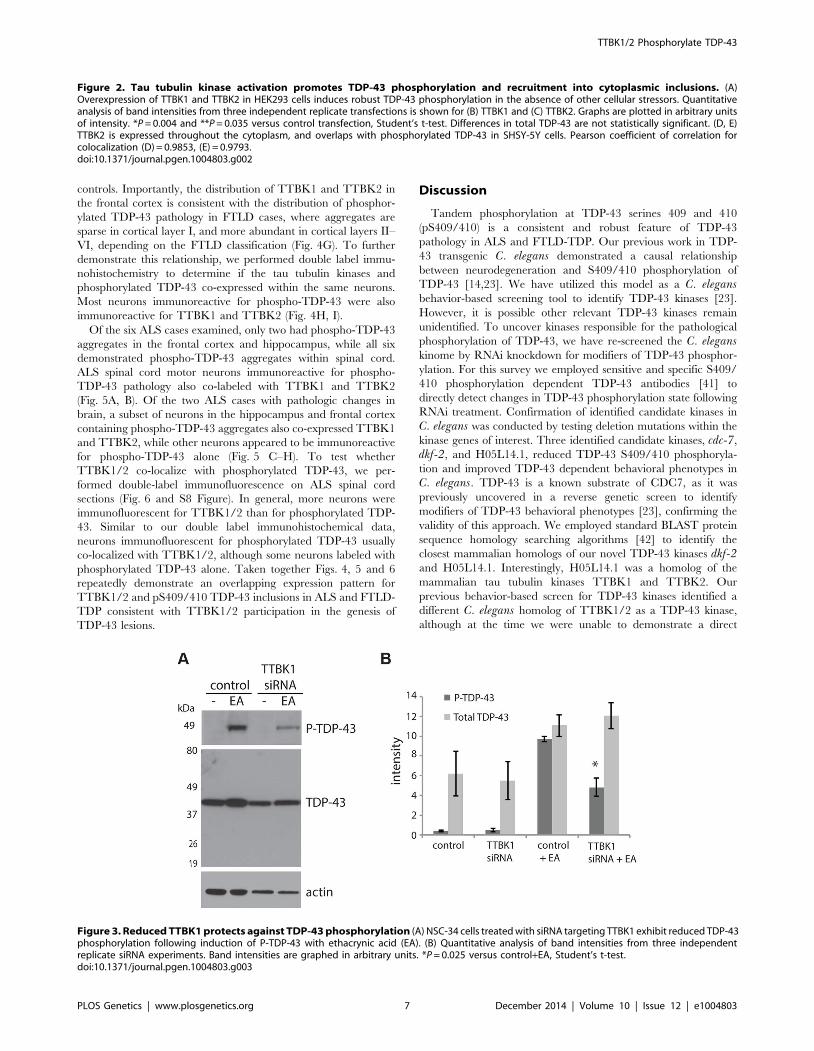
controls. Importantly, the distribution of TTBK1 and TTBK2 in
the frontal cortex is consistent with the distribution of phosphor-
ylated TDP-43 pathology in FTLD cases, where aggregates are
sparse in cortical layer I, and more abundant in cortical layers II–
VI, depending on the FTLD classification (Fig. 4G). To further
demonstrate this relationship, we performed double label immu-
nohistochemistry to determine if the tau tubulin kinases and
phosphorylated TDP-43 co-expressed within the same neurons.
Most neurons immunoreactive for phospho-TDP-43 were also
immunoreactive for TTBK1 and TTBK2 (Fig. 4H, I).
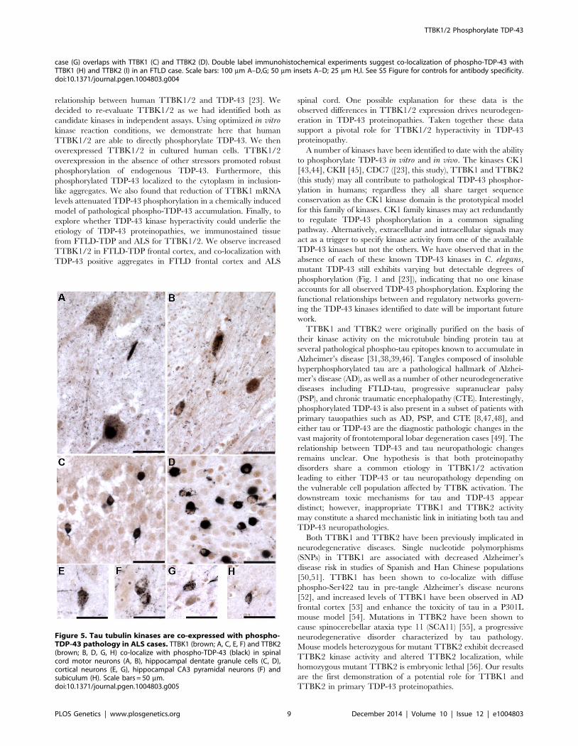
Of the six ALS cases examined, only two had phospho-TDP-43
aggregates in the frontal cortex and hippocampus, while all six
demonstrated phospho-TDP-43 aggregates within spinal cord.
ALS spinal cord motor neurons immunoreactive for phospho-
TDP-43 pathology also co-labeled with TTBK1 and TTBK2
(Fig. 5A, B). Of the two ALS cases with pathologic changes in
brain, a subset of neurons in the hippocampus and frontal cortex
containing phospho-TDP-43 aggregates also co-expressed TTBK1
and TTBK2, while other neurons appeared to be immunoreactive
for phospho-TDP-43 alone (Fig. 5 C–H). To test whether
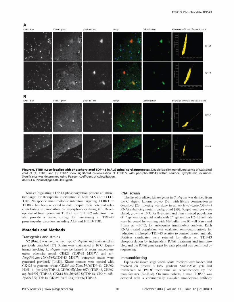
TTBK1/2 co-localize with phosphorylated TDP-43, we per-
formed double-label immunofluorescence on ALS spinal cord
sections (Fig. 6 and S8 Figure). In general, more neurons were
immunofluorescent for TTBK1/2 than for phosphorylated TDP-
43. Similar to our double label immunohistochemical data,
neurons immunofluorescent for phosphorylated TDP-43 usually
co-localized with TTBK1/2, although some neurons labeled with
phosphorylated TDP-43 alone. Taken together Figs. 4, 5 and 6
repeatedly demonstrate an overlapping expression pattern for
TTBK1/2 and pS409/410 TDP-43 inclusions in ALS and FTLD-
TDP consistent with TTBK1/2 participation in the genesis of
TDP-43 lesions.
Discussion
Tandem phosphorylation at TDP-43 serines 409 and 410
(pS409/410) is a consistent and robust feature of TDP-43
pathology in ALS and FTLD-TDP. Our previous work in TDP-
43 transgenic C. elegans demonstrated a causal relationship
between neurodegeneration and S409/410 phosphorylation of
TDP-43 [14,23]. We have utilized this model as a C. elegansbehavior-based screening tool to identify TDP-43 kinases [23].
However, it is possible other relevant TDP-43 kinases remain
unidentified. To uncover kinases responsible for the pathological
phosphorylation of TDP-43, we have re-screened the C. eleganskinome by RNAi knockdown for modifiers of TDP-43 phosphor-
ylation. For this survey we employed sensitive and specific S409/
410 phosphorylation dependent TDP-43 antibodies [41] to
directly detect changes in TDP-43 phosphorylation state following
RNAi treatment. Confirmation of identified candidate kinases in
C. elegans was conducted by testing deletion mutations within the
kinase genes of interest. Three identified candidate kinases, cdc-7,
dkf-2, and H05L14.1, reduced TDP-43 S409/410 phosphoryla-
tion and improved TDP-43 dependent behavioral phenotypes in
C. elegans. TDP-43 is a known substrate of CDC7, as it was
previously uncovered in a reverse genetic screen to identify
modifiers of TDP-43 behavioral phenotypes [23], confirming the
validity of this approach. We employed standard BLAST protein
sequence homology searching algorithms [42] to identify the
closest mammalian homologs of our novel TDP-43 kinases dkf-2and H05L14.1. Interestingly, H05L14.1 was a homolog of the
mammalian tau tubulin kinases TTBK1 and TTBK2. Our
previous behavior-based screen for TDP-43 kinases identified a
different C. elegans homolog of TTBK1/2 as a TDP-43 kinase,
although at the time we were unable to demonstrate a direct
Figure 2. Tau tubulin kinase activation promotes TDP-43 phosphorylation and recruitment into cytoplasmic inclusions. (A)Overexpression of TTBK1 and TTBK2 in HEK293 cells induces robust TDP-43 phosphorylation in the absence of other cellular stressors. Quantitativeanalysis of band intensities from three independent replicate transfections is shown for (B) TTBK1 and (C) TTBK2. Graphs are plotted in arbitrary unitsof intensity. *P = 0.004 and **P = 0.035 versus control transfection, Student’s t-test. Differences in total TDP-43 are not statistically significant. (D, E)TTBK2 is expressed throughout the cytoplasm, and overlaps with phosphorylated TDP-43 in SHSY-5Y cells. Pearson coefficient of correlation forcolocalization (D) = 0.9853, (E) = 0.9793.doi:10.1371/journal.pgen.1004803.g002
Figure 3. Reduced TTBK1 protects against TDP-43 phosphorylation (A) NSC-34 cells treated with siRNA targeting TTBK1 exhibit reduced TDP-43phosphorylation following induction of P-TDP-43 with ethacrynic acid (EA). (B) Quantitative analysis of band intensities from three independentreplicate siRNA experiments. Band intensities are graphed in arbitrary units. *P = 0.025 versus control+EA, Student’s t-test.doi:10.1371/journal.pgen.1004803.g003
TTBK1/2 Phosphorylate TDP-43
PLOS Genetics | www.plosgenetics.org 7 December 2014 | Volume 10 | Issue 12 | e1004803
Figure 4. Upregulated Tau tubulin kinases are also co-expressed with phospho-TDP-43 pathology. Representative photomicrographsdepicting TTBK1 (A, C) and TTBK2 (B, D) immunoreactivity in cortical neurons in normal (A, B) and FTLD-TDP Type B (C, D) cases. The cellulardistribution is both cytoplasmic and nuclear (insets), and immunoreactivity appears to be more widespread in FTLD cases relative to normal controls.Cortical layers I-VI are indicated (C). Quantification of immunostaining demonstrated a statistically significant increase in both TTBK1 (E) and TTBK2 (F)in FTLD cases compared to normal controls (**P = 0.003; ***P,0.0001). The distribution of phospho-TDP-43 immunoreactivity in the cortex of an FTLD
TTBK1/2 Phosphorylate TDP-43
PLOS Genetics | www.plosgenetics.org 8 December 2014 | Volume 10 | Issue 12 | e1004803
relationship between human TTBK1/2 and TDP-43 [23]. We
decided to re-evaluate TTBK1/2 as we had identified both as
candidate kinases in independent assays. Using optimized in vitrokinase reaction conditions, we demonstrate here that human
TTBK1/2 are able to directly phosphorylate TDP-43. We then
overexpressed TTBK1/2 in cultured human cells. TTBK1/2
overexpression in the absence of other stressors promoted robust
phosphorylation of endogenous TDP-43. Furthermore, this
phosphorylated TDP-43 localized to the cytoplasm in inclusion-
like aggregates. We also found that reduction of TTBK1 mRNA
levels attenuated TDP-43 phosphorylation in a chemically induced
model of pathological phospho-TDP-43 accumulation. Finally, to
explore whether TDP-43 kinase hyperactivity could underlie the
etiology of TDP-43 proteinopathies, we immunostained tissue
from FTLD-TDP and ALS for TTBK1/2. We observe increased
TTBK1/2 in FTLD-TDP frontal cortex, and co-localization with
TDP-43 positive aggregates in FTLD frontal cortex and ALS
spinal cord. One possible explanation for these data is the
observed differences in TTBK1/2 expression drives neurodegen-
eration in TDP-43 proteinopathies. Taken together these data
support a pivotal role for TTBK1/2 hyperactivity in TDP-43
proteinopathy.
A number of kinases have been identified to date with the ability
to phosphorylate TDP-43 in vitro and in vivo. The kinases CK1
[43,44], CKII [45], CDC7 ([23], this study), TTBK1 and TTBK2
(this study) may all contribute to pathological TDP-43 phosphor-
ylation in humans; regardless they all share target sequence
conservation as the CK1 kinase domain is the prototypical model
for this family of kinases. CK1 family kinases may act redundantly
to regulate TDP-43 phosphorylation in a common signaling
pathway. Alternatively, extracellular and intracellular signals may
act as a trigger to specify kinase activity from one of the available
TDP-43 kinases but not the others. We have observed that in the
absence of each of these known TDP-43 kinases in C. elegans,mutant TDP-43 still exhibits varying but detectable degrees of
phosphorylation (Fig. 1 and [23]), indicating that no one kinase
accounts for all observed TDP-43 phosphorylation. Exploring the
functional relationships between and regulatory networks govern-
ing the TDP-43 kinases identified to date will be important future
work.
TTBK1 and TTBK2 were originally purified on the basis of
their kinase activity on the microtubule binding protein tau at
several pathological phospho-tau epitopes known to accumulate in
Alzheimer’s disease [31,38,39,46]. Tangles composed of insoluble
hyperphosphorylated tau are a pathological hallmark of Alzhei-
mer’s disease (AD), as well as a number of other neurodegenerative
diseases including FTLD-tau, progressive supranuclear palsy
(PSP), and chronic traumatic encephalopathy (CTE). Interestingly,
phosphorylated TDP-43 is also present in a subset of patients with
primary tauopathies such as AD, PSP, and CTE [8,47,48], and
either tau or TDP-43 are the diagnostic pathologic changes in the
vast majority of frontotemporal lobar degeneration cases [49]. The
relationship between TDP-43 and tau neuropathologic changes
remains unclear. One hypothesis is that both proteinopathy
disorders share a common etiology in TTBK1/2 activation
leading to either TDP-43 or tau neuropathology depending on
the vulnerable cell population affected by TTBK activation. The
downstream toxic mechanisms for tau and TDP-43 appear
distinct; however, inappropriate TTBK1 and TTBK2 activity
may constitute a shared mechanistic link in initiating both tau and
TDP-43 neuropathologies.
Both TTBK1 and TTBK2 have been previously implicated in
neurodegenerative diseases. Single nucleotide polymorphisms
(SNPs) in TTBK1 are associated with decreased Alzheimer’s
disease risk in studies of Spanish and Han Chinese populations
[50,51]. TTBK1 has been shown to co-localize with diffuse
phospho-Ser422 tau in pre-tangle Alzheimer’s disease neurons
[52], and increased levels of TTBK1 have been observed in AD
frontal cortex [53] and enhance the toxicity of tau in a P301L
mouse model [54]. Mutations in TTBK2 have been shown to
cause spinocerebellar ataxia type 11 (SCA11) [55], a progressive
neurodegenerative disorder characterized by tau pathology.
Mouse models heterozygous for mutant TTBK2 exhibit decreased
TTBK2 kinase activity and altered TTBK2 localization, while
homozygous mutant TTBK2 is embryonic lethal [56]. Our results
are the first demonstration of a potential role for TTBK1 and
TTBK2 in primary TDP-43 proteinopathies.
case (G) overlaps with TTBK1 (C) and TTBK2 (D). Double label immunohistochemical experiments suggest co-localization of phospho-TDP-43 withTTBK1 (H) and TTBK2 (I) in an FTLD case. Scale bars: 100 mm A–D,G; 50 mm insets A–D; 25 mm H,I. See S5 Figure for controls for antibody specificity.doi:10.1371/journal.pgen.1004803.g004
Figure 5. Tau tubulin kinases are co-expressed with phospho-TDP-43 pathology in ALS cases. TTBK1 (brown; A, C, E, F) and TTBK2(brown; B, D, G, H) co-localize with phospho-TDP-43 (black) in spinalcord motor neurons (A, B), hippocampal dentate granule cells (C, D),cortical neurons (E, G), hippocampal CA3 pyramidal neurons (F) andsubiculum (H). Scale bars = 50 mm.doi:10.1371/journal.pgen.1004803.g005
TTBK1/2 Phosphorylate TDP-43
PLOS Genetics | www.plosgenetics.org 9 December 2014 | Volume 10 | Issue 12 | e1004803
Kinases regulating TDP-43 phosphorylation present an attrac-
tive target for therapeutic intervention in both ALS and FTLD-
TDP. No specific small molecule inhibitors targeting TTBK1 or
TTBK2 has been reported to date, despite their potential roles
contributing to tauopathies by hyperphosphorylating tau. Devel-
opment of brain penetrant TTBK1 and TTBK2 inhibitors may
also provide a viable strategy for intervening in TDP-43
proteinopathy disorders including ALS and FTLD-TDP.
Materials and Methods
Transgenics and strainsN2 (Bristol) was used as wild type C. elegans and maintained as
previously described [57]. Strains were maintained at 16uC. Exper-
iments involving C. elegans were performed at room temperature
unless otherwise noted. CK423 (TDP-43 M337V) and eri-1(mg366);lin-15b(n744);TDP-43 M337V transgenic strains were
generated previously [14,23]. Kinase mutants were crossed with
CK423 to generate strains CK566 cdc-7(tm4391);TDP-43, CK602
H05L14.1(tm4720);TDP-43, CK600 dkf-2(tm4076);TDP-43, CK597
nsy-1(ok593);TDP-43, CK613 kin-20(ok505);TDP-43, CK574 mlk-1(ok2471);TDP-43, CK623 F39F10.3(tm4396);TDP-43.
RNAi screenThe list of predicted kinase genes in C. elegans was derived from
the C. elegans kinome project [58], with library construction as
described [23]. Testing was done in an eri-1(2/2);lin-15(2/2)RNAi enhancing mutant background [59]. Staged embryos were
plated, grown at 16uC for 8–9 days, and then a mixed population
of 1st generation gravid adults with 2nd generation L2–L3 animals
were harvested by washing with M9 buffer into 96 well plates and
frozen at 280uC, for subsequent immunoblot analysis. Each
RNAi treated population was evaluated semi-quantitatively for
reduction in phospho-TDP-43 relative to control treated animals.
Positives candidates were retested for effects on TDP-43
phosphorylation by independent RNAi treatment and immuno-
blot, and the RNAi gene target for each plasmid was confirmed by
sequencing.
ImmunoblottingEquivalent mixed-stage worm lysate fractions were loaded and
resolved on precast 4–15% gradient SDS-PAGE gels and
transferred to PVDF membrane as recommended by the
manufacturer (Bio-Rad). On immunoblots, human TDP-43 was
detected with a commercially available monoclonal antibody
Figure 6. TTBK1/2 co-localize with phosphorylated TDP-43 in ALS spinal cord aggregates. Double-label immunofluorescence of ALS spinalcord of (A) TTBK1 and (B) TTBK2 show significant co-localization of TTBK1/2 with phospho-TDP-43 within neuronal cytoplasmic inclusions.Significance was determined using Pearson coefficient of colocalization.doi:10.1371/journal.pgen.1004803.g006
TTBK1/2 Phosphorylate TDP-43
PLOS Genetics | www.plosgenetics.org 10 December 2014 | Volume 10 | Issue 12 | e1004803
ab57105 (Abcam) directed against human TDP-43 amino acids 1–
261. TDP-43 phosphorylated at S409/S410 was detected by a
monoclonal antibody called anti phospho TDP-43 (pS409/410)
available from Cosmobio (catalog # TIP-PTD-M01). C. elegansb-tubulin levels were measured using monoclonal antibody E7 as a
loading control as previously described [60,61]. TTBK1 was
detected by Abcam rabbit polyclonal antibody ab103944 at
1:1000 dilution. TTBK2 was detected by Abgent rabbit polyclonal
antibody AP12162a at 1:1000 dilution. HRP labeled goat anti-
mouse IgG was the secondary antibody (GE Healthcare) and used
at a dilution of 1:4000. Dilutions were: 1:7500 for ab57105, 1:1000
for pS409/410, and 1:10000 for E7. Immunoblots shown are
representative of at least 3 independent experiments. Quantitation
was performed using ImageJ image processing and analysis
software.
Kinase assaysGST-TDP-43 (WT) and GST-TDP-43 (M337V) fusion proteins
were purified from BL21 (DE3) expression host cells as previously
described [62]. Active kinase enzymes were obtained commer-
cially via purification from SF9 cells for PRKD2, TTBK1 and
TTBK2 (Signalchem). Enzyme assays were carried out in a kinase
reaction buffer containing 25 mM MOPS, 12.25 mM glycerol-
phosphate, 25 mM MgCl, 5 mM EGTA, 2 mM EDTA, 0.25 mM
DTT and 50 mM ATP.
Cell linesHEK 293 cells (ATCC) were cultured in Dulbecco’s modified
Eagle medium (DMEM) supplemented with 10% defined fetal
bovine serum (FBS) and penicillin (50 IU/ml)–streptomycin
(50 mg/ml). NSC-34 cells (Cedarlane Labs) and SHSY-5Y cells
(ATCC) were cultured in DMEM/HAM’s F12 (50/50) with 10%
FBS and penicillin (50 IU/ml)–streptomycin (50 mg/ml).
Immunofluorescence for cultured cellsCells were seeded onto poly-D-Lysine coated (Sigma Aldrich)
12 mm round glass cover slips in 24-well plates. Cells were
transfected with the plasmid encoding TTBK2-GFP with
GenePorter 2 (Genlantis) using the manufacturer’s protocol. Cells
were fixed for imaging in 4% formaldehyde 96 hours after
transfection. Cells were washed 365 min in PBS/Ca2+/Mg2+,
then blocked in antibody buffer (PBS, 0.5% Triton X-100, 1 mM
EDTA, 0.1% BSA, 0.05% NaN2)+10% normal goat serum.
Primary antibody was applied and incubated for 1 hour at room
temperature (Cosmo Bio; 1:1000). Cells were washed 365 min in
PBS/Ca2+/Mg2+, then re-blocked for 10 min. Appropriate
secondary antibody was applied and incubated for 20 min at
room temperature. Cells were again washed 365 min in PBS/
Ca2+/Mg2+, counterstained with 300 nM DAPI and mounted
with ProLong Gold antifade. Microscopy was performed on a
Delta Vision microscope (Applied Precision, Inc) using a 606 oil
immersion objective, a sCMOS camera, and 262 binning. Image
analysis was performed using softWoRx 6.0 Beta software.
RNA interferenceHEK 293 cells were treated with 150 mM ethacrynic acid (EA)
for 5 hours to induce endogenous TDP-43 phosphorylation [36].
NSC-34 cells were grown in differentiation medium (DMEM/
HAM’s F12 (50/50), 1% FBS, 1% non-essential amino acids
(NEAA), penicillin (50 IU/ml)–streptomycin (50 mg/ml)) for one
day prior to treatment with 50 mM EA for 5 hours. TTBK1
siRNA construct was MMC.RNAI.N001162864.12.1 (Integrated
DNA Technologies). RNAi experiments were carried out as per
protocol in the TriFECTa Dicer-Substrate RNAi manual (Inte-
grated DNA Technologies).
TransfectionTransfection of plasmids containing full-length TTBK1
(pWO:TTBK1) and TTBK2 (TTBK2 GFP pFLAP dest) sequenc-
es [63] was performed as specified by the manufacturer using the
Geneporter 2 Transfection Reagent (Genlantis).
Quantitative reverse-transcription PCRRNA was purified from flash-frozen cell pellets using TRIzol
Reagent (Life Technologies) according to the manufacturer’s
protocol. cDNA was made using iScript Reverse Transcription
Supermix (Bio-Rad). qPCR was performed on an 7900HT Real
Time PCR System (Applied Biosystems) using iTaq Universal
SYBR Green Supermix (Bio-Rad).
Ethics statement: Post mortem human tissueDe-identified post-mortem brain tissue used in this study was
determined to be an exempt from IRB review by the VA Puget
Sound Health Care System Human Research Protection Program
Director on December 29, 2011. Tissue used for these studies was
obtained from the University of Washington Alzheimer’s Disease
Research Center brain bank (Seattle, WA), and the Indiana
Alzheimer Disease Center brain bank(Indianapolis, IN), where
consent for autopsy and permission for use of tissue in scientific
experiments was obtained. FTLD and ALS cases were selected on the
basis of having an autopsy-confirmed diagnosis of FTLD and FTLD-
related disorders or ALS. Control samples were from de-identified
neurologically healthy control participants, who were of a similar age.
Immunohistochemistry and immunofluorescence fortissue
Primary antibodies used for immunohistochemistry were anti-
TTBK1 (Abcam, 1:100), anti-TTBK2 (Abgent, 1:200), and anti-
phospho TDP-43 409/410 (CosmoBio, 1:1000)). In order to
minimize variability, sections from all cases (normal and affected
subjects) were stained simultaneously for each antibody. Immu-
nostained sections were analyzed using the computerized image
analysis system, MicroComputer Imaging Device (MCID, Imag-
ing Research, St. Catherines, Ontario, Canada). Blinded assess-
ment of optical density measurements were obtained relative to the
proportional area for TTBK1 and TTBK2 immunostaining in
frontal cortex grey matter (three separate readings per case). Data
were averaged and are represented as mean +/2 SEM. A two
tailed Student’s t-test was used to assess differences in TTBK1 and
TTBK2 expression between cases and controls. For double label
immunohistochemistry experiments, sections were first immuno-
stained with anti-phospho TDP-43 and reaction product was
visualized with nickel enhanced DAB (black). Sections were then
immunostained with anti-TTBK1 or TTBK2 and visualized with
DAB alone (brown). For double label immunofluorescence
experiments, AlexaFluor 488 goat anti-rabbit and AlexaFluor
594 goat anti-mouse secondary antibodies (Molecular Probes)
were used and autofluoresence was quenched with 0.1% Sudan
Black [64]. To demonstrate specificity of the TTBK antibodies,
TTBK1 and TTBK2 were blocked with 50-fold amount of
immunizing peptide overnight at 4uC before proceeding with the
immunostaining protocol (see S5 Figure).
Supporting Information
S1 Figure Immunoblot results from primary kinase RNAi
screen. Populations of RNAi treated C. elegans were harvested
TTBK1/2 Phosphorylate TDP-43
PLOS Genetics | www.plosgenetics.org 11 December 2014 | Volume 10 | Issue 12 | e1004803
into 96-well plates prior to immunoblot analysis. Gene names and
locations of kinases tested are presented in Table S1. Two rows
from each plate were tested in alternating wells for each
immunoblot. Labels above individual wells describe Row and
Column information for each sample. Plate numbers are indicated
at the left of the immunoblot. a-tubulin antibody is used as a load
control. Candidates confirmed on repeat testing are boxed in blue.
(PDF)
S2 Figure Full immunoblots of total TDP-43 levels. Full
immunoblots from Fig. 1, showing low (3 minute exposure) and
high molecular weight species (15 minute exposure) of total TDP-43.
(PDF)
S3 Figure H05L14.1 and dkf-2 mammalian homologs identified
by BLAST. (A, B) Cladogram of vertebrate homologs of the C.elegans proteins H05L14.1 and dkf-2. The entire C. elegansamino acid sequences of H05L14.1 or dkf-2 were compared
against non-redundant reference sequences from the RefSeq
protein database (7-20-2014, NCBI). (A) 2878 active hits were
identified by BLAST with similarity to H05L14.1, and subse-
quently filtered to include only sequences from C. elegans or
phylum chordata. The top 50 hits underwent multiple sequence
alignment, alignment refinement, phylogenetic reconstruction,
and are displayed in a cladogram, with branch support values in
red [27]. Related human gene and gene identifier is boxed. Homo
sapiens GI# 58761548 is TTBK1. (B) 5000 active hits were
identified with similarity to dkf-2, filtered, and graphed as above.
Homo sapiens GI# 5031689 is PRKD3. (C) H05L14.1 kinase
domain has 40% identity to human TTBK1 and TTBK2. (D) dkf-2 has more than 70% identity to human PRKD2 and PRKD3.
Sequence identity was calculated using Clustal W method for
multiple sequence alignment. (E) Alignment report for H05L14.1,
TTBK1, and TTBK2 kinase domain, including boxes around
sequence that matches the consensus. (F) Alignment report for dkf-2, PRKD2, and PRKD3. Reports were generated using Lasergene
MegAlign software for protein sequence analysis and alignment.
(PDF)
S4 Figure In vitro kinase assay controls. (A) Tau is a known
substrate of TTBK2 [65]. To test enzyme activity, approximately
1 mg of non-phosphorylated recombinant human tau purified from
E. coli were incubated with equivalent amounts of TTBK2
enzyme purified from cultured cells by two commercial suppliers
(Origene catalog #LY406582 and Signalchem #T18-11G).
Phosphorylation was assessed by reactivity with AT270, a
phospho-tau antibody recognizing tau phosphorylated at
Thr181. (B) Purified TTBK1, TTBK2, and CDC7 can also
phosphorylate TDP-43 at serines 403 and 404 (CosmoBio,
#CAC-TIP-PTD-P05) in an in vitro kinase assay. (C) Histone
H1 is a known substrate of PRKD2 [34]. To confirm PRKD2
activity, human PRKD2 (SignalChem #P76-10) purified from
cultured cells was incubated with purified recombinant Histone
H1. We observed phosphorylation of Histone H1 as detected by
reactivity with pT146 specific antibody (Bioss Catalog # bs-
3176R). (D) Purified CDC7, TTBK1, TTBK2, or PRKD2 were
incubated singly or pairwise with radiolabeled phosphate. TTBK1
can robustly auto-phosphorylate, while TTBK2 and PRKD2 are
also capable of auto-phosphorylation. Pairwise combinations of
CDC7, TTBK1, TTBK2, and PRKD2 did not exhibit any
increase or variety in phosphorylation beyond baseline levels of
auto-phosphorylation for each kinase.
(PDF)
S5 Figure (A) Phosphorylated TDP-43 is localized in a large
discrete cytoplasmic aggregate following TTBK2 overexpression
in HEK293 cells. (B) TTBK2 and phosphorylated TDP-43 co-
localize in cells overexpressing TTBK2.
(PDF)
S6 Figure (A) Treatment of HEK293 cells with a variety of
cellular stressors failed to produce phosphorylated TDP-43.
Bafilomycin and wortmannin are inhibitors of autophagy, PSI is
a general proteasome inhibitor, cadmium chloride is a heavy
metal, taxol is an inhibitor of microtubule dynamics, rotenone
blocks the mitochondrial electron transport chain (creating
reactive oxygen species (ROS)), pepstatin inhibits aspartic
proteases, and paraquat catalyzes formation of ROS. (B) TTBK1
is reduced by nearly 80% following siRNA treatment in NSC-34
cells. Quantitative PCR measurements (qPCR) for TTBK1
mRNA levels are displayed in arbitrary units for an untreated
control and cells treated with TTBK1 siRNA. (C) siRNA targeting
TTBK1 reduce levels of TTBK1 protein, as detected by
immunoblot. (D) TTBK1 protein levels are reduced by an average
of 46%, following siRNA treatment in NSC-34 cells. Quantitation
of TTBK1 protein levels from three independent experiments is
graphed in arbitrary units of band intensity.
(PDF)
S7 Figure Antibody Validation. (A) Studies examining expression
of TTBK1 used commercially sourced antibodies including Anti-
TTBK1 (Sigma-Aldrich, SAB3500002), Anti TTBK#1 (Abgent,
AP4947a), Anti-TTBK1 (Abcam, ab103944). (B) Antibodies
tested for TTBK2 were TTBK2 Antibody N-term (Abgent,AP12162a), Anti-Tau tubulin kinase 1 antibody (Abcam, ab67839),
and TTBK2 Polyclonal Antibody (Proteintech, 15072-1-AP).
Antibodies underlined/bold above were used in further experiments
and are boxed in red in the figure. (C–J) Peptide blocking
experiments with the cognate immunizing peptide further demon-
strates specificity of the selected TTBK1 and TTBK2 antibodies.
Anti-TTBK1 (Abcam, ab103944) (C–F) and anti-TTBK2
(Abgent, AP12162a) (G–J) were pre-incubated with a 50 fold
excess of the blocking peptide (D, F, H, J) before proceeding with the
immunostaining protocol and compared with immunostaining
using antibody alone (C, E, G, I). ALS spinal cord (C, D, G, H)
and FTLD frontal cortex (E, F, I, J). Scale bar = 100 um.
(PDF)
S8 Fig TTBK1/2 co-localize with phosphorylated TDP-43 in
aggregates in ALS spinal cord. Double-label immunofluorescence of
ALS spinal cord demonstrates additional neurons that significantly
co-localize TTBK1 (upper panel) or TTBK2 (lower panel) with
phospho-TDP-43 within neuronal cytoplasmic inclusions. Signifi-
cance was determined using Pearson coefficient of colocalization.
(PDF)
S1 Table Kinase genes tested by immunoblot. 96-well plate
locations of each RNAi treated population of C. elegans prior to
testing by immunoblot (S1 Figure). Control RNAi for each plate
are highlighted in purple. L4440: empty vector RNAi control.
unc-22: positive control for effective RNAi treatments, causing a
strong paralyzed phenotype in treated TDP-43 worms. TDP-43:
RNAi targeting the TDP-43 transgene is a positive control for
suppression of TDP-43 phenotypes. Kinase RNAi treatments that
caused C. elegans sterility or growth arrest, causing insufficient
sample for protein detection by immunoblot, are highlighted in
green. Kinase RNAi treatments that decreased TDP-43 phos-
phorylation in initial testing are highlighted in blue. Kinase RNAi
treatments that reproducibly decreased TDP-43 phosphorylation
in multiple independent experiments have names that are bolded
and underlined.
(XLSX)
TTBK1/2 Phosphorylate TDP-43
PLOS Genetics | www.plosgenetics.org 12 December 2014 | Volume 10 | Issue 12 | e1004803
Acknowledgments
We thank the reviewers and editors for helpful comments and suggestions.
We thank the National Bioresource Project (Japan) and C. elegans Genetics
Center for providing strains. We thank Aleen Saxton and Jennifer Hilton
for outstanding technical assistance. We thank Andrew Fire for C. elegansexpression plasmids as well as Sarah Goetz and Kathryn Anderson for the
TTBK2 expression plasmid. We thank the Developmental Studies
Hybridoma Bank (NICHD) for the b-tubulin antibody E7. We thank
Julie Ahringer and Mark Vidal for production of RNAi libraries from
which the kinase targeting clones were retrieved.
Author Contributions
Conceived and designed the experiments: NFL TDB BCK. Performed the
experiments: NFL PJM TJS LG EL. Analyzed the data: NFL PJM TJS LG
JBL BCK. Contributed reagents/materials/analysis tools: BG MAR TJM
JRM. Wrote the paper: NFL PJM TJS LG BG MAR TJM TDB JBL
BCK.
References
1. Arai T, Hasegawa M, Akiyama H, Ikeda K, Nonaka T, et al. (2006) TDP-43 is a
component of ubiquitin-positive tau-negative inclusions in frontotemporal lobardegeneration and amyotrophic lateral sclerosis. Biochem Biophys Res Commun
351: 602–611.
2. Neumann M, Sampathu DM, Kwong LK, Truax AC, Micsenyi MC, et al.
(2006) Ubiquitinated TDP-43 in frontotemporal lobar degeneration andamyotrophic lateral sclerosis. Science 314: 130–133.
3. Rutherford NJ, Zhang YJ, Baker M, Gass JM, Finch NA, et al. (2008) Novelmutations in TARDBP (TDP-43) in patients with familial amyotrophic lateral
sclerosis. PLoS Genet 4: e1000193.
4. Sreedharan J, Blair IP, Tripathi VB, Hu X, Vance C, et al. (2008) TDP-43
mutations in familial and sporadic amyotrophic lateral sclerosis. Science 319:1668–1672.
5. Kabashi E, Valdmanis PN, Dion P, Spiegelman D, McConkey BJ, et al. (2008)TARDBP mutations in individuals with sporadic and familial amyotrophic
lateral sclerosis. Nat Genet 40: 572–574.
6. Van Deerlin VM, Leverenz JB, Bekris LM, Bird TD, Yuan W, et al. (2008)
TARDBP mutations in amyotrophic lateral sclerosis with TDP-43 neuropa-thology: a genetic and histopathological analysis. Lancet Neurol 7: 409–416.
7. Kuhnlein P, Sperfeld AD, Vanmassenhove B, Van Deerlin V, Lee VM, et al.(2008) Two German kindreds with familial amyotrophic lateral sclerosis due to
TARDBP mutations. Arch Neurol 65: 1185–1189.
8. McKee AC, Gavett BE, Stern RA, Nowinski CJ, Cantu RC, et al. (2010) TDP-
43 proteinopathy and motor neuron disease in chronic traumatic encephalop-athy. J Neuropathol Exp Neurol 69: 918–929.
9. Schwab C, Arai T, Hasegawa M, Yu S, McGeer PL (2008) Colocalization of
transactivation-responsive DNA-binding protein 43 and huntingtin in inclusions
of Huntington disease. J Neuropathol Exp Neurol 67: 1159–1165.
10. Nakashima-Yasuda H, Uryu K, Robinson J, Xie SX, Hurtig H, et al. (2007) Co-
morbidity of TDP-43 proteinopathy in Lewy body related diseases. ActaNeuropathol 114: 221–229.
11. Higashi S, Iseki E, Yamamoto R, Minegishi M, Hino H, et al. (2007)
Concurrence of TDP-43, tau and alpha-synuclein pathology in brains of
Alzheimer’s disease and dementia with Lewy bodies. Brain Res 1184: 284–294.
12. Amador-Ortiz C, Lin WL, Ahmed Z, Personett D, Davies P, et al. (2007) TDP-43 immunoreactivity in hippocampal sclerosis and Alzheimer’s disease. Ann
Neurol 61: 435–445.
13. Wilson RS, Yu L, Trojanowski JQ, Chen EY, Boyle PA, et al. (2013) TDP-43
pathology, cognitive decline, and dementia in old age. JAMA Neurol 70: 1418–1424.
14. Liachko NF, Guthrie CR, Kraemer BC (2010) Phosphorylation PromotesNeurotoxicity in a Caenorhabditis elegans Model of TDP-43 Proteinopathy.
J Neurosci 30: 16208–16219.
15. Kabashi E, Lin L, Tradewell ML, Dion PA, Bercier V, et al. (2010) Gain and
loss of function of ALS-related mutations of TARDBP (TDP-43) cause motordeficits in vivo. Hum Mol Genet 19: 671–683.
16. Lu Y, Ferris J, Gao FB (2009) Frontotemporal dementia and amyotrophic lateralsclerosis-associated disease protein TDP-43 promotes dendritic branching. Mol
Brain 2: 30.
17. Wegorzewska I, Bell S, Cairns NJ, Miller TM, Baloh RH (2009) TDP-43 mutant
transgenic mice develop features of ALS and frontotemporal lobar degeneration.Proc Natl Acad Sci U S A 106: 18809–18814.
18. Stallings NR, Puttaparthi K, Luther CM, Burns DK, Elliott JL (2010)Progressive motor weakness in transgenic mice expressing human TDP-43.
Neurobiol Dis 40: 404–414.
19. Zhou H, Huang C, Chen H, Wang D, Landel CP, et al. (2010) transgenic rat
model of neurodegeneration caused by mutation in the TDP gene. PLoS Genet6: e1000887.
20. Lee EB, Lee VM, Trojanowski JQ (2012) Gains or losses: molecular mechanisms
of TDP43-mediated neurodegeneration. Nat Rev Neurosci 13: 38–50.
21. Hasegawa M, Arai T, Nonaka T, Kametani F, Yoshida M, et al. (2008)
Phosphorylated TDP-43 in frontotemporal lobar degeneration and amyotrophic
lateral sclerosis. Ann Neurol 64: 60–70.
22. Neumann M, Kwong LK, Lee EB, Kremmer E, Flatley A, et al. (2009)Phosphorylation of S409/410 of TDP-43 is a consistent feature in all sporadic
and familial forms of TDP-43 proteinopathies. Acta Neuropathol 117: 137–149.
23. Liachko NF, McMillan PJ, Guthrie CR, Bird TD, Leverenz JB, et al. (2013)
CDC7 inhibition blocks pathological TDP-43 phosphorylation and neurode-generation. Ann Neurol 74: 39–52.
24. Salado IG, Redondo M, Bello ML, Perez C, Liachko NF, et al. (2014) Protein
Kinase CK-1 Inhibitors As New Potential Drugs for Amyotrophic LateralSclerosis. J Med Chem 57: 2755–2772.
25. Manning G (2005) Genomic overview of protein kinases. WormBook: 1–19.
26. Altschul SF, Gish W, Miller W, Myers EW, Lipman DJ (1990) Basic local
alignment search tool. J Mol Biol 215: 403–410.
27. Dereeper A, Guignon V, Blanc G, Audic S, Buffet S, et al. (2008) Phylogeny.fr:robust phylogenetic analysis for the non-specialist. Nucleic Acids Res 36: W465–
469.
28. Ikezu S, Ikezu T (2014) Tau-tubulin kinase. Front Mol Neurosci 7: 33.
29. Ren M, Feng H, Fu Y, Land M, Rubin CS (2009) Protein kinase D is an
essential regulator of C. elegans innate immunity. Immunity 30: 521–532.
30. Fu Y, Rubin CS (2011) Protein kinase D: coupling extracellular stimuli to theregulation of cell physiology. EMBO Rep 12: 785–796.
31. Sato S, Cerny RL, Buescher JL, Ikezu T (2006) Tau-tubulin kinase 1 (TTBK1),
a neuron-specific tau kinase candidate, is involved in tau phosphorylation andaggregation. J Neurochem 98: 1573–1584.
32. Flotow H, Graves PR, Wang AQ, Fiol CJ, Roeske RW, et al. (1990) Phosphate
groups as substrate determinants for casein kinase I action. J Biol Chem 265:14264–14269.
33. Streets AJ, Needham AJ, Gill SK, Ong AC (2010) Protein kinase D-mediated
phosphorylation of polycystin-2 (TRPP2) is essential for its effects on cell growth
and calcium channel activity. Mol Biol Cell 21: 3853–3865.
34. Sturany S, Van Lint J, Muller F, Wilda M, Hameister H, et al. (2001) Molecularcloning and characterization of the human protein kinase D2. A novel member
of the protein kinase D family of serine threonine kinases. J Biol Chem 276:3310–3318.
35. Takahashi M, Tomizawa K, Sato K, Ohtake A, Omori A (1995) A novel tau-
tubulin kinase from bovine brain. FEBS Lett 372: 59–64.
36. Iguchi Y, Katsuno M, Takagi S, Ishigaki S, Niwa J, et al. (2012) Oxidative stressinduced by glutathione depletion reproduces pathological modifications of TDP-
43 linked to TDP-43 proteinopathies. Neurobiol Dis 45: 862–870.
37. Rizzardini M, Lupi M, Bernasconi S, Mangolini A, Cantoni L (2003)
Mitochondrial dysfunction and death in motor neurons exposed to theglutathione-depleting agent ethacrynic acid. J Neurol Sci 207: 51–58.
38. Takahashi M, Tomizawa K, Sato K, Ohtake A, Omori A (1995) A novel tau-
tubulin kinase from bovine brain. FEBS Lett 372: 59–64.
39. Tomizawa K, Omori A, Ohtake A, Sato K, Takahashi M (2001) Tau-tubulinkinase phosphorylates tau at Ser-208 and Ser-210, sites found in paired helical
filament-tau. Febs Letters 492: 221–227.
40. Mackenzie IR, Neumann M, Baborie A, Sampathu DM, Du Plessis D, et al.(2011) A harmonized classification system for FTLD-TDP pathology. Acta
Neuropathol 122: 111–113.
41. Inukai Y, Nonaka T, Arai T, Yoshida M, Hashizume Y, et al. (2008) Abnormal
phosphorylation of Ser409/410 of TDP-43 in FTLD-U and ALS. FEBS Lett582: 2899–2904.
42. Wheeler DL, Barrett T, Benson DA, Bryant SH, Canese K, et al. (2005)
Database resources of the National Center for Biotechnology Information.Nucleic Acids Res 33: D39–45.
43. Kametani F, Nonaka T, Suzuki T, Arai T, Dohmae N, et al. (2009)
Identification of casein kinase-1 phosphorylation sites on TDP-43. Biochem
Biophys Res Commun 382: 405–409.
44. Choksi DK, Roy B, Chatterjee S, Yusuff T, Bakhoum MF, et al. (2014) TDP-43Phosphorylation by casein kinase Ie promotes oligomerization and enhances
toxicity in vivo. Hum Mol Genet 23: 1025–1035.
45. Carlomagno Y, Zhang Y, Davis M, Lin WL, Cook C, et al. (2014) CaseinKinase II Induced Polymerization of Soluble TDP-43 into Filaments Is Inhibited
by Heat Shock Proteins. PLoS One 9: e90452.
46. Hanger DP, Betts JC, Loviny TLF, Blackstock WP, Anderton BH (1998) Newphosphorylation sites identified in hyperphosphorylated tau (paired helical
filament-tau) from Alzheimer’s disease brain using nanoelectrospray mass
spectrometry. Journal of Neurochemistry 71: 2465–2476.
47. Uryu K, Nakashima-Yasuda H, Forman MS, Kwong LK, Clark CM, et al.(2008) Concomitant TAR-DNA-binding protein 43 pathology is present in
Alzheimer disease and corticobasal degeneration but not in other tauopathies.J Neuropathol Exp Neurol 67: 555–564.
48. Yokota O, Davidson Y, Bigio EH, Ishizu H, Terada S, et al. (2010)
Phosphorylated TDP-43 pathology and hippocampal sclerosis in progressivesupranuclear palsy. Acta Neuropathol 120: 55–66.
TTBK1/2 Phosphorylate TDP-43
PLOS Genetics | www.plosgenetics.org 13 December 2014 | Volume 10 | Issue 12 | e1004803

49. Josephs KA, Hodges JR, Snowden JS, Mackenzie IR, Neumann M, et al. (2011)
Neuropathological background of phenotypical variability in frontotemporaldementia. Acta Neuropathol 122: 137–153.
50. Vazquez-Higuera JL, Martınez-Garcıa A, Sanchez-Juan P, Rodrıguez-Rodrı-
guez E, Mateo I, et al. (2011) Genetic variations in tau-tubulin kinase-1 arelinked to Alzheimer’s disease in a Spanish case-control cohort. Neurobiol Aging
32: 550.e555–559.51. Yu NN, Yu JT, Xiao JT, Zhang HW, Lu RC, et al. (2011) Tau-tubulin kinase-1
gene variants are associated with Alzheimer’s disease in Han Chinese. Neurosci
Lett 491: 83–86.52. Lund H, Cowburn RF, Gustafsson E, Stromberg K, Svensson A, et al. (2013)
Tau-tubulin kinase 1 expression, phosphorylation and co-localization withphospho-Ser422 tau in the Alzheimer’s disease brain. Brain Pathol 23: 378–389.
53. Sato S, Xu J, Okuyama S, Martinez LB, Walsh SM, et al. (2008) Spatial learningimpairment, enhanced CDK5/p35 activity, and downregulation of NMDA
receptor expression in transgenic mice expressing tau-tubulin kinase 1.
J Neurosci 28: 14511–14521.54. Xu J, Sato S, Okuyama S, Swan RJ, Jacobsen MT, et al. (2010) Tau-tubulin
kinase 1 enhances prefibrillar tau aggregation and motor neuron degeneration inP301L FTDP-17 tau-mutant mice. FASEB J 24: 2904–2915.
55. Houlden H, Johnson J, Gardner-Thorpe C, Lashley T, Hernandez D, et al.
(2007) Mutations in TTBK2, encoding a kinase implicated in tau phosphory-lation, segregate with spinocerebellar ataxia type 11. Nat Genet 39: 1434–1436.
56. Bouskila M, Esoof N, Gay L, Fang EH, Deak M, et al. (2011) TTBK2 kinasesubstrate specificity and the impact of spinocerebellar-ataxia-causing mutations
on expression, activity, localization and development. Biochem J 437: 157–167.57. Brenner S (1974) The genetics of Caenorhabditis elegans. Genetics 77: 71–94.
58. Plowman GD, Sudarsanam S, Bingham J, Whyte D, Hunter T (1999) The
protein kinases of Caenorhabditis elegans: a model for signal transduction in
multicellular organisms. Proc Natl Acad Sci U S A 96: 13603–13610.
59. Wang D, Kennedy S, Conte D, Kim JK, Gabel HW, et al. (2005) Somatic
misexpression of germline P granules and enhanced RNA interference in
retinoblastoma pathway mutants. Nature 436: 593–597.
60. Kraemer BC, Schellenberg GD (2007) SUT-1 enables tau-induced neurotoxicity
in C. elegans. Hum Mol Genet 16: 1959–1971.
61. Guthrie CR, Schellenberg GD, Kraemer BC (2009) SUT-2 potentiates tau-
induced neurotoxicity in Caenorhabditis elegans. Hum Mol Genet 18: 1825–
1838.
62. Buratti E, Brindisi A, Giombi M, Tisminetzky S, Ayala YM, et al. (2005) TDP-
43 binds heterogeneous nuclear ribonucleoprotein A/B through its C-terminal
tail: an important region for the inhibition of cystic fibrosis transmembrane
conductance regulator exon 9 splicing. J Biol Chem 280: 37572–37584.
63. Goetz SC, Liem KF, Anderson KV (2012) The spinocerebellar ataxia-associated
gene Tau tubulin kinase 2 controls the initiation of ciliogenesis. Cell 151: 847–
858.
64. Oliveira VC, Carrara RC, Simoes DL, Saggioro FP, Carlotti CG, et al. (2010)
Sudan Black B treatment reduces autofluorescence and improves resolution of in
situ hybridization specific fluorescent signals of brain sections. Histol Histopathol
25: 1017–1024.
65. Kitano-Takahashi M, Morita H, Kondo S, Tomizawa K, Kato R, et al. (2007)
Expression, purification and crystallization of a human tau-tubulin kinase 2 that
phosphorylates tau protein. Acta Crystallogr Sect F Struct Biol Cryst Commun
63: 602–604.
TTBK1/2 Phosphorylate TDP-43
PLOS Genetics | www.plosgenetics.org 14 December 2014 | Volume 10 | Issue 12 | e1004803