the role of sulfatide lipid domains in the membrane pore-forming activity of cobra cardiotoxin
TRANSCRIPT
Biochimica et Biophysica Acta 1818 (2012) 1378–1385
Contents lists available at SciVerse ScienceDirect
Biochimica et Biophysica Acta
j ourna l homepage: www.e lsev ie r .com/ locate /bbamem
The role of sulfatide lipid domains in the membrane pore-forming activity ofcobra cardiotoxin
Po-Long Wu a,c, Chang-Ru Chiu a, Wei-Ning Huang b,⁎, Wen-Guey Wu a,⁎⁎a Department of Life Science, National Tsing Hua University, Hsinchu, Taiwanb Department of Biotechnology, Yuanpei University, Hsinchu, Taiwanc Environment and Biotechnology Department, Refining and Manufacturing Research Institute, CPC Corporation, Chiayi, Taiwan
⁎ Correspondence to: W.N. Huang, Department of BiotNo. 306, Yuanpei Street, Hsinchu 30015, Taiwan. Tefax: +886 03 6102312.⁎⁎ Correspondence to: W.G. Wu, Department of LifeUniversity 101, Section 2, Kuang-Fu Road, Hsinchu5731040; fax: +886 03 5717237.
E-mail addresses: [email protected] ([email protected] (W.-G. Wu).
0005-2736/$ – see front matter © 2012 Elsevier B.V. Aldoi:10.1016/j.bbamem.2012.02.018
a b s t r a c t
a r t i c l e i n f oArticle history:Received 15 September 2011Received in revised form 20 January 2012Accepted 15 February 2012Available online 23 February 2012
Keywords:CardiotoxinPoreLipid domainSulfatide
Cobra CTX A3, the major cardiotoxin (CTX) from Naja atra, is a cytotoxic, basic β-sheet polypeptide that isknown to induce a transient membrane leakage of cardiomyocytes through a sulfatide-dependent CTXmembrane pore formation and internalization mechanism. The molecular specificity of CTX A3-sulfatideinteraction at atomic levels has also been shown by both nuclear magnetic resonance (NMR) and X-ray dif-fraction techniques to reveal a role of CTX-induced sulfatide conformational changes for CTX A3 binding anddimer formation. In this study, we investigate the role of sulfatide lipid domains in CTX pore formation byvarious biophysical methods, including fluorescence imaging and atomic force microscopy, and suggest animportant role of liquid-disordered (ld) and solid-ordered (so) phase boundary in lipid domains to facilitatethe process. Fluorescence spectroscopic studies on the kinetics of membrane leakage and CTX oligomeriza-tion further reveal that, although most CTXs can oligomerize on membranes, only a small fraction of CTXsoligomerizations form leakage pores. We therefore suggest that CTX binding at the boundary between theso and so/ld phase coexistence sulfatide lipid domains could form effective pores to significantly enhancethe CTX-induced membrane leakage of sulfatide-containing phosphatidylcholine vesicles. The model is con-sistent with our earlier observations that CTX may penetrate and lyse the bilayers into small aggregates at alipid/protein molar ratio of about 20 in the ripple Pβ′ phase of phosphatidylcholine bilayers and suggest anovel mechanism for the synergistic action of cobra secretary phospholipase A2 and CTXs.
© 2012 Elsevier B.V. All rights reserved.
1. Introduction
Sphingolipids are ubiquitous constituents of all mammalian plas-ma membranes. A diverse array of pathogens and protein toxins areknown to target sphingolipids [1–3]. For example, the gangliosideGM1, globo-triaosylceramide, and globo-tetraosylceramide havebeen shown to be the receptor for the cholera toxin from Vibrio cholera[4], shiga toxin from Escherichia coli and the pig edemadisease toxin, re-spectively [5]. Interestingly, in additional to the lipid binding specificity,the formations of sphingolipid domains, or the so-called lipid rafts, havealso been suggested to significantly modulate important biological ac-tivities [6]. Thus, δ-lysin, a secreted peptide from Staphylococcus aureus,preferentially binds to liquid-disordered (ld) domains, which concen-trates the toxin and leads to dye efflux from lipid vesicles [7]. Melittin,
echnology, Yuanpei University,l.: +886 03 5381183x8160;
Science, National Tsing Hua30013, Taiwan. Tel.: +886 3
. Huang),
l rights reserved.
a toxin frombee venom, increasesmembrane leakage of sphingomyelincontaining vesicles because it forms transmembrane pores when thesolid-ordered (so) and liquid-disordered (ld) phase, i.e., so/ld phase, co-exist [8]. The membrane binding affinity of sea anemone-derived equi-natoxin II is also strongly enhanced by the presence of sphingomyelinand forms pores only under the liquid-ordered (lo)/ld coexistencephase [9]. How the existence of sphingolipid domains could modulatebiological activities of pore formation toxins in the plasma membraneis of current biophysical interests [10–13].
Cobra cardiotoxins (CTXs) are 60–62 amino acid amphiphilicpolypeptides consisting of extended β-sheets with a three-fingeredloop folding topology [14]. CTXs are water-soluble proteins andhave strong binding affinities on negatively charged membrane [15].In vivo, CTXs cause systolic heart arrest, severe tissue necrosis, and/or blindness [16]. In vitro, CTXs cause general cytotoxicity in manycell types such as bacterial cells, human erythrocytes, cancer cells,and cardiomyocytes [17–20]. Although other membrane targetssuch as glycosaminoglycans or integrins have been shown to bindto distinct type of CTXs and modulate CTX activities [21–25], thelipid binding ability of CTXs, followed by their pore forming activity,is believed to play important roles in the CTX-induced toxicity. As amembrane acting toxin, CTXs induce membrane aggregation, fusion,
1379P.-L. Wu et al. / Biochimica et Biophysica Acta 1818 (2012) 1378–1385
and leakage of phospholipid vesicles [26–28] and therefore alsonamed as cytotoxins [14]. However, recent studies on the action ofCTX A3 (a major CTX from Taiwan cobra venom) on cardiomyocyteshave suggested sulfatide as its specific target. Monoclonal antibodiesraised against sulfatide (sulfogalactosylceramide, SGC) are capableof inhibiting the action of CTX A3 to prevent membrane leakage andcell internalization [29,30]. CTX A3 forms pores specifically insulfatide-containing vesicles with pore sizes and lifetimes in therange of about 30 Å and 10−2 s, respectively [31]. The crystal structureof the CTXA3/sulfatide complex reveals an unexpected orientation forthe sulfatide fatty chains and sheds light on a possible mechanism oflipid-mediated toxin translocation [32]. Since sulfatides exist in theouter leaflet of most eukaryotic plasma membranes including malegerm cells, myelin sheath cells, and epithelial cells [33] and have beenclassified as a component of lipid rafts [34,35], it is interesting toknow whether sulfatide lipid domains are also involved in its poreforming activity.
In this study, we investigate the role of sulfatide lipid domains inCTX pore formation by various biophysical methods, including fluo-rescence imaging and atomic force microscopy. A phase diagram ofsulfatide/1-palmitoyl-2-oleoyl-sn-glycerol-3-phosphocholine (POPC)mixture is also presented and correlated with CTX-induced mem-brane leakage activity. By comparing how phosphatidylserine (PS)-and sulfatides-containing membranes could differentially modulatethe oligomerization process of CTXs and their related membraneleakage activity, we demonstrate the existence of sulfatide lipid do-mains and its important role in regulating CTX pore formation. Theresults suggest that effective CTX pore formation followed by CTXoligomerization only occurs at the sulfatide lipid domain boundary.Since secretary phospholipase A2 is also ubiquitously present incobra venom and has been shown recently to restructure membranesin the presence of lipid domains [11], a novel mechanism is proposedto explain the synergistic action between phospholipase A2 and CTXs.
2. Materials and methods
2.1. Materials and purification
All lipids, 1-palmitoyl-2-oleoyl-sn-glycerol-3-phosphocholine (POPC),1-palmitoyl-2-oleoyl-sn-glycero-3-phospho-L-serine/sodiumsalt (POPS),and sulfatides/porcine brain/ammonium salt were obtained com-mercially from Avanti Polar Lipids (Alabaster, AL). Cholesterol, 6-carboxyfluoresein (CF), rhodamine B isothiocyanate and 1,6-diphenyl-1,3,5-hexatriene (DPH) were purchased from Sigma Aldrich (St. Louis,MO). 1,1′-dioctadecyl-3,3,3′,3′-tetramethylindocarbocyanine perchlorate(DiIC18)was purchased from Invitrogen (U.S.A.). CTXA3 and rhodamine-labeled CTX A3 (Rh-A3) were purified and conjugated as previously de-scribed [31]. Briefly, CTX A3 was purified by applying crude venoms(from Snake Education Farm, Tainan, Taiwan) to a SP-Sephadex C-25ion exchange column chromatograph followed by HPLC on a reverse-phase C-18 column. The purity of all toxins was verified by SDS-PAGE,HPLC and mass spectrometry.
2.2. Membrane vesicle leakage assay and CTX oligomerizationmeasurement
For leakage assays, lipidswere dried under nitrogen gas and vacuumand then hydrated with 10 mM Tris (pH 7.4) containing 75 mM NaCland 50 mM CF. Lipid mixtures were extruded through polycarbonatefilters (pore size 0.1 μm) to obtain homogeneous large unilamellarvesicles (LUVs). The residual fluorescence molecules on the outsideof the vesicles were removed using a Sepharose CL-4B column with10 mM Tris (pH 7.4), 150 mM NaCl buffer solution. The CF leakagewas calculated using the following expression: leakage=(Ft−Fi)/(Ff−Fi), where Fi is the initial fluorescence before adding proteins,Ft is the fluorescence reading at time t, and Ff is the final fluorescence
determined by adding 0.02% Triton X-100 [36]. Fluorescence was ex-cited at 480 nm and emitted at 520 nm.
For evaluation of temperature effect on leakage, the cuvette in-cluding vesicle solutions was first incubated at the desired tempera-ture by a circulating water system for 10 min and then the desiredamount of CTX A3 was added. The temperatures were monitored di-rectly inside the cuvette with a thermocouple thermometer. Differentsamples were used for each temperature measuring experiment.
For oligomerization assays, LUVs were prepared with a 10 mM Tris(pH 7.4), 150 mM NaCl buffer solution without going through theprocedure with the CL-4B column purification. Varying concentra-tions of Rh-A3 were added to the vesicle solution (10 μM) and theability of CTX A3 to oligomerize is monitored by fluorescence reso-nance energy transfer between identical probes (homo-FRET) by ex-citing and emitting at 550 nm and 580 nm, respectively.
2.3. Monolayer penetration
Monolayer experiments were done on a Langmuir minitrough(Joyce-Laebl Ltd.) as previously reported [26]. Briefly, surface pressurewas determined in a fixed-area, circular Petri dish. The measurementswere carried out at the desired temperature and under constant stirring.Lipid mixtures, dissolved in a methanol/chloroform solution, were gent-ly spread onto the air/water interface in the trough and the amounts oflipid mixture were controlled to obtain the desired initial pressure. Thesubphase solution was prepared in the same manner as the LUVs. Thedesired amount of CTX A3 (50 nM) was then added for CTX penetrationmeasurement.
2.4. Phase diagram determination of sulfatide/POPC dispersions
The fluorescence anisotropy measurements were carried out on anSLM-4800 spectrofluorometer. The phase diagram for sulfatide/POPCmultilamellar vesicles was determined according to Rodrigo F. M. etal. [37]. Briefly, a molar ratio of 0.1% DPH was used as the probe forthefluorescence anisotropymeasurements. TheDPH containing samplewas excited at 360 nm, and the emitted wavelength was recorded at431 nm. The recorded intensities were used to compute the fluores-cence anisotropy br> according to the equation of br>=(I∥− I⊥)/(I∥+2I⊥), where I∥ and I⊥ represent the polarized fluorescence intensityin parallel and perpendicular directions, respectively.
2.5. Fluorescence imaging microscopy
Thefluorescence imagingwas performed as previously described [38].Briefly, the experiments were performed in a home-built, wide-field ge-ometry inverted microscope (Olympus IX70; 100× objective). Lightfrom a continuous-wave diode-pumped YAG laser was used to excite at532 nm and fluorescence images were recorded by an electron multiply-ing charge-coupled device (EMCCD; Andor Ixon DV-887BI). Andor Soliswas used for image acquisition and storage.
2.6. Atomic force microscopy
Supported bilayers were prepared from small unilamellar vesicles(SUV) on mica by the calcium chloride fusion method [39]. Lipid filmswere dried under vacuum for at least 16 h and then hydrated withbuffer containing 10 mM Tris–HCl (pH 7.4) and 150 mM NaCl. Aftersonication, supported bilayers were prepared by fusion of SUVs(~1 mM) with 10 mM calcium chloride on mica for 1 h. AFM mea-surements for bilayer samples were carried out on a Picoscan atomicforce microscope (Molecular Imaging, MI) in Mac mode with Type IIMAClevers (62-014; Molecular Imaging) at 25 °C.
1380 P.-L. Wu et al. / Biochimica et Biophysica Acta 1818 (2012) 1378–1385
3. Results
3.1. CTX-induced membrane leakage on sulfatides or POPS containingPOPC large unilamellar vesicles
We have previously shown by fluorescence spectroscopic mea-surement on the membrane pore forming activities by comparingthe retention ratio between fluorescence probe with different molecu-lar weight such as dextran probe of FD-70 and FD-4 and concludedthat the CTX A3-induce pore in sulfatide containing vesicles is~25–30 Å diameter [31]. The CTX A3 pores in sulfatide containingPOPC vesicles are similar to the CTX A3 pores observed in cardiomyo-cyte membranes. But, it is more stable and smaller than those observedin the less selective PS containing membranes.
Similar conclusion can also be reached based on the kinetic mea-surement of the CTX A3-induced membrane leakage of 6-CF probe(Fig. 1A and B), showing that CTX A3 is more potent in inducing vesicleleakage in sulfatide containing vesicle. For instance, at 20% leakage ofthe 6-CF trapped in POPS-containing vesicles, 200 nM CTX A3 was re-quired. But, about one-tenth the concentration of CTX A3 can generatea similar effect on sulfatide-containing vesicles. Under these conditions,
0 50 100 150 200 2500
50
100
150
0
7
14
21
Leak
age
rate
(%
/min
)
CTX A3 (nM)
Leak
age
(%)
0 10 20 30 40 500
50
100
150
0
20
40
60
Leak
age
rate
(%
/min
)
CTX A3 (nM)
Leak
age
(%)
0 50 100 150 2000
15
30
45
60
50%
Lea
kage
tim
e (s
)
CTX A3 (nM)
A
B
C
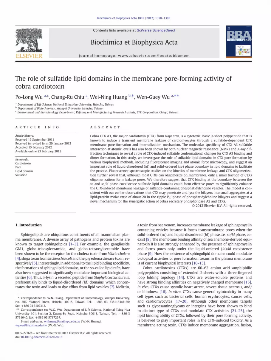
Fig. 1. Fluorescence spectroscopic investigation on the kinetics of membrane poreforming activities as monitored by the CTX A3-induced leakage of 6-CF probe in oneto one molar ratio of (A) POPS- or (B) sulfatide-containing POPC membrane vesicles:The initial rate (left y-axis) and total amount (right y-axis) of membrane leakage of6-CF in LUV (10 μM) were studied at 25 °C and plotted against the designated CTXA3 concentrations. While the initial leakage rates increase as a square of CTX concen-tration, the total amount of leakage depends linearly on the CTX amount added tothe vesicles. (C) At higher CTX concentration, the time required to induce leakage of50% 6-CF probe from POPS-( ) or sulfatide-(◆) containing vesicles becomes shorter.
the initial rate for CTX A3-induced leakage was ~80%/min and 25%/minfor POPS- and sulfatide-containing vesicles, respectively. Thus, the fas-ter initial leakage rate of the POPS containing vesicles, an indication ofa larger size of CTX A3 pore, is also observed to exhibit a shorter time re-quired to achieve 50% leakage of the vesicle content (Fig. 1C). The half-time required for the leakage process decreases as the concentration ofCTX A3 increases, suggesting that the reaction order of leakage washigher than one.
It is noted that, in the studied CTX A3 concentration from nM tosub-μM range, the initial rate of CTX A3-induced vesicle leakage in-creased as the square of CTX A3 concentration on both POPS and sul-fatide containing vesicles. In contrast, the percentage of final vesicularleakage depended only linearly on CTX A3 concentration. Therefore,under the studied experimental condition, the bimolecular interac-tion of CTX A3 was involved in the process of pore formation andthe percentage of leakage was proportional to the number of pores.
3.2. Kinetics of CTX oligomerization as reflected by the kinetics of fluorescenceresonance energy transfer of CTX
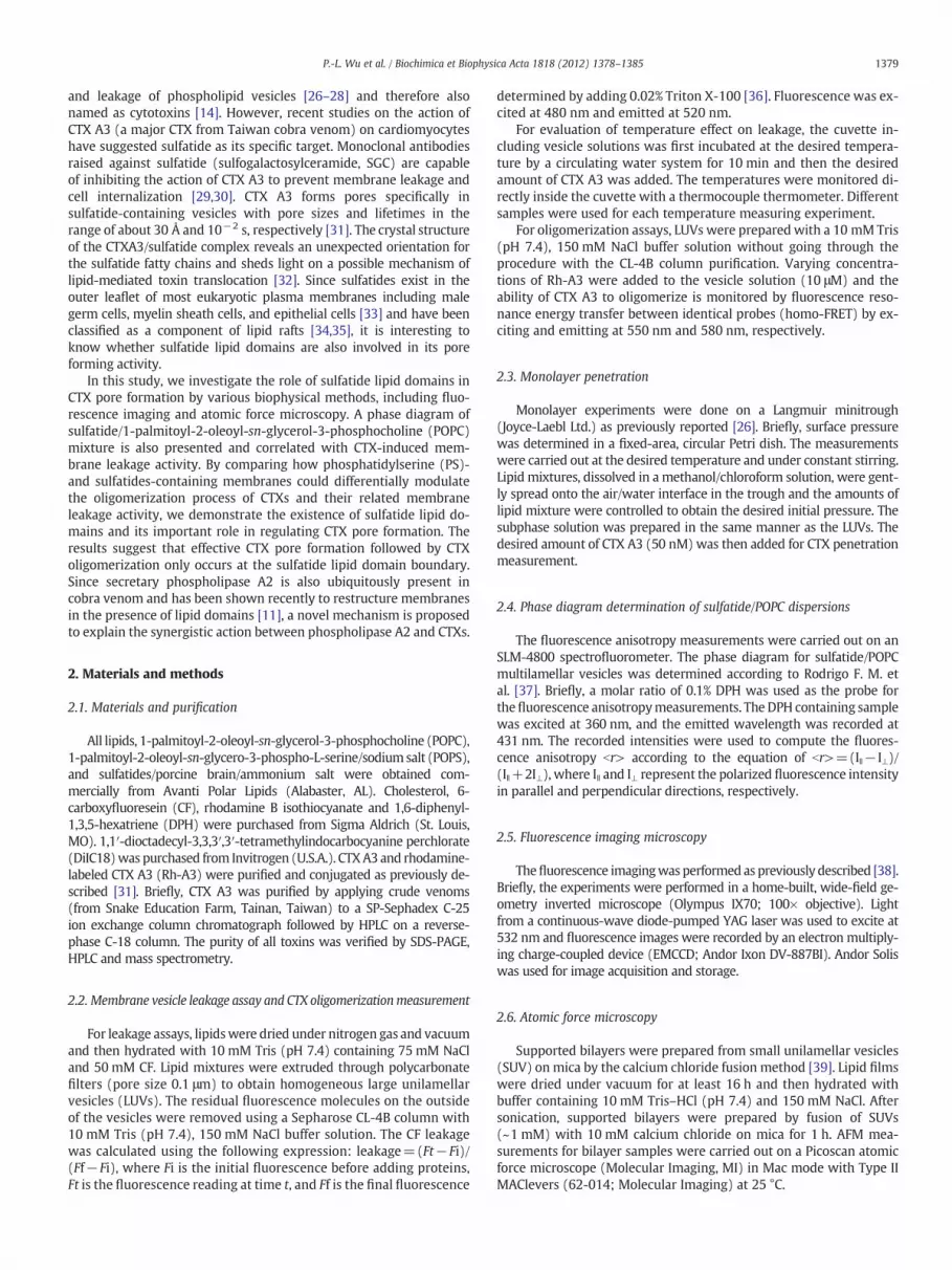
In order to see how the detected CTX A3 pore formation is relatedto the oligomerization process of CTX A3 on the studied membranevesicles, we measure the fluorescence resonance energy transfer be-tween identical probes (homo-FRET) for CTX A3 labeled with rhoda-mine, i.e., Rh-A3. Fig. 2A and B shows the dose-dependent homo-FRETof Rh-A3 by monitoring its fluorescence intensity decay as a functionof time in POPS and sulfatide-containing vesicles, respectively. When1.25 nM of Rh-A3 was added into the POPS-containing vesicles, thedegree of CTX oligomerization can be seen to increase as reflectedby the fluorescence intensity decay to indicate an increase of itshomo-FRET as a function of time. In consistent with this interpreta-tion, as the concentrations of Rh-A3 increase to 100 nM, the time re-quired for the fluorescence energy decays by 50%, i.e., t1/2, for Rh-A3on POPS-containing vesicles also decreases to ~2 s (Fig. 2C). Appar-ently, the t1/2 required for CTX oligomerization is much shorter thanthe time required for observing CTX-induced leakage of 50% 6-CFprobe, i.e., 30 s at the studied concentration of 100 nM (see Fig. 1C).It suggests that CTX oligomerization detected by the homo-FRETmethod is not a rate limiting process for the CTX pore formation onPOPS containing vesicles.
When similar experiments are performed on sulfatide containingvesicles, we were surprised to find that, while the fluorescence inten-sities of Rh-A3 also decayed as a function of time, there was no signifi-cant concentration dependent effect within the studied concentrationrange between 1.25 nMand 100 nM (Fig. 2B). The t1/2 remains constant(~240 s) throughout the studied concentration range (Fig. 2C) andappeared to be longer than the time required for 50% leakage ofsulfatide-containing vesicles, i.e. ~70 s or less for CTX concentrationlower than 100 nM. Apparently, most of the CTX oligomerization asreflected by the kinetics of homo-FRET is not related to the CTX A3pore formation to account for the observed membrane leakage process.One of the possible explanations is that effective pore formation occursmuch earlier than the CTX oligomerization detected by this homo-FRETmethod and/or only a small amount of CTX oligomerization could formeffective pore in sulfatide containing vesicles. The slow kinetics of Rh-A3 homo-FRET further suggests that, upon adding to the sulfatide con-taining vesicles, most CTX A3 might bind at a diffusion-limited areasuch as the gel phase of sulfatide lipid domain.
3.3. CTX A3 binding at sulfatide lipid domains in sulfatide containingPOPC vesicles
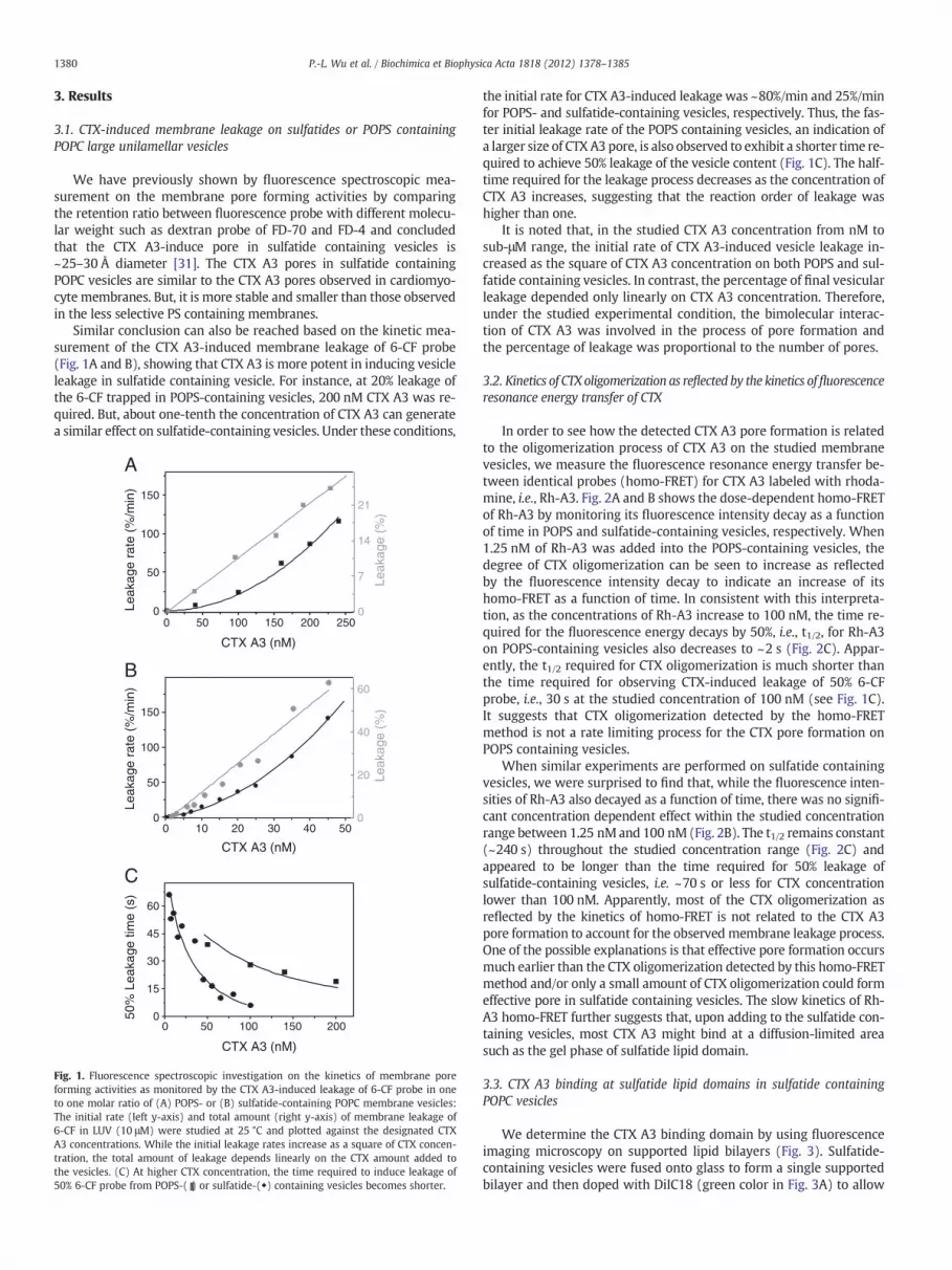
We determine the CTX A3 binding domain by using fluorescenceimaging microscopy on supported lipid bilayers (Fig. 3). Sulfatide-containing vesicles were fused onto glass to form a single supportedbilayer and then doped with DiIC18 (green color in Fig. 3A) to allow
0 25 50 75 1000
30
60
240
t 1/2
(sec
onds
)
Rh-A3 (nM)
0 100 200 3000.0
0.2
0.4
0.6
0.8
1.0
Flu
. Int
./ R
h-A
35 nM
30 nM
100 nM
1.25 nM
Time (second)
0 300 600 900 12000.0
0.2
0.4
0.6
0.8
1.0
Time (seconds)
Flu
. Int
./ R
h-A
3
1.25~100 nM
A
B
C
Fig. 2. The kinetics of oligomerization using homo-FRET assays. The fluorescence intensitydecay as function of time after different concentrations of Rh-A3 is added to (A) 50% POPS-containing vesicles or (B) 50% sulfatide-containing vesicles. The concentrations of vesiclesare 10 μM. The leakage experiments were performed at 25 °C. (C) The time (t1/2) requiredfor decay of half of the fluorescence intensity as a function of Rh-A3 concentration in 50%POPS ( ) or 50% sulfatide (◆) containing vesicles.
A B
C D
Fig. 3. Thefluorescencemicroscopic images of CTXA3 binding to supported bilayers. (A) Thesupported bilayers composed of 50% sulfatidemembranes (sulfatide/POPC=1:1 inmolar ra-tion) were dopedwith 1/500 DiIC18 (green), a fluorescence probe identified by FRAP exper-iments as a tracer for the liquid disordered (ld) phase. (B) The fluorescence image ofRhodamine labeled CTX A3 (Rh-A3) bound to supported bilayers. Rh-A3 (50 nM) wasadded to the water phase of the membrane at 3 min. (C) Superposition of the two imagesfrom A and B, showing a complementary pattern of the area for the CTX A3 binding regionand lipid ld phase region. (D) Enlarged image of panel C, showing the tubule-like structureof sulfatide lipid domain in 50% sulfatide/POPC dispersions.
1381P.-L. Wu et al. / Biochimica et Biophysica Acta 1818 (2012) 1378–1385
the visualization of liquid disorder ld phase [40]. Interestingly, asshown in Fig. 3C, the red color area responsible for the Rh-A3 bindingto sulfatide containing membranes exhibits almost perfect comple-mentary to the green color area responsible for the DiIC18 partition-ing ld phase (Fig. 3). The mutual exclusive binding of the twofluorescence probes of DiIC18 and Rh-A3 was tested by the statisticanalysis of Pearson coefficient [41]. Pearson's coefficient was foundto be negative (−0.15), suggesting strongly that most Rh-A3 indeedbind to lipid domains outside the DiIC18 partitioning ld phase. Al-though this observation is consistent with the conjecture that Rh-A3binds to gel-state sulfatide domain to account for the unexpectedlarge t1/2 on sulfatide containing membranes, it is not clear howsuch a binding could lead to the more effective CTX A3 pore formationresponsible for the CTX-induced membrane leakage of the sulfatidecontaining vesicles.
3.4. Phase diagram of sulfatide containing membranes and its relationshipwith CTX-induced leakage activity
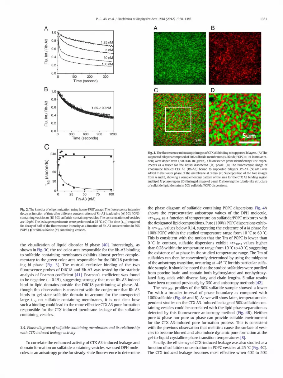
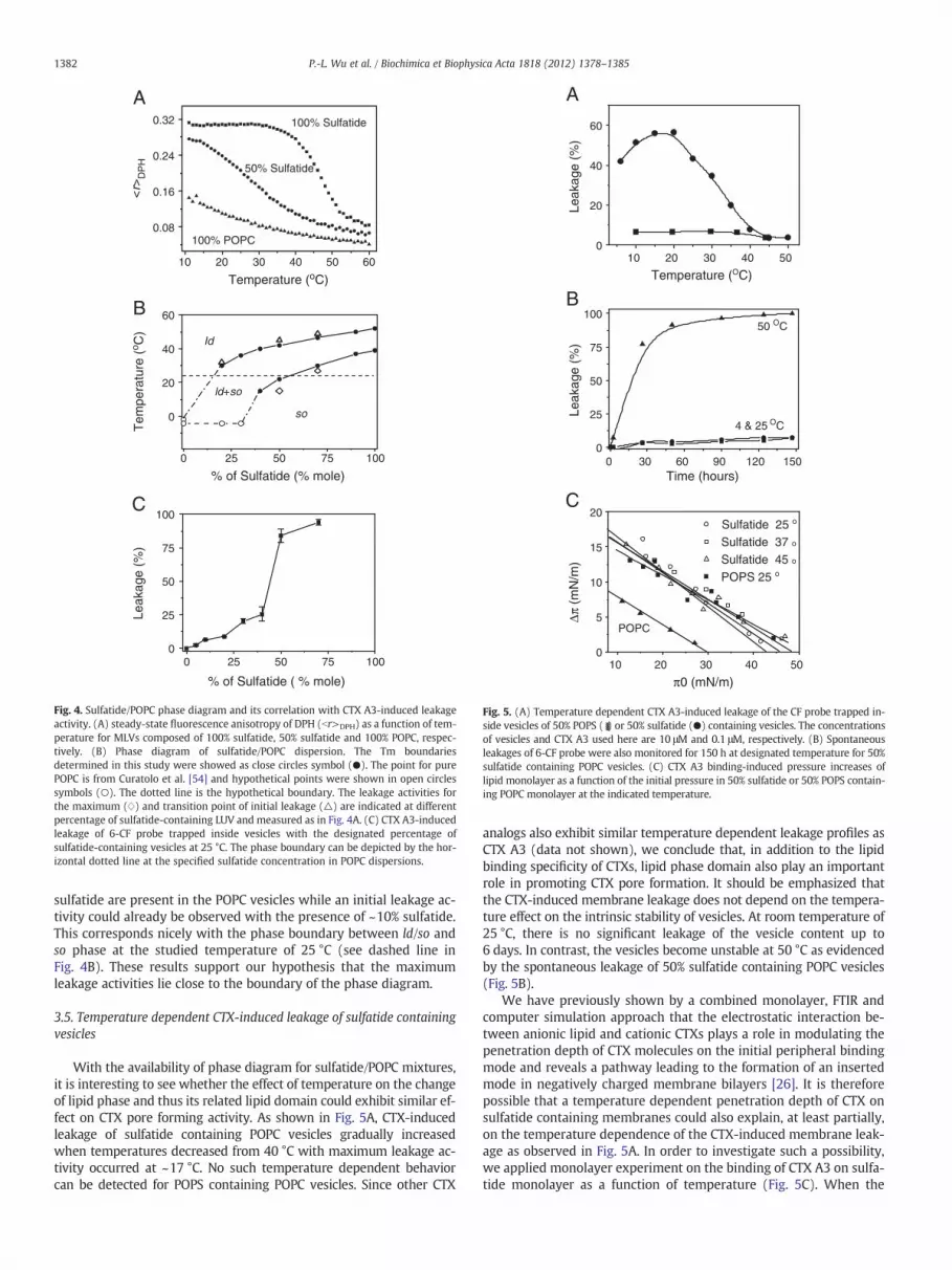
To correlate the enhanced activity of CTX A3-induced leakage anddomain formation on sulfatide containing vesicles, we used DPH mole-cules as an anisotropy probe for steady-state fluorescence to determine
the phase diagram of sulfatide containing POPC dispersions. Fig. 4Ashows the representative anisotropy values of the DPH molecule,br>DPH, as a function of temperature on sulfatide/POPC mixtures withthe designated lipid compositions. Pure (100%) POPCdispersions exhib-it br>DPH values below 0.14, suggesting the existence of a ld phase for100% POPC within the studied temperature range from 10 °C to 60 °C.This is consistent with the notion that the Tm of POPC is lower than0 °C. In contrast, sulfatide dispersions exhibit br>DPH values higherthan 0.28 within the temperature range from 10 °C to 40 °C, suggestingthe existence of so phase in the studied temperature range. The Tm ofsulfatides can then be conveniently determined by using the midpointof the anisotropy transition, occurring at ~45 °C for this particular sulfa-tide sample. It should be noted that the studied sulfatides were purifiedfrom porcine brain and contain both hydroxylated and nonhydroxy-lated fatty acids with diverse fatty acid chain lengths. Similar resultshave been reported previously by DSC and anisotropy methods [42].
The br>DPH profiles of the 50% sulfatide sample showed a lowerTm with a broader interval of phase boundary as compared with100% sulfatide (Fig. 4A and B). As we will show later, temperature de-pendent studies on the CTX A3-induced leakage of 50% sulfatide con-taining vesicles could be correlated with the lipid phase separation asdetected by this fluorescence anisotropy method (Fig. 4B). Neitherpure ld phase nor pure so phase can provide suitable environmentfor the CTX A3-induced pore formation process. This is consistentwith the previous observation that melittins cause the surface of vesi-cles to become blurred and also induce dynamic pore formation at thegel-to-liquid crystalline phase transition temperatures [8].
Finally, the efficiency of CTX-induced leakage was also studied as afunction of sulfatide concentration in POPC vesicles at 25 °C (Fig. 4C).The CTX-induced leakage becomes most effective when 40% to 50%
A
B
2010 30 40 50 60
0.08
0.16
0.24
0.32
Temperature (oC)
Tem
pera
ture
(o C
)<
r>D
PH
100% POPC
50% Sulfatide
100% Sulfatide
0 25 50 75 1000
25
50
75
100
% of Sulfatide ( % mole)
Leak
age
(%)
C
0 25 50 75 100
0
20
40
60
% of Sulfatide (% mole)
so
Id
ld+so
Fig. 4. Sulfatide/POPC phase diagram and its correlation with CTX A3-induced leakageactivity. (A) steady-state fluorescence anisotropy of DPH (br>DPH) as a function of tem-perature for MLVs composed of 100% sulfatide, 50% sulfatide and 100% POPC, respec-tively. (B) Phase diagram of sulfatide/POPC dispersion. The Tm boundariesdetermined in this study were showed as close circles symbol (●). The point for purePOPC is from Curatolo et al. [54] and hypothetical points were shown in open circlessymbols (○). The dotted line is the hypothetical boundary. The leakage activities forthe maximum (w) and transition point of initial leakage (△) are indicated at differentpercentage of sulfatide-containing LUV and measured as in Fig. 4A. (C) CTX A3-inducedleakage of 6-CF probe trapped inside vesicles with the designated percentage ofsulfatide-containing vesicles at 25 °C. The phase boundary can be depicted by the hor-izontal dotted line at the specified sulfatide concentration in POPC dispersions.
10 20 30 40 500
5
10
15
20
π0 (mN/m)
Δπ (m
N/m
)
Sulfatide 25 o
Sulfatide 37 oSulfatide 45 oPOPS 25 o
POPC
10 20 30 40 500
20
40
60
Temperature (OC)
Leak
age
(%)
A
C
B
0 30 60 90 120 1500
25
50
75
100
Time (hours)
Leak
age
(%)
4 & 25 OC
50 OC
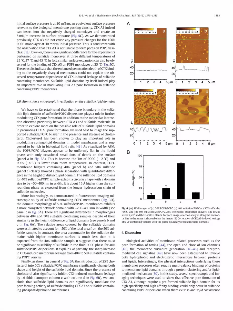
Fig. 5. (A) Temperature dependent CTX A3-induced leakage of the CF probe trapped in-side vesicles of 50% POPS ( ) or 50% sulfatide (●) containing vesicles. The concentrationsof vesicles and CTX A3 used here are 10 μM and 0.1 μM, respectively. (B) Spontaneousleakages of 6-CF probe were also monitored for 150 h at designated temperature for 50%sulfatide containing POPC vesicles. (C) CTX A3 binding-induced pressure increases oflipid monolayer as a function of the initial pressure in 50% sulfatide or 50% POPS contain-ing POPC monolayer at the indicated temperature.
1382 P.-L. Wu et al. / Biochimica et Biophysica Acta 1818 (2012) 1378–1385
sulfatide are present in the POPC vesicles while an initial leakage ac-tivity could already be observed with the presence of ~10% sulfatide.This corresponds nicely with the phase boundary between ld/so andso phase at the studied temperature of 25 °C (see dashed line inFig. 4B). These results support our hypothesis that the maximumleakage activities lie close to the boundary of the phase diagram.
3.5. Temperature dependent CTX-induced leakage of sulfatide containingvesicles
With the availability of phase diagram for sulfatide/POPC mixtures,it is interesting to see whether the effect of temperature on the changeof lipid phase and thus its related lipid domain could exhibit similar ef-fect on CTX pore forming activity. As shown in Fig. 5A, CTX-inducedleakage of sulfatide containing POPC vesicles gradually increasedwhen temperatures decreased from 40 °C with maximum leakage ac-tivity occurred at ~17 °C. No such temperature dependent behaviorcan be detected for POPS containing POPC vesicles. Since other CTX
analogs also exhibit similar temperature dependent leakage profiles asCTX A3 (data not shown), we conclude that, in addition to the lipidbinding specificity of CTXs, lipid phase domain also play an importantrole in promoting CTX pore formation. It should be emphasized thatthe CTX-induced membrane leakage does not depend on the tempera-ture effect on the intrinsic stability of vesicles. At room temperature of25 °C, there is no significant leakage of the vesicle content up to6 days. In contrast, the vesicles become unstable at 50 °C as evidencedby the spontaneous leakage of 50% sulfatide containing POPC vesicles(Fig. 5B).
We have previously shown by a combined monolayer, FTIR andcomputer simulation approach that the electrostatic interaction be-tween anionic lipid and cationic CTXs plays a role in modulating thepenetration depth of CTX molecules on the initial peripheral bindingmode and reveals a pathway leading to the formation of an insertedmode in negatively charged membrane bilayers [26]. It is thereforepossible that a temperature dependent penetration depth of CTX onsulfatide containing membranes could also explain, at least partially,on the temperature dependence of the CTX-induced membrane leak-age as observed in Fig. 5A. In order to investigate such a possibility,we applied monolayer experiment on the binding of CTX A3 on sulfa-tide monolayer as a function of temperature (Fig. 5C). When the
A
a b
1383P.-L. Wu et al. / Biochimica et Biophysica Acta 1818 (2012) 1378–1385
initial surface pressure is at 30 mN/m, an equivalent surface pressurerelevant to the biological membrane packing density, CTX A3 indeedcan insert into the negatively charged monolayer and create an8 mN/m increase in surface pressure (Fig. 5C). As we demonstratedpreviously, CTX A3 did not cause any pressure changes for the 100%POPC monolayer at 30 mN/m initial pressure. This is consistent withthe observation that CTX A3 is not unable to form pores on POPC vesi-cles [31]. However, there is no significant difference for the experimentsperformed on sulfatide monolayer at three different temperatures of25 °C, 37 °C and 45 °C. In fact, similar surface expansion can also be ob-served for the binding of CTX A3 on POPS monolayer at 25 °C (Fig. 5C).These results indicate that the enhanced penetration depth of CTX bind-ing to the negatively charged membranes could not explain the ob-served temperature-dependence of CTX-induced leakage of sulfatidecontaining membranes. Sulfatide lipid domains by itself indeed playan important role in modulating CTX A3 pore formation in sulfatidecontaining POPC membranes.
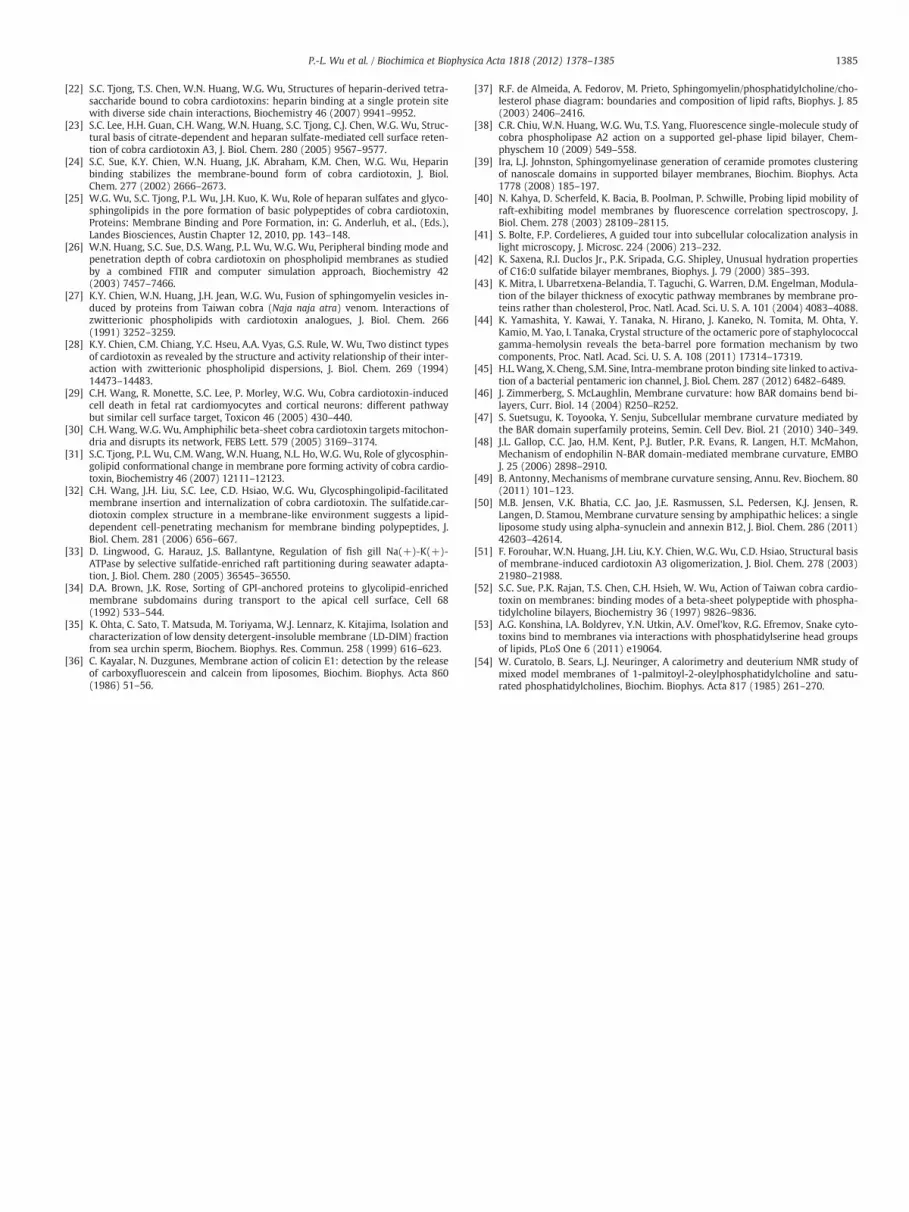
a b c d0
25
50
75
100
Leak
age
(%)
B
c d
Fig. 6. (A) AFM images of (a) 50% POPS/POPC (b) 40% sulfatide/POPC (c) 50% sulfatide/POPC, and (d) 50% sulfatide/25%POPC/25% cholesterol supported bilayers. The imagesize is 5 μm2 and the z-scale is 50 nm. For each image, a section analysis along the horizon-tal line in the image is shown below the image. (B) Correlation of CTX A3-induced leakageof 6-CF containing vesicles with the phase boundary of sulfatide lipid domains.
3.6. Atomic force microscopic investigation on the sulfatide lipid domains
We have so far established that the phase boundary in the sulfa-tide lipid domain of sulfatide/POPC dispersions plays a role in furthermodulating CTX pore formation, in addition to the molecular interac-tion observed previously between CTX A3 and sulfatide molecule. Inorder to explore more on the possible role of sulfatide lipid domainsin promoting CTX A3 pore formation, we used AFM to image the sup-ported sulfatide/POPC bilayer in the presence and absence of choles-terol. Cholesterol has been shown to play an important role inmodulating sphingolipid domains in model membranes and is sug-gested to be rich in biological lipid rafts [43]. As visualized by AFM,the POPS/POPC bilayers appear to be uniformly flat in the liquidphase with only occasional small dots of debris on the surface(panel a in Fig. 6A). This is because the Tm of POPC (−2 °C) andPOPS (14 °C) is lower than room temperature. In contrast, POPCmembrane bilayers containing 40% (panel b) and 50% sulfatide(panel c) clearly showed a phase separation with quantitative differ-ence in the height of distinct lipid domain. The sulfatide lipid domainsfor 40% sulfatide/POPC sample exhibit a circular shape with a domainsize to be ~50–400 nm in width. It is about 15 Å higher than the sur-rounding phase as expected from the longer hydrocarbon chain ofsulfatide molecules.
More interestingly, as demonstrated in fluorescence imaging mi-croscopic study of sulfatide containing POPC membranes (Fig. 3D),the domain morphology of 50% sulfatide/POPC membranes exhibitsa more elongated network domain with ~200–400 nm in width (seepanel c in Fig. 6A). There are significant differences in morphologiesbetween 40% and 50% sulfatide containing samples despite of theirsimilarity in the height difference of lipid domains (see panels b andc in Fig. 6A). The relative areas covered by the sulfatide domainswere estimated to account for ~50% of the total area from the 50% sul-fatide sample. In contrast, the area accountable for the sulfatide do-mains with higher membrane surface is much less than it isexpected from the 40% sulfatide sample. It suggests that there mustbe significant miscibility of sulfatide in the fluid POPC phase for 40%sulfatide/POPC dispersions. It explains, at partially, the sharp increasein CTX-induced membrane leakage from 40% to 50% sulfatide contain-ing POPC vesicles.
Finally, as shown in panel d of Fig. 6A, the introduction of 25% cho-lesterol into 50% sulfatide/POPC membrane significantly change bothshape and height of the sulfatide lipid domain. Since the presence ofcholesterol also significantly inhibit CTX-induced membrane leakageby ~6-folds (compare column c with column d in Fig. 6B), we con-clude that sulfatide lipid domains can significantly modulate thepore forming activity of sulfatide binding CTX A3 on sulfatide contain-ing phosphatidylcholine membranes.
4. Discussion
Biological activities of membrane-related processes such as thepore formation of toxins [44], the open and close of ion channels[45], the membrane curvature generation [46–48] and receptor-mediated cell signaling [49] have now been established to involveboth hydrophobic and electrostatic interactions between proteinsand lipids. Interestingly, the physical interactions underlying thesemembranes processes often require multi-valency bindings of proteinsto membrane lipid domains through a protein-clustering and/or lipid-mediated mechanism [50]. In this study, several spectroscopic and im-aging techniques were used to show that effective pore formation ofCTX A3, although require a pre-formed sulfatide lipid domain for itshigh specificity and high affinity binding, could only occur in sulfatidecontaining POPC dispersions when there exist so and so/ld coexistence

1384 P.-L. Wu et al. / Biochimica et Biophysica Acta 1818 (2012) 1378–1385
phase of sulfatide lipid domains. Protein binding and oligomerizationson the bilayer surface, as reflected by the kinetics of CTX A3 homo-FRET measurement, are not the rate-limiting process. Most of the CTXA3 molecules are bound on sulfatide-containing so domain (Fig. 3)and can only slowly oligomerize due to the slow diffusion of so domain.Based on our previous studies, about 8 molecules of CTX A3 can form apore and induced all-or-none leakage on vesicles [51]. A 100 nm diame-ter LUV is composed of about 105 lipids and 200 CTX A3 molecules areneeded for 20% leakage of sulfatide-containing vesicles. This impliesthat the oligomerizations of most CTX A3molecules on membrane sur-face are not effective to form pore. Since the pore forming activity ap-pears to correlate well with the presence of phase boundary ofsulfatide lipid domains (Fig. 6), we hypothesize that the exposure ofthe hydrophobic region in the mismatch boundary sites between dis-tinct lipid phase domain may play an important role to facilitate thepenetration of CTX A3 into lipid bilayers to form correct membraneoligomerization and pores.
Three lines of evidence are consistent with the hypothesis. First, bythe combined 31P and 2H NMR investigation, in conjunction with DPHfluorescence anisotropy and DSC studies, on the interaction of CTX A3with zwitterionic dipalmitoyl PC (DPPC) dispersion, we showed thatCTX A3may penetrate and lyse the DPPC bilayers into small aggregatesat a lipid/protein molar ratio of ~20 in the ripple Pβ′ phase [52]. Thissuggests that CTX A3 may undergo a re-distribution between penetra-tion and peripheral binding states depending on the presence of distinctlipid phase coexistence. Second, by using the single molecule fluores-cence imaging method, we have shown that amphiphilic proteinssuch as cobra phospholipase A2may preferentially bind to the packingdefect regions of lipid bilayers to facilitate its enzymatic action onthe PC molecules. The phase boundary of distinct lipid phase do-mains, such as the sulfatide lipid domains in sulfatide/POPC disper-sions, allows the exposure of the mismatched hydrophobic regionand therefore may serve as a packing defect region to facilitate thebinding and penetration of amphiphilic CTX A3 molecules. Finally,as we demonstrated in this work, the CTX A3-induced vesicle leak-age of sulfatide/POPC dispersions correlates well with the phaseboundary of distinct lipid domains.
Many evidences suggest the existence of bilayer packing defect inbiological membrane surfaces due to the hydrophobic mismatch ofmembrane proteins with lipid bilayers and also between distinctlipid domains. Cholesterol in eukaryotic cell membranes has beenproposed to play a role to modulate bilayer thickness and its relatedbiological functions. Specifically, variation of cholesterol concentra-tion in the glycosphingolipid raft domain has been shown to eitherpositively or negatively regulate biological activities of membraneproteins. Herein, we showed that the introduction of cholesterolinto the sulfatide lipid domains inhibits CTX A3-induced membraneleakage of sulfatide/POPC dispersions (Fig. 6). This observation is con-sistent with the fact that depriving cholesterol molecules from theplasma membrane of cardiomyocytes could also enhance CTXA3-induced membrane leakage and CTX A3 internalization.
Phospholipase A2 and CTXs are the two major components ofcobra venoms and have been suggested to act synergistically for therespective cytotoxicity on many cells. Interestingly, the presence oflipid domains has been shown recently to induce specificity in the hy-drolytic activity of phospholipase A2, resulting in marked differencesin the physical properties of the membrane end-product [11]. Thus,the involvement of lipid domains in both the enzymatic activity ofphospholipase A2 and CTX A3-induced membrane leakage leads to anew possibility that the synergistic action between cobra phospholi-pase A2 and CTXs may also be modulated by the presence of lipiddomains.
It should be pointed out that other lipid components may also beinvolved in the cytotoxicity of CTXs. For instance, it was reported re-cently that the leakage ability in PS-containing membranes inducedby CTX from Naja kaouthia was much more pronounced than those
measured for the anionic lipid of sulfatide [53]. Since there are morethan seven CTX homologues in Taiwan cobra venom, understandingthe combinatorial action of CTX homologues against a distinct typeof lipid domains may be needed to fully appreciate the action mecha-nism of cobra venoms against different cell types. Considering the di-verse types of molecules in the plasma membranes and extracellularmatrix have now been shown to be a potential target of CTX action[20], future investigation to understand how snake venomworks ondif-ferent cell typesmay help to establishmethods for the understanding ofthe biological functions in the newly established proteomic world.
Acknowledgements
We thank Linda Johnston for access to atomic force microscopy fa-cilities (NRC, Canada). We also thank Dr. Da-Shin Wang for readingthe manuscript and editorial suggestions. This work was supportedby a grant from the NSC.
References
[1] S. Manes, G. del Real, A.C. Martinez, Pathogens: raft hijackers, Nat. Rev. Immunol.3 (2003) 557–568.
[2] A. Singh, M. Del Poeta, Lipid signalling in pathogenic fungi, Cell. Microbiol. 13(2011) 177–185.
[3] L. Abrami, M. Fivaz, F.G. van der Goot, Adventures of a pore-forming toxin at thetarget cell surface, Trends Microbiol. 8 (2000) 168–172.
[4] S.V. Heyningen, Cholera toxin: interaction of subunitswith gangliosideGM1, Science183 (1974) 656–657.
[5] S. DeGrandis, H. Law, J. Brunton, C. Gyles, C.A. Lingwood, Globotetraosylceramideis recognized by the pig edema disease toxin, J. Biol. Chem. 264 (1989)12520–12525.
[6] D. Lingwood, K. Simons, Lipid rafts as a membrane-organizing principle, Science327 (2010) 46–50.
[7] A. Pokorny, P.F. Almeida, Permeabilization of raft-containing lipid vesicles bydelta-lysin: a mechanism for cell sensitivity to cytotoxic peptides, Biochemistry44 (2005) 9538–9544.
[8] M.J. Gomara, S. Nir, J.L. Nieva, Effects of sphingomyelin on melittin pore forma-tion, Biochim. Biophys. Acta 1612 (2003) 83–89.
[9] P. Schon, A.J. Garcia-Saez, P. Malovrh, K. Bacia, G. Anderluh, P. Schwille, Equina-toxin II permeabilizing activity depends on the presence of sphingomyelin andlipid phase coexistence, Biophys. J. 95 (2008) 691–698.
[10] M. Milescu, F. Bosmans, S. Lee, A.A. Alabi, J.I. Kim, K.J. Swartz, Interactions be-tween lipids and voltage sensor paddles detected with tarantula toxins, Nat.Struct. Mol. Biol. 16 (2009) 1080–1085.
[11] C. Leidy, J. Ocampo, L. Duelund, O.G. Mouritsen, K. Jorgensen, G.H. Peters, Mem-brane restructuring by phospholipase A2 is regulated by the presence of lipid do-mains, Biophys. J. 101 (2011) 90–99.
[12] H. Lee, J.A. Rotolo, J. Mesicek, T. Penate-Medina, A. Rimner, W.C. Liao, X. Yin, G.Ragupathi, D. Ehleiter, E. Gulbins, D. Zhai, J.C. Reed, A. Haimovitz-Friedman, Z.Fuks, R. Kolesnick, Mitochondrial ceramide-rich macrodomains functionalizeBax upon irradiation, PLoS One 6 (2011) e19783.
[13] M. Kulma, M. Herec, W. Grudzinski, G. Anderluh, W.I. Gruszecki, K. Kwiatkowska,A. Sobota, Sphingomyelin-rich domains are sites of lysenin oligomerization: im-plications for raft studies, Biochim. Biophys. Acta 1798 (2010) 471–481.
[14] T.S. Chen, F.Y. Chung, S.C. Tjong, K.S. Goh, W.N. Huang, K.Y. Chien, P.L. Wu, H.C.Lin, C.J. Chen, W.G. Wu, Structural difference between group I and group IIcobra cardiotoxins: X-ray, NMR, and CD analysis of the effect of cis-proline con-formation on three-fingered toxins, Biochemistry 44 (2005) 7414–7426.
[15] J. Dufourcq, J.F. Faucon, Specific binding of a cardiotoxin from Naja mossambicamossambica to charged phospholipids detected by intrinsic fluorescence, Bio-chemistry 17 (1978) 1170–1176.
[16] A.L. Harvey, Snake Toxins, 1991, pp. 259–298.[17] N. Zusman, T.M. Miklas, T. Graves, G.E. Dambach, R.A. Hudson, On the interaction
of cobra venom protein cardiotoxins with erythrocytes, Biochem. Biophys. Res.Commun. 124 (1984) 629–636.
[18] A. Zaheer, B.M. Braganca, Comparative study of three basic polypeptides fromsnake venoms in relation to their effects on the cell membrane of normal andtumor cells, Cancer Biochem. Biophys. 5 (1980) 41–46.
[19] W.F. Tzeng, Y.H. Chen, Suppression of snake-venom cardiotoxin-induced cardio-myocyte degeneration by blockage of Ca2+ influx or inhibition of non-lysosomal proteinases, Biochem. J. 256 (1988) 89–95.
[20] L.W. Chen, P.H. Kao, Y.S. Fu, S.R. Lin, L.S. Chang, Membrane-damaging activity ofTaiwan cobra cardiotoxin 3 is responsible for its bactericidal activity, Toxicon58 (2011) 46–53.
[21] P.L.Wu, S.C. Lee, C.C. Chuang, S.Mori, N. Akakura,W.G.Wu, Y. Takada, Non-cytotoxiccobra cardiotoxin A5 binds to alpha(v)beta3 integrin and inhibits bone resorption.Identification of cardiotoxins as non-RGD integrin-binding proteins of the Ly-6family, J. Biol. Chem. 281 (2006) 7937–7945.
1385P.-L. Wu et al. / Biochimica et Biophysica Acta 1818 (2012) 1378–1385
[22] S.C. Tjong, T.S. Chen, W.N. Huang, W.G. Wu, Structures of heparin-derived tetra-saccharide bound to cobra cardiotoxins: heparin binding at a single protein sitewith diverse side chain interactions, Biochemistry 46 (2007) 9941–9952.
[23] S.C. Lee, H.H. Guan, C.H. Wang, W.N. Huang, S.C. Tjong, C.J. Chen, W.G. Wu, Struc-tural basis of citrate-dependent and heparan sulfate-mediated cell surface reten-tion of cobra cardiotoxin A3, J. Biol. Chem. 280 (2005) 9567–9577.
[24] S.C. Sue, K.Y. Chien, W.N. Huang, J.K. Abraham, K.M. Chen, W.G. Wu, Heparinbinding stabilizes the membrane-bound form of cobra cardiotoxin, J. Biol.Chem. 277 (2002) 2666–2673.
[25] W.G. Wu, S.C. Tjong, P.L. Wu, J.H. Kuo, K. Wu, Role of heparan sulfates and glyco-sphingolipids in the pore formation of basic polypeptides of cobra cardiotoxin,Proteins: Membrane Binding and Pore Formation, in: G. Anderluh, et al., (Eds.),Landes Biosciences, Austin Chapter 12, 2010, pp. 143–148.
[26] W.N. Huang, S.C. Sue, D.S. Wang, P.L. Wu, W.G. Wu, Peripheral binding mode andpenetration depth of cobra cardiotoxin on phospholipid membranes as studiedby a combined FTIR and computer simulation approach, Biochemistry 42(2003) 7457–7466.
[27] K.Y. Chien, W.N. Huang, J.H. Jean, W.G. Wu, Fusion of sphingomyelin vesicles in-duced by proteins from Taiwan cobra (Naja naja atra) venom. Interactions ofzwitterionic phospholipids with cardiotoxin analogues, J. Biol. Chem. 266(1991) 3252–3259.
[28] K.Y. Chien, C.M. Chiang, Y.C. Hseu, A.A. Vyas, G.S. Rule, W. Wu, Two distinct typesof cardiotoxin as revealed by the structure and activity relationship of their inter-action with zwitterionic phospholipid dispersions, J. Biol. Chem. 269 (1994)14473–14483.
[29] C.H. Wang, R. Monette, S.C. Lee, P. Morley, W.G. Wu, Cobra cardiotoxin-inducedcell death in fetal rat cardiomyocytes and cortical neurons: different pathwaybut similar cell surface target, Toxicon 46 (2005) 430–440.
[30] C.H. Wang, W.G. Wu, Amphiphilic beta-sheet cobra cardiotoxin targets mitochon-dria and disrupts its network, FEBS Lett. 579 (2005) 3169–3174.
[31] S.C. Tjong, P.L. Wu, C.M. Wang, W.N. Huang, N.L. Ho, W.G. Wu, Role of glycosphin-golipid conformational change in membrane pore forming activity of cobra cardio-toxin, Biochemistry 46 (2007) 12111–12123.
[32] C.H. Wang, J.H. Liu, S.C. Lee, C.D. Hsiao, W.G. Wu, Glycosphingolipid-facilitatedmembrane insertion and internalization of cobra cardiotoxin. The sulfatide.car-diotoxin complex structure in a membrane-like environment suggests a lipid-dependent cell-penetrating mechanism for membrane binding polypeptides, J.Biol. Chem. 281 (2006) 656–667.
[33] D. Lingwood, G. Harauz, J.S. Ballantyne, Regulation of fish gill Na(+)-K(+)-ATPase by selective sulfatide-enriched raft partitioning during seawater adapta-tion, J. Biol. Chem. 280 (2005) 36545–36550.
[34] D.A. Brown, J.K. Rose, Sorting of GPI-anchored proteins to glycolipid-enrichedmembrane subdomains during transport to the apical cell surface, Cell 68(1992) 533–544.
[35] K. Ohta, C. Sato, T. Matsuda, M. Toriyama, W.J. Lennarz, K. Kitajima, Isolation andcharacterization of low density detergent-insoluble membrane (LD-DIM) fractionfrom sea urchin sperm, Biochem. Biophys. Res. Commun. 258 (1999) 616–623.
[36] C. Kayalar, N. Duzgunes, Membrane action of colicin E1: detection by the releaseof carboxyfluorescein and calcein from liposomes, Biochim. Biophys. Acta 860(1986) 51–56.
[37] R.F. de Almeida, A. Fedorov, M. Prieto, Sphingomyelin/phosphatidylcholine/cho-lesterol phase diagram: boundaries and composition of lipid rafts, Biophys. J. 85(2003) 2406–2416.
[38] C.R. Chiu, W.N. Huang, W.G. Wu, T.S. Yang, Fluorescence single-molecule study ofcobra phospholipase A2 action on a supported gel-phase lipid bilayer, Chem-physchem 10 (2009) 549–558.
[39] Ira, L.J. Johnston, Sphingomyelinase generation of ceramide promotes clusteringof nanoscale domains in supported bilayer membranes, Biochim. Biophys. Acta1778 (2008) 185–197.
[40] N. Kahya, D. Scherfeld, K. Bacia, B. Poolman, P. Schwille, Probing lipid mobility ofraft-exhibiting model membranes by fluorescence correlation spectroscopy, J.Biol. Chem. 278 (2003) 28109–28115.
[41] S. Bolte, F.P. Cordelieres, A guided tour into subcellular colocalization analysis inlight microscopy, J. Microsc. 224 (2006) 213–232.
[42] K. Saxena, R.I. Duclos Jr., P.K. Sripada, G.G. Shipley, Unusual hydration propertiesof C16:0 sulfatide bilayer membranes, Biophys. J. 79 (2000) 385–393.
[43] K. Mitra, I. Ubarretxena-Belandia, T. Taguchi, G. Warren, D.M. Engelman, Modula-tion of the bilayer thickness of exocytic pathway membranes by membrane pro-teins rather than cholesterol, Proc. Natl. Acad. Sci. U. S. A. 101 (2004) 4083–4088.
[44] K. Yamashita, Y. Kawai, Y. Tanaka, N. Hirano, J. Kaneko, N. Tomita, M. Ohta, Y.Kamio, M. Yao, I. Tanaka, Crystal structure of the octameric pore of staphylococcalgamma-hemolysin reveals the beta-barrel pore formation mechanism by twocomponents, Proc. Natl. Acad. Sci. U. S. A. 108 (2011) 17314–17319.
[45] H.L.Wang, X. Cheng, S.M. Sine, Intra-membrane proton binding site linked to activa-tion of a bacterial pentameric ion channel, J. Biol. Chem. 287 (2012) 6482–6489.
[46] J. Zimmerberg, S. McLaughlin, Membrane curvature: how BAR domains bend bi-layers, Curr. Biol. 14 (2004) R250–R252.
[47] S. Suetsugu, K. Toyooka, Y. Senju, Subcellular membrane curvature mediated bythe BAR domain superfamily proteins, Semin. Cell Dev. Biol. 21 (2010) 340–349.
[48] J.L. Gallop, C.C. Jao, H.M. Kent, P.J. Butler, P.R. Evans, R. Langen, H.T. McMahon,Mechanism of endophilin N-BAR domain-mediated membrane curvature, EMBOJ. 25 (2006) 2898–2910.
[49] B. Antonny, Mechanisms of membrane curvature sensing, Annu. Rev. Biochem. 80(2011) 101–123.
[50] M.B. Jensen, V.K. Bhatia, C.C. Jao, J.E. Rasmussen, S.L. Pedersen, K.J. Jensen, R.Langen, D. Stamou, Membrane curvature sensing by amphipathic helices: a singleliposome study using alpha-synuclein and annexin B12, J. Biol. Chem. 286 (2011)42603–42614.
[51] F. Forouhar, W.N. Huang, J.H. Liu, K.Y. Chien, W.G. Wu, C.D. Hsiao, Structural basisof membrane-induced cardiotoxin A3 oligomerization, J. Biol. Chem. 278 (2003)21980–21988.
[52] S.C. Sue, P.K. Rajan, T.S. Chen, C.H. Hsieh, W. Wu, Action of Taiwan cobra cardio-toxin on membranes: binding modes of a beta-sheet polypeptide with phospha-tidylcholine bilayers, Biochemistry 36 (1997) 9826–9836.
[53] A.G. Konshina, I.A. Boldyrev, Y.N. Utkin, A.V. Omel'kov, R.G. Efremov, Snake cyto-toxins bind to membranes via interactions with phosphatidylserine head groupsof lipids, PLoS One 6 (2011) e19064.
[54] W. Curatolo, B. Sears, L.J. Neuringer, A calorimetry and deuterium NMR study ofmixed model membranes of 1-palmitoyl-2-oleylphosphatidylcholine and satu-rated phosphatidylcholines, Biochim. Biophys. Acta 817 (1985) 261–270.