the role of protein denaturation energetics and molecular chaperones in the aggregation and...
TRANSCRIPT

The Role of Protein Denaturation Energetics andMolecular Chaperones in the Aggregation andMistargeting of Mutants Causing Primary HyperoxaluriaType INoel Mesa-Torres1., Israel Fabelo-Rosa2., Debora Riverol2, Cristina Yunta3, Armando Albert3,
Eduardo Salido2*, Angel L. Pey1*
1 Department of Physical Chemistry, Faculty of Sciences, University of Granada, Granada, Spain, 2 Centre for Biomedical Research on Rare Diseases, Instituto Tecnologıas
Biomedicas, University of La Laguna, Tenerife, Spain, 3 Department of Crystallography and Structural Biology, Instituto de Quımica Fısica ‘‘Rocasolano’’, Consejo Superior
de Investigaciones Cientıficas, Madrid, Spain
Abstract
Primary hyperoxaluria type I (PH1) is a conformational disease which result in the loss of alanine:glyoxylate aminotransferase(AGT) function. The study of AGT has important implications for protein folding and trafficking because PH1 mutants maycause protein aggregation and mitochondrial mistargeting. We herein describe a multidisciplinary study aimed tounderstand the molecular basis of protein aggregation and mistargeting in PH1 by studying twelve AGT variants. Expressionstudies in cell cultures reveal strong protein folding defects in PH1 causing mutants leading to enhanced aggregation, andin two cases, mitochondrial mistargeting. Immunoprecipitation studies in a cell-free system reveal that most mutantsenhance the interactions with Hsc70 chaperones along their folding process, while in vitro binding experiments show nochanges in the interaction of folded AGT dimers with the peroxisomal receptor Pex5p. Thermal denaturation studies bycalorimetry support that PH1 causing mutants often kinetically destabilize the folded apo-protein through significantchanges in the denaturation free energy barrier, whereas coenzyme binding overcomes this destabilization. Modeling of themutations on a 1.9 A crystal structure suggests that PH1 causing mutants perturb locally the native structure. Our worksupport that a misbalance between denaturation energetics and interactions with chaperones underlie aggregation andmistargeting in PH1, suggesting that native state stabilizers and protein homeostasis modulators are potential drugs torestore the complex and delicate balance of AGT protein homeostasis in PH1.
Citation: Mesa-Torres N, Fabelo-Rosa I, Riverol D, Yunta C, Albert A, et al. (2013) The Role of Protein Denaturation Energetics and Molecular Chaperones in theAggregation and Mistargeting of Mutants Causing Primary Hyperoxaluria Type I. PLoS ONE 8(8): e71963. doi:10.1371/journal.pone.0071963
Editor: Eugene A. Permyakov, Russian Academy of Sciences, Institute for Biological Instrumentation, Russian Federation
Received June 13, 2013; Accepted July 5, 2013; Published August 27, 2013
Copyright: � 2013 Mesa-Torres et al. This is an open-access article distributed under the terms of the Creative Commons Attribution License, which permitsunrestricted use, distribution, and reproduction in any medium, provided the original author and source are credited.
Funding: This work was supported by the Spanish ministry of Science and Innovation (RYC2009-04147 and CSD2009-00088 to ALP, SAF2011-23933 to ES, andCSD2006-00015, S2010/BMD-2457 and BFU2011-25384 to AA) and Junta de Andalucia (P11CTS-7187 ALP); FPI predoctoral fellowships from the Spanish ministryof Science and Innovation to IF-R and NM-T. The funders had no role in study design, data collection and analysis, decision to publish, or preparation of themanuscript.
Competing Interests: The authors have declared that no competing interests exist.
* E-mail: [email protected] (ES); [email protected] (ALP)
Introduction
Primary hyperoxaluria type I (PH1) is an autosomal recessive
inborn error of metabolism caused by mutations in the AGXT
gene, coding for the enzyme alanine-glyoxylate aminotransferase
(AGT). AGT catalyzes the transamination of L-alanine to
pyruvate and glyoxylate to glycine in the presence of pyridoxal
59-phosphate (PLP) as cofactor [1]. AGT deficiency causes
glyoxylate accumulation that is subsequently oxidized to oxalate,
leading to the production of calcium oxalate crystals that result in
progressive renal failure, and eventually, a life-threatening
systemic build-up of oxalate known as oxalosis [2]. Liver and
kidney transplantation is the only curative option to date, but this
aggressive treatment poses significant morbidity and mortality [3].
A limited number of patients with specific genotypes have been
reported to respond to pharmacological doses of pyridoxine, even
though the molecular mechanisms involved in the response are
unclear [4,5,6,7]. AGXT has two polymorphic variants, the most
frequent (‘‘wild-type’’; AGT WT) called the major allele (haplotype)
and a less common polymorphic variant called the minor allele
(referred to as AGT LM) which appears in 20% of control subjects
and 46% of PH1 patients [1]. The minor allele shows two single
amino acid substitutions (p.P11L and p.I340M) among other
genomic changes. Even though the minor allele does not cause
PH1 by itself, it is known to exacerbate the deleterious effects of
additional mutations [1,4,7]. About 150 mutations in the AGXT
gene have been described in PH1 patients, 50% of them being
missense mutations [1], and a few of them, such as p.G170R and
p.I244T, are relatively common. Several molecular mechanisms
seem to contribute to AGT loss-of-function in PH1 at the protein
level: i) Mitochondrial mistargeting, where the AGT enzyme is
imported to mitochondria [2]; ii) Protein aggregation [4,8,9]; iii)
Accelerated proteasomal degradation [8]; iv) Catalytic defects [4].
However, the molecular details underlying protein mistargeting,
aggregation and degradation in PH1 remain unclear. Beyond their
PLOS ONE | www.plosone.org 1 August 2013 | Volume 8 | Issue 8 | e71963
interest in PH1, some of these mutations have important
implications in cell biology and genetics. AGT mistargeting
mutations are unique models to try to understand some of the
principles behind enzyme compartmentalization in the cell, which
also has relevant evolutionary connotations [10]. In addition, the
necessary synergy between common polymorphisms and disease-
causing mutations of the AGXT minor haplotype is one of the best
characterized examples of such interaction in human genetics [2].
Human genetic diseases are often caused by alterations in
protein homeostasis [11]. The ability of a protein to fold into its
native and functional conformation relies on intrinsic physico-
chemical properties (thermodynamic stability, folding, unfolding,
misfolding and aggregation rates) in the crowded intracellular
milieu, as well as in the interaction of different conformations
populated along the folding/unfolding process with elements of
the protein homeostasis network, an array of pathways involved in
the control of protein synthesis, folding, post-translational modi-
fication, trafficking, disaggregation and degradation [11,12,13,14].
In the context of protein homeostasis, we have recently suggested a
role of the low kinetic stability of the apo-AGT (which shows no
coenzyme bound [6]) and enhanced interactions with Hsc70,
Hsp90 and Hsp60 chaperones [6,9,15] in the aggregation (for the
I244T variant -p.P11L, p.I244T and p.I340M in cis -) and
mistargeting (for the G170R variant -p.P11L, p.G170R and
p.I340M in cis -) of PH1 mutations. Whether these aberrant
protein features are specific to these mutations and/or intrinsic to
a certain pathogenic mechanism (aggregation vs. mistargeting)
remains unclear. From the perspective of the protein homeostasis
network, understanding the role of specific protein features in the
disease-causing mechanisms is required to develop new therapeu-
tic strategies aimed to restore protein function [11,13,14].
However, owing to the large complexity and interactivity of the
protein homeostasis pathways, involving at least 800 different
proteins [13], a detailed characterization of protein homeostasis
defects in PH1 represents a remarkable challenge.
In this work, we have performed a multidisciplinary character-
ization of four polymorphic variants (WT, p.P11L, p.I340M and
p.P11L/I340M or minor allele, LM) and six known PH1 mutations
present in the minor allele (named p.H83R, p.F152I, p.G170R,
p.I244T, p.P319L and p.A368T through this paper). We also
analyzed two rare variants of uncertain pathogenicity: p.R197Q
and p.A295T, both in the minor allele. Our main aim is to shed light
on the complex mutational effects of AGT folding and stability in
PH1 from biochemical, biophysical, cell and structural biology
perspectives required to deeply understand PH1 as a conforma-
tional disease. Our results show that PH1 causing mutations
associated with aggregation and mistargeting display common
alterations in protein folding, stability and interaction with
molecular chaperones. This suggests that the protein homeostasis
pathways involved in both mechanisms are shared and, conse-
quently, that the final fate of the mutant proteins is likely
determined by their specific regulatory elements. Nevertheless, our
results indicate that native state stability and molecular chaperones
are key points to understand PH1 pathogenesis, that might be
targeted pharmacologically to restore protein homeostasis in PH1
patients.
Materials and Methods
Construction, expression and purification of Pex5p-pbdand AGT proteins in E.coli
Cloning of the PTS1-binding domain (amino acids 235–602 in
reference sequence NM_000319) of human Pex5 (Pex5p-pbd) was
performed after reverse transcription of normal human liver
mRNA and PCR amplification with primers BgNPEX5-F:
AGATCTCATATGGAGTTTGAACGAGCCAAG and
SlRPEX5-R: TGCGACGAATTCACTGGGGCAGGCCAAAC.
The NdeI and EcoRI sites designed at the 59end of the primers
were used to clone the amplification product into pCOLDII
expression vector. The AGT expression constructs were generated
as previously described [6], using site-directed mutagenesis,
standard subcloning procedures and confirmed by sequencing.
E. coli BL21strain containing pCOLDII plasmids encoding AGT
and Pex5p-pbd proteins were grown in the presence of ampicillin
0.1 mg/ml and induced with 0.4 mM IPTG for 6 h at 4uC. His-
tagged AGT and Pex5-pbd proteins were purified from soluble
extracts using IMAC-columns (GE Healthcare) as recommended
by the manufacturer. Proteins were further purified by size-
exclusion chromatography (SEC) as previously described [6].
Holo- and apo-AGT were prepared and stored as previously
described [6]. Protein concentration was measured spectrophoto-
metrically using e280(1 mg/ml) = 1.069 (AGT) and 1.243 (Pex5p-
pbd), calculated from their sequences [16].
Spectroscopic analysesAll spectroscopic analyses were performed in 20 mM Na-
Hepes, 200 mM NaCl pH 7.4 at 25uC. The hydrodynamic
behavior of dimeric holo-AGT proteins was evaluated by dynamic
light scattering (DLS) using 5 mM AGT (in subunit) and 50 mM
PLP in a Zetasizer Nano ZS (Malvern Inc.). UV-visible absorption
spectra were acquired in an Agilent 8453 diode-array spectro-
photometer using 3 mm path length cuvettes and 20 mM AGT.
Near-UV/Visible circular dichroism measurements were per-
formed as described [6]. Fluorescence measurements were
performed as previously described [6,17] with some minor
modifications (see SI text).
Enzyme kinetic analysisThe AGT overall transaminase activity was customarily
measured as described [18]. Briefly, 5 mg/ml of AGT incubated
in Na-Phosphate 0.1 M pH 8 buffer at 25uC in the presence of
150 mM PLP, 0–5 mM glyoxylate and the reaction was triggered
by adding 0–100 mM L-Alanine. Pyruvate formed in the reactions
was measured in a Tecan Infinite M200 Pro microplate reader by
a coupled NADH:lactate dehydrogenase assay after 2 min
reaction at 25uC. Global fittings of activity measurements were
performed using a double-displacement mechanism [17].
Differential scanning calorimetry (DSC)DSC measurements were performed and analyzed using a two-
state irreversible kinetic model as described [6] (a detailed
description of DSC fittings can be found in the SI text).
Denaturation rate constants k are determined from the profiles
of excess heat capacity vs. temperature profiles using equation 1:
k~n:Cp(exc)
DH{vHw
ð1Þ
Where Cp(exc) and ,H. are the excess heat capacity and the
excess enthalpy at each temperature, n and DH stand for the scan
rate and the calorimetric enthalpy, respectively. The temperature
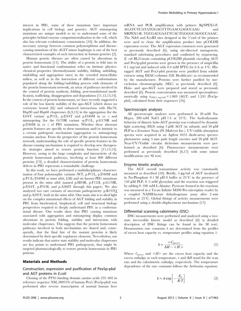
dependence of the rate constants follows the Arrhenius equation:
k~A:exp {Ea
R:T
� �ð2Þ
Protein Misfolding in Primary Hyperoxaluria I
PLOS ONE | www.plosone.org 2 August 2013 | Volume 8 | Issue 8 | e71963

Where Ea is the activation energy and T is the absolute
temperature in K.
Mutational effects on the activation free energy (DDG ),
enthalpy (DDH ) and entropy (DDS ) where determined on
the basis of the transition state theory as described in [19]. The
values of DDH and DDS where considered to be constant
within the temperature range involved in extrapolations based on
the highly linear Arrhenius plots found in all cases as well as the
nice agreement between the values obtained upon determination
of these parameters at 37uC (physiological temperature) and at
60uC (approximately the average and median Tm value for all
AGT enzymes; data not shown). Mutational effects on activation
energetic parameters were determined using equations (3)–(5):
DDG=~{R:T :lnk(37oC)(mut)
k(37oC)(WT)
� �ð3Þ
DDH=~Ea(mut){Ea(WT) ð4Þ
{TDDS=~DDG={DDH= ð5Þ
Expression and characterization of AGT variants inchinese hamster ovary (CHO) cells
CHO cells (ATTC, USA) were grown in a-MEM (Lonza,
Germany) supplemented with glutamine, penicillin/streptomycin
and 5% fetal bovine serum. Cell transfections were performed
using AGT cDNA variants subcloned in pCIneo plasmids
(Promega, USA) in 6-well plates with Transfast reagent (Promega,
USA), following manufacturer’s guidelines. After 24 h, cells were
passed to 100 mm Petri dishes containing 4 glass coverslips for
immunofluorescence studies. Cultures were harvested 48 h after
transfection.
Cells were sonicated in lysis buffer (100 mM potassium
phosphate pH 8.0, 250 mM sucrose, 0.05% triton X-100,
100 mM PLP) and centrifuged at 4uC, 5000 g for 10 min
(Beckman, USA) to obtain soluble fractions (supernatants). The
pellets were washed with phosphate buffer saline (PBS) and
resuspended in RIPA buffer. The protein concentration was
measured by using bicinchoninic acid (BCA), and equal amounts
of total protein were run in 10% acrylamide-SDS gels. The upper
portion of the gel (above 70 kDa) was stained with Coomassie blue
and scanned to control for equal protein loading, while the
remaining of the gel was transferred to nitrocellulose membranes
for western analysis. Purified AGT protein were included to
calibrate the amount of AGT present in the cell lysates.
Membranes were probed with anti-AGT rabbit serum followed
by HRP-conjugated anti-rabbit IgG (Jackson ImmunoResearch,
USA) and enhanced chemiluminescence substrate (Roche, Ger-
many). Chemiluminescent signals were measured in VersaDoc
4000 MP and ChemiDoc MP devices and analysed using
ImageLab Software (BioRad, Hercules, CA).
AGT activities were measured using cell lysates containing
100 mg total protein incubated in 100 mM K-Phosphate buffer
pH 8.0 at 37uC for 30 min in the presence of 40 mM PLP, 10 mM
glyoxylate and 40 mM L-Alanine. Pyruvate formed in the
reactions was measured by a coupled NADH:lactate dehydroge-
nase assay [18].
For immunofluorescence confocal microscopy, cells grown on
two coverslips were pulse-labeled in vivo with 100 nM MitoTracker
Red (Invitrogen USA) during 15 min, followed by additional
15 min chase in label-free medium. PBS, pH 7.4, was used as a
buffer in all subsequent washes and incubations. Cells were fixed
in 3% paraformaldehyde at room temperature for 10 min. and
permeabilized with 0.1% Triton X100-PBS. For AGT labeling on
mitotracker stained cells, rabbit anti-human AGT (a gift from Dr.
Danpure, University College London, UK) and Alexa Fluor 488
goat anti-rabbit IgG (Invitrogen, USA) were used. To label AGT
and peroxisomes on the same cells, guinea pig anti-human AGT
(also provided by Dr. Danpure) and rabbit anti-human PMP70
(Abcam, UK) were used, followed by incubations with Alexa Fluor
488 goat anti-guinea pig IgG (Invitrogen, USA) and Alexa Fluor
555 goat anti-rabbit IgG (invitrogen, USA). The coverslips were
mounted with PBS-glycerol. The images were taken with a 606objective in a confocal laser-scanning microscope (Olympus
Inverted IX81, Japan).
In vitro expression of AGT variants in a cell-free systemand interaction with Hsc70 chaperones
Cell-free expression of AGT variants was performed in rabbit
reticulocyte lysates (TnT system, Promega, USA) at 30uC for 2 h
using 35S-Met and AGT cDNA variants subcloned in pCIneo
plasmids. Protein synthesis was stopped with 100 mg/ml cyclo-
heximide, and 1/25th of the reaction product was set aside for
analysis. Hsc70 immunoprecipitation was carried out with the
remaining TnT product, using rat-Hsc70 antibodies (Abcam,
Cambridge, UK) as previously described [9]. The immunopre-
cipitated proteins and the initial TnT products were denatured in
Laemmli’s buffer and analyzed by SDS-PAGE and fluorography.
Molecular modelingp.I340M crystallization assays were carried out on a 60-well
microbath under oil (Terasaki plates) at 291 K. Crystals were
obtained using a precipitant solution containing 15% PEG 3350;
0.1M Bis-Tris pH 5.2, 150 mM Li2SO4 and 5% w/v octyl-b-D-
glucoside as additive. The best crystal forms were obtained by
mixing drops of 1 ml protein solution, 2 ml precipitant solution and
0.45 ml additive. In general, the crystals appear and grow in the
following 24 hours. Crystals were mounted in a fiber loop,
transferred to the cryoprotectant (20% glycerol on the crystal
mother liquor solution) and flash-frozen at 100 K in a nitrogen gas
steam. p.I340M crystals diffraction data set was collected using a
ADSC Q4 CCD detector at ID14.4 beamline of the European
Synchrotron Radiation Facility (Grenoble). Diffraction data were
processed with XDS [20] and scaled with SCALA from the CCP4
package (Collaborative Computational Project, Number 4) [21]. A
summary of the diffraction protocol, data-collection and refine-
ment statistics are given in Table S2. p.I340M diffracted to 1.90 A
resolution and belonged to space group P212121, with unit-cell
parameters a = 54.5, b = 103.5, c = 153. The p.I340M structure
was solved by molecular replacement using Phaser [22] with the
coordinates from the native protein (PDB 1H0C [23]). Several
cycles of restrained refinement with PHENIX [24] and iterative
model building with COOT [24,25] yielded to the final model
with an R/Rfree 0.17/0.20. The water structure was also modeled.
The stereochemistry of the models was verified with MolProbity
[26]. Ribbon figures were produced using PyMol [27]. The
coordinates and structure factor amplitudes have been deposited
in the Protein Data Bank (PDB code: 2YOB).
Statistical analysisFor statistical comparison, a two-sample Student’s t-test was
performed.
Protein Misfolding in Primary Hyperoxaluria I
PLOS ONE | www.plosone.org 3 August 2013 | Volume 8 | Issue 8 | e71963
Results
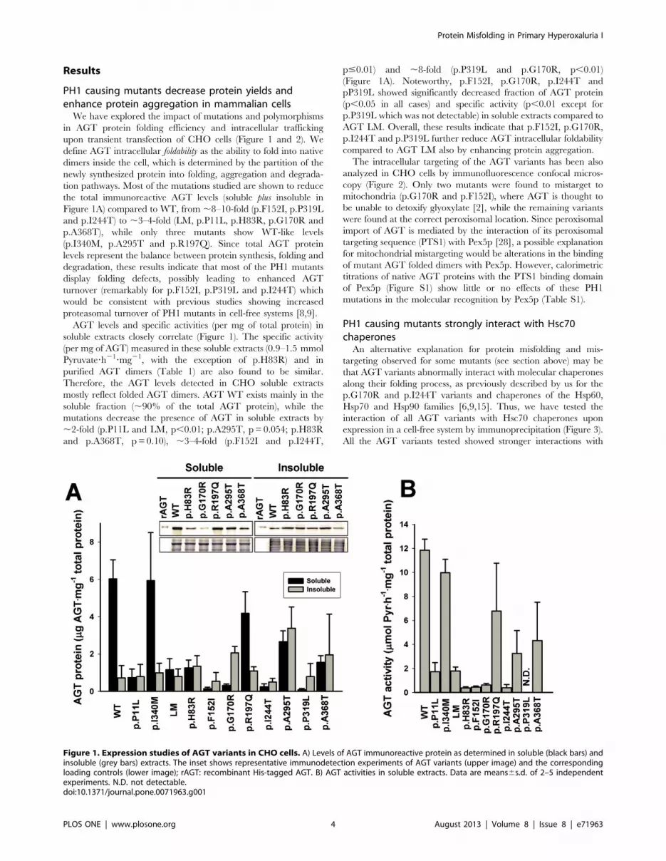
PH1 causing mutants decrease protein yields andenhance protein aggregation in mammalian cells
We have explored the impact of mutations and polymorphisms
in AGT protein folding efficiency and intracellular trafficking
upon transient transfection of CHO cells (Figure 1 and 2). We
define AGT intracellular foldability as the ability to fold into native
dimers inside the cell, which is determined by the partition of the
newly synthesized protein into folding, aggregation and degrada-
tion pathways. Most of the mutations studied are shown to reduce
the total immunoreactive AGT levels (soluble plus insoluble in
Figure 1A) compared to WT, from ,8–10-fold (p.F152I, p.P319L
and p.I244T) to ,3–4-fold (LM, p.P11L, p.H83R, p.G170R and
p.A368T), while only three mutants show WT-like levels
(p.I340M, p.A295T and p.R197Q). Since total AGT protein
levels represent the balance between protein synthesis, folding and
degradation, these results indicate that most of the PH1 mutants
display folding defects, possibly leading to enhanced AGT
turnover (remarkably for p.F152I, p.P319L and p.I244T) which
would be consistent with previous studies showing increased
proteasomal turnover of PH1 mutants in cell-free systems [8,9].
AGT levels and specific activities (per mg of total protein) in
soluble extracts closely correlate (Figure 1). The specific activity
(per mg of AGT) measured in these soluble extracts (0.9–1.5 mmol
Pyruvate?h21?mg21, with the exception of p.H83R) and in
purified AGT dimers (Table 1) are also found to be similar.
Therefore, the AGT levels detected in CHO soluble extracts
mostly reflect folded AGT dimers. AGT WT exists mainly in the
soluble fraction (,90% of the total AGT protein), while the
mutations decrease the presence of AGT in soluble extracts by
,2-fold (p.P11L and LM, p,0.01; p.A295T, p = 0.054; p.H83R
and p.A368T, p = 0.10), ,3–4-fold (p.F152I and p.I244T,
p#0.01) and ,8-fold (p.P319L and p.G170R, p,0.01)
(Figure 1A). Noteworthy, p.F152I, p.G170R, p.I244T and
pP319L showed significantly decreased fraction of AGT protein
(p,0.05 in all cases) and specific activity (p,0.01 except for
p.P319L which was not detectable) in soluble extracts compared to
AGT LM. Overall, these results indicate that p.F152I, p.G170R,
p.I244T and p.P319L further reduce AGT intracellular foldability
compared to AGT LM also by enhancing protein aggregation.
The intracellular targeting of the AGT variants has been also
analyzed in CHO cells by immunofluorescence confocal micros-
copy (Figure 2). Only two mutants were found to mistarget to
mitochondria (p.G170R and p.F152I), where AGT is thought to
be unable to detoxify glyoxylate [2], while the remaining variants
were found at the correct peroxisomal location. Since peroxisomal
import of AGT is mediated by the interaction of its peroxisomal
targeting sequence (PTS1) with Pex5p [28], a possible explanation
for mitochondrial mistargeting would be alterations in the binding
of mutant AGT folded dimers with Pex5p. However, calorimetric
titrations of native AGT proteins with the PTS1 binding domain
of Pex5p (Figure S1) show little or no effects of these PH1
mutations in the molecular recognition by Pex5p (Table S1).
PH1 causing mutants strongly interact with Hsc70chaperones
An alternative explanation for protein misfolding and mis-
targeting observed for some mutants (see section above) may be
that AGT variants abnormally interact with molecular chaperones
along their folding process, as previously described by us for the
p.G170R and p.I244T variants and chaperones of the Hsp60,
Hsp70 and Hsp90 families [6,9,15]. Thus, we have tested the
interaction of all AGT variants with Hsc70 chaperones upon
expression in a cell-free system by immunoprecipitation (Figure 3).
All the AGT variants tested showed stronger interactions with
Figure 1. Expression studies of AGT variants in CHO cells. A) Levels of AGT immunoreactive protein as determined in soluble (black bars) andinsoluble (grey bars) extracts. The inset shows representative immunodetection experiments of AGT variants (upper image) and the correspondingloading controls (lower image); rAGT: recombinant His-tagged AGT. B) AGT activities in soluble extracts. Data are means6s.d. of 2–5 independentexperiments. N.D. not detectable.doi:10.1371/journal.pone.0071963.g001
Protein Misfolding in Primary Hyperoxaluria I
PLOS ONE | www.plosone.org 4 August 2013 | Volume 8 | Issue 8 | e71963

Figure 2. Immunolocalization studies of AGT variants in CHO cells. WT (upper left panel), LM (lower left panel), p.G170R (upper right panel)and p.F152I (lower right panel). In each panel, the upper row shows mitochondrial immunolocalization (AGT variant, Mitotracker probe and theirmerge), while lower row shows peroxisomal immunolocalization (AGT variant, PMP70 and their merge).doi:10.1371/journal.pone.0071963.g002
Table 1. Functional properties and hydrodynamic diameter of AGT variants.
AGT Variant Vmax (mmol?h21?mg21)aKM(Alanine)
(mM)a KM(Glyoxilate) (mM)a Kd(PLP) (nM)bkon(PLP)
(M21?s21)koff(PLP) (105)(s21)
Diameter(mean±s.d.; innm)c
WT 2.2260.09 19.561.4 245629 100630 (14006800) 197633 28618 8.162.3
p.P11L 2.4360.08 18.361.2 185622 (1070690) 15061 1661 9.862.8
p.I340M 3.0360.10 22.361.3 277627 172642 N.det. N.det. 8.260.1
LM 2.2660.09 19.861.5 197627 157615 N.det. N.det. 7.860.1
p.H83R 0.14160.005 42.462.4 156619 (11806380) 14706160 174653 8.962.3
p.F152I 1.9060.19 15.763.1 201668 (4906300) 2990670 146690 8.960.2
p.G170R 2.5460.13 21.962.0 293642 (13806170) 12562 1762 6.660.9
p.R197Q 1.8460.06 18.161.2 168621 156634 N.det. N.det. 9.960.7
p.I244T 2.8260.20 16.862.4 193649 (30606240) 5161 1661 7.160.7
p.A295T 2.1060.10 18.561.3 292630 71616 N.det. N.det. 9.560.4
p.P319L 1.5460.13 16.262.7 212660 (2406360) 26867 6610 7.361.0
p.A368T 2.1260.13 23.062.5 297650 134626 N.det. N.det. 8.260.2
aEnzyme kinetic parameters were obtained from global fittings of 2–4 independent experimental series using a double-displacement mechanism;bKd values were estimated from single titrations except for AGT WT protein where Kd value is the mean6s.d. from three independent titrations. Data in parenthesis areKd estimates obtained from the kinetic binding experiments (Kd = koff/kon).cHydrodynamic diameter of holo-AGT variants determined by dynamic light scattering (DLS). Data are mean6s.d. of 3–9 independent measurements.doi:10.1371/journal.pone.0071963.t001
Protein Misfolding in Primary Hyperoxaluria I
PLOS ONE | www.plosone.org 5 August 2013 | Volume 8 | Issue 8 | e71963

Hsc70 chaperones (p,0.01 in all cases), except p.I340M.
Polymorphic p.P11L and LM showed a 4-fold increase in
immunoprecipitated AGT compared to the WT protein, while
disease-causing mutants showed somewhat higher levels, from 4.8-
fold (p.H83R, p = 0.46 vs. LM) to 7-fold (p.P319L, p = 0.078 vs.
LM). Owing to the significant variability in these experiments, the
changes detected are not robust enough to claim mutation-specific
differences compared to AGT LM, but the trend observed suggests
that disease-causing mutations may increase the interaction with
Hsc70 chaperones along their folding process vs. LM protein.
PH1 causing mutants do not generally perturb AGToligomerization and function
To probe whether PH1 mutants may affect AGT oligomeriza-
tion and catalytic properties (as recently found for some PH1
mutants [4]), we have expressed recombinant AGT variants in
E.coli and purified AGT dimers to homogeneity. Size-exclusion
chromatography analysis showed a single peak for all variants with
an elution volume consistent with a <90 kDa dimeric form (data
not shown). The molecular dimensions of purified dimers were
further studied by dynamic light scattering (DLS), showing a
hydrodynamic diameter of 8.162.3 nm for holo-WT and
8.461.1 nm for the rest of holo-AGT variants (Table 1). Within
the experimental uncertainty, these results are consistent with the
size of dimeric AGT WT previously reported by DLS [29] and
also imply no noticeable perturbation of dimer size and/or dimer-
monomer equilibrium by these mutations.
Thus, we evaluated the effect of these variants on the
functionality of the AGT protein. Enzyme kinetic analyses based
on a double-displacement mechanism ([17]; see Figure 4A–B for
representative examples) were performed for all the AGT variants
and the enzyme kinetic parameters obtained are compiled in
Table 1. All the AGT variants tested showed similar specific
activity (Vmax) to that found for AGT WT with the exception of
p.H83R mutant, which displayed a ,15-fold reduction in specific
activity. No large changes in Km values for L-Ala and glyoxylate
were found among the AGT variants studied. The environment of
the bound coenzymes to WT and p.H83R was characterized by
Near-UV and visible absorption and circular dichroism spectros-
copies, revealing a ,10–15 nm blue shift in the absorbance and
dichroic bands of bound PLP and PMP (Figure 4C–D), which
supports a distorted coenzyme binding mode to the p.H83R
mutant. We have further tested this hypothesis by incubating holo-
WT and p.H83R (at a final monomer concentration of 36 mM) in
the presence of L-Ala for 1 h at 25uC and analyzed the the
amount of PMP released by UV-absorption spectroscopy upon
filtration using microfilters of 10 kDa cut-off. Under these
conditions, ,3% of the PMP was released in the WT enzyme
(consistent with tight binding of PMP along the overall transam-
ination reaction previously reported; [17]), while ,44% of PMP
was released in the p.H83R, indicating a large decrease in PMP
binding affinity for this mutant. Thus, we conclude that the large
decrease in catalytic performance of p.H83R is caused by a
distortion in the binding mode of PLP and PMP coenzymes.
We have also measured the binding affinity of the apo-variants
for PLP using fluorescence spectroscopy (Figure S2). Direct
equilibrium measurements allowed to determine the Kd values
for the WT and some stable mutants at 30uC (Table 1). All the
AGT variants studied under equilibrium conditions showed
affinity for PLP similar to the WT protein (Kd about 100 nM).
Under these conditions, aggregation in the absence of PLP was
found for p.P11L, p.H83R, p.F152I, p.G170R, p.I244T and
p.P319L mutants. Lowering the temperature or adding 10%
glycerol did not prevent aggregation in the time scale required for
AGT:PLP equilibration. Alternatively, we estimated the affinity
for PLP by performing kinetic binding experiments under pseudo-
first order conditions (Figure S2B) providing only an upper limit
for the Kd values mostly due to the large uncertainties associated to
the determination of the koff (Table 1). No clear differences were
observed for the estimated Kd values for PLP between WT and the
mutants studied using this kinetic approach (Table 1).
PH1 mutations kinetically destabilize the apo-AGT formThermal denaturation of AGT shows a single transition for all
AGT variants studied by differential scanning calorimetry (DSC;
Figure 5A). Denaturation of AGT variants is a purely kinetic
process, which is well described by the irreversible two-state
conversion of the native dimer to a final aggregated state (NRF).
This kinetic process is characterized by a denaturation rate
constant k [6,30], which is inversely proportional to its half-life for
denaturation. In this scenario, protein kinetic stability (as a given
denaturation rate or half-life under certain experimental condi-
tions) is determined by the height of the free energy barrier that
AGT must cross from the native state to reach the transition state
of the denaturation rate-limiting step [7]. Thus, mutational and
PLP effects on AGT thermal transition (Tm and Ea values) are
translated into changes in kinetic stability (Table 2 and Figure S3).
Remarkably, removal of bound PLP has a dramatic effect on the
stability of most of AGT variants, reducing by ,25uC and 4–5
orders of magnitude the Tm values and the corresponding kinetic
stabilities (Table 2 and Figure S3).
DSC is particularly suitable to study the stability of AGT
enzymes because it provides not only a Tm value, which can be
Figure 3. Interaction of AGT variants with Hsc70 chaperones ina cell-free system. Representative autoradiograms of AGT proteinslabeled with 35S-Met are shown for several AGT variants (E: total AGTsynthesized in extracts; I: AGT immunoprecipitated using anti-Hsc70antibodies; note that 1 ml of the TnT reaction was loaded in E lanes,while the protein immunoprecipitated from 6 ml TnT lysate was loadedin I lanes). Data in the lower panel are expressed as percentage ofimmunoprecipitated AGT compared to the total AGT synthesized, andare mean6s.d. from three independent experiments.doi:10.1371/journal.pone.0071963.g003
Protein Misfolding in Primary Hyperoxaluria I
PLOS ONE | www.plosone.org 6 August 2013 | Volume 8 | Issue 8 | e71963

alternatively obtained by other techniques, but also allow
determine accurately the rate constants as a function of
temperature, to compare kinetic stabilities over widely different
time scales (in this work half-lives for denaturation range from
several minutes to thousands of years, extrapolated to physiolog-
ical temperature; Table 2) and provide insightful parameters of the
denaturation process such as denaturation enthalpies and activa-
tion energies [6,19,30]. For instance, the temperature dependence
of denaturation enthalpies shows a common behavior for holo-
and apo-AGT enzymes (Figure 6A), yielding a global slope (i.e. a
denaturation heat capacity) of 10.260.6 kcal?mol21?K21 which is
slightly lower than the theoretical value expected for global
unfolding of the AGT dimer (14.8 kcal?mol21?K21; based on
[31]). This analysis supports that AGT thermal denaturation
involves a large loss of tertiary structure, possibly reflecting
denaturation of both domains in AGT, and also that all the AGT
enzymes studied here in their holo- and apo-forms denature to a
similar extent.
DSC scans are used to determine denaturation rates (k) or half-
lives at different temperatures by building Arrhenius plots
(Figure 5B) which allow study kinetic stabilities at physiological
temperature (Table 2). Most of the holo variants are kinetically
stable at 37uC with extrapolated half-lives ranging from ,12 to
,80000 years (Table 2), with the exception of p.H83R, with a
very short half-life of ,4 h likely due to a distortion in the binding
mode of PLP (Figure 4). The apo-proteins are much less stable,
remarkably the p.G170R, p.A295T, p.H83R, p.F152I, p.I244T
and p.P319L enzymes, which reduce ,20–150-fold the kinetic
stability vs. the LM variant, and they denature on a time scale of a
few minutes to hours (Table 2). Indeed, the half-lives for enzyme
inactivation upon incubation of apo-proteins at 37uC and standard
activity measurements agree very well with those obtained from
our DSC analyses (Table 2). This significant kinetic destabilization
of the apo vs. holo-forms in most of PH1 mutants is not due to
Figure 4. Functional characterization of WT and p.H83R. A andB) Enzyme activity measurements for WT (A) and p.H83R (B) at differentL-Ala concentrations in the presence of 0.25 mM (diamonds), 0.5 mM(down triangles), 1 mM (up triangles) and 2 mM (circles) glyoxylate.Data in panel A are means6s.d from four independent measurementswhile data in panel B are means from two independent measurements.Lines are best fits for the different glyoxylate concentrations obtainedfrom global fittings using a double-displacement mechanism. C and D)Absorption (C) and circular dichroism (D) spectra for holo-WT (black)and holo-p.H83R (grey) acquired upon incubation for at least 10 min inthe absence (continuous lines; PLP bound) or presence (dashed lines;PMP bound) of 500 mM L-alanine.doi:10.1371/journal.pone.0071963.g004
Figure 5. Thermal denaturation of AGT variants studied bydifferential scanning calorimetry (DSC). A) Representative DSCtraces obtained at 3uC/min; Lines are best-fits from a two-stateirreversible denaturation model with first-order kinetics [6]; B) Arrheniusplots for the irreversible denaturation of AGT variants, indicating alsothe extrapolated rate constants at physiological temperature (interceptwith the vertical dotted line). Symbols are: WT (circle), LM (triangle) andp.F152I (square). Data for holo proteins are shown as closed symbolsand for apo proteins as open symbols.doi:10.1371/journal.pone.0071963.g005
Protein Misfolding in Primary Hyperoxaluria I
PLOS ONE | www.plosone.org 7 August 2013 | Volume 8 | Issue 8 | e71963
large changes in the affinity for PLP (Table 1), but rather, it
suggests that cofactor binding overcomes some destabilizing
interactions present in the native state of the apo-forms [7].
Regarding the polymorphic variants, p.P11L and LM are
kinetically destabilizing, while p.I340M enhances AGT kinetic
stability compared to the WT protein, either in the apo- or the
holo-form (Table 2).
The large kinetic stabilization induced by PLP binding, as well
as the destabilization induced by some mutants in their apo-form
must originate from changes in the height of the denaturation free
energy barrier (i.e. the free energy difference between the native
and denaturation transition state [6]). Our DSC analyses also
provide information on the reaction order of AGT denaturation
by determining the m value (i.e. reaction order is 1/m), and hence,
on the oligomerization state of the denaturation transition state: a
m value close to one indicates first-order kinetics, involving a
dimeric transition state for AGT, while a value close to two
supports that the transition state is monomeric. As we show in
Table 2, most of the AGT variants display first-order denaturation
kinetics (m<1), with only small deviations for a few apo-forms.
Thus, the AGT denaturation transition state is essentially a
partially unfolded dimer for all AGT variants, indicating that other
unfolding steps (dimer dissociation and monomer unfolding) must
occur after the denaturation rate-limiting step, and hence, these
steps do not contribute to the effect of mutations, polymorphisms
or PLP binding on AGT kinetic stability. PLP mediated kinetic
stabilization arise from its preferential binding and stabilization of
the native AGT dimeric structure, raising the denaturation free
energy barrier by ,7 kcal?mol21 (see [6] and Table 1).
Structural and energetic basis of AGT kineticdestabilization by PH1 mutations
A plot of the activation energies for denaturation of all AGT
enzymes as a function of their Tm value (which exponentially
correlate well with their kinetic stabilities; see Figure S3) shows
that those AGT enzymes with lower kinetic stability display lower
values of Ea (Figure 6B). The high linearity of Arrhenius plots
(Figure 5B) indicates a negligible activation heat capacity, and
thus, Ea values can be considered essentially as temperature-
independent. Hence, the results shown in Figure 6B can be
rationalized as mutational effects on the energetic balance (entropic
and enthalpic contributions) of the free energy barrier for
denaturation (previously shown for denaturation of other protein
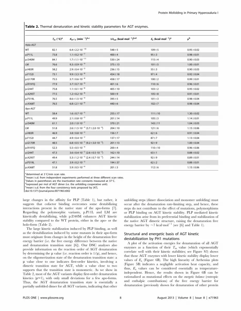
Table 2. Thermal denaturation and kinetic stability parameters for AGT enzymes.
Tm (6C)a K376C (min21)b,c Dhcal (kcal?mol21)b,d Ea (kcal?mol21)e mb
Holo-AGT
WT 82.1 6.462.2?10210 54865 10965 0.9560.02
p.P11L 73.8 1.160.2?1027 40064 9563 0.9860.01
p.I340M 84.1 1.761.1?10211 530624 11564 0.9060.03
LM 76.6 9.360.9?1029 375613 10165 1.0060.01
p.H83R 58.2 2.960.4?1023 236615 5563 0.9060.03
p.F152I 73.1 9.963.3?1028 454618 9764 0.9260.04
p.G170R 75.5 3.760.6?1028 458617 10062 0.9960.01
p.R197Q 77.9 3.760.7?1029 40166 11066 0.9660.01
p.I244T 75.8 1.160.1?1028 405610 10362 0.9560.02
p.A295T 77.5 1.260.2?1028 50069 10568 0.9160.01
p.P319L 76.5 8.661.5?1029 39565 10163 0.9860.04
p.A368T 76.5 6.862.1?1029 44066 10267 0.9860.04
Apo-AGT
WT 58.4 1.660.7?1025 255617 111610 1.3060.02
p.P11L 49.9 2.160.8?1023 201614 10563 1.1460.01
p.I340M 61.1 2.061.0?1027 370621 143611 1.0460.03
LM 51.8 2.661.5?1024 (3.762.0?1024) 204610 12166 1.1560.06
p.H83R 46.9 3.860.8?1022 15667 6266 0.9160.04
p.F152I 44.7 4.960.4?1022 137617 8767 1.1360.04
p.G170R 48.5 6.860.5?1023 (9.263.0?1023) 201613 9269 1.0060.04
p.R197Q 52.3 5.560.5?1024 20064 110610 0.9660.06
p.I244T 47.3 3.660.4?1022 (2.660.5?1022) 162610 6361 0.8860.02
p.A295T 49.4 5.361.2?1023 (2.460.7?1023) 244614 9269 0.8960.01
p.P319L 47.1 3.960.2?1022 194637 6262 0.8860.01
p.A368T 51.8 5.960.5?1024 20163 11266 1.1560.06
adetermined at 3uC/min scan rate.bmean6s.d. from independent experiments performed at three different scan rates.cValues in parentheses are the inactivation rate constants measured at 37uC.dexpressed per mol of AGT dimer (i.e. the unfolding cooperative unit).emean6s.d. from the four consistency tests proposed by [47].doi:10.1371/journal.pone.0071963.t002
Protein Misfolding in Primary Hyperoxaluria I
PLOS ONE | www.plosone.org 8 August 2013 | Volume 8 | Issue 8 | e71963

systems; [19,32,33]). We have thus calculated the changes in the
activation enthalpies (DDH ) and entropies (2TDDS ) and
plotted them as a function of the mutational effects on the kinetic
stability (DDG ) for the holo-(Figure 6C) and apo-(Figure 6D)
enzymes. We found that changes in kinetic stability (DDG ) arise
from large changes of opposite sign in DDH and 2DDTS that
largely cancel out for holo- and apo-AGT enzymes. Since DDH
reflect structural differences (,solvent exposure) between the
native and the transition states, the analyses shown in Figure 6
support that the most destabilizing mutants decrease the
magnitude of the structural change occurring between the native
and denaturation transition states. We must note that there is no
experimental evidence of large structural changes in the native
dimer upon mutation: they show similar hydrodynamic radius
and, with the exception of p.H83R, similar enzyme activities
(Table 1) as well as denaturation enthalpies consistent with
similarly folded native states (Table 2), and thus, these changes
may primarily affect the denaturation transition state. Using
structure-energetics relationships, a value of 260 kcal?mol21 for
DDH (found for several AGT mutants; see Figure 6) is translated
into a difference of ,90 folded residues or ,8500 A2 of solvent
exposed surface [31] between the native and transition state (per
dimer), supporting the existence of significant mutational effects on
the structure and energetics of the denaturation transition state
(see [33] for a similar situation with mutants of human
phosphoglycerate kinase 1).
Structure of p.I340M and modeling of the AGT variantsThe crystal structure of p.I340M has been determined at an
unprecedented 1.9 A resolution to improve our modeling efforts
on PH1 causing mutants. The structures of p.I340M and the WT
(PDB code 1H0C [23]) are nearly identical. AGT homodimer has
each protomer folded into a large N-terminal domain, a smaller C-
terminal domain and a 22 amino acid long unstructured N-
terminal tail that grabs the other subunit within the dimer
(Figure 7A). None of the substitutions observed in the same
Figure 6. Structure-energetics relationships for thermal denaturion of holo- and apo-AGT enzymes. A) Temperature dependence ofdenaturation enthalpies (DH) for holo- (closed symbols) and apo-(open symbols) proteins. The linear fit provides the value of DCp
( = 10.260.6 kcal.mol21). B) Activation energy (Ea) plotted vs. the Tm for holo- (closed symbols) and apo-(open symbols) AGT enzymes. C and D)changes in activation enthalpic and entropic contributions to AGT kinetic stability as a function of changes in activation free energies for holo-(C) andapo-(D) AGT enzymes. Lines in C and D are meant to guide the eye and have no theoretical meaning.doi:10.1371/journal.pone.0071963.g006
Protein Misfolding in Primary Hyperoxaluria I
PLOS ONE | www.plosone.org 9 August 2013 | Volume 8 | Issue 8 | e71963
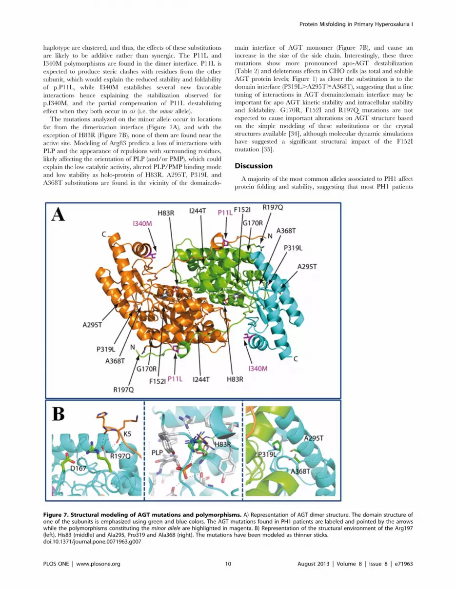
haplotype are clustered, and thus, the effects of these substitutions
are likely to be additive rather than synergic. The P11L and
I340M polymorphisms are found in the dimer interface. P11L is
expected to produce steric clashes with residues from the other
subunit, which would explain the reduced stability and foldability
of p.P11L, while I340M establishes several new favorable
interactions hence explaining the stabilization observed for
p.I340M, and the partial compensation of P11L destabilizing
effect when they both occur in cis (i.e. the minor allele).
The mutations analyzed on the minor allele occur in locations
far from the dimerization interface (Figure 7A), and with the
exception of H83R (Figure 7B), none of them are found near the
active site. Modeling of Arg83 predicts a loss of interactions with
PLP and the appearance of repulsions with surrounding residues,
likely affecting the orientation of PLP (and/or PMP), which could
explain the low catalytic activity, altered PLP/PMP binding mode
and low stability as holo-protein of H83R. A295T, P319L and
A368T substitutions are found in the vicinity of the domain:do-
main interface of AGT monomer (Figure 7B), and cause an
increase in the size of the side chain. Interestingly, these three
mutations show more pronounced apo-AGT destabilization
(Table 2) and deleterious effects in CHO cells (as total and soluble
AGT protein levels; Figure 1) as closer the substitution is to the
domain interface (P319L.A295T$A368T), suggesting that a fine
tuning of interactions in AGT domain:domain interface may be
important for apo AGT kinetic stability and intracellular stability
and foldability. G170R, F152I and R197Q mutations are not
expected to cause important alterations on AGT structure based
on the simple modeling of these substitutions or the crystal
structures available [34], although molecular dynamic simulations
have suggested a significant structural impact of the F152I
mutation [35].
Discussion
A majority of the most common alleles associated to PH1 affect
protein folding and stability, suggesting that most PH1 patients
Figure 7. Structural modeling of AGT mutations and polymorphisms. A) Representation of AGT dimer structure. The domain structure ofone of the subunits is emphasized using green and blue colors. The AGT mutations found in PH1 patients are labeled and pointed by the arrowswhile the polymorphisms constituting the minor allele are highlighted in magenta. B) Representation of the structural environment of the Arg197(left), His83 (middle) and Ala295, Pro319 and Ala368 (right). The mutations have been modeled as thinner sticks.doi:10.1371/journal.pone.0071963.g007
Protein Misfolding in Primary Hyperoxaluria I
PLOS ONE | www.plosone.org 10 August 2013 | Volume 8 | Issue 8 | e71963

may show defective AGT function due to alterations in its protein
homeostasis (this work; [4,6,9,36]). To understand the different
mechanisms underlying PH1, we have integrated in this work
concepts from protein structure, biophysics, biochemistry and cell
biology to provide a comprehensive view on the mutational effects
on protein folding, assembly, transport, mistargeting and degra-
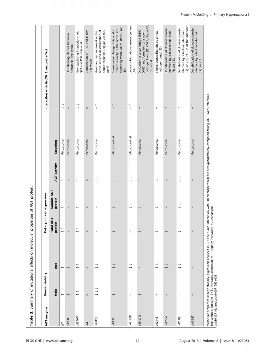
dation of the AGT protein (summarized in Table 3). Our work
delineates important checkpoints in AGT protein homeostasis,
such as the stability of the apo-proteins and the recognition of
folding intermediates by molecular chaperones (Figure 8) that
might be specifically targeted to restore AGT function in PH1
patients. We also provide insight to current potential therapies for
PH1 such as pyridoxine supplementation [4,5,6,37].
PLP binding to AGT enhances native state kinetic stability by
4–5 orders of magnitude ([6]; this work), making holo-proteins
highly kinetically stable at physiological temperature, with the only
exception of the catalytic mutant p.H83R. Interestingly, we found
that p.F152I, p.G170R, p.I244T, p.A295T and p.P319L are
markedly destabilized as apo-proteins compared to the non-
pathogenic LM variant (Table 2), and actually, they denature at a
relatively fast rate at physiological temperature. As we have
previously discussed for p.G170R [6], the low kinetic stability of
these mutants in the apo-form may have important implications
for PH1 pathogenesis, since it is likely that a significant fraction of
AGT may transiently exist as apo-protein in vivo, and thus, it might
be susceptible to intracellular irreversible alterations such as
mitochondrial import, protein aggregation and degradation.
Consequently, four of these variants (p.F152I, p.G170R,
p.I244T and p.P319L) show evident signs of misfolding and,
possibly, accelerated turnover in CHO cells, while two of them
(p.G170R and p.F152I) also cause protein mitochondrial mis-
targeting (Table 3). This indicates some degree of correlation
between the apo- stability and protein mitochondrial import and/
or intracellular aggregation. According to this interpretation, we
propose a beneficial effect of pyridoxine supplementation in
patients carrying these four mutations (F152I, G170R, I244T and
P319L). In fact, p.F152I and p.G170R have been described as
pyridoxine-responsive genotypes in PH1 patients [38,39].
Our DSC analyses provide molecular insights on the effect of
PH1 causing mutants and PLP binding on the AGT kinetic
stability, denaturation mechanism and structural/energetic fea-
tures of its denaturation free energy barrier previously unexplored.
Within the set of PH1 mutants studied here, only the thermal
stability of p.F152I and p.G170R have been reported earlier (by
circular dichroism and inactivation experiments) showing thermal
destabilization (lower Tm) mainly for their apo-forms [35,40,41].
Our detailed DSC kinetic analyses further show that the kinetic
overstabilization exerted by PLP bound to p.F152I, p.G170R,
p.I244T, p.A295T and p.P319L as holo-proteins arises from subtle
changes in the enthalpic and entropic contributions to the
denaturation free energy barrier, since a similar pattern of
enthalpy/entropy compensation is found for holo- and apo-AGT
proteins (Figure 6C and 6D). Moreover, these effects must concern
mainly to the structure and energetics of the dimeric transition
state for denaturation, and thus, mutational effects on dimer
dissociation and full monomer unfolding do not contribute to the
relevant kinetic stability of AGT enzymes since they occur after
the denaturation rate-limiting step.
Figure 8. Folding and misfolding checkpoints of PH1 causing mutants.doi:10.1371/journal.pone.0071963.g008
Protein Misfolding in Primary Hyperoxaluria I
PLOS ONE | www.plosone.org 11 August 2013 | Volume 8 | Issue 8 | e71963
Ta
ble
3.
Sum
mar
yo
fm
uta
tio
nal
eff
ect
so
nm
ole
cula
rp
rop
ert
ies
of
AG
Tp
rote
in.
AG
Te
nz
ym
eK
ine
tic
sta
bil
ity
Eu
ka
ryo
tic
cell
ex
pre
ssio
nIn
tera
ctio
nw
ith
Hsc
70
Str
uct
ura
le
ffe
ct
Ho
loA
po
To
tal
AG
Tp
rote
inS
olu
ble
AG
Tp
rote
inA
GT
act
ivit
yT
arg
eti
ng
WT
Pe
roxi
som
alQ
Q
p.P
11
LQ
Q=
==
Pe
roxi
som
al=
De
stab
ilizi
ng
clas
he
sb
etw
ee
np
roto
me
rs(t
his
wo
rk)
p.I3
40
Mq
qP
ero
xiso
mal
Ne
wst
abili
zin
gin
tera
ctio
ns
wit
hQ
23
and
D5
2(t
his
wo
rk)
LM=
==
==
Pe
roxi
som
al=
Co
mb
inat
ion
of
P1
1L
and
I34
0M
(th
isw
ork
)
p.H
83
RQ
==
Pe
roxi
som
al=
qSt
ruct
ura
lre
arra
ng
em
en
tat
the
acti
vesi
tean
dd
est
abili
zati
on
of
bo
un
dco
fact
ors
(Fig
ure
7B
;th
isw
ork
)
p.F
15
2I
Mit
och
on
dri
al=
qC
on
serv
ativ
ech
ang
e(t
his
wo
rk);
Cre
ate
sa
cavi
tyin
the
acti
vesi
ted
isp
laci
ng
W1
08
wh
ich
stac
ksP
MP
[35
]
p.G
17
0R
=Q
Q=
Mit
och
on
dri
al=
qLo
calc
on
form
atio
nal
rear
ran
ge
me
nt
[34
]
p.R
19
7Q
q=
Pe
roxi
som
al=
qD
isru
pti
on
of
asa
ltb
rid
ge
(R1
97
-D
16
7)
and
form
atio
no
fa
ne
wh
ydro
ge
nb
on
d(Q
19
7-K
5;F
igu
re7
B;
this
wo
rk)
p.I2
44
T=
QP
ero
xiso
mal
=q
a-h
elix
de
stab
iliza
tio
nb
ya
ne
wh
ydro
ge
nb
on
d[2
3]
p.A
29
5T
=Q
==
Pe
roxi
som
alq
De
stab
iliza
tio
no
fd
om
ain
:do
mai
nin
terf
ace
by
ab
ulk
ier
sid
e-c
hai
n(F
igu
re7
B).
p.P
31
9L
=Q
Pe
roxi
som
alq
De
stab
iliza
tio
no
fd
om
ain
:do
mai
nin
terf
ace
by
ab
ulk
ier
sid
e-c
hai
n(F
igu
re7
B).
P3
19
isat
the
inte
rfac
e.
p.A
36
8T
==
==
=P
ero
xiso
mal
=q
De
stab
iliza
tio
no
fd
om
ain
:do
mai
nin
terf
ace
by
ab
ulk
ier
sid
e-c
hai
n(F
igu
re7
B).
Mo
lecu
lar
pro
pe
rtie
s(k
ine
tic
stab
ility
,e
xpre
ssio
nan
alys
es
inC
HO
cells
and
inte
ract
ion
wit
hH
sc7
0ch
ape
ron
es)
are
sem
iqu
anti
tati
vely
com
par
ed
taki
ng
AG
TLM
asre
fere
nce
.T
he
sym
bo
lsin
dic
ate
:q
/Q,
incr
eas
ed
/de
cre
ase
d;
=q
,sl
igh
tly
incr
eas
ed
;=
,u
nch
ang
ed
.d
oi:1
0.1
37
1/j
ou
rnal
.po
ne
.00
71
96
3.t
00
3
Protein Misfolding in Primary Hyperoxaluria I
PLOS ONE | www.plosone.org 12 August 2013 | Volume 8 | Issue 8 | e71963

The intracellular homeostasis of AGT protein seems to rely on a
delicate balance between protein folding, misfolding, degradation
and intracellular trafficking. Importantly, the AGT LM protein, a
variant which is not disease-causing but it is known to sensitize
AGT towards deleterious mutations, is shown to notably reduce
protein kinetic stability and to enhance protein misfolding and
degradation, while most of disease-causing mutations further
exacerbate at least some of these defects (Table 3). The partial
correlation between these molecular defects suggests that multiple
elements in the protein homeostasis networks play a role in
determining the fate of PH1 mutants (including chaperones,
cochaperones and regulatory proteins [13] and vitamin B6 salvage
enzymes implicated in the recycling and targeting of PLP to apo-
enzymes [42]). This complexity in the homeostasis of AGT
proteins, as well as individual differences in the protein homeo-
stasis network (which may even occur among isogenic individuals
[43]), may explain inter-individual variability in clinical presenta-
tions and residual activities for patients sharing a given genotype
[7,44] and the different fate of mutant proteins (aggregation vs.
mitochondrial mistargeting) when expressed under different
experimental conditions (this work and [36]). Despite our findings,
those specific events and interactions responsible for the partition
between protein mitochondrial mistargeting, aggregation and
degradation remain elusive.
In the present work, we show that all PH1 mutations of the minor
haplotype strongly interact with Hsc70 chaperones, adding to our
previous work on p.I244T and p.G170R that also showed
enhanced interactions with Hsc90 [6,9] and bacterial Hsp60
[15]. We have recently reported that p.G170R interacts with
Hsc70 and Hsp90 chaperones through a molten globule folding
intermediate [6], while p.I244T interacts with Hsp60 chaperones
in partially folded monomeric state with the folded N-terminal and
C-terminal domains in an open conformation [15]. We thus
propose that the last steps involving docking of tertiary structure
elements and acquisition of the dimeric quaternary structure are
crucial for proper folding of AGT. Moreover, enhanced interac-
tion of PH1 mutants with these molecular chaperones suggest a
rougher folding landscape for these mutants (with a higher
population of kinetic/equilibrium intermediates [13,45,46]). Thus,
molecular chaperones emerge as an important checkpoint in the
folding of PH1 mutants, likely by partitioning AGT folding
intermediates into productive formation of native dimers and
peroxisomal import, presentation of partially folded states to the
mitochondrial import systems, aggregation and proteasomal
degradation [7]. Hsp70, Hsp60 and Hsp90 chaperone systems
are known to cooperate in assisting protein folding, and the
regulation of chaperone activity by cochaperones and regulatory
proteins may lead to different fates (i.e. folding vs. degradation) for
the client proteins [13]. Overall, all these results suggest that at
least these three chaperone systems (Hsp60, Hsp70 and Hsp90) are
potential targets for correction of the folding defects displayed by
PH1 mutants. Consequently, the detailed characterization of the
chaperone requirements for efficient folding of PH1 mutants will
open new approaches for therapeutic intervention in PH1. We
have already initiated such studies in cell and animal models of
PH1 (ongoing work).
Conclusions
In this work, we present a multidisciplinary approach that
provides clues on the protein homeostasis defects displayed by
PH1 causing mutations leading to protein aggregation and
mistargeting. We observe a significant correlation between
mutation-induced kinetic destabilization of the apo-AGT dimer,
kinetic trapping by molecular chaperones and intracellular protein
foldability and mistargeting. Detailed kinetic and structure-
energetics analyses also show that cofactor induced overstabiliza-
tion of some mutants is caused by subtle changes in the enthalpic/
entropic contributions to denaturation free energy barriers, which
may also explain the pyridoxine responsiveness found in patients
carrying these mutations. We propose that native state kinetic
stabilizers and protein homeostasis modulators may be suitable
pharmacological therapies to correct folding and stability defects
in PH1.
Supporting Information
Figure S1 Interaction of Pex5p-pbd and AGT-WT byisothermal titration calorimetry (ITC). A) Raw calorimetric
data; B) Binding isotherm (the line shows the best-fit to one-
independent-type-of sites).
(TIF)
Figure S2 Equilibrium (A) and kinetic (B) PLP bindingexperiments to apo-AGT. Line in panel A shows the best fit to
a 1:1 equilibrium binding model; Line in panel B are linear fits of
the experimental data, the slope providing the value of kon and the
y-intercept the value of koff. Data are from means6s.d. from three
independent experiments.
(TIF)
Figure S3 Exponential relationship between the kineticstability at physiological temperature and Tm values forholo- (open symbols) and apo-(closed symbols) AGTenzymes.(TIF)
Table S1 Thermodynamic binding parameters for theinteraction between holo-AGT variants with Pex5p-pbd.(DOC)
Table S2 Data collection and refinement statistics.(DOC)
Materials S1 Materials and Methods.(DOC)
Acknowledgments
We thank Dr. J.A. Gavira and Ms. R. Fernandez for their assistance in
DLS experiments and Ms. B. Rodriguez for her help with E. coli
expression.
Author Contributions
Conceived and designed the experiments: ALP ES AA. Performed the
experiments: NM-T IF-R CY AA ES ALP. Analyzed the data: NM-T IF-R
CY AA ES ALP. Contributed reagents/materials/analysis tools: ALP ES
AA DR. Wrote the paper: ALP ES AA.
References
1. Williams EL, Acquaviva C, Amoroso A, Chevalier F, Coulter-Mackie M, et al.
(2009) Primary hyperoxaluria type 1: update and additional mutation analysis of
the AGXT gene. Hum Mutat 30: 910–917.
2. Danpure CJ (2006) Primary hyperoxaluria type 1: AGT mistargeting highlights
the fundamental differences between the peroxisomal and mitochondrial protein
import pathways. Biochim Biophys Acta 1763: 1776–1784.
3. Cochat P, Hulton SA, Acquaviva C, Danpure CJ, Daudon M, et al. (2012)
Primary hyperoxaluria Type 1: indications for screening and guidance for
diagnosis and treatment. Nephrol Dial Transplant 27: 1729–1736.
4. Cellini B, Montioli R, Voltattorni CB (2011) Human liver peroxisomal
alanine:glyoxylate aminotransferase: characterization of the two allelic forms
and their pathogenic variants. Biochim Biophys Acta 1814: 1577–1584.
Protein Misfolding in Primary Hyperoxaluria I
PLOS ONE | www.plosone.org 13 August 2013 | Volume 8 | Issue 8 | e71963

5. Lumb MJ, Birdsey GM, Danpure CJ (2003) Correction of an enzyme trafficking
defect in hereditary kidney stone disease in vitro. Biochem J 374: 79–87.6. Pey AL, Salido E, Sanchez-Ruiz JM (2011) Role of low native state kinetic
stability and interaction of partially unfolded states with molecular chaperones in
the mitochondrial protein mistargeting associated with primary hyperoxaluria.Amino Acids 41: 1233–1245.
7. Salido E, Pey AL, Rodriguez R, Lorenzo V (2012) Primary hyperoxalurias:Disorders of glyoxylate detoxification. Biochim Biophys Acta 1822: 1453–1464.
8. Coulter-Mackie MB, Lian Q (2006) Consequences of missense mutations for
dimerization and turnover of alanine:glyoxylate aminotransferase: study of aspectrum of mutations. Mol Genet Metab 89: 349–359.
9. Santana A, Salido E, Torres A, Shapiro LJ (2003) Primary hyperoxaluria type 1in the Canary Islands: a conformational disease due to I244T mutation in the
P11L-containing alanine:glyoxylate aminotransferase. Proc Natl Acad Sci U S A100: 7277–7282.
10. Martin W (2010) Evolutionary origins of metabolic compartmentalization in
eukaryotes. Philos Trans R Soc Lond B Biol Sci 365: 847–855.11. Powers ET, Morimoto RI, Dillin A, Kelly JW, Balch WE (2009) Biological and
chemical approaches to diseases of proteostasis deficiency. Annu Rev Biochem78: 959–991.
12. Balch WE, Morimoto RI, Dillin A, Kelly JW (2008) Adapting proteostasis for
disease intervention. Science 319: 916–919.13. Hartl FU, Bracher A, Hayer-Hartl M (2011) Molecular chaperones in protein
folding and proteostasis. Nature 475: 324–332.14. Gomes CM (2012) Protein misfolding in disease and small molecule therapies.
Curr Top Med Chem 12: 2460–2469.15. Albert A, Yunta C, Arranz R, Pena A, Salido E, et al. (2010) Structure of GroEL
in complex with an early folding intermediate of alanine glyoxylate
aminotransferase. J Biol Chem 285: 6371–6376.16. Pace CN, Vajdos F, Fee L, Grimsley G, Gray T (1995) How to measure and
predict the molar absorption coefficient of a protein. Protein Sci 4: 2411–2423.17. Cellini B, Bertoldi M, Montioli R, Paiardini A, Borri Voltattorni C (2007)
Human wild-type alanine:glyoxylate aminotransferase and its naturally occur-
ring G82E variant: functional properties and physiological implications.Biochem J 408: 39–50.
18. Rumsby G, Weir T, Samuell CT (1997) A semiautomated alanine:glyoxylateaminotransferase assay for the tissue diagnosis of primary hyperoxaluria type 1.
Ann Clin Biochem 34 (Pt 4): 400–404.19. Rodriguez-Larrea D, Minning S, Borchert TV, Sanchez-Ruiz JM (2006) Role of
solvation barriers in protein kinetic stability. J Mol Biol 360: 715–724.
20. Kabsch W (2010) Xds. Acta Crystallogr D Biol Crystallogr 66: 125–132.21. (1994) The CCP4 suite: programs for protein crystallography. Acta
Crystallogr D Biol Crystallogr 50: 760–763.22. McCoy AJ, Grosse-Kunstleve RW, Adams PD, Winn MD, Storoni LC, et al.
(2007) Phaser crystallographic software. J Appl Crystallogr 40: 658–674.
23. Zhang X, Roe SM, Hou Y, Bartlam M, Rao Z, et al. (2003) Crystal structure ofalanine:glyoxylate aminotransferase and the relationship between genotype and
enzymatic phenotype in primary hyperoxaluria type 1. J Mol Biol 331: 643–652.24. Adams PD, Afonine PV, Bunkoczi G, Chen VB, Davis IW, et al. (2010)
PHENIX: a comprehensive Python-based system for macromolecular structuresolution. Acta Crystallogr D Biol Crystallogr 66: 213–221.
25. Emsley P, Cowtan K (2004) Coot: model-building tools for molecular graphics.
Acta Crystallogr D Biol Crystallogr 60: 2126–2132.26. Chen VB, Arendall WB 3rd, Headd JJ, Keedy DA, Immormino RM, et al.
(2010) MolProbity: all-atom structure validation for macromolecular crystallog-raphy. Acta Crystallogr D Biol Crystallogr 66: 12–21.
27. DeLano WL (2002) Pymol. DeLano Scientific LLC.
28. Fodor K, Wolf J, Erdmann R, Schliebs W, Wilmanns M (2012) Molecularrequirements for peroxisomal targeting of alanine-glyoxylate aminotransferase as
an essential determinant in primary hyperoxaluria type 1. PLoS Biol 10:
e1001309.
29. Cellini B, Montioli R, Paiardini A, Lorenzetto A, Maset F, et al. (2010)
Molecular defects of the glycine 41 variants of alanine glyoxylate aminotrans-
ferase associated with primary hyperoxaluria type I. Proc Natl Acad Sci U S A
107: 2896–2901.
30. Sanchez-Ruiz JM (1992) Theoretical analysis of Lumry-Eyring models in
differential scanning calorimetry. Biophys J 61: 921–935.
31. Robertson AD, Murphy KP (1997) Protein Structure and the Energetics of
Protein Stability. Chem Rev 97: 1251–1268.
32. Costas M, Rodriguez-Larrea D, De Maria L, Borchert TV, Gomez-Puyou A, et
al. (2009) Between-species variation in the kinetic stability of TIM proteins
linked to solvation-barrier free energies. J Mol Biol 385: 924–937.
33. Pey AL, Mesa-Torres N, Chiarelli LR, Valentini G (2013) Structural and
Energetic Basis of Protein Kinetic Destabilization in Human Phosphoglycerate
Kinase 1 Deficiency. Biochemistry 52: 1160–1170.
34. Djordjevic S, Zhang X, Bartlam M, Ye S, Rao Z, et al. (2010) Structural
implications of a G170R mutation of alanine:glyoxylate aminotransferase that is
associated with peroxisome-to-mitochondrion mistargeting. Acta Crystallogr
Sect F Struct Biol Cryst Commun 66: 233–236.
35. Cellini B, Montioli R, Paiardini A, Lorenzetto A, Voltattorni CB (2009)
Molecular Insight into the Synergism between the Minor Allele of Human Liver
Peroxisomal Alanine:Glyoxylate Aminotransferase and the F152I Mutation.
J Biol Chem 284: 8349–8358.
36. Fargue S, Lewin J, Rumsby G, Danpure CJ (2013) Four of the most common
mutations in primary hyperoxaluria type 1 unmask the cryptic mitochondrial
targeting sequence of alanine:glyoxylate aminotransferase encoded by the
polymorphic minor allele. J Biol Chem 288: 2475–2484.
37. Fargue S, Rumsby G, Danpure CJ (2013) Multiple mechanisms of action of
pyridoxine in primary hyperoxaluria type 1. Biochim Biophys Acta.
38. Monico CG, Rossetti S, Olson JB, Milliner DS (2005) Pyridoxine effect in type I
primary hyperoxaluria is associated with the most common mutant allele.
Kidney Int 67: 1704–1709.
39. van Woerden CS, Groothoff JW, Wijburg FA, Annink C, Wanders RJ, et al.
(2004) Clinical implications of mutation analysis in primary hyperoxaluria type
1. Kidney Int 66: 746–752.
40. Cellini B, Lorenzetto A, Montioli R, Oppici E, Voltattorni CB (2010) Human
liver peroxisomal alanine:glyoxylate aminotransferase: Different stability under
chemical stress of the major allele, the minor allele, and its pathogenic G170R
variant. Biochimie 92: 1801–1811.
41. Pittman AM, Lage MD, Poltoratsky V, Vrana JD, Paiardini A, et al. (2012)
Rapid profiling of disease alleles using a tunable reporter of protein misfolding.
Genetics 192: 831–842.
42. di Salvo ML, Contestabile R, Safo MK (2011) Vitamin B(6) salvage enzymes:
mechanism, structure and regulation. Biochim Biophys Acta 1814: 1597–1608.
43. Casanueva MO, Burga A, Lehner B (2012) Fitness trade-offs and environmen-
tally induced mutation buffering in isogenic C. elegans. Science 335: 82–85.
44. Danpure CJ, editor(2001) Primary hyperoxalurias.
45. Chakraborty K, Chatila M, Sinha J, Shi Q, Poschner BC, et al. (2010)
Chaperonin-catalyzed rescue of kinetically trapped states in protein folding. Cell
142: 112–122.
46. Kerner MJ, Naylor DJ, Ishihama Y, Maier T, Chang HC, et al. (2005)
Proteome-wide analysis of chaperonin-dependent protein folding in Escherichia
coli. Cell 122: 209–220.
47. Sanchez-Ruiz JM, Lopez-Lacomba JL, Cortijo M, Mateo PL (1988) Differential
scanning calorimetry of the irreversible thermal denaturation of thermolysin.
Biochemistry 27: 1648–1652.
Protein Misfolding in Primary Hyperoxaluria I
PLOS ONE | www.plosone.org 14 August 2013 | Volume 8 | Issue 8 | e71963