the role of micrornas in human liver cancers
TRANSCRIPT
iatc
T
FA
0©d
The Role of MicroRNAs in Human Liver Cancers
Chiara Braconi,a Jon C. Henry,a Takayuki Kogure,b Thomas Schmittgen,a and Tushar Patelb
Hepatocellular carcinoma (HCC) is a primary malignancy of the liver of global importance. Recentstudies of the expression and role of microRNA (miRNA) in HCC are providing new insights intodisease pathogenesis. In addition, therapeutic efforts targeting specific miRNAs are being evaluatedin animal models of HCC. The potential of miRNAs as biomarkers of disease or prognostic markersis being explored. Herein, we review studies of miRNA expression in human HCC, and discussrecent advances in knowledge about the involvement and role of selected miRNAs in diseasepathogenesis, as biomarkers, or as therapeutic targets for HCC.Semin Oncol 38:752-763. © 2011 Published by Elsevier Inc.
Hepatocellular cancer (HCC) is a primary malig-nancy arising within the liver. This disease isthe third most common cause of cancer deaths
n men worldwide. The incidence and mortality of HCCre increasing in many countries. These concerningrends are the opposite of those for several other can-ers.1 The data underscore the unmet need for effective
treatments for HCC. HCC often arises within a diseasedliver and represents a lethal complication of cirrhosis.The poor prognosis of HCC patients reflects the lack ofunderstanding of liver carcinogenesis and the failure todevelop interventions that are aimed at blocking orreversing the steps of malignant transformation. Molec-ular targeted therapies focused on critical pathways areneeded. Indeed, global gene expression profile studieshave revealed the activation of several pathways thatdrive uncontrolled growth and aberrant survival signal-ing. Several key pathways have been identified. Re-cently, the mixed kinase inhibitor sorafenib has beenapproved for use in HCC. However, no novel genemarkers or molecular prognostic factors have enteredinto clinical practice, emphasizing the need for newmarkers for diagnosis and prognosis of HCC patients.
aThe Ohio State University, Columbus, OH.bMayo Clinic, Jacksonville, FL.
his study was supported by Grant No. DK069370 from the NationalInstitutes of Health (TP).
inancial disclosures: None of the authors has any financial disclosures.ddress correspondence to Tushar Patel, MBChB, Mayo Clinic, 4500San Pablo Rd, Jacksonville, FL 32224. E-mail: [email protected]
270-9295/ - see front matter2011 Published by Elsevier Inc.
oi:10.1053/j.seminoncol.2011.08.001
Seminar752
microRNA EXPRESSION IN LIVER CANCERS
MicroRNAs (miRNAs) are a family of genes encodingsmall RNA molecules that play key roles in controllinggene expression. Mature miRNAs are the result of se-quential processing of primary miRNA transcripts (pri-miRNA), mediated by two RNA III enzymes that acteither in the nucleus (Drosha) or in the cytoplasm(Dicer).2 Mature miRNAs can negatively regulate pro-tein expression of specific mRNA by either transla-tional inhibition or mRNA degradation. These mecha-nisms have been extensively reviewed elsewhere.3
Widespread alterations of miRNAs occur across thehuman genome in a broad array of human cancers, andmiRNA expression has been implicated in cancer gen-esis and progression. In fact, miRNAs may functioneither as tumor-suppressor genes or as oncogenes, bytargeting and silencing mRNAs involved in carcinogen-esis. Recent studies show that miRNA expression canbe more useful than mRNA based profiling for identi-fying tissue type of tumor origin.
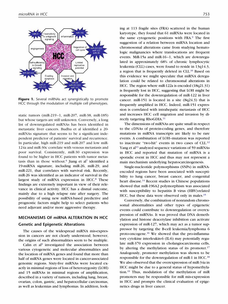
miRNAs might contribute to tumor progression, bymodulating the expression of gene products involvedin phenotypic characteristics of malignant cells, such asinvasion and uncontrolled growth (Figure 1). Mu-rakami et al observed that some miRNAs are associatedwith the degree of differentiation in hepatocellularcarcinoma. MiR-222, miR-106a, miR-92, miR-17–5p,miR-20, and miR-18 are increased in poorly versus mod-erately versus well-differentiated hepatomas.4 The find-ing of alterations in miRNA and target gene expressionin HCC and other cancers highlights potential mecha-nisms of tumor progression caused by a loss of ability tocontrol proliferation or by gain of ability to survive inhostile environments related to these genetic changesin tumor cells. A comparison between nonmetastaticversus metastatic HCCs performed by Budhu et al5
identified a few miRNAs that are upregulated in meta-
s in Oncology, Vol 38, No 6, December 2011, pp 752-763
fivmppn
sclella
ism
ebhswH
pi
microRNA in HCC 753
static tumors (miR-219–1, miR-207, miR-38, miR-185)but whose targets are still unknown. Conversely, a longlist of downregulated miRNAs has been identified inmetastatic liver cancers. Budhu et al identified a 20-miRNAs signature that seems to be a significant inde-pendent predictor of patients’ survival and recurrence.In particular, high miR-219 and miR-207 and low miR-124a and miR-30c correlate with venous metastasis andpoor survival. Consistently, miR-30 expression wasfound to be higher in HCC patients with tumor metas-tasis than in those without.6 Jiang et al7 identified a19-miRNA signature, including miR-26, miR-29, andmiR-221, that correlates with survival risk. Recently,miR-26 was identified as an indicator of survival in thelargest study of miRNA expression in HCC.8 These
ndings are extremely important in view of their rele-ance in clinical activity. HCC has a dismal outcome,ainly due to a high relapse rate after surgery. Theossibility of using new miRNA-based predictive andrognostic factors might help to select patients whoeed adjuvant and/or more aggressive therapy.
MECHANISMS OF miRNA ALTERATION IN HCC
Genetic and Epigenetic Alterations
The causes of the widespread miRNA mis-expres-sion in cancers are not clearly understood; however,the origins of such abnormalities seem to be multiple.
Calin et al9 investigated the association betweenvarious cytogenetic and molecular abnormalities andthe location of miRNA genes and found that more thanhalf of miRNA genes were located in cancer-associatedgenomic regions. Sixty-five miRNAs were located ex-actly in minimal regions of loss of heterozygosity (LOH)and 15 miRNAs in minimal regions of amplification,described in a variety of tumors, including lung, breast,ovarian, colon, gastric, and hepatocellular carcinomas,
Figure 1. Several miRNAs act synergistically to promoteHCC through the modulation of multiple cell phenotypes.
as well as leukemias and lymphomas. In addition, look- n
ing at 113 fragile sites (FRA) scattered in the humankaryotype, they found that 61 miRNAs were located inthe same cytogenetic positions with FRA.9 The firstuggestion of a relation between miRNA location andhromosomal alterations came from studying hemato-ogic malignancies where translocations are frequentvents. MiR-15a and miR-16–1, which are downregu-ated in approximately 68% of chronic lymphocyticeukemia (CLL) cases, were found to reside in 13q14.3,
region that is frequently deleted in CLL.10 Based onthis evidence we might speculate that miRNA deregu-lation could be related to chromosomal alterations inHCC. The region where miR-122a is encoded (18q21.31)is frequently lost in HCC, suggesting that LOH might beresponsible for the downregulation of miR-122 in livercancer. miR-151 is located in a site (8q24.3) that isfrequently amplified in HCC. Indeed, miR-151 expres-sion is correlated with intrahepatic metastasis of HCCand increases HCC cell migration and invasion by di-rectly targeting RhoGDIA.11
The dimensions of miRNAs are quite small in respectto the cDNAs of protein-coding genes, and thereforemutations in miRNA transcripts are likely to be rareevents. A combination of LOH–mutation was reportedto inactivate “two-hit” events in two cases of CLL.12
Yang et al13 analyzed sequence variations of 59 miRNAsn HCC and reported that mutation of miRNA is aporadic event in HCC and thus may not represent aain mechanism underlying hepatocarcinogenesis.Single-nucleotide polymorphisms (SNPs) in miRNA-
ncoded regions have been associated with suscepti-ility to lung cancer, breast cancer, and congenitaleart disease.14 Recent studies in Chinese populationshowed that miR-196A2 polymorphism was associatedith susceptibility to hepatitis B virus (HBV)-relatedCC, but these data were observed only in males.15
Conversely, the combination of nonrandom chromo-somal abnormalities and other types of epigeneticevents could contribute to downregulation or overex-pression of miRNAs. It was proved that DNA demeth-ylation and histone deacetylase inhibition can activateexpression of miR-127, which may act as a tumor sup-pressor by targeting the B-cell leukemia/lymphoma 6proto-oncogene.16 We showed that the pro-inflamma-tory cytokine interleukin-6 (IL-6) may potentially regu-late miR-370 expression in cholangiocarcinoma cells,by altering the methylation status of its promoter.17
Analogously, promoter methylation was shown to beresponsible for the downregulation of miR-1 in HCC.18
We also observed that the overexpression of miR-34a inHCC might be due to a general status of hypomethyla-tion.19 Thus, modulation of the methylation of miR
romoters may be responsible for miR mis-expressionn HCC and prompts the clinical evaluation of epige-
etics drugs in liver cancer.
Dndg
cgmgthHtie
is
ensbt
ipnolmm
bhm1mt
pceHtipmp
mta
754 C. Braconi et al
Viral Disease
Chronic infection with HBV predisposes to the de-velopment of HCC. A salient feature of these chronicinfections is the integration of subgenomic HBV-DNAfragments into many different locations within the hostDNA. HBV integration is considered one of the eventsinvolved in hepatocarcinogenesis. Several critical genesinvolved in control of cell growth and adhesion, such ascyclin A1, retinoic acid receptor, calmodulin 1, MLL2,SERCA-1, and p53, have been identified adjacent to inte-grated HBV-DNA. However, because HBV-DNA integratesinto random positions of the human genome, all HBV-DNA integration does not directly transform hepatocytes.Recently, the discovery of noncoding RNAs suggested thehypothesis that HBV-DNA integration can exert an onco-genetic role not only by promoting genetic instabilitywith alteration of protein encoding genes but also bymodulating the expression of regulatory genes such asmiRNAs.
HBV integration is associated with genetic instabilityat different locus sited near FRA.20 For example, HBV-
NA integrates at 1p36, near FRA1A. Liver dysplasticodules reportedly have gains in 1p and 1q,21 buturing tumor progression there is LOH in these re-ions,22 suggesting that the consequences of HBV inte-
gration and associated genetic instability are often dy-namic during carcinogenesis. MiR-34a is encoded onchromosome 1p36 and is frequently upregulated inHCC (Table 1). The mechanism by which this miRNA isupregulated is still under investigation. p53, whichseems to positively regulate miR-34 in other tumortypes, is often silenced in HCC and thus the relation-ship between miR-34a and p53 in HCC is not fullyunderstood. HBV-DNA integration near the miR-34a sitemight provide a further reasonable explanation for theupregulation of this miRNA in HCC. Likely, HBV inte-gration acts synergistically on different regulatorygenes that have an additional effect on cell prolifer-ation and transformation. In fact, integration atFRA1A may also silence expression of miRNA-200a,which is known to be decreased in HCC compared tonon-tumor tissue (Table 1).
Interestingly, other mechanisms involving miRNAmay contribute to interactions between HBV and hepa-tocytes. Jin et al23 have recently shown that HBV en-odes a miRNA in the precore–core region of itsenome. Although it was speculated that this viral (v)-iRNA could affect the expression pattern of host cell
enes, only the viral polymerase gene was experimen-ally found as its target. Therefore, conversely to whatappens to v-miRNAs encoded by other viruses, theBV-miRNA does not seem to participate in the hepa-
itis-induced transformation of hepatocytes. However,t might influence the capability of the virus to prolif-
rate and integrate into the host genome, modulatingndirectly the expression of cellular miRNAs as de-cribed above.
HBV-related HCCs have higher levels of miR-143xpression, whose induction seems to be caused byuclear factor kappaB (NF-�B) activation. miR-143 washown to promote HCC metastasis in an animal modely targeting the fibronectin type III domain containinghe 3B gene, which regulates cell motility.24
In the Western world, most cases of HCC are relatedto hepatitis C virus (HCV) infection. Remarkable efforthas been put into assessing the effects of miRNAs onHCV replication (see below). However, additional evi-dence shows that miRNA expression may be modu-lated by HCV. Several miRNA expression profiling stud-ies show that HCV-related HCCs have a differentmiRNA expression signature when compared to HCV-negative HCCs. Ura et al25observed that some miRNAs,ncluding miR-26 and miR-29, were commonly re-ressed in HBV- or HCV-related HCC in comparison toormal liver tissues. Conversely, the expression ofther miRNAs could differentiate HBV- from HCV-re-
ated HCCs, and thereby suggest differences in theechanisms by which these viruses may modulateiRNA expression.25 Studies in HCV-infected HCC cell
lines confirmed the HCV-dependent modulation ofmiRNA expression.26�28 Less clear is the mechanism
y which HCV modulates miRNA expression. Weave shown that overexpression of HCV-proteins canodulate miRNA expression. Upregulation of miR-
93b in HCV-positive cells may regulate apoptosis byodulating the expression of the anti-apoptotic pro-
ein Mcl-1.28
miR-155 was found to be overexpressed in HCV-replicating cells.26 Interestingly a correlation between
re-miR-155 (BIC) and serum HCV RNA has been re-ently described in HCV-infected patients. The lowestxpression of BIC was found in patients that eliminatedCV RNA from both serum and macrophages.29 Even
hough additional studies are warranted to evaluate thenteraction between HCV and miR-155, these data areromising for the development of novel miRNA-basedolecular markers in the assessment of HCV-positiveatients.
SELECTED miRNAs AND ROLE INPATHOGENESIS OF LIVER CANCERS
miR-221/-222
miR-221 and miR-222 are encoded in tandem from agene cluster located on chromosome X (Xp11.3) andhave identical 5= regions that enable them to target thesame genes.30 They behave as oncogenes in several
alignancies. In HCC miR-221 and miR-222 were foundo be significantly upregulated when compared withdjacent liver tissues (Table 1). Murakami et al4 com-
pared miRNA expression in tumors with different de-
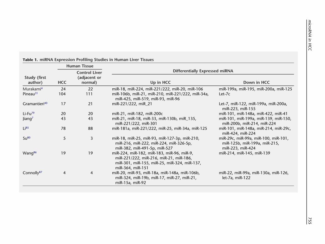
Table 1. miRNA Expression Profiling Studies in Human Liver Tissues
Study (firstauthor)
Human TissueDifferentially Expressed miRNA
HCC
Control Liver(adjacent or
normal) Up in HCC Down in HCC
Murakami4 24 22 miR-18, miR-224, miR-221/222, miR-20, miR-106 miR-199a, miR-195, miR-200a, miR-125Pineau31 104 111 miR-106b, miR-21, miR-210, miR-221/222, miR-34a,
miR-425, miR-519, miR-93, miR-96Let-7c
Gramantieri40 17 21 miR-221/222, miR_21 Let-7, miR-122, miR-199a, miR-200a,miR-223, miR-155
Li-Fu79 20 20 miR-21, miR-182, miR-200c miR-101, miR-148a, miR-422, miR-41Jiang7 43 43 miR-21, miR-18, miR-33, miR-130b, miR_135,
miR-221/222, miR-301miR-101, miR-199a, miR-139, miR-150,
miR-200b, miR-214, miR-224Li85 78 88 miR-181a, miR-221/222, miR-25, miR-34a, miR-125 miR-101, miR-148a, miR-214, miR-29c,
miR-424, miR-224Su80 5 3 miR-18, miR-25, miR-93, miR-127-3p, miR-210,
miR-216, miR-222, miR-224, miR-326-5p,miR-382, miR-491-5p, miR-527
miR-29c, miR-99a, miR-100, miR-101,miR-125b, miR-199a, miR-215,miR-223, miR-424
Wang86 19 19 miR-224, miR-182, miR-183, miR-96, miR-9,miR-221/222, miR-216, miR-21, miR-186,miR-301, miR-155, miR-25, miR-324, miR-137,miR-364, miR-151
miR-214, miR-145, miR-139
Connolly87 4 4 miR-20, miR-93, miR-18a, miR-148a, miR-106b,miR-324, miR-19b, miR-17, miR-27, miR-21,miR-15a, miR-92
miR-22, miR-99a, miR-130a, miR-126,let-7a, miR-122
microRN
Ain
HC
C755
vaCigtpaChphlwsdt
eHawrs
M
fs
spc
Celbti
docd
756 C. Braconi et al
grees of differentiation and found miR-222 as one ofthose miRNAs that significantly increases in poorly dif-ferentiated HCC. Recently, miR-221 and miR-222 werefound to be consistently dysregulated through eachstep going from human normal liver to cirrhosis to fullblown HCC.31 These miRNA enhance tumorigenesis initro and in vivo by regulating several targets. HCC hasdisrupted expression of p27Kip1, a member of theip/Kip family of cyclin-dependent kinase (CDK) inhib-
tors that function to negatively control cell cycle pro-ression.32 Recent data strongly support the hypothesishat miR-221/-222 regulate p27Kip1 expression at aost-transcriptional level.31,33 Cell cycle progression islso affected by miR-221 through the modulation ofDKN1C/p57, which was shown to correlate with aigher biological aggressiveness, advanced stage,oor differentiation, larger size, portal invasion, andigh proliferative activity in HCC. Furthermore, a
ow CDKN1C/p57 labeling index is associated withorse outcomes and lower disease-free survival after
urgery, suggesting that miR-221/-222–dependentownregulation of CDKN1C/p57 might contribute tohe progression of HCC.34
In addition to the modulation of cell cycle, miR-221contributes to the progression of HCC through theinhibition of apoptosis. Bmf is a pro-apoptotic memberof the Bcl-2 family that triggers caspase activation inHCC. Upregulation of miR-221 is responsible for thelack of Bmf expression in human HCC tissues.35 How-ver, the multiple targets of miR-221 may explain whyCCs with higher miR-221 levels were associated withshorter time to recurrence, whereas low Bmf levelsere associated with a trend toward shorter time to
ecurrence; this difference did not achieve statisticalignificance.35 Furthermore, miR-221/-222 can affect
the expression of DNA damage inducible transcript 4which is a modulator of the mammalian target of rapa-mycin (mTOR) pathway.31 Wong et al36 studied geneexpression profiling in HCC cells treated with anti–miR-222 and observed that Akt signaling was the majorpathway implicated in the miR-222–affected biologicalnetworks. They further proved protein phosphatase 2Asubunit B to be a target of miR-222.36 Garofalo et al37
showed the miR-221/-222 target PTEN and TIMP3tumor suppressors, and enhance cellular migrationthrough the activation of the AKT pathway and metal-lopeptidases. Thus, it is likely that miR-221/-222 can actsynergistically on several targets, which results in themodulation of the PI3K-akt-mTOR pathway.
The MET proto-oncogene encodes the tyrosine ki-nase receptor for hepatocyte growth factor (HGF), apotent mitogen and motogen for epithelial cells. MET isoverexpressed in HCC. Patients with high MET HCChave a significantly shorter 5-year survival than patientswith low MET HCC.38 Recent evidence suggests that
ET induces miR-221/-222 through AP-1 transcription
actor, providing the molecular basis for the prognosticignificance of miR-221/-222.37
miR-122
One of the most abundant miRNAs in the liver ismiR-122a. It is expressed in normal hepatocytes but isdownregulated in up to 70% of human HCCs (Table 1).MiR-122 is described as a liver-specific miRNA. Theexpression of miR-122 can be used as a marker of liverdevelopment and it undergoes liver-specific expressionduring embryonic development. A role for miR-122 inhepatocarcinogenesis is suggested by the differentialexpression of miR-122a in HCC versus non-tumor cir-rhotic hepatic tissue, and by the studies by Kutay et al39
of miRNA expression in murine HCC. In these studies,miR-122a was decreased by 50% between 36 and 54weeks of a folate-methyl–deficient diet, when neoplas-tic transformation also occurred. In fact, none of theseanimals that were switched to a folate-methyl–adequatediet after 36 weeks developed HCC or had aberrantexpression of miR-122a in the liver.
Deregulation of cell cycle progression can contrib-ute to hepatocarcinogenesis. Cyclin G1 promotes cellcycle progression and it may be associated withgenomic instability. In HCC cells, an inverse correlationhas been observed between miR122a and cyclin G1,which is a validated target of this miRNA. In experi-mental hepatocarcinogenesis, loss of cyclin G1 is asso-ciated with a significantly lower tumor incidence aftercarcinogenic challenges.40 Thus, deregulated expres-ion of cyclin G1, in response to altered miR122a ex-ression, could contribute to the pathogenesis of liverancer.
While the adult liver is the major site for miR-122,AT-1 (cationic amino acid transporter-1) is universallyxpressed in all adult mammalian tissues except theiver. During mouse embryonic development, miR-122ecomes expressed in the liver as CAT-1 mRNA beginso decrease, suggesting that CAT-1 mRNA could be ann vivo target for degradation directed by miR-122.41
CAT-1 is a carrier protein required in the regeneratingliver for the transport of cationic amino acids andpolyamines in the late G1 phase, a process that isessential for liver cells to enter mitosis.41 Given these
ata, modulation of expression of CAT-1 may be an-ther mechanism through which this miRNA regulatesell cycle in normal and transformed hepatocytes. Ad-itionaly studies by Bai et al42 identified several other
targets of miR-122, including ADAM10, SRF, and IGFR1.All of these proteins are upregulated in HCC and areinvolved in the modulation of cell adhesion, motility,and growth, providing additional evidence for the con-tribution of miR-122 to hepatocarcinogenesis.
These observations acquire clinical relevance inview of the correlation between overall survival and
miR-122 expression levels in human HCCs.43 Interest-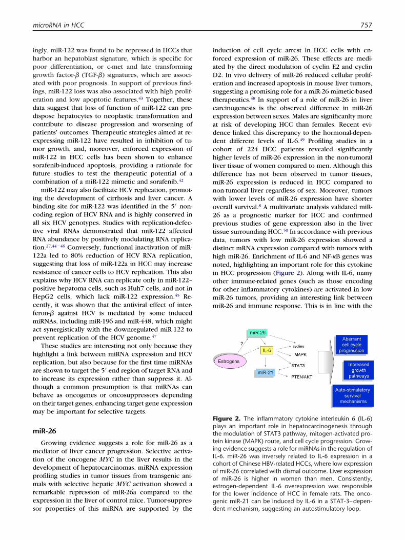
aieddcpemmsfc
cfmap
dpm
ceadd
2ptddhniofmm
microRNA in HCC 757
ingly, miR-122 was found to be repressed in HCCs thatharbor an hepatoblast signature, which is specific forpoor differentiation, or c-met and late transforminggrowth factor-� (TGF-�) signatures, which are associ-ted with poor prognosis. In support of previous find-ngs, miR-122 loss was also associated with high prolif-ration and low apoptotic features.43 Together, theseata suggest that loss of function of miR-122 can pre-ispose hepatocytes to neoplastic transformation andontribute to disease progression and worsening ofatients’ outcomes. Therapeutic strategies aimed at re-xpressing miR-122 have resulted in inhibition of tu-or growth, and, moreover, enforced expression ofiR-122 in HCC cells has been shown to enhance
orafenib-induced apoptosis, providing a rationale foruture studies to test the therapeutic potential of aombination of a miR-122 mimetic and sorafenib.42
miR-122 may also facilitate HCV replication, promot-ing the development of cirrhosis and liver cancer. Abinding site for miR-122 was identified in the 5= non-coding region of HCV RNA and is highly conserved inall six HCV genotypes. Studies with replication-defec-tive viral RNAs demonstrated that miR-122 affectedRNA abundance by positively modulating RNA replica-tion.27,44�46 Conversely, functional inactivation of miR-122a led to 80% reduction of HCV RNA replication,suggesting that loss of miR-122a in HCC may increaseresistance of cancer cells to HCV replication. This alsoexplains why HCV RNA can replicate only in miR-122–positive hepatoma cells, such as Huh7 cells, and not inHepG2 cells, which lack miR-122 expression.45 Re-ently, it was shown that the antiviral effect of inter-eron-� against HCV is mediated by some inducediRNAs, including miR-196 and miR-448, which might
ct synergistically with the downregulated miR-122 torevent replication of the HCV genome.47
These studies are interesting not only because theyhighlight a link between miRNA expression and HCVreplication, but also because for the first time miRNAsare shown to target the 5=-end region of target RNA andto increase its expression rather than suppress it. Al-though a common presumption is that miRNAs canbehave as oncogenes or oncosuppressors dependingon their target genes, enhancing target gene expressionmay be important for selective targets.
miR-26
Growing evidence suggests a role for miR-26 as amediator of liver cancer progression. Selective activa-tion of the oncogene MYC in the liver results in the
evelopment of hepatocarcinomas. miRNA expressionrofiling studies in tumor tissues from transgenic ani-als with selective hepatic MYC activation showed a
remarkable repression of miR-26a compared to theexpression in the liver of control mice. Tumor-suppres-
sor properties of this miRNA are supported by theinduction of cell cycle arrest in HCC cells with en-forced expression of miR-26. These effects are medi-ated by the direct modulation of cyclin E2 and cyclinD2. In vivo delivery of miR-26 reduced cellular prolif-eration and increased apoptosis in mouse liver tumors,suggesting a promising role for a miR-26 mimetic-basedtherapeutics.48 In support of a role of miR-26 in liverarcinogenesis is the observed difference in miR-26xpression between sexes. Males are significanlty moret risk of developing HCC than females. Recent evi-ence linked this discrepancy to the hormonal-depen-ent different levels of IL-6.49 Profiling studies in a
cohort of 224 HCC patients revealed significantlyhigher levels of miR-26 expression in the non-tumoralliver tissue of women compared to men. Although thisdifference has not been observed in tumor tissues,miR-26 expression is reduced in HCC compared tonon-tumoral liver regardless of sex. Moreover, tumorswith lower levels of miR-26 expression have shorteroverall survival.8 A multivariate analysis validated miR-6 as a prognostic marker for HCC and confirmedrevious studies of gene expression also in the liverissue surrounding HCC.50 In accordance with previousata, tumors with low miR-26 expression showed aistinct mRNA expression compared with tumors withigh miR-26. Enrichment of IL-6 and NF-�B genes wasoted, highlighting an important role for this cytokine
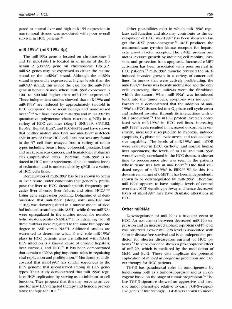
n HCC progression (Figure 2). Along with IL-6, manyther immune-related genes (such as those encodingor other inflammatory cytokines) are activated in lowiR-26 tumors, providing an interesting link betweeniR-26 and immune response. This is in line with the
Figure 2. The inflammatory cytokine interleukin 6 (IL-6)plays an important role in hepatocarcinogenesis throughthe modulation of STAT3 pathway, mitogen-activated pro-tein kinase (MAPK) route, and cell cycle progression. Grow-ing evidence suggests a role for miRNAs in the regulation ofIL-6. miR-26 was inversely related to IL-6 expression in acohort of Chinese HBV-related HCCs, where low expressionof miR-26 correlated with dismal outcome. Liver expressionof miR-26 is higher in women than men. Consistently,estrogen-dependent IL-6 overexpression was responsiblefor the lower incidence of HCC in female rats. The onco-genic miR-21 can be induced by IL-6 in a STAT-3–depen-
dent mechanism, suggesting an autostimulatory loop.
mfcr
rpirmtsAhal
iw
scioop
tttg
lmt
ptd
toisammtbrksd
l1tasit
758 C. Braconi et al
correlation found between miR-26 expression and theresponse to interferon-� in HBV-related HCC.8 Thus,
iR-26 might represent a promising prognostic markeror HCC, able to distinguish those tumors that areharacterized by a poorer outcome but a favorableesponse to adjuvant therapy with interferon-�.
miR-21
miR-21 is consistently identified as being signifi-cantly overexpressed in HCC cells when compared tonormal liver and cirrhotic cells both in animals and inhumans. An important role for miR-21 in growth regu-lation is suggested by studies in which it was shown toregulate cell cycle progression and apoptosis.51,52 Theole of miR-21 has been studied in several differentathologic conditions and processes that are character-
zed by an increased cell proliferation. However, theole of aberrant expression of miR-21 has been studiedost widely in tumors. The contribution of this miR-21
o the pathogenesis of cancer is suggested by studieshowing deregulated expression in several neoplasms.berrant expression has been observed in cervical,ead and neck, pancreatic, and breast cancers, as wells in glioblastoma, cholangiocarcinoma, CLL, diffusearge B-cell lymphoma, and other tumors.53 In HCC,miR-21 is highly overexpressed (Table 1) and can con-tribute to tumor growth and spread by modulating theexpression of gene products involved in phenotypiccharacteristics of cancer cells such as cell growth, mi-gration, and invasion.51 miR-21 was found to be in-creased in the early stages of liver regeneration, sug-gesting a contribution of this miRNA to hepatocyteproliferation.54
miR-21 was shown to target PTEN in HCC cells.PTEN is a tumor-suppressor gene that is frequentlydecreased in liver cancer and correlates with poorprognosis. Overexpression of miR-21 was found to si-lence PTEN, leading to phosphorylation of FAK andmetalloproteinases, which resulted in promotion ofcell migration and invasion.51 Other targets of miR-21nclude the programmed cell death 4 (PDCD4) gene,
hich is reduced in HCC; peli-1, which regulates NF-�Bsignaling; and tropomyosin 1, a tumor-suppressor pro-tein reduced in HCV-associated HCC.52,55
The anti-apoptotic activity of miR-21 is also sup-ported by the increased expression of this miRNA fol-lowing cytotoxic drugs. In fact, we have previouslyshown that miR-21 modulates gemcitabine-induced ap-optosis by PTEN-dependent activation of PI3-kinase sig-naling in cholangiocarcinoma cells.56 Rossi et al57 havehown that 5-fluorouracil (5-FU) upregulates miR-21 inolorectal cancer cells and suggested that the drug-nduced miRNA dysregulation could be the expressionf cellular response to the toxic effects of 5-FU. In viewf these results, miR-21 might be further studied as a
otential target of therapeutic strategies. The combina-ion of anti–miR-21 therapy with more classical chemo-herapy agents might help to overcome chemoresis-ance, promote cell death, and limit liver cancerrowth and metastasis.
HCC represents a classical case of inflammation-inked cancer. IL-6 is a multifunctional cytokine that
ediates the hepatic response to systemic inflamma-ion and likely contributes to hepatocarcinogenesis.58
High serum level of IL-6 seems to be a risk factor for thedevelopment of HCC.49,59 Recent data that postulate a
otential role for miR-21 in the regulation of inflamma-ory cytokine production and the evidence of an IL-6–ependent induction of miR-21 in myeloma cells60 sug-
gest a link between overexpression of miR-21 andcytokines in the development and maintenance of livercancer.
miR-155
niR-155 was found within the BIC gene on chromo-some 21 in humans. The genomic structure of humanBIC consists of three exons, However, it lacks a largeopen reading frame and therefore its sole function maybe to give rise to miR-155 encoded within exon 3.MiR-155 is involved in regulating the innate immuneresponse. It is induced by inflammatory stimuli such asthe bacterial endotoxin61 and it has been identified as aarget of synthetic viral intermediates and of interfer-n-�, the host antiviral response cytokine.62 These data
ndicate that immune cells, such as macrophages, re-pond to viral cues by strongly upregulating miR-155nd suggest that miR-155 is a component of the pri-ary macrophage response to different types of inflam-atory mediators. It appears that miR-155 exerts con-
rol of the mammalian immune system, at least in part,y regulating cytokine production.63 miR-155 is up-egulated in diffuse large B-cell lymphoma, CLL, Hodg-in disease, and pancreatic and colorectal cancers,howing that it can behave as an oncogene in severalifferent malignancies.64
HCC frequently develops in a background of chronicinflammatory disease of the liver. As a component ofthe inflammatory response, miR-155 links inflammationand cancer. miR-155 was upregulated in mice treatedwith a choline-deficient, low-methionine amino acid(CDAA) diet, which induces steatohepatitis at earlystages and leads to the formation of pre-neoplasticnodules after 65 weeks and hepatocellular adenomasand carcinomas after 84 weeks.65 miR-155 expressionevel correlated positively with the number of miR-55–positive cells, as well as the extent of inflamma-ion in these mice, suggesting that miR-155 could play
causal role in CDAA diet–induced pathogenesis. Inupport of this hypothesis, miR-155 was found to bencreased in human HCC, while its tumor-suppressorarget C/EBPB was reduced.65 In addition, miR-155 was
significantly upregulated in chronic liver disease com-
cHtlfnt
tcmsaHilmcwb
ddsmolH
oMac
fcls
microRNA in HCC 759
pared to normal liver and high miR-155 expression innon-tumoral tissues was associated with poor overallsurvival in HCC patients.66
miR-199a* (miR-199a-3p)
The miR-199a gene is located on chromosomes 1and 19. miR-199a-1 is located in an intron of the Dy-namin 2 (DNM2) gene on chromosome 19p13.2.miRNA genes may be expressed as either the maturestrand or the miRNA* strand. Although the miRNAstrand is generally expressed at higher levels than themiRNA* strand, this is not the case for the miR-199agene in hepatic tissues, where miR-199a* expression is100- to 300-fold higher than miR-199a expression.7
Three independent studies showed that miR-199a andmiR-199a* are reduced by approximately twofold inHCC compared to adjacent benign and nondiseasedliver.4,7,40 We have assayed miR-199a and miR-199a* byquantitative polymerase chain reaction (qPCR) in avariety of HCC cell lines (Skep-1, SNU449, SNU182,HepG2, Hep38, Huh7, and PLC/PRF5) and have shownthat neither mature miR-199a nor miR-199a* is detect-able in any of these HCC cell lines nor was any foundin the 37 cell lines assayed from a variety of tumortypes including breast, lung, colorectal, prostate, headand neck, pancreas cancers, and hematologic malignan-cies (unpublished data). Therefore, miR-199a* is re-duced in HCC tumor specimens, albeit at modest levelsof reduction, and is undetectable by qPCR in a varietyof HCC cells lines.
Deregulation of miR-199a* has been shown to occurin liver tissue under conditions that generally predis-pose the liver to HCC. Steatohepatitis frequently pre-cedes liver fibrosis, liver failure, and often HCC.67,68
Using gene expression profiling, Dolganiuc et al dem-onstrated that miR-199a* (along with miR-182 and�183) was downregulated in a murine model of alco-hol-induced steatohepatitis (ASH), while three miRNAswere upregulated in the murine model for nonalco-holic steatohepatitis (NASH).69 It is intriguing that allthree miRNAs were regulated to the same but oppositedegree in ASH versus NASH. Additional studies arewarranted to determine what, if any, role miR-199a*plays in HCC patients who are inflicted with NASH.HCV infection is a known cause of chronic hepatitis,liver cirrhosis, and HCC.70 It has been demonstratedthat certain miRNAs play important roles in regulatingviral replication and proliferation.45 Murakami et al dis-overed that miR-199a* has similar sequences to theCV genome that is conserved among all HCV geno-
ypes. Their study demonstrated that miR-199a* regu-ates HCV replication by serving as an inhibitor to cellunction. They propose that this may serve as an ave-ue for new HCV-targeted therapy and hence a preven-
ative therapy for HCC.71Other possibilities exist in which miR-199a* regu-lates cell function and also may contribute to the de-velopment of HCC. miR-199a* has been shown to tar-get the MET proto-oncogene.72 MET produces theransmembrane tyrosine kinase receptor for hepato-yte growth factor receptor. The c-MET protein pro-otes invasive growth by inducing cell motility, inva-
ion, and protection from apoptosis. Increased c-METctivation has been associated with poor survival inCC patients.73 miR-199a* mimetic reversed the MET-
nduced invasive growth in a variety of cancer cellines. In tumors that were actively proliferating, the
iR-199a/a* locus was heavily methylated and the onlyells expressing these miRNAs were the fibroblastsithin the tumor. When miR-199a* was introducedack into the tumor cells, apoptosis was induced.72
Fornari et al demonstrated that the addition of miR-199a* to HCC tissues led to a G1-phase cell cycle arrestand reduced invasion through its interactions with c-MET production.74 The mTOR protein inversely corre-lated with miR-199a* in HCC cell lines. IncreasingmiR-199a* levels resulted in increased doxorubicin sen-sitivity, increased susceptibility to hypoxia, inducedapoptosis, G1-phase cell cycle arrest, and reduced inva-sive capability. The levels of miR-199a* and mTORwere evaluated in HCC, cirrhotic, and normal humanliver specimens; the levels of mTOR and miR-199a*were inversely correlated in the HCC tissues. A shortertime to reoccurrence also was seen in the patientswhose tissue was low in miR-199a*.74 One final vali-
ated target of miR-199a* is ERK.72 While this is aownstream target of c-MET, it has been independentlyhown to be downregulated by miR-199a*. ThereforeiR-199a* appears to have multiple levels of control
ver the c-MET signaling pathway and hence decreasedevels of miR-199a* may have dramatic alterations inCC.
Other miRNAs
Downregulation of miR-29 is a frequent event inHCC. An association between decreased miR-29b ex-pression and an increased alpha-fetoprotein (AFP) levelwas observed. Lower miR-29b level is associated withshorter disease-free survival and is an independent pre-dictor for shorter disease-free survival of HCC pa-tients.75 In vitro evidence shows a pro-apoptotic effect
f miR-29, which is mediated by the modulation ofcl-1 and Bcl-2. These data implicate the potential
pplication of miR-29 in prognosis prediction and can-er therapy for HCC patients.
TGF-� has paradoxical roles in tumorigenesis byunctioning both as a tumor-suppressor and as an on-ogene based on the stage of tumor progression.76 Theate TGF-� signature showed an aggressive and inva-ive tumor phenotype relative to early TGF-�–respon-
sive genes.43 Interestingly, TGF-� was shown to modu-
gtpcom1c
amm
tc
saorm
760 C. Braconi et al
late also miRNA expression. In particular, miR-181 isinduced by TGF-�, likely as result of a late TGF-�response (afer 24 hours).77 Indeed, miR-181 has onco-enic properties that mediate liver carcinogenesishrough the TIMP-3–mediated modulation of metallo-roteinases and enhanced chemoresistance of HCCells.77 Along with miR-181, TGF-� can also modulatether miRNAs that may be involved in HCC, such asiR-23, miR-27, miR-24, miR-200, miR-21, miR-99, miR-
06, miR-125, miR-101, miR-222, and miR-193. In ac-ordance with TGF-�’s effects on prognosis, miR-23a,
miR-27a, and miR-24 closely correlated with intrahe-patic metastasis of HCC. Similarly, HCC tissues withliver cirrhosis also had a higher expression of miR-23aand miR-27a compared with those without liver cirrho-sis.78
miR-101 is a tumor-suppressor miRNA that modu-lates cell migration and cell growth by acting on severaltargets including FOS and Mcl-1.79,80
Members of the Let-7 family have been found to bereduced in HCC.81 In particular, Let-7g was shown toct as a tumor-suppressor gene in liver cancer cells, byodulating the ability of the cells to proliferate and toigrate.81,82 Indeed, Let-7g was reduced in human HCC
with metastasis in comparison with those with no dis-tant metastasis and was predictive of poor survival inHCC patients.81 These effects seem to be mediated byhe targeting of several genes, including Bcl-xL, type 1ollagen alpha2, and likely c-Myc.81�83
miRNA IN DIAGNOSIS AND PROGNOSIS OFHCC
Several recent studies have been performed ofmiRNA profiling in human HCC. Similar to observationsin other cancers, these studies have revealed alteredexpression of several miRNAs (Table 1). Further defi-nition of the miRNAs that are aberrantly expressed inHCC and elucidation of their contribution to the patho-physiology of HCC is likely to be useful in establishinga role for miRNA. On the basis of these profiling stud-ies, signatures that are associated with cancer or pre-cancerous changes can be identified. miRNA expres-sion has been studied in HCC tissues in comparisonwith adjacent non-neoplastic liver tissues. Among themiRNAs that are aberrantly expressed there are severalthat are increased in expression, including miR-21,miR-221/-222, and miR-181, whereas others are signif-icantly decreased in expression, such as miR-122, miR-101, miR-29, miR-200, miR-148a, and mir-199a (Table1). Thus, expression profiling in the absence of a de-fined role may be useful for the diagnosis of HCC or indefining prognosis. Moreover, identification of a role ofaberrantly expressed miRNAs in cellular processes as-sociated with cancer such as proliferation, apoptosis,
or deregulated intracellular signaling is likely to provideadditional insight into the pathogenesis of HCC thatmay eventually yield newer approaches to therapy.
miRNA AS THERAPEUTIC TARGETS FOR HCC
A therapeutic approach for HCC that targets miRNAwould be highly innovative. Such an approach wouldbe promising given the evidence to date of the involve-ment of miRNA in HCC pathogenesis and biology. Sys-temic administration of antisense LNA (locked nucleicacid) oligonucleotides to miR-122 was recently shownto modulate miRNA and target gene expression in theliver and result in the loss of HCV with minimaltoxicities in a non-human primate.84 This studyhowed the feasibility of this approach for targetingberrantly overexpressed miRNA in the liver. In an-ther recent study, the therapeutic value of a miRNAeplacement strategy was described for miR-26a, aiRNA whose expression is frequently lost in HCC.48
miR-26a can target cyclins D2 and E2 and induce G1
arrest when expressed in human liver cancer cells.Using a mouse model of HCC, miR-26 replacementdelivered using a self-complementary adeno-associatedvirus vector dramatically suppressed disease progres-sion. Tumor-specific apoptosis was activated. Thesefindings support the potential of targeting tumor-sup-pressing miRNAs or aberrantly expressed miRNA as apowerful and highly specific anticancer therapeuticmodality for HCC.
REFERENCES1. Edwards BK, Ward E, Kohler BA, et al. Annual report to
the nation on the status of cancer, 1975–2006, featuringcolorectal cancer trends and impact of interventions(risk factors, screening, and treatment) to reduce futurerates. Cancer. 2010;116:544–73.
2. Cullen BR. Transcription and processing of human mi-croRNA precursors. Mol Cell. 2004;16:861–5.
3. Bartel DP. MicroRNAs: genomics, biogenesis, mecha-nism, and function. Cell. 2004;116:281–97.
4. Murakami Y, Yasuda T, Saigo K, et al. Comprehensiveanalysis of microRNA expression patterns in hepatocel-lular carcinoma and non-tumorous tissues. Oncogene.2006;25:2537–45.
5. Budhu A, Jia HL, Forgues M, et al. Identification of me-tastasis-related microRNAs in hepatocellular carcinoma.Hepatology. 2008;47:897–907.
6. Yao J, Liang L, Huang S, et al. MicroRNA-30d promotestumor invasion and metastasis by targeting Galphai2 inhepatocellular carcinoma. Hepatology. 2010;51:846–56.
7. Jiang J, Gusev Y, Aderca I, et al. Association of MicroRNAexpression in hepatocellular carcinomas with hepatitisinfection, cirrhosis, and patient survival. Clin CancerRes. 2008;14:419–27.
8. Ji J, Shi J, Budhu A, et al. MicroRNA expression, survival,and response to interferon in liver cancer. N Engl J Med.2009;361:1437–47.
9. Calin GA, Sevignani C, Dumitru CD, et al. Human mi-
croRNA genes are frequently located at fragile sites and
microRNA in HCC 761
genomic regions involved in cancers. Proc Natl Acad SciU S A. 2004;101:2999–3004.
10. Calin GA, Dumitru CD, Shimizu M, et al. Frequent dele-tions and down-regulation of micro- RNA genes miR15and miR16 at 13q14 in chronic lymphocytic leukemia.Proc Natl Acad Sci U S A. 2002;99:15524–9.
11. Ding J, Huang S, Wu S, et al. Gain of miR-151 on chro-mosome 8q24.3 facilitates tumour cell migration andspreading through downregulating RhoGDIA. Nat CellBiol. 2010;12:390–9.
12. Calin GA, Ferracin M, Cimmino A, et al. A MicroRNAsignature associated with prognosis and progression inchronic lymphocytic leukemia. N Engl J Med. 2005;353:1793–801.
13. Yang J, Zhou F, Xu T, et al. Analysis of sequence varia-tions in 59 microRNAs in hepatocellular carcinomas.Mutat Res. 2008;638:205–9.
14. Fasanaro P, Greco S, Ivan M, Capogrossi MC, Martelli F.microRNA: emerging therapeutic targets in acute isch-emic diseases. Pharmacol Ther. 2010;125:92–104.
15. Qi P, Dou TH, Zhou FG, Gu X, Wang H, Gao CF. Asso-ciation of a variant in MIR 196A2 with susceptibility tohepatocellular carcinoma in male Chinese patients withchronic hepatitis B virus infection. Hum Immunol. 2010;71:621–6.
16. Saito Y, Liang G, Egger G, et al. Specific activation ofmicroRNA-127 with downregulation of the proto-onco-gene BCL6 by chromatin-modifying drugs in human can-cer cells. Cancer Cell. 2006;9:435–43.
17. Meng F, Wehbe-Janek H, Henson R, Smith H, Patel T.Epigenetic regulation of microRNA-370 by interleukin-6in malignant human cholangiocytes. Oncogene. 2008;27:378–86.
18. Datta J, Kutay H, Nasser MW, et al. Methylation mediatedsilencing of MicroRNA-1 gene and its role in hepatocel-lular carcinogenesis. Cancer Res. 2008;68:5049–58.
19. Meng FY, Braconi C, Swenson E, Khrapenko L, Patel T.Epigenetic activation of miR-34a contributes to an inva-sive phenotype in hepatocellular cancer. Hepatology.2008;48:387A–8A.
20. Feitelson MA, Lee J. Hepatitis B virus integration, fragilesites, and hepatocarcinogenesis. Cancer Lett. 2007;252:157–70.
21. Raidl M, Pirker C, Schulte-Hermann R, et al. Multiplechromosomal abnormalities in human liver (pre)neopla-sia. J Hepatol. 2004;40:660–8.
22. Zondervan PE, Wink J, Alers JC, et al. Molecular cytoge-netic evaluation of virus-associated and non-viral hepato-cellular carcinoma: analysis of 26 carcinomas and 12concurrent dysplasias. J Pathol. 2000;192:207–15.
23. Jin WB, Wu FL, Kong D, Guo AG. HBV-encoded mi-croRNA candidate and its target. Comput Biol Chem.2007;31:124–6.
24. Zhang X, Liu S, Hu T, Liu S, He Y, Sun S. Up-regulatedmicroRNA-143 transcribed by nuclear factor kappa Benhances hepatocarcinoma metastasis by repressing fi-bronectin expression. Hepatology. 2009;50:490–9.
25. Ura S, Honda M, Yamashita T, et al. Differential mi-croRNA expression between hepatitis B and hepatitis Cleading disease progression to hepatocellular carcinoma.Hepatology. 2009;49:1098–112.
26. Liu X, Wang T, Wakita T, Yang W. Systematic identifica-
tion of microRNA and messenger RNA profiles inhepatitis C virus-infected human hepatoma cells. Virol-ogy. 2010;398:57–67.
27. Randall G, Grakoui A, Rice CM. Clearance of replicatinghepatitis C virus replicon RNAs in cell culture by smallinterfering RNAs. Proc Natl Acad Sci U S A. 2003;100:235–40.
28. Braconi C, Valeri N, Gasparini P, et al. Hepatitis C virusproteins modulate microRNA expression and chemosen-sitivity in malignant hepatocytes. Clin Cancer Res. 2010;16:957–66.
29. Sidorkiewicz M, Grek M, Jozwiak B, Majda-StanislawskaE, Piekarska A, Bartkowiak J. Expression of microRNA-155 precursor in peripheral blood mononuclear cellsfrom hepatitis Cpatients after antiviral treatment. ActaVirol. 2010;54:75–8.
30. le SC, Nagel R, Egan DA, et al. Regulation of thep27(Kip1) tumor suppressor by miR-221 and miR-222promotes cancer cell proliferation. EMBO J. 2007;26:3699–708.
31. Pineau P, Volinia S, McJunkin K, et al. miR-221 overex-pression contributes to liver tumorigenesis. Proc NatlAcad Sci U S A. 2010;107:264–9.
32. Koff A. How to decrease p27Kip1 levels during tumordevelopment. Cancer Cell. 2006;9:75–6.
33. Visone R, Russo L, Pallante P, et al. MicroRNAs (miR)-221and miR-222, both overexpressed in human thyroid pap-illary carcinomas, regulate p27Kip1 protein levels andcell cycle. Endocr Relat Cancer. 2007;14:791–8.
34. Fornari F, Gramantieri L, Ferracin M, et al. MiR-221 con-trols CDKN1C/p57 and CDKN1B/p27 expression inhuman hepatocellular carcinoma. Oncogene. 2008;27:5651–61.
35. Gramantieri L, Fornari F, Ferracin M, et al. MicroRNA-221targets Bmf in hepatocellular carcinoma and correlateswith tumor multifocality. Clin Cancer Res. 2009;15:5073–81.
36. Wong QW, Ching AK, Chan AW, et al. MiR-222 overex-pression confers cell migratory advantages in hepatocel-lular carcinoma through enhancing AKT signaling. ClinCancer Res. 2010;16:867–75.
37. Garofalo M, Di LG, Romano G, et al. miR-221&222 reg-ulate TRAIL resistance and enhance tumorigenicitythrough PTEN and TIMP3 downregulation. Cancer Cell.2009;16:498–509.
38. Ueki T, Fujimoto J, Suzuki T, Yamamoto H, Okamoto E.Expression of hepatocyte growth factor and its receptorc-met proto-oncogene in hepatocellular carcinoma.Hepatology. 1997;25:862–6.
39. Kutay H, Bai S, Datta J, et al. Downregulation of miR-122in the rodent and human hepatocellular carcinomas.J Cell Biochem. 2006;99:671–8.
40. Gramantieri L, Ferracin M, Fornari F, et al. Cyclin G1 is atarget of miR-122a, a microRNA frequently down-regu-lated in human hepatocellular carcinoma. Cancer Res.2007;67:6092–9.
41. Chang J, Nicolas E, Marks D, et al. miR-122, a mammalianliver-specific microRNA, is processed from hcr mRNAand may downregulate the high affinity cationic aminoacid transporter CAT-1. RNA Biol. 2004;1:106–13.
42. Bai S, Nasser MW, Wang B, et al. MicroRNA-122 inhibits
tumorigenic properties of hepatocellular carcinoma cells
762 C. Braconi et al
and sensitizes these cells to sorafenib. J Biol Chem.2009;284:32015–27.
43. Coulouarn C, Factor VM, Andersen JB, Durkin ME, Thor-geirsson SS. Loss of miR-122 expression in liver cancercorrelates with suppression of the hepatic phenotypeand gain of metastatic properties. Oncogene. 2009;28:3526–36.
44. Henke JI, Goergen D, Zheng J, et al. microRNA-122stimulates translation of hepatitis C virus RNA. EMBO J.2008;27:3300–10.
45. Jopling CL, Norman KL, Sarnow P. Positive and negativemodulation of viral and cellular mRNAs by liver-specificmicroRNA miR-122. Cold Spring Harb Symp Quant Biol.2006;71:369–76.
46. Zhang R, Wang L, Yu GR, Zhang X, Yao LB, Yang AG.MicroRNA-122 might be a double-edged sword in hepa-tocellular carcinoma. Hepatology. 2009;50:1322–3.
47. Pedersen IM, Cheng G, Wieland S, et al. Interferon mod-ulation of cellular microRNAs as an antiviral mechanism.Nature. 2007;449:919–22.
48. Kota J, Chivukula RR, O’Donnell KA, et al. TherapeuticmicroRNA delivery suppresses tumorigenesis in a mu-rine liver cancer model. Cell. 2009;137:1005–17.
49. Naugler WE, Sakurai T, Kim S, et al. Gender disparity inliver cancer due to sex differences in MyD88-dependentIL-6 production. Science. 2007;317:121–4.
50. Llovet JM, Ricci S, Mazzaferro V, et al. Sorafenib inadvanced hepatocellular carcinoma. N Engl J Med. 2008;359:378–90.
51. Meng F, Henson R, Wehbe-Janek H, Ghoshal K, Jacob ST,Patel T. MicroRNA-21 regulates expression of the PTENtumor suppressor gene in human hepatocellular cancer.Gastroenterology. 2007;133:647–58.
52. Zhang H, Ozaki I, Mizuta T, et al. Involvement of pro-grammed cell death 4 in transforming growth factor-beta1-induced apoptosis in human hepatocellular carci-noma. Oncogene. 2006;25:6101–12.
53. Selcuklu SD, Donoghue MT, Spillane C. miR-21 as a keyregulator of oncogenic processes. Biochem Soc Trans.2009;37:918–25.
54. Marquez RT, Wendlandt E, Galle CS, Keck K, McCaffreyAP. MicroRNA-21 is upregulated during the proliferativephase of liver regeneration, targets Pellino-1, and inhibitsNF-kappaB signaling. Am J Physiol Gastrointest LiverPhysiol. 2010;298:G535–41.
55. Zhu S, Si ML, Wu H, Mo YY. MicroRNA-21 targets thetumor suppressor gene tropomyosin 1 (TPM1). J BiolChem. 2007;282:14328–36.
56. Meng F, Henson R, Lang M, et al. Involvement of humanmicro-RNA in growth and response to chemotherapy inhuman cholangiocarcinoma cell lines. Gastroenterology.2006;130:2113–29.
57. Rossi L, Bonmassar E, Faraoni I. Modification of miR geneexpression pattern in human colon cancer cells follow-ing exposure to 5-fluorouracil in vitro. Pharmacol Res.2007;56:248–53.
58. Moran DM, Mattocks MA, Cahill PA, Koniaris LG,McKillop IH. Interleukin-6 mediates G(0)/G(1) growtharrest in hepatocellular carcinoma through a STAT 3-de-pendent pathway. J Surg Res. 2008;147:23–33.
59. Soresi M, Giannitrapani L, D’Antona F, et al. Interleukin-6
and its soluble receptor in patients with liver cirrhosisand hepatocellular carcinoma. World J Gastroenterol.2006;12:2563–8.
60. Loffler D, Brocke-Heidrich K, Pfeifer G, et al. Interleu-kin-6 dependent survival of multiple myeloma cells in-volves the Stat3-mediated induction of microRNA-21through a highly conserved enhancer. Blood. 2007;110:1330–3.
61. Tili E, Michaille JJ, Cimino A, et al. Modulation of miR-155 and miR-125b levels following lipopolysaccharide/TNF-alpha stimulation and their possible roles in regulat-ing the response to endotoxin shock. J Immunol. 2007;179:5082–9.
62. O’Connell RM, Taganov KD, Boldin MP, Cheng G, Balti-more D. MicroRNA-155 is induced during the macro-phage inflammatory response. Proc Natl Acad Sci U S A.2007;104:1604–9.
63. Thai TH, Calado DP, Casola S, et al. Regulation of thegerminal center response by microRNA-155. Science.2007;316:604–8.
64. Faraoni I, Antonetti FR, Cardone J, Bonmassar E. miR-155gene: a typical multifunctional microRNA. Biochim Bio-phys Acta. 2009;1792:497–505.
65. Wang B, Majumder S, Nuovo G, et al. Role of microRNA-155 at early stages of hepatocarcinogenesis induced bycholine-deficient and amino acid-defined diet in C57BL/6mice. Hepatology. 2009;50:1152–61.
66. Budhu A, Yu Z, Forgues M, Tang ZY, Croce C, Wang XW.Immune cell-related MICRORNA-155 is associated withhuman liver cirrhosis and hepatocellular carcinoma.ILCA Annual Conference (abstract). Milan, Italy, Sept4–6, 2009.
67. Bugianesi E, Leone N, Vanni E, et al. Expanding thenatural history of nonalcoholic steatohepatitis: fromcryptogenic cirrhosis to hepatocellular carcinoma. Gas-troenterology. 2002;123:134–40.
68. Morgan TR, Mandayam S, Jamal MM. Alcohol and hep-atocellular carcinoma. Gastroenterology. 2004;127:S87–96.
69. Dolganiuc A, Petrasek J, Kodys K, et al. MicroRNA ex-pression profile in Lieber-DeCarli diet-induced alcoholicand methionine choline deficient diet-induced nonalco-holic steatohepatitis models in mice. Alcohol Clin ExpRes. 2009;33:1704–10.
70. Wasley A, Alter MJ. Epidemiology of hepatitis C: geo-graphic differences and temporal trends. Semin LiverDis. 2000;20:1–16.
71. Murakami Y, Aly HH, Tajima A, Inoue I, Shimotohno K.Regulation of the hepatitis C virus genome replication bymiR-199a. J Hepatol. 2009;50:453–60.
72. Kim S, Lee UJ, Kim MN, et al. MicroRNA miR-199a*regulates the MET proto-oncogene and the downstreamextracellular signal-regulated kinase 2 (ERK2). J BiolChem. 2008;283:18158–66.
73. Kaposi-Novak P, Lee JS, Gomez-Quiroz L, Coulouarn C,Factor VM, Thorgeirsson SS. Met-regulated expressionsignature defines a subset of human hepatocellular car-cinomas with poor prognosis and aggressive phenotype.J Clin Invest. 2006;116:1582–95.
74. Fornari F, Milazzo M, Chieco P, et al. MiR-199a–3p reg-ulates mTOR and c-Met to influence the doxorubicinsensitivity of human hepatocarcinoma cells. Cancer Res.
2010;70:5184–93.
8
8
8
8
8
8
microRNA in HCC 763
75. Xiong Y, Fang JH, Yun JP, et al. Effects of microRNA-29on apoptosis, tumorigenicity, and prognosis of hepato-cellular carcinoma. Hepatology. 2010;51:836–45.
76. Massague J. A very private TGF-beta receptor embrace.Mol Cell. 2008;29:149–50.
77. Wang B, Hsu SH, Majumder S, et al. TGFbeta-mediated upregu-lation of hepatic miR-181b promotes hepatocarcinogenesis bytargeting TIMP3. Oncogene. 2010;29:1787–97.
78. Huang S, He X, Ding J, et al. Upregulation of miR-23aapproximately 27a approximately 24 decreases trans-forming growth factor-beta-induced tumor-suppressiveactivities in human hepatocellular carcinoma cells. Int JCancer. 2008;123:972–8.
79. Li S, Fu H, Wang Y, et al. MicroRNA-101 regulates ex-pression of the v-fos FBJ murine osteosarcoma viral on-cogene homolog (FOS) oncogene in human hepatocel-lular carcinoma. Hepatology. 2009;49:1194–202.
80. Su H, Yang JR, Xu T, et al. MicroRNA-101, down-regu-lated in hepatocellular carcinoma, promotes apoptosisand suppresses tumorigenicity. Cancer Res. 2009;69:1135–42.
81. Ji J, Zhao L, Budhu A, et al. Let-7g targets collagen typeI alpha2 and inhibits cell migration in hepatocellular
carcinoma. J Hepatol. 2010;52:690–7.2. Lan FF, Wang H, Chen YC, et al. Hsa-let-7g inhibitsproliferation of hepatocellular carcinoma cells by down-regulation of c-Myc and up-regulation of p16(INK4A). IntJ Cancer. 2011;128:319–31.
3. Shimizu S, Takehara T, Hikita H, et al. The let-7 family ofmicroRNAs inhibits Bcl-xL expression and potentiatessorafenib-induced apoptosis in human hepatocellularcarcinoma. J Hepatol. 2010;52:698–704.
4. Lanford RE, Hildebrandt-Eriksen ES, Petri A, et al. Ther-apeutic silencing of microRNA-122 in primates withchronic hepatitis C virus infection. Science. 2010;327:198–201.
5. Li W, Xie L, He X, et al. Diagnostic and prognosticimplications of microRNAs in human hepatocellular car-cinoma. Int J Cancer. 2008;123:1616–22.
6. Wang Y, Lee AT, Ma JZ, et al. Profiling microRNA ex-pression in hepatocellular carcinoma reveals microRNA-224 up-regulation and apoptosis inhibitor-5 as a mi-croRNA-224-specific target. J Biol Chem. 2008;283:13205–15.
7. Connolly E, Melegari M, Landgraf P, et al. Elevated ex-pression of the miR-17–92 polycistron and miR-21 inhepadnavirus-associated hepatocellular carcinoma con-tributes to the malignant phenotype. Am J Pathol. 2008;
173:856–64.