the role of age of acquisition and language usage in early, high-proficient bilinguals: an fmri...
TRANSCRIPT

The Role of Age of Acquisition and LanguageUsage in Early, High-Proficient Bilinguals:
An fMRI Study During Verbal Fluency
Daniela Perani,1,2* Jubin Abutalebi,3 Eraldo Paulesu,5 Simona Brambati,1
Paola Scifo,4 Stefano F. Cappa,1,2 and Ferruccio Fazio2,5
1Universita Vita-Salute San Raffaele, Milan, Italy2Istituto di Bioimmagini e Fisiologia Molecolare, Consiglio Nazionale delle Ricerche, Milan, Italy
3Clinica Neurologica, Universita Vita-Salute San Raffaele, Milan, Italy4Istituto Scientifico San Raffaele, Milan, Italy
5Universita Milano-Bicocca, Milan, Italy
� �
Abstract: We assessed the effects of age of acquisition and language exposure on the cerebral correlatesof lexical retrieval in high-proficient, early-acquisition bilinguals. Functional MRI was used to studySpanish–Catalan bilinguals who acquired either Spanish or Catalan as a first language in the first years oflife. Subjects were exposed to the second language at 3 years of age, and have used both languages in dailylife since then. Subjects had a comparable level of proficiency in the comprehension of both languages.Lexical retrieval with the verbal fluency task resulted in the well-established pattern of left hemisphericactivation centered on the inferior frontal region. The effect of age of acquisition was assessed by dividingthe subjects into two groups, on the basis of the language acquired first (Catalan-born or Spanish-bornbilinguals). Functional comparisons indicated that less extensive brain activation was associated withlexical retrieval in the language acquired earlier in life. The two groups were also different in languageusage/exposure, as assessed with a specific questionnaire; in particular, the exposure to the secondlanguage (Spanish) was less intensive in the case of Catalans. This was reflected in a significant interac-tion, indicating a more extensive activation in Catalans during production in Spanish. Overall, theseresults indicate that, during a production task, both age of acquisition and language exposure affect thepattern of brain activation in bilinguals, even if both languages are acquired early and with a comparablelevel of proficiency. Hum. Brain Mapping 19:170–182, 2003. © 2003 Wiley-Liss, Inc.
Key words: language acquisition; bilinguals; functional neuroimaging
� �
INTRODUCTION
The effect of environmental input in shaping theneural organization of language is controversial. At ageneral level, modifications of cerebral language rep-resentation due to external influences may be consid-ered as an instance of cerebral plasticity (Neville andBavelier, 1998). While plasticity after brain damage inthe adult human brain has been repeatedly observedin sensory and motor pathways (Donoghue, 1995),functional reorganization involving the language ar-
Contract grant sponsor: European Commission, DG XII; Contractgrant number: PSS-1046.*Correspondence to: Prof. Daniela Perani, Universita Vita-SaluteSan Raffaele Istituto di Bioimmagini e Fisiologia Molecolare, CNR,Via Olgettina 60, 20132 Milano, Italy. E-mail: [email protected] for publication 25 February 2002; Accepted 14 February2003DOI 10.1002/hbm.10110
� Human Brain Mapping 19:170–182(2003) �
© 2003 Wiley-Liss, Inc.

eas has only been suggested by studies of recoveryfrom aphasia (Weiller et al., 1995). The study of thecerebral organization of language representation inbilinguals may provide another model to test thishypothesis.
Several environmental factors have been consideredto affect the neural organization of language, such asthe age of second language (L2) acquisition and thedegree of proficiency attained in each of the spokenlanguages. As for the first factor, a large literaturesuggests that linguistic abilities are sensitive to the ageof exposure to language. People who learn a languagelater, particularly after late infancy or puberty, do notgenerally achieve the same level of proficiency asyoung learners (Birdsong, 1999; Johnson and New-port, 1989). The causes of these age effects on languageperformance are controversial, with explanationsranging from the postulation of a biologically based“critical period” for language acquisition, to an em-phasis on differences between infant and adult learn-ing contexts (Lenneberg, 1967). The performance dif-ferences may entail the hypothesis that the neuralrepresentation of a second language differs as a func-tion of its age of acquisition.
Furthermore, proficiency appears to play an impor-tant role in L2 organization. Several psycholinguisticstudies have shown that, with increasing proficiency,the mental processing of L2 changes in late languagelearners. For instance, in initial stages of L2 acquisi-tion, lexical items of the second language are pro-cessed by directly translating them through theirequivalents in the first language, whereas in laterlearning stages, when proficiency increases, they be-come more concept-mediated without translation (Du-four and Kroll, 1995; Schreuder and Weltens, 1993).Hence, first language and second language lexicalitems are thought to access a common semantic sys-tem, as a bilingual becomes more proficient in thesecond language. Thus, it may be expected that alsothe increasing proficiency of late learners should entaila reorganization of language areas in the bilingualbrain.
Only a few functional neuroimaging studies in bi-linguals have specifically addressed these interestingissues (Chee et al., 1999a,b, 2001; Dehaene et al., 1997;Illes et al., 1999; Kim et al., 1997; Klein et al., 1994,1995; Perani et al., 1996, 1998; Price et al., 1999). Theresults of these neuroimaging studies were recentlysummarized (Abutalebi et al., 2001). The conclusiondrawn was that second languages learned later in life(after age 6–9) and with less attained proficienciesappeared to be associated with non-overlapping, lessreproducible cerebral substrates within groups of bi-
linguals, compared to the relatively fixed left-hemi-spheric network observed in groups of monolinguals.On the other hand, when proficiency was kept con-stant in these studies, age of acquisition per se did notseem to have a major impact on brain representationsof L2, at least at the macroscopic (brain area) level.Specifically, in the case of word generation and, ingeneral, of production tasks, there was some evidencethat a lower degree of language proficiency may beassociated with differences in brain activity in anteriorbrain structures, such as Broca’s area and the basalganglia (Chee et al., 1999a; Yetkin et al., 1996). Con-versely, in the case of comprehension, the proficiency-related differences involved the temporal lobes, inparticular the temporal pole (Dehaene et al., 1997;Perani et al., 1996, 1998). A crucial finding highlightedby these studies is that the differences appeared to goin different directions: more extensive cerebral activa-tions being associated with production in the less pro-ficient language, smaller activations when compre-hending the less proficient language. It washypothesized that, in the case of effortful tasks (suchas word generation), this difference may be attributedto the recruitment of additional resources. In the caseof language comprehension, the less extensive activa-tion associated with the less proficient language mayreflect a more limited elaboration of the linguisticmaterial (Abutalebi et al., 2001).
None of the imaging studies have taken into con-sideration the role of environmental exposure on ce-rebral language representation, or its relationship tothe first and second language in early bilinguals. It iswell known from psycholinguistic studies that exer-cise, usage, and experience may increase performancein L2 production and comprehension (Green, 1998).All this is applicable to late bilinguals, and all latebilingual speakers are well aware of these phenomena.
Keeping this in mind, our aim was twofold. Wesought first to investigate any differences in brainactivation in early, high-proficient bilinguals whomaster both languages very early in life but withdifferent age of acquisition; in previous studies, a pos-sible subtle effect of age of acquisition may have beenobscured by the large effect of the proficiency level.Second, we investigated the functional effect on neuralrepresentations of differential language usage in ev-eryday life.
Using functional magnetic resonance imaging(fMRI), we addressed these issues in a group of well-defined, early-acquisition, high-proficiency bilingualsby evaluating the cerebral pattern of activation duringlexical search and retrieval. The language learned firstwas defined as L1, and the language usage/exposure
� Age of Acquisition and Language Exposure �
� 171 �

was determined by means of measurements in differ-ent daily contexts. These environmental factors maybe considered crucial in affecting the neural organiza-tion of language. Their possible effect on the cerebrallanguage representation has never been assessed us-ing functional neuroimaging.
The participants (six Spanish-born and five Catalan-born bilinguals) acquired either Spanish or Catalan asthe first language in the first years of life. They wereexposed to the second language after the age of 3years, and since then they have used both languagesin their daily lives. Indeed, the subjects all live inBarcelona (Catalonia, Spain), which is a highly bilin-gual society where Catalan and Spanish languages areboth used in social contexts. The groups differ in termsof the amount of daily L2 usage (either Catalan orSpanish being more prevalent in everyday life), asassessed by formal investigation. The two groups hada comparable level of proficiency in language compre-hension.
SUBJECTS AND METHODS
Subjects
Eleven healthy, right-handed subjects (as verifiedusing the Edinburgh Inventory; Oldfield, 1971) partic-ipated in this study. This group included men (agerange, 20–27 years) from a naturally bilingual society,in which highly skilled bilinguals represent a largeproportion of the population. All subjects were re-cruited on a voluntary basis and gave written, in-formed consent. The experimental protocol followedthe guidelines for human research developed by eth-ical committees of the participating institutions andconformed to the Helsinki Declaration.
Subject selection and behavioral evaluation
Subjects were chosen from a pool of more than 80subjects who were selected through behavioral testsand directed interviews at the University of Barcelona.These screening tests were designed to find bilingualswho fulfilled the following criteria: (1) both languagesacquired in early childhood, and (2) both languagesspoken equally well since acquisition. In the firstround of subject selection, participants were askedquestions concerning the languages they were ex-posed to in the very first years of life, i.e., languagesspoken (to them) by the father, mother, brothers, sis-ters, and if any other person was living in their home(e.g., grandmother). Only participants exposed exclu-sively to one language (either Spanish or Catalan)
during the first years of life (before age 3) were se-lected. Effectively, participants were raised as mono-linguals until age 3 years. All subjects acquired thesecond language after the age of 3 (when they at-tended kindergarten), and since then they have usedboth languages and are highly proficient in both. Inthe second round of subject selection, the level ofproficiency in native and second language was for-mally assessed (see Perani et al., 1998).
The participants were asked to specify, using a de-tailed questionnaire, their present use of each lan-guage. They were asked to estimate how many hoursper day they were exposed to each language. Thequestionnaire covered the following areas: media (TVand radio), family (with each member), university(classmates and teachers), friends (not classmates),girlfriend, reading (newspapers and books), other ac-tivities (hobbies, sports, music, etc.) (see Table I).
Scanning procedures
MRI scans were performed on a 1.5 T General Elec-tric Signal Horizon System (GE, Milwaukee, WI),equipped with echo-speed gradient coils and ampli-fier hardware, using a standard quadrature head-coil.Before fMRI scans, scout spin-echo sagittal scans (flipangle 90 degrees, TE � 20 msec, TR � 500 msec, FOV� 240 � 240 mm, matrix 256 � 192) were acquired tovisualize the anterior and posterior commissures on amidline sagittal section and to facilitate data acquisi-tion roughly along the bicommissural plane. A struc-tural spin-echo data set matched to the fMRI images(TE � 20 msec, TR � 600 msec) was also acquired,prior to fMRI data collection, in order to facilitatesubsequent stereotactic normalization with SPM soft-ware of the MRI images. Field homogeneity was ad-justed by means of “global shimming” for each sub-ject.
Activation images were acquired using an echo pla-nar imaging (EPI) gradient echo sequence (flip angle90 degrees, TE � 60 msec, TR � 3,000 msec, FOV� 280 � 280, matrix 64 � 64). The selected volumesconsisted of 24 contiguous transverse images, 4-mmthick, which were acquired every 3 sec. The volumematrix was then resample to 64 � 64 � 24 resulting ina final voxel size of 4.375 � 4.375 � 4 mm.
Image processing
After image reconstruction, raw-data visualizationand pre-processing were performed with Analyze-5software (BRU, Mayo Foundation, Rochester, MN).All subsequent data analysis was performed in Matlab
� Perani et al. �
� 172 �

v. 4.2 (MathWorks, Natick, MA) using Statistical Para-metric Mapping software (SPM-99; Wellcome Depart-ment of Cognitive Neurology, London, UK). The fMRIscans were realigned to account for any movementduring the experiment and then stereotactically nor-malized into the standard stereotactic space imple-mented in SPM-99 (Montreal Neurological Institute),to allow inter-subject data averaging and comparisonsacross tasks. Stereotactic normalization was first per-formed for the spin-echo high-resolution structuralMRI volume, which was in the same space as the
functional activation volumes. The normalization pa-rameters identified for this structural volume werethen applied to the functional images by SPM-99. Atthis stage, the data matrix was interpolated to producevoxels of dimensions 2 � 2 � 4 mm. After stereotacticnormalization, the common stereotactic space coveredby our data involved complete planes from �20 to�60 from the bicommissural plane. The stereotacti-cally normalized scans were smoothed through aGaussian filter of 10 � 10 � 10 mm to reduce residualanatomical discrepancies between subjects, and im-
TABLE IA. Total everyday exposure to Catalan language, by daily activity
Subject TV Radio Teacher Classmates Family Friends Fiancee Hobby ReadingExposureto Catalan
L1: CatalanX.S. 15.70 0.41 17.71 5.31 33.06 11.81 0.00 4.13 4.72 92.85B.P. 9.28 0.00 9.94 1.66 15.47 5.52 19.34 1.33 2.32 64.86J.P. 8.83 0.85 0.00 1.01 7.06 21.01 28.25 5.05 1.77 73.83R.G. 13.24 1.58 9.01 3.15 25.22 12.68 0.00 0.00 1.58 66.46J.T. 5.51 1.73 31.73 7.05 16.45 4.46 11.75 8.22 2.88 89.78Mean 77.56
L1: SpanishJ.J.R. 6.33 2.85 9.05 2.72 0.00 6.33 0.00 1.45 5.07 33.80F.J.G. 1.54 0.62 6.61 11.01 0.00 0.00 0.00 3.70 0.62 24.10J.C. 4.00 4.67 14.28 6.67 0.00 9.33 4.67 1.33 4.00 48.95O.N. 2.24 0.00 11.54 7.69 0.00 19.23 7.18 6.41 0.90 55.19J.C. 10.71 0.00 18.37 6.12 0.00 0.82 10.20 0.00 7.14 53.36S.E. 5.05 0.00 9.61 0.48 0.00 0.60 0.00 0.00 0.67 16.41Mean 38.63
TABLE IB. Total everyday usage of Spanish language, by daily activity
Subject TV Radio Teacher Classmates Family Friends Fiancee Hobby ReadingExposureto Catalan
L1: CatalanX.S. 0.00 0.00 5.00 2.15 0.00 0.00 0.00 0.00 0.00 7.15B.P. 3.18 0.00 14.13 3.80 0.00 5.52 0.00 3.10 5.41 35.14J.P. 10.33 0.00 4.16 2.17 0.00 5.71 0.00 0.00 3.80 26.17R.G. 7.24 0.00 6.01 6.11 0.00 12.08 0.00 0.00 2.10 33.54J.T. 1.20 0.00 3.10 2.40 0.00 0.42 0.00 0.00 3.10 10.22Mean 22.44
L1: SpanishJ.J.R. 6.33 6.65 13.57 4.08 10.80 6.33 8.67 2.17 7.60 66.20F.J.G. 5.67 2.89 1.10 11.01 19.97 11.01 13.46 5.21 5.58 75.90J.C. 2.66 1.30 3.59 2.66 26.13 7.38 0.00 1.33 6.00 51.05O.N. 2.24 0.00 6.92 6.92 8.78 12.82 0.00 5.02 2.11 44.81J.C. 15.20 0.00 5.52 6.12 12.02 0.35 3.37 1.30 2.76 46.64S.E. 5.05 0.00 12.61 4.48 33.80 3.80 18.70 5.08 0.07 83.59Mean 61.37
Values are expressed as the percent of time (daily) each subject was aware of the Catalan or Spanish language in their regular dailyactivities. Values were obtained from a detailed questionnaire, which asked each participant to estimate time of exposure to language(hr/day) for several common activities.
� Age of Acquisition and Language Exposure �
� 173 �

prove signal to noise ratio. Global differences in fMRIsignal were co-varied out for all voxels. High-passfiltering was used to remove artefactual contributionto fMRI signal such as, for example, physiologicalnoise from cardiac and respiratory cycles.
Experimental paradigm
Blood oxygenation level measurements were col-lected in one single frame of 240 consecutive, T2*-weighted fMRI multislice scans, during which thephonemic verbal fluency task was alternated for 12times to a rest condition, where subjects were in-structed to empty the mind and to avoid inner speech.Each epoch (12 verbal fluency epochs and 12 restepochs) lasted 30 sec. In the phonemic verbal fluencytask, a letter was called out loud to the subject imme-diately before the first scan. Subjects were requested togenerate words covertly, without articulation, asmany words as possible that began with that letterover the course of 30 sec but to move on to the nextletter in the alphabet as soon as they were strugglingto produce examples in order to maintain a high rateof production. They were asked to recall the wordsthey had generated after the experiment, and the ex-aminer recorded these. Half of the subjects generatedwords in Spanish during the first six verbal fluencyepochs and in Catalan during the last six verbal flu-ency epochs, according to the following block design:
R-Span-R-Span-R-Span-R-Span-R-Span-R-Span-
R-Cat-R-Cat-R-Cat-R-Cat-R-Cat-R-Cat
�with R � rest, Span � Spanish, Cat � Catalan�.
The other subjects performed the fluency task be-ginning with Catalan and ending with Spanish
R-Cat-R-Cat-R-Cat-R-Cat-R-Cat-R-Cat-R-Span-
R-Span-R-Span-R-Span-R-Span-R-Span.
This was done in order to neutralize potential con-founds of language. The task sequence was random-ized across subjects.
Statistical comparisons
The present experiment is based on a classic block-subtraction method, grouping together the conditionsconcerning each language, Catalan and Spanish. Thus,the experiment was treated as a block design (fluencyfor a given language and rest). To emphasize the
commonalties and differences among the activationpatterns of the subjects, statistical analyses were per-formed on a group basis according to the implemen-tation of the general linear model for fMRI data de-vised by Friston et al. (1995). Random effect analysisas implemented in SPM99 was applied to the statisti-cal analysis.
In order to assess brain activity attributed to generalphonological word retrieval processes, not specificallyassociated to one language, the main effect of phonemicfluencies as opposed to the baseline conditions (restcondition) was first calculated within the entire groupof subjects (Catalan � Spanish vs. Rest in all subjects).
Functional differences between the two languageswere computed by direct comparisons between lan-guages (i.e., L1–L2 and vice versa) for each group ofbilinguals. By doing so, four different contrasts re-sulted: two investigating the effect of L1 as opposed toL2 (Catalan vs. Spanish in the Catalan group, andSpanish vs. Catalan in the Spaniard group) and twoexploring the effects of L2 as opposed to L1 (respec-tively, Spanish vs. Catalan in Catalans, and Catalan vs.Spanish in Spaniards).
In addition, in order to evaluate the functional effectof the reduced exposure to the Spanish language in theCatalan group, the interaction effect was assessed. Inthis specific contrast, Catalans when generating wordsin Spanish were compared to Spaniards when gener-ating words in Catalan:
�Spanish–rest� in Catalans �Catalan–rest� in Spaniards
Comparisons of means were made for all voxels byusing the t statistics, thus generating statistical para-metric maps of the t values SPM {t}, which weretransformed to Z distribution maps. A standardthreshold of P � 0.001, corrected, was adopted for themain effect of word retrieval vs. baseline. Direct con-trasts among different languages were masked on thevoxels identified by the main effect (thresholded at P� 0.001) and threshold at P � 0.001. The interactioneffects were calculated at P � 0.01 and masked withthe main effect threshold at P � 0.001.
RESULTS
Behavioral data
Eleven subjects were selected who fulfilled our cri-teria. All of them acquired either Spanish (six subjects)or Catalan (five subjects) as first language up to 3years of life. After the age of 3 years they became
� Perani et al. �
� 174 �
exposed to the second language, and consequently,they can be considered as early acquisition bilinguals(Fabbro, 1999). Since then, living in Barcelona, theyused both languages in everyday life (all of themcurrently attended university classes where, in aver-age, half of the courses are taught in Spanish and theother half in Catalan).
The degree of language proficiency was objectivelyassessed with a comprehension test as described inPerani et al. (1998). In more details, language profi-ciency of the participants was assessed with a word-translation task that included three lists of high-, me-dium-, and low-frequency words, respectively.Subjects had to translate from L2 to L1. Subject per-formance was compared using ANOVA (the groupwas a between-group factor and the word list typewas a within-group factor). The ANOVA revealed noeffect of word frequency and group. Proficiency wasalso measured with a story comprehension task in L1and L2 through a questionnaire. ANOVA (one be-tween-group factor and two within-group factors: lan-guage, replica) showed no group or language effects.
All 11 subjects had a high and comparable degree oflanguage proficiency for both languages. Also, thenumber of words produced in the fluency task (asrecalled after the scanning session) was comparable.Spaniards produced, on average, 32 words in Catalanand 30 words in Spanish; Catalans produced, on av-erage, 37 words in Catalan and 34 words in Spanish.No difference was significant.
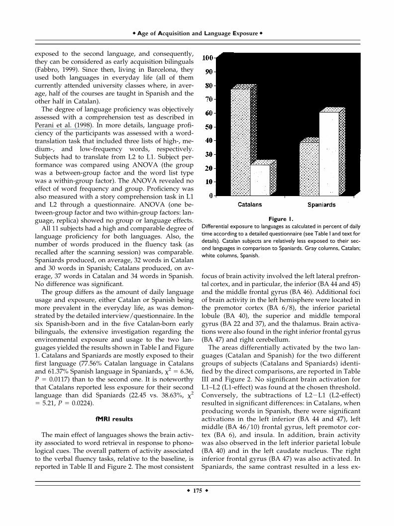
The group differs as the amount of daily languageusage and exposure, either Catalan or Spanish beingmore prevalent in the everyday life, as was demon-strated by the detailed interview/questionnaire. In thesix Spanish-born and in the five Catalan-born earlybilinguals, the extensive investigation regarding theenvironmental exposure and usage to the two lan-guages yielded the results shown in Table I and Figure1. Catalans and Spaniards are mostly exposed to theirfirst language (77.56% Catalan language in Catalansand 61.37% Spanish language in Spaniards, �2 � 6.36,P � 0.0117) than to the second one. It is noteworthythat Catalans reported less exposure for their secondlanguage than did Spaniards (22.45 vs. 38.63%, �2
� 5.21, P � 0.0224).
fMRI results
The main effect of languages shows the brain activ-ity associated to word retrieval in response to phono-logical cues. The overall pattern of activity associatedto the verbal fluency tasks, relative to the baseline, isreported in Table II and Figure 2. The most consistent
focus of brain activity involved the left lateral prefron-tal cortex, and in particular, the inferior (BA 44 and 45)and the middle frontal gyrus (BA 46). Additional fociof brain activity in the left hemisphere were located inthe premotor cortex (BA 6/8), the inferior parietallobule (BA 40), the superior and middle temporalgyrus (BA 22 and 37), and the thalamus. Brain activa-tions were also found in the right inferior frontal gyrus(BA 47) and right cerebellum.
The areas differentially activated by the two lan-guages (Catalan and Spanish) for the two differentgroups of subjects (Catalans and Spaniards) identi-fied by the direct comparisons, are reported in TableIII and Figure 2. No significant brain activation forL1–L2 (L1-effect) was found at the chosen threshold.Conversely, the subtractions of L2�L1 (L2-effect)resulted in significant differences: in Catalans, whenproducing words in Spanish, there were significantactivations in the left inferior (BA 44 and 47), leftmiddle (BA 46/10) frontal gyrus, left premotor cor-tex (BA 6), and insula. In addition, brain activitywas also observed in the left inferior parietal lobule(BA 40) and in the left caudate nucleus. The rightinferior frontal gyrus (BA 47) was also activated. InSpaniards, the same contrast resulted in a less ex-
Figure 1.Differential exposure to languages as calculated in percent of dailytime according to a detailed questionnaire (see Table I and text fordetails). Catalan subjects are relatively less exposed to their sec-ond languages in comparison to Spaniards. Gray columns, Catalan;white columns, Spanish.
� Age of Acquisition and Language Exposure �
� 175 �

tended pattern of brain activity within the left fron-tal lobe: in the inferior (BA 45) and middle frontalgyrus (BA 46/10) and in the insula.
The interaction analysis showed significant brainactivation foci in the left hemisphere, namely in themiddle frontal gyrus (BA 46/10) and left inferior pa-rietal lobule (BA 40). This difference may be consid-ered to reflect the effect of lower exposure to theSpanish language in Catalans (Table IV).
DISCUSSION
We examined the effects of age of acquisition and ofexposure on the cerebral correlates of lexical searchand retrieval in a population of early-acquisition,high-proficient bilinguals. We will first address howthe age of L2 acquisition in early bilinguals is respon-sible for differences in cerebral activation patterns,and then, how usage/exposure may also be a crucialfactor.
Our results underline that the age of language ac-quisition is a crucial factor, even in early bilingualswith a very high degree of proficiency for L1 and L2.Indeed, the direct comparisons between L1 vs. L2 inour groups showed no significant activation foci at thechosen threshold, while the reverse contrast (L2 � L1)resulted in significant differences in brain activity.
Classically, among researchers, the effects of age-of-onset of L2 acquisition were highly debated and theso-called “critical period” for language acquisition hasbeen a constant source of controversy (Elman et al.,1997). The critical period hypothesis proposed by Len-neberg (1967), which considers puberty as the limit,relies on the main assumption that age-related effectsreported in L2 studies are the results of maturationalchanges in brain structures, which are involved inacquiring a language. L2 learning after the criticalperiod was postulated to be different in nature frompre-pubertal learning, because of the requirement fora more conscious, labored effort. Other psycholin-guists argued against a native-like performance, evenif L2 is learned before the end of the critical period (forreview, see Harley and Wang, 1997). For instances,Hyltenstam (1992) showed that the onset for L2 acqui-sition after age 7 may lead to incompleteness not onlyin grammar but in the lexicon as well. Furthermore, inJohnson and Newport’s studies (1989, 1991) native-like performance was achieved only among bilingualswith age of L2 acquisition between 3 and 7 years,whereas Ioup (1989) found incomplete acquisitionamong immigrants who arrived between age 6 to 9years in the United States. It becomes clear that it isdifficult to set a specific age at which the critical periodterminates, but most researchers converge by setting it
TABLE II. Main effect for both languages
x y z Z-value
L inferior frontal gyrus (BA 44) �44 10 32 5.70�54 12 24 4.88
L inferior frontal gyrus (BA 45) �36 36 4 5.50�30 24 4 5.45�42 28 0 5.06
L middle frontal gyrus (BA 46) �36 36 20 4.62L premotor cortex (BA 6/8) �40 4 60 4.51
�46 12 52 4.23�42 8 56 4.20
L pre-SMA �2 8 56 4.16�2 20 44 3.22
L inferior parietal lobule (BA 40) �42 �28 40 4.49�32 �36 44 4.53�32 �32 36 4.19
L superior temporal gyrus (BA 22) �54 �34 16 3.46L middle temporal gyrus (BA 37) �48 �54 �8 4.87L thalamus �6 �10 8 4.23R inferior frontal gyrus (BA 47) 46 24 �4 4.17R cerebellum 6 �46 �16 3.91
In both groups of bilinguals, Spanish and Catalan fluency conditions were pooled and compared tothe baseline condition. This contrast indicated brain activity attributed to general phonologicalword-retrieval processes not specifically associated with one language.
� Perani et al. �
� 176 �

at age 6 to 7 years, hence, bilinguals who acquire theirL2 before that age are called “early bilinguals” (Fab-bro, 1999).
Our behavioral results did not indicate crucial dif-ferences in language processing and language usewhen early bilinguals acquire L2 after age 3. Indeed,language processing, as revealed by the evaluation ofproficiency in comprehension, was comparableamong our subjects (furthermore, the fluency task per-formance, measured according to the number ofwords produced in L1 and L2, was comparable forboth groups). However, the present data indicate that,at the macroscopic cerebral level, additional neuralresources are involved when generating words in L2
at a comparable level of language performance to thatof L1. A possible interpretation is that the generationof words, in the language acquired first in life, is lesseffortful, and is thus reflected, at the neural level, bythe engagement of less extensive neural resources,even when proficiency is held constant. Because pho-nemic fluency is a relatively difficult, effortful task, thedifference between L2 and L1 (in the L2 � L1 subtrac-tion) may be attributed to the recruitment of addi-tional resources within a dedicated network.
This interpretation must be reconciled with the lim-ited findings derived from other imaging investiga-tions of language production in early bilinguals (Cheeet al., 1999a; Kim et al., 1997). Kim et al. (1997) studiedearly and late bilinguals. Of these, six had been ex-posed to L1 and L2 during infancy (the exact age of L2acquisition however, was not provided) and six beganlearning L2 after puberty. The experimental task re-quired covert extended language production (dis-course). The authors found Broca’s area differentiallyactivated for the two groups: overlapping activationsfor both languages in early learners, and spatiallysegregated activations in late learners. However, amajor problem for the interpretation of these results isthat no formal assessment of language proficiency wasconducted. Since there is a general negative correla-tion between age of acquisition and proficiency (John-son and Newport, 1989), these two variables are con-founded in this experiment. Kim et al.’s conclusionwas that age of acquisition is a major factor in thecortical organization of second language processing.The overlapping activations in Broca’s area for L1 andL2 in early bilinguals are in contrast with the differ-ential activation for L1 and L2 in early bilinguals inour study. Several factors may account for this differ-ence. The study by Kim and coworkers focused onlyon Broca’s area, whereas the activation differencesbetween L2 and L1 in our study were not only foundin Broca’s area but also extended to other regions inthe lateral frontal cortex. In addition, task-related ef-fects should be taken into consideration. The produc-tion of extended speech, as in Kim et al.’s study (1997),relies on a more “natural” elaboration of the linguisticmaterial; in contrast, most of the linguistic processingevaluated in bilinguals is related to phonological tasks(Flege et al., 1999), or to morphosyntactic processing(Weber-Fox and Neville, 1996). The differences inbrain activation between our study and Kim et al.’sstudy may thus reflect the task differences. In general,verbal fluency task are considered to assess the abilityto access lexical items through a number of strategicsearch processes, which may involve the retrieval ofphonological, lexical, and semantic knowledge (Tro-
Figure 2.Brain activity during phonological fluency task. A: Main effects oflanguages (L1 and L2) vs. baseline in the whole group of bilinguals.B: Direct comparison between L2 (Catalan) vs. L1 (Spanish) inSpaniards. C: Direct comparison between L2 (Spanish) vs. L1(Catalan) in Catalans.
� Age of Acquisition and Language Exposure �
� 177 �

ster, 1998). The multi-component nature of this testmakes it sensitive not only to frontal lobe damage(Milner, 1964), but also to schizophrenia (Spence et al,2000) and dementia (Monsch et al., 1992).
Different considerations apply to Chee et al.’s study(1999a). Using fMRI in Mandarin–English bilinguals,they found no differences within the left prefrontalcortex when comparing a word stem completion taskin early bilinguals and late bilinguals with high degreeof language proficiency. A group of early bilinguals(L2 acquisition before age 6) were compared to latebilinguals (L2 acquisition after age 12). Subjects werestudied when producing words cued by a word stempresented visually. The authors predicted that the pro-cessing of Mandarin would require neural resourcesdistinct from English, since Mandarin has an ideo-graphic writing system. However, the pattern of brain
activation in response to Mandarin was similar to thatobserved for English and, furthermore, this was truefor both early and late bilinguals with high profi-ciency. A possible explanation for these findings maybe found, again, in the nature of task: from a cognitiveviewpoint, word stem completion in Chee et al.’sstudy, is a completely different task than word gener-ation on phonological and semantic cueing.
While ideally suited for the investigation of primingeffects, word-stem completion on the basis of writtenword stems relies extensively on orthographic knowl-edge, and may be less demanding in terms of linguis-tic requirements (Buckner et al., 1995, 2000). More-over, age of L2 acquisition of Chee et al.’s subjectsvaried from age 2 to age 6, whereas our study includesonly subjects who learned L2 at age 3, when admittedto kindergarten.
It is noteworthy that differences in age of acquisi-tion were found in another class of tasks, involvingaccess to morphosyntactic knowledge. Weber-Fox andNeville (1996) reported an ERP study of syntactic andlexical–conceptual violations in bilinguals where sub-jects were categorized by their age of first exposure toL2: at 1–3 years of age, 4–6 years, 7–10 years, 11–13years or greater than 16 years. Later exposure to L2was significantly associated with worse performance,at least for syntactic violations. Even the group of
TABLE III. Direct comparison between languages
x y z Z-value
L1 vs. L2Catalans: No voxels above thresholdSpaniards: No voxels above threshold
L2 vs. L1Catalans
L insula �28 16 8 4.15�32 14 0 3.73
L inferior frontal gyrus (BA 44) �36 18 28 3.41L middle frontal gyrus (BA 46/10) �40 44 8 3.95
�46 40 16 3.57L inferior frontal gyrus (BA 47) �32 18 �4 3.69L caudate nucleus �16 2 20 3.57L premotor cortex (BA 6) �40 4 60 3.71
�48 4 52 3.40L inferior parietal (BA 40) �44 �28 44 3.46R inferior frontal gyrus (BA 47) 48 20 �8 3.58
SpaniardsL inferior frontal gyrus (BA 45) �48 36 8 3.10L middle frontal gyrus (BA 46/10) �40 44 8 3.61L insula �34 16 12 3.51
Table demonstrates the L1 effect in Catalans (Catalan vs. Spanish) and Spaniards (Spanish vs. Catalan), and the L2 effect for Catalans(Spanish vs. Catalan) and Spaniards (Catalan vs. Spanish)
TABLE IV. Language exposure effects
x y zZ-
value
L middle frontal gyrus (BA 46/10) �40 46 4 2.44L inferior parietal lobule (BA 40) �44 �28 40 2.64
The data refer to the interaction effect in Catalans generating wordsin Spanish, a language to which they are not intensively exposed,compared to Spaniards producing word in Catalan.
� Perani et al. �
� 178 �

those exposed to L2 as young as 1–3 showed a differ-ent pattern to syntactic phrase structure violations.Combining these data with our results, one may con-clude that even when a L2 is learned very early in life,different neural resources may act for its processing,which may not be necessarily be associated to a worselanguage performance at the behavioral level. Thesecerebral differences may be particularly sensitive toeffortful language tasks, such as word generation onphonological cueing, or morphosyntactic tasks.
The present data show that a differential exposureto a given language may intervene on cerebral repre-sentations in multilinguals, even in the case when thedegree of proficiency is kept constant. That differentialexposure, reflected in a more intensive and frequentusage of a given language, may be a crucial factor hasalready been demonstrated in aphasiology. In partic-ular, recovery studies have clearly indicated that oneof the polyglot’s languages may recover in a differentmanner from the others (Albert and Obler, 1978; Para-dis, 1998). More than a century ago, Pitres (1895) de-scribed the influence of the language spoken in thepatient’s environment on preferential recovery, focus-ing on what has come to be known as Pitres’s law.According to the latter, the language spoken mostfrequently and intensively before brain damage(which is not necessarily the language learnt earlier inlife) is the one to recover first. The more extensivebrain activity observed in the left lateral frontal cortexfor Catalans, when generating words in Spanish, thesecond language with lower usage for this group, incomparison with the more limited activation found inthe Spaniards for word finding in Catalan (Fig. 2), mayreflect this environmental factor. Indeed, the interac-tion effect confirms this hypothesis: the brain activityassociated with fluency in L2 selectively the left lateralfrontal and parietal cortex for the Catalans than for theSpaniards. Catalans in Catalonia, albeit highly profi-cient in Spanish since early childhood, are less ex-posed to Spanish than Spaniards living in Catalonia totheir L2 (Catalan).
A previous fMRI experiment based on phonemicfluency in multilinguals (Yetkin et al., 1996), showedlarger foci of brain activity for the “less fluent” lan-guages. The experimental group was composed of sixheterogeneous subjects, fluent in at least two lan-guages and non-fluent in a third language. “Fluent”was empirically defined as speaking the language cur-rently and for at least 5 years, whereas “non-fluent”was used for languages studied for 2 to 4 years andwithout regular use in the everyday life. Moreover,English was always labeled as L1, despite the fact thatthe native language was not English in two of the six
subjects. While the resulting findings are difficult tointerpret given the lack of control of important vari-ables such as the age of language acquisition and levelof proficiency, they seem to be in agreement with the“exposure” hypothesis.
The present findings indicate that activity in the leftfrontal cortex during verbal fluency in bilinguals isless extensive in the case of the language acquiredfirst, which is associated with more extensive usage/exposure. Many functional neuroimaging studieshave implicated the left frontal convexity in languageprocessing tasks. For instance, increased blood flowwas observed specifically during semantic monitoring(Demonet et al., 1992), word generation (Martin et al.,1996; Petersen et al., 1988), semantic and phonemicfluency (Frith et al., 1991; Mummery et al., 1996;Paulesu et al., 1997), and with verbal working memorytasks (Smith et al., 1996). More recently, studyingword generation tasks of increasing task difficulty,Thompson-Schill and colleagues (1997, 1999) sug-gested that brain activity in these areas, that is, Brod-mann areas 45, 46, and 47 in the left inferior andmiddle frontal gyrus, is presumably associated withthe general selection processes of information amongcompeting alternatives from semantic memory. Theseresearchers underscored that these cortical areaswould be differentially engaged on the basis of taskdifficulty. When the word-to-be-generated possessesmore competing alternatives, among which the subjecthas to select, a larger left prefrontal network wasinvolved. With increasing task difficulty, more neuralactivity should thereafter be evoked. Likewise, theeffect of experience on task performance resulted indecreased neural activity (Thompson-Schill et al.,1999). A substantial body of information, particularlyfrom the past 10–15 years of research, has demon-strated that neuronal properties in auditory, sensory,visual, and motor cortices can be reorganized in theadult human brain, either in response to lesions or toexperience alone. Representations in a particular brainarea are said to contract (or expand) so that they canoccupy a smaller (or larger) plot of neural “real es-tate,” an effect termed “representational plasticity”(Donoghue, 1995). Practice-related changes in a word-generation task have been observed in a study wherethe prefrontal brain areas were less active in the well-practiced automatic condition (Raichle et al., 1994).Later, Petersen et al. (1998) further investigated theeffects of practice on a verbal task using PET. Theobserved reduced brain activity in the left frontal lobefollowing practice was putatively related to process-ing differences between high and low practice perfor-mance in verb generation. That experience alone may
� Age of Acquisition and Language Exposure �
� 179 �

decrease brain activity in the left prefrontal cortex washighlighted by a recent PET study (Petersson et al.,1999) during the recall of abstract designs. Specifically,practice-related decreases were observed in the leftlateral frontal cortex (BA 9, 10, and 46).
It may be hypothesized that similar changes, whichwe interpret to reflect long-term cognitive plasticity,were responsible for the effects of age of acquisitionand of exposure in our study. As a task becomes moreautomatic, performance will gradually rely less onprefrontal support, and decreases in left prefrontalactivity might reflect a decreasing dependence on con-trolled processing. Here, we particularly stress thatpractice, in the case of bilingual language processing,is not directly equivalent to proficiency (in terms ofabsolute level of fluency), but rather to the more in-tensive use/exposure of a language. Intensive expo-sure to a language in a bilingual environment will leadto a lower activation threshold (Green, 1986) andtherefore to a higher degree of automaticity, i.e., de-creased dependence on controlled and attentional pro-cessing. Supposedly, this would be reflected in de-creased activity of the prefrontal region. We suggestthat some components of the neural network subserv-ing word generation have a dynamic role, and this willbe reflected in functional reorganization of the lan-guage-processing network during intensive exposure.
Our findings are also compatible with the frame-work proposed by Green (1986). Green’s model oflanguage control presupposes the existence of recip-rocally inter-dependent language subsystems for theselection process among multiple languages in bilin-guals. Language selection is regulated by a device (theso-called “language-specifier”) that prevents codemixing. This stage is followed by the operation of aresource generator, which, following the specificationof the language, activates the appropriate languageoutput system. In short, if the polyglot intends tospeak in L1, the language specifier selects the intendedlanguage. Then, the resource generator activates thesubsystems for word output in L1 and at the sametime suppresses the word output subsystem for L2and vice versa, in order to avoid code mixing. In termsof energy resources, the model suggests that languageselection should be performed with less effort in thecase of the more available language. This is a conceptadapted by many clinical researchers in order to ex-plain specific recovery patterns in polyglot aphasia(Aglioti and Fabbro, 1993; Aglioti et al., 1996;Abutalebi et al., 2000). For instance, it has beenclaimed that one of the languages may selectivelyrecover because of a lower activation threshold, whichis strongly linked to language exposure (Fabbro, 1999).
This is the first anatomo-functional evidence thatboth age of acquisition and amount of exposure to alanguage may affect the extent of activation during alexical search and retrieval task. The finding of lessextensive activity in the left prefrontal cortex is con-sistent with the selection and use of a more availableand “automatic” language. Repeated activation of thecortical representations in a language may be expectedto strengthen the neocortical connections in such away that the neural network could support lexicalretrieval with a less extensive engagement of prefron-tal regions.
CONCLUSION
Functional neuroimaging with PET and fMRI offersus a direct window into the complex mechanism ofinteraction among language systems in the humanbrain, and thus appears to be a valuable tool to at-tempt to delineate the general principles of cerebrallanguage organization in bilinguals. In broad outlines,we underline the fact that the bilingual brain cannot beviewed as the sum of two monolingual language sys-tems, but rather considered as a unique and complexneural system which may differ in individual cases.There are several variables that may be responsible forthe above-mentioned differences; among them theprominent role of degree of proficiency was under-lined by neuroimaging studies carried out in the lastyears. In the present investigation in early high profi-cient bilinguals, we addressed the issues of age ofsecond language acquisition and level of languageusage/exposure. We found differences in brain acti-vation patterns for a word generation task. These dif-ferences appear to be associated with these two vari-ables never specifically addressed in past imagingstudies. Interestingly, these variables appear to affectthe macroscopic neural level during lexical retrieval:in the case of word generation more extensive cerebralactivations are associated with the language acquiredas second even if mastered with equal level of profi-ciency. This effect appears to be modulated by expo-sure, as it is less evident in bilinguals who are moreextensively exposed (in percentage) to the second lan-guage. The knowledge about how language is orga-nized in the human brain and the possible variationsassociated with multiple languages and crucial func-tional variables may have important implications forall contexts in which multi-lingual interactions are atplay. For example, the finding that language exposureis a crucial factor for the neural representation ofmultiple languages may provide important inputs tothe educational field, such as in the case of second
� Perani et al. �
� 180 �

language teaching. Our findings also suggest that thebest approach to language-impaired bilinguals may bebased on the language to which the subject was moreintensively exposed pre-morbidly.
ACKNOWLEDGMENTS
We thank Prof. Jacques Mehler, Prof. Nuria Sebas-tian Galles, and Dr. Emanuel Dupoux who contrib-uted to the investigation in many different and usefulways. In particular, we thank Prof. Elizabeth Bates fora critical reading of the manuscript.
REFERENCES
Abutalebi J, Miozzo A, Cappa, SF (2000): Do subcortical structurescontrol language selection in bilinguals? Evidence from patho-logical language mixing. Neurocase 6:101–106.
Abutalebi J, Cappa SF, Perani D (2001): The bilingual brain asrevealed by functional neuroimaging. Bilingual Lang Cognit4:179–190.
Aglioti S, Fabbro F (1993): Paradoxical selective recovery in a bilin-gual aphasic following subcortical lesion. Neuroreport 4:1359–1362.
Aglioti S, Beltramello A, Girardi F, Fabbro F (1996): Neurolinguisticand follow-up study of an unusual pattern of recovery frombilingual subcortical aphasia. Brain 119:1551–1564.
Albert ML, Obler LK (1978): The bilingual brain. New York: Aca-demic Press.
Beatty WW (1998): Cortical and subcortical influences on clusteringand switching in the performance of verbal fluency tasks. Neuro-psychologia 36:295–304.
Birdsong D (1999): Second language acquisition and the criticalperiod hypothesis. Mahwah, NJ: Lawrence Erlbaum Associates.
Buckner RL, Raichle ME, Petersen SE (1995): Dissociation of humanprefrontal cortical areas across different speech production tasksand gender groups. J Neurophysiol 74:2163–73.
Buckner RL, Koutstaal W, Schacter DL, Rosen BR (2000): FunctionalMRI evidence for a role of frontal and inferior temporal cortex inamodal components of priming. Brain 123:620–40.
Chee MWL, Tan EWL, Thiel T (1999a): Mandarin and English singleword processing studied with functional Magnetic ResonanceImaging. J Neurosci 19:3050–3056.
Chee MWL, Caplan D, Soon CS, Sriram N, Tan EWL, Thiel T,Weekes B (1999b): Processing of visually presented sentences inMandarin and English studied with fMRI. Neuron 23:127–137.
Chee MWL, Hon N, Ling Lee H, Soon CS (2001): Relative languageproficiency modulates BOLD signal change when Bilinguals per-form semantic judgments. Neuroimage 13:1155–1163.
Dehaene SD, Dupoux E, Mehler J, Cohen L, Paulesu E, Perani D, vande Moortele PF, Lehericy S, Le Bihan D (1997): Anatomicalvariability in the cortical representation of first and second lan-guages. Neuroreport 8:3809–3815.
Demonet JF, Chollet F, Ramsay S, Cardebat D, Nespoulous JL, WiseR, Rascol A, Frackowiak R (1992): The anatomy of phonologicaland semantic processing in normal subjects. Brain 115:1753–68.
Donoghue JP (1995): Plasticity of adult sensorimotor representa-tions. Curr Opin Neurobiol 5:749–754.
Dufour R, Kroll JFK (1995): Matching words to concepts in twolanguages: A test of the concept mediation model of bilingualrepresentation. Mem Cogn 23:166–180.
Elman JL, Bates E, Johnson MH, Karmiloff-Smith A, Parisi D, Plun-kett K (1996): Rethinking Innateness: a connectionist perspectiveon development. Cambridge, MA: MIT Press.
Fabbro F. (1999): The neurolinguistics of bilingualism. An introduc-tion. Hove, East Sussex, UK: Psychology Press.
Flege JE, Yeni-Komshian GH, Liu S (1999): Age constraints onsecond-language acquisition. J Mem Lang 41:78–104.
Friston KJ, Holmes AP, Worsley KJ, Poline JB, Frith CD, FrackowiakRSJ (1995): Statistical parametric maps in functional imaging: ageneral linear approach. Hum Brain Mapp 2:189–210.
Frith CD, Friston KJ, Liddle PF, Frackowiak RS (1991): A PET studyof word finding. Neuropsychologia 29:1137–1148.
Green DW (1986): Control, activation, and resource: a frameworkand a model for the control of speech in bilinguals. Brain Lang27:210–223.
Green DW (1998): Mental control of the bilingual lexico-semanticsystem. Bilingualism 1:67–81.
Harley B, Wang W (1997): The critical period hypothesis: where arewe now? In: De Groot AMB, Kroll JFK, editors. Tutorials inbilingualism. Psycholinguistic perspectives. Mahwah, NJ: LEAPublishers.
Hyltenstam K (1992): Non-native features of near-native speakers:On the ultimate attainment of childhood L2 learners. In: RJHarris, editor. Cognitive processing in bilinguals. Amsterdam:Elsevier.
Illes J, Francis WS, Desmond JE, Gabrieli JDE, Glover GH, PoldrackR, Lee CJ, Wagner AD (1999): Convergent cortical representationof semantic processing in bilinguals. Brain Lang 70:347–363.
Ioup G (1989): Immigrant children who have failed to acquire nativeEnglish. In: Gass, Madden, Preston, Selinker, editors. Variationin second language acquisition, Vol 2. Clevedon, UK: Multilin-gual Matters.
Johnson JS, Newport EL (1989): Critical period effects in secondlanguage learning: the influence of maturational state on theacquisition of English as a second language. Cogn Psychol 21:60–99.
Johnson JS, Newport EL (1991): Critical period effects on universalproperties of language: the status of subjacency in the acquisi-tion of a second language. Cognition 39:215–258.
Kim KHS, Relkin NR, Lee KM, Hirsch J (1997): Distinct cortical areasassociated with native and second languages. Nature 388:171–174.
Klein D, Zatorre R, Milner B, Meyer E, Evans A (1994): Left putami-nal activation when speaking a second language: evidence fromPET. Neuroreport 5:2295–2297.
Klein D, Milner B, Zatorre R, Meyer E, Evans A (1995): The neuralsubstrates underlying word generation: A bilingual functional-imaging study. Proc Natl Acad Sci U S A 92:2899–2903.
Lenneberg EH (1967): Biological foundations of language. NewYork: Wiley.
Martin A, Wiggs CL, Ungerleider LG, Haxby JV (1996): Neuralcorrelates of category-specific knowledge Nature 379:649–652.
Milner B (1964): Some effects of frontal lobectomy in man. In War-ren JM, Akert K, editors. The frontal granular cortex and behav-ior. New York: McGraw Hill. p 313–334.
Monsch AU, Bondi MW, Butters N, Salmon DP, Katzman R, Thal LJ(1992):. Comparisons of verbal fluency tasks in the detection ofdementia of the Alzheimer type. Arch Neurol 49:1253–1258.
Mummery CJ, Patterson K, Hodges JR. Wise RJS (1996): Generatinga ‘tiger’ as an animal name or a word beginning with T: differ-ences in brain activations. Proc R Soc Lond B 263:989–995.
Neville HJ, Bavelier D (1998): Neural organization and plasticity oflanguage. Curr Opin Neurobiol 8:254–258.
� Age of Acquisition and Language Exposure �
� 181 �

Oldfield RC (1971): The assessment and analysis of handedness: theEdinburgh Inventory. Neuropsychologia 9:97–113.
Paulesu E, Goldacre B, Scifo P, Cappa SF, Gilardi MC, Castiglioni I,Perani D, Fazio F. (1997): Functional heterogeneity of left inferiorfrontal cortex as revealed by fMRI. Neuroreport 8:2011–2016.
Paradis M (1998): Language and communication in multilinguals.In: Stemmer B, Whitaker H, editors. Handbook of neurolinguis-tics. San Diego, CA: Academic Press. p 417–430.
Perani D, Dehaene S, Grassi F, Cohen L, Cappa SF, Dupoux E, FazioF, Mehler J (1996): Brain processing of native and foreign lan-guages. Neuroreport 7:2439–2444.
Perani D, Paulesu E, Sebastian-Galles N, Dupoux E, Dehaene S,Bettinardi V, Cappa SF, Fazio F, Mehler J (1998): The bilingualbrain: proficiency and age of acquisition of the second language.Brain 121:1841–1852.
Petersen SE, Fox PT, Posner MI, Mintun M, Raichle ME (1988):Positron emission tomographic studies of single word process-ing. Nature 331:585–589.
Petersen SE, van Mier H, Fiez JA, Raichle ME (1998): The effects ofpractice on the functional anatomy of task performance. ProcNatl Acad Sci U S A 95:853–860.
Petersson KM, Elfgren C, Ingvar M (1999): Dynamic changes in thefunctional anatomy of the human brain during recall of abstractdesigns related to practice. Neuropsychologia 37:567–587.
Pitres A. (1895): Etude sur l’aphasie chez les polyglottes. Rev Med15:873–899.
Price CJ, Green D, von Studnitz R (1999): A functional imagingstudy of translation and language switching. Brain 122:2221–2236.
Raichle ME, Fiez JA, Videen TO, MacLeod AM, Pardo JV, Fox PT,Petersen SE (1994): Practice related changes in human brain func-tional anatomy during nonmotor learning. Cereb Cortex 4:8–26.
Schreuder R, Weltens B (1993): The bilingual lexicon. Amsterdam:John Benjamins Publishing.
Smith EE, Jonides J, Koeppe RA (1996): Dissociating verbal andspatial working memory using PET. Cereb Cortex 6:11–20.
Spence SA, Liddle PF, Stefan MD, Hellewell JS, Sharma T, FristonKJ, Hirsch SR, Frith CD, Murray RM, Deakin JF, Grasby PM(2000): Functional anatomy of verbal fluency in people withschizophrenia and those at genetic risk. Focal dysfunction anddistributed disconnectivity reappraised. Br J Psychiatry 176:52–60.
Thompson-Schill SL, D’Esposito M, Aguirre GK, Farah MJ (1997):Role of left inferior prefrontal cortex in retrieval of semanticknowledge: a reevaluation. Proc Natl Acad Sci U S A 94:14692–14797.
Thompson-Schill SL, D’Esposito M, Kan IP (1999): Thompson-SchillSL, D’Esposito M, Kan IP. 1999. Effects of repetition and com-petition on activity in left prefrontal cortex during word gener-ation. Neuron 23:513–522.
Troster AI, Fields JA, Testa JA, Paul RH, Blanco CR, Hames, KA,Salmon DP (1998): Cortical and subcortical influences on clus-tering and switching in the performance of verbal fluency tasks.Neuropsychologia 26:295–304.
Yetkin O, Yetkin FZ, Haughton VM, Cox RW (1996): Use of func-tional MR to map language in multilingual volunteers. Am JNeuroradiol 17:473–477.
Weber-Fox CM, Neville HJ (1996): Maturational constraints onfunctional specialization for language processing: ERP andbehavioral evidence in bilingual speakers. J Cogn Neurosci8:231–256.
Weiller C, Isensee C, Rijntjes M, Huber W, Mueller S, Bier D,Dutschka K, Woods RP, Noth J, Diener HC (1995): Recoveryfrom Wernicke’s aphasia: a positron emission tomographicstudy. Ann Neurol 37:723–732.
� Perani et al. �
� 182 �