the refeeding syndrome: a neglected but potentially serious
TRANSCRIPT
Vol.:(0123456789)1 3
Internal and Emergency Medicine https://doi.org/10.1007/s11739-020-02525-7
IM - REVIEW
The Refeeding Syndrome: a neglected but potentially serious condition for inpatients. A narrative review
Valentina Ponzo1 · Marianna Pellegrini1 · Iolanda Cioffi2 · Luca Scaglione3 · Simona Bo1
Received: 29 July 2020 / Accepted: 30 September 2020 © The Author(s) 2020
AbstractThe Refeeding Syndrome (RFS) is a potentially serious, but still overlooked condition, occurring in individuals who are rapidly fed after a period of severe undernourishment. RFS derives from an abnormal electrolyte and fluid shifts leading to many organ dysfunctions. Symptoms generally appear within 2–5 days of re-feeding and may be absent/mild or severe and life threating, depending on the pre-existing degree of malnutrition and comorbidities. The lack of a standard definition and the nonspecificity of the symptoms make both incidence estimate and diagnosis difficult. In 2020, the American Society for Parenteral and Enteral Nutrition (ASPEN) proposed a unifying definition for the RFS and its severity classification. The awareness of the condition is crucial for identifying patients at risk, preventing its occurrence, and improving the manage-ment. The objectives of this narrative review were to summarize the current knowledge and recommendations about the RFS and to provide useful tips to help physicians to recognize and prevent the syndrome.
Keywords Hypophosphatemia · Hypokalemia · Hypomagnesemia · Malnutrition · Refeeding syndrome · Thiamine
Introduction
Malnutrition is a frequent and often unrecognized condition among inpatients [1, 2]. Indeed, 20–50% of individuals are at risk of malnutrition or already malnourished at hospital admission, but malnutrition is diagnosed in 7% only [3]. Older age, low socioeconomic status, lack of organizational support, chronic systemic or psychiatric diseases, polyther-apy, poor diet, reduced absorption capacity, excessive nutri-ent losses are the most frequent conditions underlying mal-nutrition [4]. The management of malnourished inpatients can be difficult due to the risk of metabolic impairment after
the start of nutrition [5]. The adverse outcomes of refeed-ing were firstly reported during the World War II in rapidly re-fed prisoners who had starved for five to six months [6]. People who have fasted for a long time, developed heart, and/or respiratory failure, peripheral edema, neurological symptoms, and death after the introduction of excessive or even appropriate calorie amount [6–8]. In the 80 s, the term ‘refeeding syndrome’ (RFS) was introduced to describe severe hypophosphatemia and other electrolyte/metabolic abnormalities and the related cardiovascular and pulmonary manifestations leading to death occurring in two chroni-cally malnourished patients who received aggressive dex-trose‐based parenteral nutrition (PN) [9]. Since then, many cases of RFS have been described as a rare, but severe and potentially fatal complication related to re-feeding (either orally, enterally or parenterally) of individuals who have fasted or consumed very few calories over a long period of time [10, 11]. Among the diseases or conditions predispos-ing to malnutrition and consequently to RFS after re-feeding, anorexia nervosa [12–14], cancer [15, 16], critical illnesses [13, 17–20], and frailty in the elderly [21–27] are the most frequently implicated.
The switch from a catabolic to an anabolic state may be the cause of the clinical manifestations of the RFS, even though the pathophysiological mechanisms are still not fully
Electronic supplementary material The online version of this article (https ://doi.org/10.1007/s1173 9-020-02525 -7) contains supplementary material, which is available to authorized users.
* Simona Bo [email protected]
1 Department of Medical Sciences, University of Torino, c.so AM Dogliotti 14, 10126 Turin, Italy
2 Department of Clinical Medicine and Surgery, Federico II University Hospital, Naples, Italy
3 Internal Medicine Unit, Città della Salute e della Scienza Hospital of Torino, Turin, Italy
Internal and Emergency Medicine
1 3
understood [28]. Furthermore, the lack of a clear definition accounts for the difficulty of diagnosis and uncertainties in treatment [2, 29]. Therefore, the RFS is a potentially seri-ous condition, often overlooked by many physicians [30]. This is of particular concern because of the high prevalence of hospital malnutrition often underestimated even in the internal medicine wards [31, 32].
The objectives of this narrative review are to summarize the knowledge on the RFS and to focus on the most useful topics for the clinical practice.
Methods
The following databases were queried: PubMed (National Library of Medicine), the Cochrane Library, Excerpta Medica dataBASE (EMBASE), and the Cumulative Index to Nursing and Allied Health Literature (CINAHL). The search strategy was performed using the following key-words: refeeding syndrome OR phosphate, potassium, mag-nesium AND anorexia nervosa, cancer, critically ill patients, elderly. The filters ‘humans’ and ‘adults’ were used. Hand searching the references of the identified studies and reviews was carried out too.
Incidence rates for RFS
The lack of a universally recognized RFS definition makes it difficult to obtain precise estimates of its incidence [33]. Indeed, either hypophosphatemia only or multiple electro-lyte abnormalities (with different cut offs) with or without clinical manifestations have been considered in its definition [34, 35]. The reported incidence rates ranged between 0 and 80%, depending on the definition and the patient population studied [34]. RFS has been described in 48% of severely malnourished patients, in 34% of intensive care unit (ICU) patients, in 33% of patients with anorexia nervosa (AN), in 25% of cancer inpatients, and in 9.5% of patients hospi-talized for malnutrition from gastrointestinal fistulae [10, 12, 33, 36]. Many factors may lead to underestimation of RFS incidence rate, such as insufficient monitoring of the patients’ electrolytes after nutrition starting, lack of consul-tation by experts in clinical nutrition, the nonspecificity of the clinical manifestations of the syndrome in patients with multiple co-morbidity and the physician unawareness [11].
Population at risk for RFS
To identify patients at risk for RFS is necessary evaluating the risk of malnutrition by validated screening tools first, and then assessing the diagnosis and grading the severity
of malnutrition [5, 33, 37, 38]. Distinguishing malnutri-tion from the other related conditions, such as starvation, cachexia, cancer cachexia, and sarcopenia, is important from a clinical point of view (Table 1) [39–44]. The screening for the risk of malnutrition should be performed in inpatients within the first 24–48 h through validated screening tools, such as the Nutritional Risk Screening 2002 (NRS-2002), the Mini Nutritional Assessment-Short Form (MNA-SF), the Malnutrition Universal Screening Tool (MUST), the Short Nutritional Assessment Questionnaire (SNAQ) [5, 37, 39]. If an individual is identified to be at risk of malnutrition, an extensive nutritional assessment for diagnosis and evaluation of the severity of malnutrition should be carried out by an expert in nutrition [39, 40].
A great number of diseases or conditions predisposes to malnutrition [21, 28, 33, 34, 37, 39, 45–47]. These pre-disposing conditions can be divided into the following categories: predisposing to disease-related malnutrition with inflammation (chronic diseases leading to catabolic inflammatory responses); predisposing to disease-related malnutrition without inflammation (acute disease and injury-related malnutrition); and predisposing to malnutri-tion in the absence of diseases (hunger, socioeconomic, or psychologic-related conditions, drugs) [39], as summarized in Supplementary Table 1.
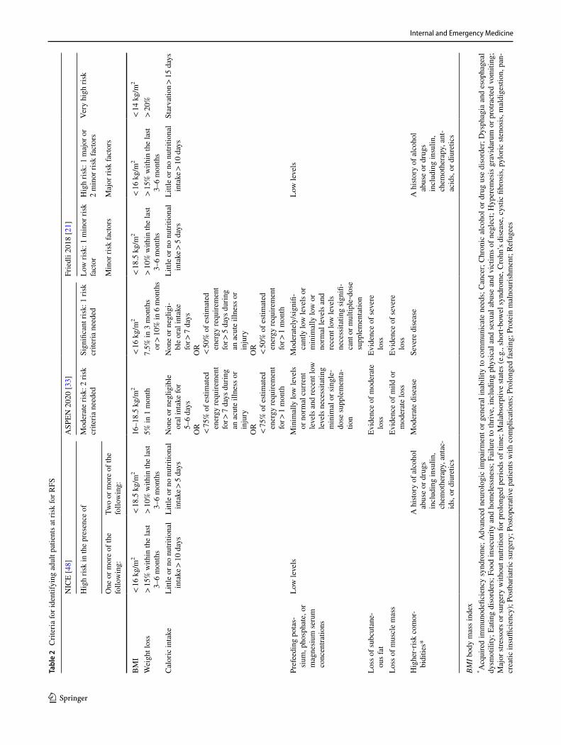
In the presence of severe underweight or weight loss, prolonged fasting period, and/or low electrolyte concentra-tions, the risk of RFS is particularly high [30]. In 2006, the National Institute for Health and Clinical Excellence (NICE) guidelines [48] reported the risk factors to identify people at low or high risk for RSF. In 2018 Friedli et Coll added the very high-risk category [21]. Recently, the American Soci-ety for Parenteral and Enteral Nutrition (ASPEN) published updated consensus criteria for identifying adult patients at risk for RFS [33]. These criteria are presented in Table 2.
Diagnosis of RFS
The difficulty in RFS diagnosing is due to the discrepancy between the onset of the symptoms and the occurring of metabolic shift (see below), and the nonspecific nature of its clinical manifestations [46]. There is a great heterogene-ity among the published definitions of RFS, ranging from hypophosphatemia alone [18, 19, 22, 24, 27, 49–54] to the presence of severe low-serum electrolyte levels along with fluid balance abnormalities and/or organ dysfunction [16, 21, 34, 55]. Only hypophosphatemia has been universally recognized as a feature of the syndrome [38]. Friedli et Coll. proposed diagnostic criteria for imminent or manifest RFS, based on the electrolyte blood concentrations and clinical symptoms to standardize its prevention and treatment [21]. According to this definition, “imminent” RFS is present
Internal and Emergency Medicine
1 3
when a shift in electrolytes occurs within 72 h after the start of nutritional treatment (i.e., > 30% decrease in blood phos-phate from baseline or phosphate values < 0.6 mmol/L or any two other electrolyte shifts below normal range) [21]. “Manifest” RFS is considered if any electrolyte shift occurs in conjunction with typical clinical symptoms (see below) [21].
More recently, the ASPEN proposed diagnostic crite-ria for distinguishing mild, moderate or severe RFS [33] (Table 3). The extent of the decrease in the serum levels of one or more electrolytes (among phosphate, potassium, or magnesium) defines RFS severity: 10–20% (mild RFS), 20–30% (moderate RFS), > 30% and/or organ dysfunction and/or thiamine deficiency (severe RFS) [33]. Thus, either hypophosphatemia and/or hypokalemia and/or hypomagne-semia qualify the presence of the RFS. The timing of onset is determinant for the diagnosis, since the RFS develops shortly (from hours up to 5 days) after having substantially increased the energy provision to individuals who have been undernourished [33].
Pathophysiology and clinical manifestations
The pathophysiology of the RFS is probably related to the shift from the catabolic to the anabolic metabolic pathways occurring after the re-start of feeding in undernourished
subjects. During early starvation, blood glucose and insu-lin levels decline while glucagon concentrations increase by stimulating glycogenolysis in the liver. When glycogen reserves become depleted, gluconeogenesis is stimulated in the liver, using amino acids derived from muscle break-down [56]. During prolonged fasting, the body switches to use fats as the main sources of energy with a decrease in basal metabolic rate of 20–25% [57]. Increased lipolysis in fat reserves leads to the production of ketones that are used by the brain as preferred fuel during starvation [29, 56]. During prolonged fasting, several intracellular min-erals become severely depleted, particularly phosphate, potassium, and magnesium. However, the concentrations of these minerals may remain within the normal range in the serum because there is a reduction in their renal excre-tion and because of the phosphate outflow from the cells into the blood, leading to normal blood phosphate levels despite depleted storages [21].
Symptoms generally appear within 2–5 days of re-feed-ing and may range from absent/mild to a severe and life-threating clinical syndrome, depending on the pre-existing degree of malnutrition and comorbidity [10, 11, 45]. All the body organs may be involved, leading to cardiac, res-piratory, hematologic, gastrointestinal, neurologic, and musculoskeletal manifestations, until death [10, 21, 58].
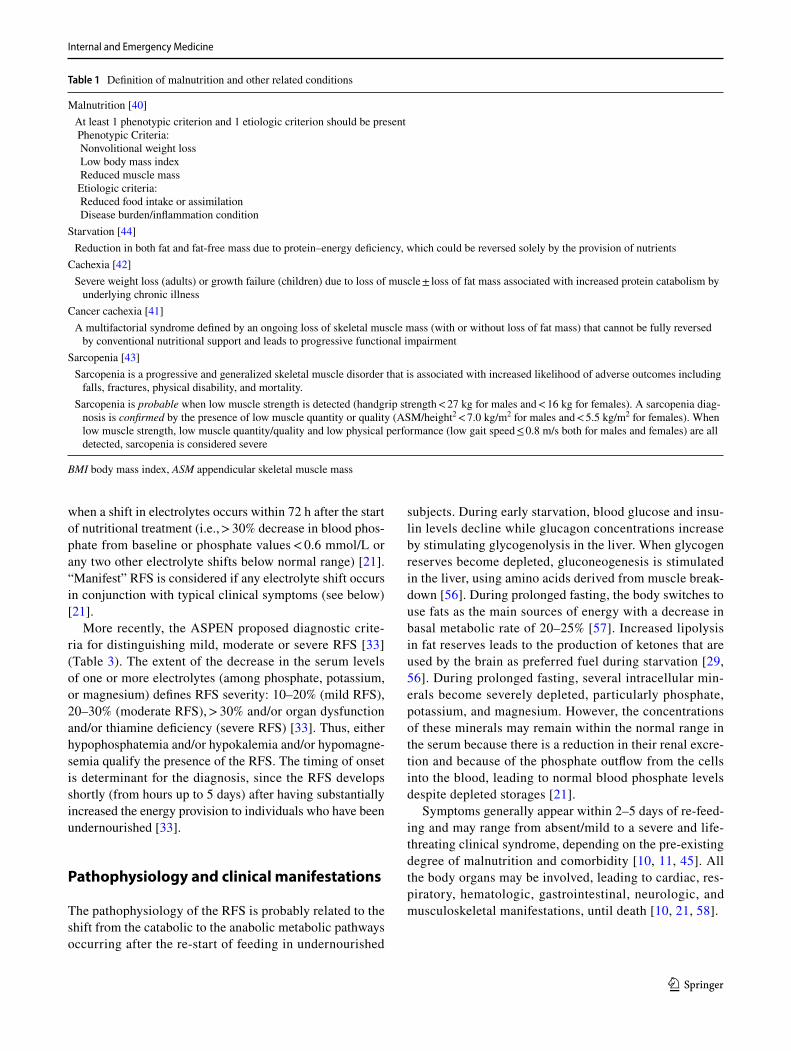
Table 1 Definition of malnutrition and other related conditions
BMI body mass index, ASM appendicular skeletal muscle mass
Malnutrition [40] At least 1 phenotypic criterion and 1 etiologic criterion should be present Phenotypic Criteria: Nonvolitional weight loss Low body mass index Reduced muscle mass Etiologic criteria: Reduced food intake or assimilation Disease burden/inflammation condition
Starvation [44] Reduction in both fat and fat-free mass due to protein–energy deficiency, which could be reversed solely by the provision of nutrients
Cachexia [42] Severe weight loss (adults) or growth failure (children) due to loss of muscle ± loss of fat mass associated with increased protein catabolism by
underlying chronic illnessCancer cachexia [41] A multifactorial syndrome defined by an ongoing loss of skeletal muscle mass (with or without loss of fat mass) that cannot be fully reversed
by conventional nutritional support and leads to progressive functional impairmentSarcopenia [43] Sarcopenia is a progressive and generalized skeletal muscle disorder that is associated with increased likelihood of adverse outcomes including
falls, fractures, physical disability, and mortality. Sarcopenia is probable when low muscle strength is detected (handgrip strength < 27 kg for males and < 16 kg for females). A sarcopenia diag-
nosis is confirmed by the presence of low muscle quantity or quality (ASM/height2 < 7.0 kg/m2 for males and < 5.5 kg/m2 for females). When low muscle strength, low muscle quantity/quality and low physical performance (low gait speed ≤ 0.8 m/s both for males and females) are all detected, sarcopenia is considered severe
Internal and Emergency Medicine
1 3
Tabl
e 2
Crit
eria
for i
dent
ifyin
g ad
ult p
atie
nts a
t ris
k fo
r RFS
BMI b
ody
mas
s ind
ex* A
cqui
red
imm
unod
efici
ency
syn
drom
e; A
dvan
ced
neur
olog
ic im
pairm
ent o
r gen
eral
inab
ility
to c
omm
unic
ate
need
s; C
ance
r; C
hron
ic a
lcoh
ol o
r dru
g us
e di
sord
er; D
ysph
agia
and
eso
phag
eal
dysm
otili
ty; E
atin
g di
sord
ers;
Foo
d in
secu
rity
and
hom
eles
snes
s; F
ailu
re to
thriv
e, in
clud
ing
phys
ical
and
sex
ual a
buse
and
vic
tims
of n
egle
ct; H
yper
emes
is g
ravi
daru
m o
r pro
tract
ed v
omiti
ng;
Maj
or st
ress
ors o
r sur
gery
with
out n
utrit
ion
for p
rolo
nged
per
iods
of t
ime;
Mal
abso
rptiv
e st
ates
(e.g
., sh
ort‐b
owel
synd
rom
e, C
rohn
’s d
isea
se, c
ystic
fibr
osis
, pyl
oric
sten
osis
, mal
dige
stion
, pan
-cr
eatic
insu
ffici
ency
); Po
stbar
iatri
c su
rger
y; P
osto
pera
tive
patie
nts w
ith c
ompl
icat
ions
; Pro
long
ed fa
sting
; Pro
tein
mal
nour
ishm
ent;
Refu
gees
NIC
E [4
8]A
SPEN
202
0 [3
3]Fr
iedl
i 201
8 [2
1]
Hig
h ris
k in
the
pres
ence
of
Mod
erat
e ris
k: 2
risk
cr
iteria
nee
ded
Sign
ifica
nt ri
sk: 1
risk
cr
iteria
nee
ded
Low
risk
: 1 m
inor
risk
fa
ctor
Hig
h ris
k: 1
maj
or o
r 2
min
or ri
sk fa
ctor
sVe
ry h
igh
risk
One
or m
ore
of th
e fo
llow
ing:
Two
or m
ore
of th
e fo
llow
ing:
Min
or ri
sk fa
ctor
sM
ajor
risk
fact
ors
BM
I <
16 k
g/m
2 <
18.5
kg/
m2
16–1
8.5
kg/m
2 <
16 k
g/m
2 <
18.5
kg/
m2
< 16
kg/
m2
< 14
kg/
m2
Wei
ght l
oss
> 15
% w
ithin
the
last
3–6
mon
ths
> 10
% w
ithin
the
last
3–6
mon
ths
5% in
1 m
onth
7.5%
in 3
mon
ths
or >
10%
in 6
mon
ths
> 10
% w
ithin
the
last
3–6
mon
ths
> 15
% w
ithin
the
last
3–6
mon
ths
> 20
%
Cal
oric
inta
keLi
ttle
or n
o nu
tritio
nal
inta
ke >
10 d
ays
Littl
e or
no
nutri
tiona
l in
take
> 5
days
Non
e or
neg
ligib
le
oral
inta
ke fo
r 5–
6 da
ysO
R <
75%
of e
stim
ated
en
ergy
requ
irem
ent
for >
7 da
ys d
urin
g an
acu
te il
lnes
s or
inju
ryO
R <
75%
of e
stim
ated
en
ergy
requ
irem
ent
for >
1 m
onth
Non
e or
neg
ligi-
ble
oral
inta
ke
for >
7 da
ysO
R <
50%
of e
stim
ated
en
ergy
requ
irem
ent
for >
5 da
ys d
urin
g an
acu
te il
lnes
s or
inju
ryO
R <
50%
of e
stim
ated
en
ergy
requ
irem
ent
for >
1 m
onth
Littl
e or
no
nutri
tiona
l in
take
> 5
days
Littl
e or
no
nutri
tiona
l in
take
> 10
day
sSt
arva
tion >
15 d
ays
Pref
eedi
ng p
otas
-si
um, p
hosp
hate
, or
mag
nesi
um se
rum
co
ncen
tratio
ns
Low
leve
lsM
inim
ally
low
leve
ls
or n
orm
al c
urre
nt
leve
ls a
nd re
cent
low
le
vels
nec
essi
tatin
g m
inim
al o
r sin
gle‐
dose
supp
lem
enta
-tio
n
Mod
erat
ely/
sign
ifi-
cant
ly lo
w le
vels
or
min
imal
ly lo
w o
r no
rmal
leve
ls a
nd
rece
nt lo
w le
vels
ne
cess
itatin
g si
gnifi
-ca
nt o
r mul
tiple‐d
ose
supp
lem
enta
tion
Low
leve
ls
Loss
of s
ubcu
tane
-ou
s fat
Evid
ence
of m
oder
ate
loss
Evid
ence
of s
ever
e lo
ssLo
ss o
f mus
cle
mas
sEv
iden
ce o
f mild
or
mod
erat
e lo
ssEv
iden
ce o
f sev
ere
loss
Hig
her‐
risk
com
or-
bidi
ties*
A h
istor
y of
alc
ohol
ab
use
or d
rugs
in
clud
ing
insu
lin,
chem
othe
rapy
, ant
ac-
ids,
or d
iure
tics
Mod
erat
e di
seas
eSe
vere
dis
ease
A h
istor
y of
alc
ohol
ab
use
or d
rugs
in
clud
ing
insu
lin,
chem
othe
rapy
, ant
-ac
ids,
or d
iure
tics
Internal and Emergency Medicine
1 3
Insulin and carbohydrate metabolism
Rapid refeeding in a starved patient causes the metabolic and hormonal changes underlying the syndrome [59]. The provision of nutrients, above all carbohydrates, increases insulin secretion and promotes a sudden shift from fat to carbohydrates metabolism. Insulin stimulates the sodium potassium ATPase symporter, with magnesium as co-fac-tor, which transports glucose and potassium into the cells and moves out sodium. Moreover, insulin release stimu-lates anabolic processes that require minerals (promoting cellular uptake of phosphate, potassium, and magnesium) and coenzymes, such as thiamine [29]. The electrolyte shift, along with the depletion of the mineral pool, could lead to profound hypophosphatemia and low extracellular magnesium and potassium concentrations, but not neces-sarily to the depletion of all together. Furthermore, insulin has an anti-natriuretic effect on renal tubules causing a decrease in urinary sodium and water excretion [59]. This determines a rapid fluid overload that can lead to conges-tive cardiac failure, arrhythmia, and pulmonary edema.
Hypophosphatemia
The phosphate is predominantly an intracellular mineral that plays a key role in energy production and transfer (as a component of adenosine triphosphate (ATP) [58] and it is necessary for many enzymatic processes of cellular metabolic pathways [60]. During refeeding, the increased phosphate consumption due to enhanced production of phosphorylated intermediates results in reduced generation of ATP and 2,3-diphosphoglycerate with impaired cardiac and respiratory functions, and decreased oxygen release to the tissues (Table 4).
Hypokalemia
Potassium is an intracellular mineral and it is crucial for the maintenance of the sodium–potassium membrane gradient; hypokalemia causes imbalance in the electro-chemical membrane potential and impaired transmission
of electrical impulses resulting in arrhythmias, cardiac arrest, and neurologic symptoms [61–63].
Hypomagnesemia
Magnesium plays a role as a cofactor for the phosphorylation of ATP and it is important for the maintenance of neuro-muscular and enzymatic functions. Its depletion results in increased renal losses of potassium, aggravating hypoka-lemia with arrhythmias and ECG abnormalities, and in abdominal discomfort and neuromuscular symptoms [64].
Thiamine deficit
Thiamine is another cofactor in ATP production. Its increased consumption during refeeding by the enhanced activity of enzymes implicated in the carbohydrate metabo-lism may lead to neurologic disorders (dry beriberi, Wer-nicke encephalopathy and Korsakoff’s syndrome), car-diovascular disorders, and metabolic acidosis (due to the conversion of glucose into lactate) [65] (Table 4).
Prevention and treatment
The identification of patients at risk for RFS is the first step to prevent the onset of the syndrome, and to avoid an exces-sive nutritional replenishment in those individuals [21, 66]. Risk factors should be carefully investigated before starting either oral, enteral, or parenteral nutrition, because every route of calorie administration is implicated in the occur-rence of the RFS [33, 58]. Well-trained medical staff and specialized nutritional support teams, consisting of physi-cians, dieticians, nurses, and pharmacists, positively impact on the patient outcomes [48]. However, a multidisciplinary team is not available in all hospital settings, and often the evaluation of the risk for RFS is left to the clinician’s critical sense at the time of starting nutritional support [11, 33, 36, 38, 67]. After defining the degree of RFS risk, the rate of fluid and nutrition administration, the correction of electro-lyte imbalances, and the supplementation of vitamins and micronutrients (zinc, iron, selenium) can be determined [36] (Table 5). If a prolonged nutritional support is required,
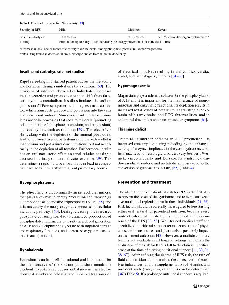
Table 3 Diagnostic criteria for RFS severity [33]
*Decrease in any (one or more) of electrolyte serum levels, among phosphate, potassium, and/or magnesium**Resulting from the decrease in any electrolyte and/or from thiamine deficiency
Severity of RFS Mild Moderate Severe
Serum electrolytes* 10–20% less 20–30% less > 30% less and/or organ dysfunction**Timing From hours up to 5 days after increasing the energy provision in an individual at risk
Internal and Emergency Medicine
1 3
adjustments over time in accordance with the patient clinical conditions might be necessary [58].
Several therapeutic approaches have been proposed to prevent or treat the RSF [10, 21, 28, 36, 45, 46, 48, 67, 68] (Fig. 1). Since hypophosphatemia occurs after refeed-ing, according to the grade of RSF risk, phosphate may be administered preventively before the initiation of nutri-tional therapy, even if blood levels are in the low-normal range [21]. Similarly, thiamine is essential in carbohydrates
metabolism and should be supplemented before restart feeding even in the case of normal blood levels [21]. An excessive administration of glucose by stimulating insulin production leads to the consumption of electrolytes (mainly phosphate) through the anabolic pathways. Starting re-feed-ing very gradually, independently of the route of adminis-tration, is therefore mandatory [58]. Owing to the risk of fluid overload, sodium and hydration should be provided cautiously, until the patient is metabolically stable [38]. In
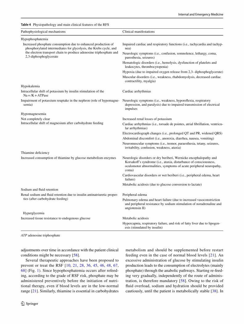
Table 4 Physiopathology and main clinical features of the RFS
ATP adenosine triphosphate
Pathophysiological mechanisms Clinical manifestations
Hypophosphatemia Increased phosphate consumption due to enhanced production of
phosphorylated intermediates for glycolysis, the Krebs cycle, and the electron transport chain to produce adenosine triphosphate and 2,3-diphosphoglycerate
Impaired cardiac and respiratory functions (i.e., tachycardia and tachyp-nea)
Neurologic symptoms (i.e., confusion, somnolence, lethargy, coma, paresthesia, seizures)
Hematologic disorders (i.e., hemolysis, dysfunction of platelets and leukocytes, thrombocytopenia)
Hypoxia (due to impaired oxygen release from 2,3- diphosphoglycerate)Muscular disorders (i.e., weakness, rhabdomyolysis, decreased cardiac
contractility, myalgia)HypokalemiaIntracellular shift of potassium by insulin stimulation of the
Na + /K + ATPaseCardiac arrhythmias
Impairment of potassium reuptake in the nephron (role of hypomagne-semia)
Neurologic symptoms (i.e., weakness, hyporeflexia, respiratory depression, and paralysis) due to impaired transmission of electrical impulses
HypomagnesemiaNot completely clearIntracellular shift of magnesium after carbohydrate feeding
Increased renal losses of potassiumCardiac arrhythmias (i.e., torsade de pointes, atrial fibrillation, ventricu-
lar arrhythmias)Electrocardiograph changes (i.e., prolonged QT and PR, widened QRS)Abdominal discomfort (i.e., anorexia, diarrhea, nausea, vomiting)Neuromuscular symptoms (i.e., tremor, paraesthesia, tetany, seizures,
irritability, confusion, weakness, ataxia)Thiamine deficiencyIncreased consumption of thiamine by glucose metabolism enzymes Neurologic disorders or dry beriberi, Wernicke encephalopathy and
Korsakoff’s syndrome (i.e., ataxia, disturbance of consciousness, oculomotor abnormalities, symptoms of acute peripheral neuropathy, coma)
Cardiovascular disorders or wet beriberi (i.e., peripheral edema, heart failure)
Metabolic acidosis (due to glucose conversion to lactate)Sodium and fluid retentionRenal sodium and fluid retention due to insulin antinatriuretic proper-
ties (after carbohydrate feeding)Peripheral edemaPulmonary edema and heart failure (due to increased vasoconstriction
and peripheral resistance by sodium stimulation of noradrenaline and angiotensin II)
HyperglycemiaIncreased tissue resistance to endogenous glucose Metabolic acidosis
Hypercapnia, respiratory failure, and risk of fatty liver due to lipogen-esis (stimulated by insulin)
Internal and Emergency Medicine
1 3
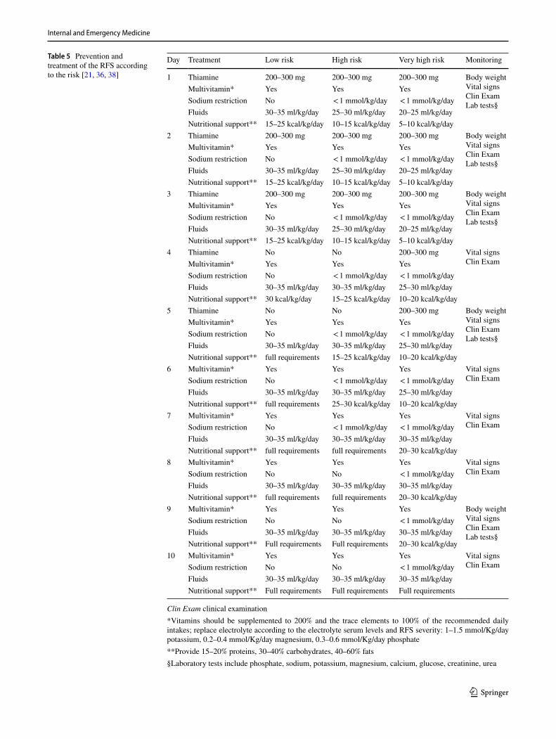
Table 5 Prevention and treatment of the RFS according to the risk [21, 36, 38]
Clin Exam clinical examination*Vitamins should be supplemented to 200% and the trace elements to 100% of the recommended daily intakes; replace electrolyte according to the electrolyte serum levels and RFS severity: 1–1.5 mmol/Kg/day potassium, 0.2–0.4 mmol/Kg/day magnesium, 0.3–0.6 mmol/Kg/day phosphate**Provide 15–20% proteins, 30–40% carbohydrates, 40–60% fats§Laboratory tests include phosphate, sodium, potassium, magnesium, calcium, glucose, creatinine, urea
Day Treatment Low risk High risk Very high risk Monitoring
1 Thiamine 200–300 mg 200–300 mg 200–300 mg Body weightVital signsClin ExamLab tests§
Multivitamin* Yes Yes YesSodium restriction No < 1 mmol/kg/day < 1 mmol/kg/dayFluids 30–35 ml/kg/day 25–30 ml/kg/day 20–25 ml/kg/dayNutritional support** 15–25 kcal/kg/day 10–15 kcal/kg/day 5–10 kcal/kg/day
2 Thiamine 200–300 mg 200–300 mg 200–300 mg Body weightVital signsClin ExamLab tests§
Multivitamin* Yes Yes YesSodium restriction No < 1 mmol/kg/day < 1 mmol/kg/dayFluids 30–35 ml/kg/day 25–30 ml/kg/day 20–25 ml/kg/dayNutritional support** 15–25 kcal/kg/day 10–15 kcal/kg/day 5–10 kcal/kg/day
3 Thiamine 200–300 mg 200–300 mg 200–300 mg Body weightVital signsClin ExamLab tests§
Multivitamin* Yes Yes YesSodium restriction No < 1 mmol/kg/day < 1 mmol/kg/dayFluids 30–35 ml/kg/day 25–30 ml/kg/day 20–25 ml/kg/dayNutritional support** 15–25 kcal/kg/day 10–15 kcal/kg/day 5–10 kcal/kg/day
4 Thiamine No No 200–300 mg Vital signsClin ExamMultivitamin* Yes Yes Yes
Sodium restriction No < 1 mmol/kg/day < 1 mmol/kg/dayFluids 30–35 ml/kg/day 30–35 ml/kg/day 25–30 ml/kg/dayNutritional support** 30 kcal/kg/day 15–25 kcal/kg/day 10–20 kcal/kg/day
5 Thiamine No No 200–300 mg Body weightVital signsClin ExamLab tests§
Multivitamin* Yes Yes YesSodium restriction No < 1 mmol/kg/day < 1 mmol/kg/dayFluids 30–35 ml/kg/day 30–35 ml/kg/day 25–30 ml/kg/dayNutritional support** full requirements 15–25 kcal/kg/day 10–20 kcal/kg/day
6 Multivitamin* Yes Yes Yes Vital signsClin ExamSodium restriction No < 1 mmol/kg/day < 1 mmol/kg/day
Fluids 30–35 ml/kg/day 30–35 ml/kg/day 25–30 ml/kg/dayNutritional support** full requirements 25–30 kcal/kg/day 10–20 kcal/kg/day
7 Multivitamin* Yes Yes Yes Vital signsClin ExamSodium restriction No < 1 mmol/kg/day < 1 mmol/kg/day
Fluids 30–35 ml/kg/day 30–35 ml/kg/day 30–35 ml/kg/dayNutritional support** full requirements full requirements 20–30 kcal/kg/day
8 Multivitamin* Yes Yes Yes Vital signsClin ExamSodium restriction No No < 1 mmol/kg/day
Fluids 30–35 ml/kg/day 30–35 ml/kg/day 30–35 ml/kg/dayNutritional support** full requirements full requirements 20–30 kcal/kg/day
9 Multivitamin* Yes Yes Yes Body weightVital signsClin ExamLab tests§
Sodium restriction No No < 1 mmol/kg/dayFluids 30–35 ml/kg/day 30–35 ml/kg/day 30–35 ml/kg/dayNutritional support** Full requirements Full requirements 20–30 kcal/kg/day
10 Multivitamin* Yes Yes Yes Vital signsClin ExamSodium restriction No No < 1 mmol/kg/day
Fluids 30–35 ml/kg/day 30–35 ml/kg/day 30–35 ml/kg/dayNutritional support** Full requirements Full requirements Full requirements
Internal and Emergency Medicine
1 3
case of overt symptoms, energy and fluid intakes should be reduced and adapted to the clinical conditions [30].
Specific conditions might require special attention.
Anorexia nervosa
Most inpatients with AN are at high risk for RFS [12]; refeeding is the first step of the treatment and must be managed very cautiously [66, 69]. International guidelines are based mainly on clinical experience, due to the lack of well-designed trials in inpatients with AN [70, 71]. At hos-pital admission, the recommended calorie provision ranges from 5–20 kcal/kg to 30–40 kcal/kg [70, 71]. A progres-sive increase of 5–10 kcal/kg/day (if high risk of RFS) or 10–20 kcal/kg/day (if moderate risk of RFS) could be carried out after the stabilization of the clinical conditions (e.g., improvement of electrocardiographic abnormalities, correction of electrolyte imbalance, replacement of thia-mine and vitamins, and stabilization of comorbidities)
[48, 66, 72]. Caloric provision could increase up to 70–100 kcal/kg per day if patients have increased energy requirement such as in case of inappropriate behaviors (throwing or hiding food, vomiting, intense exercise, etc.) [71]. Refeeding with a lower calorie provision and a slow energy increase may be a better approach for severely malnourished patients with chronic comorbidity, while higher caloric intakes might be reserved for moderately malnourished patients with acute illnesses [69]. Preventive supplementation with phosphate, potassium, magnesium, thiamine and other vitamins, trace elements, and minerals as well as sodium and fluid restriction are recommended too [66, 71]. Both meal-based approaches (with or without oral nutritional supplements) and combined approaches with nasogastric feeding can be used in inpatients requir-ing higher caloric intakes [69, 73]. Parenteral nutrition is not recommended unless no other form of refeeding is possible [69].
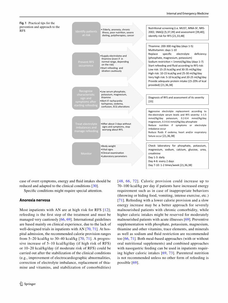
Fig. 1 Practical tips for the prevention and approach to the RFS • Elderly, anorexia, chronic
illness, poor nutri�on, severe die�ng, polytherapies, cancer
Iden�fy pa�ents at risk
•Supply electrolytes and thiamine (even if in normal range, dependingon the risk)
•Start refeeding and idra�on cau�ously
Prevent RFS occurrence
•Low serum phosphate, potassium, magnesium, thiamine
•Alert if: tachycardiatachypnea, oedema, confusion, ECG altera�ons
Recognize characteris�c
sign and symptoms a�er
star�ng refeeding
•A�er about 3 days withoutsigns and symptoms, stop worryng about RFS.
Treat electrolyte imbalances and
manage refeeding
•Body weight•Vital signs•Clinical examina�on•Laboratory parameters
Monitor
Nutri�onal screening (i.e. MUST, MNA-SF, NRS-2002, SNAQ) [5,37,39] and assessment [39,40]; iden�fy risk for RFS [21,33,48]
Thiamine: 200-300 mg/day (days 1-5) Mul�vitamin: days 1-10 Replace specific electrolyte deficiency (phosphate, magnesium, potassium) Sodium restric�on < 1mmol/kg/day (days 1-7) Start refeeding and fluid according to RFS risk: Low risk: 15-25 kcal/kg and 30-35 ml/kg/day High risk: 10-15 kcal/kg and 25-30 ml/kg/day Very high risk: 5-10 kcal/kg and 20-25 ml/kg/day Provide adequate protein intake (15-20% of kcal provided) [21,36,38]
Aggressive electrolyte replacement according to the electrolyte serum levels and RFS severity: 1-1.5 mmol/Kg/day potassium, 0.2-0.4 mmol/Kg/day magnesium, 0.3-0.6 mmol/Kg/day phosphate Reduce nutri�on if symptoms or electrolyte imbalance occur Reduce fluids if oedema, heart and/or respiratory failure occur [21,36,38]
Diagnosis of RFS and assessment of its severity [33]
Check laboratory for phosphate, potassium, magnesium, sodium, calcium, glucose, urea, crea�nine Day 1-3: daily Day 4-6: every 2 days Day 7-10: 1-2 �mes/week [21,36,38]
Internal and Emergency Medicine
1 3
Cancer
Up to 50–80% of patients with advanced cancer are at high risk of developing RFS [74], in particular individuals with head and neck cancer [75, 76]. Cancer cachexia cannot be arrested or reversed by any known form of nutritional, hormonal, or pharmacological treatment [77]. There are no specific guidelines on how to re-feed cancer patients at risk for RFS, being NICE recommendations [48] the most frequently used [29, 75, 76, 78, 79]. In patients eating lit-tle or nothing for more than 5 days, refeeding should be started with no more than 50% of the caloric requirements, with ≤ 10 kcal/kg/day in high-risk patients and ≤ 5 kcal/kg/day in very high-risk patients (BMI < 14 kg/m2 or negligi-ble intake for 2 weeks or more) [48]. Owing to the poten-tial benefit of protein intake on muscle anabolism, cancer patients should receive a protein intake of 1 g/kg/day up to 1.5 g/kg/day [79]. When oral refeeding is possible, the use of oral nutritional supplements can be useful in reaching nutritional goals [76]; if oral feeding is either impossible or insufficient, enteral, or parenteral nutrition should be considered [76], with slow progressive caloric increase to reach the full needs within 4–7 days [74]. In the case of cancer cachexia, a very cautious refeeding should begin by initially supplying about 25% of the estimated calorie requirement [77], with a very gradual caloric increase over several days, and a careful monitoring of phosphate and electrolytes serum levels [80].
Conclusions
This narrative review provides the latest information on the management of RFS in light of the current evidence. Although RFS is a frequent condition that can have serious consequences above all in specific categories of inpatients, it is often undiagnosed and overlooked by physicians. Its knowledge is essential to avoid rapid and excessive nourish-ing of at-risk patients; thus, preventing serious complica-tions, long hospital stays, and the increase in health costs.
Author contribution All authors contributed to the study conception and design. Idea for the article: SB. Writing—original draft preparation MP; Writing—review and editing: VP, IC, LS; Supervision: LS, SB. All authors read and approved the final manuscript.
Funding Open access funding provided by Università degli Studi di Torino within the CRUI-CARE Agreement. The authors did not receive financial support for the research, authorship, and/or publication of this article.
Data availability Not applicable.
Code availability Not applicable.
Compliance with ethical standards
Conflicts of interest The authors declare that they have no conflict of interest.
Statement of human and animal rights This article does not contain any studies with human participants or animals performed by any of the authors.
Informed consent For this type of study, formal consent is not required.
Consent to participate Not applicable.
Consent for publication Not applicable.
Open Access This article is licensed under a Creative Commons Attri-bution 4.0 International License, which permits use, sharing, adapta-tion, distribution and reproduction in any medium or format, as long as you give appropriate credit to the original author(s) and the source, provide a link to the Creative Commons licence, and indicate if changes were made. The images or other third party material in this article are included in the article’s Creative Commons licence, unless indicated otherwise in a credit line to the material. If material is not included in the article’s Creative Commons licence and your intended use is not permitted by statutory regulation or exceeds the permitted use, you will need to obtain permission directly from the copyright holder. To view a copy of this licence, visit http://creat iveco mmons .org/licen ses/by/4.0/.
References
1. Vest MT, Papas MA, Shapero M, McGraw P, Capizzi A, Jurkovitz C (2018) Characteristics and outcomes of adult inpatients with malnutrition. J Parenter Enteral Nutr 42:1009–1016. https ://doi.org/10.1002/jpen.1042
2. Janssen G, Pourhassan M, Lenzen-Großimlinghaus R, Jäger M, Schäfer R, Spamer C, Cuvelier I, Volkert D, Wirth R (2019) The refeeding syndrome revisited: you can only diagnose what you know. Eur J Clin Nutr 73:1458–1463. https ://doi.org/10.1038/s4143 0-019-0441-x
3. Lanctin DP, Merced-Nieves F, Mallett RM, Arensberg MB, Guenter P, Sulo S, Platts-Mills TF (2019) Prevalence and eco-nomic burden of malnutrition diagnosis among patients presenting to united states emergency departments. Acad Emerg Med. https ://doi.org/10.1111/acem.13887
4. Barker LA, Gout BS, Crowe TC (2011) Hospital malnutrition: prevalence, identification and impact on patients and the health-care system. Int J Environ Res Public Health 8:514–527. https ://doi.org/10.3390/ijerp h8020 514
5. Reber E, Gomes F, Bally L, Schuetz P, Stanga Z (2019) Nutri-tional management of medical inpatients. J Clin Med 8:1130. https ://doi.org/10.3390/jcm80 81130
6. Schnitker MA, Mattman PE, Bliss TL (1951) A clinical study of malnutrition in Japanese prisoners of war. Ann Intern Med 35:69–96. https ://doi.org/10.7326/0003-4819-35-1-69
7. Keys A, Brožek J, Henschel A, Mickelsen O, Taylor HL (1950) The biology of human starvation, Vols. 1 & 2
8. Netherlands, Committee on Malnutrition, Burger GCE, Drum-mond JC, Sandstead HR (1948) Malnutrition and starvation in Western Netherlands: September 1944–July 1945 Pt. 1. General State Print. Office, The Hague
Internal and Emergency Medicine
1 3
9. Weinsier RL, Krumdieck CL (1981) Death resulting from overzealous total parenteral nutrition: the refeeding syndrome revisited. Am J Clin Nutr 34:393–399. https ://doi.org/10.1093/ajcn/34.3.393
10. Boateng AA, Sriram K, Meguid MM, Crook M (2010) Refeeding syndrome: treatment considerations based on collective analy-sis of literature case reports. Nutrition 26:156–167. https ://doi.org/10.1016/j.nut.2009.11.017
11. Skipper A (2012) Refeeding syndrome or refeeding hypophos-phatemia: a systematic review of cases. Nutr Clin Pract 27:34–40. https ://doi.org/10.1177/08845 33611 42791 6
12. Brown CA, Sabel AL, Gaudiani JL, Mehler PS (2015) Predic-tors of hypophosphatemia during refeeding of patients with severe anorexia nervosa. Int J Eat Disord 48:898–904. https ://doi.org/10.1002/eat.22406
13. Vignaud M, Constantin J-M, Ruivard M, Villemeyre-Plane M, Futier E, Bazin J-E, Annane D, AZUREA group (AnorexieRea Study Group) (2010) Refeeding syndrome influences outcome of anorexia nervosa patients in intensive care unit: an observational study. Crit Care 14:R172. https ://doi.org/10.1186/cc927 4
14. Yamazaki T, Inada S, Yoshiuchi K (2019) Body mass index cut-off point associated with refeeding hypophosphatemia in adults with eating disorders. Int J Eat Disord 52:1322–1325. https ://doi.org/10.1002/eat.23177
15. Grasso S, Ferro Y, Migliaccio V et al (2013) Hypokalemia dur-ing the early phase of refeeding in patients with cancer. Clinics 68:1413–1415. https ://doi.org/10.6061/clini cs/2013(11)05
16. Rasmussen SO, Kristensen MB, Wessel I, Andersen JR (2016) Incidence and risk factors of refeeding syndrome in head and neck cancer patients-an observational study. Nutr Cancer 68:1320–1329. https ://doi.org/10.1080/01635 581.2016.12251 03
17. Marik PE, Bedigian MK (1996) Refeeding hypophosphatemia in critically ill patients in an intensive care unit. A prospective study. Arch Surg 131:1043–1047. https ://doi.org/10.1001/archs urg.1996.01430 22003 7007
18. Doig GS, Simpson F, Heighes PT, Bellomo R, Chesher D, Cat-erson ID, Reade MC, Harrigan PWJ, Refeeding Syndrome Trial Investigators Group (2015) Restricted versus continued standard caloric intake during the management of refeeding syndrome in critically ill adults: a randomised, parallel-group, multicentre, single-blind controlled trial. Lancet Respir Med 3:943–952. https ://doi.org/10.1016/S2213 -2600(15)00418 -X
19. Fuentes E, Yeh DD, Quraishi SA et al (2017) hypophosphatemia in enterally fed patients in the surgical intensive care unit: com-mon but unrelated to timing of initiation or aggressiveness of nutrition delivery. Nutr Clin Pract 32:252–257. https ://doi.org/10.1177/08845 33616 66298 8
20. Olthof LE, Koekkoek WACK, van Setten C, Kars JCN, van Blok-land D, van Zanten ARH (2018) Impact of caloric intake in criti-cally ill patients with, and without, refeeding syndrome: a retro-spective study. Clin Nutr 37:1609–1617. https ://doi.org/10.1016/j.clnu.2017.08.001
21. Friedli N, Stanga Z, Culkin A, Crook M, Laviano A, Sobotka L, Kressig RW, Kondrup J, Mueller B, Schuetz P (2018) Manage-ment and prevention of refeeding syndrome in medical inpatients: an evidence-based and consensus-supported algorithm. Nutrition 47:13–20. https ://doi.org/10.1016/j.nut.2017.09.007
22. Dror Y, Almashanu S, Lubart E, Sela B-A, Shimoni L, Segal R (2013) The impact of refeeding on blood fatty acids and amino acid profiles in elderly patients: a metabolomic analysis. JPEN J Parenter Enteral Nutr 37:109–116. https ://doi.org/10.1177/01486 07112 44326 0
23. Gaudiani JL, Sabel AL, Mascolo M, Mehler PS (2012) Severe anorexia nervosa: outcomes from a medical stabilization unit. Int J Eat Disord 45:85–92. https ://doi.org/10.1002/eat.20889
24. Gaudiani JL, Brinton JT, Sabel AL, Rylander M, Catanach B, Mehler PS (2016) Medical outcomes for adults hospitalized with severe anorexia nervosa: an analysis by age group. Int J Eat Disord 49:378–385. https ://doi.org/10.1002/eat.22437
25. Henderson S, Boyce F, Sumukadas D, Witham MD (2010) Changes in serum magnesium and phosphate in older hospital-ised patients–correlation with muscle strength and risk factors for refeeding syndrome. J Nutr Health Aging 14:872–876. https ://doi.org/10.1007/s1260 3-010-0261-0
26. Kagansky N, Levy S, Koren-Morag N, Berger D, Knobler H (2005) Hypophosphataemia in old patients is associated with the refeeding syndrome and reduced survival. J Intern Med 257:461–468. https ://doi.org/10.1111/j.1365-2796.2005.01457 .x
27. Lubart E, Leibovitz A, Dror Y, Katz E, Segal R (2009) Mortality after nasogastric tube feeding initiation in long-term care elderly with oropharyngeal dysphagia–the contribution of refeeding syn-drome. Gerontology 55:393–397. https ://doi.org/10.1159/00021 8162
28. Stanga Z, Brunner A, Leuenberger M, Grimble RF, Shenkin A, Allison SP, Lobo DN (2008) Nutrition in clinical practice-the refeeding syndrome: illustrative cases and guidelines for pre-vention and treatment. Eur J Clin Nutr 62:687–694. https ://doi.org/10.1038/sj.ejcn.16028 54
29. Mehanna HM, Moledina J, Travis J (2008) Refeeding syndrome: what it is, and how to prevent and treat it. BMJ 336:1495–1498. https ://doi.org/10.1136/bmj.a301
30. Friedli N, Odermatt J, Reber E, Schuetz P, Stanga Z (2020) Refeeding syndrome: update and clinical advice for prevention, diagnosis and treatment. Curr Opin Gastroenterol 36:136–140. https ://doi.org/10.1097/MOG.00000 00000 00060 5
31. Rinninella E, Cintoni M, De Lorenzo A, Addolorato G, Vas-sallo G, Moroni R, Miggiano GAD, Gasbarrini A, Mele MC (2018) Risk, prevalence, and impact of hospital malnutrition in a tertiary care referral university hospital: a cross-sectional study. Intern Emerg Med 13:689–697. https ://doi.org/10.1007/s1173 9-018-1884-0
32. Finocchiaro C, Fanni G, Bo S (2019) Clinical impact of hos-pital malnutrition. Intern Emerg Med 14:7–9. https ://doi.org/10.1007/s1173 9-018-1987-7
33. da Silva JSV, Seres D, Sabino K et al (2020) ASPEN Consen-sus recommendations for refeeding syndrome. Nutr Clin Pract 35(2):178–195. https ://doi.org/10.1002/ncp.10474
34. Friedli N, Stanga Z, Sobotka L, Culkin A, Kondrup J, Laviano A, Mueller B, Schuetz P (2017) Revisiting the refeeding syn-drome: results of a systematic review. Nutrition 35:151–160. https ://doi.org/10.1016/j.nut.2016.05.016
35. Khan LUR, Ahmed J, Khan S, MacFie J (2011) Refeeding syn-drome: a literature review. Gastroenterol Res Pract. https ://doi.org/10.1155/2011/41097 1
36. McKnight CL, Newberry C, Sarav M, Martindale R, Hurt R, Daley B (2019) Refeeding syndrome in the critically ill: a lit-erature review and clinician’s guide. Curr Gastroenterol Rep 21:58. https ://doi.org/10.1007/s1189 4-019-0724-3
37. Pourhassan M, Cuvelier I, Gehrke I, Marburger C, Modreker MK, Volkert D, Willschrei H-P, Wirth R (2018) Risk factors of refeeding syndrome in malnourished older hospitalized patients. Clin Nutr 37:1354–1359. https ://doi.org/10.1016/j.clnu.2017.06.008
38. Reber E, Friedli N, Vasiloglou MF, Schuetz P, Stanga Z (2019) Management of refeeding syndrome in medical inpatients. J Clin Med 8:2202. https ://doi.org/10.3390/jcm81 22202
39. Cederholm T, Barazzoni R, Austin P et al (2017) ESPEN guide-lines on definitions and terminology of clinical nutrition. Clin Nutr 36:49–64. https ://doi.org/10.1016/j.clnu.2016.09.004
40. Cederholm T, Jensen GL, Correia MITD et al (2019) GLIM criteria for the diagnosis of malnutrition – A consensus report
Internal and Emergency Medicine
1 3
from the global clinical nutrition community. Clin Nutr 38:1–9. https ://doi.org/10.1016/j.clnu.2018.08.002
41. Fearon K, Strasser F, Anker SD et al (2011) Definition and classification of cancer cachexia: an international consensus. Lancet Oncol 12:489–495. https ://doi.org/10.1016/S1470 -2045(10)70218 -7
42. Evans WJ, Morley JE, Argilés J et al (2008) Cachexia: a new definition. Clin Nutr 27:793–799. https ://doi.org/10.1016/j.clnu.2008.06.013
43. Cruz-Jentoft AJ, Bahat G, Bauer J et al (2019) Sarcopenia: revised European consensus on definition and diagnosis. Age Ageing 48:16–31. https ://doi.org/10.1093/agein g/afy16 9
44. Thomas DR (2007) Loss of skeletal muscle mass in aging: examining the relationship of starvation, sarcopenia and cachexia. Clin Nutr 26:389–399. https ://doi.org/10.1016/j.clnu.2007.03.008
45. Walmsley RS (2013) Refeeding syndrome: screening, incidence, and treatment during parenteral nutrition. J Gastroenterol Hepatol 28(Suppl 4):113–117. https ://doi.org/10.1111/jgh.12345
46. Pulcini CD, Zettle S, Srinath A (2016) Refeeding syndrome. Pedi-atr Rev 37:516–523. https ://doi.org/10.1542/pir.2015-0152
47. Michalsen A, Li C (2013) Fasting therapy for treating and prevent-ing disease—current state of evidence. Forsch Komplementmed 20:444–453. https ://doi.org/10.1159/00035 7765
48. Guidance | Nutrition support for adults: oral nutrition support, enteral tube feeding and parenteral nutrition | Guidance | NICE. https ://www.nice.org.uk/guida nce/cg32/chapt er/1-Guida nce#scree ning-for-malnu triti on-and-the-risk-of-malnu triti on-in-hospi tal-and-the-commu nity. Accessed 2 Jun 2020
49. Marvin VA, Brown D, Portlock J, Livingstone C (2008) Factors contributing to the development of hypophosphataemia when refeeding using parenteral nutrition. Pharm World Sci 30:329–335. https ://doi.org/10.1007/s1109 6-007-9180-5
50. Elnenaei MO, Alaghband-Zadeh J, Sherwood R, Awara MA, Moniz C, le Roux CW (2011) Leptin and insulin growth factor 1: diagnostic markers of the refeeding syndrome and mortality. Br J Nutr 106:906–912. https ://doi.org/10.1017/S0007 11451 10010 97
51. Rigaud D, Tallonneau I, Brindisi M-C, Vergès B (2012) Prognosis in 41 severely malnourished anorexia nervosa patients. Clin Nutr 31:693–698. https ://doi.org/10.1016/j.clnu.2012.02.016
52. Chen L-J, Chen H-L, Bair M-J, Wu C-H, Lin I-T, Lee Y-K, Chu C-H (2014) Refeeding syndrome in Southeastern Taiwan: our experience with 11 cases. World J Gastroenterol 20:10525–10530. https ://doi.org/10.3748/wjg.v20.i30.10525
53. Kraaijenbrink BVC, Lambers WM, Mathus-Vliegen EMH, Siegert CEH (2016) Incidence of refeeding syndrome in internal medicine patients. Neth J Med 74:116–121
54. Pantoja F, Fragkos KC, Patel PS, Keane N, Samaan MA, Barnova I, Di Caro S, Mehta SJ, Rahman F (2019) Refeeding syndrome in adults receiving total parenteral nutrition: an audit of prac-tice at a tertiary UK centre. Clin Nutr 38:1457–1463. https ://doi.org/10.1016/j.clnu.2018.06.967
55. Rio A, Whelan K, Goff L, Reidlinger DP, Smeeton N (2013) Occurrence of refeeding syndrome in adults started on artificial nutrition support: prospective cohort study. BMJ Open 3:e002173. https ://doi.org/10.1136/bmjop en-2012-00217 3
56. Mehanna H, Nankivell PC, Moledina J, Travis J (2009) Refeeding syndrome—awareness, prevention and management. Head Neck Oncol 1:4. https ://doi.org/10.1186/1758-3284-1-4
57. McCray S, Walker S, Parrish CR (2005) Much ado about refeed-ing. Pract Gastroenterol 28:26–44
58. Crook MA, Hally V, Panteli JV (2001) The importance of the refeeding syndrome. Nutrition 17:632–637. https ://doi.org/10.1016/s0899 -9007(01)00542 -1
59. Hearing SD (2004) Refeeding syndrome. BMJ 328:908–909. https ://doi.org/10.1136/bmj.328.7445
60. Weisinger JR, Bellorín-Font E (1998) Magnesium and phospho-rus. Lancet 352:391–396
61. McDonough AA, Youn JH (2017) Potassium homeostasis: the knowns, the unknowns, and the health benefits. Physiology (Bethesda) 32:100–111. https ://doi.org/10.1152/physi ol.00022 .2016
62. Elliott TL, Braun M (2017) Electrolytes: potassium disorders. FP Essent 459:21–28
63. Kardalas E, Paschou SA, Anagnostis P, Muscogiuri G, Siasos G, Vryonidou A (2018) Hypokalemia: a clinical update. Endocr Connect 7:R135–R146. https ://doi.org/10.1530/EC-18-0109
64. Ahmed F, Mohammed A (2019) Magnesium: the forgotten elec-trolyte-a review on hypomagnesemia. Med Sci (Basel). https ://doi.org/10.3390/medsc i7040 056
65. Polegato BF, Pereira AG, Azevedo PS, Costa NA, Zornoff LAM, Paiva SAR, Minicucci MF (2019) Role of thiamine in health and disease. Nutr Clin Pract 34:558–564. https ://doi.org/10.1002/ncp.10234
66. Skowrońska A, Sójta K, Strzelecki D (2019) Refeeding syndrome as treatment complication of anorexia nervosa. Psychiatr Pol 53:1113–1123. https ://doi.org/10.12740 /PP/Onlin eFirs t/90275
67. Aubry E, Friedli N, Schuetz P, Stanga Z (2018) Refeeding syn-drome in the frail elderly population: prevention, diagnosis and management. Clin Exp Gastroenterol 11:255–264. https ://doi.org/10.2147/CEG.S1364 29
68. Kraft MD, Btaiche IF, Sacks GS (2005) Review of the refeed-ing syndrome. Nutr Clin Prac 20:625–633. https ://doi.org/10.1177/01154 26505 02000 6625
69. Garber AK, Sawyer SM, Golden NH, Guarda AS, Katzman DK, Kohn MR, Le Grange D, Madden S, Whitelaw M, Redgrave GW (2016) A systematic review of approaches to refeeding hospital-ized patients with anorexia nervosa. Int J Eat Disord 49:293–310. https ://doi.org/10.1002/eat.22482
70. Resmark G, Herpertz S, Herpertz-Dahlmann B, Zeeck A (2019) Treatment of anorexia nervosa-new evidence-based guidelines. J Clin Med. https ://doi.org/10.3390/jcm80 20153
71. Cuerda C, Vasiloglou MF, Arhip L (2019) Nutritional manage-ment and outcomes in malnourished medical inpatients: anorexia nervosa. J Clin Med. https ://doi.org/10.3390/jcm80 71042
72. Robinson P, Rhys Jones W (2018) MARSIPAN: management of really sick patients with anorexia nervosa. BJPsych Advances 24:20–32. https ://doi.org/10.1192/bja.2017.2
73. Rizzo SM, Douglas JW, Lawrence JC (2019) Enteral nutrition via nasogastric tube for refeeding patients with anorexia nervosa: a systematic review. Nutr Clin Pract 34:359–370. https ://doi.org/10.1002/ncp.10187
74. Szeja N, Grosicki S (2020) Refeeding syndrome in hematological cancer patients - current approach. Expert Rev Hematol 13:201–212. https ://doi.org/10.1080/17474 086.2020.17277 38
75. Windpessl M, Mayrbaeurl B, Baldinger C, Tiefenthaller G, Prischl FC, Wallner M, Thaler J (2017) Refeeding syndrome in oncol-ogy: report of four cases. World J Oncol 8:25–29. https ://doi.org/10.14740 /wjon1 007w
76. Kaderbay A, Atallah I, Fontaine E, Chobert-Bakouline M, Schmitt S, Mitariu P, Righini CA (2018) Malnutrition and refeeding syn-drome prevention in head and neck cancer patients: from theory to clinical application. Eur Arch Otorhinolaryngol 275:1049–1058. https ://doi.org/10.1007/s0040 5-018-4935-2
77. Palesty JA, Dudrick SJ (2011) Cachexia, malnutrition, the refeed-ing syndrome, and lessons from Goldilocks. Surg Clin North Am 91:653–673. https ://doi.org/10.1016/j.suc.2011.02.007
78. Ahmed S, Travis J, Mehanna H (2011) Re-feeding syndrome in head and neck—prevention and management. Oral Oncol 47:792–796. https ://doi.org/10.1016/j.oralo ncolo gy.2010.06.009
Internal and Emergency Medicine
1 3
79. Arends J, Bachmann P, Baracos V et al (2017) ESPEN guidelines on nutrition in cancer patients. Clin Nutr 36:11–48. https ://doi.org/10.1016/j.clnu.2016.07.015
80. Arends J, Baracos V, Bertz H et al (2017) ESPEN expert group recommendations for action against cancer-related malnu-trition. Clin Nutr 36:1187–1196. https ://doi.org/10.1016/j.clnu.2017.06.017
Publisher’s Note Springer Nature remains neutral with regard to jurisdictional claims in published maps and institutional affiliations.