the prb/e2f cell-cycle pathway mediates cell death in parkinson's disease
TRANSCRIPT
The pRb/E2F cell-cycle pathway mediates cell deathin Parkinson’s diseaseGunter U. Hoglinger*†‡§, Joshua J. Breunig¶�, Candan Depboylu*, Caroline Rouaux**, Patrick P. Michel†‡,Daniel Alvarez-Fischer*, Anne-Laurence Boutillier**, James DeGregori††, Wolfgang H. Oertel*,Pasko Rakic§¶�, Etienne C. Hirsch†‡, and Stephane Hunot†‡§
*Department of Experimental Neurology, Philipps University, 35039 Marburg, Germany; †Department of Experimental Neurology and Therapeutics, UniteMixte de Recherche 679, Institut National de la Sante et de la Recherche Medicale, 75013 Paris, France; ‡Faculte de Medecine, Unite Mixte de Recherche 679,Universite Pierre et Marie Curie-Paris, 75013 Paris, France; ¶Department of Neurobiology, Yale University School of Medicine, and �Kavli Institute forNeuroscience, Yale University, New Haven, CT 06510; **Laboratoire de Signalisations Moleculaires et Neurodegenerescence, Unite Mixtede Recherche 692, Institut National de la Sante et de la Recherche Medicale, F-67085 Strasbourg, France; and ††Department of Biochemistryand Molecular Genetics, University of Colorado, Denver, CO 80262
Contributed by Pasko Rakic, December 29, 2006 (sent for review September 25, 2006)
The mechanisms leading to degeneration of dopaminergic neurons(DNs) in the substantia nigra of patients with Parkinson’s disease(PD) are not completely understood. Here, we show, in the post-mortem human tissue, that these neurons aberrantly expressmitosis-associated proteins, including the E2F-1 transcription fac-tor, and appear to duplicate their nuclear DNA. We further dem-onstrate that the dopaminergic neurotoxin 1-methyl-4-phenyl-1,2,3,6-tetrahydropyridine injected into mice and application of itsactive metabolite 1-methyl-4-phenylpyridinium to mesencephaliccultures activate the retinoblastoma–E2F pathway in postmitoticDNs. We also find that cell death rather than mitotic divisionfollowed the toxin-induced replication of DNA, as determined byBrdU incorporation in DNs. In addition, blocking E2F-1 transcriptionprotected cultured DNs against 1-methyl-4-phenylpyridinium tox-icity. Finally, E2F-1-deficient mice were significantly more resistantto 1-methyl-4-phenyl-1,2,3,6-tetrahydropyridine-induced dopami-nergic cell death than their wild-type littermates. Altogether, BrdUincorporation in mature neurons and lack of evidence for newbornneurons argue against neuronal turnover in normal conditions orduring pathological states in the substantia nigra. Instead, ourresults demonstrate that mitosis-like signals are activated in ma-ture DNs in patients with PD and mediate neuronal death inexperimental models of the disease. Inhibition of mitosis-likesignals may therefore provide strategies for neuroprotection in PD.
adult neurogenesis � neurodegeneration � retinoblastoma �apoptosis � dopamine
A progressive degeneration of dopaminergic neurons (DNs)in the substantia nigra pars compacta (SNc) accounts for
the debilitating motor symptoms in Parkinson’s disease (PD).Although several drugs provide temporary symptomatic benefit,no therapy is currently available to halt disease progression,because the mechanisms that initiate and propagate neuronalcell death are incompletely understood (1).
The present study was inspired by findings that in a number ofdiverse neurodegenerative diseases mature neurons activate themolecular program that normally guides proliferating cellsthrough the cell cycle to mitotic division, (2–5) and that cellcycle-related signals are implicated in the molecular pathwaysleading to apoptosis in some neurodegenerative conditions(6–9). There is some evidence that in PD DNs activate themolecular cell-cycle program. Phosphorylation of the retinoblas-toma protein (pRb), a molecular trigger of cell-cycle progres-sion, is observed in DNs in the postmortem SNc of PD patients(10). Likewise, cyclin E promotes S-phase entry and is a sub-strate of the ubiquitin-ligase parkin, mutations of which havebeen linked to familial PD (11). Other genes that underliefamilial forms of PD have also been linked to cancer or impli-cated in cell-cycle regulation (12). In the 6-hydroxydopamine ratmodel of PD, cell-cycle markers are expressed in DNs (13).
Finally, inhibition of the cell cycle-related kinase cdk2 providessome neuroprotection in the 1-methyl-4-phenyl-1,2,3,6-tetrahydropyridine (MPTP) mouse model of PD (14). Together,the available data suggest that cell cycle-associated mechanismsare linked to the process of neurodegeneration in PD. However,there have also been reports suggesting constitutive and lesion-induced neurogenesis in the adult SNc (15). Therefore, theobserved cell-cycle events could be related to de novo generationof DNs as well.
Thus, it has not been firmly established whether cell cycle-related signals in DNs in the SNc in PD play a role in cell deathprocesses or are indicative of neurogenesis. In the present studywe use human postmortem tissue and in vivo and in vitro modelsof PD to examine this unresolved issue.
ResultsThis study consists of a series of experiments defined to explorepossible relationships between the molecular pathways of the cellcycle and cell death.
Cell-Cycle Activation in DNs in PD. We examined DNs in the SNc inpostmortem tissue from PD patients for aberrant cell-cycleactivation.
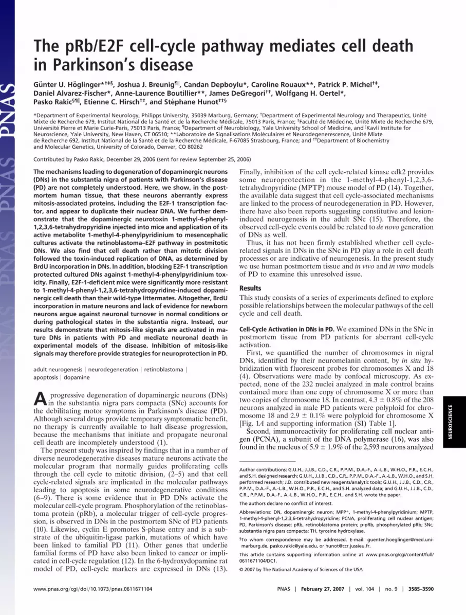
First, we quantified the number of chromosomes in nigralDNs, identified by their neuromelanin content, by in situ hy-bridization with fluorescent probes for chromosomes X and 18(4). Observations were made by confocal microscopy. As ex-pected, none of the 232 nuclei analyzed in male control brainscontained more than one copy of chromosome X or more thantwo copies of chromosome 18. In contrast, 4.3 � 0.8% of the 208neurons analyzed in male PD patients were polyploid for chro-mosome 18 and 2.9 � 0.1% were polyploid for chromosome X[Fig. 1A and supporting information (SI) Table 1].
Second, immunoreactivity for proliferating cell nuclear anti-gen (PCNA), a subunit of the DNA polymerase (16), was alsofound in the nucleus of 5.9 � 1.9% of the 2,593 neurons analyzed
Author contributions: G.U.H., J.J.B., C.D., C.R., P.P.M., D.A.-F., A.-L.B., W.H.O., P.R., E.C.H.,and S.H. designed research; G.U.H., J.J.B., C.D., C.R., P.P.M., D.A.-F., A.-L.B., W.H.O., and S.H.performed research; J.D. contributed new reagents/analytic tools; G.U.H., J.J.B., C.D., C.R.,P.P.M., D.A.-F., A.-L.B., W.H.O., P.R., E.C.H., and S.H. analyzed data; and G.U.H., J.J.B., C.D.,C.R., P.P.M., D.A.-F., A.-L.B., W.H.O., P.R., E.C.H., and S.H. wrote the paper.
The authors declare no conflict of interest.
Abbreviations: DN, dopaminergic neuron; MPP�, 1-methyl-4-phenylpyridinium; MPTP,1-methyl-4-phenyl-1,2,3,6-tetrahydropyridine; PCNA, proliferating cell nuclear antigen;PD, Parkinson’s disease; pRb, retinoblastoma protein; p-pRb, phosphorylated pRb; SNc,substantia nigra pars compacta; TH, tyrosine hydroxylase.
§To whom correspondence may be addressed. E-mail: [email protected], [email protected], or [email protected].
This article contains supporting information online at www.pnas.org/cgi/content/full/0611671104/DC1.
© 2007 by The National Academy of Sciences of the USA
www.pnas.org�cgi�doi�10.1073�pnas.0611671104 PNAS � February 27, 2007 � vol. 104 � no. 9 � 3585–3590
NEU
ROSC
IEN
CE
in PD patients (Fig. 1B). Under identical staining conditions, nosuch immunoreactivity was observed in 11,634 neurons analyzedin controls.
Third, immunoreactivity for E2F-1, a mitosis-linked transcrip-tion factor (16), was found predominantly in the cytoplasm of3.5 � 0.9% of the 1,569 neurons analyzed in PD patients (Fig.1C). Under identical staining conditions, no such staining wasobserved in 1,482 neurons analyzed in controls.
These observations provide strong evidence that aberrantcell-cycle processes are activated in DNs in PD.
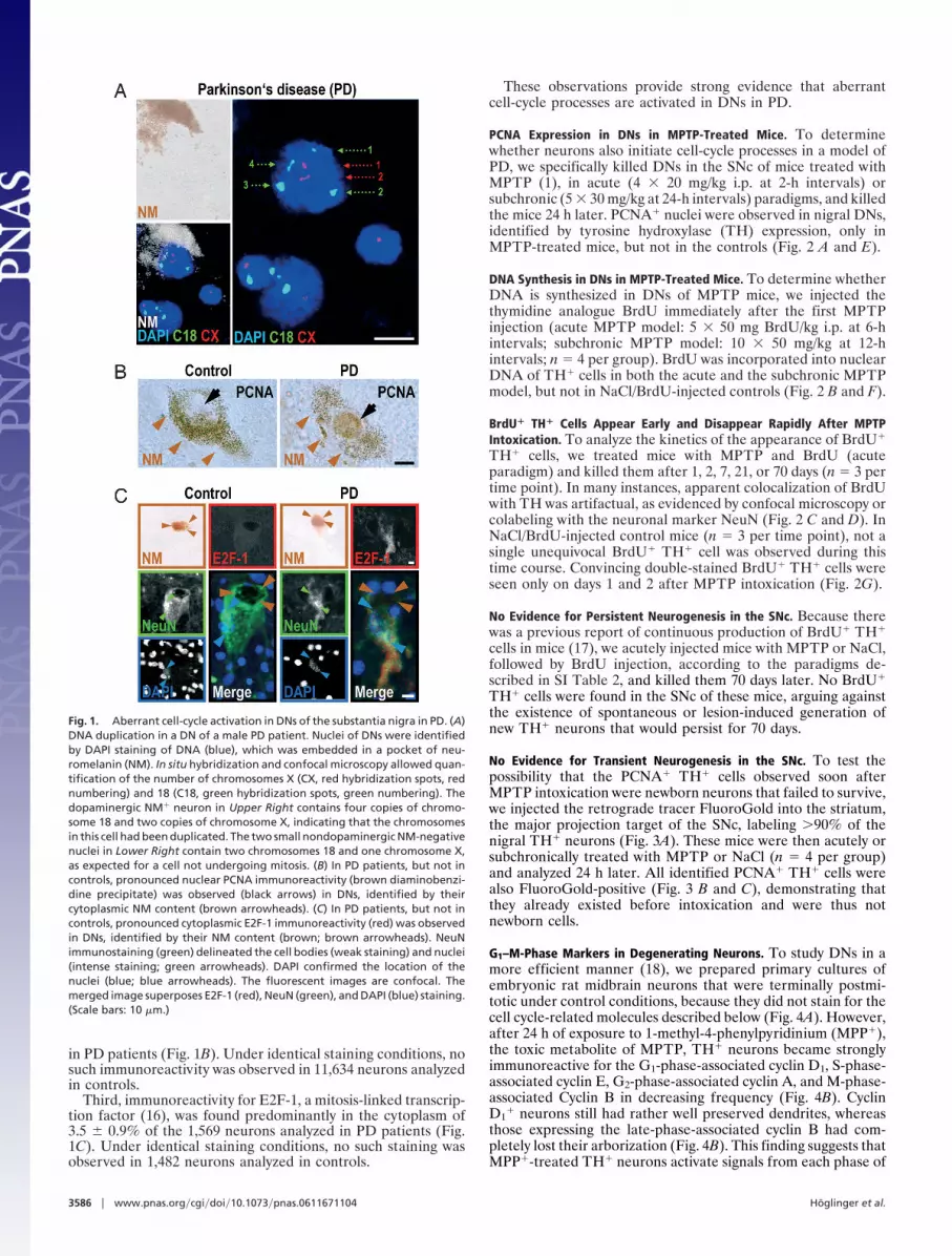
PCNA Expression in DNs in MPTP-Treated Mice. To determinewhether neurons also initiate cell-cycle processes in a model ofPD, we specifically killed DNs in the SNc of mice treated withMPTP (1), in acute (4 � 20 mg/kg i.p. at 2-h intervals) orsubchronic (5 � 30 mg/kg at 24-h intervals) paradigms, and killedthe mice 24 h later. PCNA� nuclei were observed in nigral DNs,identified by tyrosine hydroxylase (TH) expression, only inMPTP-treated mice, but not in the controls (Fig. 2 A and E).
DNA Synthesis in DNs in MPTP-Treated Mice. To determine whetherDNA is synthesized in DNs of MPTP mice, we injected thethymidine analogue BrdU immediately after the first MPTPinjection (acute MPTP model: 5 � 50 mg BrdU/kg i.p. at 6-hintervals; subchronic MPTP model: 10 � 50 mg/kg at 12-hintervals; n � 4 per group). BrdU was incorporated into nuclearDNA of TH� cells in both the acute and the subchronic MPTPmodel, but not in NaCl/BrdU-injected controls (Fig. 2 B and F).
BrdU� TH� Cells Appear Early and Disappear Rapidly After MPTPIntoxication. To analyze the kinetics of the appearance of BrdU�
TH� cells, we treated mice with MPTP and BrdU (acuteparadigm) and killed them after 1, 2, 7, 21, or 70 days (n � 3 pertime point). In many instances, apparent colocalization of BrdUwith TH was artifactual, as evidenced by confocal microscopy orcolabeling with the neuronal marker NeuN (Fig. 2 C and D). InNaCl/BrdU-injected control mice (n � 3 per time point), not asingle unequivocal BrdU� TH� cell was observed during thistime course. Convincing double-stained BrdU� TH� cells wereseen only on days 1 and 2 after MPTP intoxication (Fig. 2G).
No Evidence for Persistent Neurogenesis in the SNc. Because therewas a previous report of continuous production of BrdU� TH�
cells in mice (17), we acutely injected mice with MPTP or NaCl,followed by BrdU injection, according to the paradigms de-scribed in SI Table 2, and killed them 70 days later. No BrdU�
TH� cells were found in the SNc of these mice, arguing againstthe existence of spontaneous or lesion-induced generation ofnew TH� neurons that would persist for 70 days.
No Evidence for Transient Neurogenesis in the SNc. To test thepossibility that the PCNA� TH� cells observed soon afterMPTP intoxication were newborn neurons that failed to survive,we injected the retrograde tracer FluoroGold into the striatum,the major projection target of the SNc, labeling �90% of thenigral TH� neurons (Fig. 3A). These mice were then acutely orsubchronically treated with MPTP or NaCl (n � 4 per group)and analyzed 24 h later. All identified PCNA� TH� cells werealso FluoroGold-positive (Fig. 3 B and C), demonstrating thatthey already existed before intoxication and were thus notnewborn cells.
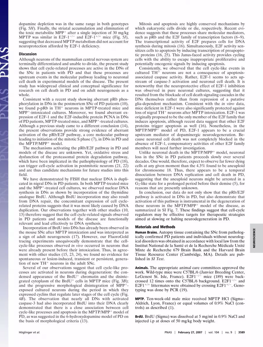
G1–M-Phase Markers in Degenerating Neurons. To study DNs in amore efficient manner (18), we prepared primary cultures ofembryonic rat midbrain neurons that were terminally postmi-totic under control conditions, because they did not stain for thecell cycle-related molecules described below (Fig. 4A). However,after 24 h of exposure to 1-methyl-4-phenylpyridinium (MPP�),the toxic metabolite of MPTP, TH� neurons became stronglyimmunoreactive for the G1-phase-associated cyclin D1, S-phase-associated cyclin E, G2-phase-associated cyclin A, and M-phase-associated Cyclin B in decreasing frequency (Fig. 4B). CyclinD1
� neurons still had rather well preserved dendrites, whereasthose expressing the late-phase-associated cyclin B had com-pletely lost their arborization (Fig. 4B). This finding suggests thatMPP�-treated TH� neurons activate signals from each phase of
Fig. 1. Aberrant cell-cycle activation in DNs of the substantia nigra in PD. (A)DNA duplication in a DN of a male PD patient. Nuclei of DNs were identifiedby DAPI staining of DNA (blue), which was embedded in a pocket of neu-romelanin (NM). In situ hybridization and confocal microscopy allowed quan-tification of the number of chromosomes X (CX, red hybridization spots, rednumbering) and 18 (C18, green hybridization spots, green numbering). Thedopaminergic NM� neuron in Upper Right contains four copies of chromo-some 18 and two copies of chromosome X, indicating that the chromosomesin this cell had been duplicated. The two small nondopaminergic NM-negativenuclei in Lower Right contain two chromosomes 18 and one chromosome X,as expected for a cell not undergoing mitosis. (B) In PD patients, but not incontrols, pronounced nuclear PCNA immunoreactivity (brown diaminobenzi-dine precipitate) was observed (black arrows) in DNs, identified by theircytoplasmic NM content (brown arrowheads). (C) In PD patients, but not incontrols, pronounced cytoplasmic E2F-1 immunoreactivity (red) was observedin DNs, identified by their NM content (brown; brown arrowheads). NeuNimmunostaining (green) delineated the cell bodies (weak staining) and nuclei(intense staining; green arrowheads). DAPI confirmed the location of thenuclei (blue; blue arrowheads). The fluorescent images are confocal. Themerged image superposes E2F-1 (red), NeuN (green), and DAPI (blue) staining.(Scale bars: 10 �m.)
3586 � www.pnas.org�cgi�doi�10.1073�pnas.0611671104 Hoglinger et al.

the cell cycle as they degenerate. The fact that fewer cells werecyclin B� than cyclin D1
� may signify that some cells die beforereaching an M-phase-like state.
G1/S-Phase Transition in Degenerating Neurons. As a next step, wesought out to determine whether cells progressed toward S phasein the expected manner. The S phase is typically initiated byphosphorylation of pRb, leading to the release of E2F transcription
factors. Unbound E2F proteins autoinduce E2F gene expressionand transactivate E2F-target genes, such as PCNA, which is re-quired for DNA synthesis (16). After MPP� intoxication, but not incontrol conditions, numerous TH� cells were immunoreactive forphosphorylated pRb (p-pRb), E2F-1, PCNA, or BrdU (Fig. 4C),suggesting that DNA replication in degenerating TH� neuronsfollows the expected program. Fewer cells were positive for p-pRb,E2F-1, or PCNA� than for BrdU, however. This discrepancy mightbe caused by the lower probability of detecting transiently activatedsignals in cells passing through the G1/S transition than BrdU thatbecomes permanently incorporated.
Co-Occurrence of BrdU Incorporation and Caspase-3 Activation. Totest whether the cell cycle and cell death were activated simul-taneously or mutually exclusively, we stained for BrdU andcaspase-3. Among the 643 MPP�-intoxicated TH� neuronsanalyzed in vitro, 88% of those containing activated caspase-3also had BrdU� nuclei, suggesting that the programs for cellcycle and apoptosis are activated concomitantly (Fig. 4D).
Cell-Cycle Signals in Apoptotic TH� Cells in the SNc. We then askedwhether the cell-cycle signals that we observed in vitro wererecapitulated in vivo. TH� apoptotic cells, identified accordingto morphological criteria (13) 24 h after acute MPTP intoxica-tion in vivo, were p-pRb� and E2F-1�, suggesting that, like invitro, degenerating neurons activate cell-cycle signals in vivo (SIFig. 6).
Caspase Inhibition Does Not Prevent BrdU Incorporation. To under-stand the hierarchy of cell cycle/cell death signaling, we inhibitedcaspase signaling. In MPP�-intoxicated TH� neurons in vitro,inhibition of caspases with the broad-spectrum inhibitor zVAD-fmk did not prevent BrdU incorporation (Fig. 5 A and B),suggesting that caspase activation is not functionally upstream ofDNA synthesis.
E2F-1 Suppression Prevents MPP�-Induced Caspase-3 Activation. Wethen tested, conversely, whether cell-cycle activation is upstream
Fig. 2. Nuclear DNA synthesis in DNs in the SNc of mice 24 h after MPTPintoxication. (A) Nuclear PCNA immunoreactivity (red) in a DN, identified byTH expression (green) is shown. An overview (ov) and an inset showing ahigher magnification of the cell in a confocal reconstruction of the xy, xz, andyz planes are shown. (B) BrdU immunoreactivity (green) in a TH� neuron (red).Overview (ov) and detail of the boxed area showing the cell in a confocalreconstruction of the xy, xz, and yz planes. The superposition of BrdU and THappears yellow, demonstrating that both markers colocalize in the samecellular compartment, suggesting a breakdown of the nuclear membrane.Note the disintegrated TH� cytoplasm and the condensed BrdU� chromatin,suggesting that the cell is in a state of degeneration. (C and D) Examples ofartifactual BrdU-TH colocalization. (C) A BrdU� nucleus (green) that errone-ously appears to belong to a TH� neuron (red) when seen with conventionalmicroscopy: an overview (ov) and an enlarged detail in the boxed area areshown. However, confocal microscopy of sections 1 �m apart shows that theBrdU� nucleus is juxtaposed to the TH� cell and that the nucleus of the TH� cell(best seen at 5 �m; *) is not BrdU�. (D) A BrdU� nucleus (green) that errone-ously appears to belong to a TH� neuron (white), when observed by confocalmicroscopy. An overview (ov) and an enlarged detail are shown). However,the lack of counterstaining with the neuronal marker NeuN (red) demon-strates that the BrdU� nucleus (arrowhead) is not neuronal in nature, incontrast to the neighboring TH� BrdU� cell (arrow). The TH� immunoreactiv-ity in the vicinity of the BrdU� nucleus is contained in the neurites of neigh-boring DNs. (Scale bars: 10 �m.) (E and F) Absolute numbers of PCNA� TH� cells(E) and BrdU� TH� cells (F) per SNc of mice 24 h after NaCl injection (control)or after intoxication with MPTP in the acute (aMPTP) or subchronic (cMPTP)paradigm. *, P � 0.05, ANOVA followed by post hoc test, vs. NaCl. (G) Absolutenumbers of BrdU� TH� cells per SNc of mice at different time points (d � days)after MPTP intoxication in the acute paradigm (aMPTP). Two-way ANOVAshowed a significant effect of treatment (MPTP vs. NaCl, P � 0.001), time (P �0.01), and treatment � time interaction (P � 0.01). *, P � 0.05, post hoc test,vs. NaCl.
Fig. 3. PCNA-expressing dopaminergic cells in the SNc of MPTP mice are notnewborn neurons, but preexist. (A) TH� neurons (green) in the SNc werelabeled by striatal injection of the retrograde tracer FluoroGold (FG, blue) 3days before MPTP intoxication. (B) Overview (ov) showing a cell (arrow) in theSNc of a mouse 24 h after acute MPTP intoxication expressing both TH (green)and PCNA (red). The presence of FluoroGold (blue) demonstrated that the cellexisted before the lesion, ruling out the possibility that it was a stem cell-derived newborn dopaminergic cell. (C) An enlarged detail from the boxedarea in B shown as a confocal reconstruction in the xy, xz, and yz plane, toconfirm the colocalization of TH, PCNA, and FG. [Scale bars: 100 �m (A); 10 �m(B and C).]
Hoglinger et al. PNAS � February 27, 2007 � vol. 104 � no. 9 � 3587
NEU
ROSC
IEN
CE

of caspase activation in the MPP� model in vitro. E2F inductionis a bottleneck event upstream of PCNA expression and DNAreplication (14). E2F-1 and E2F-4 are the most abundant E2Ffamily members in the brain, but E2F-1, unlike E2F-4, ispredominately regulated by p-pRb. Furthermore, E2F-1 hasbeen shown to be the E2F family member that most potentlysensitizes cells for apoptosis (16, 19). Thus, we chose to inhibitE2F-1 expression by transfection of cultured neurons with avalidated E2F-1 antisense oligonucleotide; transfection effi-ciency was 80% (20). E2F-1 sense oligonucleotides, missenseoligonucleotides (data not shown), and omission of oligonucle-otides (data not shown) were used as controls. The antisenseoligonucleotide, but not the controls, decreased E2F-1 expres-sion, activation of caspase-3, and loss of TH� cells in MPP�-intoxicated cultures (Fig. 5 C–E), suggesting that E2F-1 signalingis upstream of caspase activation and cell death in this model.
E2F-1�/� Mice Are Protected Against MPTP. Finally, we testedwhether E2F-1-deficiency in mice (19) could mitigate MPTPtoxicity in vivo. Indeed, after both acute and subchronic MPTPintoxication, stereological cell counts demonstrated that therewas significantly less nigral TH� cell loss in E2F-1�/� mice thanin E2F-1�/� mice (Fig. 5 F and G). Yet, the extent of striatal
Fig. 4. Cell-cycle events do not occur in postmitotic TH� midbrain neurons invitro under control conditions (A), but are observed after MPP� intoxication(B–D). (A) In control cultures, cell cycle-associated proteins, such as cyclin D1
(CyD1, green) were never expressed in TH� neurons (red). (B) However, after24 h of intoxication with MPP�, the G1-phase-associated CyD1, the S-phase-associated cyclin E (CyE), the G2-phase-associated cyclin A (CyA), and theM-phase-associated cyclin B (CyB) (green) were expressed in TH� cells (red).The graph shows the percentage of TH� cells expressing CyD1, CyE, CyA, andCyB after 24 h of MPP� intoxication. (C) Immunoreactivity for the S-phase-associated markers p-pRb, E2F-1, PCNA, and BrdU (green) was detected in TH�
DNs (red) after 24 h of MPP� intoxication. Visualization of chromatin withDAPI (blue) demonstrated the perinuclear expression of E2F-1 (green) in TH�
neurons (red). The graph shows the percentage of TH� cells immunoreactivefor p-pRb, E2F-1, PCNA, and BrdU after 24 h of MPP� intoxication. (D) A 24-htreatment of cultured neurons with MPP� led to the appearance of morpho-logically disintegrated cells (structure, gray) with condensed chromatin (DAPI,white) and dopaminergic phenotype (TH�, red) that were immunopositive forboth the activated form of the downstream effector caspase-3 (aC3, blue) andBrdU (green). The graph shows the quantification of the presence (�) orabsence (�) of BrdU and aC3 immunoreactivity in TH� cells after 24 h of MPP�
treatment. [Scale bars: 20 �m (A and B); 10 �m (C and D).]
Fig. 5. E2F-1 signaling is upstream of MPP�-induced caspase-3 activation, andE2F-1 deficiency provides protection against MPTP. (A and B) Caspase activationis not upstream of cell-cycle signaling. Caspase inhibition (CI) with 200 �MzVAD-fmk during a 24-h intoxication of TH� neurons (red) with MPP� preventedthe appearance of immunoreactivity for the activated form of caspase-3 (aC3,green) (A),butnotthe incorporationofBrdU(green) (B).DAPI (blue) stainsnuclei.The superposition of red and green appears yellow. The superposition of red,green, and blue appears white. The graphs show the percentage of TH� cellsimmunoreactive for aC3 (A) and BrdU (B). *, P � 0.05; n.s., not significant. (C–E)E2F-1 inhibition protects neurons in vitro from MPP�. Inhibition of E2F-1 expres-sion with antisense (AS), but not sense (S) oligonucleotides during a 24-h intox-ication of TH� DNs (red) with MPP� attenuated the appearance of immunore-activity for E2F-1 (green) (C) and aC3 (green) (D) and the MPP�-induced loss ofTH� cell (E).DAPI (blue) stainsnuclei.Thegraphs showthepercentageofTH� cellsimmunoreactive for E2F-1 (C) and aC3 (D) and the percentage of surviving TH�
cells (E) after transfection with E2F-1 sense (S) or antisense (AS) oligonucleotidesand grown for 24 h in the absence (C) or the presence (M) of MPP�. *, P � 0.05;
***, P � 0.001; n.s., not significant. (F–I) E2F-1 deficiency protects neurons in vivofrom MPTP. (F) Dopaminergic (TH�) cell bodies in the SNc, assessed 7 days afterMPTP intoxication in both the subchronic (cMPTP) and acute paradigm (aMPTP),weresignificantlyprotected inE2F-1�/� micecomparedwithE2F-1�/� mice.*,P�0.05;n.s.,not significant. (G)RepresentativephotomicrographsofTH� cellbodiesin the SNc in E2F-1�/� mice compared with E2F-1�/� mice 7 days after MPTPintoxication. (H) The striatal dopamine depletion, assessed 7 days after MPTPintoxication in both paradigms did not differ between E2F-1�/� and E2F-1�/�
mice. (I) The time course of the accumulation and elimination of the toxicMPTP-metabolite MPP� after an injection of MPTP was identical in E2F-1�/� miceand wild-type littermates (E2F-1�/�). [Scale bars: 10 �m (A-D); 100 �m (E and G).]
3588 � www.pnas.org�cgi�doi�10.1073�pnas.0611671104 Hoglinger et al.
dopamine depletion was in the same range in both genotypes(Fig. 5H). Finally, the striatal accumulation and elimination ofthe toxic metabolite MPP� after a single injection of 30 mg/kgMPTP was similar in E2F-1�/� and E2F-1�/� mice (Fig. 5I),suggesting that decreased MPTP metabolism did not account forneuroprotection afforded by E2F-1 deficiency.
DiscussionAlthough neurons of the mammalian central nervous system areterminally differentiated and unable to divide, the present studyshows that cell cycle-related processes are active in the DNs ofthe SNc in patients with PD and that these processes areupstream events in the molecular pathway leading to neuronalcell death in experimental models of the disease. The presentstudy has widespread clinical and conceptual significance forresearch on cell death in PD and on adult neurogenesis as awhole.
Consistent with a recent study showing aberrant pRb phos-phorylation in DNs in the postmortem SNc of PD patients (10),we found p-pRb in TH� neurons in MPTP-treated mice andMPP�-intoxicated cultures. Moreover, we found aberrant ex-pression of E2F-1 and the E2F-inducible protein PCNA in DNsof PD patients, MPTP-treated mice, and MPP�-treated cultures.Although a previous study came to a different conclusion (14),the present observations provide strong evidence of aberrantactivation of the pRb/E2F pathway, a core molecular pathwayleading to initiation of the mitotic S phase (7), in DNs in PD andthe MPTP/MPP� model.
The mechanisms activating the pRb/E2F pathway in PD andmodels of the disease are unknown. Yet, oxidative stress anddysfunction of the proteasomal protein degradation pathway,which have been implicated in the pathophysiology of PD (18),can trigger cell-cycle activation in postmitotic neurons (21, 22)and are thus candidate mechanisms for future studies into thisfield.
We have demonstrated by FISH that nuclear DNA is dupli-cated in nigral DNs in PD patients. In both MPTP-treated miceand the MPP�-treated cell cultures, we observed nuclear DNAsynthesis in DNs as shown by incorporation of the thymidineanalogue BrdU. Although BrdU incorporation may also resultfrom DNA repair, the concomitant expression of cell cycle-related proteins suggests that it was most likely caused by DNAduplication. Our observations together with other studies (10,13) therefore suggest that the cell cycle-related signals observedin PD patients and models of the disease are functionallyrelevant and lead effectively to DNA synthesis.
Incorporation of BrdU into DNs has already been observed inthe mouse SNc after MPTP intoxication and was interpreted asa sign of adult neurogenesis (17). However, our FluoroGoldtracing experiments unequivocally demonstrate that the cell-cycle-like processes observed in vivo occurred in neurons thatwere already present before their intoxication. Thus, in agree-ment with other studies (15, 23, 24), we found no evidence forspontaneous or lesion-induced, transient or persistent, genera-tion of new TH� neurons in the adult SNc.
Several of our observations suggest that cell cycle-like pro-cesses are activated in neurons during degeneration: the con-densed appearance of the BrdU� chromatin and the disinte-grated cytoplasm of the BrdU� cells in MPTP mice (Fig. 3B);and the progressive morphological disintegration of MPP�-exposed cultured neurons during the period in which theyexpressed cyclins that regulate later stages of the cell cycle (Fig.4B). The observation that nearly all DNs with activatedcaspase-3 had also incorporated BrdU into their DNA clearlydemonstrated that there is a close association between cellcycle-like processes and apoptosis in the MPTP/MPP� model ofPD, as was suggested in the 6-hydroxydopamine model of PD onthe basis of morphological criteria (13).
Mitosis and apoptosis are highly conserved mechanisms bywhich eukaryotic cells divide or die, respectively. Recent evi-dence suggests that these processes share molecular mediators,such as pRb and the E2F family of transcription factors (6–9).The transcriptional activity of E2F prepares cells for DNAsynthesis during mitosis (16). Simultaneously, E2F activity sen-sitizes cells to apoptosis by inducing transcription of proapopto-tic proteins (16, 25). This Janus-faced activity provides cyclingcells with the ability to escape inappropriate proliferative andpotentially oncogenic signals by inducing apoptosis.
Consistently, we observed that the cell cycle-like events incultured TH� neurons are not a consequence of apoptosis-associated caspase activity. Rather, E2F-1 seems to acts up-stream of caspase-3 activation and neuronal cell death. It isnoteworthy that the neuroprotective effect of E2F-1 inhibitionwas observed in pure neuronal cultures, suggesting that itresulted from the blockade of cell death signaling cascades in theDNs themselves rather than from repression of a putativeglia-dependent mechanism. Consistent with the in vitro data,mice deficient in E2F-1 were also significantly protected againstloss of nigral TH� neurons after MPTP intoxication. E2F-1 wasoriginally proposed to be the only member of the E2F family thatinduces apoptosis, although recent data suggest that other E2Fproteins trigger apoptosis as well (16). Nonetheless, in theMPTP/MPP� model of PD, E2F-1 appears to be a crucialupstream mediator of dopaminergic neurodegeneration. Be-cause neuronal cell death was not completely blocked in theabsence of E2F-1, compensatory activities of other E2F familymembers will need further investigation.
Unlike neuronal death in the MPTP/MPP� model, neuronalloss in the SNc in PD patients proceeds slowly over severaldecades. One would, therefore, expect to observe far fewer dyingcells at any given moment than the 4.3% found to be aneuploidfor chromosome 18. Thus, there appears to be a temporaldissociation between DNA replication and cell death in PD,suggesting that the aneuploid neurons might be arrested in aG2-like state for a prolonged period before their demise (5), forreasons that are presently unknown.
In conclusion, our data do not only show that the pRb/E2Fpathway is activated in DNs in PD, but also demonstrate thatactivation of this pathway is instrumental in the degeneration ofthese neurons in the MPTP/MPP� model of the disease, assummarized in SI Fig. 7. These findings suggest that cell-cycleregulators may be effective targets for therapeutic strategiesaimed at slowing or halting neurodegeneration in PD.
Materials and MethodsHuman Brains. Autopsy tissue containing the SNc from patholog-ically confirmed PD patients and individuals without neurolog-ical disorders was obtained in accordance with local law from theInstitut National de la Sante et de la Recherche Medicale UniteMixte de Recherche 679 Brain Bank and the Harvard BrainTissue Resource Center (Cambridge, MA). Details are pub-lished in SI Text.
Animals. The appropriate animal care committees approved thework. Wild-type mice were C57BL/6 (Janvier Breeding Center,LeGenest St. Isle, France). E2F1�/� mice (189) were back-crossed 12 times onto the C57BL/6 background. E2F1�/� andE2F1�/� littermates were obtained by crossing E2F1�/�. Geno-typing was done by PCR (19).
MPTP. Ten-week-old male mice received MPTP HCl (Sigma–Aldrich, Lyon, France) or equal volumes of 0.9% NaCl (con-trols) as described (1).
BrdU. BrdU (Sigma) was dissolved at 5 mg/ml in 0.9% NaCl andinjected i.p at doses of 50 mg/kg body weight.
Hoglinger et al. PNAS � February 27, 2007 � vol. 104 � no. 9 � 3589
NEU
ROSC
IEN
CE
FluoroGold. FluoroGold (0.4 �l 4%) was injected in the striatum(coordinates: A �1.2, L �1.6, V �2.5 mm) of anesthetized mice(10 ml/kg of 1% ketamine/0.2% xylazine).
Tissue Preparation. For immunohistochemistry, mice were killedwith 100 mg/kg pentobarbital i.p. and perfused transcardially.Postfixed frozen brains were cut in 20-�m coronal sections. ForHPLC, mice were killed by cervical dislocation. The dissectedstriata were homogenized in 500 �l of 0.4 M perchloric acid,centrifuged (20 min, 13,000 � g, 4°C), and passed through a0.2-�m filter.
HPLC. Dopamine was measured by RP-HPLC with electrochem-ical detection (potential 750 mV) under isocratic conditions withan Ag/AgCl reference electrode. MPP� was determined byHPLC with UV detection (wavelength 295 nm).
Cell Culture, Immunohistochemistry, and Image Analysis. Details arepublished in SI Text.
Antisense Oligonucleotides. Thirty minutes before intoxication,mouse cultures were transfected with 0.35 �M of mouse-specificE2F-1 antisense (5�-GAA GCG TTT GGT GGT CAG AT-3�),E2F-1 sense (5�-ATC TGA CCA CCA AAC GCT TC-3�), ormissense (5�-TTG CCT CCC TTT GAA AAA TG-3�) oligo-nucleotides or without oligonucleotides as described (20).
In Situ Hybridization. Ten-micrometer cryostat sections of humanSNc were fixed at �20°C in 3:1 methanol/acetic acid, rinsed ina graded alcohol series, digested with 0.5% pepsin in 0.01 M HCl(13 min, 37°C), washed in PBS, postfixed in 1% paraformalde-hyde (5 min), washed, and dehydrated in a graded alcohol series.Nuclei and DNA probes (CEP; Vysis, Des Plaines, IL) weredenatured (76°C, 2 min) and hybridized overnight in a humid-ified chamber (37°C). Sections were rinsed in 0.4� saline-sodiumcitrate with 0.3% Nonidet P-40 (American Bioanalytical, Natick,MA) (72°C, 2 min) and 2� SSC with 0.1% Nonidet P-40 (20°C,5 min). Autofluorescence was reduced with Sudan black. Vysiscontrol slides and female tissue (for chromosome X probes) werehybridized to ensure sensitivity and specificity.
Statistics. Data are shown as mean � SEM. Normal parametricdata were compared with the two-sided, unpaired t test orANOVA followed by post hoc Student-Newmann-Keuls test.P � 0.05 was considered significant.
We thank S. Stei for technical assistance and the Harvard Brain TissueResource Center for postmortem tissue. This work was supported byHarvard Brain Tissue Resource Center Grant R24 MH 68855, GermanMinistry of Education and Research Grants BMBF-01GN0513 and01GO0201, European Union Grant LSHM-CT-2003-503330, the PeterHofmann Project, Fondation pour la Recherche Medicale GrantACE20030307094, the Institut National de la Sante et de la RechercheMedicale, and the Public Health Service (P.R.).
1. Dauer W, Przedborski S (2003) Neuron 39:889–909.2. Jordan-Sciutto KL, Wang G, Murphey-Corb M, Wiley CA (2002) J Neurosci
22:2185–2195.3. Ranganathan S, Bowser R (2003) Am J Pathol 162:823–835.4. Yang Y, Geldmacher DS, Herrup K (2001) J Neurosci 21:2661–2668.5. Yang Y, Mufson EJ, Herrup K (2003) J Neurosci 23:2557–2563.6. Nguyen MD, Mushynski WE, Julien JP (2002) Cell Death Differ 9:1294–1306.7. Greene LA, Biswas SC, Liu DX (2004) Cell Death Differ 11:49–60.8. Herrup K, Neve R, Ackerman SL, Copani A (2004) J Neurosci 24:9232–9239.9. Kuan CY, Schloemer AJ, Lu A, Burns KA, Wenig W-L, Williams M-T, Strauss
KI, Vorhees CV, Flavell RA, Davis RJ, et al. (2004) J Neurosci 24:10763–10772.10. Jordan-Sciutto KL, Dorsey R, Chalovich EM, Hammond RR, Achim CL
(2003) J Neuropat Exp Neurol 62:68–74.11. Staropoli JF, McDermott C, Martinat C, Schulman B, Demireva E, Abeliovich
A (2003) Neuron 37:735–749.12. West AB, Dawson VL, Dawson TM (2005) Trends Neurosci 28:348–352.13. El-Khodor BF, Oo TF, Kholodilov N, Burke RE (2003) Exp Neurol
179:17–27.14. Smith PD, Crocker SJ, Jackson-Lewis V, Jordan-Sciutto KL, Hayley S, Mount
MP, O’Hare MJ, Callaghan S, Slack RS, Przedborski S, et al. (2003) Proc NatlAcad Sci USA 100:13650–13655.
15. Borta A, Hoglinger GU (2007) J Neurochem 100:587–595.16. DeGregori J (2002) Biochim Biophys Acta 1602:131–150.17. Zhao M, Momma S, Delfani K, Carlen M, Cassidy RM, Johansson CB, Brismar
H, Shupliakov O, Frisen J, Janson AM (2002) Proc Natl Acad Sci USA100:7925–7930.
18. Hoglinger GU, Carrard G, Michel PP, Friguet B, Hirsch EC (2003) J Neuro-chem 86:1297–1307.
19. Field SJ, Tsai F-Y, Kuo F, Zubiaga AM, Kaelin WG, Livingston DM, OrkinSH, Greenberg ME (1996) Cell 85:549–561.
20. Trinh E, Boutillier AL, Loeffler JP (2001) Mol Cell Neurosci 17:342–353.21. Rideout HJ, Wang Q, Park DS, Stefanis L (2003) J Neurosci 23:1237–1245.22. Klein JA, Longo-Guess CM, Rossmann MP, Seburn KL, Hurd RE, Frankel
WN, Bronson RT, Ackerman SL (2002) Nature 419:367–374.23. Lie DC, Dziewczapolski G, Willhoite AR, Kaspar BK, Shults CW, Gage FH
(2002) J Neurosci 22:6639–6649.24. Frielingsdorf H, Schwarz K, Brundin P, Mohapel P (2004) Proc Natl Acad Sci
USA 101:10177–10182.25. Liu DX, Greene LA (2001) Neuron 32:425–438.
3590 � www.pnas.org�cgi�doi�10.1073�pnas.0611671104 Hoglinger et al.