the peripheral nervous system (pns) and reflex activity
TRANSCRIPT
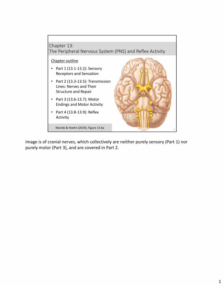
Chapter 13: The Peripheral Nervous System (PNS) and Reflex Activity
1
Chapter outline
• Part 1 (13.1-13.2): Sensory Receptors and Sensation
• Part 2 (13.3-13.5): Transmission Lines: Nerves and Their Structure and Repair
• Part 3 (13.6-13.7): Motor Endings and Motor Activity
• Part 4 (13.8-13.9): Reflex Activity
Marieb & Hoehn (2019), Figure 13.6a
Image is of cranial nerves, which collectively are neither purely sensory (Part 1) nor purely motor (Part 3), and are covered in Part 2.
1
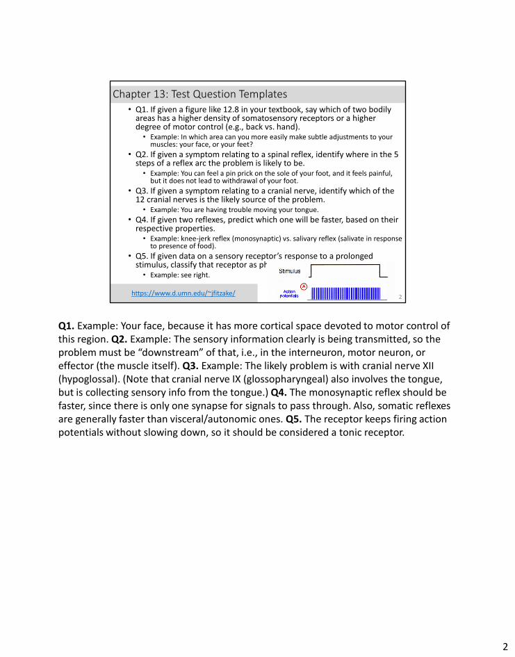
Chapter 13: Test Question Templates• Q1. If given a figure like 12.8 in your textbook, say which of two bodily
areas has a higher density of somatosensory receptors or a higher degree of motor control (e.g., back vs. hand).• Example: In which area can you more easily make subtle adjustments to your
muscles: your face, or your feet?
• Q2. If given a symptom relating to a spinal reflex, identify where in the 5 steps of a reflex arc the problem is likely to be.• Example: You can feel a pin prick on the sole of your foot, and it feels painful,
but it does not lead to withdrawal of your foot.
• Q3. If given a symptom relating to a cranial nerve, identify which of the 12 cranial nerves is the likely source of the problem.• Example: You are having trouble moving your tongue.
• Q4. If given two reflexes, predict which one will be faster, based on their respective properties.• Example: knee-jerk reflex (monosynaptic) vs. salivary reflex (salivate in response
to presence of food).
• Q5. If given data on a sensory receptor’s response to a prolonged stimulus, classify that receptor as phasic or tonic.• Example: see right.
2https://www.d.umn.edu/~jfitzake/
Q1. Example: Your face, because it has more cortical space devoted to motor control of this region. Q2. Example: The sensory information clearly is being transmitted, so the problem must be “downstream” of that, i.e., in the interneuron, motor neuron, or effector (the muscle itself). Q3. Example: The likely problem is with cranial nerve XII (hypoglossal). (Note that cranial nerve IX (glossopharyngeal) also involves the tongue, but is collecting sensory info from the tongue.) Q4. The monosynaptic reflex should be faster, since there is only one synapse for signals to pass through. Also, somatic reflexes are generally faster than visceral/autonomic ones. Q5. The receptor keeps firing action potentials without slowing down, so it should be considered a tonic receptor.
2
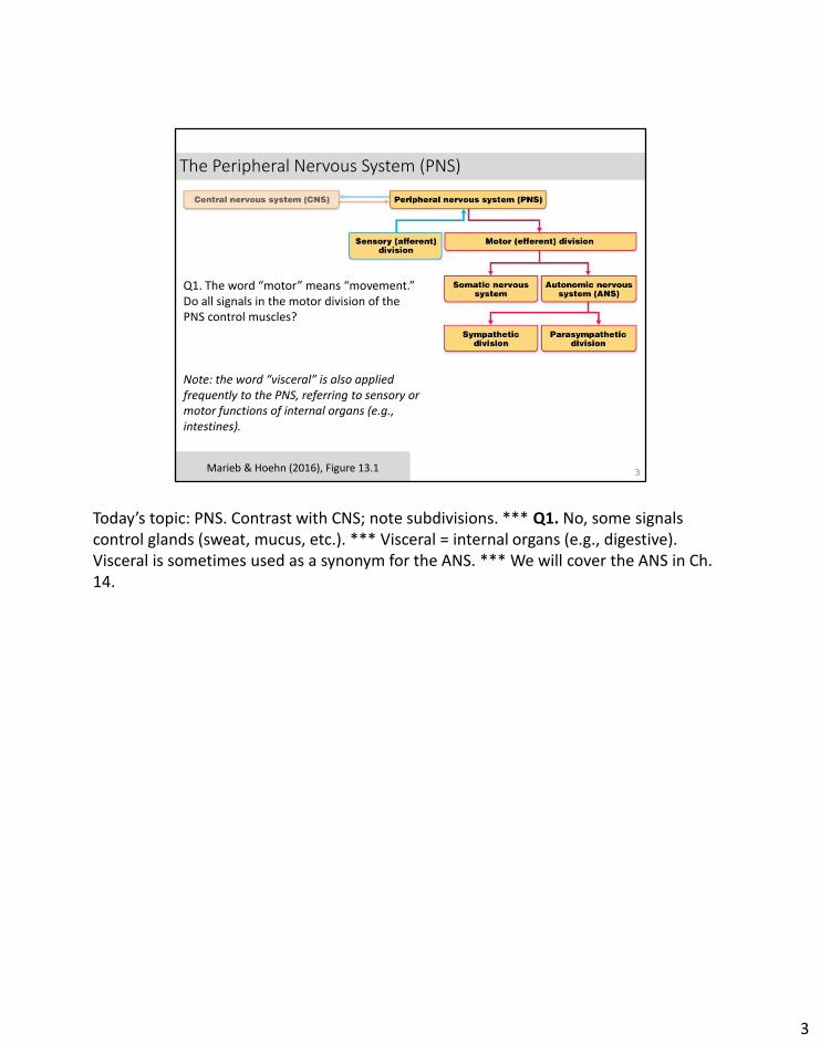
The Peripheral Nervous System (PNS)
3Marieb & Hoehn (2016), Figure 13.1
Q1. The word “motor” means “movement.” Do all signals in the motor division of the PNS control muscles?
Note: the word “visceral” is also applied frequently to the PNS, referring to sensory or motor functions of internal organs (e.g., intestines).
Today’s topic: PNS. Contrast with CNS; note subdivisions. *** Q1. No, some signals control glands (sweat, mucus, etc.). *** Visceral = internal organs (e.g., digestive). Visceral is sometimes used as a synonym for the ANS. *** We will cover the ANS in Ch. 14.
3
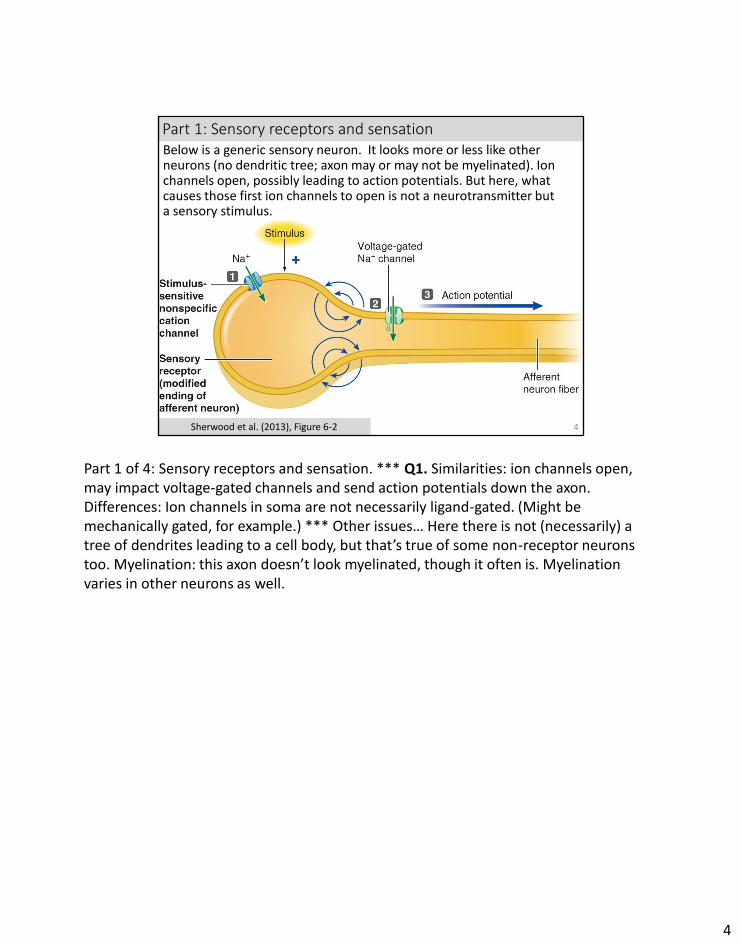
4Sherwood et al. (2013), Figure 6-2
Part 1: Sensory receptors and sensation Below is a generic sensory neuron. It looks more or less like other neurons (no dendritic tree; axon may or may not be myelinated). Ion channels open, possibly leading to action potentials. But here, what causes those first ion channels to open is not a neurotransmitter but a sensory stimulus.
Part 1 of 4: Sensory receptors and sensation. *** Q1. Similarities: ion channels open, may impact voltage-gated channels and send action potentials down the axon. Differences: Ion channels in soma are not necessarily ligand-gated. (Might be mechanically gated, for example.) *** Other issues… Here there is not (necessarily) a tree of dendrites leading to a cell body, but that’s true of some non-receptor neurons too. Myelination: this axon doesn’t look myelinated, though it often is. Myelination varies in other neurons as well.
4
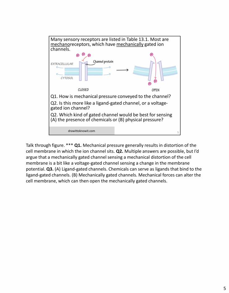
Many sensory receptors are listed in Table 13.1. Most are mechanoreceptors, which have mechanically gated ion channels.
Q1.
Q1. How is mechanical pressure conveyed to the channel?
Q2. Is this more like a ligand-gated channel, or a voltage-gated ion channel?
Q2. Which kind of gated channel would be best for sensing (A) the presence of chemicals or (B) physical pressure?
5drawittoknowit.com
Talk through figure. *** Q1. Mechanical pressure generally results in distortion of the cell membrane in which the ion channel sits. Q2. Multiple answers are possible, but I’d argue that a mechanically gated channel sensing a mechanical distortion of the cell membrane is a bit like a voltage-gated channel sensing a change in the membrane potential. Q3. (A) Ligand-gated channels. Chemicals can serve as ligands that bind to the ligand-gated channels. (B) Mechanically gated channels. Mechanical forces can alter the cell membrane, which can then open the mechanically gated channels.
5
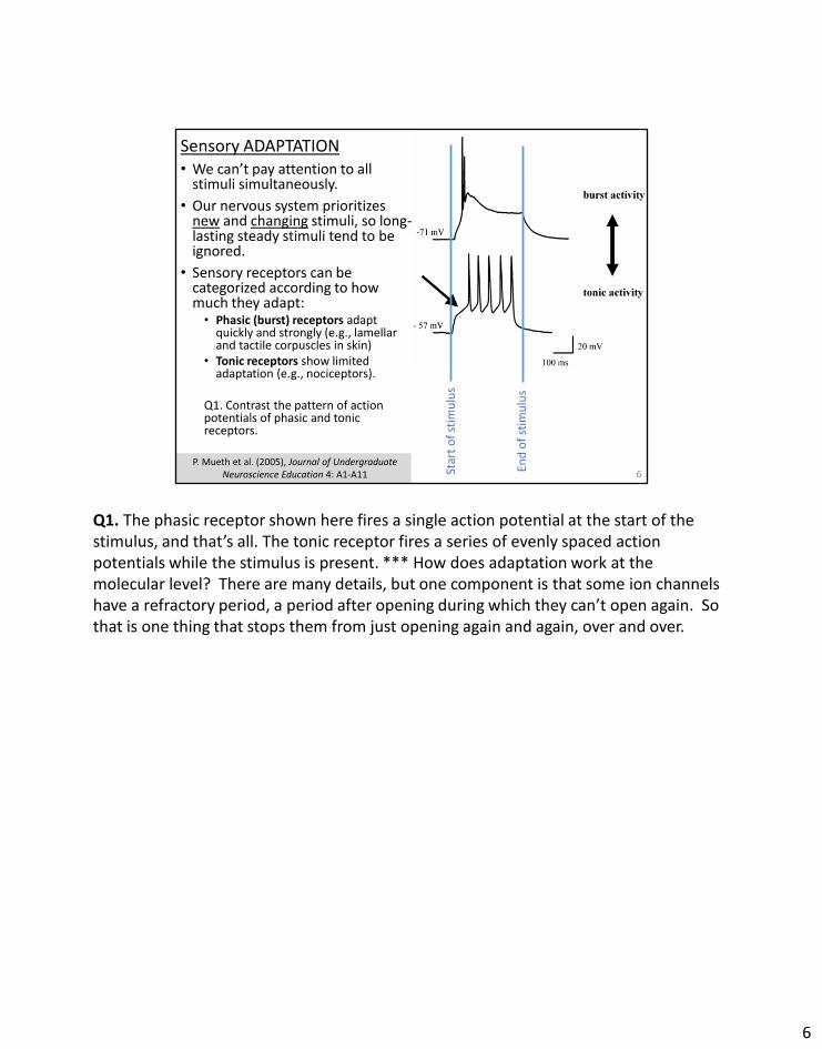
Sensory ADAPTATION• We can’t pay attention to all
stimuli simultaneously.
• Our nervous system prioritizes new and changing stimuli, so long-lasting steady stimuli tend to be ignored.
• Sensory receptors can be categorized according to how much they adapt:• Phasic (burst) receptors adapt
quickly and strongly (e.g., lamellar and tactile corpuscles in skin)
• Tonic receptors show limited adaptation (e.g., nociceptors).
Q1. Contrast the pattern of action potentials of phasic and tonic receptors.
6P. Mueth et al. (2005), Journal of Undergraduate
Neuroscience Education 4: A1-A11 Star
t o
f st
imu
lus
End
of
stim
ulu
sQ1. The phasic receptor shown here fires a single action potential at the start of the stimulus, and that’s all. The tonic receptor fires a series of evenly spaced action potentials while the stimulus is present. *** How does adaptation work at the molecular level? There are many details, but one component is that some ion channels have a refractory period, a period after opening during which they can’t open again. So that is one thing that stops them from just opening again and again, over and over.
6
Part 2: Transmission lines: nerves and their structure and repair
7Marieb & Hoehn (2019), Figure 13.4
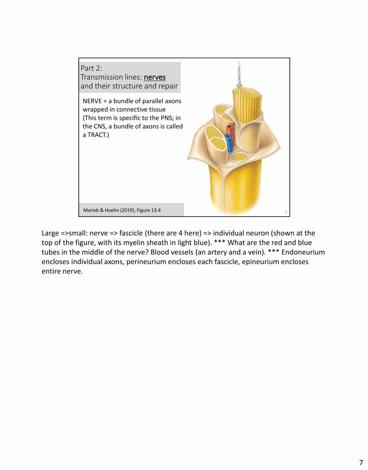
NERVE = a bundle of parallel axons wrapped in connective tissue(This term is specific to the PNS; in the CNS, a bundle of axons is called a TRACT.)
Large =>small: nerve => fascicle (there are 4 here) => individual neuron (shown at the top of the figure, with its myelin sheath in light blue). *** What are the red and blue tubes in the middle of the nerve? Blood vessels (an artery and a vein). *** Endoneurium encloses individual axons, perineurium encloses each fascicle, epineurium encloses entire nerve.
7
Peripheral nerve organization: nerve plexuses
8Marieb & Hoehn (2019), Figure 13.7and slideplayer.com/slide/6372151/
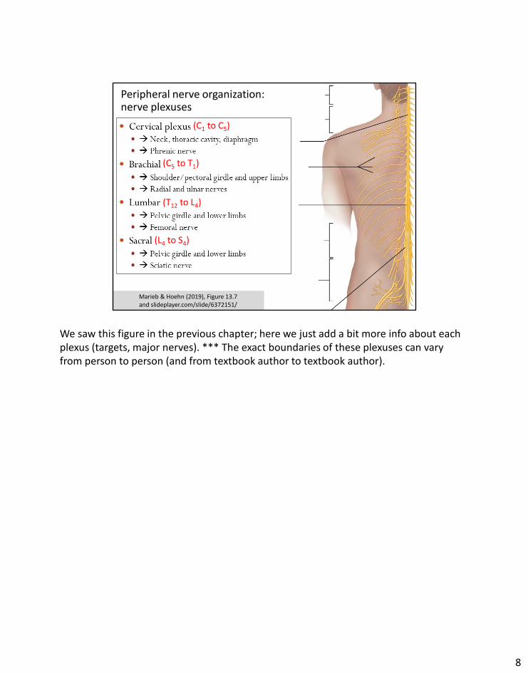
(C1 to C5)
(C5 to T1)
(T12 to L4)
(L4 to S4)
We saw this figure in the previous chapter; here we just add a bit more info about each plexus (targets, major nerves). *** The exact boundaries of these plexuses can vary from person to person (and from textbook author to textbook author).
8
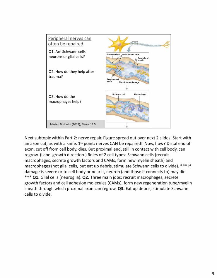
Peripheral nerves can often be repaired
9Marieb & Hoehn (2019), Figure 13.5
Q1. Are Schwann cells neurons or glial cells?
Q2. How do they help after trauma?
Q3. How do the macrophages help?
Next subtopic within Part 2: nerve repair. Figure spread out over next 2 slides. Start with an axon cut, as with a knife. 1st point: nerves CAN be repaired! Now, how? Distal end of axon, cut off from cell body, dies. But proximal end, still in contact with cell body, can regrow. (Label growth direction.) Roles of 2 cell types: Schwann cells (recruit macrophages, secrete growth factors and CAMs, form new myelin sheath) and macrophages (not glial cells, but eat up debris, stimulate Schwann cells to divide). *** If damage is severe or to cell body or near it, neuron (and those it connects to) may die. *** Q1. Glial cells (neuroglia). Q2. Three main jobs: recruit macrophages, secrete growth factors and cell adhesion molecules (CAMs), form new regeneration tube/myelin sheath through which proximal axon can regrow. Q3. Eat up debris, stimulate Schwann cells to divide.
9
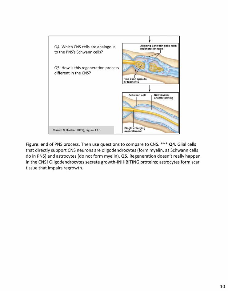
10
Q4. Which CNS cells are analogous to the PNS’s Schwann cells?
Q5. How is this regeneration process different in the CNS?
Marieb & Hoehn (2019), Figure 13.5
Figure: end of PNS process. Then use questions to compare to CNS. *** Q4. Glial cells that directly support CNS neurons are oligodendrocytes (form myelin, as Schwann cells do in PNS) and astrocytes (do not form myelin). Q5. Regeneration doesn’t really happen in the CNS! Oligodendrocytes secrete growth-INHIBITING proteins; astrocytes form scar tissue that impairs regrowth.
10
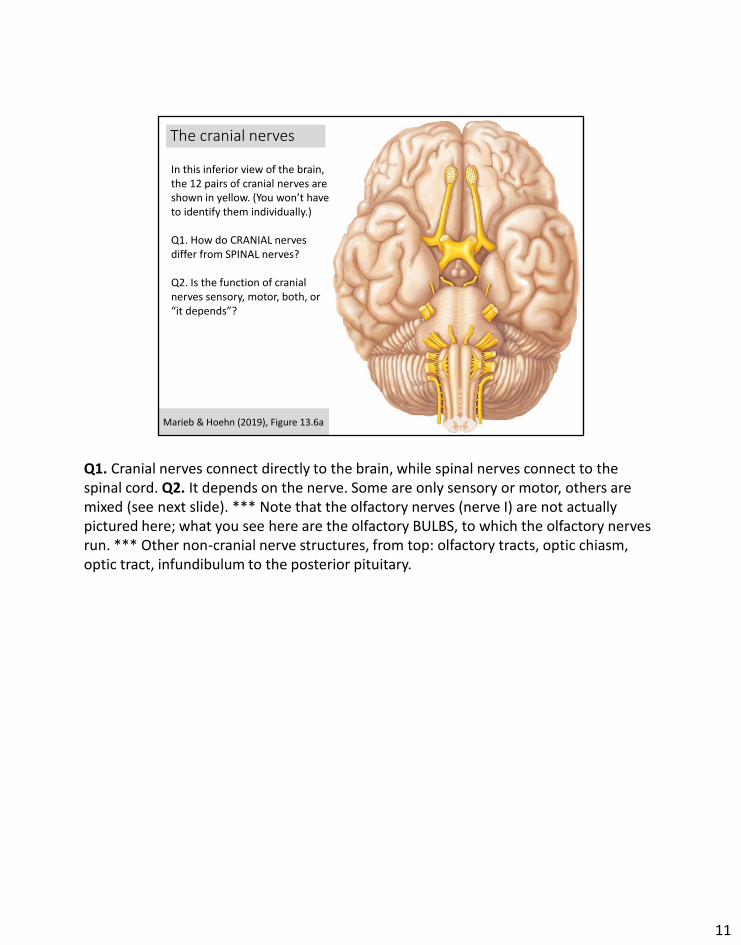
The cranial nerves
11
In this inferior view of the brain, the 12 pairs of cranial nerves are shown in yellow. (You won’t have to identify them individually.)
Q1. How do CRANIAL nerves differ from SPINAL nerves?
Q2. Is the function of cranial nerves sensory, motor, both, or “it depends”?
Marieb & Hoehn (2019), Figure 13.6a
Q1. Cranial nerves connect directly to the brain, while spinal nerves connect to the spinal cord. Q2. It depends on the nerve. Some are only sensory or motor, others are mixed (see next slide). *** Note that the olfactory nerves (nerve I) are not actually pictured here; what you see here are the olfactory BULBS, to which the olfactory nerves run. *** Other non-cranial nerve structures, from top: olfactory tracts, optic chiasm, optic tract, infundibulum to the posterior pituitary.
11
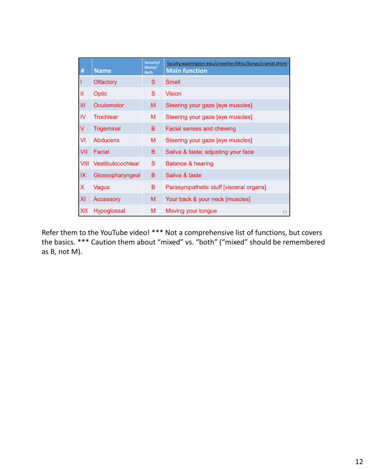
# Name
Sensory/ Motor/Both Main function
I Olfactory S Smell
II Optic S Vision
III Oculomotor M Steering your gaze [eye muscles]
IV Trochlear M Steering your gaze [eye muscles]
V Trigeminal B Facial senses and chewing
VI Abducens M Steering your gaze [eye muscles]
VII Facial B Saliva & taste; adjusting your face
VIII Vestibulocochlear S Balance & hearing
IX Glossopharyngeal B Saliva & taste
X Vagus B Parasympathetic stuff [visceral organs]
XI Accessory M Your back & your neck [muscles]
XII Hypoglossal M Moving your tongue 12
faculty.washington.edu/crowther/Misc/Songs/cranial.shtml
Refer them to the YouTube video! *** Not a comprehensive list of functions, but covers the basics. *** Caution them about “mixed” vs. “both” (“mixed” should be remembered as B, not M).
12
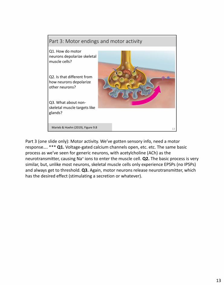
Part 3: Motor endings and motor activity
13Marieb & Hoehn (2019), Figure 9.8
Q1. How do motor neurons depolarize skeletal muscle cells?
Q2. Is that different from how neurons depolarize other neurons?
Q3. What about non-skeletal muscle targets like glands?
Part 3 (one slide only): Motor activity. We’ve gotten sensory info, need a motor response…. *** Q1. Voltage-gated calcium channels open, etc. etc. The same basic process as we’ve seen for generic neurons, with acetylcholine (ACh) as the neurotransmitter, causing Na+ ions to enter the muscle cell. Q2. The basic process is very similar, but, unlike most neurons, skeletal muscle cells only experience EPSPs (no IPSPs) and always get to threshold. Q3. Again, motor neurons release neurotransmitter, which has the desired effect (stimulating a secretion or whatever).
13
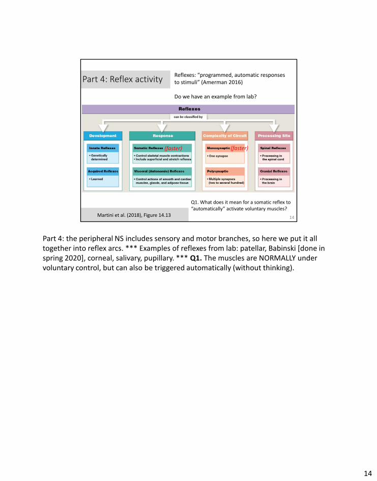
Part 4: Reflex activity
14
Reflexes: “programmed, automatic responses to stimuli” (Amerman 2016)
Do we have an example from lab?
Martini et al. (2018), Figure 14.13
(faster)(faster)
Q1. What does it mean for a somatic reflex to “automatically” activate voluntary muscles?
Part 4: the peripheral NS includes sensory and motor branches, so here we put it all together into reflex arcs. *** Examples of reflexes from lab: patellar, Babinski [done in spring 2020], corneal, salivary, pupillary. *** Q1. The muscles are NORMALLY under voluntary control, but can also be triggered automatically (without thinking).
14
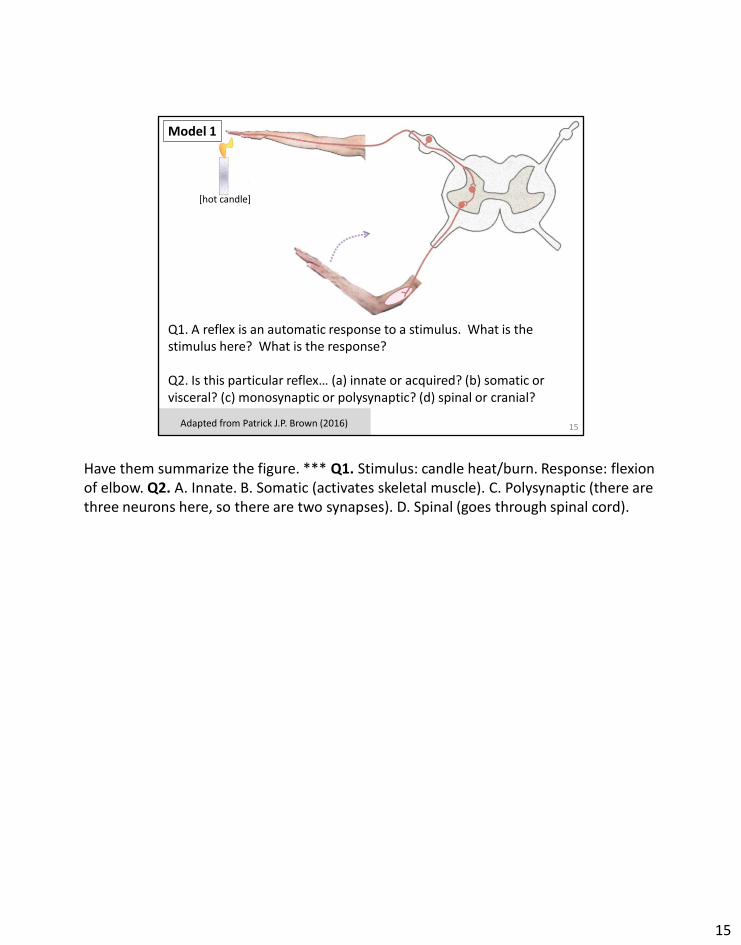
15Adapted from Patrick J.P. Brown (2016)
[hot candle]
Q1. A reflex is an automatic response to a stimulus. What is the stimulus here? What is the response?
Q2. Is this particular reflex… (a) innate or acquired? (b) somatic or visceral? (c) monosynaptic or polysynaptic? (d) spinal or cranial?
Model 1
Have them summarize the figure. *** Q1. Stimulus: candle heat/burn. Response: flexion of elbow. Q2. A. Innate. B. Somatic (activates skeletal muscle). C. Polysynaptic (there are three neurons here, so there are two synapses). D. Spinal (goes through spinal cord).
15
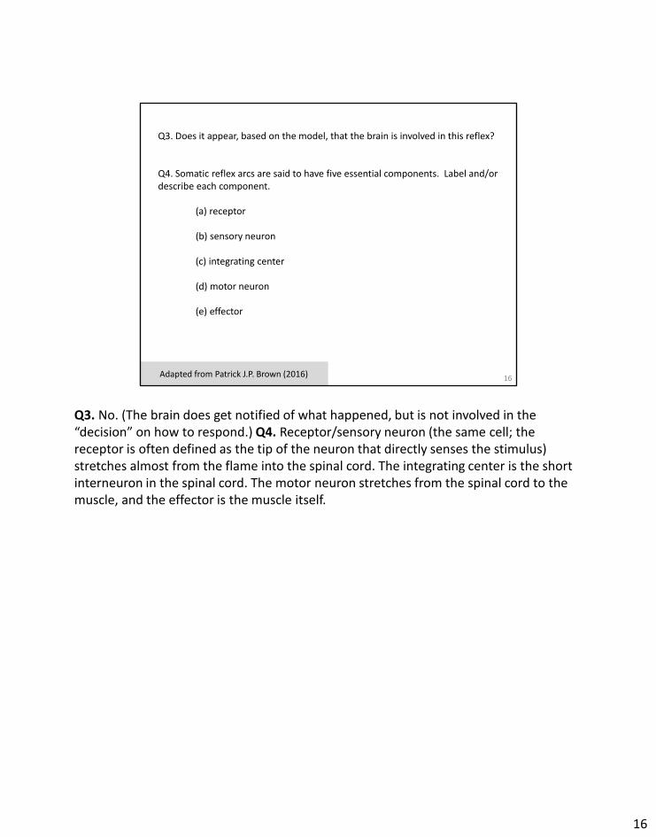
16Adapted from Patrick J.P. Brown (2016)
Q3. Does it appear, based on the model, that the brain is involved in this reflex?
Q4. Somatic reflex arcs are said to have five essential components. Label and/or describe each component.
(a) receptor
(b) sensory neuron
(c) integrating center
(d) motor neuron
(e) effector
Q3. No. (The brain does get notified of what happened, but is not involved in the “decision” on how to respond.) Q4. Receptor/sensory neuron (the same cell; the receptor is often defined as the tip of the neuron that directly senses the stimulus) stretches almost from the flame into the spinal cord. The integrating center is the short interneuron in the spinal cord. The motor neuron stretches from the spinal cord to the muscle, and the effector is the muscle itself.
16
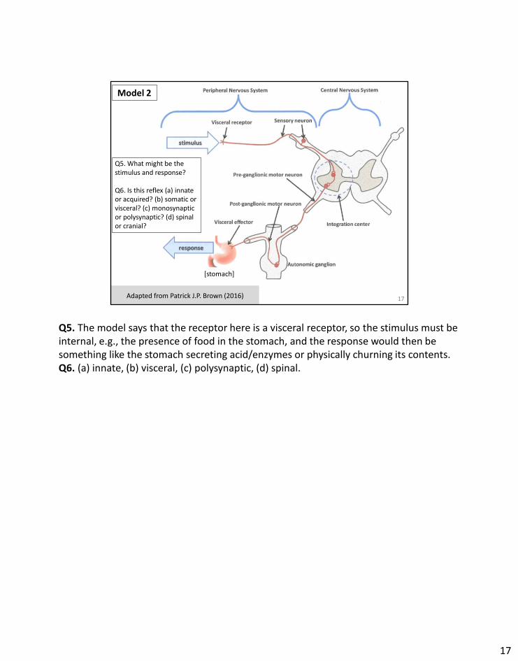
17Adapted from Patrick J.P. Brown (2016)
Q5. What might be the stimulus and response?
Q6. Is this reflex (a) innate or acquired? (b) somatic or visceral? (c) monosynaptic or polysynaptic? (d) spinal or cranial?
[stomach]
Model 2
Q5. The model says that the receptor here is a visceral receptor, so the stimulus must be internal, e.g., the presence of food in the stomach, and the response would then be something like the stomach secreting acid/enzymes or physically churning its contents. Q6. (a) innate, (b) visceral, (c) polysynaptic, (d) spinal.
17
18Adapted from Patrick J.P. Brown (2016)
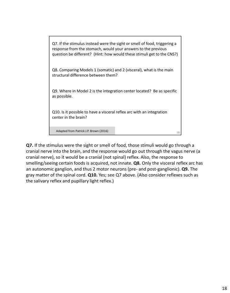
Q7. If the stimulus instead were the sight or smell of food, triggering a response from the stomach, would your answers to the previous question be different? (Hint: how would these stimuli get to the CNS?)
Q8. Comparing Models 1 (somatic) and 2 (visceral), what is the main structural difference between them?
Q9. Where in Model 2 is the integration center located? Be as specific as possible.
Q10. Is it possible to have a visceral reflex arc with an integration center in the brain?
Q7. If the stimulus were the sight or smell of food, those stimuli would go through a cranial nerve into the brain, and the response would go out through the vagus nerve (a cranial nerve), so it would be a cranial (not spinal) reflex. Also, the response to smelling/seeing certain foods is acquired, not innate. Q8. Only the visceral reflex arc has an autonomic ganglion, and thus 2 motor neurons (pre- and post-ganglionic). Q9. The gray matter of the spinal cord. Q10. Yes; see Q7 above. (Also consider reflexes such as the salivary reflex and pupillary light reflex.)
18
19
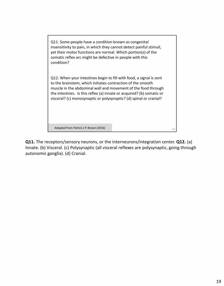
Q11. Some people have a condition known as congenital insensitivity to pain, in which they cannot detect painful stimuli, yet their motor functions are normal. Which portion(s) of the somatic reflex arc might be defective in people with this condition?
Q12. When your intestines begin to fill with food, a signal is sent to the brainstem, which initiates contraction of the smooth muscle in the abdominal wall and movement of the food through the intestines. Is this reflex (a) innate or acquired? (b) somatic or visceral? (c) monosynaptic or polysynaptic? (d) spinal or cranial?
Adapted from Patrick J.P. Brown (2016)
Q11. The receptors/sensory neurons, or the interneurons/integration center. Q12. (a) Innate. (b) Visceral. (c) Polysynaptic (all visceral reflexes are polysynaptic, going through autonomic ganglia). (d) Cranial.
19
Chapter 13: additional resources
• Regarding the neuromuscular junction, the “Study Area” of the Pearson Mastering website, the Interactive Physiology page has an animation of this (see Muscular System section).
• Other suggestions? Let me know…
20
20
Answer key for Suggested Lecture Outline file
• You should already have access to answers to some of the questions (Check Your Understanding, online Practice Quiz, online Practice Test)
• Answers to pre-lecture questions and end-of-chapter Review Questions will be in the Presenter Notes that accompany this slide.
21
ANSWERS TO PRE-LECTURE QUESTIONS… PL1. Answers will vary. PL2. Some neurons do cross between the PNS and CNS. The axons of sensory neurons stretch from PNS nerves into the CNS gray matter. Motor neurons have their dendrites and cell bodies in the CNS gray matter, but their axons stretch out into the PNS. PL3. Mixed nerves contain both sensory neurons and motor neurons. Mixed nerves are extremely common in the human body. All dorsal and ventral rami are mixed, for example. A few of the cranial nerves are mixed nerves: V (trigeminal), VII (facial), IX (glossopharyngeal), and X (vagus). PL4. A reflex arc is a chain of cells that enables reflexes, i.e., predictable and fast responses to stimuli. According to your textbook, there are 5 components of a reflex arc: receptor, sensory neuron, integration center, motor neuron, effector. PL5. Sensory info from your eyes is carried by cranial nerve II (optic). Motor info for your eye muscles is carried by cranial nerves III (oculomotor), IV (trochlear), and VI (abducens). (VI controls the lateral rectus muscle, IV controls the superior oblique muscle, and III controls the other muscles: inferior rectus, medial rectus, inferior oblique, superior oblique. You don’t need to know which of these specific muscles go with which specific nerves.) (It could also be argued that cranial nerve VII (facial) affects the eye, since a branch of it controls the lacrimal gland, which lubricates the eye with tears. You don’t need to remember that!) *** ANSWERS TO REVIEW QUESTIONS (pages 529-530)… 5: e. 7 (1) f;
21
(2) i; (3) b; (4) g, h, l; (5) e; (6) i; (7) c; (8) k; (9) l; (10) c,d,f,k. 12: (a) A plexus is a network. A nerve plexus is a branching nerve network formed by ventral ra-mi from several spinal nerves. (b) The cervical plexus originates from the ventral rami of C1 to C5 spinal nerves; the brachial plexus originates from the ventral rami of C5 to T1; the lumbar plexus originates from the ventral rami of T12 to L4 (your text says L1-L4, but other sources say T12-L4); and the sacral plexus originates from L4 to S4. 19: In the PNS, damaged fibers can be replaced or repaired by physical and chemical processes directed by macrophages and Schwann cells. In the CNS, oligodendrocytes do not aid fiber regeneration because they have growth-inhibiting proteins on their surface, allowing dam-aged fibers to collapse and die. (pp. 497–498)
21