the parkinson-associated protein pink1 interacts with beclin1 and promotes autophagy
TRANSCRIPT
The Parkinson-associated protein PINK1 interacts withBeclin1 and promotes autophagy
S Michiorri1,2,8, V Gelmetti1,8, E Giarda3, F Lombardi1, F Romano1,2, R Marongiu1, S Nerini-Molteni3, P Sale4, R Vago3, G Arena1,
L Torosantucci1, L Cassina3, MA Russo2, B Dallapiccola1, EM Valente*,1,5 and G Casari*,3,6,7
Mutations in the PINK1 gene cause autosomal recessive Parkinson’s disease. The PINK1 gene encodes a protein kinase that ismitochondrially cleaved to generate two mature isoforms. In addition to its protective role against mitochondrial dysfunction andapoptosis, PINK1 is also known to regulate mitochondrial dynamics acting upstream of the PD-related protein Parkin. Recentdata showed that mitochondrial Parkin promotes the autophagic degradation of dysfunctional mitochondria, and that stablePINK1 silencing may have an indirect role in mitophagy activation. Here we report a new interaction between PINK1 and Beclin1,a key pro-autophagic protein already implicated in the pathogenesis of Alzheimer’s and Huntington’s diseases. Both PINK1N- and C-terminal are required for the interaction, suggesting that full-length PINK1, and not its cleaved isoforms, interacts withBeclin1. We also demonstrate that PINK1 significantly enhances basal and starvation-induced autophagy, which is reduced byknocking down Beclin1 expression or by inhibiting the Beclin1 partner Vps34. A mutant, PINK1W437X, interaction of which withBeclin1 is largely impaired, lacks the ability to enhance autophagy, whereas this is not observed for PINK1G309D, a mutant withdefective kinase activity but unaltered ability to bind Beclin1. These findings identify a new function of PINK1 and furtherstrengthen the link between autophagy and proteins implicated in the neurodegenerative process.Cell Death and Differentiation (2010) 17, 962–974; doi:10.1038/cdd.2009.200; published online 8 January 2010
Parkinson’s disease (PD) is a frequent neurodegenerativedisorder resulting from massive degeneration of the dopami-nergic neurons in the substantia nigra. Although most casesare sporadic, several genes are known to cause familial PD,especially with early onset.1
Mutations in the PINK1 gene are the second most frequentcause of autosomal recessive PD after those in the Parkingene.2,3 The PINK1 gene encodes a serine–threonine kinasewith an N-terminal mitochondrial import sequence, firstcharacterized as a protein aimed at maintaining mitochondrialintegrity and preventing apoptosis in response to cellularstressors.2,4–8 This neuroprotective role is partly exertedthrough phosphorylation of the mitochondrial chaperon,TRAP1, although cytoplasm-restricted PINK1 was alsoshown to protect against MPTP damage.9,10 The full-lengthPINK1 (PINK1-FL) is processed within mitochondria togenerate two mature proteins;4,11 all three isoforms localizeboth to the mitochondria and cytosol, their relative ratio beingregulated by several factors.10–13
Increasing data have demonstrated that absence offunctional PINK1 induces abnormalities of mitochondrial
morphology.6,14,15 In several studies (mostly in Drosophila),PINK1 was shown to promote fission acting upstream of theFis1–Drp1 machinery, and the mitochondrial phenotypeobserved in PINK1 knockout flies or silenced cells wasassociated to reduced fission.16,17 Subsequent studies inmammalian cell systems contradicted these results, demon-strating that mutant or silenced PINK1 resulted in increasedfragmentation.15,17–19
A major progress in this field has come from the finding thatPINK1 interacts with Parkin to regulate mitochondrialdynamics. Parkin overexpression was found to rescue themitochondrial abnormalities observed in PINK1 knockout fliesand silenced cells but not vice versa, suggesting that Parkinmay act downstream of PINK1 in a common pathway.14,15,19 Ithas been recently shown that Parkin is directly recruited todysfunctional mitochondria mediating their eliminationthrough mitophagy,20 and that stable silencing of PINK1induces an increase in oxidative stress levels, which activatesboth mitophagy and mitochondrial fission.19 Mitophagy is atightly regulated autophagy-based mechanism that has acentral role in mitochondria quality control, by selectively
Received 02.4.09; revised 06.11.09; accepted 17.11.09; Edited by M Piacentini; published online 08.1.10
1Casa Sollievo della Sofferenza Hospital, CSS-Mendel Institute, Rome, Italy; 2Department of Experimental Medicine, Sapienza University, Rome, Italy; 3HumanMolecular Genetics Unit, San Raffaele Scientific Institute, DIBIT, Milan, Italy; 4IRCCS San Raffaele Pisana, Rome, Italy; 5Department of Medical and Surgical PaediatricSciences, University of Messina, Messina, Italy; 6Vita-Salute San Raffaele University School of Medicine, Milan, Italy and 7National Institute of Neurosciences,Milan, Italy*Correspondening authors: EM Valente or G Casari, Neurogenetics Unit, CSS-Mendel Institute, viale Regina Margherita 261, 00198 Rome, Italy.Tel: þ 39 06 44160537; Fax: þ 39 06 44160548; E-mail: [email protected] or [email protected] authors contributed equally to this work.Keywords: PINK1; Beclin1; Parkinson’s disease; autophagy; mitochondriaAbbreviations: 3MA, 3-methyladenine; aa, amino acid; ER, endoplasmic reticulum; FL, full length; FLIM, Fluorescence Lifetime Imaging Microscopy; FRET,Fluorescence Resonance Energy Transfer; LC3, Microtubule-associated protein light chain 3; Mfn1, Mitofusin 1; MPTP, 1-methyl-4-phenyl-1,2,3, 6-tetrahydropyridine;OMM, outer mitochondrial membrane; PD, Parkinson’s disease; PI3K, phosphatidylinositol 30-kinase; PINK1, PTEN induced putative kinase 1; TRAP1, TNF receptor-associated protein 1
Cell Death and Differentiation (2010) 17, 962–974& 2010 Macmillan Publishers Limited All rights reserved 1350-9047/10 $32.00
www.nature.com/cdd
eliminating damaged mitochondria before they could activateapoptosis.21 Indeed, autophagy is being increasingly recog-nized as an essential homeostatic process to clear misfoldedor aggregated proteins and to ensure organellar turnover.The role of autophagy seems even more relevant in agingnon-mitotic cells, such as neurons, in which damagedcellular components cannot be diluted through recurring celldivision cycles.22
Compelling evidence now indicates a protective role ofautophagy against neurodegeneration. Dysfunctional auto-phagy has been implicated in a growing number of neurode-generative diseases, including PD, that share the pathoge-netic pathways of mitochondrial abnormalities and misfoldedprotein damage.22–24 Interestingly, the fission protein Fis1was found to activate the autophagy pathway,25 and it hasbeen proposed that mitochondria fusion/fission machineryand mitophagy could represent a ‘quality control axis’,regulating mitochondrial dynamics and function.26 Althoughin PINK1-deficient cells mitophagy seems to depend onmitochondrial fission,19 Parkin-activated mitophagy seemsindependent from its effects on mitochondrial morphology,suggesting that the two mechanisms can be regulatedtogether or in parallel.20
Here, we present evidence that PINK1-FL, but not itscleaved isoforms or a mutant protein truncated at theC-terminus, interacts with the pro-autophagic protein Beclin1and enhances autophagy. This effect results from an increaseof the autophagic flux and is, at least, partly mediated throughthe Beclin1–Vps34 complex, suggesting a main role forPINK1 in this fundamental cellular pathway.
Results
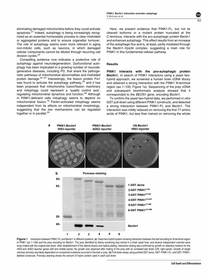
PINK1 interacts with the pro-autophagic proteinBeclin1. In search of PINK1 interactors using a yeast two-hybrid approach, we screened a human brain cDNA libraryand obtained a strong interaction with the PINK1 N-terminalregion (aa 1–100; Figure 1a). Sequencing of the prey cDNAand subsequent bioinformatic analysis showed that itcorresponded to the BECN1 gene, encoding Beclin1.
To confirm the yeast two-hybrid data, we performed in vitroGST pull down using different PINK1 constructs, and detecteda strong interaction between PINK1-FL and Beclin1. Theinteraction was mildly reduced on removing the first 77 aminoacids of PINK1, but less than halved on removing the whole
Figure 1 Interaction between PINK1-FL and Beclin1 in different systems. (a) Yeast two-hybrid system showing interaction between the bait encoding for N-terminal regionof PINK1 (aa 1–100) and the prey encoding for Beclin1. The prey identified by library screening was cloned in a fresh yeast host, and several independent colonies werearray-mated with the original bait strain. After establishment of the diploid strains and replica plating, interaction testing was confirmed by growth on selective medium for theHIS3 and ADE2 reporter genes (left and middle panel). No growth was observed after mating of the prey with an unrelated bait strain (UR, right panel). Growth of singlecolonies (arrows) was likely dependent on occasional mutations occurred in the bait or prey. (b) Pull-down assay using purified GST alone, GST–PINK1-FL- and GST–PINK1-deleted constructs. Ponceau staining shows the amount of fusion protein used in each pull down
PINK1–Beclin1 interaction promotes autophagyS Michiorri et al
963
Cell Death and Differentiation
N-terminus (1–112 aa), and completely abolished when theC-terminus was also deleted (Figure 1b). These resultsconfirmed that PINK1-FL binds Beclin1 in vitro, and sug-gested that the interaction involves both its N- and C-terminalregions. To assess whether Beclin1 could be a substrateof PINK1, we performed an in vitro kinase assay usingGST–PINK177–581 and observed no phosphorylation ofBeclin1 (data not shown).
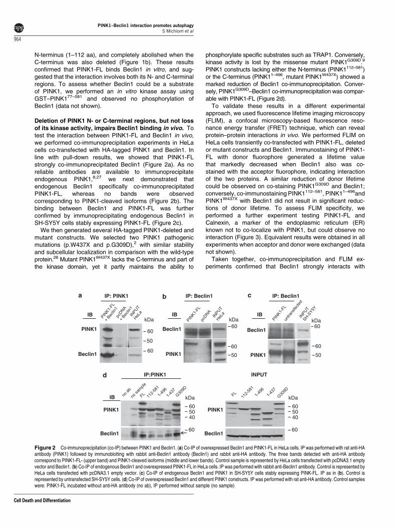
Deletion of PINK1 N- or C-terminal regions, but not lossof its kinase activity, impairs Beclin1 binding in vivo. Totest the interaction between PINK1-FL and Beclin1 in vivo,we performed co-immunoprecipitation experiments in HeLacells co-transfected with HA-tagged PINK1 and Beclin1. Inline with pull-down results, we showed that PINK1-FLstrongly co-immunoprecipitated Beclin1 (Figure 2a). As noreliable antibodies are available to immunoprecipitateendogenous PINK1,8,27 we next demonstrated thatendogenous Beclin1 specifically co-immunoprecipitatedPINK1-FL, whereas no bands were observedcorresponding to PINK1-cleaved isoforms (Figure 2b). Thebinding between Beclin1 and PINK1-FL was furtherconfirmed by immunoprecipitating endogenous Beclin1 inSH-SY5Y cells stably expressing PINK1-FL (Figure 2c).
We then generated several HA-tagged PINK1-deleted andmutant constructs. We selected two PINK1 pathogenicmutations (p.W437X and p.G309D),2 with similar stabilityand subcellular localization in comparison with the wild-typeprotein.28 Mutant PINK1W437X lacks the C-terminus and part ofthe kinase domain, yet it partly maintains the ability to
phosphorylate specific substrates such as TRAP1. Conversely,kinase activity is lost by the missense mutant PINK1G309D.9
PINK1 constructs lacking either the N-terminus (PINK1112–581)or the C-terminus (PINK11–496, mutant PINK1W437X) showed amarked reduction of Beclin1 co-immunoprecipitation. Conver-sely, PINK1G309D–Beclin1 co-immunoprecipitation was compar-able with PINK1-FL (Figure 2d).
To validate these results in a different experimentalapproach, we used fluorescence lifetime imaging microscopy(FLIM), a confocal microscopy-based fluorescence reso-nance energy transfer (FRET) technique, which can revealprotein–protein interactions in vivo. We performed FLIM onHeLa cells transiently co-transfected with PINK1-FL, deletedor mutant constructs and Beclin1. Immunostaining of PINK1-FL with donor fluorophore generated a lifetime valuethat markedly decreased when Beclin1 also was co-stained with the acceptor fluorophore, indicating interactionof the two proteins. A similar reduction of donor lifetimecould be observed on co-staining PINK1G309D and Beclin1;conversely, co-immunostaining PINK1112–581, PINK11–496andPINK1W437X with Beclin1 did not result in significant reduc-tions of donor lifetime. To assess FLIM specificity, weperformed a further experiment testing PINK1-FL andCalnexin, a marker of the endoplasmic reticulum (ER)known not to co-localize with PINK1, but could observe nointeraction (Figure 3). Equivalent results were obtained in allexperiments when acceptor and donor were exchanged (datanot shown).
Taken together, co-immunoprecipitation and FLIM ex-periments confirmed that Beclin1 strongly interacts with
IP: PINK1 IP: Beclin1 IP: Beclin1
50
60
kDaIB
PINK1
Beclin1
kDa
60
IB
Beclin1
PINK1
Beclin1
PINK1
kDa60
60
50
IB
60
50
INPUTIP:PINK1
IB
PINK1
Beclin160
kDa
605040
PINK1
Beclin160
kDa
605040
60
Figure 2 Co-immunoprecipitation (co-IP) between PINK1 and Beclin1. (a) Co-IP of overexpressed Beclin1 and PINK1-FL in HeLa cells. IP was performed with rat anti-HAantibody (PINK1) followed by immunoblotting with rabbit anti-Beclin1 antibody (Beclin1) and rabbit anti-HA antibody. The three bands detected with anti-HA antibodycorrespond to PINK1-FL- (upper band) and PINK1-cleaved isoforms (middle and lower bands). Control sample is represented by HeLa cells transfected with pcDNA3.1 emptyvector and Beclin1. (b) Co-IP of endogenous Beclin1 and overexpressed PINK1-FL in HeLa cells. IP was performed with rabbit anti-Beclin1 antibody. Control is represented byHeLa cells transfected with pcDNA3.1 empty vector. (c) Co-IP of endogenous Beclin1 and PINK1 in SH-SY5Y cells stably expressing PINK-FL. IP as in (b). Control isrepresented by untransfected SH-SY5Y cells. (d) Co-IP of overexpressed Beclin1 and different PINK1 constructs. IP was performed with rat anti-HA antibody. Control sampleswere: PINK1-FL incubated without anti-HA antibody (no ab), IP performed without sample (no sample)
PINK1–Beclin1 interaction promotes autophagyS Michiorri et al
964
Cell Death and Differentiation
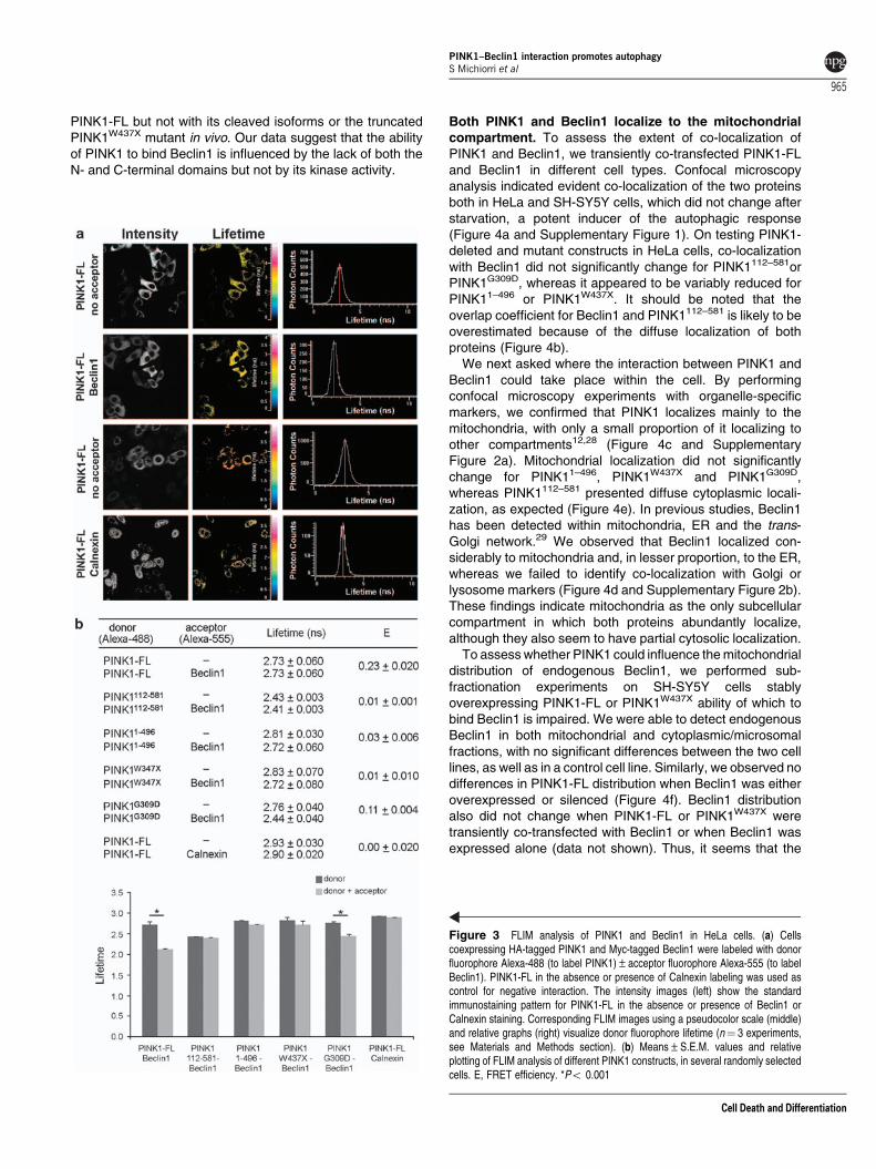
PINK1-FL but not with its cleaved isoforms or the truncatedPINK1W437X mutant in vivo. Our data suggest that the abilityof PINK1 to bind Beclin1 is influenced by the lack of both theN- and C-terminal domains but not by its kinase activity.
Both PINK1 and Beclin1 localize to the mitochondrialcompartment. To assess the extent of co-localization ofPINK1 and Beclin1, we transiently co-transfected PINK1-FLand Beclin1 in different cell types. Confocal microscopyanalysis indicated evident co-localization of the two proteinsboth in HeLa and SH-SY5Y cells, which did not change afterstarvation, a potent inducer of the autophagic response(Figure 4a and Supplementary Figure 1). On testing PINK1-deleted and mutant constructs in HeLa cells, co-localizationwith Beclin1 did not significantly change for PINK1112–581orPINK1G309D, whereas it appeared to be variably reduced forPINK11–496 or PINK1W437X. It should be noted that theoverlap coefficient for Beclin1 and PINK1112–581 is likely to beoverestimated because of the diffuse localization of bothproteins (Figure 4b).
We next asked where the interaction between PINK1 andBeclin1 could take place within the cell. By performingconfocal microscopy experiments with organelle-specificmarkers, we confirmed that PINK1 localizes mainly to themitochondria, with only a small proportion of it localizing toother compartments12,28 (Figure 4c and SupplementaryFigure 2a). Mitochondrial localization did not significantlychange for PINK11–496, PINK1W437X and PINK1G309D,whereas PINK1112–581 presented diffuse cytoplasmic locali-zation, as expected (Figure 4e). In previous studies, Beclin1has been detected within mitochondria, ER and the trans-Golgi network.29 We observed that Beclin1 localized con-siderably to mitochondria and, in lesser proportion, to the ER,whereas we failed to identify co-localization with Golgi orlysosome markers (Figure 4d and Supplementary Figure 2b).These findings indicate mitochondria as the only subcellularcompartment in which both proteins abundantly localize,although they also seem to have partial cytosolic localization.
To assess whether PINK1 could influence the mitochondrialdistribution of endogenous Beclin1, we performed sub-fractionation experiments on SH-SY5Y cells stablyoverexpressing PINK1-FL or PINK1W437X ability of which tobind Beclin1 is impaired. We were able to detect endogenousBeclin1 in both mitochondrial and cytoplasmic/microsomalfractions, with no significant differences between the two celllines, as well as in a control cell line. Similarly, we observed nodifferences in PINK1-FL distribution when Beclin1 was eitheroverexpressed or silenced (Figure 4f). Beclin1 distributionalso did not change when PINK1-FL or PINK1W437X weretransiently co-transfected with Beclin1 or when Beclin1 wasexpressed alone (data not shown). Thus, it seems that the
Figure 3 FLIM analysis of PINK1 and Beclin1 in HeLa cells. (a) Cellscoexpressing HA-tagged PINK1 and Myc-tagged Beclin1 were labeled with donorfluorophore Alexa-488 (to label PINK1)±acceptor fluorophore Alexa-555 (to labelBeclin1). PINK1-FL in the absence or presence of Calnexin labeling was used ascontrol for negative interaction. The intensity images (left) show the standardimmunostaining pattern for PINK1-FL in the absence or presence of Beclin1 orCalnexin staining. Corresponding FLIM images using a pseudocolor scale (middle)and relative graphs (right) visualize donor fluorophore lifetime (n¼ 3 experiments,see Materials and Methods section). (b) Means±S.E.M. values and relativeplotting of FLIM analysis of different PINK1 constructs, in several randomly selectedcells. E, FRET efficiency. *Po 0.001
PINK1–Beclin1 interaction promotes autophagyS Michiorri et al
965
Cell Death and Differentiation
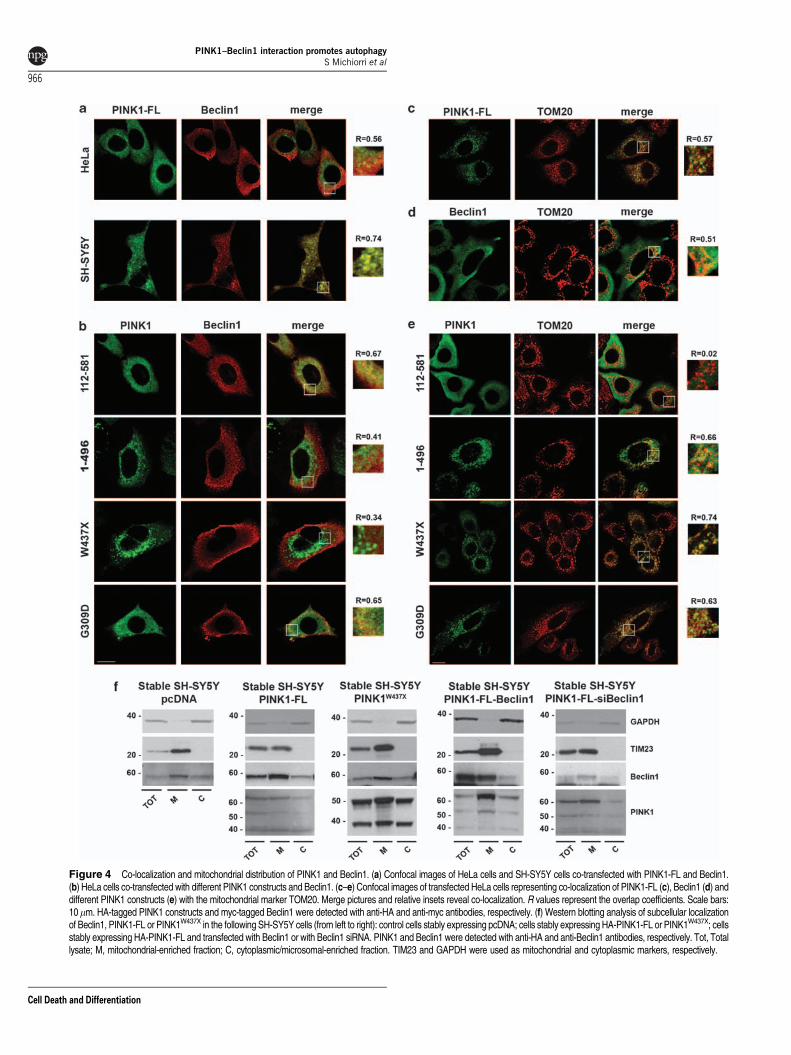
Figure 4 Co-localization and mitochondrial distribution of PINK1 and Beclin1. (a) Confocal images of HeLa cells and SH-SY5Y cells co-transfected with PINK1-FL and Beclin1.(b) HeLa cells co-transfected with different PINK1 constructs and Beclin1. (c–e) Confocal images of transfected HeLa cells representing co-localization of PINK1-FL (c), Beclin1 (d) anddifferent PINK1 constructs (e) with the mitochondrial marker TOM20. Merge pictures and relative insets reveal co-localization. R values represent the overlap coefficients. Scale bars:10mm. HA-tagged PINK1 constructs and myc-tagged Beclin1 were detected with anti-HA and anti-myc antibodies, respectively. (f) Western blotting analysis of subcellular localizationof Beclin1, PINK1-FL or PINK1W437X in the following SH-SY5Y cells (from left to right): control cells stably expressing pcDNA; cells stably expressing HA-PINK1-FL or PINK1W437X; cellsstably expressing HA-PINK1-FL and transfected with Beclin1 or with Beclin1 siRNA. PINK1 and Beclin1 were detected with anti-HA and anti-Beclin1 antibodies, respectively. Tot, Totallysate; M, mitochondrial-enriched fraction; C, cytoplasmic/microsomal-enriched fraction. TIM23 and GAPDH were used as mitochondrial and cytoplasmic markers, respectively.
PINK1–Beclin1 interaction promotes autophagyS Michiorri et al
966
Cell Death and Differentiation
interaction between PINK1 and Beclin1 is not relevant for themitochondrial distribution of both proteins.
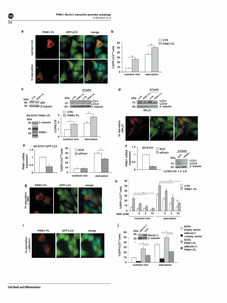
PINK1-FL, but not pathogenic mutant PINK1W437X,enhances autophagy. As the main role of Beclin1 is topromote autophagosome formation and maturation,29,30 wereasoned that PINK1 could interact with Beclin1 to modulateautophagy. Two complementary strategies, based onimmunofluorescence and western blotting, were optimizedto detect autophagy. We first transiently overexpressedPINK1-FL in SH-SY5Y cells stably expressing GFP–LC3under nutrient-rich and starvation conditions, and comparedthe number of PINK1-expressing and non-expressingcells that were positive for GFP–LC3 vacuoles, indicative ofactivated autophagy (Figure 5a). In nutrient-rich conditions,PINK1-FL overexpression induced a nearly three-foldincrease in the number of vacuole-positive cells comparedwith untransfected cells. Under starvation, the number ofautophagic untransfected cells increased about three timescompared with basal condition, and PINK1-FL overexpressionproduced a further significant increase of autophagic cellscompared with starved control (Figure 5b). Notably, we did notobserve any co-localization of PINK1 with the GFP–LC3dots under nutrient-rich and starvation conditions(Supplementary Figure 3).
To confirm the ability of PINK1 to enhance autophagy, wemonitored the conversion of endogenous LC3 from thecytoplasmic (LC3-I, 18 kDa) to the membrane-bound form(LC3-II, 16 kDa) in SH-SY5Y cells stably expressing PINK1. Inthis system, we quantified autophagy by assessing the LC3-II/LC3-I ratio, as we observed that, in SH-SY5Y cells, autophagyactivation was variably associated with LC3-II increase and/orLC3-I decrease.31 Under both nutrient-rich and starvationconditions, PINK1-FL cells showed a significant increase ofthe LC3-II/LC3-I ratio compared with control cells. Activationof autophagy was further confirmed by showing reduction ofP62 levels in cells expressing PINK1 versus controls(Figure 5c).
To explore whether the observed results actually repre-sented an increase of autophagy, we treated cells with thelysosomal protease inhibitor ammonium chloride, (NH4Cl),that prevents the degradation of autophagosomes. Immuno-fluorescence analysis showed that GFP–LC3 punctaedramatically increased under starvation conditions. Inwestern blotting, treatment with NH4Cl induced an evidentaccumulation of LC3-II in both cell lines (Figure 5d). Thesefindings suggest that PINK1-induced autophagy results froma true enhancement of the autophagic flux and not from theblockage of autophagic degradation.32
We next knocked down PINK1 expression using RNAiin GFP–LC3 and wild-type SH-SY5Y cells, obtaining 60and 80% decline of PINK1 mRNA expression, respectively.In immunofluorescence experiments, PINK1 silencinginduced a significant decrease of starvation-induced autop-hagy when compared with control cells, which was confirmedby a reduction of the LC3-II/LC3-I ratio in western blotting(Figures 5e and f).
To assess whether PINK1-related autophagy is mediatedby the Beclin1–Vps34 complex, we first evaluated GFP–LC3vacuole-positive cells in presence of 3-methyladenine (3MA),
a compound known to block autophagy by inhibiting theactivity of class III PI3K–Vps34,33 a Beclin1 effector thatregulates autophagosome formation and maturation.30 Weshowed that, in PINK1-expressing cells, increasing concen-trations of 3MA were able to significantly decrease basal andstarvation-induced autophagy (Figures 5g and h). To furtherverify the direct involvement of Beclin1, we then knockeddown Beclin1 by siRNA. Beclin1 silencing also induced asignificant reduction of autophagy in cells overexpressingPINK1, although the proportion of autophagic cells remainedhigher than that of control cells (Figures 5i and j).
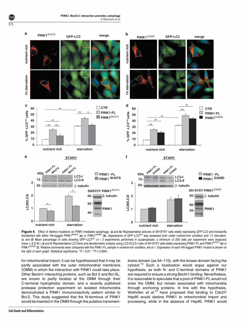
To assess the effect of distinct mutations on PINK1-relatedautophagy, we next monitored the proportion of autophagic cellsand endogenous LC3 conversion in SH-SY5Y cells expressingPINK1-FL and either the W437X or the G309D mutant.
In immunofluorescence, the percentage of vacuole-positivecells was significantly lower in cells expressing PINK1W437X
than those expressing PINK1-FL in nutrient-rich conditions,and overall comparable with untransfected controls understarvation (Figures 6a and c). Conversely, overexpression ofPINK1G309D resulted in an increase in the number ofautophagic cells that did not significantly differ from PINK1-FL both under nutrient-rich and starvation conditions (Figures6b and d). Similar results were obtained with LC3 westernblotting (Figures 6e and f).
Taken together, our data show that PINK1-FL is ableto significantly enhance basal and starvation-inducedautophagy, and that this effect is at least partly mediated byBeclin1. The PINK1-mediated autophagy is greatly reducedin presence of PINK1W437X, ability of which to bind Beclin1is largely impaired, but not in presence of PINK1G309D,a protein with defective kinase activity but unaltered Beclin1binding.
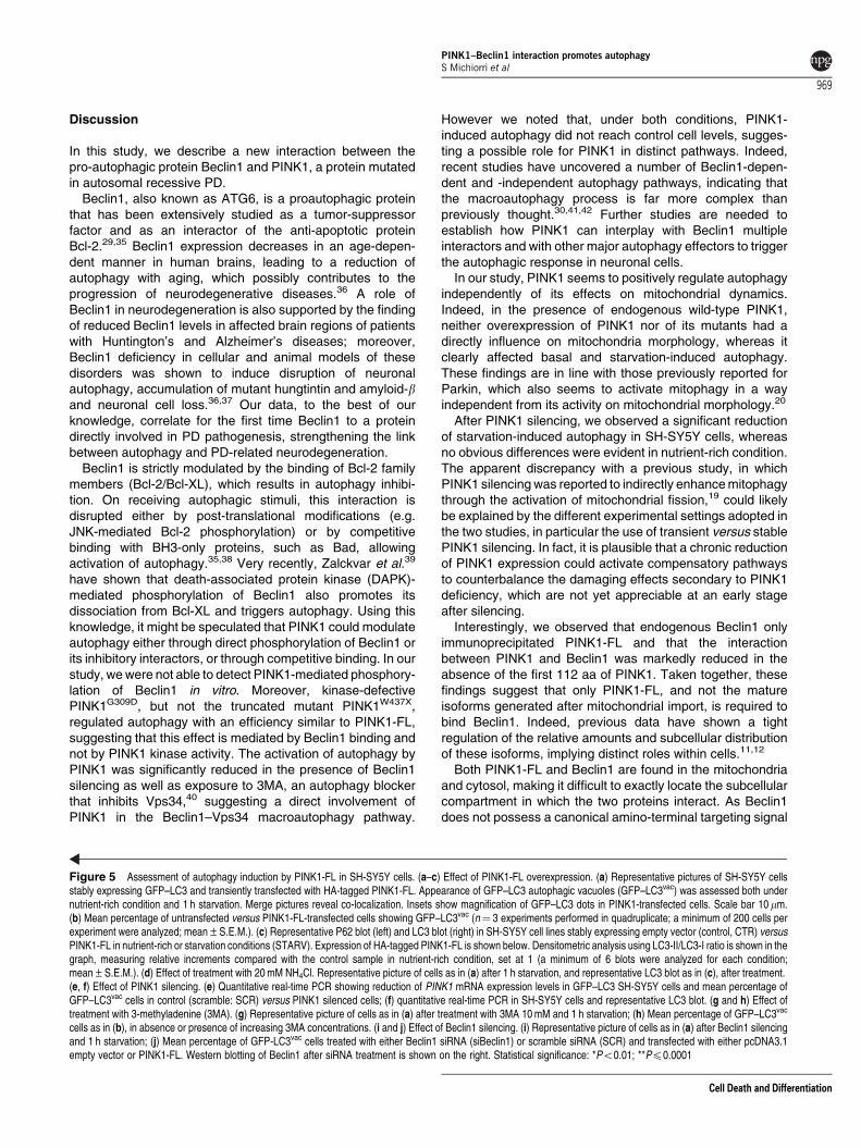
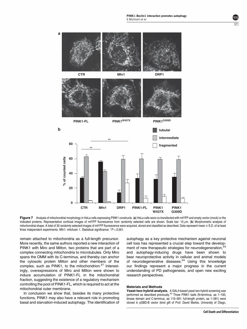
PINK1 overexpression does not affect mitochondrialmorphology. We next sought to assess whether theobserved effects of PINK1 on autophagy could bemediated by its activity on mitochondrial dynamics.
Mitochondrial morphology was analyzed in HeLa cells byco-transfecting a mitochondrially targeted yellow fluorescentprotein (mtYFP) with mitofusin1 and DRP1 as positivecontrols for the induction of fusion or fission, and with eitherPINK1-FL, PINK1W437X or PINK1G309D and performing liveimaging using confocal microscopy. For quantitative analysisof mitochondrial morphology, we distinguished between cellswith tubular, intermediate or fragmented mitochondria, asdescribed previously.34 Co-transfection of mtYFP with eachconstruct was confirmed by immunofluorescence analysis(data not shown).
Live imaging of cells overexpressing mitofusin1 showeda higher number of cells with tubular mitochondria thanmock-transfected cells, whereas the expression of DRP1resulted in a significant increase of fragmented mitochondria.Transfection of mtYFP with PINK1-FL or mutants did notresult in visible morphological changes of the mitochondrialnetwork in comparison with control cells (Figure 7),suggesting that, at least in this experimental setting, PINK1is not directly involved in the regulation of mitochondrialdynamics.
PINK1–Beclin1 interaction promotes autophagyS Michiorri et al
967
Cell Death and Differentiation
1h s
tarv
atio
n+N
H4C
l
1h s
tarv
atio
n
+ 3M
A
GFP-LC3PINK1-FL merge
GFP-LC3PINK1-FL merge
1h s
tarv
atio
n+
siB
eclin
1GFP-LC3PINK1-FL merge
nu
trie
nt
rich
1h s
tarv
atio
n
%G
FP
-LC
3vac ce
lls%
GF
P-L
C3va
c cells
0
20
30
40
50
60
10
3MA (mM)
nutrient rich starvation
2 5 10- 2 5 10-
CTRPINK1 -FL
*
*
*
*
nutrient rich starvation0
20
30
40
50
60
10
60 -
50 - β−tubulinBeclin-1
kDa *
0
10
20
30
40
50
60
nutrient rich starvation
%G
FP
-LC
3vac ce
lls
**
**
CTRPINK1-FL
SCR+empty vectorsiBeclin1+empty vectorSCR+PINK1-FLsiBeclin1+PINK1-FL
*
starvationnutrient rich
%G
FP
-LC
3vac ce
lls
40
0
20
30
50
10
*
PIN
K1
mR
NA
exp
ress
ion
0.5
0
1
1.5SH-SY5Y GFP-LC3
SCRsiPink1
0.5
0
1
1.5 SH-SY5Y
PIN
K1
mR
NA
exp
ress
ion
15 -
STARV
50 -
kDa
LC3II/LC3I 1.4 0.9
SH-SY5Y PINK1-FL
PINK1
50 -
60 -
50 -
kDa
50 -
STARV
CTRPINK1-FL
LC
3II/L
C3I
nutrient rich starvation0
1
2
3*
*
15 -
kDaLC3-ILC3-II
NH4Cl
STARV
SCRsiPink1
50-
15-
kDa
50 -
kDa60 -
??β−tubulin
β−tubulin
β−tubulin
p62LC3-ILC3-IIβ−tubulin
LC3-ILC3-IIβ−tubulin
PINK1–Beclin1 interaction promotes autophagyS Michiorri et al
968
Cell Death and Differentiation
Discussion
In this study, we describe a new interaction between thepro-autophagic protein Beclin1 and PINK1, a protein mutatedin autosomal recessive PD.
Beclin1, also known as ATG6, is a proautophagic proteinthat has been extensively studied as a tumor-suppressorfactor and as an interactor of the anti-apoptotic proteinBcl-2.29,35 Beclin1 expression decreases in an age-depen-dent manner in human brains, leading to a reduction ofautophagy with aging, which possibly contributes to theprogression of neurodegenerative diseases.36 A role ofBeclin1 in neurodegeneration is also supported by the findingof reduced Beclin1 levels in affected brain regions of patientswith Huntington’s and Alzheimer’s diseases; moreover,Beclin1 deficiency in cellular and animal models of thesedisorders was shown to induce disruption of neuronalautophagy, accumulation of mutant hungtintin and amyloid-band neuronal cell loss.36,37 Our data, to the best of ourknowledge, correlate for the first time Beclin1 to a proteindirectly involved in PD pathogenesis, strengthening the linkbetween autophagy and PD-related neurodegeneration.
Beclin1 is strictly modulated by the binding of Bcl-2 familymembers (Bcl-2/Bcl-XL), which results in autophagy inhibi-tion. On receiving autophagic stimuli, this interaction isdisrupted either by post-translational modifications (e.g.JNK-mediated Bcl-2 phosphorylation) or by competitivebinding with BH3-only proteins, such as Bad, allowingactivation of autophagy.35,38 Very recently, Zalckvar et al.39
have shown that death-associated protein kinase (DAPK)-mediated phosphorylation of Beclin1 also promotes itsdissociation from Bcl-XL and triggers autophagy. Using thisknowledge, it might be speculated that PINK1 could modulateautophagy either through direct phosphorylation of Beclin1 orits inhibitory interactors, or through competitive binding. In ourstudy, we were not able to detect PINK1-mediated phosphory-lation of Beclin1 in vitro. Moreover, kinase-defectivePINK1G309D, but not the truncated mutant PINK1W437X,regulated autophagy with an efficiency similar to PINK1-FL,suggesting that this effect is mediated by Beclin1 binding andnot by PINK1 kinase activity. The activation of autophagy byPINK1 was significantly reduced in the presence of Beclin1silencing as well as exposure to 3MA, an autophagy blockerthat inhibits Vps34,40 suggesting a direct involvement ofPINK1 in the Beclin1–Vps34 macroautophagy pathway.
However we noted that, under both conditions, PINK1-induced autophagy did not reach control cell levels, sugges-ting a possible role for PINK1 in distinct pathways. Indeed,recent studies have uncovered a number of Beclin1-depen-dent and -independent autophagy pathways, indicating thatthe macroautophagy process is far more complex thanpreviously thought.30,41,42 Further studies are needed toestablish how PINK1 can interplay with Beclin1 multipleinteractors and with other major autophagy effectors to triggerthe autophagic response in neuronal cells.
In our study, PINK1 seems to positively regulate autophagyindependently of its effects on mitochondrial dynamics.Indeed, in the presence of endogenous wild-type PINK1,neither overexpression of PINK1 nor of its mutants had adirectly influence on mitochondria morphology, whereas itclearly affected basal and starvation-induced autophagy.These findings are in line with those previously reported forParkin, which also seems to activate mitophagy in a wayindependent from its activity on mitochondrial morphology.20
After PINK1 silencing, we observed a significant reductionof starvation-induced autophagy in SH-SY5Y cells, whereasno obvious differences were evident in nutrient-rich condition.The apparent discrepancy with a previous study, in whichPINK1 silencing was reported to indirectly enhance mitophagythrough the activation of mitochondrial fission,19 could likelybe explained by the different experimental settings adopted inthe two studies, in particular the use of transient versus stablePINK1 silencing. In fact, it is plausible that a chronic reductionof PINK1 expression could activate compensatory pathwaysto counterbalance the damaging effects secondary to PINK1deficiency, which are not yet appreciable at an early stageafter silencing.
Interestingly, we observed that endogenous Beclin1 onlyimmunoprecipitated PINK1-FL and that the interactionbetween PINK1 and Beclin1 was markedly reduced in theabsence of the first 112 aa of PINK1. Taken together, thesefindings suggest that only PINK1-FL, and not the matureisoforms generated after mitochondrial import, is required tobind Beclin1. Indeed, previous data have shown a tightregulation of the relative amounts and subcellular distributionof these isoforms, implying distinct roles within cells.11,12
Both PINK1-FL and Beclin1 are found in the mitochondriaand cytosol, making it difficult to exactly locate the subcellularcompartment in which the two proteins interact. As Beclin1does not possess a canonical amino-terminal targeting signal
Figure 5 Assessment of autophagy induction by PINK1-FL in SH-SY5Y cells. (a–c) Effect of PINK1-FL overexpression. (a) Representative pictures of SH-SY5Y cellsstably expressing GFP–LC3 and transiently transfected with HA-tagged PINK1-FL. Appearance of GFP–LC3 autophagic vacuoles (GFP–LC3vac) was assessed both undernutrient-rich condition and 1 h starvation. Merge pictures reveal co-localization. Insets show magnification of GFP–LC3 dots in PINK1-transfected cells. Scale bar 10mm.(b) Mean percentage of untransfected versus PINK1-FL-transfected cells showing GFP–LC3vac (n¼ 3 experiments performed in quadruplicate; a minimum of 200 cells perexperiment were analyzed; mean±S.E.M.). (c) Representative P62 blot (left) and LC3 blot (right) in SH-SY5Y cell lines stably expressing empty vector (control, CTR) versusPINK1-FL in nutrient-rich or starvation conditions (STARV). Expression of HA-tagged PINK1-FL is shown below. Densitometric analysis using LC3-II/LC3-I ratio is shown in thegraph, measuring relative increments compared with the control sample in nutrient-rich condition, set at 1 (a minimum of 6 blots were analyzed for each condition;mean±S.E.M.). (d) Effect of treatment with 20 mM NH4Cl. Representative picture of cells as in (a) after 1 h starvation, and representative LC3 blot as in (c), after treatment.(e, f) Effect of PINK1 silencing. (e) Quantitative real-time PCR showing reduction of PINK1 mRNA expression levels in GFP–LC3 SH-SY5Y cells and mean percentage ofGFP–LC3vac cells in control (scramble: SCR) versus PINK1 silenced cells; (f) quantitative real-time PCR in SH-SY5Y cells and representative LC3 blot. (g and h) Effect oftreatment with 3-methyladenine (3MA). (g) Representative picture of cells as in (a) after treatment with 3MA 10 mM and 1 h starvation; (h) Mean percentage of GFP–LC3vac
cells as in (b), in absence or presence of increasing 3MA concentrations. (i and j) Effect of Beclin1 silencing. (i) Representative picture of cells as in (a) after Beclin1 silencingand 1 h starvation; (j) Mean percentage of GFP-LC3vac cells treated with either Beclin1 siRNA (siBeclin1) or scramble siRNA (SCR) and transfected with either pcDNA3.1empty vector or PINK1-FL. Western blotting of Beclin1 after siRNA treatment is shown on the right. Statistical significance: *Po0.01; **Pp0.0001
PINK1–Beclin1 interaction promotes autophagyS Michiorri et al
969
Cell Death and Differentiation
for mitochondrial import, it can be hypothesized that it may bepartly associated with the outer mitochondrial membrane(OMM) in which the interaction with PINK1 could take place.Other Beclin1-interacting proteins, such as Bcl-2 and Bcl-XL,are known to partly localize at the OMM through theirC-terminal hydrophobic domain, and a recently publishedprotease protection experiment on isolated mitochondriademonstrated a PINK1 immunoreactivity pattern similar toBcl-2. This study suggested that the N-terminus of PINK1would be inserted in the OMM through the putative transmem-
brane domain (aa 94–110), with the kinase domain facing thecytosol.13 Such a localization would argue against ourhypothesis, as both N- and C-terminal domains of PINK1are required to ensure a strong Beclin1 binding. Nevertheless,it is reasonable to speculate that a pool of PINK1-FL would notenter the OMM, but remain associated with mitochondriathrough anchoring proteins. In line with this hypothesis,Weihofen et al.12 have proposed that binding to Cdc37/Hsp90 would destine PINK1 to mitochondrial import andprocessing, while in the absence of Hsp90, PINK1 would
nu
trie
nt r
ich
1h s
tarv
atio
n
GFP-LC3 mergePINK1G309D
0
1
2
3
starvationnutrient rich
LC
3II/L
C3I
LC3-ILC3-II
LC3-ILC3-II
50-
60-
50-
SH-SY5Y PINK1G309D
PINK1
PINK1 -FLPINK1 G309D
PINK1W437XGFP-LC3 merge
1h s
tarv
atio
nn
utr
ien
t ric
h
**
**
**
nutrient rich starvation
**
10
0
20
30
50
40
60
% G
FP
-L
C3va
c ce
lls
10
0
20
30
50
40
60
% G
FP
-L
C3va
c cel
ls*
**
CTR
PINK1-FL
PINK1W437X
nutrient rich starvation
****
****
**
CTRPINK1-FLPINK1G309D
?β−tubulin
PINK1
50 -
40 -
50 -SH-SY5Y PINK1W437X
β−tubulin β−tubulin
β−tubulin
STARV
0
1
2
3
starvationnutrient rich
LC
3II/L
C3I
PINK1 -FLPINK1 W437X
*
*
15 -
50 -
kDa
15-
50-
kDa
STARV
Figure 6 Effect of distinct mutations on PINK1-mediated autophagy. (a and b) Representative pictures of SH-SY5Y cells stably expressing GFP-LC3 and transientlytransfected with either HA-tagged PINK1W437X (a) or PINK1G309D (b). Appearance of GFP–LC3vac was assessed both under nutrient-rich condition and 1 h starvation.(c and d) Mean percentage of cells showing GFP–LC3vac (n¼ 3 experiments performed in quadruplicate; a minimum of 200 cells per experiment were analyzed;mean±S.E.M.). (e and f) Representative LC3 blots and densitometric analysis using LC3-II/LC3-I ratio of SH-SY5Y cells stably expressing PINK1-FL and PINK1W437X (e) orPINK1G309D (f). Relative increments were compared with the PINK1-FL sample in nutrient-rich condition, set at 1. Expression of each HA-tagged PINK1 mutant is shown onthe right of each graph. Statistical significance: *Po0.01; **Pp0.0001.
PINK1–Beclin1 interaction promotes autophagyS Michiorri et al
970
Cell Death and Differentiation
remain attached to mitochondria as a full-length precursor.More recently, the same authors reported a new interaction ofPINK1 with Miro and Milton, two proteins that are part of acomplex connecting mitochondria to microtubules. Only Mirospans the OMM with its C-terminus, and thereby can anchorthe cytosolic protein Milton and other members of thecomplex, such as PINK1, to the mitochondrion.27 Interest-ingly, overexpressions of Miro and Milton were shown toinduce accumulation of PINK1-FL in the mitochondrialfraction, suggesting the existence of a regulatory mechanismcontrolling the pool of PINK1-FL, which is required to act at themitochondrial outer membrane.
In conclusion we show that, besides its many protectivefunctions, PINK1 may also have a relevant role in promotingbasal and starvation-induced autophagy. The identification of
autophagy as a key protective mechanism against neuronalcell loss has represented a crucial step toward the develop-ment of new therapeutic strategies for neurodegeneration,23
and autophagy-inducing drugs have been shown tobear neuroprotective activity in cellular and animal modelsof neurodegenerative diseases.24 Using this knowledgeour findings represent a major progress in the currentunderstanding of PD pathogenesis, and open new excitingresearch perspectives.
Materials and MethodsYeast-two-hybrid analysis. A GAL4-based yeast two-hybrid screening wasperformed as described previously.43 Three PINK1 baits (N-terminus, aa 1–100;kinase domain and C-terminus, aa 110–581; full-length protein, aa 1–581) werecloned in pGBD-B vector (kind gift of Prof. David Markie, University of Otago,
CTR DRP1Mfn1
PINK1-FL PINK1W437X PINK1G309D
PINK1G309D
PINK1W437X
PINK1-FLMfn1CTR DRP1
% o
f co
un
ted
cel
ls
**
**
tubular
intermediate
fragmented80
60
40
20
0
Figure 7 Analysis of mitochondrial morphology in HeLa cells expressing PINK1 constructs. (a) HeLa cells were co-transfected with mtYFP and empty vector (mock) or theindicated proteins. Representative confocal images of mtYFP fluorescence from randomly selected cells are shown. Scale bar: 10 mm. (b) Morphometric analysis ofmitochondrial shape. A total of 30 randomly selected images of mtYFP fluorescence were acquired, stored and classified as described. Data represent mean±S.D. of at leastthree independent experiments. Mfn1: mitofusin 1. Statistical significance: *Po0.001.
PINK1–Beclin1 interaction promotes autophagyS Michiorri et al
971
Cell Death and Differentiation
New Zealand) by gap repair, and used to screen a pre-transformed human braincDNA library (Human Brain BD Matchmaker, BD Biosciences, San Jose, CA).Potential interactions were checked for the activation of two reporter genes (HIS3 andADE2) by growth on selective medium. Specificity and reproducibility of interactionswere further confirmed by re-cloning interacting preys in pACT2-B vector and matingthem with the original bait strain and with an unrelated bait strain (b2-microglobulin, aa30–96). For positive clones, interaction sequence tags were determined bysequencing the PCR fragments obtained from prey inserts and the correspondinggenes were identified by bioinformatic search of nucleic acid databases.
Eukaryotic expression vectors. The PINK1 constructs were all tagged atC-terminus with HA epitope. The constructs PINK1-FL, PINK1W437X and PINK1G309D
have been described previously.28 The PINK11–496 and PINK1112–581 constructs weregenerated from PINK1-FL cDNA by introducing, respectively, an EcoRI restrictionsite at position 497 and a start codon at position 112 using the QuickChange II XLSite-Directed Mutagenesis Kit (Stratagene, La Jolla, CA, USA). The two fragments,1–496 and 112–581, were then subcloned in pcDNA 3.1(þ ) (Invitrogen, Carlsbad,CA, USA) and HA-tagged at the C-terminus as described.28 The Beclin1 cDNAomitting the stop codon was amplified from human cDNA using primers 50-CCCAAGCTTGGGATGGAAGGGTCTAAGAC-30 (sense; HindIII tail) and 50-GGAATTCTTTGTTATAAAATTGTGAGGACAC-30 (antisense; EcoRI tail) and cloned into theHindIII and EcoRI sites of pcDNA3.1(þ ). The Myc tag was introduced at theC-terminus and the stop codon was inserted into sequence. The pEGFPC1-LC3plasmid was a kind gift from Dr. Francesca De Marchi (International Centre forGenetic Engineering and Biotechnology, Trieste, Italy).
Human PINK1 shRNA expression plasmid was constructed using the pSilencer3.1 H1-hygro system (Applied Biosystems, Foster City, CA, USA). Sense andantisense oligonucleotides were synthetized as previously described.12 ThepSilencer hygro vector expressing a scramble shRNA was used as negative control.
Analysis of mitochondrial morphology was performed using pEYFP-Mito(Clontech, Carlsbad, CA, USA), human mitofusin1 (pCB6Myc-MFN1) and humanDRP1 (pcDNA3.1-DRP1) plasmids, kindly provided by Prof. Luca Scorrano(University of Geneva Medical School, Geneva, Switzerland and Dulbecco-TelethonInstitute, Padua, Italy).
Cell cultures, stable expression and treatments. Human HeLa andSH-SY5Y cells were cultured in Dulbecco’s modified Eagle’s medium (DMEM;Invitrogen), supplemented with 2 mM L-glutamine, 200 U/ml penicillin, 200 mg/mlstreptomycin, 1 mM sodium pyruvate and 10% heat inactivated FBS at 37 1C in 95%humidifier air and 5% CO2. For serum and amino-acid deprivation studies(starvation), cells were cultured in serum-free Earle’s Balanced Salt Solutionsmedium (EBSS; Invitrogen) at 37 1C for 1 h in the presence or absence of 20 mMNH4Cl or 3MA at concentrations of 2 mM, 5 mM and 10 mM (Sigma Aldrich,St Louis, MO, USA).
Stable transfectants expressing PINK1-FL–HA, PINK1W437X–HA, PINK1G309D
–HA and GFP–LC3 cDNA were obtained after transfection of respective expressionvectors in SH-SY5Y human neuroblastoma cells. Transfected cells were cultured asbulk in a selective medium containing 1 mg/ml G418 (Invitrogen) for 20 days,and then screened for protein expression by western blotting. As a control, a cell linestably expressing the pCDNA3.1 empty vector was also created following thesame protocol.
To knock down endogenous PINK1 expression, wild-type and GFP–LC3 SH-SY5Y cells were transfected with PINK1 or scramble shRNA expression vectors.After 72 h, PINK1 mRNA expression was verified by quantitative real time PCR.
For Beclin1 knockdown, siRNA duplex oligoribonucleotides were purchased fromInvitrogen (BECN1 Stealth Select RNAi; HSS112742). The Stealth RNAi NegativeControl Duplex (Invitrogen) was used as negative control. The SH-SY5Y cells eitherstably expressing GFP–LC3 or PINK1-FL were transfected with 100 pmol siRNA,according to manufacturer’s instructions. To increase Beclin1 silencing efficiency,siRNA transfection was repeated after 24 h, co-transfecting PINK1-FL expressionvector in GFP–LC3 SH-SY5Y cells. Beclin1 knockdown and PINK1 expression werechecked 48 h from the second transfection by western blotting. All transfectionshave been performed using Lipofectamine 2000 transfection reagent (Invitrogen).
GST pull down. The GST –pull down was performed by standard techniques.The GST–PINK1112–581 and GST–PINK1112–496 constructs have been describedpreviously.28 The GST–PINK1-FL, GST–PINK177–581 and GST–PINK177–496
constructs were generated from pcDNA3.1 PINK1-FL or pcDNA3.1 PINK11–496
by PCR using a sense oligonucleotide carrying the restriction site BamHI at position
1 and 77 of the corresponding plasmid and a antisense oligonucleotide, EcoRI, inframe with the GST protein. The GST-tagged constructs were expressed in theEscherichia coli strain BL21 and GST–PINK1 fusion proteins were produced underthe induction by 0.1 mM isopropyl thiogalactopyranoside (Sigma) for 3 h at roomtemperature. Recombinant proteins were purified by affinity absorption usingglutathione–Sepharose 4B (Amersham Biosciences AB, Uppsala, Sweden)according to the manufacturer’s instructions, and then incubated for 2 h at 40 1Cwith lysates from HeLa cells expressing Beclin1–Myc followed by extensivewashings. Pull-down products were separated by SDS-PAGE and analyzed bywestern blotting with anti-Myc antibody.
Q-RT-PCR. Total RNA was extracted using the RNeasy kit (Qiagen, Hilden,Germany) and then reverse transcribed with SuperScript II Reverse Transcriptase(Invitrogen). Resulting cDNAs were quantified by real-time PCR using SYBR greenmaster mix (Applied Biosystems) on the HT-7900 platform (Applied Biosystems)using the following primers: PINK1-Fw: 50-CAAGAGGCTCAGCTACCTGCAC-30;PINK1-Rev: 50-TGTCTCACGTCTGGAGGCACT-30; GAPDH-Fw: 50-CGCTTCGCTCTCTGCTCCT-30; GAPDH-Rev: 50-CCTTCACCTTCCCCATGGT-30. The relativeexpression was calculated using the DDCt method, and normalized to GAPDHexpression.
Western blotting analysis. For western blotting analysis, 50mg of sampleswere subjected to SDS-PAGE and probed with the following antibodies: rat anti-HA(Roche Diagnostics, Indianapolis, IN, USA) or rabbit anti-HA (Sigma) forthe detection of PINK1 proteins, goat anti-Myc or rabbit anti-Beclin1 (NovusBiologicals, Littleton, CO, USA) for Beclin1 protein, mouse anti-LC3 (MBLInternational, Woburn, MA, USA), mouse anti-P62 (BD Biosciences), mouse anti-b-tubulin (Sigma), rabbit anti-GAPDH (Santa Cruz Biotechnology, Santa Cruz,CA, USA) and mouse anti-TIM23 (BD Biosciences). All samples were normalized tob-tubulin.
Densitometric analysis of LC3 blots was performed using the software QuantityOne 4.6.5 (Bio-Rad, Hercules, CA, USA). Activation of autophagy was evaluatedusing LC3-II/LC3-I ratio, measuring relative increments of each sample comparedwith the control sample in nutrient-rich condition, set at 1.32
Co-immunoprecipitation assay. The HeLa cells were either transfectedwith vectors expressing PINK1-FL or co-transfected with vectors expressing PINK1-FL or each PINK1 mutant and Beclin1. Both HeLa cells and SH-SY5Y cells stablyexpressing PINK1-FL were lysed in 1% Triton X-100 buffer (10 mM HEPES (pH7.5), 142.5 mM KCl, 5 mM MgCl2, 1 mM EDTA), containing protease andphosphatase inhibitor cocktails (Pierce, Rockford, IL, USA). Total cell extractswere centrifuged at 6000� g for 15 min.
To immunoprecipitate endogenous Beclin1, 1 mg of the lysate was incubatedovernight at 4 1C with 4 mg of rabbit polyclonal anti-Beclin1 antibody (Novus) andprotein A Agarose (Roche). To immunoprecipitate PINK1, 500mg of the lysateswere incubated overnight at 4 1C with 1mg of rat monoclonal anti-HA antibody(Roche) and protein G Sepharose (Roche). The immune complexes were thenwashed three times with lysis buffer without Triton X-100 by centrifugation. Sampleswere heated in SDS sample buffer and processed by western blotting.
Immunofluorescence and confocal microscopy. Immuno-fluorescence analysis was performed as described previously28 using thefollowing antibodies: anti-HA (Covance, Berkeley, CA, USA) to detect PINK1proteins, anti-Myc (Novus Biologicals) to detect Beclin1, anti-Calnexin (Sigma), anti-TOM20 (BD Biosciences), anti-LAMP1 (Developmental Studies Hybridoma Bank,Iowa City, IA, USA), anti-Golgin (Molecular Probes, Eugene, OR, USA). Primaryantibodies were visualized using the appropriate secondary antibodies conjugatedwith either Alexa Fluor 488 or Alexa Fluor 555 (Molecular Probes). All images wereacquired using a confocal microscope (PCM Eclipse TE300, Nikon Instruments,Tokyo, Japan). Merged images were obtained by EZ2000 software. The OverlapCoefficient (R) was calculated on several randomly selected cells from differentslides, using WCIF ImageJ software (www.uhnresearch.ca/facilities/wcif/imagej/).For each co-localization showing an R value 40.30, overlay masks are presentedin Supplementary Figure 2c.
FRET–FLIM assay. The FRET–FLIM measurements rely on the observationthat fluorescence lifetime of a donor fluorophore is shorter in close proximity(o10 nm) of an acceptor fluorophore, in which the energy transfer only depends onthe distance of the two protein-interacting domains and not on their relative
PINK1–Beclin1 interaction promotes autophagyS Michiorri et al
972
Cell Death and Differentiation

concentration. The FRET–FLIM assays were performed as previously described.Briefly, fixed HeLa cells transiently transfected with PINK1 constructs and Beclin1were labeled with appropriate primary and secondary antibodies (see above).Samples were examined with a C1 confocal microscope attached to a TE2000inverted microscope and equipped with a LIMO lifetime imaging system (Nikon).Cells were excited under two-photon excitation at 750 nm with a Ti:sapphire ultrafastlaser source (Mai Tai Laser 750–850, Spectra Physics, CA, USA). Photons wereaccumulated for 60 s and data were analyzed using EZ LIMO Software 3.40 (Nikon).For each condition, a minimum of three experiments in duplicate were performed,analyzing 7–8 cells from each slide for statistical analysis. The FRET efficiency (E)was calculated as: E¼ (1�tDa/tD) where tD and tDA are the lifetimes of thedonor alone and the donor in the presence of acceptor, respectively.44
Subcellular fractionation. Mitochondrial and cytosolic subcellular fractionswere isolated by cell disruption followed by differential centrifugation and washing asdescribed previously,4 with some modifications. Briefly, homogenates werecentrifuged at 750� g for 10 min at 4 1C, and supernatants were thencentrifuged at 10 000� g for 15 min at 4 1C. Pellets contained the enrichedmitochondrial fraction, whereas supernatants contained the cytoplasmic/microsomal fraction. Equal amounts of proteins from the two fractions wereanalyzed by western blotting. Beclin1 was detected with anti-Beclin1 antibody,whereas PINK1 was detected using rat monoclonal anti-HA antibody. Anti-TIM23and anti-GAPDH antibodies were used as molecular markers of the two fractions.
Analysis of GFP–LC3 immunofluorescence. The SH-SY5Y cellsstably expressing GFP–LC3 were transfected with HA-tagged PINK1-FL,PINK1W437X or PINK1G309D, and immunostained with anti-HA antibody to markPINK1-expressing cells. In each acquired field, GFP–LC3 cells not expressingPINK1 were used as controls. For PINK1 silencing, GFP–LC3 SH-SY5Y cells wereeither transfected with PINK1 or scramble shRNA expression plasmid. For Beclin1silencing, GFP–LC3 SH-SY5Y cells were transfected with either HA-tagged PINK1-FL or pcDNA3.1 with or without Beclin1 or scramble siRNA. Autophagy wasquantified by counting the percentage of cells exhibiting the accumulation ofGFP–LC3 vacuoles (GFP–LC3vac). Cells mostly presenting a diffuse distribution ofGFP–LC3 were considered non-autophagic, whereas cells presenting severalintense GFP–LC3 vacuoles were classified as autophagic.44 Experiments wereperformed in nutrient-rich condition and after starvation, in absence or presence oftreatments. Optimal setting for starvation-induced autophagy was established in a4-h time course experiment. Each experiment set was repeated at least three timesin quadruplicate and a minimum of 200 cells per experiment were analyzed. EachGFP–LC3 staining was read by two independent investigators blinded to theexperimental setting.
Mitochondrial morphology analysis by live imaging. Analysis ofmitochondrial morphology was performed as described.34 Cells were co-transfectedwith plasmids expressing EYFP–Mito and empty vector (control), regulators ofmitochondrial dynamics (human Mitofusin1 or human DRP1) or PINK1 constructs(PINK1-FL, PINK1W437X or PINK1G309D), using a 1:2 ratio. Confocal microscopywas performed using Perkin Elmer (Foster City, CA, USA) UltraVIEW Spinning DiskConfocal Microscope and EMCCD Hamamatsu C9100 imaging camera. Thissystem is equipped with a stage incubator from OkoLab (www.okolab.com),allowing to work with live cells maintained under stable conditions of temperature,CO2 and humidity. A total of 30 randomly selected images of cells expressingmtYFP for each transfection were acquired. Morphometric analysis of mitochondrialnetwork was performed examining 100–150 cells for each experimental condition.Cells were divided into three classes according to mitochondrial shape: tubularmitochondrial network, intermediate or fragmented mitochondrial structures.Experiments were repeated at least three times, and analysis was performed bythree operators independently. Co-transfection efficiency was assessed performingin parallel live imaging and immunofluorescence analyses on the same samples.
Statistical analysis. Data are expressed as means±S.E.M. values.Student’s t-test or ANOVA were used for calculation of P-values as appropriate.The distribution of values for each variable was tested for normality usingKolmogorov–Smirnov test.
Conflict of interest
The authors declare no conflict of interest.
Acknowledgements. We are grateful to Dr. Sylvia Lombardo for technicalsupport, Dr. Alessandro Ferraris for statistical analysis support, Prof. David Markiefor help in designing the yeast two-hybrid experiment and for providing the pGBD-Bvector, Dr. Francesca De Marchi for providing GFP–LC3 (pEGFPC1-LC3) and Prof.Luca Scorrano for providing human Mitofusin1 (pCB6Myc-MFN1) and human WTDRP1 (pcDNA3.1-DRP1) plasmids. This study was supported with grants fromTelethon Foundation (GGP07210 to EMV and GC), Italian Ministry of Health(Ricerca Corrente 2008–2009, Ricerca Finalizzata 2004 and 2006 and ex art.56 toBD) and Fondazione Cariplo (to GC). The support of Fondazione Livio Patrizi andTransgenomics is also gratefully acknowledged. The sponsors had no role in studydesign, data collection and analysis, decision to publish or preparation of the paper.
1. Gasser T. Mendelian forms of Parkinson’s disease. Biochim Biophys Acta 2009; 1792:587–596.
2. Valente EM, Abou-Sleiman PM, Caputo V, Muqit MM, Harvey K, Gispert S et al. Hereditaryearly-onset Parkinson’s disease caused by mutations in PINK1. Science 2004; 304:1158–1160.
3. Tan EK, Skipper LM. Pathogenic mutations in Parkinson disease. Hum Mutat 2007; 28:641–653.
4. Petit A, Kawarai T, Paitel E, Sanjo N, Maj M, Scheid M et al. Wild-type PINK1 preventsbasal and induced neuronal apoptosis, a protective effect abrogated by Parkinson disease-related mutations. J Biol Chem 2005; 280: 34025–34032.
5. Wang HL, Chou AH, Yeh TH, Li AH, Chen YL, Kuo YL et al. PINK1 mutants associatedwith recessive Parkinson’s disease are defective in inhibiting mitochondrial release ofcytochrome c. Neurobiol Dis 2007; 28: 216–226.
6. Wood-Kaczmar A, Gandhi S, Yao Z, Abramov AS, Miljan EA, Keen G et al. PINK1 isnecessary for long term survival and mitochondrial function in human dopaminergicneurons. PLoS ONE 2008; 3: e2455.
7. Deng H, Jankovic J, Guo Y, Xie W, Le W. Small interfering RNA targeting the PINK1induces apoptosis in dopaminergic cells SH-SY5Y. Biochem Biophys Res Commun 2005;337: 1133–1138.
8. Sandebring A, Thomas KJ, Beilina A, van der BM, Cleland MM, Ahmad R et al.Mitochondrial alterations in PINK1 deficient cells are influenced by calcineurin-dependentdephosphorylation of dynamin-related protein 1. PLoS ONE 2009; 4: e5701.
9. Pridgeon JW, Olzmann JA, Chin LS, Li L. PINK1 protects against oxidative stress byphosphorylating mitochondrial chaperone TRAP1. PLoS Biol 2007; 5: e172.
10. Haque ME, Thomas KJ, D’Souza C, Callaghan S, Kitada T, Slack RS et al. CytoplasmicPink1 activity protects neurons from dopaminergic neurotoxin MPTP. Proc Natl Acad SciUSA 2008; 105: 1716–1721.
11. Lin W, Kang UJ. Characterization of PINK1 processing, stability, and subcellularlocalization. J Neurochem 2008; 106: 464–474.
12. Weihofen A, Ostaszewski B, Minami Y, Selkoe DJ. Pink1 Parkinson mutations, the Cdc37/Hsp90 chaperones and Parkin all influence the maturation or subcellular distribution ofPink1. Hum Mol Genet 2008; 17: 602–616.
13. Zhou C, Huang Y, Shao Y, May J, Prou D, Perier C et al. The kinase domainof mitochondrial PINK1 faces the cytoplasm. Proc Natl Acad Sci USA 2008; 105:12022–12027.
14. Tan JM, Dawson TM. Parkin blushed by PINK1. Neuron 2006; 50: 527–529.15. Exner N, Treske B, Paquet D, Holmstrom K, Schiesling C, Gispert S et al. Loss-of-function
of human PINK1 results in mitochondrial pathology and can be rescued by parkin.J Neurosci 2007; 27: 12413–12418.
16. Yang Y, Ouyang Y, Yang L, Beal MF, McQuibban A, Vogel H et al. Pink1 regulatesmitochondrial dynamics through interaction with the fission/fusion machinery. Proc NatlAcad Sci USA 2008; 105: 7070–7075.
17. Park J, Lee G, Chung J. The PINK1–Parkin pathway is involved in the regulationof mitochondrial remodeling process. Biochem Biophys Res Commun 2009; 378:518–523.
18. Lutz AK, Exner N, Fett ME, Schlehe JS, Kloos K, Laemmermann K et al. Loss of parkin orPINK1 function increases DRP1-dependent mitochondrial fragmentation. J Biol Chem2009; 284: 22938–22951.
19. Dagda RK, Cherra III SJ, Kulich SM, Tandon A, Park D, Chu CT. Loss of PINK1 functionpromotes mitophagy through effects on oxidative stress and mitochondrial fission. J BiolChem 2009; 284: 13843–13855.
20. Narendra D, Tanaka A, Suen DF, Youle RJ. Parkin is recruited selectively to impairedmitochondria and promotes their autophagy. J Cell Biol 2008; 183: 795–803.
21. Kim I, Rodriguez-Enriquez S, Lemasters JJ. Selective degradation of mitochondria bymitophagy. Arch Biochem Biophys 2007; 462: 245–253.
22. Komatsu M, Ueno T, Waguri S, Uchiyama Y, Kominami E, Tanaka K. Constitutiveautophagy: vital role in clearance of unfavorable proteins in neurons. Cell Death Differ2007; 14: 887–894.
23. Martinez-Vicente M, Cuervo AM. Autophagy and neurodegeneration: when the cleaningcrew goes on strike. Lancet Neurol 2007; 6: 352–361.
24. Winslow AR, Rubinsztein DC. Autophagy in neurodegeneration and development. BiochimBiophys Acta 2008; 1782: 723–729.
PINK1–Beclin1 interaction promotes autophagyS Michiorri et al
973
Cell Death and Differentiation
25. Gomes LC, Scorrano L. High levels of Fis1, a pro-fission mitochondrial protein, triggerautophagy. Biochim Biophys Acta 2008; 1777: 860–866.
26. Twig G, Hyde B, Shirihai OS. Mitochondrial fusion, fission and autophagy asa quality control axis: the bioenergetic view. Biochim Biophys Acta 2008; 1777:1092–1097.
27. Weihofen A, Thomas KJ, Ostaszewski BL, Cookson MR, Selkoe DJ. Pink1 forms amultiprotein complex with Miro and Milton, linking Pink1 function to mitochondrial trafficking(dagger). Biochemistry 2009; 48: 2045–2052.
28. Silvestri L, Caputo V, Bellacchio E, Atorino L, Dallapiccola B, Valente EM et al.Mitochondrial import and enzymatic activity of PINK1 mutants associated to recessiveParkinsonism. Hum Mol Genet 2005; 14: 3477–3492.
29. Cao Y, Klionsky DJ. Physiological functions of Atg6/Beclin 1: a unique autophagy-relatedprotein. Cell Res 2007; 17: 839–849.
30. Matsunaga K, Saitoh T, Tabata K, Omori H, Satoh T, Kurotori N et al. Two Beclin 1-bindingproteins, Atg14L and Rubicon, reciprocally regulate autophagy at different stages. Nat CellBiol 2009; 11: 385–396.
31. Gimenez-Xavier P, Francisco R, Santidrian AF, Gil J, Ambrosio S. Effects of dopamine onLC3-II activation as a marker of autophagy in a neuroblastoma cell model. Neurotoxicology2009; 30: 658–665.
32. Mizushima N, Yoshimori T. How to interpret LC3 immunoblotting. Autophagy 2007; 3:542–545.
33. Klionsky DJ, Cuervo AM, Seglen PO. Methods for monitoring autophagy from yeast tohuman. Autophagy 2007; 3: 181–206.
34. Cipolat S, Martins de BO, Dal ZB, Scorrano L. OPA1 requires mitofusin 1 to promotemitochondrial fusion. Proc Natl Acad Sci USA 2004; 101: 15927–15932.
35. Pattingre S, Tassa A, Qu X, Garuti R, Liang XH, Mizushima N et al. Bcl-2 antiapoptoticproteins inhibit Beclin 1-dependent autophagy. Cell 2005; 122: 927–939.
36. Pickford F, Masliah E, Britschgi M, Lucin K, Narasimhan R, Jaeger PA et al. Theautophagy-related protein beclin 1 shows reduced expression in early Alzheimer diseaseand regulates amyloid beta accumulation in mice. J Clin Invest 2008; 118: 2190–2199.
37. Shibata M, Lu T, Furuya T, Degterev A, Mizushima N, Yoshimori T et al. Regulation of intracellularaccumulation of mutant Huntingtin by Beclin 1. J Biol Chem 2006; 281: 14474–14485.
38. Maiuri MC, Criollo A, Tasdemir E, Vicencio JM, Tajeddine N, Hickman JA et al. BH3-onlyproteins and BH3 mimetics induce autophagy by competitively disrupting the interactionbetween Beclin 1 and Bcl-2/Bcl-X(L). Autophagy 2007; 3: 374–376.
39. Zalckvar E, Berissi H, Mizrachy L, Idelchuk Y, Koren I, Eisenstein M et al. DAP-kinase-mediated phosphorylation on the BH3 domain of beclin 1 promotes dissociation of beclin 1from Bcl-X(L) and induction of autophagy. EMBO Rep 2009; 10: 285–292.
40. Petiot A, Ogier-Denis E, Blommaart EF, Meijer AJ, Codogno P. Distinct classes ofphosphatidylinositol 30-kinases are involved in signaling pathways that controlmacroautophagy in HT-29 cells. J Biol Chem 2000; 275: 992–998.
41. Nishida Y, Arakawa S, Fujitani K, Yamaguchi H, Mizuta T, Kanaseki T et al. Discovery ofAtg5/Atg7-independent alternative macroautophagy. Nature 2009; 461: 654–658.
42. Chu CT, Zhu J, Dagda R. Beclin 1-independent pathway of damage-induced mitophagyand autophagic stress: implications for neurodegeneration and cell death. Autophagy2007; 3: 663–666.
43. Semple JI, Prime G, Wallis LJ, Sanderson CM, Markie D. Two-hybrid reporter vectors forgap repair cloning. Biotechniques 2005; 38: 927–934.
44. Hallworth R, Currall B, Nichols MG, Wu X, Zuo J. Studying inner ear protein–proteininteractions using FRET and FLIM. Brain Res 2006; 1091: 122–131.
Supplementary Information accompanies the paper on Cell Death and Differentiation website (http://www.nature.com/cdd)
PINK1–Beclin1 interaction promotes autophagyS Michiorri et al
974
Cell Death and Differentiation