the painful knee after total knee arthroplasty: evaluation and
TRANSCRIPT
The painful knee after total knee arthroplasty: evaluation and managementAlberto Momoli, Stefano Giaretta, Martino Modena, Gian Mario Micheloni Orthopedic and Traumatology Unit, Ospedale San Bortolo, Vicenza, Italy
Summary. Total knee arthroplasty (TKA) is the treatment of choice for end-stage osteoarthritis of the knee. The aging of population and the need to maintain high quality of life have increased the demand for TKA. Although considered a successful procedure, 15-30% of patients presenting persistent pain. The management of these patients requires a clinical, laboratory and radiological assessment in order to address the underlying aetiology. There are several causes of pain, divided in joint and non-joint related, which should be diagnosed and treated promptly. Patients with unexplained pain should be treated conservatively since a plausible reason has been identified. (www.actabiomedica.it)
Key words: knee, arthroplasty, painful , evaluation, management
Acta Biomed 2017; Vol. 88, Supplement 2: 60-67 DOI: 10.23750/abm.v88i2 -S.6515 © Mattioli 1885
O r i g i n a l a r t i c l e
Introduction
Total knee arthroplasty (TKA) is a very successful treatment for knee osteoarthritis (OA), a progressive musculoskeletal disorder that affects an ever-growing proportion of the population. The demand for pros-thetic surgery increasing not only due to the aging of the population, but also for obtains quality of life pres-ervation (1). The indications of TKA are expanding also to younger patients such as implants and surgical techniques continue to improve. Usually this surgery leads to a significative improvement of symptoms; reg-istries and meta-analysis report a satisfaction rate of 80 to 85% (2). Nevertheless many patients suffer for different symptoms after this procedure (3) and several studies indicate a dissatisfaction rate of 15-30% after 3 months, in particular due to lack of functional im-provement and persistent pain (4,5). Analysing these patients, most have no identifiable causes of pain and the symptoms getting worse with time despite treat-ments (6,7). A painful articulation could have a good objective evaluation, range of motion and correct im-plant positioning on x-rays.
The evaluation of painful TKA needs consensus regarding the definition of pain; in literature recent studies conducted utilizing the minimal clinical im-portant difference (MCID) and the patient acceptable symptoms state (PASS) shows concordance and reli-ability in post TKA outcome evaluation (8). Unfortu-nately the majority of studies are based on heterogenic values and subjects leading to difficult comparison.
Another focus is the time of pain evaluation and in these terms lack of standardization doesn’t allow to statistical analysis and strong evidences.
Although these critical issues, the correct evalu-ation of painful TKA includes: clinical evaluation, se-rological investigation, diagnostic imaging and micro-biological analysis in order to recognize the underlying cause.
Clinical evaluation
The history of symptoms pays a central role in the investigation: if the pain is the same before and after surgery, the cause could be not related to knee OA
12-momoli.indd 60 06/06/17 10:27
The painful knee after total knee arthroplasty 61
and the implant doesn’t improve the condition, such as in case of avascular necrosis of the femoral head, hip arthritis, arterial insufficiency, aneurysm, thrombosis and diabetes neuropathy. Pain onset in first days af-ter surgery should be investigated for acute infection, prostheses instability o misalignment. Inflammatory pain is usually continuous while when it appears with movement suggests a mechanic cause. Second onset pain could be related to loosing of the components, late posterior instability in posterostabilized TKA or late infections (that could be without typical signs like heat, redness and swelling). In case of persistent pain, also without increase of joint volume, chronic infections caused by anaerobic germs should be suspected (9).
Scar neuromas, tendinitis and bursitis of pes an-serinus and femoral biceps are identified by palpation around the joint. In such cases local anesthetic injec-tion improves rapidly symptoms and pain.
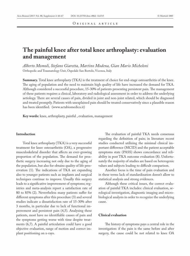
Palpation is painful also in case of overhang, in particular due to protrusion of tibial component in the medial region (Figure 1).
Evaluation of the vascular and neurological sta-tus is mandatory in order to find out neuritis, radicular compression or vascular insufficiency.
In case of abnormal pain, complex regional pain syndrome (CRPS) should be considered. The preva-lence is 21% at one, 13% a three and 12,7% at six months after TKA (10). Common risk factors are pre-operative pain, anxiety and depression. Trophic changes, motor disturbance, oedema and joint stiffness characterizing this condition, usually pain is diffuse, with burning sensation that worsen with movement and cold.
Laboratory evaluation
Laboratory tests are mandatory when infection is suspected, in particular inflammatory activity while hemograms and leukograms are not specific especially in implants with chronic infections.
Assay of erythrocyte sedimentation rate (ESR), C-reactive protein (CRP) and procalcitonin (PCT) are commonly used to prove the suspicion of infec-tion; nevertheless they present high sensitivity but low specificity, with high rate of false positives. The ESR
peak is 5-7 days after surgery, while CRP peak is 2-3 days after surgery. Baseline values are reached respec-tively after three months and three weeks. High levels of ESR and CRP are related to infection with a sensi-tivity of 0.95, specificity of 0.93 and a negative predic-tive value of 0.97 (11). In early postoperative days pay an important role serum level of interleukin 6 (IL-6) cause its rapid peak that comes baseline after 48 to 72 hours.
Test of joint puncture is mandatory for suspected infection (12) with leukocytes count and cultures of aerobic and anaerobic bacteria. Results higher than 2500 leukocytes per high magnification field and about 60% of polymorphonulear leukocytes (PMN) are indicative of infection with a sensitivity and speci-ficity of 98% (9).
Positive culture should be compared to the symp-toms and blood samples, if contamination is suspected
Figure 1. Under load x-rays show TKA with overhang of the tibial component
12-momoli.indd 61 06/06/17 10:27
A. Momoli, S. Giaretta, M. Modena, et al.62
repetition of puncture is suggested. Parvizi et al (13) published a diagnostic algorithm for TKA infection based on at least three aspiration, characterized by ma-jor and minor criteria.
Recently several studies have purposed the assess-ment of α-defensin in the articular samples with en-couraging result, but large-scale evidences are needed for state its significance for the diagnosis of peripros-thetic joint infections (14).
Radiological evaluation
Under load full leg antero-posterior, lateral and axial patella view x-rays are necessary to evaluate a painful TKA. Possible findings are the presence of ra-diolucency, varus-valgus malalignment, malrotation, periosteal reaction, gas in soft tissues, signs of loosen-ing, joint space asymmetry, component sizes, polyeth-ylene abrasion, stress fracture and heterotopic ossifi-cation. Lateral view shows tibial slope, patellar height related to joint line and sagittal alignment of femoral and tibial components. Also examination of preopera-
tive x-rays is important for determinate previous joint line, posterior femoral offset and patellar position.
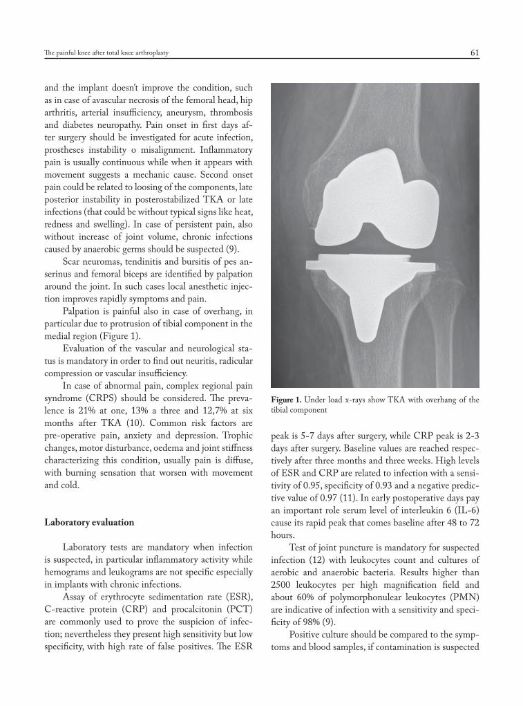
In case of evidence of loosening or overload at prostheses-bone interface a Technetium-99m scintig-raphy is indicated (2). This is not a screening tool and present high sensitivity but low specificity. Because of the physiological bone remodelling before one year after surgery, is not suggested in this period. Evalua-tion of serial examination and amount of uptake, dif-fuse and disproportional, should be indicating TKA loosening (Figure 2). Even after these results, with this exam is impossible to differentiate between septic o aseptic loosening. Association with leukocytes la-belled with Indium-111 scintigraphy improve sensitiv-ity and specificity to 85% (15)
Ultrasonography (US) is conducted if abnormali-ties in superficial soft tissue are suspected, particular collateral ligament lesions and tendon injuries.
Computer tomography (CT) pays a fundamental role in description of osteolysis areas (16) and in case of suspected fracture. Moreover should be used for di-agnosis of malrotation of femoral or tibial components.
Figure 2. Bone scintigraphy shows high uptake at the rigth knee TKA
12-momoli.indd 62 06/06/17 10:27
The painful knee after total knee arthroplasty 63
Management of pain
According to the literature, pain after TKA is due to in to joint related, non-joint related and unex-plained causes.
Joint related causes:Infections, instability, loosening of implant, frac-
tures, femoropatellar problems, other causes (compo-nent overhang, irritation of lateral facet of the patella, patellar clunck syndrome, popliteal tendon dysfunc-tion).
• Regarding infections decision-making process depend on time of onset, organism, conditions of tis-sues, host situation and whether the infection is super-ficial or deep.
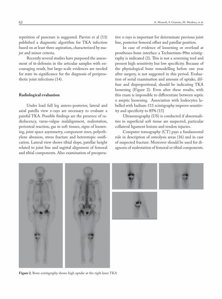
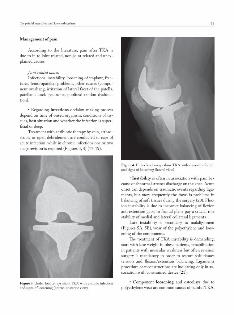
Treatment with antibiotic therapy by vein, arthro-scopic or open debridement are conducted in case of acute infection, while in chronic infections one or two stage revision is required (Figures 3, 4) (17-19).
• Instability is often in association with pain be-cause of abnormal stresses discharge on the knee. Acute onset can depends on traumatic events regarding liga-ments, but more frequently the focus is problems in balancing of soft tissues during the surgery (20). Flex-ion instability is due to incorrect balancing of flexion and extension gaps, in frontal plane pay a crucial role stability of medial and lateral collateral ligaments.
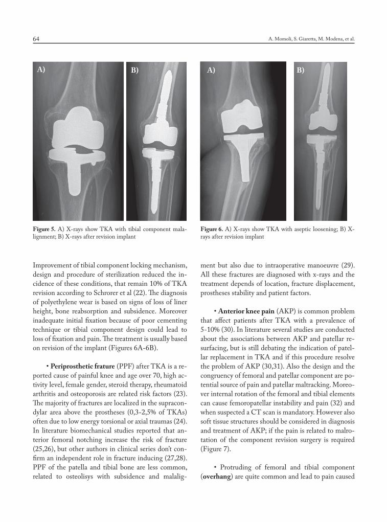
Late instability is secondary to malalignment (Figures 5A, 5B), wear of the polyethylene and loos-ening of the components.
The treatment of TKA instability is demanding, start with lose weight in obese patients, rehabilitation in patients with muscular weakness but often revision surgery is mandatory in order to restore soft tissues tension and flexion/extension balancing. Ligaments procedure or reconstructions are indicating only in as-sociation with constrained device (21).
• Component loosening and osteolisys due to polyethylene wear are common causes of painful TKA.
Figure 3. Under load x-rays show TKA with chronic infection and signs of loosening (antero-posterior view)
Figure 4. Under load x-rays show TKA with chronic infection and signs of loosening (lateral view)
12-momoli.indd 63 06/06/17 10:27
A. Momoli, S. Giaretta, M. Modena, et al.64
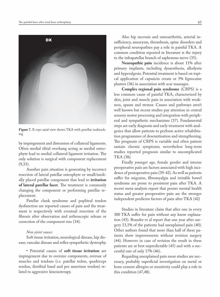
Improvement of tibial component locking mechanism, design and procedure of sterilization reduced the in-cidence of these conditions, that remain 10% of TKA revision according to Schrorer et al (22). The diagnosis of polyethylene wear is based on signs of loss of liner height, bone reabsorption and subsidence. Moreover inadequate initial fixation because of poor cementing technique or tibial component design could lead to loss of fixation and pain. The treatment is usually based on revision of the implant (Figures 6A-6B).
• Periprosthetic frature (PPF) after TKA is a re-ported cause of painful knee and age over 70, high ac-tivity level, female gender, steroid therapy, rheumatoid arthritis and osteoporosis are related risk factors (23). The majority of fractures are localized in the supracon-dylar area above the prostheses (0,3-2,5% of TKAs) often due to low energy torsional or axial traumas (24). In literature biomechanical studies reported that an-terior femoral notching increase the risk of fracture (25,26), but other authors in clinical series don’t con-firm an independent role in fracture inducing (27,28). PPF of the patella and tibial bone are less common, related to osteolisys with subsidence and malalig-
ment but also due to intraoperative manoeuvre (29). All these fractures are diagnosed with x-rays and the treatment depends of location, fracture displacement, prostheses stability and patient factors.
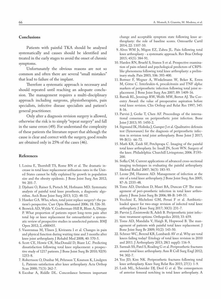
• Anterior knee pain (AKP) is common problem that affect patients after TKA with a prevalence of 5-10% (30). In literature several studies are conducted about the associations between AKP and patellar re-surfacing, but is still debating the indication of patel-lar replacement in TKA and if this procedure resolve the problem of AKP (30,31). Also the design and the congruency of femoral and patellar component are po-tential source of pain and patellar maltracking. Moreo-ver internal rotation of the femoral and tibial elements can cause femoropatellar instability and pain (32) and when suspected a CT scan is mandatory. However also soft tissue structures should be considered in diagnosis and treatment of AKP; if the pain is related to malro-tation of the component revision surgery is required (Figure 7).
• Protruding of femoral and tibial component (overhang) are quite common and lead to pain caused
Figure 5. A) X-rays show TKA with tibial component mala-lignment; B) X-rays after revision implant
A) B)
Figure 6. A) X-rays show TKA with aseptic loosening; B) X-rays after revision implant
A) B)
12-momoli.indd 64 06/06/17 10:27
The painful knee after total knee arthroplasty 65
by impingement and distension of collateral ligaments. Often medial tibial overhang acting as medial osteo-phyte lead to medial collateral ligament irritation. The only solution is surgical with component replacement (9,33).
Another pain situation is generating by incorrect resection of lateral patellar osteophyte or small/medi-ally placed patellar component that lead to irritation of lateral patellar facet. The treatment is commonly changing the component or performing patellar re-placement.
Patellar clunk syndrome and popliteal tendon dysfunction are reported causes of pain and the treat-ment is respectively with eventual resection of the fibrosis after observation and arthroscopic release or correction of the component size (34).
Non-joint causes:Soft tissue irritation, neurological disease, hip dis-
ease, vascular disease and reflex sympathetic dystrophy.
• Potential causes of soft tissue irritation are impingement due to oversize components, overuse of muscles and tendons (i.e. patellar tedon, quadriceps tendon, iliotibial band and pes anserinus tendon) re-lated to aggressive kinesioterapy.
Also hip necrosis and osteoarthritis, arterial in-sufficiency, aneurysm, thrombosis, spine disorders and peripheral neuropathies pay a role in painful TKA. A common condition reported in literature is the injury to the infrapatellar branch of saphenous nerve (35).
Neuropathic pain incidence is about 11% after primary implants, including dysaesthesia, allodynia and hyperalgesia. Potential treatment is based on topi-cal application of capsaicin cream or 5% lignocaine plasters (36) in association with scar massages.
Complex regional pain syndrome (CRPS) is a less common cause of painful TKA, characterized by skin, joint and muscle pain in association with weak-ness, spasm and tremor. Causes and pathways aren’t well known but recent studies pay attention in central sensory motor processing and integration with periph-eral and sympathetic mechanisms (37). Fundamental steps are early diagnosis and early treatment with anal-gesics that allow patients to perform active rehabilita-tion programmes of desensitisation and strengthening. The prognosis of CRPS is variable and often patient sustain chronic symptoms; nevertheless long-term studies reported prognosis similar to uncomplicated TKA (38).
Finally younger age, female gender and intense preoperative pain are factors associated with high inci-dence of postoperative pain (39-42). As well as patients suffer for migraine, fibromyalgia and irritable bowel syndrome are prone to persistent pain after TKA. A recent meta-analysis report that poorer mental health status and greater preoperative pain are the stronger independent predictor factors of pain after TKA (42)
Studies in literature claim that after one in every 300 TKA suffer for pain without any know explana-tion (43). Brander et al report that one year after sur-gery 13,1% of the patients had unexplained pain (40). Other authors found that more than half of these pa-tients show improvements without revision surgery (44). However in case of revision the result in these patients are at best unpredictable (45) and with a suc-cessful rate of only 17% (46).
Regarding unexplained pain more studies are nec-essary, probably superficial investigation on metal or bone cement allergies or sensitivity could play a role in this condition (47,48).
Figure 7. X-rays axial view shows TKA with patellar maltrack-ing
12-momoli.indd 65 06/06/17 10:27
A. Momoli, S. Giaretta, M. Modena, et al.66
Conclusions
Patients with painful TKA should be analysed systematically and causes should be identified and treated in the early stages to avoid the onset of chronic symptoms.
Unfortunately the obvious reasons are not so common and often there are several “small mistakes” that lead to failure of the implant.
Therefore a systematic approach is necessary and should repeated until reaching an adequate conclu-sion. The management requires a multi-disciplinary approach including surgeons, physiotherapists, pain specialists, infective disease specialists and patient’s general practitioner.
Only after a diagnosis revision surgery is allowed, otherwise the risk is to simply “repeat surgery” and fall in the same errors (49). For understand the complexity of these patients the literature report that although the cause is clear and correct with the surgery, good results are obtained only in 25% of the cases (46).
References
1. Losina E, Thornhill TS, Rome BN et al. The dramatic in-crease in total knee replacement utilization rates in the Unit-ed States cannot be fully explained by growth in population size and the obesity epidemic. J Bone Joint Surg Am 2012; 94: 201-7.
2. Djahani O, Rainer S, Pietsch M, Hofmann MD. Systematic analysis of painful total knee prosthesis, a diagnostic algo-rithm. Arch Bone Joint Surg 2013; 1(2): 48-52.
3. Hawker GA. Who, when, total joint replace surgery?: the pa-tient’s perspective. Curr Opin Rheumatol 2006; 18: 526-30.
4. Beswick AD, Wylde V, Gooberman-Hill R, Blom A, Dieppe P. What proportion of patients report long-term pain after total hip or knee replacement for osteoarthritis? a system-atic review of prospective studies in unselected patients. BMJ Open 2012; 2, e000435.
5. Vuorenmaa M, Ylinen J, Kiviranta I et al. Changes in pain and physical function during waiting time and 3 months after knee joint arthroplasty. J Rehabil Med 2008; 40: 570-5.
6. Scott CE, Howie CR, MacDonald D, Biant LC. Predicting dissatisfaction following total knee replacement: a prospec-tive study of 1217 patients. J Bone Joint Surg Br 2010; 92(9): 1253-8.
7. Robertsson O, Dunbar M, Pehrsson T, Knutson K, Lindgren L. Patients satisfaction after knee arthroplasty. Acta Orthop Scan 2000; 71(3): 262-7.
8. Escobar A, Riddle DL. Concordance between important
change and acceptable symptom state following knee ar-throplasty: the role of baseline scores. Osteoarthr Cartil 2014; 22: 1107-10.
9. Alves WM Jr, Migon EZ, Zabeu JL. Pain following total knee arthroplasty - a systematic approach. Rev Bras Orthop 2015; 45(5): 384-91.
10. Harden RN, Bruehl S, Stanos S et al. Prospective examina-tion of pain-related and psychological predictors of CRPS-like phenomena following total knee arthroplasty: a prelim-inary study. Pain 2003; 106: 393-400.
11. Bottner F, Wegner A, Winkelmann W, Beker K, Erren M, Götze C. Interleukin-6, procalcitonin and TNF alpha: markers of periprosthetic infection following total joint re-placement. J Bone Joint Surg Am 2007; 89: 1409-16.
12. Barrak RL, Jennings RW, Wolfe MW, Bertot AJ. The Cov-entry Award: the value of preoperative aspiration before total knee revision. Clin Orthop and Relat Res 1997; 345: 8-16.
13. Parvizi J, Gerke T, Chen AF. Proceedings of the interna-tional consensus on periprosthetic joint infection. Bone Joint J 2013; 95: 1450-2.
14. Sigmund IK, Holinka J, Gamper J et al. Qualitative defensin test (Synovasure) for the diagnosis of periprosthetic infec-tion in revision total joint arthroplasty. Bone Joint J 2017; 99-B(1): 66-72.
15. Math KR, Zaidi SF, Petchprapa C. Imaging of the painful total knee arthroplasty. In: Insall JN, Scott WN. Surgery of the knee. Philadelphia: Churchill Livingstone; 2006. P.193-200.
16. Sofka CM. Current applications of advanced cross-sectional imaging techniques in evaluating the painful arthroplasty. Skeletal Radiol 2007; 36(3): 183-93.
17. Leone JM, Hanssen AD. Management of infection at the site of a total knee arthroplasty. J Bone Joint Surg Am 2005; 87-A: 2335-48.
18. Toms AD, Davidson D, Masri BA, Duncan CP. The man-agement of peri-prosthetic infection in total knee arthro-plasty. J Bone Joint Surg Br 2006; 88-B: 149-55.
19. Vecchini E, Micheloni GM, Perusi F et al. Antibiotic-loaded spacer for two-stage revision of infected total knee arthroplasty. J Knee Surg 2017; 30(3): 231-7.
20. Parvizi J, Zmistowski B, Adeli B. Periprosthetic joint infec-tion: treatment options. Orthopedics 2010; 33: 659.
21. Toms AD, Mandalia V, Haigh R, Hopwood B. The man-agement of patients with painful total knee replacement. J Bone Joint Surg Br 2009; 91(2): 143-50.
22. Schroer WC, Berend KR, Lombardi AV et al. Why are total knees failing today? Etiology of total knee revision in 2010 and 2011. J Arthroplasty 2013; 28(1 suppl): 116-9.
23. Sarmah SS, Patel S, Reading G et al. Periprosthetic fractures around total knee arthroplasty. Ann R Coll Surg Engl 2012; 94: 302-7.
24. Yoo JD, Kim NK. Periprosthetic fractures following total knee arthroplasty. Knee Surg Relat Res 2015; 27(1): 1-9.
25. Lesh ML, Schneider DJ, Deol G et al. The consequences of anterior femoral notching in total knee arthroplasty. A
12-momoli.indd 66 06/06/17 10:27
The painful knee after total knee arthroplasty 67
biomechanical study. J Bone Joint Surg Am 2000; 82: 1096-101.
26. Zalzal P, Backstein D, Gross AE, Papini M. Notching of the anterior femoral cortex during total knee arthroplasty characteristics that increase local stresses. J Arthroplasty 2006; 21: 737-43.
27. Ritter MA, Thong AE, Keating EM et al. The effect of fem-oral notching during total knee arthroplasty on the preva-lence of post-operative femoral fractures and on clinical outcome. J Bone Joint Surg Am 2005; 87: 2411-4.
28. Gujarathi N, Putti AB, Abboud RJ et al. Risk of peripros-thetic fractures after anterior femoral notching. Acta Or-thop 2009; 80: 553-6.
29. Dennis DA. Periprosthetic fractures following total knee arthroplasty. Instr Course Lect 2001; 50: 379-89.
30. Breugem SJM, Haverkamp D. Anterior knee pain after total knee arthroplasty: what can cause this pain? World J Orthop 2014; 5(3): 163-70.
31. He JY, Jiang LS, Dai LY. Is patellar resurfacing superior than nonresurfacing in total knee arthroplasty? A meta-analysis of randomized trials. Knee 2011; 18: 137-44.
32. Berger RA, Crossett LS, Jacobs JJ, Rubash HE. Malrota-tion causing patella-femoral complications after total knee arthroplasty. Clin Orthop Relat Res 1998; (356): 144-53.
33. Mahoney OM, Kinsey T. Overhang of the femoral com-ponent in total knee arthroplasty: risk factors and clinical consequences. J Bone Joint Surg Am 2010; 92(5): 1115-21.
34. Allardyce TJ, Scuderi GR, Insall JN. Arthroscopic treat-ment of popliteus tendon dysfunction following total knee replacement. J Arthroscopy 1997; 12: 353-5.
35. Clendenen S, Greengrass R, Whalen J, O’Connor MI. In-frapatellar saphenous neuralgia after TKA can be improved with ultrasound-guided local treatments. Clin Orthop Relat Res 2015; 473(1): 119-25.
36. Finnerup NB, Otto M, McQuay HJ, Jensen TS, Sindrup SH. Algorithm for neuropathic pain treatment: an evidence based proposal. Pain 2005; 118: 289-305.
37. Janig W, Baron R. Experimental approach to CRPS. Pain 2004; 108: 3-7.
38. Burns AW, Parker DA, Coolican MR, Rayartnam K. Com-plex regional pain syndrome complicating total knee arthro-plasty. J Orthop Surg (Hong Kong) 2006; 14: 280-3.
39. Lavand’homme PM, Grosu I, France MN, Thienpont E. Pain trajectories identify patients at risk of persistent pain
after knee arthroplasty: an observational study. Clin Orthop Relat Res 2014; 472: 1409-15.
40. Brander VA, Stulberg SD, Adams AD et al. Predicting total knee replacement pain: a prospective, observational study. Clin Orthop Relat Res 2003; 416: 27-36.
41. Hischmann MT, Testa E, Amsler F, Friederich NF. The unhappy total knee arthroplasty (TKA) patient: higher WOMAC and lower KSS in depressed patients prior and after TKA. Knee Surg Sports Traumatol Arthrosc 2013; 21(10): 2405-11.
42. Lewis GN, Rice DA, McNair PJ, Kluger M. Prectors of persistent pain after total knee arthroplasty: a systematic re-view and meta-analisys. Br J Anaesth 2015; 114(4): 551-61.
43. Brassard MF, Insall JN, Scuderi GR, Faris PM. Compli-cation of total knee arthroplasty. In: Insall JN, Scott WN: Surgery of the knee. Philadelphia: Churchill Livingstone; 2006: 1753.
44. Elson DW, Brenkel IJ. A conservative approach is feasible in unexplained pain after knee replacement: a selected co-hort study. J Bone Joint Surg Br 2007; 89-B: 1042-5.
45. Jacobs MA, Hungerford DS, Krackow KA, Lennox DW. Revision total knee arthroplasty for aseptic failure. Clin Or-thop 1988; 226: 78-85.
46. Mont MA, Serna FK, Krackow KA, Hungerford DS. Ex-ploration of radiographically normal total knee replace-ments for unexplained pain. Clin Orthop Relat Res 1996; 331: 216-20.
47. Faschingbauer M, Renner L, Boettner F. Allergy in total knee replacement. Does it exist?: Review article. HSS J 2017; 13(1): 12-19.
48. Preston S, Petrera M, Kim C, Zywiel MG, Gandhi R. To-wards an understanding of the painful total knee: what is the role of patient biology?. Curr Rev Musculoskelet Med 2016; 9: 388-95.
49. Vince KG. Why knees fail. J Arthroplasty 2003;18:39-44.
Received: 20 April 2017Accepted: 21 May 2017Correspondence:Alberto Momoli, MDOrthopedic and Traumatology Unit,Ospedale San Bortolo, Vicenza, ItalyE-mail: [email protected]
12-momoli.indd 67 06/06/17 10:27