the nuclear localization of swi/snf proteins is subjected to oxygen regulation
TRANSCRIPT
Cell & BioscienceDastidar et al. Cell & Bioscience 2012, 2:30http://www.cellandbioscience.com/content/2/1/30
RESEARCH Open Access
The nuclear localization of SWI/SNF proteins issubjected to oxygen regulationRanita Ghosh Dastidar†, Jagmohan Hooda†, Ajit Shah†, Thai M Cao, Robert Michael Henke and Li Zhang*
Abstract
Background: Hypoxia is associated with many disease conditions in humans, such as cancer, stroke and traumaticinjuries. Hypoxia elicits broad molecular and cellular changes in diverse eukaryotes. Our recent studies suggest thatone likely mechanism mediating such broad changes is through changes in the cellular localization of importantregulatory proteins. Particularly, we have found that over 120 nuclear proteins with important functions rangingfrom transcriptional regulation to RNA processing exhibit altered cellular locations under hypoxia. In this report, wedescribe further experiments to identify and evaluate the role of nuclear protein relocalization in mediating hypoxiaresponses in yeast.
Results: To identify regulatory proteins that play a causal role in mediating hypoxia responses, we characterized thetime courses of relocalization of hypoxia-altered nuclear proteins in response to hypoxia and reoxygenation. Wefound that 17 nuclear proteins relocalized in a significantly shorter time period in response to both hypoxia andreoxygenation. Particularly, several components of the SWI/SNF complex were fast responders, and analysis of geneexpression data show that many targets of the SWI/SNF proteins are oxygen regulated. Furthermore, confocalfluorescent live cell imaging showed that over 95% of hypoxia-altered SWI/SNF proteins accumulated in the cytosolin hypoxic cells, while over 95% of the proteins were nuclear in normoxic cells, as expected.
Conclusions: SWI/SNF proteins relocalize in response to hypoxia and reoxygenation in a quick manner, and theirrelocalization likely accounts for, in part or in whole, oxygen regulation of many SWI/SNF target genes.
Keywords: Hypoxia response, Oxygen regulation, SWI/SNF, Live cell imaging, Protein localization
BackgroundLiving organisms ranging from yeast to mammals use oxy-gen to generate their cellular energy supply and tosynthesize important biomolecules. Hence, they need to re-spond effectively to changes in oxygen levels in the environ-ment, particularly to hypoxia [1,2]. In humans, hypoxia isresponsible for death or damage by the ischemia accom-panying heart attack, stroke, and traumatic injuries [3-5].The molecular and cellular events induced by changes inoxygen levels are very broad in eukaryotes. For example,over 20% of yeast genes change their transcript levels in re-sponse to hypoxia [6]. In the human arterial endothelialcells, more than 8% of all genes alter their transcript levelsby at least 1.5-fold in response to hypoxia [7]. In the human
* Correspondence: [email protected]†Equal contributorsDepartment of Molecular and Cell Biology, Center for Systems Biology,University of Texas at Dallas, Mail Stop RL11 800 W Campbell Road,Richardson, TX 75080, USA
© 2012 Ghosh Dastidar et al.; licensee BioMedCreative Commons Attribution License (http:/distribution, and reproduction in any medium
primary astrocytes, more than 5% of the genes alter theirtranscript levels by at least 2-fold in response to hypoxia[8]. Such broad changes in gene expression likely involvecoordinated actions of multiple pathways and regulators.Previous studies have identified several transcrip-
tional regulators, including Mga1 and Rox1, that canmediate oxygen regulation of gene expression in theyeast Saccharomyces cerevisiae [9,10]. However, theseregulators can account for the regulation of only a fractionof hypoxia-regulated genes [6]. Many other regulators arelikely involved in mediating oxygen regulation. Recently, inan effort to systematically identify proteins that can mediateoxygen regulation and signaling, we performed a genome-wide screen for proteins that exhibit altered cellular distri-bution patterns in response to hypoxia and reoxygenation[11]. We found that over 200 proteins alter their cellularlocations in response to hypoxia. Particularly, under hyp-oxia, a good number (at least 121) of nuclear proteins donot localize to the nucleus, but accumulate in the cytosol.
Central Ltd. This is an Open Access article distributed under the terms of the/creativecommons.org/licenses/by/2.0), which permits unrestricted use,, provided the original work is properly cited.
Dastidar et al. Cell & Bioscience 2012, 2:30 Page 2 of 13http://www.cellandbioscience.com/content/2/1/30
In response to reoxygenation, they readily localize to thenucleus. Notably, many of these hypoxia-redistributed nu-clear proteins are subunits of key regulatory complexesinvolved in chromatin remodeling (such as the SWI/SNFcomplex) [12-14], in transcriptional regulation (such as theSAGA complex) [15], and in splicing (such as the MRPcomplex) [16]. Hence, it is conceivable that some of thesecomplexes can play a dominant role in mediating oxygenregulation of gene expression.To further assess the roles of these regulators in medi-
ating oxygen signaling and regulation, we examined thetime course characteristics of the relocalization of theseproteins in response to hypoxia and reoxygenation. Wefound a small group of nuclear proteins relocalized in asignificantly shorter time period in response to bothhypoxia and reoxygenation, when compared to otherproteins. These proteins include three components ofthe SWI/SNF complex. Furthermore, using confocalfluorescent imaging of live cells, we quantitatively char-acterized the effect of hypoxia on the distribution ofSWI/SNF proteins. We found that in live hypoxic cells,over 95% of Swi3, Snf5, Snf6, Snf11, Snf12 and Swp82were in the cytosol, while over 95% of hypoxia-unaffected proteins, such as Swi2 and Taf14, were in thenucleus. These results suggest that hypoxia can signifi-cantly alter the composition and property of the SWI/SNFcomplex and mediate oxygen regulation of gene expression.
ResultsAmong the hypoxia-redistributed nuclear proteins wepreviously identified, some are likely involved in mediat-ing oxygen signaling and regulation of gene expression.Particularly, proteins that change their locations in rela-tively shorter time periods are likely the regulators that
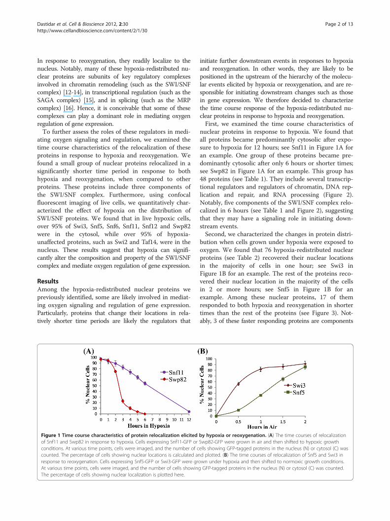
Figure 1 Time course characteristics of protein relocalization elicitedof Snf11 and Swp82 in response to hypoxia. Cells expressing Snf11-GFP orconditions. At various time points, cells were imaged, and the number of ccounted. The percentage of cells showing nuclear locations is calculated anresponse to reoxygenation. Cells expressing Snf5-GFP or Swi3-GFP were grAt various time points, cells were imaged, and the number of cells showinThe percentage of cells showing nuclear localization is plotted here.
initiate further downstream events in responses to hypoxiaand reoxygenation. In other words, they are likely to bepositioned in the upstream of the hierarchy of the molecu-lar events elicited by hypoxia or reoxygenation, and are re-sponsible for initiating downstream changes such as thosein gene expression. We therefore decided to characterizethe time course response of the hypoxia-redistributed nu-clear proteins in response to hypoxia and reoxygenation.First, we examined the time course characteristics of
nuclear proteins in response to hypoxia. We found thatall proteins became predominantly cytosolic after expo-sure to hypoxia for 12 hours; see Snf11 in Figure 1A foran example. One group of these proteins became pre-dominantly cytosolic after only 6 hours or shorter times;see Swp82 in Figure 1A for an example. This group has48 proteins (see Table 1). They include several transcrip-tional regulators and regulators of chromatin, DNA rep-lication and repair, and RNA processing (Figure 2).Notably, five components of the SWI/SNF complex relo-calized in 6 hours (see Table 1 and Figure 2), suggestingthat they may have a signaling role in initiating down-stream events.Second, we characterized the changes in protein distri-
bution when cells grown under hypoxia were exposed tooxygen. We found that 76 hypoxia-redistributed nuclearproteins (see Table 2) recovered their nuclear locationsin the majority of cells in one hour; see Swi3 inFigure 1B for an example. The rest of the proteins reco-vered their nuclear location in the majority of the cellsin 2 or more hours; see Snf5 in Figure 1B for anexample. Among these nuclear proteins, 17 of themresponded to both hypoxia and reoxygenation in shortertimes than the rest of the proteins (see Figure 3). Not-ably, 3 of these faster responding proteins are components
by hypoxia or reoxygenation. (A) The time courses of relocalizationSwp82-GFP were grown in air and then shifted to hypoxic growthells showing GFP-tagged proteins in the nucleus (N) or cytosol (C) wasd plotted. (B) The time courses of relocalization of Snf5 and Swi3 inown under hypoxia and then shifted to normoxic growth conditions.g GFP-tagged proteins in the nucleus (N) or cytosol (C) was counted.
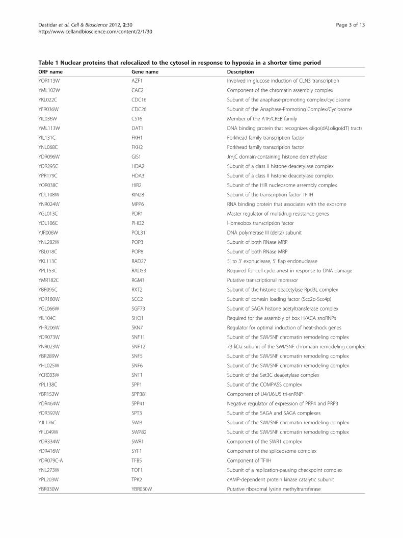
Table 1 Nuclear proteins that relocalized to the cytosol in response to hypoxia in a shorter time period
ORF name Gene name Description
YOR113W AZF1 Involved in glucose induction of CLN3 transcription
YML102W CAC2 Component of the chromatin assembly complex
YKL022C CDC16 Subunit of the anaphase-promoting complex/cyclosome
YFR036W CDC26 Subunit of the Anaphase-Promoting Complex/Cyclosome
YIL036W CST6 Member of the ATF/CREB family
YML113W DAT1 DNA binding protein that recognizes oligo(dA).oligo(dT) tracts
YIL131C FKH1 Forkhead family transcription factor
YNL068C FKH2 Forkhead family transcription factor
YDR096W GIS1 JmjC domain-containing histone demethylase
YDR295C HDA2 Subunit of a class II histone deacetylase complex
YPR179C HDA3 Subunit of a class II histone deacetylase complex
YOR038C HIR2 Subunit of the HIR nucleosome assembly complex
YDL108W KIN28 Subunit of the transcription factor TFIIH
YNR024W MPP6 RNA binding protein that associates with the exosome
YGL013C PDR1 Master regulator of multidrug resistance genes
YDL106C PHO2 Homeobox transcription factor
YJR006W POL31 DNA polymerase III (delta) subunit
YNL282W POP3 Subunit of both RNase MRP
YBL018C POP8 Subunit of both RNase MRP
YKL113C RAD27 5' to 3' exonuclease, 5' flap endonuclease
YPL153C RAD53 Required for cell-cycle arrest in response to DNA damage
YMR182C RGM1 Putative transcriptional repressor
YBR095C RXT2 Subunit of the histone deacetylase Rpd3L complex
YDR180W SCC2 Subunit of cohesin loading factor (Scc2p-Scc4p)
YGL066W SGF73 Subunit of SAGA histone acetyltransferase complex
YIL104C SHQ1 Required for the assembly of box H/ACA snoRNPs
YHR206W SKN7 Regulator for optimal induction of heat-shock genes
YDR073W SNF11 Subunit of the SWI/SNF chromatin remodeling complex
YNR023W SNF12 73 kDa subunit of the SWI/SNF chromatin remodeling complex
YBR289W SNF5 Subunit of the SWI/SNF chromatin remodeling complex
YHL025W SNF6 Subunit of the SWI/SNF chromatin remodeling complex
YCR033W SNT1 Subunit of the Set3C deacetylase complex
YPL138C SPP1 Subunit of the COMPASS complex
YBR152W SPP381 Component of U4/U6.U5 tri-snRNP
YDR464W SPP41 Negative regulator of expression of PRP4 and PRP3
YDR392W SPT3 Subunit of the SAGA and SAGA complexes
YJL176C SWI3 Subunit of the SWI/SNF chromatin remodeling complex
YFL049W SWP82 Subunit of the SWI/SNF chromatin remodeling complex
YDR334W SWR1 Component of the SWR1 complex
YDR416W SYF1 Component of the spliceosome complex
YDR079C-A TFB5 Component of TFIIH
YNL273W TOF1 Subunit of a replication-pausing checkpoint complex
YPL203W TPK2 cAMP-dependent protein kinase catalytic subunit
YBR030W YBR030W Putative ribosomal lysine methyltransferase
Dastidar et al. Cell & Bioscience 2012, 2:30 Page 3 of 13http://www.cellandbioscience.com/content/2/1/30
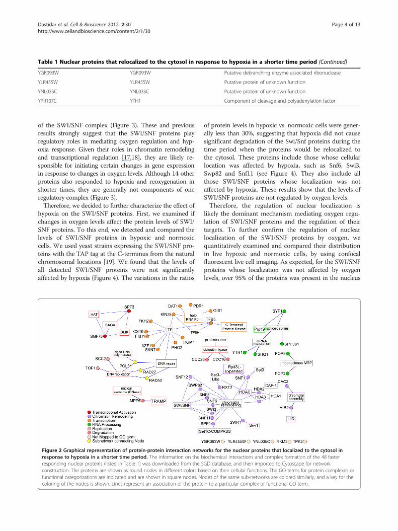
Table 1 Nuclear proteins that relocalized to the cytosol in response to hypoxia in a shorter time period (Continued)
YGR093W YGR093W Putative debranching enzyme associated ribonuclease
YLR455W YLR455W Putative protein of unknown function
YNL035C YNL035C Putative protein of unknown function
YPR107C YTH1 Component of cleavage and polyadenylation factor
Dastidar et al. Cell & Bioscience 2012, 2:30 Page 4 of 13http://www.cellandbioscience.com/content/2/1/30
of the SWI/SNF complex (Figure 3). These and previousresults strongly suggest that the SWI/SNF proteins playregulatory roles in mediating oxygen regulation and hyp-oxia response. Given their roles in chromatin remodelingand transcriptional regulation [17,18], they are likely re-sponsible for initiating certain changes in gene expressionin response to changes in oxygen levels. Although 14 otherproteins also responded to hypoxia and reoxygenation inshorter times, they are generally not components of oneregulatory complex (Figure 3).Therefore, we decided to further characterize the effect of
hypoxia on the SWI/SNF proteins. First, we examined ifchanges in oxygen levels affect the protein levels of SWI/SNF proteins. To this end, we detected and compared thelevels of SWI/SNF proteins in hypoxic and normoxiccells. We used yeast strains expressing the SWI/SNF pro-teins with the TAP tag at the C-terminus from the naturalchromosomal locations [19]. We found that the levels ofall detected SWI/SNF proteins were not significantlyaffected by hypoxia (Figure 4). The variations in the ratios
Figure 2 Graphical representation of protein-protein interaction netwresponse to hypoxia in a shorter time period. The information on the bresponding nuclear proteins (listed in Table 1) was downloaded from the Sconstruction. The proteins are shown as round nodes in different colors bafunctional categorizations are indicated and are shown in square nodes. Nocoloring of the nodes is shown. Lines represent an association of the prote
of protein levels in hypoxic vs. normoxic cells were gener-ally less than 30%, suggesting that hypoxia did not causesignificant degradation of the Swi/Snf proteins during thetime period when the proteins would be relocalized tothe cytosol. These proteins include those whose cellularlocation was affected by hypoxia, such as Snf6, Swi3,Swp82 and Snf11 (see Figure 4). They also include allthose SWI/SNF proteins whose localization was notaffected by hypoxia. These results show that the levels ofSWI/SNF proteins are not regulated by oxygen levels.Therefore, the regulation of nuclear localization is
likely the dominant mechanism mediating oxygen regu-lation of SWI/SNF proteins and the regulation of theirtargets. To further confirm the regulation of nuclearlocalization of the SWI/SNF proteins by oxygen, wequantitatively examined and compared their distributionin live hypoxic and normoxic cells, by using confocalfluorescent live cell imaging. As expected, for the SWI/SNFproteins whose localization was not affected by oxygenlevels, over 95% of the proteins was present in the nucleus
orks for the nuclear proteins that localized to the cytosol iniochemical interactions and complex formation of the 48 fasterGD database, and then imported to Cytoscape for networksed on their cellular functions. The GO terms for protein complexes ordes of the same sub-networks are colored similarly, and a key for thein to a particular complex or functional GO term.
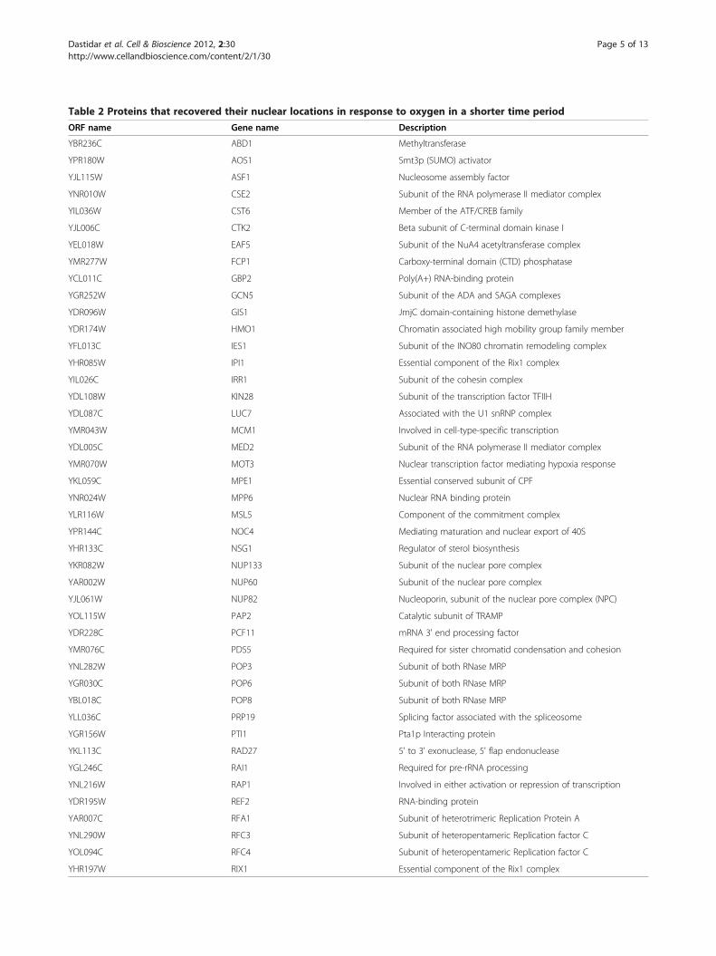
Table 2 Proteins that recovered their nuclear locations in response to oxygen in a shorter time period
ORF name Gene name Description
YBR236C ABD1 Methyltransferase
YPR180W AOS1 Smt3p (SUMO) activator
YJL115W ASF1 Nucleosome assembly factor
YNR010W CSE2 Subunit of the RNA polymerase II mediator complex
YIL036W CST6 Member of the ATF/CREB family
YJL006C CTK2 Beta subunit of C-terminal domain kinase I
YEL018W EAF5 Subunit of the NuA4 acetyltransferase complex
YMR277W FCP1 Carboxy-terminal domain (CTD) phosphatase
YCL011C GBP2 Poly(A+) RNA-binding protein
YGR252W GCN5 Subunit of the ADA and SAGA complexes
YDR096W GIS1 JmjC domain-containing histone demethylase
YDR174W HMO1 Chromatin associated high mobility group family member
YFL013C IES1 Subunit of the INO80 chromatin remodeling complex
YHR085W IPI1 Essential component of the Rix1 complex
YIL026C IRR1 Subunit of the cohesin complex
YDL108W KIN28 Subunit of the transcription factor TFIIH
YDL087C LUC7 Associated with the U1 snRNP complex
YMR043W MCM1 Involved in cell-type-specific transcription
YDL005C MED2 Subunit of the RNA polymerase II mediator complex
YMR070W MOT3 Nuclear transcription factor mediating hypoxia response
YKL059C MPE1 Essential conserved subunit of CPF
YNR024W MPP6 Nuclear RNA binding protein
YLR116W MSL5 Component of the commitment complex
YPR144C NOC4 Mediating maturation and nuclear export of 40S
YHR133C NSG1 Regulator of sterol biosynthesis
YKR082W NUP133 Subunit of the nuclear pore complex
YAR002W NUP60 Subunit of the nuclear pore complex
YJL061W NUP82 Nucleoporin, subunit of the nuclear pore complex (NPC)
YOL115W PAP2 Catalytic subunit of TRAMP
YDR228C PCF11 mRNA 3' end processing factor
YMR076C PDS5 Required for sister chromatid condensation and cohesion
YNL282W POP3 Subunit of both RNase MRP
YGR030C POP6 Subunit of both RNase MRP
YBL018C POP8 Subunit of both RNase MRP
YLL036C PRP19 Splicing factor associated with the spliceosome
YGR156W PTI1 Pta1p Interacting protein
YKL113C RAD27 5' to 3' exonuclease, 5' flap endonuclease
YGL246C RAI1 Required for pre-rRNA processing
YNL216W RAP1 Involved in either activation or repression of transcription
YDR195W REF2 RNA-binding protein
YAR007C RFA1 Subunit of heterotrimeric Replication Protein A
YNL290W RFC3 Subunit of heteropentameric Replication factor C
YOL094C RFC4 Subunit of heteropentameric Replication factor C
YHR197W RIX1 Essential component of the Rix1 complex
Dastidar et al. Cell & Bioscience 2012, 2:30 Page 5 of 13http://www.cellandbioscience.com/content/2/1/30
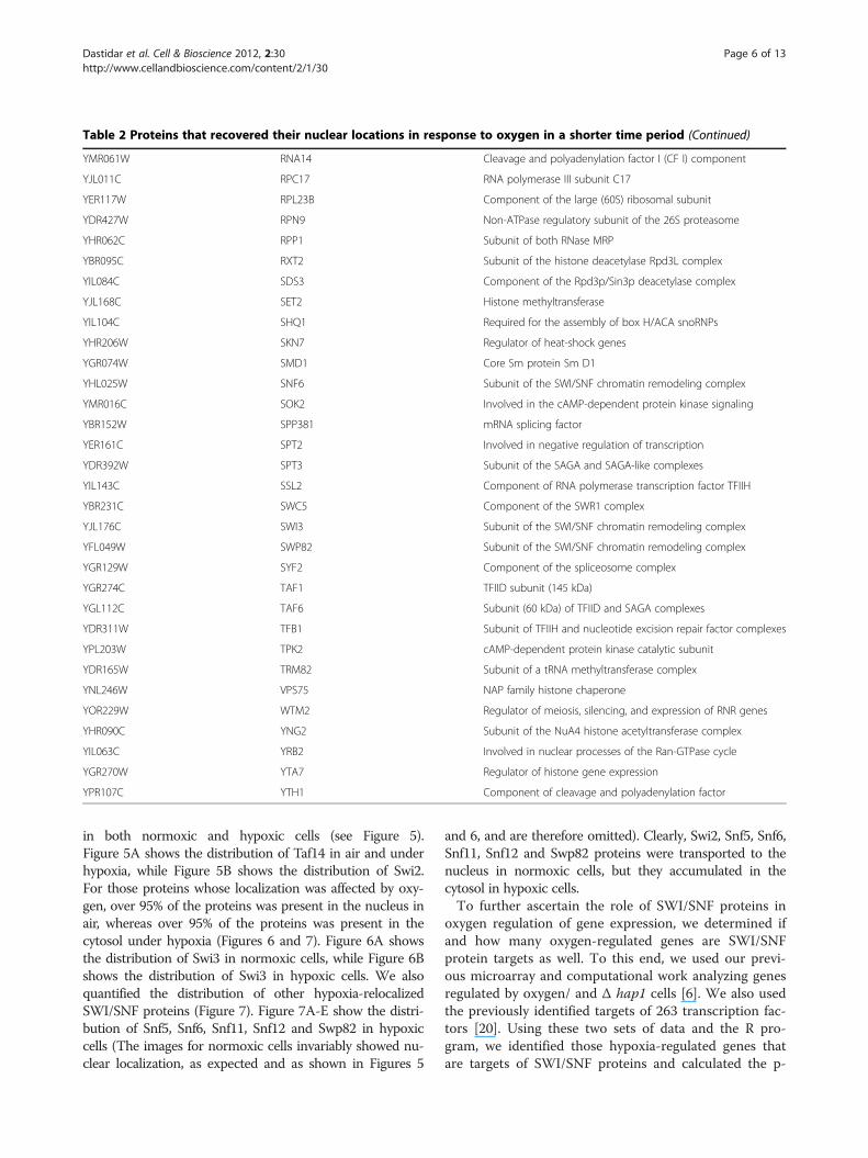
Table 2 Proteins that recovered their nuclear locations in response to oxygen in a shorter time period (Continued)
YMR061W RNA14 Cleavage and polyadenylation factor I (CF I) component
YJL011C RPC17 RNA polymerase III subunit C17
YER117W RPL23B Component of the large (60S) ribosomal subunit
YDR427W RPN9 Non-ATPase regulatory subunit of the 26S proteasome
YHR062C RPP1 Subunit of both RNase MRP
YBR095C RXT2 Subunit of the histone deacetylase Rpd3L complex
YIL084C SDS3 Component of the Rpd3p/Sin3p deacetylase complex
YJL168C SET2 Histone methyltransferase
YIL104C SHQ1 Required for the assembly of box H/ACA snoRNPs
YHR206W SKN7 Regulator of heat-shock genes
YGR074W SMD1 Core Sm protein Sm D1
YHL025W SNF6 Subunit of the SWI/SNF chromatin remodeling complex
YMR016C SOK2 Involved in the cAMP-dependent protein kinase signaling
YBR152W SPP381 mRNA splicing factor
YER161C SPT2 Involved in negative regulation of transcription
YDR392W SPT3 Subunit of the SAGA and SAGA-like complexes
YIL143C SSL2 Component of RNA polymerase transcription factor TFIIH
YBR231C SWC5 Component of the SWR1 complex
YJL176C SWI3 Subunit of the SWI/SNF chromatin remodeling complex
YFL049W SWP82 Subunit of the SWI/SNF chromatin remodeling complex
YGR129W SYF2 Component of the spliceosome complex
YGR274C TAF1 TFIID subunit (145 kDa)
YGL112C TAF6 Subunit (60 kDa) of TFIID and SAGA complexes
YDR311W TFB1 Subunit of TFIIH and nucleotide excision repair factor complexes
YPL203W TPK2 cAMP-dependent protein kinase catalytic subunit
YDR165W TRM82 Subunit of a tRNA methyltransferase complex
YNL246W VPS75 NAP family histone chaperone
YOR229W WTM2 Regulator of meiosis, silencing, and expression of RNR genes
YHR090C YNG2 Subunit of the NuA4 histone acetyltransferase complex
YIL063C YRB2 Involved in nuclear processes of the Ran-GTPase cycle
YGR270W YTA7 Regulator of histone gene expression
YPR107C YTH1 Component of cleavage and polyadenylation factor
Dastidar et al. Cell & Bioscience 2012, 2:30 Page 6 of 13http://www.cellandbioscience.com/content/2/1/30
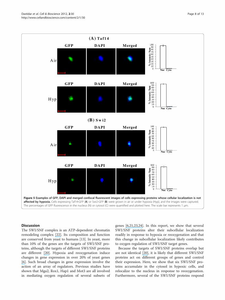
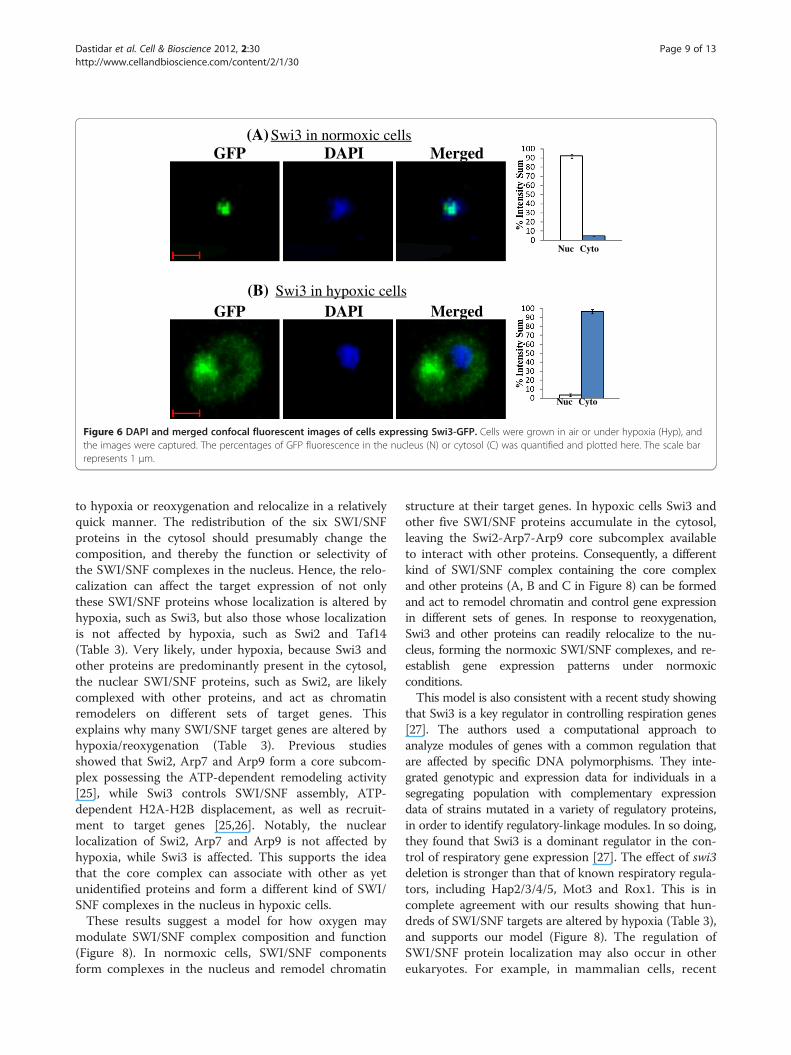
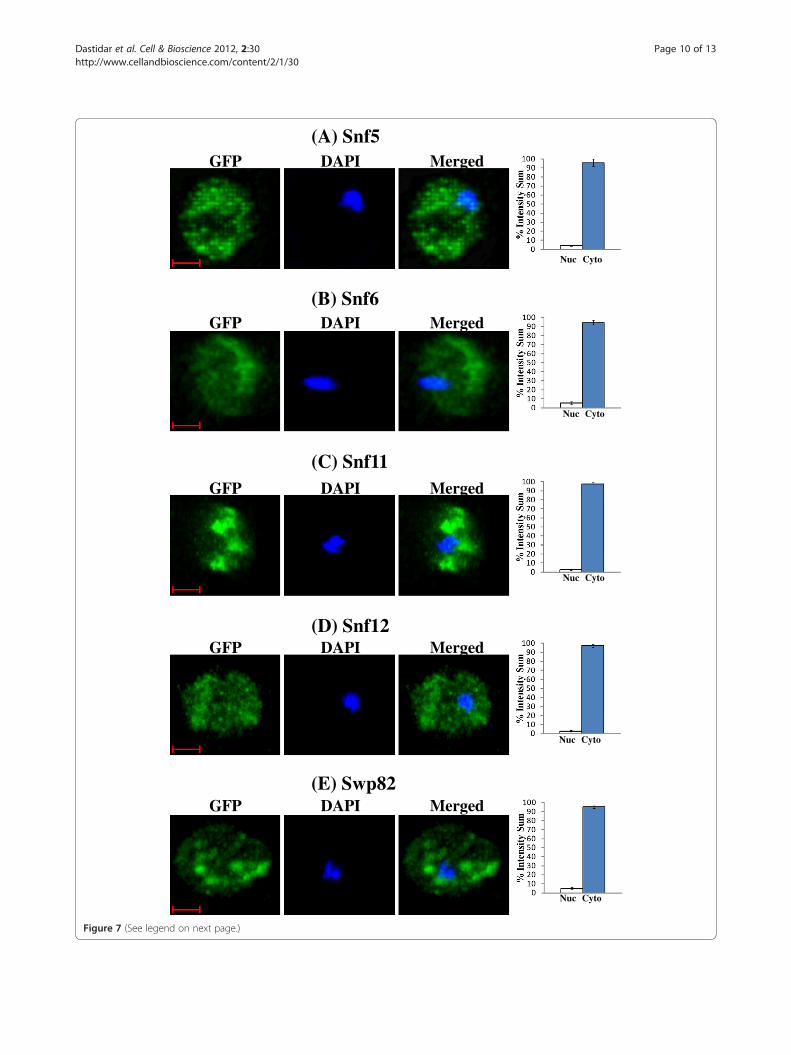
in both normoxic and hypoxic cells (see Figure 5).Figure 5A shows the distribution of Taf14 in air and underhypoxia, while Figure 5B shows the distribution of Swi2.For those proteins whose localization was affected by oxy-gen, over 95% of the proteins was present in the nucleus inair, whereas over 95% of the proteins was present in thecytosol under hypoxia (Figures 6 and 7). Figure 6A showsthe distribution of Swi3 in normoxic cells, while Figure 6Bshows the distribution of Swi3 in hypoxic cells. We alsoquantified the distribution of other hypoxia-relocalizedSWI/SNF proteins (Figure 7). Figure 7A-E show the distri-bution of Snf5, Snf6, Snf11, Snf12 and Swp82 in hypoxiccells (The images for normoxic cells invariably showed nu-clear localization, as expected and as shown in Figures 5
and 6, and are therefore omitted). Clearly, Swi2, Snf5, Snf6,Snf11, Snf12 and Swp82 proteins were transported to thenucleus in normoxic cells, but they accumulated in thecytosol in hypoxic cells.To further ascertain the role of SWI/SNF proteins in
oxygen regulation of gene expression, we determined ifand how many oxygen-regulated genes are SWI/SNFprotein targets as well. To this end, we used our previ-ous microarray and computational work analyzing genesregulated by oxygen/ and Δ hap1 cells [6]. We also usedthe previously identified targets of 263 transcription fac-tors [20]. Using these two sets of data and the R pro-gram, we identified those hypoxia-regulated genes thatare targets of SWI/SNF proteins and calculated the p-
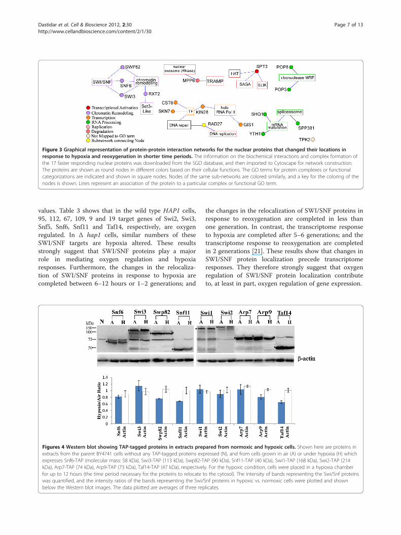
Figure 3 Graphical representation of protein-protein interaction networks for the nuclear proteins that changed their locations inresponse to hypoxia and reoxygenation in shorter time periods. The information on the biochemical interactions and complex formation ofthe 17 faster responding nuclear proteins was downloaded from the SGD database, and then imported to Cytoscape for network construction.The proteins are shown as round nodes in different colors based on their cellular functions. The GO terms for protein complexes or functionalcategorizations are indicated and shown in square nodes. Nodes of the same sub-networks are colored similarly, and a key for the coloring of thenodes is shown. Lines represent an association of the protein to a particular complex or functional GO term.
Dastidar et al. Cell & Bioscience 2012, 2:30 Page 7 of 13http://www.cellandbioscience.com/content/2/1/30
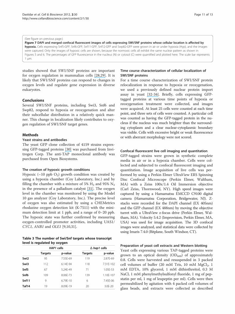
values. Table 3 shows that in the wild type HAP1 cells,95, 112, 67, 109, 9 and 19 target genes of Swi2, Swi3,Snf5, Snf6, Snf11 and Taf14, respectively, are oxygenregulated. In Δ hap1 cells, similar numbers of theseSWI/SNF targets are hypoxia altered. These resultsstrongly suggest that SWI/SNF proteins play a majorrole in mediating oxygen regulation and hypoxiaresponses. Furthermore, the changes in the relocaliza-tion of SWI/SNF proteins in response to hypoxia arecompleted between 6–12 hours or 1–2 generations; and
Figures 4 Western blot showing TAP-tagged proteins in extracts prepextracts from the parent BY4741 cells without any TAP-tagged proteins expexpresses Snf6-TAP (molecular mass: 58 kDa), Swi3-TAP (113 kDa), Swp82-TkDa), Arp7-TAP (74 kDa), Arp9-TAP (73 kDa), Taf14-TAP (47 kDa), respectivelfor up to 12 hours (the time period necessary for the proteins to relocate twas quantified, and the intensity ratios of the bands representing the Swi/below the Western blot images. The data plotted are averages of three rep
the changes in the relocalization of SWI/SNF proteins inresponse to reoxygenation are completed in less thanone generation. In contrast, the transcriptome responseto hypoxia are completed after 5–6 generations; and thetranscriptome response to reoxygenation are completedin 2 generations [21]. These results show that changes inSWI/SNF protein localization precede transcriptomeresponses. They therefore strongly suggest that oxygenregulation of SWI/SNF protein localization contributeto, at least in part, oxygen regulation of gene expression.
ared from normoxic and hypoxic cells. Shown here are proteins inressed (N), and from cells grown in air (A) or under hypoxia (H) whichAP (90 kDa), Snf11-TAP (40 kDa), Swi1-TAP (168 kDa), Swi2-TAP (214y. For the hypoxic condition, cells were placed in a hypoxia chambero the cytosol). The intensity of bands representing the Swi/Snf proteinsSnf proteins in hypoxic vs. normoxic cells were plotted and shownlicates.
CytoNuc
CytoNuc
Nuc Cyto
GFP DAPI Merged
(A) Ta f1 4
Nuc Cyto
GFP DAPI Merged
A ir
H yp
A ir
H yp
GFP DAPI Merged
(B) S w i2
GFP DAPI Merged
Figure 5 Examples of GFP, DAPI and merged confocal fluorescent images of cells expressing proteins whose cellular localization is notaffected by hypoxia. Cells expressing Taf14-GFP (A) or Swi2-GFP (B) were grown in air or under hypoxia (Hyp), and the images were captured.The percentages of GFP fluorescence in the nucleus (N) or cytosol (C) were quantified and plotted here. The scale bar represents 1 μm.
Dastidar et al. Cell & Bioscience 2012, 2:30 Page 8 of 13http://www.cellandbioscience.com/content/2/1/30
DiscussionThe SWI/SNF complex is an ATP-dependent chromatinremodeling complex [22]. Its composition and functionare conserved from yeast to humans [13]. In yeast, morethan 10% of the genes are the targets of SWI/SNF pro-teins, although the targets of different SWI/SNF proteinsare different [20]. Hypoxia and reoxygenation inducechanges in gene expression in over 20% of yeast genes[6]. Such broad changes in gene expression involve theaction of an array of regulators. Previous studies haveshown that Mga2, Rox1, Hap1 and Mot3 are all involvedin mediating oxygen regulation of several subsets of
genes [6,21,23,24]. In this report, we show that severalSWI/SNF proteins alter their subcellular localizationreadily in response to hypoxia or reoxygenation and thatthis change in subcellular localization likely contributesto oxygen regulation of SWI/SNF target genes.Because the targets of SWI/SNF proteins overlap but
are not identical [20], it is likely that different SWI/SNFproteins act on different groups of genes and controltheir expression. Here, we show that six SWI/SNF pro-teins accumulate in the cytosol in hypoxic cells, andrelocalize to the nucleus in response to reoxygenation.Furthermore, several of the SWI/SNF proteins respond
GFP DAPI Merged
Nuc Cyto
Swi3 in hypoxic cells
Nuc Cyto
GFP DAPI MergedSwi3 in normoxic cells (A)
(B)
Figure 6 DAPI and merged confocal fluorescent images of cells expressing Swi3-GFP. Cells were grown in air or under hypoxia (Hyp), andthe images were captured. The percentages of GFP fluorescence in the nucleus (N) or cytosol (C) was quantified and plotted here. The scale barrepresents 1 μm.
Dastidar et al. Cell & Bioscience 2012, 2:30 Page 9 of 13http://www.cellandbioscience.com/content/2/1/30
to hypoxia or reoxygenation and relocalize in a relativelyquick manner. The redistribution of the six SWI/SNFproteins in the cytosol should presumably change thecomposition, and thereby the function or selectivity ofthe SWI/SNF complexes in the nucleus. Hence, the relo-calization can affect the target expression of not onlythese SWI/SNF proteins whose localization is altered byhypoxia, such as Swi3, but also those whose localizationis not affected by hypoxia, such as Swi2 and Taf14(Table 3). Very likely, under hypoxia, because Swi3 andother proteins are predominantly present in the cytosol,the nuclear SWI/SNF proteins, such as Swi2, are likelycomplexed with other proteins, and act as chromatinremodelers on different sets of target genes. Thisexplains why many SWI/SNF target genes are altered byhypoxia/reoxygenation (Table 3). Previous studiesshowed that Swi2, Arp7 and Arp9 form a core subcom-plex possessing the ATP-dependent remodeling activity[25], while Swi3 controls SWI/SNF assembly, ATP-dependent H2A-H2B displacement, as well as recruit-ment to target genes [25,26]. Notably, the nuclearlocalization of Swi2, Arp7 and Arp9 is not affected byhypoxia, while Swi3 is affected. This supports the ideathat the core complex can associate with other as yetunidentified proteins and form a different kind of SWI/SNF complexes in the nucleus in hypoxic cells.These results suggest a model for how oxygen may
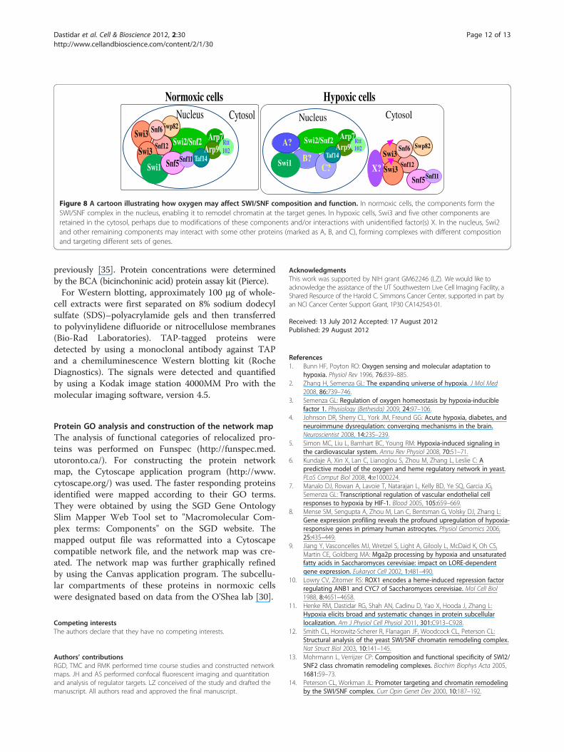
modulate SWI/SNF complex composition and function(Figure 8). In normoxic cells, SWI/SNF componentsform complexes in the nucleus and remodel chromatin
structure at their target genes. In hypoxic cells Swi3 andother five SWI/SNF proteins accumulate in the cytosol,leaving the Swi2-Arp7-Arp9 core subcomplex availableto interact with other proteins. Consequently, a differentkind of SWI/SNF complex containing the core complexand other proteins (A, B and C in Figure 8) can be formedand act to remodel chromatin and control gene expressionin different sets of genes. In response to reoxygenation,Swi3 and other proteins can readily relocalize to the nu-cleus, forming the normoxic SWI/SNF complexes, and re-establish gene expression patterns under normoxicconditions.This model is also consistent with a recent study showing
that Swi3 is a key regulator in controlling respiration genes[27]. The authors used a computational approach toanalyze modules of genes with a common regulation thatare affected by specific DNA polymorphisms. They inte-grated genotypic and expression data for individuals in asegregating population with complementary expressiondata of strains mutated in a variety of regulatory proteins,in order to identify regulatory-linkage modules. In so doing,they found that Swi3 is a dominant regulator in the con-trol of respiratory gene expression [27]. The effect of swi3deletion is stronger than that of known respiratory regula-tors, including Hap2/3/4/5, Mot3 and Rox1. This is incomplete agreement with our results showing that hun-dreds of SWI/SNF targets are altered by hypoxia (Table 3),and supports our model (Figure 8). The regulation ofSWI/SNF protein localization may also occur in othereukaryotes. For example, in mammalian cells, recent
GFP DAPI Merged
(A) Snf5
Nuc Cyto
(B) Snf6GFP DAPI Merged
Nuc Cyto
(C) Snf11GFP DAPI Merged
Nuc Cyto
Nuc Cyto
(D) Snf12GFP DAPI Merged
Nuc Cyto
GFP DAPI Merged(E) Swp82
Figure 7 (See legend on next page.)
Dastidar et al. Cell & Bioscience 2012, 2:30 Page 10 of 13http://www.cellandbioscience.com/content/2/1/30
(See figure on previous page.)Figure 7 DAPI and merged confocal fluorescent images of cells expressing SWI/SNF proteins whose cellular location is affected byhypoxia. Cells expressing Snf5-GFP, Snf6-GFP, Snf11-GFP, Snf12-GFP and Swp82-GFP were grown in air or under hypoxia (Hyp), and the imageswere captured. Only the images of hypoxic cells are shown, because the normoxic cells all exhibit the same nuclear pattern as shown inFigures 5 and 6. The percentages of GFP fluorescence in the nucleus (N) or cytosol (C) were quantified and plotted here. The scale bar represents1 μm.
Dastidar et al. Cell & Bioscience 2012, 2:30 Page 11 of 13http://www.cellandbioscience.com/content/2/1/30
studies showed that SWI/SNF proteins are importantfor oxygen regulation in mammalian cells [28,29]. It islikely that SWI/SNF proteins can respond to changes inoxygen levels and regulate gene expression in diverseeukaryotes.
ConclusionsSeveral SWI/SNF proteins, including Swi3, Snf6 andSwp82, respond to hypoxia or reoxygenation and altertheir subcellular distribution in a relatively quick man-ner. This change in localization likely contributes to oxy-gen regulation of SWI/SNF target genes.
MethodsYeast strains and antibodiesThe yeast GFP clone collection of 4159 strains expres-sing GFP-tagged proteins [30] was purchased from Invi-trogen Corp. The anti-TAP monoclonal antibody waspurchased from Open Biosystems.
The creation of hypoxic growth conditionsHypoxic (~10 ppb O2) growth condition was created byusing a hypoxia chamber (Coy Laboratory, Inc.) and byfilling the chamber with a mixture of 5% H2 and 95% N2
in the presence of a palladium catalyst [31]. The oxygenlevel in the chamber was monitored by using the Model10 gas analyzer (Coy Laboratory, Inc.). The precise levelof oxygen was also estimated by using a CHEMetricsrhodazine oxygen detection kit (K-7511) with the mini-mum detection limit at 1 ppb, and a range of 0–20 ppb.The hypoxic state was further confirmed by measuringoxygen-controlled promoter activities, including UAS1/CYC1, ANB1 and OLE1 [9,10,31].
Table 3 The number of Swi/Snf targets whose transcriptlevel is regulated by oxygen
HAP1 cells Δ hap1 cells
Targets p-value Targets p-value
Swi2 95 7.55E-69 119 2.87E-69
Swi3 112 6.14E-94 118 7.31E-102
Snf5 67 5.24E-49 71 1.05E-53
Snf6 109 8.06E-73 139 1.10E-107
Snf11 9 6.79E-10 6 7.45E-06
Taf14 19 8.09E-19 20 3.0E-20
Time course characterization of cellular localization ofSWI/SNF proteinsFor a time course characterization of SWI/SNF proteinrelocalization in response to hypoxia or reoxygenation,we used a previously defined nuclear protein importassay in yeast [32-34]. Briefly, cells expressing GFP-tagged proteins at various time points of hypoxia orreoxygenation treatment were collected, and imageswere acquired. At least 25 cells were counted at each timepoint, and three sets of cells were counted. A particular cellwas counted as having the GFP-tagged protein in the nu-cleus if the nucleus was much brighter than the surround-ing cytoplasm and a clear nuclear-cytoplasmic boundarywas visible. Cells with excessive bright or weak fluorescenceor with aberrant morphology were not scored.
Confocal fluorescent live cell imaging and quantitationGFP-tagged strains were grown in synthetic completemedia in air or in a hypoxia chamber. Cells were col-lected and subjected to confocal fluorescent imaging andquantitation. Image acquisition of live cells was per-formed by using a Perkin Elmer UltraView ERS SpinningDisc Confocal Microscope (Perkin Elmer, Waltham,MA) with a Zeiss 100x/1.4 Oil Immersion objective(Carl Zeiss, Thornwood, NY). High speed images werecaptured by using a Hamamatsu EMCCD C9100 digitalcamera (Hamamatsu Corporation, Bridgewater, NJ). Z-stacks were recorded for the DAPI channel (EX 405nm)and the GFP channel (EX 488nm) by moving the objectiveturret with a UltraView z-focus drive (Perkin Elmer, Wal-tham, MA). Volocity 5.4.2 (Improvision, Perkin Elmer, MA,USA) was used for image acquisition. The 3D confocalimages were analyzed, and statistical data were collected byusing Imaris 7.4.0 (Bitplane, South Windsor, CT).
Preparation of yeast cell extracts and Western blottingYeast cells expressing various TAP-tagged proteins weregrown to an optical density (OD600) of approximately0.8. Cells were harvested and resuspended in 3 packedcell volumes of buffer (20 mM Tris, 10 mM MgCl2, 1mM EDTA, 10% glycerol, 1 mM dithiothreitol, 0.3 MNaCl, 1 mM phenylmethylsulfonyl fluoride, 1 mg of pep-statin per ml, 1 mg of leupeptin per ml). Cells were thenpermeabilized by agitation with 4 packed cell volumes ofglass beads, and extracts were collected as described
Swi3Swp82
Swi3Snf12 Swi2/Snf2
Swi1
Swi3Snf5
Arp7
Snf11 Taf14
Rtt102Arp9
Snf6
Nucleus Cytosol Nucleus Cytosol
Swi2/Snf2
Swi1
Arp7
Taf14
Rtt102Arp9A?
B?C?
Swp82Swi3
Snf12Swi3
Snf5Snf11
Snf6
X?
Normoxic cells Hypoxic cells
Figure 8 A cartoon illustrating how oxygen may affect SWI/SNF composition and function. In normoxic cells, the components form theSWI/SNF complex in the nucleus, enabling it to remodel chromatin at the target genes. In hypoxic cells, Swi3 and five other components areretained in the cytosol, perhaps due to modifications of these components and/or interactions with unidentified factor(s) X. In the nucleus, Swi2and other remaining components may interact with some other proteins (marked as A, B, and C), forming complexes with different compositionand targeting different sets of genes.
Dastidar et al. Cell & Bioscience 2012, 2:30 Page 12 of 13http://www.cellandbioscience.com/content/2/1/30
previously [35]. Protein concentrations were determinedby the BCA (bicinchoninic acid) protein assay kit (Pierce).For Western blotting, approximately 100 μg of whole-
cell extracts were first separated on 8% sodium dodecylsulfate (SDS)–polyacrylamide gels and then transferredto polyvinylidene difluoride or nitrocellulose membranes(Bio-Rad Laboratories). TAP-tagged proteins weredetected by using a monoclonal antibody against TAPand a chemiluminescence Western blotting kit (RocheDiagnostics). The signals were detected and quantifiedby using a Kodak image station 4000MM Pro with themolecular imaging software, version 4.5.
Protein GO analysis and construction of the network mapThe analysis of functional categories of relocalized pro-teins was performed on Funspec (http://funspec.med.utoronto.ca/). For constructing the protein networkmap, the Cytoscape application program (http://www.cytoscape.org/) was used. The faster responding proteinsidentified were mapped according to their GO terms.They were obtained by using the SGD Gene OntologySlim Mapper Web Tool set to "Macromolecular Com-plex terms: Components" on the SGD website. Themapped output file was reformatted into a Cytoscapecompatible network file, and the network map was cre-ated. The network map was further graphically refinedby using the Canvas application program. The subcellu-lar compartments of these proteins in normoxic cellswere designated based on data from the O'Shea lab [30].
Competing interestsThe authors declare that they have no competing interests.
Authors’ contributionsRGD, TMC and RMK performed time course studies and constructed networkmaps. JH and AS performed confocal fluorescent imaging and quantitationand analysis of regulator targets. LZ conceived of the study and drafted themanuscript. All authors read and approved the final manuscript.
AcknowledgmentsThis work was supported by NIH grant GM62246 (LZ). We would like toacknowledge the assistance of the UT Southwestern Live Cell Imaging Facility, aShared Resource of the Harold C. Simmons Cancer Center, supported in part byan NCI Cancer Center Support Grant, 1P30 CA142543-01.
Received: 13 July 2012 Accepted: 17 August 2012Published: 29 August 2012
References1. Bunn HF, Poyton RO: Oxygen sensing and molecular adaptation to
hypoxia. Physiol Rev 1996, 76:839–885.2. Zhang H, Semenza GL: The expanding universe of hypoxia. J Mol Med
2008, 86:739–746.3. Semenza GL: Regulation of oxygen homeostasis by hypoxia-inducible
factor 1. Physiology (Bethesda) 2009, 24:97–106.4. Johnson DR, Sherry CL, York JM, Freund GG: Acute hypoxia, diabetes, and
neuroimmune dysregulation: converging mechanisms in the brain.Neuroscientist 2008, 14:235–239.
5. Simon MC, Liu L, Barnhart BC, Young RM: Hypoxia-induced signaling inthe cardiovascular system. Annu Rev Physiol 2008, 70:51–71.
6. Kundaje A, Xin X, Lan C, Lianoglou S, Zhou M, Zhang L, Leslie C: Apredictive model of the oxygen and heme regulatory network in yeast.PLoS Comput Biol 2008, 4:e1000224.
7. Manalo DJ, Rowan A, Lavoie T, Natarajan L, Kelly BD, Ye SQ, Garcia JG,Semenza GL: Transcriptional regulation of vascular endothelial cellresponses to hypoxia by HIF-1. Blood 2005, 105:659–669.
8. Mense SM, Sengupta A, Zhou M, Lan C, Bentsman G, Volsky DJ, Zhang L:Gene expression profiling reveals the profound upregulation of hypoxia-responsive genes in primary human astrocytes. Physiol Genomics 2006,25:435–449.
9. Jiang Y, Vasconcelles MJ, Wretzel S, Light A, Gilooly L, McDaid K, Oh CS,Martin CE, Goldberg MA: Mga2p processing by hypoxia and unsaturatedfatty acids in Saccharomyces cerevisiae: impact on LORE-dependentgene expression. Eukaryot Cell 2002, 1:481–490.
10. Lowry CV, Zitomer RS: ROX1 encodes a heme-induced repression factorregulating ANB1 and CYC7 of Saccharomyces cerevisiae. Mol Cell Biol1988, 8:4651–4658.
11. Henke RM, Dastidar RG, Shah AN, Cadinu D, Yao X, Hooda J, Zhang L:Hypoxia elicits broad and systematic changes in protein subcellularlocalization. Am J Physiol Cell Physiol 2011, 301:C913–C928.
12. Smith CL, Horowitz-Scherer R, Flanagan JF, Woodcock CL, Peterson CL:Structural analysis of the yeast SWI/SNF chromatin remodeling complex.Nat Struct Biol 2003, 10:141–145.
13. Mohrmann L, Verrijzer CP: Composition and functional specificity of SWI2/SNF2 class chromatin remodeling complexes. Biochim Biophys Acta 2005,1681:59–73.
14. Peterson CL, Workman JL: Promoter targeting and chromatin remodelingby the SWI/SNF complex. Curr Opin Genet Dev 2000, 10:187–192.
Dastidar et al. Cell & Bioscience 2012, 2:30 Page 13 of 13http://www.cellandbioscience.com/content/2/1/30
15. Wu P-YJ, Ruhlmann C, Winston F, Schultz P: Molecular Architecture of theS. cerevisiae SAGA Complex. Molecular Cell 2004, 15:199–208.
16. Chamberlain JR, Lee Y, Lane WS, Engelke DR: Purification andcharacterization of the nuclear RNase P holoenzyme complex revealsextensive subunit overlap with RNase MRP. Genes Dev 1998,12:1678–1690.
17. Weissman B, Knudsen KE: Hijacking the chromatin remodeling machinery:impact of SWI/SNF perturbations in cancer. Cancer Res 2009,69:8223–8230.
18. Wilson BG, Roberts CW: SWI/SNF nucleosome remodellers and cancer. NatRev Cancer 2011, 11:481–492.
19. Ghaemmaghami S, Huh WK, Bower K, Howson RW, Belle A, Dephoure N,O'Shea EK, Weissman JS: Global analysis of protein expression in yeast.Nature 2003, 425:737–741.
20. Hu Z, Killion PJ, Iyer VR: Genetic reconstruction of a functionaltranscriptional regulatory network. Nat Genet 2007, 39:683–687.
21. Lai LC, Kosorukoff AL, Burke PV, Kwast KE: Metabolic-state-dependentremodeling of the transcriptome in response to anoxia and subsequentreoxygenation in Saccharomyces cerevisiae. Eukaryot Cell 2006,5:1468–1489.
22. Clapier CR, Cairns BR: The biology of chromatin remodeling complexes.Annu Rev Biochem 2009, 78:273–304.
23. Jiang Y, Vasconcelles MJ, Wretzel S, Light A, Martin CE, Goldberg MA: MGA2is involved in the low-oxygen response element-dependent hypoxicinduction of genes in Saccharomyces cerevisiae. Mol Cell Biol 2001,21:6161–6169.
24. Kastaniotis AJ, Zitomer RS: Rox1 mediated repression. Oxygen dependentrepression in yeast. Adv Exp Med Biol 2000, 475:185–195.
25. Yang X, Zaurin R, Beato M, Peterson CL: Swi3p controls SWI/SNF assemblyand ATP-dependent H2A-H2B displacement. Nat Struct Mol Biol 2007,14:540–547.
26. Dechassa ML, Zhang B, Horowitz-Scherer R, Persinger J, Woodcock CL,Peterson CL, Bartholomew B: Architecture of the SWI/SNF-nucleosomecomplex. Mol Cell Biol 2008, 28:6010–6021.
27. Gat-Viks I, Meller R, Kupiec M, Shamir R: Understanding gene sequencevariation in the context of transcription regulation in yeast. PLoS Genet2010, 6:e1000800.
28. Kenneth NS, Mudie S, van Uden P, Rocha S: SWI/SNF regulates the cellularresponse to hypoxia. J Biol Chem 2009, 284:4123–4131.
29. Johnson AB, Barton MC: Hypoxia-induced and stress-specific changes inchromatin structure and function. Mutat Res 2007, 618:149–162.
30. Huh WK, Falvo JV, Gerke LC, Carroll AS, Howson RW, Weissman JS, O'SheaEK: Global analysis of protein localization in budding yeast. Nature 2003,425:686–691.
31. Hon T, Dodd A, Dirmeier R, Gorman N, Sinclair PR, Zhang L, Poyton RO: AMechanism of Oxygen Sensing in Yeast: Multiple Oxygen-ResponsiveSteps in the Heme Biosynthetic Pathway Affect Hap1 activity. J. Biol.Chem 2003, 278:50771–50780.
32. Shulga N, Roberts P, Gu Z, Spitz L, Tabb MM, Nomura M, Goldfarb DS: Invivo nuclear transport kinetics in Saccharomyces cerevisiae: a role forheat shock protein 70 during targeting and translocation. J Cell Biol 1996,135:329–339.
33. Fahrenkrog B, Hubner W, Mandinova A, Pante N, Keller W, Aebi U: Theyeast nucleoporin Nup53p specifically interacts with Nic96p and isdirectly involved in nuclear protein import. Mol Biol Cell 2000,11:3885–3896.
34. Leslie DM, Timney B, Rout MP, Aitchison JD: Studying nuclear proteinimport in yeast. Methods 2006, 39:291–308.
35. Zhang L, Hach A, Wang C: Molecular Mechanism Governing HemeSignaling in Yeast: a Higher-Order Complex Mediates Heme Regulationof the Transcriptional Activator HAP1. Mol Cell Biol 1998, 18:3819–3828.
doi:10.1186/2045-3701-2-30Cite this article as: Dastidar et al.: The nuclear localization of SWI/SNFproteins is subjected to oxygen regulation. Cell & Bioscience 2012 2:30.
Submit your next manuscript to BioMed Centraland take full advantage of:
• Convenient online submission
• Thorough peer review
• No space constraints or color figure charges
• Immediate publication on acceptance
• Inclusion in PubMed, CAS, Scopus and Google Scholar
• Research which is freely available for redistribution
Submit your manuscript at www.biomedcentral.com/submit