the medial prefrontal cortex is both necessary and sufficient for the acquisition of conditioned...
TRANSCRIPT
The Medial Prefrontal Cortex is Both Necessary and Sufficientfor the Acquisition of Conditioned Defeat
Chris M. Markham, Cloe A. Luckett, and Kim L. HuhmanThe Neuroscience Institute, Georgia State University, Atlanta, GA USA, 161 Jesse Hill Jr. Drive,Suite 832, Atlanta, GA 30303
AbstractWe have previously demonstrated that the basolateral amygdala (BLA) is a key component of aneural circuit mediating memory formation for emotionally relevant stimuli in an ethologically-based model of conditioned fear, termed conditioned defeat (CD). In this model, subjects aresocially defeated by a larger, more aggressive hamster. Upon subsequent exposure to a smaller,non-aggressive intruder, the defeated animal will show high levels of submissive behaviors andfail to defend its territory. Here we examined whether the medial prefrontal cortex (mPFC), anarea with extensive connections with the amygdala, is also a component of this circuit. Temporaryinactivation of the mPFC using muscimol, a GABAA receptor agonist, significantly enhanced theacquisition but not expression of CD, while blockade of GABAA receptors in the mPFC usingbicuculline, a GABAA antagonist, impaired acquisition of CD. Given these findings, we nextsought to test whether plasticity related to the defeat experience occurs in the mPFC. We infusedanisomycin, a protein synthesis inhibitor, in the mPFC but this treatment did not alter theacquisition of CD. In our final experiment, we demonstrated that bicuculline failed to alter theacquisition of CD. Together, these results demonstrate for the first time that while the mPFC isboth necessary and sufficient for the acquisition of CD, it does not appear to mediate plasticityrelated to the defeat experience. In contrast, while plasticity underlying CD does appear to occurin the BLA, GABAergic receptor inhibition in the BLA is not sufficient to enhance CD.
Keywordssocial stress; amygdala; defensive behavior; conditioned fear; aggression
1. IntroductionOur lab has identified the primary components of an integrated neural circuit underlyingconditioned defeat (CD), a learned response whereby socially defeated Syrian hamsters(Mesocricetus auratus) display submissive and defensive behaviors instead of territorialaggression when they are subsequently paired with a non-aggressive conspecific (Huhman etal., 2003; Potegal et al., 1993). We have also gathered substantial evidence that theamygdala, in particular the basolateral complex (BLA), is a critical component of this circuitbecause temporary inactivation of the BLA (via muscimol infusion) impairs the acquisition
© 2011 Elsevier Ltd. All rights reserved.Corresponding Author: Chris M. Markham, Ph.D., [email protected], Tel: 1.404.413.6337, Fax: 1.404.413.5471, TheNeuroscience Institute, Georgia State University, 161 Jesse Hill Jr. Drive, Suite 832, Atlanta, GA 30303, USA.Publisher's Disclaimer: This is a PDF file of an unedited manuscript that has been accepted for publication. As a service to ourcustomers we are providing this early version of the manuscript. The manuscript will undergo copyediting, typesetting, and review ofthe resulting proof before it is published in its final citable form. Please note that during the production process errors may bediscovered which could affect the content, and all legal disclaimers that apply to the journal pertain.
NIH Public AccessAuthor ManuscriptNeuropharmacology. Author manuscript; available in PMC 2013 February 1.
Published in final edited form as:Neuropharmacology. 2012 February ; 62(2): 933–939. doi:10.1016/j.neuropharm.2011.09.026.
NIH
-PA Author Manuscript
NIH
-PA Author Manuscript
NIH
-PA Author Manuscript

and expression of CD (Jasnow and Huhman 2001; Markham et al., 2010). Importantly,plasticity related to the defeat experience also appears to be mediated in the BLA becauseoverexpression of cAMP response element-binding protein (CREB) enhances the acquisitionof CD whereas pre-training infusion of anisomycin, a protein synthesis inhibitor, orifenprodil, a NMDA receptor (NR2B subunit) antagonist, both block the acquisition of CD(Day et al., 2011; Jasnow et al., 2005; Markham and Huhman, 2008). These findings areconsistent with a large number of studies showing that the amygdala is necessary for theacquisition and expression of conditioned fear in more traditional models such as auditoryfear conditioning and fear-potentiated startle (Davis, 1997, 2000; LeDoux, 2000; Maren,2001; Walker and Davis, 2002).
The prelimbic/infralimbic cortex (corresponding to the medial prefrontal cortex (mPFC)) isan area that has extensive connections with the amygdala (McDonald et al., 1996), and it is asite which we have yet to investigate for its possible role in CD. Like the amygdala, themPFC has also been implicated in fear learning, including acquisition (Morgan and LeDoux,1995; Morgan et al., 1993), expression (Sierra-Mercado et al., 2006) and extinction (Miladand Quirk, 2002; Morgan et al., 1993; Quirk et al., 2003) of conditioned fear. In addition,several lines of evidence suggest a functional interaction between the two areas whereby themPFC acts to inhibit the activity of the amygdala via glutamatergic projections (Smith et al.,2000) to GABAergic intercalated (IC) cells, which in turn induce feedforward inhibition ofthe output neurons in the amygdala (Quirk et al., 2003). While Quirk and colleagues (2003)acknowledge that it is possible that other structures receiving direct projections from themPFC (such as the periaqueductal gray (Floyd et al., 2000) and hypothalamus (Floyd et al.,2001) are responsible for the inhibition of fear responses to conditioned stimuli independentof the amygdala, this does not seem to be the case because direct stimulation of the mPFCreduces the responsiveness of amygdalar output neurons in a site-specific manner (Milad etal., 2004; Quirk et al., 2003).
The primary aim of the present series of experiments was to determine whether the mPFC ispart of the neural circuit mediating CD. If the mPFC exerts a feedforward inhibition of theamygdala, then temporary inactivation of the mPFC should enhance, while temporaryactivation should impair the acquisition of CD. We also asked whether protein synthesis inthe mPFC is necessary for the formation of CD. Finally, we also sought to determinewhether blockade of GABAergic receptors in the BLA would be sufficient to enhance theacquisition of conditioned defeat.
2. Methods and Procedures2.1 Animals and housing conditions
Subjects in all experiments were adult male Syrian hamsters (Mesocricetus auratus, CharlesRiver Laboratories, New York, NY) that weighed 120-140 g and were between 9-10 weeksold at the time of testing. Upon arrival, they were housed in groups of 5-6 per cage for oneweek (as a quarantine measure) prior to the start of any manipulation. Animals were housedin a temperature (20 ± 2° C) and humidity-controlled room with free access to food andwater and were kept on a 14:10 h light: dark cycle with lights out at 1100 h. All training andtesting sessions were performed under dim red light illumination during the first 3 hours ofthe dark phase of the light-dark cycle in order to minimize circadian effects. Residentaggressors (RA) used for defeat training were older (> 6 months), singly housed malesweighing between 170 and 195 g, while younger males (2 months), weighing between 110and 120 g, were group housed (5-6 per cage) and served as non-aggressive intruders (NAI).All procedures and protocols were approved by the Georgia State University InstitutionalAnimal Care and Use Committee and are in accordance with the standards outlined in theNational Institutes of Health Guide for Care and Use of Laboratory Animals.
Markham et al. Page 2
Neuropharmacology. Author manuscript; available in PMC 2013 February 1.
NIH
-PA Author Manuscript
NIH
-PA Author Manuscript
NIH
-PA Author Manuscript

2.2 Surgical ProceduresOne week after arrival, subjects were anesthetized with sodium pentobarbital (90 mg/kg,i.p., Lundbeck, Inc., Deerfield, IL) and were placed into a stereotaxic frame (David KopfInstruments, Tujunga, CA). For the medial prefrontal cortex, a stainless steel guide cannula(26-gauge, Plastics One, Roanoke, VA), held at a 20° angle toward the midline, wasunilaterally implanted in either the left or right hemisphere (3.2 mm rostral and ± 1.6 mmlateral relative to bregma and 3.2 mm below dura). In order to minimize tissue damage tothe area of interest, a smaller, 33-gauge injection needle (Plastics One, Roanoke, VA) thatprojected 1.2 mm below the guide cannula was used on injection day in order to reach afinal depth of 4.4 mm below dura. For the basolateral amygdala, stainless steel guidecannula identical to the ones used above were bilaterally implanted in the followingcoordinates: 0.2 mm rostral and ± 3.4 mm lateral relative to bregma and 2.1 mm below dura.Again, in order to minimize tissue damage, a 33-gauge injection needle that projected 4.2mm below the guide cannula and reaching a final depth of 6.3 mm below dura was used forthe injection procedure. Following surgery, dummy stylets were placed in the guide cannulato help maintain patency and 1 cc of warm physiological saline and 0.1 cc of ketoprofen(Fort Dodge Animal Health, Fort Dodge, IA) was injected subcutaneously in order to aid inrecovery. Hamsters were allowed 7-10 days to recover prior to the start of behavioral testingduring which they were monitored daily to ensure that there was no adverse outcome fromthe surgery. Three days prior to the start of the experiment, subjects were handled by gentlyrestraining them and removing and replacing the dummy stylet in order to maintain patencyand to habituate the subjects to the infusion procedure.
2.3 Social defeat and behavioral testingOn the day of social defeat training, animals were transported to our testing suite within thevivarium and allowed to acclimate for at least 30 min. Training sessions consisted of asingle 15 min (full defeat) or 5 min (sub-optimal defeat) exposure to the RA in theaggressor’s home cage, upon which the RA reliably attacked the experimental hamsterwithin 60 s. During the training session, attacks by the RA against the subject occur onlysporadically in bouts, and do not last for the entire duration of the session. All training andtesting sessions were recorded via a CCD camera positioned overhead. Twenty-four hoursafter defeat, subjects were transported to the testing suite and allowed to acclimate for atleast 30 min, after which a NAI was introduced into each subjects’ home cage for 5 min.The trials were recorded and scored by an observer that was blind to the experimentalconditions using the behavioral analysis software Hindsight (developed by Scott Weiss,Ph.D.). The total duration of four classes of behaviors were scored during the testingsession: (a) social behavior (stretch, approach, sniff, nose touching and flank marking), (b)non-social behavior (locomotion, exploration, grooming, nesting), (c) submissive/defensivebehaviors (flight, avoidance, tail up, upright, side defense, stretch attend, head flag,attempted escape from cage), and (d) aggressive behaviors (upright an side offense, chaseand attack).
2.4 Drug Infusion2.4.1 Experiments 1 and 2—Animals were removed from their home cage and gentlyrestrained while the dummy stylet was removed. Muscimol (Sigma-Aldrich, 2.2 nmol in 300nl saline) or vehicle control (300 nl saline) was then unilaterally infused into the left or rightmPFC over a 1.5 min period using a 1-μL syringe (Hamilton, Reno, NV) connected to anmini-infusion pump (Harvard Apparatus, Holliston, MA) via polyethylene tubing. Duringthe infusion procedure, the animal was free to move about in a small, clear polycarbonatecage. The injection needle (33-gauge) was kept in place for an additional 1 min before being
Markham et al. Page 3
Neuropharmacology. Author manuscript; available in PMC 2013 February 1.
NIH
-PA Author Manuscript
NIH
-PA Author Manuscript
NIH
-PA Author Manuscript

removed to ensure complete diffusion of the drug after which the dummy stylet wasreplaced. Testing began 10 min after drug infusion.
We chose to use muscimol, a GABAA agonist, to inactivate the mPFC/BLA for severalreasons. First, GABAA receptors are widely distributed in the central nervous system,including the mPFC, and infusion of muscimol into specific brain areas induceshyperpolarization (Martin and Ghez, 1999) and has been shown to reversibly suppressexcitatory neurotransmission (Allen et al., 2008). In addition, the resulting suppression ofneuronal activity is temporary, lasting up to several hours depending on dose (Majchrzakand DiScala, 2000). We based the dose of muscimol used in this study on our previousexperiments showing that this dosage was the most effective in altering the acquisition and/or expression of CD while at the same time not inducing unwanted behavioral side-effects(Markham and Huhman, 2008, Markham et al., 2010).
2.4.2 Experiment 3—Bicuculline methobromide (Sigma-Aldrich, 300 ng in 300 nl saline)or vehicle control (300 nl saline) was unilaterally infused into the mPFC using the sameprocedure described above. The dose of bicuculline used in this experiment was based onpilot studies ensuring that this dose did not induce behavioral side effects as well as aprevious study demonstrating that a similar dose was effective in blocking 8-OH-DPAT-induced circadian phase shifts (Mintz, et al., 1997).
2.4.3 Experiment 4—Anisomycin (Sigma-Aldrich, 1.125 nmol in 300 nl saline) or vehiclecontrol (300 nl saline) was unilaterally infused into the mPFC using the same proceduredescribed above, except that testing began 20 min after drug infusion. Anisomycin has beenshown to impair memory by disrupting protein synthesis, and we have previously used thisdose in the BLA to inhibit the acquisition of CD (Bourtchouladze et al. 1998; Markham andHuhman, 2008; Schafe and LeDoux, 2000).
2.4.4 Experiment 5—Bicuculline methobromide (Sigma-Aldrich, 200 ng in 200 nl saline)or vehicle control (200 nl saline) was bilaterally infused into the BLA using the sameprocedure described above. Testing began 10 min after infusion.
2.5.1 Site Verification—At the end of each experiment, hamsters were administered anoverdose of Sleepaway euthanasia solution (Fort Dodge Animal Health, Fort Dodge, IA)and infused with 300 nl of India ink to verify the placement of the injection needle. Thebrains were then removed and fixed in 10% buffered formalin for at least 48 hours beforebeing sectioned on a Leica CM 3050 S cryostat. Thirty μm sections were taken and stainedwith cresyl violet and coverslipped with DPX. Sections were then examined under a light-microscope (Zeiss Axioplan 2) for placement verification. Only animals with injection siteswithin 0.3 mm of the target structure were included in the analysis.
2.5.2 Site Verification Results—Figure 1a shows the injection sites for all animals inExperiments 1-4 (mPFC) while Figure 1b shows the injection sites for animals inExperiment 5 (BLA). Only injections that were localized to within 0.3 mm of the targetstructure were included in the analysis. In addition, groups were collapsed over side ofinjection because there was no evidence of laterality (no significant difference betweeninjections of the left and right mPFC). In experiments in which several injection sites weredeemed to be misses (i.e., greater than 0.3 mm from target structure), data from thesesubjects were analyzed separately as an anatomical miss control group.
2.6.1 Experiment 1 and 2: Effect of activation of GABAA receptors in the mPFCon the acquisition or expression of CD—The goal of this experiment was todetermine whether the temporary inactivation of the mPFC using the GABAA agonist
Markham et al. Page 4
Neuropharmacology. Author manuscript; available in PMC 2013 February 1.
NIH
-PA Author Manuscript
NIH
-PA Author Manuscript
NIH
-PA Author Manuscript

muscimol would significantly enhance the acquisition (Experiment 1) and/or expression(Experiment 2) of CD. Animals (Experiment 1, n=32; Experiment 2, n=30) were randomlyassigned to either the muscimol (2.2 nmol in 300 nl saline) or the vehicle (300 nl saline)groups. For animals in Experiment 1, muscimol or vehicle was infused 10 min prior to beingexposed to the RA for 15 min. We did not use a sub-optimal defeat protocol in thisexperiment because we were unsure what effect muscimol infusions would have on CD. Onthe following day, animals were tested drug-free in their own home cage against a NAI for 5min. In Experiment 2, animals were placed drug free into the home cage of a RA for 15 mindefeat training. On the following day, they were infused with either muscimol or vehicle 10min prior to be paired with a NAI in their own home cage.
In Experiment 1, injection needles were localized to within 0.3 mm of the mPFC in 25 of the32 animals (Figure 1a). Other subjects (n=5) had cannula placements that missed the targetand were located in the intermediate region of the lateral septum (LSi). These animals wereincluded in the anatomical miss control group. Additionally, the head caps on two of thesubjects became detached during the infusion process. In Experiment 2, injection needleswere localized to within 0.3 mm in 25 of 30 animals (Figure 1a). Other subjects (n=5) hadcannula placements that missed the target and were located approximately 0.5 mm lateral(n=1) or ventral to the mPFC (n=4). These animals were included in the anatomical controlgroup.
2.6.2 Experiment 3: Inhibition of GABAA receptors in the mPFC on theacquisition of CD—The goal of Experiment 3 was to determine whether antagonism ofGABAA receptors in the mPFC would impair the acquisition of CD. Animals (n=21) wererandomly assigned to either the bicuculline (300 ng in 300 nl saline) or vehicle (300 nlsaline) groups and injected 10 min prior to being placed in the home cage of a RA for 15min. On the following day, animals were tested drug-free in their own home cage against aNAI for 5 min.
Injection needles were localized to within 0.3 mm of the mPFC in 19 of the 21 animals. Onesubject had a cannula placement that missed the target and was located approximately 0.5mm caudal to the mPFC. One animal had a clogged cannula during the injection procedureand was excluded from the analysis.
2.6.3 Experiment 4: Inhibition of protein synthesis in the mPFC on theacquisition of CD—The goal of Experiment 4 was to determine whether plasticity relatedto the defeat experience is mediated, at least in part, in the mPFC. Animals (n=17) wererandomly assigned to either the anisomycin (1.125 nmol in 300 nl saline) or vehicle (300 nlsaline) and injected 20 min prior to being placed into the home cage of a RA for 15 min. Onthe following day, animals were tested drug-free in their own home cage against a NAI for 5min. Injection needles were localized to within 0.3 mm of the mPFC in all 17 animals.
2.6.4 Experiment 5: Inhibition of GABAA receptors in the BLA on theacquisition of CD—The goal of Experiment 5 was to determine whether blockade ofGABAA receptors in the BLA would potentiate the acquisition of CD. In Experiment 5,animals (n=47) were randomly assigned to either the bicuculline (100 or 200 ng in 200 nlsaline) or vehicle (200 nl saline) groups and injected 10 min prior to be being defeated by anRA for 5 min. Based on our previous finding that muscimol infusion in the BLA impairedthe acquisition of CD (Markham and Huhman, 2008), we hypothesized that bicuculline inthe BLA would enhance CD. Therefore, in order to avoid a ceiling effect, we used a sub-optimal defeat protocol for this experiment, which was comprised of a single 5-min pairingwith the RA.
Markham et al. Page 5
Neuropharmacology. Author manuscript; available in PMC 2013 February 1.
NIH
-PA Author Manuscript
NIH
-PA Author Manuscript
NIH
-PA Author Manuscript

Injection needles were localized bilaterally or unilaterally to within 0.3 mm of the BLA in37 of the 47 animals (Figure 1b). Other subjects (n=10) had cannula placements that missedthe BLA bilaterally (n=7), or had clogged cannula at the time of injection (n=3). Theseanimals were not included in the analysis. Of the remaining animals (n=37), 14 hadunilateral, while 23 had bilateral cannula placements aimed at the BLA.
2.7 Statistical analysisThe total duration (seconds) of each behavior displayed (Submissive/Defensive, Social,Nonsocial) was determined, and the mean total duration of each behavior was thencompared using separate t-tests for independent samples (Experiments 1-4) and ANOVA(Experiment 5).
3. Results3.1 Histology
Figure 2 is a photomicrograph of a representative coronal section of the mPFC (2a) andBLA (2b) showing the location of the injection needle and subsequent ink infusion. Table 1summarizes the cannula placements, including hits and misses for all experiments in thisstudy.
3.2 Experiment 1: Inactivation of the mPFC enhanced the acquisition of CDAs shown in Figure 3a, animals receiving muscimol displayed a significant increase insubmissive and defensive behaviors (t(23) = −2.54, p<0.05) and a significant reduction innon-social behaviors (t(23) = 2.56, p<0.05) compared to the vehicle control group. Nosignificant differences were observed between the two groups in total duration of socialbehaviors. No instances of aggression were observed. In addition, animals in the anatomicalmiss control group that received muscimol (n=4) exhibited levels of submission that weresimilar to the vehicle control group, indicating that the increased submission observed in themuscimol-mPFC group was site-specific.
3.3 Experiment 2: Inactivation of the mPFC did not alter the expression of CDAs shown in Figure 3b, inactivation of the mPFC prior to testing with a NAI did notsignificantly alter any of the behavioral measures, including submission and defensivebehaviors. Analysis of the anatomical control group showed that in three of the four animalswith infusion of muscimol in the LSi exhibited increased aggression, but no changes insubmission (data not shown). This finding is consistent with a recent study in our labshowing that inactivation of the lateral septum (LS) with muscimol induces aggression inpreviously defeated hamsters (McDonald et al., in preparation).
3.4 Experiment 3: Stimulation of the mPFC impaired the acquisition of CDAs shown in Figure 4, infusion of bicuculline in the mPFC inhibited the acquisition of CD,as evidenced by a significant reduction in the duration of submissive and defensivebehaviors during testing with a NAI (t(17) = 4.62, p<0.001). In addition, there was asignificant increase in social behavior in the muscimol group compared to the vehicleanimals (t(17) = −3.85, p<0.001).
3.5 Experiment 4: Inhibition of protein synthesis in the mPFC did not impair the acquisitionof CD
As shown in Figure 5, infusion of anisomycin in the mPFC did not alter the acquisition ofCD, with no significant differences in total duration of submissive/defensive, social or non-social behavior between the two groups.
Markham et al. Page 6
Neuropharmacology. Author manuscript; available in PMC 2013 February 1.
NIH
-PA Author Manuscript
NIH
-PA Author Manuscript
NIH
-PA Author Manuscript

3.6 Experiment 5: Stimulation of the BLA did not enhance the acquisition of CDAs shown in Figure 6, infusion of bicuculline did not affect the acquisition of CD, and nosignificant differences were observed between the bicuculline and vehicle groups on totalduration of submissive, aggressive, social and non-social behaviors.
4. DiscussionIn the present series of experiments, we demonstrate that the mPFC is a component of theneural circuit mediating the learned response to social defeat; temporary inactivation of themPFC enhanced, while blockade of GABAergic receptors in the mPFC impaired theacquisition of CD. We also found that while the mPFC is necessary for the acquisition ofCD, the plasticity related to the defeat experience does not appear to occur here. In addition,although we expected that disinhibition of the BLA might enhance CD following suboptimaldefeat training, this was not the case because there was no significant difference in theduration of submissive behavior exhibited by the bicuculline-infused animals compared tothe control group. This was perhaps surprising considering that we had previouslydemonstrated that inactivation, whether bilateral or unilateral, or inhibition of proteinsynthesis in the BLA was sufficient to disrupt the acquisition of CD (Markham andHuhman, 2008), and that overexpression of CREB in the BLA enhances the acquisition ofCD (Jasnow et al., 2004). Thus, these results suggest that while the mPFC is both necessaryand sufficient for CD, simple blockade of GABAergic inhibition within the BLA is notsufficient to enhance CD.
A large number of studies have suggested that the mPFC is involved in the modulation ofemotional responses to aversive stimuli (however, see also Gewirtz, et al., 1997). Forexample, an early study by Holson (1986) showed that mPFC lesions increase fear reactivityto aversive stimuli (referred to as ‘timidity’), while Morgan and LeDoux (1995)demonstrated that lesions of the mPFC enhance the acquisition of conditioned fear.Conversely, al Maskati and Zbrozyna (1989) found that electrical and chemical (D,L-homocysteic acid) stimulation of the mPFC inhibits the increased cardiovascular toneelicited by electrical stimulation of the amygdala. A more recent study by Quirk andcolleagues (2003) found that electrical stimulation of the mPFC reduces conditioned fearpreviously paired with an auditory CS. These bidirectional effects on fear reactivityfollowing manipulation of the mPFC (stimulation vs. lesion) are consistent with our findingsthat pre-training inactivation of the mPFC using muscimol enhanced submissive/defensivebehaviors, while disinhibition of this area via infusion of bicuculline impaired submission.Importantly, we believe that these effects are specific to the role of the mPFC on CDbecause an analysis of the defeat training sessions after muscimol/bicuculline infusionsrevealed no drug effects on either the amount of aggression exhibited by the RA or on thesubmissive behaviors exhibited by the subjects in response to attack, indicating that thedrugs, themselves, did not directly alter the agonistic interactions. In terms of the role of themPFC on expression to CD, our finding that temporary inactivation of the mPFC did notaffect the expression of CD was rather surprising. Indeed, evidence from a recent study(Sierra-Mercado et al., 2006) showing that inactivation of the mPFC blocks the expressionof conditioned fear using a more traditional model led us to hypothesize that the mPFCwould also be involved in the expression of CD. One possible explanation for thisdiscrepancy is that while the Sierra-Mercado study (2006) used tetrodotoxin (TTX), asodium channel blocker, as the inactivating agent, we used muscimol, a GABAA agonist.Therefore it is possible that TTX may have caused a more global inhibition of neural activitywithin the mPFC compared to muscimol, which might be more selective. Further studieswill determine whether a more global inactivation of the mPFC using TTX or lidocainemight reveal an effect of the mPFC on the expression of CD.
Markham et al. Page 7
Neuropharmacology. Author manuscript; available in PMC 2013 February 1.
NIH
-PA Author Manuscript
NIH
-PA Author Manuscript
NIH
-PA Author Manuscript

Previous work from our lab has demonstrated that plasticity related to the defeat experienceoccurs at least in part in the BLA (Markham and Huhman, 2008), and considering itsextensive connectivity with the mPFC and the sufficiency of mPFC inactivation inenhancing CD acquisition demonstrated here, we hypothesized that the mPFC may also playa role in plasticity related to CD. Several studies have demonstrated that inhibition of proteinsynthesis in the mPFC is effective in blocking extinction of fear memory (Santini et al.,2004), consolidation of remote memory (Blum et al., 2006) and inhibitory avoidance (Zhanget al., 2011). The results of Experiment 4, however, indicated that infusion of anisomycindid not impair the acquisition of CD. There are several possible explanations for thisdiscrepancy. One possibility concerns the use of the conditioned defeat model, itself, ascompared with more traditional fear conditioning models. Both the Zhang and Santinistudies used models that are based on a single sensory modality to form a simple US-CSassociation. On the other hand, CD is a more complex model that most likely involvesmultiple sensory modalities and therefore necessarily engages multiple neural pathways.Simply inactivating protein synthesis in one structure in this circuit that has been shown toproject extensively to the BLA (an area that we have shown to be involved in plasticity forCD) may not have been sufficient to impair plastic changes related to social defeat stress.Additionally, we must also acknowledge the possibility that the mPFC is indeed a criticalsite for plasticity underlying CD, but that plasticity for defeat-induced memory is notdependent on protein synthesis in this brain area.
In Experiment 5, bicuculline infusions in the BLA failed to enhance the acquisition of CD.This was an unexpected finding, considering previous data from our lab has shown thattemporary inactivation of the BLA via infusion of muscimol is effective in impairing theacquisition of CD (Jasnow and Huhman, 2001; Markham and Huhman, 2008), andoverexpression of CREB in the BLA enhances acquisition of CD (Jasnow, et al., 2004). Wehad therefore hypothesized that stimulation of this area via infusion of bicuculline wouldenhance CD. However, the present data indicate that simple disinhibition of BLA neuronsvia blocking GABAergic receptors is not sufficient to enhance CD. This finding isinconsistent with reports that infusion of bicuculline in the amygdala is sufficient to enhancethe memory formation in some models of conditioned fear (Brioni, et al., 1989). Other thansimple differences in fear conditioning models used, one explanation for this inconsistencyis that the neural and molecular mechanisms underlying CD are mediated by anotherneurotransmitter system, such as glutamate, and the simple lack of inhibitory tone in theBLA does not directly lead to stimulation of the critical excitatory output for CD. It ispossible that direct stimulation of the excitatory projection neurons in the BLA, instead ofdisinhibiting GABAergic mechanisms, may be more effective in enhancing memoryformation related to CD. This would be consistent with our previous finding thatoverexpression of CREB in the BLA significantly enhanced the acquisition of CD followinga sub-optimal defeat (Jasnow et al., 2005). This is especially significant considering CREBphosphorylation and resulting gene expression is, at least in part, dependent on glutamate-initiated activation of NMDA receptors (Rajadhyaksha et al., 1999). In line with thesearguments, we have also demonstrated that infusion of the NMDA receptor antagonistDL-2-amino-5-phosphonopentanoic acid (AP5) blocked the acquisition and expression ofCD (Jasnow et al., 2004). Additionally, several other studies support the idea that glutamatemay be responsible for mediating memory formation related to conditioned fear. Forexample, infusion of either the metabotropic glutamate receptor antagonist 2-methyl-6-(phenylethynyl)-pyridine (MPEP) or the AMPA/kainate receptor antagonist NBQX blockedthe acquisition of fear learning (Schulz et al., 2001; Walker et al., 2005). It is also possiblethat site specific differences within the amygdala may explain these findings. For example,disinhibition of the central nucleus (CeA), which is the primary output pathway of theamygdala, may be more effective in enhancing the acquisition of CD. Future studies will
Markham et al. Page 8
Neuropharmacology. Author manuscript; available in PMC 2013 February 1.
NIH
-PA Author Manuscript
NIH
-PA Author Manuscript
NIH
-PA Author Manuscript

examine this possibility by infusing bicuculline in the CeA in order to determine whetherthis will result in an enhancement of CD.
While the present data showing that pharmacological manipulation of the mPFC affected theacquisition of CD is consistent with a number of other studies, it should be noted that thereare also some contradictory data. For example, while some studies (Holson, 1986; Morganand LeDoux, 1995; Sacchetti et al., 2002; Vouimba et al., 2000) have demonstrated thatlesions and/or inactivation of the mPFC enhance conditioned fear, others have either foundno effect (Sierra-Mercado et al., 2006) or that lesions actually impair fear memory (Frysztakand Neafsey, 1991). These differences may be attributed by any number of factors,including the use of a variety of fear conditioning models, different methods used toinactivate the region of interest (i.e., permanent vs. temporary inactivation) as well as site-specific differences within the mPFC, itself. Indeed, recent evidence is beginning to suggestthat the dorsomedial prefrontal cortex (dmPFC) mediates fear acquisition (Holson, 1986;Morgan and LeDoux, 1995; Sacchetti et al., 2002), while the ventromedial prefrontal cortex(vmPFC) mediates expression and/or extinction of conditioned fear (Blum et al., 2006;Sierra-Mercado et al., 2006). Because of the ambiguity of the existing data, as well as thefact that the neural circuit mediating CD is not as well-characterized compared to moreconventional models of conditioned fear, we purposefully infused a somewhat larger volumeof the pharmacological compounds (muscimol, bicuculline, or anisomycin) in order topotentially affect the major portions of the mPFC, including the dmPFC and vmPFC. It mustbe stated that smaller volume injections aimed specifically at mPFC subdivisions mightclarify this point and should be pursued as a future direction for this work.
There is growing evidence that there is a functional interaction between the mPFC andamygdala in mediating certain aspects of conditioned fear. Indeed, our own studies havenow demonstrated that temporary inactivation of the mPFC and BLA have opposing effecton the acquisition of CD. This would seem to support the idea that the mPFC functions toinhibit the activity of the amygdala. Akirav and Maroun (2007) have recently suggestedsuch a role for the mPFC and amygdala such that under normal conditions the mPFC isactive and exerts an inhibitory tone on amygdalar output. In contrast, during times of stressthe inhibitory tone of the mPFC on the amygdala is significantly reduced, thereby activatingor enhancing the output pathways of the amygdala and leading to increased fear behaviors.In the context of the present experiment, this hypothesis is consistent, because we show thatstimulation impaired, while inactivation of the mPFC, enhanced the acquisition of CD.Importantly, these effects were obtained by activating or inhibiting mPFC activity duringsocial defeat stress. In contrast, previous studies reporting similar results have usedtraditional fear conditioning models that utilize simple CSs, such as lights or tones thatdepend on a single sensory modality. We believe the present results extend these findings byshowing that learning in a more complex behavioral system, such as that which occursduring social defeat stress, is also modulated by the inhibitory interaction between the mPFCand amygdala. The exact mechanism by which this inhibition occurs is still under debate,especially considering that the mPFC projections to the amygdala are excitatory (Smith etal., 2000). However, Quirk and colleagues (2003) have suggested that the inhibition of theamygdala via mPFC projections involve inhibitory interneurons that gate transmission ofBLA output pathways to the CeA. In support of this hypothesis, they demonstrate thatelectrical stimulation of the mPFC reduces the responsiveness of neurons in the CeA, and asmentioned above, it is possible that enhancement of CD may have been observed had weexamined the CeA. Future studies will directly examine this possibility as well as explorefurther possible functional connectivity between the mPFC and amygdala.
Markham et al. Page 9
Neuropharmacology. Author manuscript; available in PMC 2013 February 1.
NIH
-PA Author Manuscript
NIH
-PA Author Manuscript
NIH
-PA Author Manuscript

AcknowledgmentsThe authors would like to thank Alisa Norvelle, M.S., for her overall technical assistance and Anwar Lopez forassistance with the photomicrographs. This research was supported by the National Institutes of Health RO1MH62044 to KLH. All procedures were approved by the Georgia State University Animal Care and UseCommittee and comply with US law.
ReferencesAkirav I, Maroun M. The role of the medial prefrontal cortex-amygdala circuit in stress effects on the
extinction of fear. Neural. Plast. 2007; 2007:30873. [PubMed: 17502909]Allen TA, Narayanan NS, Kholodar-Smith DB, Zhao Y, Laubach M, Brown TH. Imaging the spread
of reversible brain inactivations using fluorescent muscimol. J. Neurosci. Methods. 2008; 171:30–38. [PubMed: 18377997]
al Maskati HA, Zbrozyna AW. Stimulation in prefrontal cortex area inhibits cardiovascular and motorcomponents of the defence reaction in rats. J. Auton. Nerv. Syst. 1989; 28:117–125. [PubMed:2625500]
Blum S, Hebert AE, Dash PK. A role for the prefrontal cortex in recall of recent and remote memories.Neuroreport. 2006; 17:341–344. [PubMed: 16462609]
Bourtchouladze R, Abel T, Berman N, Gordon R, Lapidus K, Kandel ER. Different trainingprocedures recruit either one or two critical periods for contextual memory consolidation, each ofwhich requires protein synthesis and PKA. Learn. Mem. 1998; 5:365–374. [PubMed: 10454361]
Brioni JD, Nagahara AH, McGaugh JL. Involvement of the amygdala GABAergic system in themodulation of memory storage. Brain Res. 1989; 487:105–112. [PubMed: 2752279]
Davis M. Neurobiology of fear responses: the role of the amygdala. J. Neuropsychiatry Clin. Neurosci.1997; 9:382–402. [PubMed: 9276841]
Davis, M. The role of the amygdala in conditioned and unconditioned fear and anxiety. In: Aggleton,JP., editor. The amygdala. Oxford University Press; New York: 2000. p. 213-288.
Day DE, Cooper MA, Markham CM, Huhman KL. NR2B subunit of the NMDA receptor in thebasolateral amygdala is necessary for the acquisition of conditioned defeat in Syrian hamsters.Behav. Brain. Res. 2011; 217:55–59. [PubMed: 20933543]
Floyd NS, Price JL, Ferry AT, Keay KA, Bandler R. Orbitomedial prefrontal cortical projections todistinct longitudinal columns of the periaqueductal gray in the rat. J. Comp. Neurol. 2000;422:556–578. [PubMed: 10861526]
Floyd NS, Price JL, Ferry AT, Keay KA, Bandler R. Orbitomedial prefrontal cortical projections tohypothalamus in the rat. J. Comp. Neurol. 2001; 432:307–328. [PubMed: 11246210]
Frysztak RJ, Neafsey EJ. The effect of medial frontal cortex lesions on respiration, “freezing”, andultrasonic vocalization during conditioned emotional responses in rats. Cereb. Cortex. 1991;1:418–425. [PubMed: 1822749]
Gewirtz JC, Falls WA, Davis M. Normal conditioning inhibition and extinction of freezing and fear-potentiated startle following electrolytic lesions of medial prefrontal cortex in rats. Behav.Neurosci. 1997; 111:712–726. [PubMed: 9267649]
Holson R,R. Mesial prefrontal cortical lesions and timidity in rats 1. Reactivity to aversive stimuli.Physiol. Behav. 1986; 37:221–230. [PubMed: 3737731]
Huhman KL, Solomon MB, Janicki M, Harmon AC, Lin SM, Israel JE, Jasnow AM. Conditioneddefeat in male and female Syrian hamsters. Horm. Behav. 2003; 44:293–299. [PubMed:14609551]
Jasnow AM, Huhman KL. Activation of GABAA receptors in the amygdala blocks the acquisition andexpression of conditioned defeat in Syrian hamsters. Brain. Res. 2001; 920:142–150. [PubMed:11716820]
Jasnow AM, Shi C, Israel JE, Davis M, Huhman KL. Memory of social defeat is facilitated by cAMPresponse element-binding protein overexpression in the amygdala. Behav. Neurosci. 2005;119:1125–1130. [PubMed: 16187840]
LeDoux JE. Emotion circuits in the brain. Ann. Rev. Neurosci. 2000; 23:155–184. [PubMed:10845062]
Markham et al. Page 10
Neuropharmacology. Author manuscript; available in PMC 2013 February 1.
NIH
-PA Author Manuscript
NIH
-PA Author Manuscript
NIH
-PA Author Manuscript

Majchrzak M, Di Scala G. GABAA and muscimol as reversible inactivation tools in learning andmemory. Neural. Plast. 2000; 7:19–29. [PubMed: 10709211]
Maren S. Neurobiology of Pavlovian fear conditioning. Annu. Rev. Neurosci. 2001; 24:897–931.[PubMed: 11520922]
Markham CM, Huhman KL. Is the medial amygdala part of the neural circuit modulating conditioneddefeat in Syrian hamsters? Learn. Mem. 2008; 15:6–12. [PubMed: 18174368]
Markham CM, Taylor SL, Huhman KL. Role of amygdala and hippocampus in the neural circuitsubserving conditioned defeat in Syrian hamsters. Learn. Mem. 2010; 17:109–116. [PubMed:20154357]
Martin JH, Ghez C. Pharmacological inactivation in the analysis of the central control of movement. J.Neurosci. Methods. 1999; 86:145–159. [PubMed: 10065983]
McDonald AJ, Mascagni F, Guo L. Projections of the medial and lateral prefrontal cortices to theamygdala: a Phaseolus vulgaris leucoagglutinin study in the rat. Neuroscience. 1996; 71:55–75.[PubMed: 8834392]
Milad MR, Quirk GJ. Neurons in the medial prefrontal cortex signal memory for fear extinction.Nature. 2002; 420:70–74. [PubMed: 12422216]
Milad MR, Vidal-Gonzalez I, Quirk GJ. Electrical stimulation of medial prefrontal cortex reducesconditioned fear in a temporally specific manner. Behav. Neurosci. 2004; 118:389–394. [PubMed:15113265]
Mintz EM, Gillespie CF, Marvel CL, Huhman KL, Albers HE. Serotonergic regulation of circadianrhythms in Syrian hamsters. Neuroscience. 1997; 79:563–569. [PubMed: 9200739]
Morgan MA, Romanski LM, LeDoux JE. Extinction of emotional learning: contribution of medialprefrontal cortex. Neurosci. Lett. 1993; 163:109–113. [PubMed: 8295722]
Morgan MA, LeDoux JE. Differential contribution of dorsal and ventral medial prefrontal cortex to theacquisition and extinction of conditioned fear in rats. Behav. Neurosci. 1995; 109:681–688.[PubMed: 7576212]
Morin, LP.; Wood, RI. A stereotaxic atlas of the Golden hamster brain. Academic Press; San Diego,CA: 2001.
Potegal M, Huhman K, Moore T, Meyerhoff J. Conditioned defeat in the Syrian golden hamster(Mesocricetus auratus). Behav. Neural. Biol. 1993; 60:93–102. [PubMed: 8117243]
Quirk GJ, Likhtik E, Pelletier JG, Paré D. Stimulation of medial prefrontal cortex decreasesresponsiveness of central amygdala output neurons. J. Neurosci. 2003; 23:8800–8807. [PubMed:14507980]
Rajadhyaksha A, Barczak A, Macias W, Leveque JC, Lewis SE, Konradi C. L-Type Ca(2+) channelsare essential for glutamate-mediated CREB phosphorylation and c-fos gene expression in striatalneurons. J. Neurosci. 2001; 19:6348–6359. [PubMed: 10414964]
Sacchetti B, Baldi E, Lorenzini CA, Bucherelli C. Differential contribution of some cortical sites to theformation of memory traces supporting fear conditioning. Exp. Brain Res. 2002; 146:223–232.[PubMed: 12195524]
Santini E, Ge H, Ren K, Peña de Ortiz S, Quirk GJ. Consolidation of fear extinction requires proteinsynthesis in the medial prefrontal cortex. J. Neurosci. 2004; 24:5704–5710. [PubMed: 15215292]
Schafe GE, LeDoux JE. Memory consolidation of auditory pavlovian fear conditioning requiresprotein synthesis and protein kinase A in the amygdala. J. Neurosci. 2000; 20:RC96. [PubMed:10974093]
Schulz B, Fendt M, Gasparini F, Lingenhöhl K, Kuhn R, Koch M. The metabotropic glutamatereceptor antagonist 2-methyl-6-(phenylethynyl)-pyridine (MPEP) blocks fear conditioning in rats.Neuropharmacology. 2001; 41:1–7. [PubMed: 11445180]
Sierra-Mercado D Jr, Cocoran KA, Lebrón-Milad K, Quirk GJ. Inactivation of the ventromedialprefrontal cortex reduces expression of conditioned fear and impairs subsequent recall ofextinction. Eur. J. Neurosci. 2006; 24:1751–1758. [PubMed: 17004939]
Smith Y, Paré JF, Paré D. Differential innervation of parvalbumin-immunoreactive interneurons of thebasolateral amygdaloid complex by cortical and intrinsic inputs. J. Comp. Neurol. 2000; 416:496–508. [PubMed: 10660880]
Markham et al. Page 11
Neuropharmacology. Author manuscript; available in PMC 2013 February 1.
NIH
-PA Author Manuscript
NIH
-PA Author Manuscript
NIH
-PA Author Manuscript

Vouimba RM, Garcia R, Baudry M, Thompson RF. Potentiation of conditioned freezing followingdorsomedial prefrontal cortex lesions does not interfere with fear reduction in mice. Behav.Neurosci. 2000; 114:720–724. [PubMed: 10959531]
Walker DL, Davis M. The role of amygdala glutamate receptors in fear learning, fear-potentiatedstartle, and extinction. Pharmacol. Biochem. Behav. 2002; 71:379–392. [PubMed: 11830172]
Walker DL, Paschall GY, Davis M. Glutamate receptor antagonist infusions into the basolateral andmedial amygdala reveal differential contributions to olfactory vs. context fear conditioning andexpression. Learn. Mem. 2005; 12:120–129. [PubMed: 15774945]
Zhang Y, Fukushima H, Kida S. Induction and requirement of gene expression in the anteriorcingulated cortex and medial prefrontal cortex for the consolidation of inhibitory avoidancememory. Mol. Brain. 2011; 4:1–11. [PubMed: 21211057]
Markham et al. Page 12
Neuropharmacology. Author manuscript; available in PMC 2013 February 1.
NIH
-PA Author Manuscript
NIH
-PA Author Manuscript
NIH
-PA Author Manuscript

Highlights: The medial prefrontal cortex is both necessary and sufficientfor the acquisition of conditioned defeat in Syrian hamsters
Infusion of muscimol into the mPFC enhanced acquisition of conditioned defeat
Infusion of bicuculline into the mPFC impaired acquisition of conditioned defeat
Infusion of anisomycin did not affect acquisition of conditioned defeat
The mPFC is an integral component mediating the effects of social defeat stress
However, plasticity to the defeat experience occurs elsewhere
Markham et al. Page 13
Neuropharmacology. Author manuscript; available in PMC 2013 February 1.
NIH
-PA Author Manuscript
NIH
-PA Author Manuscript
NIH
-PA Author Manuscript

Figure 1a and 1b.Histological reconstructions of injection sites for animals receiving infusions of muscimol/bicuculline/anisomycin into the mPFC in Experiments 1-4 (a) or bicuculline into the BLA inExperiment 5 (b). Black dots represent the site of injection into the mPFC or BLA, whilerectangles represent misplaced injection sites. Dots and rectangles may represent more thanone placement. Drawings are adapted from Morin and Wood (2001).
Markham et al. Page 14
Neuropharmacology. Author manuscript; available in PMC 2013 February 1.
NIH
-PA Author Manuscript
NIH
-PA Author Manuscript
NIH
-PA Author Manuscript

Figure 2a and 2b.Representative photomicrograph is shown of a coronal brain section of the mPFC (2a) andBLA (2b). The needle tract and ink injection are clearly visible and indicate an injection siteapproximately 3.2 mm (mPFC) and 0.2 mm (BLA) rostral to bregma. Injection volume ofthe ink was identical to the drug/vehicle volume used in the experiments. Abbreviations:mPFC – medial prefrontal cortex, Cg1 – cingulate cortex, area 1, PrL – prelimbic cortex, IL– infralimbic cortex, E/OV – ependymal layer/olfactory ventricle, fmi – forceps minorcorpus callosum, BLA – basolateral amygdala, Pir – piriform cortex, ot – optic tract.
Markham et al. Page 15
Neuropharmacology. Author manuscript; available in PMC 2013 February 1.
NIH
-PA Author Manuscript
NIH
-PA Author Manuscript
NIH
-PA Author Manuscript
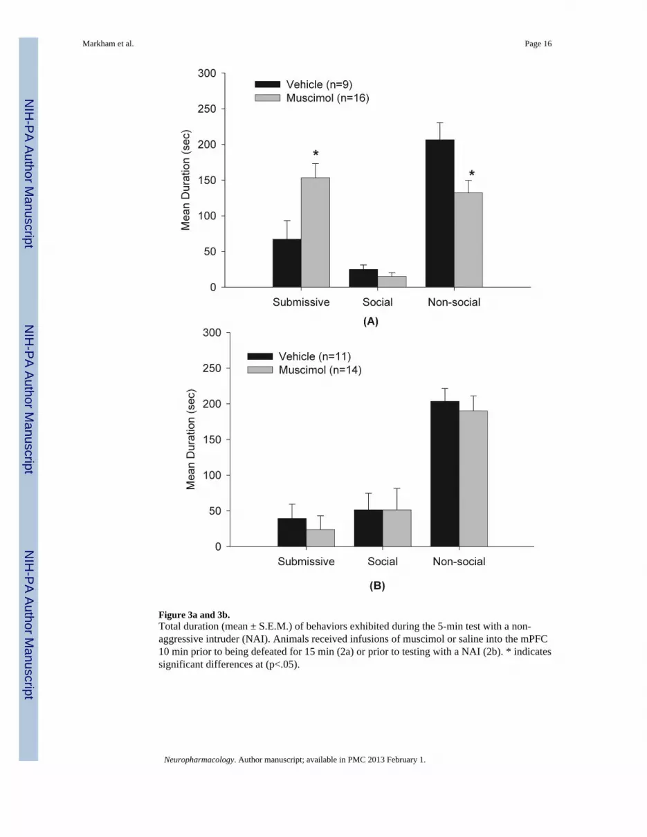
Figure 3a and 3b.Total duration (mean ± S.E.M.) of behaviors exhibited during the 5-min test with a non-aggressive intruder (NAI). Animals received infusions of muscimol or saline into the mPFC10 min prior to being defeated for 15 min (2a) or prior to testing with a NAI (2b). * indicatessignificant differences at (p<.05).
Markham et al. Page 16
Neuropharmacology. Author manuscript; available in PMC 2013 February 1.
NIH
-PA Author Manuscript
NIH
-PA Author Manuscript
NIH
-PA Author Manuscript

Figure 4.Total duration (mean ± S.E.M.) of behaviors exhibited during the 5-min test with a NAI.Animals received infusions of bicuculline or saline into the mPFC 10 min prior to beingdefeated for 15 min. * indicates significant differences at (p<.001).
Markham et al. Page 17
Neuropharmacology. Author manuscript; available in PMC 2013 February 1.
NIH
-PA Author Manuscript
NIH
-PA Author Manuscript
NIH
-PA Author Manuscript

Figure 5.Total duration (mean ± S.E.M.) of behaviors exhibited during the 5-min test with a NAI.Animals received infusions of anisomycin or saline into the mPFC 20 min prior to beingdefeated for 15 min.
Markham et al. Page 18
Neuropharmacology. Author manuscript; available in PMC 2013 February 1.
NIH
-PA Author Manuscript
NIH
-PA Author Manuscript
NIH
-PA Author Manuscript

Figure 6.Total duration (mean ± S.E.M.) of behaviors exhibited during the 5-min test with a NAI.Animals received infusions of bicuculline or saline into the BLA 10 min prior to beingdefeated for 5 min.
Markham et al. Page 19
Neuropharmacology. Author manuscript; available in PMC 2013 February 1.
NIH
-PA Author Manuscript
NIH
-PA Author Manuscript
NIH
-PA Author Manuscript

NIH
-PA Author Manuscript
NIH
-PA Author Manuscript
NIH
-PA Author Manuscript
Markham et al. Page 20
Tabl
e 1
Sum
mar
y of
his
tolo
gica
l pla
cem
ents
for
all s
ubje
cts i
n E
xper
imen
ts 1
-5
Exp
erim
ents
12
34
5
Hits
2525
1917
23
Mis
ses
55
10
21
Mis
cella
neou
s*2
01
03
Tota
l32
3021
1747
* Indi
cate
s ani
mal
s tha
t wer
e no
t inc
lude
d in
the
anal
ysis
due
to fa
ctor
s oth
er th
an m
isse
d pl
acem
ents
, suc
h as
mis
sing
cap
s or c
logg
ed c
annu
la p
rior t
o dr
ug/in
k in
fusi
on.
Neuropharmacology. Author manuscript; available in PMC 2013 February 1.