the histone deacetylase inhibitor valproic acid sensitizes diffuse large b-cell lymphoma cell lines...
TRANSCRIPT

Am J Transl Res 2013;5(2):170-183www.ajtr.org /ISSN:1943-8141/AJTR1302007
Original ArticleThe histone deacetylase inhibitor valproic acid sensitizes diffuse large B-cell lymphoma cell lines to CHOP-induced cell death
Malin Ageberg1, Karin Rydström2, Thomas Relander2, Kristina Drott1,2
1Division of Hematology and Transfusion Medicine, Lund University, BMC B13, Klinikg. 26, S-22184 Lund, Swe-den; 2Skåne Department of Oncology, Lund University Hospital, S-22185 Lund, Sweden
Received February 13, 2013; Accepted March 5, 2013; Epub March 28, 2013; Published April 8, 2013
Abstract: Epigenetic code modifications by histone deacetylase inhibitors (HDACis) have recently been proposed as potential new therapies for hematological malignancies. Diffuse large B-cell lymphoma (DLBCL) is the most com-mon form of aggressive lymphoma. At present, standard first line treatment for DLBCL patients is the antracycline-based chemotherapy regimen CHOP (cyclophosphamide, doxorubicin, vincristine and prednisone) combined with the monoclonal anti-CD20 antibody rituximab (R-CHOP). Since only 50-60% of patients reach a long-time cure by this treatment, there is an urgent need for novel treatment strategies to increase the response and long-term re-mission to initial R-CHOP therapy. In this study, we investigated the effect of the HDAC inhibitor valproic acid (VPA) on DLBCL cell lines. To elucidate the effects of VPA on chemo-sensitivity, we used a cell-line based model of CHOP-refractory DLBCL. All five DLBCL cell lines treated with VPA alone or in combination with CHOP showed decreased viability and proliferation. The VPA-induced sensitization of DLBCL cells to cytotoxic treatment resulted in increased number of apoptotic cell as judged by annexin V-positivity and the presence of cleaved caspase-3. In addition, pretreatment with VPA resulted in a significantly increased DNA-damage as compared to CHOP alone. In summary, HDAC inhibitors such as VPA, are promising therapeutic agents in combination with R-CHOP for patients with DLBCL.
Keywords: Non-Hodgkins lymphoma, valproic acid, valproate, HDAC
Introduction
Diffuse large B-cell lymphoma (DLBCL) is the most common subgroup of non-Hodgkin lym-phoma, accounting for approximately 30% of all lymphomas. DLBCLs belong to the group of aggressive lymphoma, and usually result in death within a couple months if left untreated. DLBCL is most commonly diagnosed in middle-aged and elderly individuals. Antracycline-based chemotherapy comprised of cyclophos-phamide, doxorubicin, vincristine and prednisone (CHOP) in combination with ritux-imab, a monoclonal antibody against CD20, is considered to be the standard treatment regi-men for patients with DLBCL. Despite the improved prognosis after introduction of anti-CD20 therapy to the conventional CHOP treat-ment, the 5-year progression-free survival rate remains around 60% [1-3]. Hence, the need for new therapeutic agents that augment sensitiv-
ity to R-CHOP treatment without increased tox-icity is urgent.
Along with mutations in the DNA code, malig-nant transformation can result from aberra-tions in chromatin-modifying proteins such as histone acetylases and methylases leading to altered expression or defect activation of impor-tant genes such as tumor suppressors or onco-genes. This feature is underscored by findings by Pasqualucci et al., demonstrating that the histone acetyltransferases EP300 and CREBBP are heterozygously mutated in 39% of DLBCL cases, and expressed at subnormal levels in an additional number of DLBCL cases [4]. Moreover, the histone methyltransferase MLL2 is mutated in 24% of DLBCL [5].
These data suggest that dosage of epigenetic regulators may be vital for preserving a benign phenotype. Therefore, new treatments in DLBCL

Valproic acid sensitizes to CHOP in DLBCL cell lines
171 Am J Transl Res 2013;5(2):170-183
should aim at restoring physiologic acetylation levels, and the use of inhibitors of histone acet-ylation could have a rational basis in DLBCL.
Several histone deacetylase inhibitors (HDACis) are shown to have effect on specific tumor types as single agent drugs and hematological malignancies seem to be particularly sensitive to HDAC inhibitors. Accordingly, vorinostat (Zolinza® or SAHA) and romidepsin (Istodax®) were approved by the FDA in 2006 and 2009, respectively, for the treatment of cutaneous T-cell lymphoma (CTCL) [6]. Also, in 2011, FDA approved romidepsin for the treatment of patients with peripheral T-cell lymphoma fol-lowing at least one prior therapy [7]. Vorinostat and the HDAC class I specific inhibitor, MGCD01103, has been tested as a monother-apy for the treatment of relapsed and refractory DLBCL but with limited activity [8]. Several other HDAC inhibitors are under evaluation in clinical trials both as single agents and in com-bination with chemotherapeutic drugs [9].
In 2001, valproic acid (VPA), a GABA agonist with a long history of clinical use for treatment of epilepsy and mood disorders (reviewed in [10]), was identified having HDAC inhibitory activity [11]. VPA is a short-chain fatty acid that has been shown to inhibit the class I and II HDAC enzymes [10, 12]. VPA was recently shown to bind with high affinity to the hydropho-bic active site channel of HDAC8 by van der Waals interactions [12]. Since its identification as an HDAC inhibitor, VPA has been suggested to regulate several mechanisms involved in malignant transformation such as cell cycle control, differentiation, DNA repair and apopto-sis (reviewed in [10]. In addition, VPA has also been associated with DNA methylation, as VPA can induce downregulation of chromatin main-tenance proteins but also induce direct meth-ylation of lysine 4 on histone 3 [13, 14]. The anti-tumor activity of VPA has been observed in both solid and hematological malignancies such as thyroid cancer [15], neuroblastoma [16], glioma [17], breast cancer [18], and hema-tological malignancies [19-21].
Although VPA can induce histone acetylation within 30 minutes, events that regulate chro-matin condensation status may take up to 48 hours to be completed. Consequently, pretreat-ment with VPA for 48 hours has been shown to alter chromatin structure by regulation of chro-
matin modulation proteins such as depletion of chromatin maintenance proteins DNA methyl-transferase 1 and HP1. Hence, pretreatment with VPA for 48 hours has been shown to sensi-tize to cell death induced by the topoisomerase II inhibitor epirubicin in a mouse model, corre-lating to chromatin decondensation and increased DNAdamage [13, 22, 23].
VPA is presently implicated in numerous clinical trials, both as a single agent and in association with other drugs, involving various pathologies such as mood disorders, auto-immune diseas-es and cancer. Interestingly, VPA has been eval-uated in a sequence-specific combination with FEC100 (5-fluorouracil, epirubicin and cyclo-phosphamide) as a primary therapy in a phase I/II trial for locally advanced/metastatic breast cancer [24]. Results were encouraging, with no pharmacokinetic or pharmacodynamic interac-tions. Partial response was seen in 9 of 41 patients in phase I, and objective response in 9 of 14 patients in phase II.
In this study, we have used a cell line-based model of CHOP-resistant DLBCL to investigate the ability of VPA to sensitize diffuse large B-cell lymphoma cell lines to CHOP treatment. Our results demonstrate that VPA potentiates the cytotoxic effects of CHOP treatment by induc-ing apoptosis as determined by annexin V and an increased level of cleaved caspase-3. Rituximab-mediated cellular cytotoxicity is sus-tained in the presence of VPA. In addition, we demonstrate an increased formation of topoi-somerase IIa-DNA complexes and also an increased level of γH2AX indicating higher amount of double-strand breaks (DSBs) in response to VPA. Our results support a possible novel treatment strategy of DLBCL, utilizing VPA in combination with the conventional R-CHOP protocol.
Materials and methods
Reagents
Cyclophosphamide monohydrate (C), vincris-tine sulfate (O), doxorubicin monohydrate (H), prednisolone (P), and valproic acid (VPA) was obtained from Sigma Aldrich (St Louis, MO). Prednisolone is the biologically active sub-stance of prednisone. Rituximab was obtained from local pharmacy. 7-AAD was obtained from BD Pharmingen (San Diego, CA)

Valproic acid sensitizes to CHOP in DLBCL cell lines
172 Am J Transl Res 2013;5(2):170-183
Cells and culture conditions
The human diffuse large B-cell lymphoma (DLBCL) cell lines SU-DHL-5, Karpas-422, SU-DHL-8 and WSU-NHL were purchased from the German Collection of Microorganisms and Cell Cultures (DSMZ). The diffuse large B cell lymphoma cell line ULA [25] was kindly provid-ed by Dr Berglund (Uppsala University, Uppsala, Sweden). Karpas-422, SU-DHL-5 and SU-DHL-8 was grown in RPMI 1640 (Invitrogen, Carlsbad, CA) supplemented with 20% fetal bovine serum (FBS) (Invitrogen). WSU-NHL was grown in RPMI 1640 supplemented with 10% FBS. ULA was grown in 45% Optimem (Invitrogen) and 45% IDEM (Invitrogen) supplemented with 10% FBS. All cell lines were cultured in a humidified atmo-sphere (37°C, 5% CO2).
Cell viability
Cells were seeded in a concentration of 0.8-1x10⁶ cells/ml and treated with different com-binations of substances as specified in figure legends. Cell viability was assessed after 24 h, 48 h and 72 h by trypan blue exclusion. The VPA pretreatment experiment was performed by a 24 h or a 48 h pretreatment of cells with 0.5 or 1.5 mM VPA alone or in combination with 20 µg/ml prednisolone followed by addition of CHOP. No additional prednisolone was added to cultures where prednisolone was included in the pretreatment (CHO). The CHOP regimen used consists of 10 µM cyclophosphamide monohydrate, 20 nM doxorubicin hydrochlo-ride, 2 nM vincristine sulfate and 20 µg/ml prednisolone [25]. Viability was measured 48 h, 72 h, and 96 h after start of experiment using trypan blue exclusion.
Apoptosis analysis by flow cytometry
Labeling of cells with annexin V-PE (BD Pharmingen) was performed according to the manufacturer’s instructions. 7-AAD was added according to the manufacturer’s instructions. Apoptotic cells were defined as annexin V-positive, 7-AAD-positive and Annexin V-and 7-AAD-double positive cells.
Western blot analysis
Cells (0.8x10⁶/ml) were incubated with VPA alone or in combination with CHOP. Cells were harvested after 24 h, 48 h and 72 h and
washed once with PBS and resuspended in Laemmli sample buffer (62.5 mM Tris-HCl, pH 6.8, 25% glycerol, 2% SDS, 0,01% Bromophenol Blue; Bio-Rad Laboratories, Hercules, CA). Primary antibodies used were anti-cleaved cas-pase-3 (Asp175)(5A1) from Cell Signaling Technology; anti-GAPDH (6C5), anti-p27 (C-19), anti-p21 (187), anti-actin (C-2) and anti-topoI-Ialpha (3F6) from Santa Cruz Biotechnology (Santa Cruz Biotechnology, Heidelberg, Germany); anti-γH2AX(pS139)(N1-431) from BD Pharmingen; anti-acetylated histone H3 from Millipore (Millipore, Bedford, MA); anti-his-tone 3 (ab1791) from Abcam (Abcam, Cambridge, United Kingdom). After incubation with horseradish peroxidase (HRP)-conjugated secondary antibody, antibody binding was visu-alized with enhanced chemiluminescence (EZ-ECL, Biological industries, Beit, Israel) fol-lowed by detection with hyperfilm ECL (GE Healthcare, Buckinghamshire, United Kingdom).
Cell cycle analysis
0.5-1x10⁶ cells was washed with PBS and fixed in 70% EtOH and stored at -20°C for 1-7 days. Labeling of cells for cell cycle analysis was per-formed as follows. Cells were washed and stained in propidium iodide (PI)-staining solu-tion (50 µg/ml PI, 0.05% Triton X-100, 0.1 mg/ml Rnase A). Cells were incubated in the dark at room temperature for 1 hour, thereafter ana-lyzed on a FACSCanto II flow cytometer (Becton Dickinson, San Jose, CA). Markers were set to determine the percentage of hypodiploid cells (sub-G0/G1), and cells in the G0/G1, S and G2/M phase of the cell cycle.
ADCC assay
One day prior to start of assay, WSU-NHL cells were labeled with PKH26 red fluorescent cell linker kit for general cell membrane labeling (Sigma Aldrich) according to the manufacturer’s instructions. Heat-inactivated FBS was used throughout the experiment and obtained by heating FBS to 56°C for 30 min followed by fil-tration through 0.22 µm filter. At day one, the PKH26-labelled cells were plated on a round-bottom 96-well plate at a density of 10 000 cells/well. Cells were treated with or without VPA 1.5 mM and CHOP (C: 10 µM cyclophos-phamide monohydrate; H: 20 nM doxorubicin; O: 2 nM vincristine; P: 20 µg/ml prednisolone)

Valproic acid sensitizes to CHOP in DLBCL cell lines
173 Am J Transl Res 2013;5(2):170-183
figure 1. VPA sensitizes DLBCL cell lines to CHOP treatment. The DLBCL cell lines ULA, WSU-NHL, Karpas-422, SU-DHL-8 and SU-DHL-5 were treated for 72 h with 0.1, 2 and 10 mM VPA in the absence or presence of CHOP. The cell and cell proliferation in millions of cells viability and cell proliferation in millions of cells was assessed after 24 h, 48 h, 72 h by trypan blue exclusion and normalized to untreated control cells at 0 h. Data are presented as mean ± SEM, n=3.
followed by overnight incuba-tion at 37°C. At day two, ritux-imab was added to the cells at concentrations of 0.01-10 µg/ml followed by 20 min incuba-tion at 37°C. NK cells were isolated from peripheral blood using NK cell isolation kit from MACS (Miltenyi Biotec). Briefly, isolation of MNC from periph-eral blood was performed by density gradient centrifuga-tion with lymphoprep (Axis-Shield, Oslo, Norway) thereaf-ter MNC was labeled according to the manufacturer’s instruc-tions and applied to MACS col-umns for negative selection of the NK cell population. The purity of CD56-positive NK cells was verified by FACS. NK cells were used as effector cells and added at an effector to target cell ratio of 10:1. Cells were incubated over-night thereafter the amount of dead cells was visualized by staining with 7-AAD (BD) fol-lowed by FACS analysis. Dead target cells were identified as double-positive for PKH26 and 7-AAD and used as read-out of the assay.
Band depletion assay
To monitor the amount of topoisomerase II-DNA cova-lent complexes, treated cells were lysed with an alkaline lysis solution (200 mM NaOH, 2 mM EDTA) for 30 min on ice followed by a neutralization of lysate with 2M HCl and 1.2 M Tris (pH 8). The lysate was mixed with 3x SDS sample buffer (150 mM Tris/HCl pH 6.8, 6 mM EDTA, 45% sucrose, 9% DSD, 10% b-mercaptoeth-anol, 0,03% bromophenol blue). The lysate was passed through a 23 G needle 5 times, boiled and resolved by SDS-PAGE and analyzed by western blotting.
Valproic acid sensitizes to CHOP in DLBCL cell lines
174 Am J Transl Res 2013;5(2):170-183
Statistics
Significant differences were evaluated using the Student’s unpaired t-test. All tests were two-sided. An effect was considered to statisti-cally significant at p<0.05 (*), p<0.01 (**) or p<0.001(***). Data analysis was performed with the GraphPad Prism 5.0a (GraphPad soft-ware, Inc, La Jolla, CA) or with Microsoft Excel. Data are plotted as means ± standard error of the mean (SEM).
Results
Effect of VPA on viability and proliferation of large diffuse B-cell lymphoma (DLBCL) cell lines
We have previously established a cell line-based model of CHOP refractory DLBCL [25]. Although relapsed or refractory cases of DLBCL
clude, the addition of VPA significantly increas-es CHOP-sensitivity of DLBCL cell lines.
Clinically relevant concentrations of VPA sensi-tize DLBCL cells to CHOP treatment
To further characterize the effects of VPA on DLBCL cell lines, we continued all experiments with the CHOP-resistant cell line WSU-NHL and the CHOP-sensitive cell line SU-DHL-8. VPA is used clinically in the treatment of epilepsy, and is well tolerated at continuos serum-concentra-tions up to 0.7 mM. Moreover, the maximal tol-erated dose during 3-day treatment periods in combination with FEC in a phase I/II study by Münster et al, was 140 mg/kg/day, which cor-responds to approximately 1.5 mM of total serum VPA (at day 3) [24]. Therefore, we contin-ued to characterize the effects of 0.5 mM and 1.5 mM VPA alone or in combination with CHOP
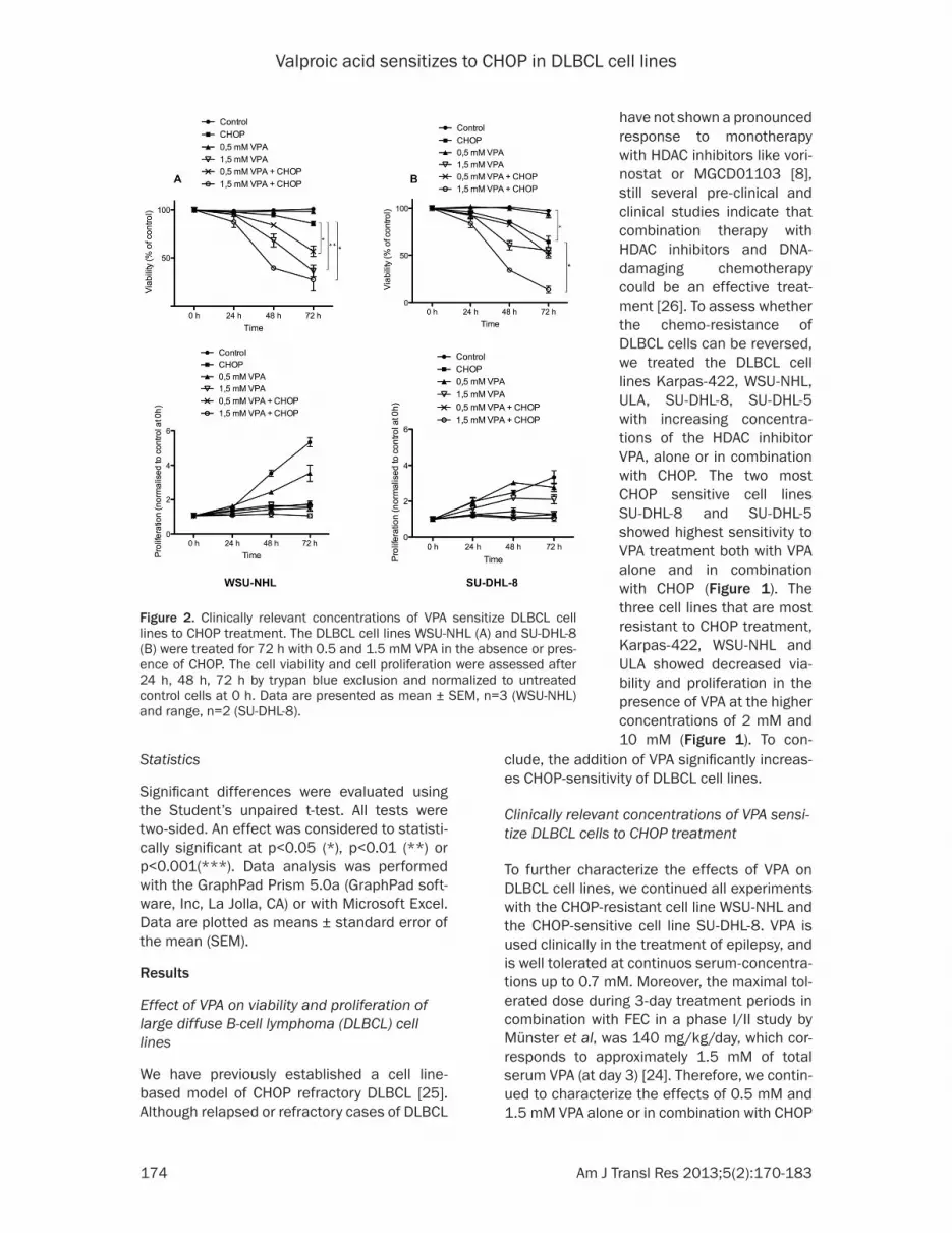
figure 2. Clinically relevant concentrations of VPA sensitize DLBCL cell lines to CHOP treatment. The DLBCL cell lines WSU-NHL (A) and SU-DHL-8 (B) were treated for 72 h with 0.5 and 1.5 mM VPA in the absence or pres-ence of CHOP. The cell viability and cell proliferation were assessed after 24 h, 48 h, 72 h by trypan blue exclusion and normalized to untreated control cells at 0 h. Data are presented as mean ± SEM, n=3 (WSU-NHL) and range, n=2 (SU-DHL-8).
have not shown a pronounced response to monotherapy with HDAC inhibitors like vori-nostat or MGCD01103 [8], still several pre-clinical and clinical studies indicate that combination therapy with HDAC inhibitors and DNA-damaging chemotherapy could be an effective treat-ment [26]. To assess whether the chemo-resistance of DLBCL cells can be reversed, we treated the DLBCL cell lines Karpas-422, WSU-NHL, ULA, SU-DHL-8, SU-DHL-5 with increasing concentra-tions of the HDAC inhibitor VPA, alone or in combination with CHOP. The two most CHOP sensitive cell lines SU-DHL-8 and SU-DHL-5 showed highest sensitivity to VPA treatment both with VPA alone and in combination with CHOP (figure 1). The three cell lines that are most resistant to CHOP treatment, Karpas-422, WSU-NHL and ULA showed decreased via-bility and proliferation in the presence of VPA at the higher concentrations of 2 mM and 10 mM (figure 1). To con-

Valproic acid sensitizes to CHOP in DLBCL cell lines
175 Am J Transl Res 2013;5(2):170-183
in WSU-NHL and SU-DHL-8. VPA treatment alone at a concentration of 1.5 mM resulted in decreased viability of both WSU-NHL and SU-DHL-8 cells (figure 2A and 2B). The pres-ence of 0.5 mM VPA alone did not considerably affect viability, but in combination with CHOP, a sensitizing effect of VPA after 72 h could be noticed as the viability decreased to 60% for WSU-NHL and to 50% for SU-DHL-8 as com-pared to 85% and 65%, respectively, for CHOP alone (figure 2A and 2B). Most striking was the additive effect of 1.5 mM VPA to CHOP, that resulted in a viability of 25% and 15% after 72 h, compared to the viability cells treated with of 1.5 mM VPA alone, that resulted in 40% and 60% viability in WSU-NHL and SU-DHL-8, respectively (figure 2A and 2B). The prolifera-tion of WSU-NHL and SU-DHL-8 was reduced in a dose-dependent manner in the presence of VPA (figure 2A and 2B). Interestingly, 0.5 mM VPA initially showed a pro-proliferative effect especially in SU-DHL-8 (figure 2B). Treatment with CHOP resulted in a proliferation arrest, which was not altered by the presence of VPA (figure 2A and 2B). In conclusion, clinically rel-evant concentrations of VPA are enough for sensitizing diffuse large Bcell lymphoma cells to CHOP treatment.
Pretreatment of DLBCL cell lines with VPA
An interesting clinical study has been per-formed, assessing the use of sequential admin-
istration of VPA and chemotherapy for patients with solid malignancies [24]. Therefore, we investigated whether pretreatment with VPA 48 h before addition of the cytotoxic combination of CHOP had the same sensitizing effect as seen for simultaneous treatment of VPA and CHOP. As seen in Tables 1 and 2, both SU-DHL-8 and WSU-NHL show significantly decreased viability for cells pretreated with 1.5 mM VPA in comparison with cells treated with VPA or CHOP alone. Taken together, sequential or simultane-ous treatment of VPA and CHOP has similar effects on cell viability. Because VPA is a well-known tranquilizer, with documented sedative effects, it could be advantageous to combine it with prednisolone, which is known to have strong invigorating effects. Moreover, predniso-lone is part of the CHOP regimen, and could easily be administered together with VPA with-out major changes in the CHOP protocol. Therefore, pretreatment with VPA and predniso-lone (20 µg/ml) for 48 h was performed before the remaining cytotoxic drugs comprising CHOP i.e. cyclophosphamide, doxorubicin and vincris-tine (CHO) were added. Table 1 and 2 show a significant decrease in viability of WSU-NHL and SU-DHL-8 pretreated with 1.5 mM VPA and prednisolone compared to cells pretreated with prednisolone alone. In conclusion, pretreat-ment with VPA alone or VPA in combination with prednisolone before addition of cytotoxic drugs has a significant negative effect on the viability of DLBCL cells.
Table 1. Pretreatment of SU-DHL-8 cells with VPA in combination with prednisolone and CHOP
SU-DHL-8 cells were treated with different combinations of VPA, prednisolone (P, 20 µg/ml)) and CHOP/CHO as illustrated as illustrated in Table 2. Cell viability was assessed at 48, 72, 96 h by trypan blue excusion. Standard error of the mean (SEM) of 3 independent experiments is shown. A two-sided t-test was performed. *P<0.05.

Valproic acid sensitizes to CHOP in DLBCL cell lines
176 Am J Transl Res 2013;5(2):170-183
VPA induces apoptosis in CHOP resistant DLBCL cells
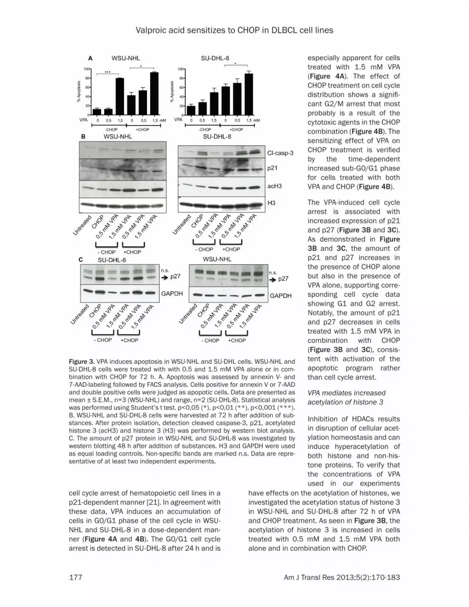
To assess if the observed cytotoxicity after VPA treatment reflects an induction of apoptosis, WSU-NHL and SU-DHL8 cells were treated with 0.5 mM or 1.5 mM VPA alone or in combination with CHOP for 72 h, followed by FACS analysis of annexin V and 7-AAD-positivity. In WSU-NHL cells, 1.5 mM VPA but not 0.5 mM VPA induced a prominent annexin V/7AAD-positivity of about 80% (figure 3A) confirming the viability data in figure 2A. A significant additive effect of 1.5 mM VPA to CHOP in WSU- NHL is confirmed by the increasing amount of annexin V and 7-AAD-positive cells (figure 3A). Treatment with CHOP alone in WSU-NHL resulted in 85% viability after 72 h as judged by trypan blue exclusion (figure 2A) but when analyzing annexin V and 7-AAD-positivity, the initiation of an apoptotic program is demonstrated by the 40% of annex-in V and 7-AAD positive cells (figure 3A). The SU-DHL-8 cell line is most likely more respon-sive to VPA than WSU-NHL as judged by higher number of annexin V-and 7-AAD-positive cells with increasing concentration of VPA as com-
pared to WSU-NHL (figure 3A). An evident addi-tive effect of VPA to CHOP is observed also in SU-DHL-8 cells (figure 3A) as judged by the amount of apoptotic annexin V-and 7-AAD-positive cells.
To further confirm that the decreased viability after VPA treatment is in fact due to apoptosis, we determined the presence of cleaved cas-pase-3 (figure 3B). The increased amount of cleaved caspase-3 in WSU-NHL cells treated with 1.5 mM VPA alone and in combination with CHOP (figure 3B) is in accordance with viability data and annexin V/7-AAD data, supporting the observed apoptotic effect of VPA with and with-out CHOP. In SU-DHL-8 cells, a strong increase in cleaved caspase-3 in the presence of CHOP is observed, consistent with the increased CHOP sensitivity of this cell lines, while the effects of VPA alone are comparable to effects in WSU-NHL cells (figure 3B).
VPA induces G1 arrest of DLBCL cells
HDAC inhibitors are reported to rapidly induce cell cycle arrest and induce tumor cell-selective apoptosis. VPA has been reported to induce
Table 2. Pretreatment of WSU-NHL cells with VPA in combination with prednisolone and CHOP
WSU-NHL cells were treated with different combinations of VPA, prednisolone (P, 20 µg/ml)) and CHOP/CHO as illustrated below. Cell viability was assessed at 48, 72, 96 h by trypan blue excusion. Standard error of the mean (SEM) of 3 independent experi-ments is shown. A two-sided t-test was performed. *P<0.05.
Valproic acid sensitizes to CHOP in DLBCL cell lines
177 Am J Transl Res 2013;5(2):170-183
cell cycle arrest of hematopoietic cell lines in a p21-dependent manner [21]. In agreement with these data, VPA induces an accumulation of cells in G0/G1 phase of the cell cycle in WSU-NHL and SU-DHL-8 in a dose-dependent man-ner (figure 4A and 4B). The G0/G1 cell cycle arrest is detected in SU-DHL-8 after 24 h and is
have effects on the acetylation of histones, we investigated the acetylation status of histone 3 in WSU-NHL and SU-DHL-8 after 72 h of VPA and CHOP treatment. As seen in figure 3B, the acetylation of histone 3 is increased in cells treated with 0.5 mM and 1.5 mM VPA both alone and in combination with CHOP.
figure 3. VPA induces apoptosis in WSU-NHL and SU-DHL cells. WSU-NHL and SU-DHL-8 cells were treated with with 0.5 and 1.5 mM VPA alone or in com-bination with CHOP for 72 h. A. Apoptosis was assessed by annexin V- and 7-AAD-labeling followed by FACS analysis. Cells positive for annexin V or 7-AAD and double positive cells were judged as apopotic cells. Data are presented as mean ± S.E.M., n=3 (WSU-NHL) and range, n=2 (SU-DHL-8). Statistical analysis was performed using Student’s t test. p<0,05 (*), p<0,01 (**), p<0,001 (***). B. WSU-NHL and SU-DHL-8 cells were harvested at 72 h after addition of sub-stances. After protein isolation, detection cleaved caspase-3, p21, acetylated histone 3 (acH3) and histone 3 (H3) was performed by western blot analysis. C. The amount of p27 protein in WSU-NHL and SU-DHL-8 was investigated by western blotting 48 h after addition of substances. H3 and GAPDH were used as equal loading controls. Non-specific bands are marked n.s. Data are repre-sentative of at least two independent experiments.
especially apparent for cells treated with 1.5 mM VPA (figure 4A). The effect of CHOP treatment on cell cycle distribution shows a signifi-cant G2/M arrest that most probably is a result of the cytotoxic agents in the CHOP combination (figure 4B). The sensitizing effect of VPA on CHOP treatment is verified by the time-dependent increased sub-G0/G1 phase for cells treated with both VPA and CHOP (figure 4B).
The VPA-induced cell cycle arrest is associated with increased expression of p21 and p27 (figure 3B and 3C). As demonstrated in figure 3B and 3C, the amount of p21 and p27 increases in the presence of CHOP alone but also in the presence of VPA alone, supporting corre-sponding cell cycle data showing G1 and G2 arrest. Notably, the amount of p21 and p27 decreases in cells treated with 1.5 mM VPA in combination with CHOP (figure 3B and 3C), consis-tent with activation of the apoptotic program rather than cell cycle arrest.
VPA mediates increased acetylation of histone 3
Inhibition of HDACs results in disruption of cellular acet-ylation homeostasis and can induce hyperacetylation of both histone and non-his-tone proteins. To verify that the concentrations of VPA used in our experiments

Valproic acid sensitizes to CHOP in DLBCL cell lines
178 Am J Transl Res 2013;5(2):170-183
VPA increases the level of covalent topo-siomerase IIα-DNA complexes and potentiates CHOP-induced DSBs
As shown in figure 3B, treatment with VPA leads to histone acetylation. It has previously been shown that this is followed by a modula-tion of genes and proteins essential for the maintenance of heterochromatin, resulting in chromatin decondensation [13]. Topoisomerase II (Topo II) is an enzyme that regulates DNA under- and overwinding. In order to carry out its
critical physiological functions, topoisomerase II generates transient double-stranded breaks in DNA, binding covalently to the DNA [27]. Chromatin decondensation induced by HDACis is associated with an increased binding of topoisomerase II to the DNA substrate and in the presence of topoisomerase II inhibitors, it is also associated with increased DNA damage and cell death [13, 28]. Doxorubicin is a Topo II inhibitor that forms covalent complexes with Topo II–induced double-stranded DNA breaks (DSBs). We used a band depletion assay to con-trol for possible differences in the extent of doxorubicin-induced Topo IIα-trapping in VPA treated cells compared to non-treated cells. Cells were treated with or without 1 mM VPA for 24 h followed by an incubation of CHOP for 24 hours. Thereafter the band depletion assay was performed to analyze the amount of Topo IIα that was trapped in covalent complexes with DNA and therefore unable to enter the gel. Hence, the proportion of Topo IIα trapped in cleavage complexes is proportional to the reduction in Topo IIa as assessed by western blotting. Both WSU-NHL and SU-DHL-8 cells treated with VPA alone and with VPA in combi-nation with CHOP had reduced levels of Topo IIα compared to untreated or CHOP-treated cells, indicating that a higher amount of Topo IIα was trapped in DNA complexes in response to VPA (figure 5B). Interestingly, treatment with VPA alone resulted in Topo IIα-trapping to a comparable extent to cells treated with a com-bination of VPA and CHOP. This may suggest that VPA-mediated HDAC inhibition alone leads to increased binding of Topo IIα to the DNA, even in the absence of topoisomerase II inhibi-tors like doxorubicin. In conclusion, our data support that VPA may increase the DNA binding of Topo IIα , which may contribute to sensitiza-tion to CHOP-treatment.
Pretreatment with VPA increases the amount of DNA double-strand breaks in CHOP treated cells
Since VPA can increase the amount of DNA cleavage complexes with Topo IIα, we investi-gated if the presence of VPA also can compro-mise the repair of DNA double-strand breaks (DSBs). The accumulation of γH2AX (histone H2AX phosphorylated on S139), is an early marker of DNA DSBs. The level of γH2AX is pro-portional to the level of free DSBs [29]. Hence,
figure 4. VPA treatment alone induces G1 arrest of WSU-NHL and SU-DHL-8 cells, and in combination with CHOP, VPA increases the number of cells in sub-G1/G0. A. WSU-NHL and B. SU-DHL-8 cells were treated with 0.,5 or 1.,5 mM VPA alone or in combi-nation with CHOP for 72 h. Cells were harvested after 24 h, 48 h and 72 h and cell cycle analysis was per-formed using propidium iodide labeling. Error bars represent S.E.M., n=3.

Valproic acid sensitizes to CHOP in DLBCL cell lines
179 Am J Transl Res 2013;5(2):170-183
we measured the levels of γH2AX as a marker to monitor the generation and repair of VPA-induced DSBs. As a positive control we used VM-16, also called etoposide, a drug reported to induce cell death with the preferential forma-tion of Topo IIα–containing cleavable complex-es. In SU-DHL-8 cells, treatment with both CHOP alone and 1 mM VPA alone resulted in an increased amount of γH2AX, and pretreatment with 1 mM VPA before addition of CHOP result-ed in an even higher amount of γH2AX (figure 5A). Interestingly, in WSU-NHL cells treated with 1 mM VPA and cells pretreated with 1 mM VPA for 24 h before addition of CHOP, an increased level of γ-H2AX was detected by western blot analysis as compared to cells treated with CHOP alone (figure 5A). Taken together, our data suggest that pretreatment with VPA does indeed potentiate CHOP-induced DNA damage in DLBCL cell lines.
VPA does not counteract rituximab-induced cellular cytotoxicity
The standard therapy for patients diagnosed with DLBCL is CHOP in combination with the monoclonal anti-CD20-antibody rituximab. Since the addition of rituximab to CHOP therapy the overall survival has increased for DLBCL patients [30]. Therefore, it is of great impor-tance that future additional drugs in the treat-ment regimen of these patients do not impede the function of the antibody-based therapy. Indeed, Shimizu et al recently reported that HDAC inhibitors including VPA increase CD20 expression on the cell surface, and augment rituximab-mediated complement dependent cytotoxicity (CDC) in lymphoma cell lines [31]. However, as the major part of the cytotoxic effect of rituximab is mediated by antibody dependent cellular cytotoxicity (ADCC) [32], we investigated the effect of VPA on rituximab-
figure 5. Co-treatment with VPA and CHOP results in an in-creased expression of γH2AX and decreased amount of Topo IIα-DNA complexes. A. SU-DHL and WSU-NHL cells were ei-ther untreated or treated with VPA and CHOP as depicted in top of figure 5. After protein isolation, the amount of γH2AX was analyzed by western blot analysis. Cells treated with 10 µM etoposide serve as control of the γH2AX antibody. B. Pro-tein from WSU-NHL cells treated as in A. was isolated as de-scribed in Materials and Methods and subjected to western blot analysis. The amount of Topo IIα was investigated. Data are representative of at least two independent experiments. GAPDH or β-actin was used as equal loading control.

Valproic acid sensitizes to CHOP in DLBCL cell lines
180 Am J Transl Res 2013;5(2):170-183
mediated ADCC. To that purpose, SU-DHL-8 and WSU-NHL cells were treated with 1.5 mM VPA alone or in combination with CHOP for 24 h followed by an incubation of cells with increas-ing concentration of rituximab. Thereafter NK cells from peripheral blood was added as effec-tor cells at a ratio of 10:1 and the viability of cells was investigated after 16 h using 7-AAD as a vibility marker. The presence of VPA had no negative effect on rituximab-mediated cellular cytotoxicity (figure 6), consistent with the data on CDC from Shimizu et al [31], and supporting its use together with the R-CHOP regimen.
Discussion
Anthracyclin-based therapy with CHOP has been the unchallenged treatment for diffuse large B-cell lymphoma since more than thirty years. Although addition of the monoclonal CD20 antibody rituximab (R-CHOP) during the last decade has led to improved overall and progression free survival, the prognosis for patients diagnosed with DLBCL is still far from satisfying, as approximately 40% of affected patients ultimately die from their disease.
ing, have been shown to potentiate DNA dam-age induced by anthracyclins such as doxorubi-cin and epirubicin in breast cancer cell lines and in mouse models [22, 23]. Moreover, it has been suggested that pretreatment with HDACis for 48 hours before treatment with anthracy-clins may give time for chromatin remodelling, resulting in maximal DNA damage [23].
In this study, we show that HDACis like valproic acid can be valuable in combination also with R-CHOP in diffuse large B-cell lymphoma cell lines. Both pretreatment and concomitant treatment with VPA sensitizes strongly to CHOP treatment at clinically relevant concentrations. Importantly, the sensitizing effect of VPA to che-motherapy was prominent also in cell lines resistant to treatment with CHOP, such as WSU-NHL. In contrast to its effects in breast cancer cell lines [23], VPA had pronounced effects in lymphoma cell lines also as a single agent.
We demonstrate anti-proliferative and pro-apoptotic activity of VPA as a single agent on DLBCL cell lines. VPA has previously been sug-gested to induce a G0/G1 cell cycle arrest by an
figure 6. VPA does not interfere with rituximab-mediated cellular cytotoxicity. A. WSU-NHL and B. SU-DHL-8 cells were labeled with PKH26, either left untreated or incubated with 1,5 mM VPA for 24 h followed by addition of varying concen-trations of rituximab. NK cells were added at an effector to target cell ratio of 10:1 thereafter the cells were incubated for an additional 16 hours. Dead target cells were identified as double positive for PKH26 and 7-AAD and used as readout of the assay. Error bars represent S.E.M., n=3.
Therefore, the clinical need of improved R-CHOP-based therapy is urgent.
Histone deacetylase inhibitors (HDACis) are a new class of therapeutic agents gaining growing attention within cancer treatment. Through mod-ification of the structural components of chroma-tin, HDACis regulate expression of tumour suppressor genes and activities of transcription factors involved in can-cer initiation and pro-gression, and are also able to regulate chroma-tin condensation. Along these lines, several in vitro studies have sug-gested that HDACis can synergize with chemo-therapy [26]. HDACis, at concentrations required for chromatin remodel-
Valproic acid sensitizes to CHOP in DLBCL cell lines
181 Am J Transl Res 2013;5(2):170-183
increased expression of p21 and p27 [33]. Our data demonstrate a dose-dependent G0/G1 arrest after 24 h of treatment, corresponding to an increased expression of p21 and p27. It could be argued that this cell cycle arrest should have protective effects to CHOP treat-ment. However, our data showing increased apoptosis both after treatment with VPA alone and with a combination of VPA and CHOP, sug-gest that the apoptotic response overrides a possible effect on cell cycle arrest. Interestingly, also levels of γH2AX and of Topo IIα cleavage complexes, indicative of DNA DSBs, increase after combination treatment with VPA and CHOP and also after VPA alone. This suggests that VPA increases DNA damage, which could contribute to the apoptotic response. Hence, VPA, both alone and in combination with CHOP, has pronounced effects on viability of lymhoma cells, which supports its use in clinical lympho-ma treatment. In addition, combination of VPA with prednisone further increased cytotoxicity. In the light of the sedative effects of VPA, this could be a clinically relevant finding, given the well-established invigorating effects of prednisone.
A possible addition of histone deacetylase inhibitors to conventional R-CHOP therapy is dependent on the sustained effect of rituximab. VPA treatment has been reported to increase the mRNA and protein level of CD20 on B-cell lymphoma cell lines [31], resulting in increased CDC. However, in contrast to the stimulatory effects on CDC, we see no effects of VPA on the rituximab-mediated ADCC of on WSU-NHL and SU-DHL-8 cells after a 24 h incubation time. This is in agreement with data by van Meerten et al showing no clear correlation between the level of CD20 expression and Rituximab-induced ADCC [34]. Still, our data indicate that VPA does not negatively affect rituximab-medi-ated ADCC, and support that VPA could be com-bined with R-CHOP in a clinical setting.
Compared to newer “second generation” HDACis such as belinostat and romidepsin, VPA is a rather weak HDACi, with HDAC inhibitory activity in the millimolar range. Still, several clinical trials have reported that its utility in cancer treatment is dependent on its HDAC inhibitory activity [35, 36], and its synergistic effects together with epirubicin correlate to its histone acetylating capacity [23]. In the pres-
ent study, the ability of VPA to inhibit HDACs, was verified by the increased expression of acetylated histone 3 after 24 h treatment with VPA at 1 mM, suggesting that VPA treatment had the desired effects.
Taken together, VPA in clinically relevant con-centrations potentiates CHOP-induced apopto-sis of lymphoma cell lines. Moreover, VPA does not interfere with rituximab-induced ADCC. Our results support VPA as a potential pretreatment therapy before R-CHOP in diffuse large B-cell lymphoma. Indeed, this concept is presently being evaluated in VALFRID, a clinical phase I trial at the Skåne Clinic of Oncology in Sweden (ClinicalTrials.gov ID NCT01622439).
Acknowledgments
This study was supported by Mrs Berta Kamprad foundation, CREATE HEALTH, the Crafoord foundation, and ALF (medical research supported by the Swedish government).
Address correspondence to: Dr. Kristina Drott, Department of Hematology and Transfusion Medicine, Lund University, BMC B13, Klinikg. 26, S-221 84 Lund, Sweden. Phone: +46-46-2220632; Fax: +46-46-184493; E-mail: [email protected]
References
[1] Huang Y, Ye S, Cao Y, Li Z, Huang J, Huang H, Cai M, Luo R and Lin T. Outcome of R-CHOP or CHOP Regimen for Germinal Center and Non-germinal Center Subtypes of Diffuse Large B-Cell Lymphoma of Chinese Patients. Scientific-WorldJournal 2012; 2012: 897178.
[2] Feugier P, Van Hoof A, Sebban C, Solal-Celigny P, Bouabdallah R, Ferme C, Christian B, Lep-age E, Tilly H, Morschhauser F, Gaulard P, Salles G, Bosly A, Gisselbrecht C, Reyes F and Coiffier B. Long-term results of the R-CHOP study in the treatment of elderly patients with diffuse large B-cell lymphoma: a study by the Groupe d’Etude des Lymphomes de l’Adulte. J Clin Oncol 2005; 23: 4117-4126.
[3] Coiffier B, Lepage E, Briere J, Herbrecht R, Tilly H, Bouabdallah R, Morel P, Van Den Neste E, Salles G, Gaulard P, Reyes F, Lederlin P and Gisselbrecht C. CHOP chemotherapy plus ritux-imab compared with CHOP alone in elderly pa-tients with diffuse large-B-cell lymphoma. N Engl J Med 2002; 346: 235-242.
[4] Pasqualucci L, Dominguez-Sola D, Chiarenza A, Fabbri G, Grunn A, Trifonov V, Kasper LH, Le-rach S, Tang H, Ma J, Rossi D, Chadburn A,

Valproic acid sensitizes to CHOP in DLBCL cell lines
182 Am J Transl Res 2013;5(2):170-183
Murty VV, Mullighan CG, Gaidano G, Rabadan R, Brindle PK and Dalla-Favera R. Inactivating mutations of acetyltransferase genes in B-cell lymphoma. Nature 2011; 471: 189-195.
[5] Pasqualucci L, Trifonov V, Fabbri G, Ma J, Rossi D, Chiarenza A, Wells VA, Grunn A, Messina M, Elliot O, Chan J, Bhagat G, Chadburn A, Gaida-no G, Mullighan CG, Rabadan R and Dalla-Fav-era R. Analysis of the coding genome of diffuse large B-cell lymphoma. Nat Genet 2011; 43: 830-837.
[6] Walkinshaw DR and Yang XJ. Histone deacety-lase inhibitors as novel anticancer therapeu-tics. Curr Oncol 2008; 15: 237-243.
[7] Coiffier B, Pro B, Prince HM, Foss F, Sokol L, Greenwood M, Caballero D, Borchmann P, Morschhauser F, Wilhelm M, Pinter-Brown L, Padmanabhan S, Shustov A, Nichols J, Carroll S, Balser J, Balser B and Horwitz S. Results from a pivotal, open-label, phase II study of ro-midepsin in relapsed or refractory peripheral T-cell lymphoma after prior systemic therapy. J Clin Oncol 2012; 30: 631-636.
[8] Boumber Y, Younes A and Garcia-Manero G. Mocetinostat (MGCD0103): a review of an iso-type-specific histone deacetylase inhibitor. Ex-pert Opin Investig Drugs 2011; 20: 823-829.
[9] Stimson L, Wood V, Khan O, Fotheringham S and La Thangue NB. HDAC inhibitor-based therapies and haematological malignancy. Ann Oncol 2009; 20: 1293-1302.
[10] Chateauvieux S, Morceau F, Dicato M and Die-derich M. Molecular and therapeutic potential and toxicity of valproic acid. J Biomed Biotech-nol 2010; 2010: Article ID 479364.
[11] Gottlicher M, Minucci S, Zhu P, Kramer OH, Schimpf A, Giavara S, Sleeman JP, Lo Coco F, Nervi C, Pelicci PG and Heinzel T. Valproic acid defines a novel class of HDAC inhibitors induc-ing differentiation of transformed cells. EMBO J 2001; 20: 6969-6978.
[12] Bermudez-Lugo JA, Perez-Gonzalez O, Rosales-Hernandez MC, Ilizaliturri-Flores I, Trujillo-Ferr-ara J and Correa-Basurto J. Exploration of the valproic acid binding site on histone deacety-lase 8 using docking and molecular dynamic simulations. J Mol Model 2012; 18: 2301-2310.
[13] Marchion DC, Bicaku E, Daud AI, Sullivan DM and Munster PN. Valproic acid alters chroma-tin structure by regulation of chromatin modu-lation proteins. Cancer Res 2005; 65: 3815-3822.
[14] Nightingale KP, Gendreizig S, White DA, Brad-bury C, Hollfelder F and Turner BM. Cross-talk between histone modifications in response to histone deacetylase inhibitors: MLL4 links his-tone H3 acetylation and histone H3K4 methyl-ation. J Biol Chem 2007; 282: 4408-4416.
[15] Catalano MG, Fortunati N, Pugliese M, Poli R, Bosco O, Mastrocola R, Aragno M and Boccuzzi G. Valproic acid, a histone deacetylase inhibi-tor, enhances sensitivity to doxorubicin in ana-plastic thyroid cancer cells. J Endocrinol 2006; 191: 465-472.
[16] Cinatl J Jr, Kotchetkov R, Blaheta R, Driever PH, Vogel JU and Cinatl J. Induction of differentia-tion and suppression of malignant phenotype of human neuroblastoma BE(2)-C cells by val-proic acid: enhancement by combination with interferon-alpha. Int J Oncol 2002; 20: 97-106.
[17] Kamitani H, Taniura S, Watanabe K, Sakamoto M, Watanabe T and Eling T. Histone acetylation may suppress human glioma cell proliferation when p21 WAF/Cip1 and gelsolin are induced. Neuro Oncol 2002; 4: 95-101.
[18] Thelen P, Schweyer S, Hemmerlein B, Wuttke W, Seseke F and Ringert RH. Expressional changes after histone deacetylase inhibition by valproic acid in LNCaP human prostate can-cer cells. Int J Oncol 2004; 24: 25-31.
[19] Kuendgen A and Gattermann N. Valproic acid for the treatment of myeloid malignancies. Cancer 2007; 110: 943-954.
[20] Lagneaux L, Gillet N, Stamatopoulos B, Del-forge A, Dejeneffe M, Massy M, Meuleman N, Kentos A, Martiat P, Willems L and Bron D. Val-proic acid induces apoptosis in chronic lym-phocytic leukemia cells through activation of the death receptor pathway and potentiates TRAIL response. Exp Hematol 2007; 35: 1527-1537.
[21] Kaiser M, Zavrski I, Sterz J, Jakob C, Fleissner C, Kloetzel PM, Sezer O and Heider U. The ef-fects of the histone deacetylase inhibitor val-proic acid on cell cycle, growth suppression and apoptosis in multiple myeloma. Haemato-logica 2006; 91: 248-251.
[22] Marchion DC, Bicaku E, Turner JG, Daud AI, Sullivan DM and Munster PN. Synergistic inter-action between histone deacetylase and topoi-somerase II inhibitors is mediated through topoisomerase IIbeta. Clin Cancer Res 2005; 11: 8467-8475.
[23] Marchion DC, Bicaku E, Daud AI, Sullivan DM and Munster PN. In vivo synergy between topoisomerase II and histone deacetylase in-hibitors: predictive correlates. Mol Cancer Ther 2005; 4: 1993-2000.
[24] Munster P, Marchion D, Bicaku E, Lacevic M, Kim J, Centeno B, Daud A, Neuger A, Minton S and Sullivan D. Clinical and biological effects of valproic acid as a histone deacetylase in-hibitor on tumor and surrogate tissues: phase I/II trial of valproic acid and epirubicin/FEC. Clin Cancer Res 2009; 15: 2488-2496.
[25] Ageberg M, Rydstrom K, Linden O, Linderoth J, Jerkeman M and Drott K. Inhibition of geranyl-
Valproic acid sensitizes to CHOP in DLBCL cell lines
183 Am J Transl Res 2013;5(2):170-183
geranylation mediates sensitivity to CHOP-in-duced cell death of DLBCL cell lines. Exp Cell Res 2011; 317: 1179-1191.
[26] Tan J, Cang S, Ma Y, Petrillo RL and Liu D. Nov-el histone deacetylase inhibitors in clinical tri-als as anti-cancer agents. J Hematol Oncol 2010; 3: 5.
[27] Deweese JE and Osheroff N. The DNA cleavage reaction of topoisomerase II: wolf in sheep’s clothing. Nucleic Acids Res 2009; 37: 738-748.
[28] Marchion DC, Bicaku E, Daud AI, Richon V, Sul-livan DM and Munster PN. Sequence-specific potentiation of topoisomerase II inhibitors by the histone deacetylase inhibitor suberoylani-lide hydroxamic acid. J Cell Biochem 2004; 92: 223-237.
[29] Motoyama N and Naka K. DNA damage tumor suppressor genes and genomic instability. Curr Opin Genet Dev 2004; 14: 11-16.
[30] Morrison VA. Evolution of R-CHOP therapy for older patients with diffuse large B-cell lympho-ma. Expert Rev Anticancer Ther 2008; 8: 1651-1658.
[31] Shimizu R, Kikuchi J, Wada T, Ozawa K, Kano Y and Furukawa Y. HDAC inhibitors augment cy-totoxic activity of rituximab by upregulating CD20 expression on lymphoma cells. Leuke-mia 2010; 24: 1760-1768.
[32] Weiner GJ. Rituximab: mechanism of action. Semin Hematol 2010; 47: 115-123.
[33] Cheng YC, Lin H, Huang MJ, Chow JM, Lin S and Liu HE. Downregulation of c-Myc is critical for valproic acid-induced growth arrest and my-eloid differentiation of acute myeloid leuke-mia. Leuk Res 2007; 31: 1403-1411.
[34] van Meerten T, van Rijn RS, Hol S, Hagenbeek A and Ebeling SB. Complement-induced cell death by rituximab depends on CD20 expres-sion level and acts complementary to anti-body-dependent cellular cytotoxicity. Clin Can-cer Res 2006; 12: 4027-4035.
[35] Kim TY, Bang YJ and Robertson KD. Histone deacetylase inhibitors for cancer therapy. Epi-genetics 2006; 1: 14-23.
[36] Thurn KT, Thomas S, Moore A and Munster PN. Rational therapeutic combinations with his-tone deacetylase inhibitors for the treatment of cancer. Future Oncol 2011; 7: 263-283.