the golgi protein acbd3, an interactor for poliovirus protein 3a, modulates poliovirus replication
TRANSCRIPT

The Golgi Protein ACBD3, an Interactor for Poliovirus Protein 3A,Modulates Poliovirus Replication
François Téoulé,a,b,c Cynthia Brisac,a,b,c* Isabelle Pelletier,a,b Pierre-Olivier Vidalain,d,e Sophie Jégouic,a,b* Carmen Mirabelli,a
Maël Bessaud,a,b* Nicolas Combelas,a,b Arnaud Autret,a,b Frédéric Tangy,d,e Francis Delpeyroux,a,b Bruno Blondela,b
Institut Pasteur, Unité de Biologie des Virus Entériques, Paris, Francea; INSERM U994, Paris, Franceb; Université Versailles Saint-Quentin, Versailles, Francec; Institut Pasteur,Unité de Génomique Virale et Vaccination, Paris, Franced; CNRS URA 3015, Paris, Francee
We have shown that the circulating vaccine-derived polioviruses responsible for poliomyelitis outbreaks in Madagascar haverecombinant genomes composed of sequences encoding capsid proteins derived from poliovaccine Sabin, mostly type 2 (PVS2),and sequences encoding nonstructural proteins derived from other human enteroviruses. Interestingly, almost all of these re-combinant genomes encode a nonstructural 3A protein related to that of field coxsackievirus A17 (CV-A17) strains. Here, weinvestigated the repercussions of this exchange, by assessing the role of the 3A proteins of PVS2 and CV-A17 and their putativecellular partners in viral replication. We found that the Golgi protein acyl-coenzyme A binding domain-containing 3 (ACBD3),recently identified as an interactor for the 3A proteins of several picornaviruses, interacts with the 3A proteins of PVS2 and CV-A17 at viral RNA replication sites, in human neuroblastoma cells infected with either PVS2 or a PVS2 recombinant encoding a3A protein from CV-A17 [PVS2-3A(CV-A17)]. The small interfering RNA-mediated downregulation of ACBD3 significantly in-creased the growth of both viruses, suggesting that ACBD3 slowed viral replication. This was confirmed with replicons. Further-more, PVS2-3A(CV-A17) was more resistant to the replication-inhibiting effect of ACBD3 than the PVS2 strain, and the aminoacid in position 12 of 3A was involved in modulating the sensitivity of viral replication to ACBD3. Overall, our results indicatethat exchanges of nonstructural proteins can modify the relationships between enterovirus recombinants and cellular interac-tors and may thus be one of the factors favoring their emergence.
Poliovirus (PV), a member of the genus Enterovirus (phyloge-netic cluster C) from the Picornaviridae family, is the etiolog-
ical agent of paralytic poliomyelitis (1). The World Health Orga-nization program for the global eradication of poliomyelitis hasbeen largely successful. However, this disease remains a publichealth issue, due partly to the rapid spread of PV in insufficientlyimmunized populations and the emergence of epidemic circulat-ing Sabin vaccine-derived PV (cVDPV), which threatens to un-dermine the eradication program (2).
Enteroviruses are nonenveloped viruses with a single-stranded,positive-sense RNA genome. All viral proteins are encoded by a singlelarge open reading frame (ORF). The resulting polyprotein is pro-cessed by viral proteases to yield mature viral proteins, including thecapsid proteins (VP1 to VP4) and the nonstructural proteins re-quired for viral replication (3). Most cVDPVs have mosaic genomescomposed of mutated PV vaccine sequences encoding capsid pro-teins and some or all sequences encoding nonstructural proteins de-rived from other human enteroviruses of species C (HEV-C), such ascoxsackievirus A (CV-A) (4). We have previously studied the cVD-PVs responsible for two poliomyelitis outbreaks in Madagascar in2002 and 2005 (5–7). These cVDPVs are all recombinant and mosthave mutated sequences encoding capsid proteins derived from thePV type 2 Sabin (PVS2) strain and some sequences in the regionencoding the nonstructural proteins which are closely related to thoseof field CV-A17 isolates (5, 8).
PV infection, like all picornavirus infections, initiates a majorremodeling of intracellular membranes, such that the cytoplasmof infected cells becomes filled with tightly associated vesicles (9,10). Virus-induced vesicles are located in the perinuclear region ofthe cell and are mostly derived from the endoplasmic reticulum(ER), Golgi apparatus, and lysosomes of the host cell through theaction of the nonstructural viral proteins 2BC and 3A (11–14).
Enterovirus RNA replication occurs on the cytoplasmic surface ofthese membranous organelles, where all the nonstructural viralproteins required for RNA replication, including 3A and its pre-cursor, 3AB, are located (15).
Analyses of enterovirus 3A proteins have led to the identifica-tion of several cellular partners of this protein. The 3A proteins ofPV and of the related coxsackievirus B3 (CV-B3) can interact withthe cellular Golgi brefeldin A-resistant guanine nucleotide ex-change factor 1 (GBF1) (16, 17), which activates the GTPase ADP-ribosylation factor 1 (Arf1) (18, 19). Arf1 regulates the recruit-ment to membranes of coat protein complex I (COP-I), which isinvolved in protein transport between the ER and the Golgi appa-ratus (20, 21). By interacting with GBF1, 3A inhibits the cellularsecretory pathway (13, 16, 17, 22). The 3A proteins of PV andCV-B3 also bind LIS1 (23), a component of a dynein motor com-plex required for Golgi apparatus integrity (24–26). The 3A-LIS1interaction may therefore also contribute to changes in proteintransport (23).
Received 30 January 2013 Accepted 19 July 2013
Published ahead of print 8 August 2013
Address correspondence to Bruno Blondel, [email protected].
* Present address: Cynthia Brisac, Massachusetts General Hospital, GastrointestinalUnit, Department of Medicine, Harvard Medical School, Boston, Massachusetts,USA; Sophie Jégouic, Biomedical Sciences Research Complex, University of St.Andrews, St Andrews, Fife, United Kingdom; Maël Bessaud, Faculté de Médecinede la Timone, Unité Emergence des Pathologies Virales, Marseille, France.
F.T. and C.B. contributed equally to this article.
Copyright © 2013, American Society for Microbiology. All Rights Reserved.
doi:10.1128/JVI.00304-13
October 2013 Volume 87 Number 20 Journal of Virology p. 11031–11046 jvi.asm.org 11031

Another Arf1 effector, in addition to COP-I, is the phosphatidyl-inositol 4-kinase III! (PI4KIII!), which catalyzes the production ofphosphatidylinositol-4-phosphate (PI4P). The membrane-anchored3A protein modulates GBF1/Arf1 activity, resulting in the preferen-tial recruitment of PI4KIII!, rather than COP-I, to sites of viral RNAreplication (27, 28). PI4KIII! recruitment leads to the enrichment ofvirus-induced membranous organelles in PI4P, which has beenshown to facilitate viral RNA replication (28).
Aichi virus (AiV), another member of the Picornaviridae fam-ily, which belongs to the genus Kobuvirus and has been implicatedin oyster-associated acute gastroenteritis (29), uses a differentstrategy to recruit PI4KIII! to the site of genome replication (30).AiV replication is dependent on the recruitment of PI4KIII! by acomplex of 3A and the 60-kDa Golgi complex-associated protein(GCP60) also known as acyl-coenzyme A binding domain-con-taining 3 (ACBD3), a protein involved in the maintenance ofGolgi apparatus structure (31). Thus, according to this model, therole of ACBD3 in the viral RNA replication machinery resemblesthat of GBF1 for enteroviruses (30). It has recently been shownthat the 3A proteins of several other picornaviruses, includingbovine kobuvirus, PV, CV-B3, and human rhinovirus 14, can becopurified with ACBD3 (32). In addition, a new host factor in-volved in PV replication, the valosin-containing protein (VCP/p97), has recently been shown to interact with the nonstructuralviral proteins 3AB and 2BC (33).
Almost all of the recombinant genomes from PVS2-derivedcVDPVs responsible for polio outbreaks in Madagascar (5, 8) en-code a nonstructural 3A protein related to that of field coxsacki-evirus A17 (CV-A17) strains. Here, we investigated the repercus-sions of this exchange, by assessing the role of the 3A proteins ofPVS2 and CV-A17 and their putative cellular partners in viralreplication. We show that although the 3A proteins of PVS2 andCV-A17 interact with ACBD3, ACBD3 does not promote viralreplication. Indeed, it actually seems to restrict this process. Thisinhibitory effect was more pronounced with PVS2 than with arecombinant PVS2 harboring a 3A protein from CV-A17 [PVS2-3A(CV-17)]. Furthermore, we have identified an amino acid inthe 3A protein involved in modulating the sensitivity of viral rep-lication to ACBD3.
MATERIALS AND METHODSCell lines, virus stocks, and viral infections. Human neuroblastomaIMR5 cells, HeLa (MRL2 clone) cells, human embryonic kidney HEK-293T cells stably expressing the simian virus 40 large T antigen and HEK-293T-T7 cells stably expressing phage T7 polymerase were cultured inDulbecco modified Eagle medium (DMEM; Gibco) supplemented with10% (vol/vol) heat-inactivated fetal bovine serum (FBS; Gibco). HumanHEp-2c cells were cultured in DMEM supplemented with 10% (vol/vol)newborn calf serum (Gibco). Cells were maintained at 37°C in a humid-ified atmosphere containing 95% air and 5% CO2.
The PVS2 strain was obtained from pT7S2 (generously provided by A.Macadam, National Institute for Biological Standards and Control(NIBSC), United Kingdom), a plasmid containing the full-length cDNAof the PVS2 genome downstream from the phage T7 RNA polymerasepromoter (34). Stocks of PVS2 and PVS2 recombinants were generated onHEp-2c cells at 34°C and were stored at "80°C until use. In all of theexperiments described here, subconfluent IMR5, HEK-293T, HeLa, orHEp-2c cells were inoculated with PVS2 or recombinant PVS2 at themultiplicity of infection (MOI) indicated, in DMEM supplemented with10% FBS, as previously described (35). Time zero postinfection (p.i.)corresponds to the time at which virus inoculation was performed. Totalvirus yields (extracellular and intracellular) from infected cells were col-
lected after freezing and thawing to release intracellular viruses. Virustiters were evaluated in HEp-2c cells by determining the number of 50%tissue culture infective dose units (TCID50) per ml, as previously de-scribed (36).
Antibodies and siRNAs. Mouse anti-actin (A4700), rabbit anti-GST (G7781), rabbit anti-FLAG (F7425), and rabbit anti-ACBD3(HPA015594) antibodies were obtained from Sigma-Aldrich. Donkey an-ti-rabbit Alexa Fluor 350 (A-10039), goat anti-mouse Alexa Fluor 488(A-11029), donkey anti-mouse Alexa Fluor 546 (A-10036), and donkeyanti-rabbit Alexa Fluor 546 (A-10040) antibodies were obtained fromInvitrogen. Mouse anti-ACBD3 (sc-101277) antibody was obtained fromSanta Cruz Biotechnology. Mouse anti-giantin (ab37266) and rabbit anti-PI4KIII! (ab109418) antibodies were obtained from Abcam. Horseradishperoxidase (HRP)-conjugated anti-mouse antibody (NA9310) and HRP-conjugated anti-rabbit (NA9340) antibodies were obtained from Amer-sham Biosciences. Mouse anti-dsRNA (MabJ2) antibody was purchasedfrom English & Scientific Consulting. Mouse anti-GBF1 antibody (catalogno. 612116) was obtained from BD Biosciences. The small interferingRNAs, against ACBD3 (siRNA-ACBD3) used throughout the presentstudy (ACBD3 ON-TARGETplus siRNA; catalog no. 64746) was pur-chased from Dharmacon. Another siRNA directed against ACBD3 (sc-78612) was obtained from Santa Cruz Biotechnology and was used in oneexperiment, as indicated in Results. A control siRNA (siRNA-ctrl; sc-37007) was also obtained from Santa Cruz Biotechnology.
ORF cloning and derived plasmids. All ORFs were amplified by stan-dard PCR methods (Ex Taq; TaKaRa) and inserted into the pDONR207plasmid (Invitrogen) with an in vitro recombination-based cloning sys-tem (Gateway System; Invitrogen) as previously described (37). Plasmidswere amplified by transforming Escherichia coli DH5#. All constructswere verified by nucleotide sequencing to ensure that no other mutationshad occurred during the cloning process.
(i) ORFs and plasmids used in the yeast two-hybrid procedure.ORFs encoding 3A(PVS2) and 3A(CV-A17) were obtained by the stan-dard PCR method above, from pBR-S2 (38) and pBR-S2/CA17 (39), re-spectively, with the forward and reverse primers described in Table 1. Forthe recombination cloning of PCR products, the 5= ends of the forwardand reverse primers were fused to attB1.1 and attB2.1 recombination se-quences, respectively, as previously described (40). The PCR productswere transferred into pDONR207 by recombination, according to themanufacturer’s instructions (BP cloning reaction; Invitrogen). The ORFsinserted into pDONR207 were then transferred, by recombination, intothe Gateway-compatible Gal4-BD-fusion yeast expression vector pD-EST32 (Invitrogen) according to the manufacturer’s instructions (LRcloning reaction; Invitrogen).
(ii) p3A(PVS2)-GST, p3A(CV-A17)-GST, and pACBD3-GST plas-mids. Plasmids encoding glutathione S-transferase (GST)-tagged 3Afrom PVS2 [p3A(PVS2)-GST] and from CV-A17 [p3A(CV-A17)-GST]were generated by transferring the ORFs encoding 3A(PVS2) and 3A(CV-A17), respectively, from pDONR207 into pDEST27 Gateway-compatibleGST-fusion expression vectors (Invitrogen) by recombination, accordingto the manufacturer’s instructions (LR cloning reaction; Invitrogen). Forthe generation of pACBD3-GST plasmids, the ORF encoding ACBD3 inpENTR221-ACBD3 (Invitrogen) was transferred to pDEST27 by recom-bination.
(iii) p3A(PVS2-V12I)-GST plasmid. The plasmid encoding a GST-tagged 3A(V12I) [p3A(PVS2-V12I)-GST], consisting of the 3A(PVS2)sequence harboring a GTT¡ATC substitution in the 3A-encoding se-quence, leading to the replacement of the Val residue in position 12 withan Ile residue, was generated by site-directed mutagenesis, with theQuikChange mutagenesis kit (Stratagene), according to the manufactur-er’s instructions. We used p3A(PVS2)-GST as the template. The forwardand reverse primers used are described in Table 2.
(iv) pACBD3-3!FLAG plasmid. The plasmid encoding the triple-flag-epitope sequence (3$FLAG)-tagged ACBD3 (pACBD3-3$FLAG) was gen-erated by transferring the ORF encoding ACBD3 from pENTR221-ACBD3
Téoulé et al.
11032 jvi.asm.org Journal of Virology

(Invitrogen) to pCI-neo Gateway-compatible 3$FLAG fusion expressionvectors (Promega) by recombination, according to the manufacturer’s in-structions (LR cloning reaction; Invitrogen).
(v) C-terminally and N-terminally truncated 3A(PVS2)-GST plas-mids. ORFs encoding 3A(PVS2) proteins with C- and N-terminaltruncations were generated from the 3A(PVS2) ORF inserted intopDONR207, by PCR with a QuikChange Lightning site-directed mu-tagenesis kit (Stratagene), according to the manufacturer’s instructions,with the forward and reverse primers described in Table 1. The truncatedPVS2 3A ORFs inserted into pDONR207 were then transferred, by recom-bination, into the pDEST27 Gateway-compatible GST-fusion expressionvector (Invitrogen), according to the manufacturer’s instructions (LRcloning reaction; Invitrogen).
Yeast two-hybrid screen. The yeast two-hybrid procedure has beendescribed elsewhere (40). Briefly, full-length 3A proteins from PVS2 orCV-A17 were inserted into the pDEST32 vector for expression as a fusionprotein with the Gal4 DNA-binding domain (Gal4-BD). These plasmidswere used to transform the AH109 yeast strain (Clontech), and Gal4-BD-fused PVS2 or CV-A17 was used as bait in a mating strategy for the screen-ing of a human spleen cDNA library. The human spleen cDNA library wasinserted into the pPC86 vector (Invitrogen) for expression as fusions withthe Gal4 activation domain (Gal4-AD) and was maintained in the Y187strain of yeast (Clontech). Transformed AH109 and Y187 yeast cells weremixed together for mating, and diploids were plated on a selective mediumlacking histidine to select for interaction-dependent transactivation of theHIS3 reporter gene. Gal4-AD-cDNA from (His%) colonies was amplified byPCR and sequenced to identify the host proteins interacting with the 3Aproteins of PVS2 or CV-A17. Mating-based screening was carried out to ob-tain 8.4-fold coverage of the complexity of the cDNA library.
Construction of PVS2-3A-FLAG, PVS2-3A(CV-A17), and PVS2-3A(CV-A17)-FLAG viruses. The fusion PCR-based procedure previouslydescribed (41) was used to produce in vitro the three modified PVS2genomes (Fig. 1A). The PVS2-3A-FLAG genome consists of the PVS2genome with a 30-nucleotide (nt) insertion after nt 5122, encoding aFLAG epitope (GDYKDDDDKG) inserted after residue 4 of the 3A pro-tein. The FLAG epitope was flanked by two Gly residues to increase itsaccessibility. The PVS2-3A(CV-A17) genome consists of the PVS2 ge-nome but with the sequence encoding the 3A protein replaced with that
encoding the 3A protein of CV-A17. The PVS2-3A(CV-A17)-FLAG ge-nome consists of the PVS2 genome but with the 3A-encoding sequencereplaced with that of 3A(CV-A17), with the FLAG-epitope insertion (de-scribed above) after residue 4.
The fusion PCR (41) allowed the synthesis of modified full-lengthgenomes placed under the control of the T7 promoter, by the fusion ofoverlapping PCR fragments (Fig. 1A) generated with forward and reverseprimers containing the 5= extension described in Table 2. We ensured thatthe final recombinant cDNA was not contaminated with full-length PVS2or CV-A17 cDNAs by performing PCR on three plasmids containingPVS2 and CV-A17 partial sequences (Table 2): pBR-&LS2 contained adefective PVS2 cDNA with a 1,205-nt deletion (nt 6251 to 7455) encom-passing part of the 3D-encoding sequence and the whole 3=-noncodingregion, pS2&5= contained a defective PVS2 cDNA with a 188-nt deletion(nt 291 to 478) within the 5=-noncoding region, and pBR-&LCA17 con-tained a defective CV-A17 cDNA genome carrying a 1,734-nt deletion (nt33 to 1766) encompassing part of the 5=-noncoding region and VP2 en-coding sequences.
Finally, the recombinant PVS2 were recovered, as previously de-scribed (41), by transfecting HEK-293T-T7 cells (kindly provided byPierre Charneau, Institut Pasteur, Paris, France), which constitutively ex-press the T7 polymerase, with the modified full-length PVS2 genomes.Transfected cells were incubated at 34°C until a cytopathogenic effect wasobserved. The supernatants containing the viruses were collected andclarified by centrifugation. The genomic modifications were checked bysequencing with the BigDye Terminator v3.1 kit (Applied Biosystems) onan ABI Prism 3140 automated sequencer (Applied Biosystems). Virusstocks were generated after two additional passages on HEp-2c cells andwere stored at "80°C until use. The stability of FLAG insertion in thePVS2-3A-FLAG and PVS2-3A(CV-A17)-FLAG recovered from the sec-ond passage was confirmed by immunoblotting whole-cell extracts ofinfected cells with an anti-FLAG antibody (data not shown). We con-firmed that FLAG epitope insertion had no effect on viral growth, bycomparison with the parental PVS2 and PVS2-3A(CV-A17), respectively(Fig. 1B).
Luciferase-encoding replicons and luciferase assays. (i) Luc-PVS2,Luc-PVS2-3A(CV-A17), and Luc-PVS2-3A(V12I). The three luciferase-encoding replicons Luc-PVS2, Luc-PVS2-3A(CV-A17), and Luc-PVS2-
TABLE 1 Primer sequences used for engineering ORFs and truncated PVS2 3A plasmids
Constructs Sensea Sequence (5=–3=)b Genome positionc
ORF 3A(PVS2) F ggggacaactttgtacaaaaaagttggcatgGGACCACTGCAGTATAAAGATCTAAAA 5110–5136*R ggggacaactttgtacaagaaagttggttaCTGGTGCCCAGCGAACAG 5370–5353*
ORF 3A(CV-A17) F ggggacaactttgtacaaaaaagttggcatgGGCCCCCTGCAGTACAAA 5128–5145†R ggggacaactttgtacaagaaagttggttaCTGCTGTCCTGCAAAGAGC 5388–5370†
C-terminal truncated PVS2 3AFor the four plasmids below F taaccaactttcttgtacaaagttgp3A(PVS2)&30-87-GST R ATCAACTGCCTGGAGCAAATCGTTG 5196–5172*p3A(PVS2)&43-87-GST R CCAGCCTTTCTTTTCACAGTAATCT 5235–5211*p3A(PVS2)&59-87-GST R CCGGTTGATGTTCCTCTCTGTTTGA 5283–5259*p3A(PVS2)&80-87-GST R GTACATAACGTACACGACACCGGCT 5349–5325*
N-terminal truncated PVS2 3AFor the five plasmids below R catgccaacttttttgtacaaacttp3A(PVS2)&1-18-GST F GAGTGTATCAACGATTTGCTCCAGG 5164–5188*p3A(PVS2)&1-29-GST F TCCCAGGAAGTGAGAGATTACTGTG 5197–5221*p3A(PVS2)&1-42-GST F ATTGTTAACATTACCAGTCAGGTTC 5236–5260*p3A(PVS2)&1-58-GST F GCGATGACTATCCTACAAGCAGTAAC 5284–5309*p3A(PVS2)&1-80-GST F AAGCTGTTCGCTGGGCACCAgtaacc 5350–5370*
a F, forward; R, reverse.b Sequences complementary to those of plasmid vectors are indicated in lowercase.c *, According to PVS2 numbering; †, according to CV-A17 67591 numbering.
ACBD3 Modulates Poliovirus Replication
October 2013 Volume 87 Number 20 jvi.asm.org 11033

3A(V12I) were generated by in vitro transcription from pT7-Luc-PVS2&P1, pT7-Luc-PVS2&P1-3A(CV-A17), and pT7-Luc-PVS2&P1-3A(V12I), respectively, constructed as described below. In vitrotranscription was performed after the linearization of these plasmids byEcoRI digestion, with a RiboMAX Large-Scale T7 RNA production sys-tem (Promega), according to the manufacturer’s instructions.
The pT7-Luc-PVS2&P1 plasmid was constructed from pT7S2, inwhich the major part of the P1-encoding region (nt 751 to 3336) wasreplaced by the firefly luciferase gene. Briefly, pT7S2 was digested at po-sitions 11097 and 3341 with AscI and PsiI, respectively, to delete both the5= NC and the P1-encoding region. A 5= NC sequence, flanked by the AscIand PsiI restriction sites at its 5= and 3= ends, respectively, was amplified byPCR from a full-length pT7S2 with the forward and reverse primers 5=-GTTCGCCAGTTAATAGTTTG-3= and 5=-CCATTTATAATGTAGTATTGTTGTTTTATCC-3=, respectively, and was inserted into pT7S2 previ-ously digested with AscI and PsiI to give pT7-PVS2&P1, with Quick Ligase(New England BioLabs). Finally, the firefly luciferase-encoding sequencewas amplified by PCR from pGL4.13 (Promega) with the forward andreverse primers 5=-ATGGAAGATGCCAAAAACATTAAG-3= and 5=-TACACGGCGATCTTGCCGCCC-3=, respectively. It was introduced intothe PsiI site of pT7-PVS2&P1 at position 750, according to the QuickLigase protocol (New England BioLabs), to generate pT7-Luc-PVS2&P1.
The pT7-Luc-PVS2&P1-3A(CV-A17) plasmid was generated frompT7-Luc-PVS2&P1, in which the sequence of the 3A of PVS2 was replacedwith that of CV-A17. An intermediate p12AAQ52P-S2Luc3ACA17-pMA-T plasmid was generated with GeneArt (Life Technologies).p12AAQ52P-S2Luc3ACA17-pMA-T carried a SnabI-NcoI (nt 4453 to5462) fragment from the PVS2 sequence, in which the 3A sequence wasreplaced with that of 3A(CV-A17). The SnabI-NcoI fragment was in-serted, with Quick Ligase (New England BioLabs), into pT7-Luc-PVS2&P1 previously digested with SnabI and NcoI, to generate pT7-Luc-PVS2&P1-3A(CV-A17).
The pT7-Luc-PVS2&P1-3A(V12I) plasmid consists of a pT7-Luc-PVS2&P1 plasmid with a GTT¡ATC substitution at nt 5143 to 5145(according to PVS2 sequence numbering) was introduced to generate aVal¡Ile substitution at amino acid 12 of 3A(PVS2). Nucleotide substitu-tions were generated by site-directed mutagenesis with the V12I.F andV12I.R primers (Table 2) and the QuikChange mutagenesis kit (Strat-agene) according to the manufacturer’s instructions.
(ii) Luciferase activity. Luciferase activity in cells transfected with lu-ciferase-encoding replicons was measured with a Bright-Glo luciferasereporter gene assay kit according to the manufacturer’s instructions(Fluoprobes) and a Tecan Infinite 200 reader.
TABLE 2 Primers sequences used to generate modified PVS2 genomes
Primera Template Sequence (5=–3=)bGenomepositionc
Fragment A pBR-&LS2pBR322-3568F* GTTCGCCAGTTAATAGTTTGS2.063.c CTTAACATCTATTTTTAGATCTTTATAtcccttgtcatcgtcatccttgtaatcaccCTGCAGTGGTCC
CTGGAATAGAGC5121–5098*
Fragment B pS2&5=S2.064.d GCTCTATTCCAGGGACCACTGCAGgattacaaggatgacgatgacaagTATAAAGATCTAAAAA
TAGATGTTAAG5122–5148*
pBR322-91R* GATTTCATACACGGTGCC
Fragment C pBR-&LS2pBR322-3568F* GTTCGCCAGTTAATAGTTTGS2.060.c GGGGGTGTAGTCTTTATGTCAATCTTTAGGTCTTTGTACTGCAGGGGGCCCTGGAATA
GAGCTTCCATGCAATT5109–5086*
Fragment D pBR-&LCA1767591.01.d AGAGAAACAGAAGATCAAACATTGGTAATTGCATGGAAGCTCTATTCCAGGGCCCCC
TGCAGTACAAAGACCTAA5128–5152†
67591.02.c CTGATAGTGGGTACATTGGGTCGTTTATTTGGCAAACCAGTGTATGCACCCTGCTGTCCTGCAAAGAGCTTGTA
5388–5365†
Fragment E pS2&5=S2.059.d CTGTTGCTGGAGTAGTGTATGTGATGTACAAGCTCTTTGCAGGACAGCAGGGTGCAT
ACACTGGTTTGCCAAAT5371–5394*
pBR322-91R* GATTTCATACACGGTGCC
Fragment F pBR-&LS2pBR322-3568F* GTTCGCCAGTTAATAGTTTGS2.066.c TCTTTGTAtcccttgtcatcgtcatccttgtaatcaccCTGCAGGGGGCCCTGGAATAGAGCTTCCATGCAATT 5109–5086*
Fragment G pBR-&LCA1767591.04.d CATTGGTAATTGCATGGAAGCTCTATTCCAGGGCCCCCTGCAGggtgattacaaggatgacgat
gacaagggaTACAAAGACCTAAAGATTGACATAAAG5140–5166†
67591.02.c CTGATAGTGGGTACATTGGGTCGTTTATTTGGCAAACCAGTGTATGCACCCTGCTGTCCTGCAAAGAGCTTGTA
5388–5365†
5 T7 extension Fused fragmentsS2.017.d GGGTAATACGACTCACTATAGGTTAAAACAGCTCTGGGGTTGTACC 1–24*S3.002.c GGCGCGCCGTTTAAACTTTTTTTTTTTTTTTTTTTTTTTTCCTCCGAATTAAAGAAAAATTTAC 7417-poly(A)*
3A V12I mutagenesis p3A(PVS2)-GST andpT7-Luc-PVS2&P1
V12I.F GAGTGTATCAACGATTTGCTCCAGG 5164–5188*V12I.R CGGAGGGGGACTGGTCTTGATATCTATTTTTAGATC 5163–5128*
a *, From Bessaud & Delpeyroux (41).b In the sequences, 5= additional overlapping extensions are indicated in italics, the FLAG-tag-encoding sequence is shown in lowercase, the T7 promoter is underlined, andmutated nucleotides are indicated in boldface.c *, According to PVS2 numbering; †, according to CV-A17 67591 numbering.
Téoulé et al.
11034 jvi.asm.org Journal of Virology

Plasmids, replicons, and siRNA transfection. HEK-293T cells in six-well or 24-well plates were transfected with plasmids (600 or 200 ng ofplasmid/well, respectively) in the presence of the JetPRIME reagent ac-cording to the manufacturer’s instructions (Polyplus transfection).
Luciferase-encoding replicons were subjected to in vitro transcriptionand the products were used to transfect HEK-293T cells in 96-well dishes(250 ng of replicon/well) in the presence of Lipofectamine 2000 (Invitro-gen), according to the manufacturer’s instructions. IMR5, HEK-293T,HeLa, or HEp-2c cells were transfected with siRNA (10 nM) in the pres-ence of Lipofectamine RNAiMAX (Invitrogen), according to the manu-facturer’s instructions. When siRNA transfection was carried out beforeviral infection or plasmid transfection, the siRNA-transfected cells werecultured for 36 h before being infected with virus or transfected withplasmids.
Pulldown assay. HEK-293T cells were plated in six-well culture dishesat a density of 106 cells per well in DMEM supplemented with 10% (vol/vol) FBS and then cultured for 24 h. Cells were transfected with 600 ng ofplasmids encoding GST-tagged proteins or GST alone, in the presence ofJetPRIME reagent (Polyplus transfection), according to the manufactur-er’s instructions. Two days after transfection, cells were either mock in-fected or infected by incubation with PVS2-3A-FLAG or PVS2-3A(CV-A17)-FLAG for 16 h. The cells were then harvested, pelleted, washed oncein phosphate-buffered saline (PBS), and resuspended in ice-cold lysis buf-fer (0.5% NP-40, 20 mM Tris-HCl [pH 8], 120 mM NaCl, and 1 mMEDTA) supplemented with Complete protease inhibitor cocktail (Roche)and then incubated for 20 min at 4°C with gentle shaking. The suspensionwas then clarified by centrifugation at 14,000 $ g for 10 min. For pull-
down analysis, cell lysates were incubated for 2 h at 4°C, with gentle shak-ing, with 25 'l of glutathione-Sepharose beads (Amersham Biosciences)for the purification of GST-tagged proteins. Beads were then washed threetimes in ice-cold lysis buffer, and proteins were recovered by boiling indenaturing loading buffer (Invitrogen). Purified complexes and proteinextracts were then analyzed by Western blotting.
Whole-cell extracts. Cells were collected, washed with PBS, and re-suspended in lysis buffer (20 mM Tris [pH 7.5], 135 mM NaCl, 2 mMEDTA, 1% NP-40, 1% sodium deoxycholate, and 0.1% sodium dodecylsulfate) supplemented with a protease inhibitor cocktail (Roche). Theextracts were then incubated on ice for 20 min, and the lysates were clar-ified by centrifugation for 10 min at 1,200 $ g.
Western blot analysis. Protein concentrations were determined witha bicinchoninic acid protein assay kit (Pierce). Samples containing equalamounts of protein were subjected to sodium dodecyl sulfate-polyacryl-amide gel electrophoresis (10 to 20% Tricine gels; Novex), as previouslydescribed (35). The proteins were transferred to nitrocellulose mem-branes (Amersham Biosciences). Nonspecific sites were blocked withnonfat milk, as previously described (35), and the membranes were incu-bated for 2 h at room temperature with the primary antibody. Membranesthen were washed in 0.1% Tween 20 in PBS (PBST; pH 7.4) and treatedwith the appropriate HRP-conjugated secondary antibody for 2 h at roomtemperature. The immunoblots were washed in PBST, and proteins weredetected with an enhanced chemiluminescence detection kit (AmershamBiosciences) and a G-box (SynGene). Anti-actin antibody was used tocontrol for equal protein loading.
FIG 1 Schematic diagram of the organization of PVS2 and PVS2-derived recombinant genomes engineered by fusion PCR. (A) For each genome, overlappingPCR fragments (labeled A to G) were generated with the primers described in Table 2. Genome fragments derived from PVS2 and CV-A17 are shown in whiteand gray, respectively. The FLAG sequence shown in black, encoding a FLAG epitope flanked by two glycine residues (GDYKDDDDKG), was inserted after nt5122, corresponding to residue 4 of 3A(PVS2) or 3A(CV-A17). (B) FLAG epitope insertion did not affect viral growth: one-step growth curves comparing PVS2with PVS2-3A(FLAG) (left panel) and PVS2-3A(CV-A17) with PVS2-3A(CV-A17)-FLAG (right panel), in IMR5 cells infected at an MOI of 10 TCID50/cell. Eachpoint represents the mean total virus titer for three independent experiments; error bars indicate the standard deviations.
ACBD3 Modulates Poliovirus Replication
October 2013 Volume 87 Number 20 jvi.asm.org 11035

Assessment of the surface expression of TNFR1. Aliquots of 4 $ 105
HEp2-c cells were harvested, pelleted, and incubated in blocking buffer(2% bovine serum albumin in PBS) with a primary mouse anti-humantumor necrosis factor receptor 1 (TNFR1) antibody (R&D Systems) for 1h at 4°C with gentle shaking. The cells were then rinsed and incubated witha secondary goat anti-mouse Alexa Fluor 488-conjugated antibody (Invit-rogen) for 1 h at 4°C. Staining with antibodies was followed by incubationwith 1 'g of propidium iodide (PI; Sigma)/ml. PI-positive cells, corre-sponding to cells with the disrupted plasma membranes, were excludedfrom the analysis. The percentage of TNFR1-positive cells was determinedby flow cytometry. We analyzed at least 10,000 cells per sample. Fluores-cence was measured with a FACScan machine (Becton Dickinson). Thedata were analyzed with CellQuest software (Becton Dickinson).
Immunofluorescence staining. IMR5 cells grown on polylysine-coated (10 'g/ml) slides were fixed by incubation for 15 min at 4°C inparaformaldehyde (4%) and were permeabilized by treatment with 0.2%Triton X-100 in PBS for 5 min. The cells were incubated for 1 h in blockingbuffer (2% bovine serum albumin in PBS) and then for 2 h at roomtemperature with the primary antibodies. The cells were washed threetimes, for 5 min each time, in blocking buffer and then incubated for 2 hwith the appropriate Alexa Fluor-conjugated secondary antibodies atroom temperature. The slides were then washed three times in PBS andmounted in ProLong Gold antifade reagent (P-36931; Invitrogen) con-taining DAPI (4=,6=-diamidino-2-phenylindole) for nuclear staining.When indicated, staining of nuclei was performed by the incubation ofslides with 1 'M TO-PRO-3 iodide (T3605; Invitrogen) for 15 min. Theslides were then washed three times in PBS and mounted in ProLong Goldantifade reagent (P-36930; Invitrogen). Images were acquired with a ZeissAxioplan 2 microscope using the Zeiss ApoTome system and Zeiss Axio-vision 4.4 software.
Statistical analysis. Data are expressed as means ( the standard de-viations for three independent experiments. A Student t test was used tocompare experimental conditions and controls. A P value of )0.05 wasconsidered significant.
Pearson correlation coefficient calculation. We assessed the degreeof colocalization of two components statistically by calculating Pearsoncorrelation coefficients for image intensity in the red and green or bluechannels, with the JACoP tool, using Costes’ automatic threshold (42).Pearson coefficient values of !0.5 are considered to indicate a significantpositive correlation.
RESULTSACBD3 is a cellular partner interacting with the 3A proteins ofPVS2 and CV-A17. We used a standard yeast two-hybrid screen-ing procedure to identify cellular proteins binding to the 3A pro-teins from PVS2 and CV-A17. Each 3A protein was used as bait forthe screening of a human spleen cDNA library. Positive yeast col-onies were selected and found to express either ACBD3 (PVS2, 32colonies; CV-A17, 14 colonies) or the cyclic AMP-responsive ele-ment-binding protein 3 (CREB3) (PVS2, 5 colonies; CV-A17, 4colonies). Since ACBD3 is associated with the Golgi apparatus andplays a role in the replication of some picornaviruses (30–32), wedecided to focus on this cellular partner to study possible differ-ences in viral replication as a function of its relationships with the3A proteins from PVS2 and CV-A17. The functional role of 3Ainteracting with CREB3 is under investigation.
We first confirmed the interaction between 3A from PVS2 orCV-A17 and endogenous ACBD3. HEK-293T cells were trans-fected with expression vectors encoding GST alone or GST fusedto the 3A protein from PVS2 [p3A(PVS2)-GST] or CV-A17[p3A(CV-A17)-GST]. The 3A-GST proteins were then purifiedwith glutathione-Sepharose beads, and samples were analyzed forthe presence of ACBD3. Cellular ACBD3 was copurified with 3A-
GST from PVS2 or CV-A17 but not with GST alone (Fig. 2A).These results confirm the interaction between 3A from PVS2 orCV-A17 and ACBD3.
Localization of 3A and ACBD3 following viral infection. Weinvestigated the distribution of 3A (from PVS2 and CV-A17) andACBD3 during viral infection, by constructing a PVS2 recombi-nant synthesizing a 3A protein tagged with a 10-amino-acid FLAGepitope, inserted into its N-terminal region after residue 4 (PVS2-3A-FLAG), and a PVS2 recombinant in which the 3A protein wasreplaced with the 3A protein from CV-A17, with a FLAG epitopein the same position [PVS2-3A(CV-A17)-FLAG]. The N-terminal
FIG 2 ACBD3 interacts with the 3A proteins of both PVS2 and CV-A17. (A)GST pulldown assay on lysates from HEK-293T cells transfected with expres-sion vectors encoding 3A(PVS2)-GST, 3A(CV-A17)-GST, or GST alone. Twodays posttransfection, the cells were collected and subjected to GST pulldownanalysis. Endogenous ACBD3 and GST were detected by Western blotting withantibodies directed against ACBD3 and GST, respectively. (B) GST pulldownassay on lysates from HEK-293T cells infected with PVS2-3A-FLAG (left) orPVS2-3A(CV-A17)-FLAG (right). Before infection, HEK-293T cells weretransfected with expression vectors encoding GST or ACBD3-GST. Cell lysateswere collected at 16 h p.i. and subjected to GST pulldown analysis. FLAG andGST were detected by Western blotting with antibodies directed against FLAGand GST, respectively.
Téoulé et al.
11036 jvi.asm.org Journal of Virology

region of 3A has been shown to tolerate small insertions, which donot prevent the generation of viruses (43–45). We first confirmedthat FLAG epitope insertion had no effect on viral growth com-pared to the parental PVS2 and PVS2-3A(CV-A17) (Fig. 1B). Wealso investigated whether the inserted tag had an effect on 3Abinding to ACBD3. HEK-293T cells were transfected with expres-sion vectors encoding GST alone or fused to the ACBD3 protein(ACBD3-GST) and then infected with PVS2-3A-FLAG or PVS2-3A(CV-A17)-FLAG, at an MOI of 50 TCID50/cell, for 16 h. GSTpulldown assays were then carried out (Fig. 2B). The 3A-FLAGproteins from PVS2 and CV-A17 and their corresponding tagged
3AB precursors were pulled down with ACBD3-GST, but not withGST alone. Thus, the insertion of a tag in the N-terminal region ofthe 3A protein had no effect on 3A binding to ACBD3, for eitherPVS2 or PVS2-3A(CV-A17).
We then investigated, by immunofluorescence microscopy,whether the 3A protein colocalized with ACBD3 in PV-infectedneuroblastoma IMR5 cells, a cell model more relevant than HEK-293T cells for PV infection. IMR5 cells were mock infected orinfected at an MOI of 50 TCID50/cell with PVS2-3A-FLAG, andthen tagged 3A and 3AB proteins and endogenous ACBD3 pro-teins were immunolabeled at the time points indicated, with anti-
FIG 3 PV 3A colocalizes with ACBD3 throughout the viral cycle, at sites of viral RNA replication, in IMR5 cells. (A) Subcellular localization of ACBD3 inuninfected cells. Cells were subjected to immunofluorescence staining with antibodies directed against giantin (green; left panel) and ACBD3 (red; middle panel),respectively. The merged image (right panel) is an overlay of the ACBD3 and the giantin images with the nuclei stained with DAPI (white). (B) Colocalization ofFLAG-tagged 3A and ACBD3 proteins during PVS2-3A-FLAG infection. Cells were mock infected or infected, and FLAG-tagged 3A/3AB and endogenousACBD3 proteins were subjected to immunofluorescence staining at the indicated times p.i. with antibodies directed against FLAG (blue; left panel) and ACBD3(red; middle panel), respectively. The merged image (right panel) is an overlay of the 3A/3AB and ACBD3 images, with the nuclei stained with TO-PRO-3(white). (C) Colocalization of ACBD3 with dsRNA during PVS2-3A-FLAG infection. Cells were mock infected or infected and endogenous ACBD3 protein anddsRNA were subjected to immunofluorescence staining (at the indicated times p.i.) with antibodies directed against ACBD3 (red; left panel) and dsRNA (green;middle panel), respectively. The merged image (right panel) is an overlay of the ACBD3 and the dsRNA images, with the nuclei stained with DAPI (white). (D)Colocalization of FLAG-tagged 3A/3AB(CV-A17) and ACBD3 proteins during PVS2-3A(CV-A17)-FLAG infection. At 6 h p.i., FLAG-tagged 3A/3AB(CV-A17)and endogenous ACBD3 proteins were subjected to immunofluorescence staining with antibodies directed against FLAG (blue; left panel) and ACBD3 (red;middle panel), respectively. The merged image (right panel) is an overlay of the 3A/3AB(CV-A17) and the ACBD3 images, with the nuclei stained withTO-PRO-3 (white). (E) Colocalization of ACBD3 with dsRNA during PVS2-3A(CV-A17)-FLAG infection. At 6 h p.i., endogenous ACBD3 protein and dsRNAwere subjected to immunofluorescence staining with antibodies directed against ACBD3 (red; left panel) and dsRNA (green; middle panel), respectively. Themerged image (right panel) is an overlay of the ACBD3 and the dsRNA images, with the nuclei stained with DAPI (white). Pearson correlation coefficients (r)were calculated to assess the degree of colocalization of ACBD3 with giantin proteins (A), 3A/3AB (B and D) or dsRNA (C and E). *, Pearson correlationcoefficient values of !0.5 are considered to indicate a significant positive correlation.
ACBD3 Modulates Poliovirus Replication
October 2013 Volume 87 Number 20 jvi.asm.org 11037
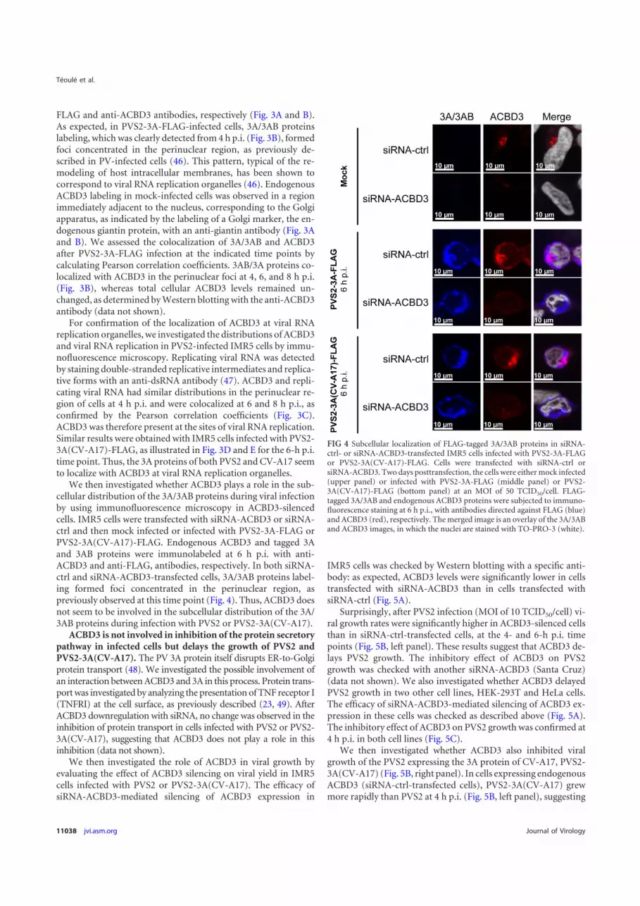
FLAG and anti-ACBD3 antibodies, respectively (Fig. 3A and B).As expected, in PVS2-3A-FLAG-infected cells, 3A/3AB proteinslabeling, which was clearly detected from 4 h p.i. (Fig. 3B), formedfoci concentrated in the perinuclear region, as previously de-scribed in PV-infected cells (46). This pattern, typical of the re-modeling of host intracellular membranes, has been shown tocorrespond to viral RNA replication organelles (46). EndogenousACBD3 labeling in mock-infected cells was observed in a regionimmediately adjacent to the nucleus, corresponding to the Golgiapparatus, as indicated by the labeling of a Golgi marker, the en-dogenous giantin protein, with an anti-giantin antibody (Fig. 3Aand B). We assessed the colocalization of 3A/3AB and ACBD3after PVS2-3A-FLAG infection at the indicated time points bycalculating Pearson correlation coefficients. 3AB/3A proteins co-localized with ACBD3 in the perinuclear foci at 4, 6, and 8 h p.i.(Fig. 3B), whereas total cellular ACBD3 levels remained un-changed, as determined by Western blotting with the anti-ACBD3antibody (data not shown).
For confirmation of the localization of ACBD3 at viral RNAreplication organelles, we investigated the distributions of ACBD3and viral RNA replication in PVS2-infected IMR5 cells by immu-nofluorescence microscopy. Replicating viral RNA was detectedby staining double-stranded replicative intermediates and replica-tive forms with an anti-dsRNA antibody (47). ACBD3 and repli-cating viral RNA had similar distributions in the perinuclear re-gion of cells at 4 h p.i. and were colocalized at 6 and 8 h p.i., asconfirmed by the Pearson correlation coefficients (Fig. 3C).ACBD3 was therefore present at the sites of viral RNA replication.Similar results were obtained with IMR5 cells infected with PVS2-3A(CV-A17)-FLAG, as illustrated in Fig. 3D and E for the 6-h p.i.time point. Thus, the 3A proteins of both PVS2 and CV-A17 seemto localize with ACBD3 at viral RNA replication organelles.
We then investigated whether ACBD3 plays a role in the sub-cellular distribution of the 3A/3AB proteins during viral infectionby using immunofluorescence microscopy in ACBD3-silencedcells. IMR5 cells were transfected with siRNA-ACBD3 or siRNA-ctrl and then mock infected or infected with PVS2-3A-FLAG orPVS2-3A(CV-A17)-FLAG. Endogenous ACBD3 and tagged 3Aand 3AB proteins were immunolabeled at 6 h p.i. with anti-ACBD3 and anti-FLAG, antibodies, respectively. In both siRNA-ctrl and siRNA-ACBD3-transfected cells, 3A/3AB proteins label-ing formed foci concentrated in the perinuclear region, aspreviously observed at this time point (Fig. 4). Thus, ACBD3 doesnot seem to be involved in the subcellular distribution of the 3A/3AB proteins during infection with PVS2 or PVS2-3A(CV-A17).
ACBD3 is not involved in inhibition of the protein secretorypathway in infected cells but delays the growth of PVS2 andPVS2-3A(CV-A17). The PV 3A protein itself disrupts ER-to-Golgiprotein transport (48). We investigated the possible involvement ofan interaction between ACBD3 and 3A in this process. Protein trans-port was investigated by analyzing the presentation of TNF receptor I(TNFRI) at the cell surface, as previously described (23, 49). AfterACBD3 downregulation with siRNA, no change was observed in theinhibition of protein transport in cells infected with PVS2 or PVS2-3A(CV-A17), suggesting that ACBD3 does not play a role in thisinhibition (data not shown).
We then investigated the role of ACBD3 in viral growth byevaluating the effect of ACBD3 silencing on viral yield in IMR5cells infected with PVS2 or PVS2-3A(CV-A17). The efficacy ofsiRNA-ACBD3-mediated silencing of ACBD3 expression in
IMR5 cells was checked by Western blotting with a specific anti-body: as expected, ACBD3 levels were significantly lower in cellstransfected with siRNA-ACBD3 than in cells transfected withsiRNA-ctrl (Fig. 5A).
Surprisingly, after PVS2 infection (MOI of 10 TCID50/cell) vi-ral growth rates were significantly higher in ACBD3-silenced cellsthan in siRNA-ctrl-transfected cells, at the 4- and 6-h p.i. timepoints (Fig. 5B, left panel). These results suggest that ACBD3 de-lays PVS2 growth. The inhibitory effect of ACBD3 on PVS2growth was checked with another siRNA-ACBD3 (Santa Cruz)(data not shown). We also investigated whether ACBD3 delayedPVS2 growth in two other cell lines, HEK-293T and HeLa cells.The efficacy of siRNA-ACBD3-mediated silencing of ACBD3 ex-pression in these cells was checked as described above (Fig. 5A).The inhibitory effect of ACBD3 on PVS2 growth was confirmed at4 h p.i. in both cell lines (Fig. 5C).
We then investigated whether ACBD3 also inhibited viralgrowth of the PVS2 expressing the 3A protein of CV-A17, PVS2-3A(CV-A17) (Fig. 5B, right panel). In cells expressing endogenousACBD3 (siRNA-ctrl-transfected cells), PVS2-3A(CV-A17) grewmore rapidly than PVS2 at 4 h p.i. (Fig. 5B, left panel), suggesting
FIG 4 Subcellular localization of FLAG-tagged 3A/3AB proteins in siRNA-ctrl- or siRNA-ACBD3-transfected IMR5 cells infected with PVS2-3A-FLAGor PVS2-3A(CV-A17)-FLAG. Cells were transfected with siRNA-ctrl orsiRNA-ACBD3. Two days posttransfection, the cells were either mock infected(upper panel) or infected with PVS2-3A-FLAG (middle panel) or PVS2-3A(CV-A17)-FLAG (bottom panel) at an MOI of 50 TCID50/cell. FLAG-tagged 3A/3AB and endogenous ACBD3 proteins were subjected to immuno-fluorescence staining at 6 h p.i., with antibodies directed against FLAG (blue)and ACBD3 (red), respectively. The merged image is an overlay of the 3A/3ABand ACBD3 images, in which the nuclei are stained with TO-PRO-3 (white).
Téoulé et al.
11038 jvi.asm.org Journal of Virology

FIG 5 ACBD3 silencing enhances PVS2 growth in IMR5, HEK-293T and HeLa cells. (A) Silencing of cellular ACBD3. IMR5 (left panel), HEK-293T (middlepanel), or HeLa (right panel) cells were transfected with 10 nM control siRNA (siRNA-ctrl) or siRNA-ACBD3. Two days after transfection with siRNA, cell lysateswere collected and ACBD3 was detected by Western blotting with an anti-ACBD3 antibody. Actin was used as a protein-loading control. (B) Effect of ACBD3silencing on PVS2 and PVS2-3A(CV-A17) growth in IMR5 cells infected at an MOI of 10 TCID50/cell. Cells were transfected with siRNA-ctrl or siRNA-ACBD3.Two days posttransfection, the cells were infected with PVS2 (left) or PVS2-3A(CV-A17) (right). (C) Effect of ACBD3 silencing on PVS2 growth in HEK-293Tor HeLa cells. Cells were transfected with siRNA-ctrl or siRNA-ACBD3. Two days posttransfection, HEK-293T cells (left panel) or HeLa cells (right panel) wereinfected with PVS2 at an MOI of 10 TCID50/cell. (D) Effect of ACBD3 silencing on PVS2 and PVS2-3A(CV-A17) growth in IMR5 cells infected at an MOI of 0.1TCID50/cell. Cells were transfected with siRNA-ctrl or siRNA-ACBD3. Two days posttransfection, cells were infected with PVS2 (left) or PVS2-3A(CV-A17)(right). (E and F) Effect of ACBD3 overexpression on PVS2 and PVS2-3A(CV-A17) growth in HEK-293T cells. Cells were transfected with 200 ng per well of avector encoding ACBD3-3$FLAG or 3$FLAG alone. Two days after transfection: (E) cell lysates were collected and ACBD3 was detected by Western blottingwith an anti-ACBD3 antibody (actin was used as a protein-loading control) or (F) cells were infected with PVS2 (left panel) or PVS2-3A(CV-A17) (right panel)at an MOI of 10 TCID50/cell. Each point represents the mean total virus titer for three independent experiments; error bars indicate standard deviations. *, P )0.05 in Student t tests comparing ACBD3-overexpressing cells with mock-transfected cells.
ACBD3 Modulates Poliovirus Replication
October 2013 Volume 87 Number 20 jvi.asm.org 11039

that the 3A protein of CV-A17 provides some advantage in termsof viral growth. Furthermore, in ACBD3-silenced cells, the growthrate of PVS2-3A(CV-A17) was slightly higher than that in siRNA-ctrl-transfected cells at the 4-h p.i. time point, but no differencewas observed at later time points (Fig. 5B, right panel). Thus, theinhibition of viral growth by endogenous ACBD3 seems to beweaker for PVS2-3A(CV-A17) than for PVS2, with the differencein viral growth abolished by the silencing of endogenous ACBD3expression. These results suggest that viral growth depends on thenonstructural 3A protein expressed, which determines the sensi-tivity of viral growth to the inhibitory effect of ACBD3. The dif-ferential effects of endogenous ACBD3 on the growth of PVS2 andPVS2-3A(CV-A17) were also observed in cells infected at a lowerMOI (0.1 TCID50/cell) (Fig. 5D).
We then investigated whether the inhibition of viral growth byACBD3 was enhanced in cells overexpressing ACBD3. We trans-fected HEK-293T cells (which are highly transfectable) with 200ng/well of an expression vector encoding a triple peptide FLAG(3$FLAG) alone (pCI-neo-3$FLAG) or fused to ACBD3(pACBD3-3$FLAG). The overexpression of ACBD3 in HEK-293T cells was checked by Western blotting with an anti-ACBD3antibody (Fig. 5E). As expected, after infection with PVS2 or PVS2-3A(CV-A17), viral growth rates were significantly lower at 4 and 6 hp.i. in ACBD3-overexpressing cells than in pCI-neo-3$FLAG-trans-fected cells (Fig. 5F). These results support the conclusion thatACBD3 inhibits the growth of PVS2 and PVS2-3A(CV-A17).
The amino acid at position 12 in the 3A protein can modulatethe sensitivity of PVS2 replication to ACBD3. The 3A proteins ofPVS2 and CV-A17 differ by only three amino acids, at positions 12(Val¡Ile), 15 (Ser¡Thr) and 86 (His¡Gln) (Fig. 6). The aminoacid difference at position 12 is the only one that results in theacquisition in the recombinant PVS2-3A(CV-A17) of a residue(Ile) present in the other Sabin strains (types 1 and 3) and refer-ence wild-type PVs 1, 2, and 3 (50–53). Furthermore, most of thePVS2 cVDPVs responsible for poliomyelitis outbreaks in Mada-gascar and cocirculating field CV-A17 strains had an Ile residue inthis position of their 3A proteins (5, 8). We therefore investigatedwhether the inhibitory effect of ACBD3 on viral growth involvedthe replication step in our model and whether this effect could bemodulated by the amino acid in position 12 of the 3A protein. Weconstructed luciferase-encoding replicons, Luc-PVS2 and Luc-PVS2-3A(CV-A17), in which the PVS2 capsid genes were replacedwith the luciferase gene in the full-length PVS2 and PVS2-3A(CV-A17) genomes, respectively. We also constructed a Luc-PVS2 rep-licon in which the Val residue in position 12 of the 3A protein wasreplaced with an Ile residue, Luc-PVS2-3A(V12I). To transfect
replicons, HEK-293T cells were used, because these cells are par-ticularly permissive to RNA transfection. HEK-293T cells treatedwith siRNA targeting ACBD3 or with the control siRNA weretransfected with each of the three luciferase-expressing replicons(Fig. 7A). In cells transfected with siRNA-ctrl, the replication lev-els of Luc-PVS2-3A(CV-A17) and Luc-PVS2-3A(V12I) were sim-ilar and higher than that of Luc-PVS2. In ACBD3-silenced cells,the three replicons had similar replication levels, higher than thosein mock-silenced cells. Similar experiments were performed inIMR5 and HeLa cells (Fig. 7B and C). As expected, the level of viralreplication was low in these cells, but confirmed the results ob-tained in HEK-293T cells.
Thus, ACBD3 inhibited the replication of both Luc-PVS2 andLuc-PVS2-3A(CV-A17). This inhibitory effect was less pro-nounced with the Luc-PVS2 replicon containing the 3A sequencefrom CV-A17 than with that containing the 3A sequence fromPVS2. Our results also suggest that the Ile residue in position 12 ofthe 3A(CV-A17) protein is associated with a lower sensitivity ofPVS2-3A(CV-A17) replication to ACBD3.
Interestingly, the residue in position 12 of the 3A sequencewas previously found to be located in the N-terminal region of3A interacting with another cellular factor important for PVreplication, GBF1 (54). We investigated whether the potentialadvantage of the PVS2-3A(CV-A17) over PVS2 resulted from amore favorable interaction of GBF1 with 3A(CV-A17) than with3A(PVS2). HEK-293T cells were transfected with expression vectorsencoding GST alone or GST fused to the 3A protein from PVS2,CV-A17, or a 3A(PVS2) protein in which the Val residue in position12 was replaced with an Ile residue [p3A(PVS2-V12I)-GST]. GBF1was copurified similarly with 3A-GST from PVS2 and CV-A17 andwith 3A(PVS2-V12I) (Fig. 8). Thus, the Ile residue in position 12 of3A from CV-A17 does not seem to modify the interaction between3A and GBF1. These results suggest that this amino acid can modu-late the sensitivity of PVS2 replication to ACBD3 without altering thebinding of 3A to GBF1.
ACBD3 does not seem to be involved in the increase inPI4KIII" recruitment to replication sites in PVS2- and PVS2-3A(CV-A17)-infected cells. In the AiV model, a complex of 3Aand ACBD3 is involved in the recruitment of PI4KIII! to mem-branous organelles to promote genome replication (30). Wetherefore investigated, by immunofluorescence microscopy,whether ACBD3 downregulated PV replication in our model byaltering the recruitment of PI4KIII! to the virus-induced fociconcentrated in the perinuclear region shown in Fig. 3B.
IMR5 cells were transfected with siRNA-ACBD3 or siRNA-ctrland then mock infected or infected with PVS2 or PVS2-3A(CA-
FIG 6 Amino acid sequence alignment of 3A proteins from enteroviruses. The sequences of 3A proteins from poliovirus vaccine strains (Sabin 1, 2, and 3),wild-type poliovirus (Mahoney, Lansing, and Leon strains), PVS2-derived cVDPVs responsible for poliomyelitis outbreaks in Madagascar in 2002 (MAD004 andMAD0029 strains), and a representative cocirculating field CV-A17 strain, shown in an alignment generated with CLC Main Workbench software v.6.0.1. TheGenBank accession numbers for each sequence are shown in brackets.
Téoulé et al.
11040 jvi.asm.org Journal of Virology

17). Endogenous ACBD3 and PI4KIII! proteins were immunola-beled at the time points indicated, with anti-ACBD3 and anti-PI4KIII! antibodies, respectively. In mock-infected cells transfectedwith siRNA-ctrl, the endogenous ACBD3 and PI4KIII! proteins co-localized in a region corresponding to the Golgi apparatus (Fig. 9). InsiRNA-ctrl-transfected cells infected with PVS2 or PVS2-3A(CA-17),PI4KIII! colocalized with ACBD3 at 6 h p.i. (Fig. 9), in foci concen-trated in the perinuclear region corresponding to the replication sitespreviously described (Fig. 3B). This colocalization was confirmed by
calculating the Pearson correlation coefficient. The silencing ofACBD3 in mock-infected cells or cells infected with PVS2 or PVS2-3A(CV-A17) had no effect on the subcellular distribution ofPI4KIII!. Thus, the downregulation of PV replication by ACBD3does not seem to involve a change in the recruitment of PI4KIII! toreplication sites in our model.
The residue in position 12 of 3A maps outside of the regionrequired for ACBD3 binding. We investigated whether theamino acid in position 12 of 3A was located in the portion of 3A
FIG 7 Effect of ACBD3 silencing on Luc-PVS2, Luc-PVS2-3A(CV-A17) and Luc-PVS2-3A(V12I) replication in HEK-293T (A), IMR5 (B), and HeLa (C) cells.Cells were transfected with siRNA-ctrl (continuous lines) or siRNA-ACBD3 (broken lines). Two days after siRNA transfection, cells were transfected withLuc-PVS2, Luc-PVS2-3A(CV-A17), or Luc-PVS2-3A(V12I) RNA, as indicated. The luciferase activity (RLU) was measured at the indicated time posttransfection(p.t.) with luciferase-encoding replicons. The data shown are the means from three independent experiments. Error bars indicate standard deviations.
ACBD3 Modulates Poliovirus Replication
October 2013 Volume 87 Number 20 jvi.asm.org 11041

mediating binding to ACBD3, by generating a series of 3A pro-teins with N- and C-terminal truncations (3A &), named accord-ing to the amino acids deleted (Fig. 10A). These truncated 3Aproteins were fused to GST, and their ability to interact with en-dogenous ACBD3 was assessed in GST pulldown assays, as describedabove. GST alone and full-length 3A fused to GST were used as neg-ative and positive controls, respectively. The deleted proteins 3A(&1-18) and 3A(&59-87) interacted with ACBD3, whereas 3A proteinswith more extensive N- or C-terminal deletions did not (Fig. 10B).These results suggest that residues 19 to 58, corresponding to theunfolded region and part of the alpha helix of the structured region ofthe 3A protein, include the region required for ACBD3 binding. Thisresult is consistent with the findings reported by Greninger et al. (55)while the present study was under review.
The residue in position 12 is located close to the region re-quired for ACBD3 binding. We therefore assessed whether theV12I substitution in 3A affected the interaction with ACBD3 in aGST pulldown assay on HEK-293T cells transfected with expres-sion vectors encoding GST alone or GST fused to 3A(PVS2),3A(CV-A17), or 3A(PVS2-V12I). ACBD3 copurified similarlywith all three forms of 3A-GST (Fig. 10C). Thus, the residue in
FIG 8 GST pulldown assay on lysates from HEK-293T cells transfected withexpression vectors encoding 3A(PVS2)-GST, 3A(CV-A17)-GST, 3A(PVS2-V12I)-GST, or GST alone. Two days posttransfection, the cells were collectedand subjected to GST pulldown analysis. Endogenous GBF1 and GST weredetected by Western blotting with antibodies directed against GBF1 and GST,respectively (the order of the samples on the gel has been modified in thisfigure).
FIG 9 Subcellular localization of PI4KIII! in siRNA-ctrl or siRNA-ACBD3-transfected IMR5 cells infected with PVS2 or PVS2-3A(CV-A17). Cells weretransfected with siRNA-ctrl or siRNA-ACBD3. Two days after transfection, cells were mock infected (upper panel) or infected with PVS2 (middle panel)or PVS2-3A(CV-A17) (bottom panel) at an MOI of 50 TCID50/cell. Six hours after infection, endogenous ACBD3 and PI4KIII! proteins were subjectedto immunofluorescence staining with antibodies directed against ACBD3 (red) and PI4KIII! (green), respectively. The merge image is an overlay of theACBD3 and the PI4KIII! images, in which the nuclei are stained with DAPI (white). Pearson correlation coefficients (r) were calculated to assess thedegree of colocalization of ACBD3 with PI4KIII!. *, Pearson correlation coefficient values of !0.5 are considered to indicate a significant positivecorrelation.
Téoulé et al.
11042 jvi.asm.org Journal of Virology

position 12 associated with the sensitivity to ACBD3 of PVS2 rep-lication seems to be located outside the region required forACBD3 binding, and the V12I substitution does not seem to affectthe interaction with ACBD3.
DISCUSSIONIn most of the genomes of the PVS2-derived cVDPVs responsiblefor polio outbreaks in Madagascar, the region encoding nonstruc-tural protein 3A is related to that of field CV-A17 strains (5, 8). Weinvestigated the repercussions of this exchange by assessing therole of the 3A proteins of PVS2 and CV-A17 and their putativecellular partners in viral replication. We showed that the 3A pro-teins of both PVS2 and CV-A17 interacted with a Golgi protein,
ACBD3. These findings are consistent with reports, publishedwhile the present study was in progress, showing that ACBD3binds 3A from AiV and other picornaviruses, including PV, hu-man rhinovirus 14, coxsackieviruses B2, B3, and B5, and bovinekobuvirus (30, 32). The 3A proteins of some other picornaviruses(cardioviruses and enterovirus 71) do not appear to associate withACBD3 (32).
In neuroblastoma IMR5 cells infected with either PVS2 or arecombinant PVS2 encoding a 3A protein from CV-A17, weshowed that ACBD3 colocalized with the 3A protein and withreplicating viral RNAs. The 3A protein also inhibits cellular pro-tein transport in PV-infected cells (48). We therefore investigatedthe possible role of an interaction between ACBD3 and 3A in this
FIG 10 Mapping of the ACBD3 binding site on 3A(PVS2). (A) Schematic representation of the 3A(PVS2) proteins with N-terminal and C-terminal truncations.Gray bars represent each of the truncated 3A(PVS2) proteins, with the deleted amino acid positions indicated. Truncated 3A(PVS2) proteins were namedaccording to the amino acid sequences deleted. The complete amino acid sequence of PVS2 3A is shown (dark bar) with the Val residue in position 12 indicatedwith a star. The conserved hydrophobic domain and structured region are indicated. The ACBD3-3A binding data are summarized on the right. The dotted linesindicate the 3A protein region containing the amino acids required for ACBD3 binding. (B) Analysis of interaction between ACBD3 and C-terminally (left panel)or N-terminally (right panel) truncated 3A(PVS2) proteins in GST pulldown assays. HEK-293T cells were transfected with expression vectors encoding truncated3A(PVS2) proteins fused to GST or with GST alone. Two days posttransfection, the cell lysates were subjected to GST pulldown assays. Endogenous ACBD3 andGST proteins were detected by Western blotting with antibodies directed against ACBD3 and GST, respectively. (C) GST pulldown assay on lysates fromHEK-293T cells transfected with expression vectors encoding 3A(PVS2)-GST, 3A(CV-A17)-GST, 3A(PVS2-V12I)-GST, or GST alone. Two days posttransfec-tion, the cells were collected and subjected to GST pulldown analysis. Endogenous ACBD3 and GST were detected by Western blotting with antibodies directedagainst ACBD3 and GST, respectively.
ACBD3 Modulates Poliovirus Replication
October 2013 Volume 87 Number 20 jvi.asm.org 11043
process. The downregulation of ACBD3 levels with siRNA did notseem to affect the inhibition of protein transport in cells infectedwith PVS2 or PVS2-3A(CV-A17), suggesting that ACBD3 is notinvolved in this inhibition (data not shown). However, ACBD3downregulation led to a significant increase in viral growth forPVS2 and PVS2-3A(CV-A17) at early time points after infection.Similar results were obtained with replicons derived from PVS2and PVS2-3A(CV-A17), in ACBD3-silenced cells. Our results,therefore, strongly suggest that ACBD3 delayed viral growth at thelevel of replication in our model. Furthermore, this inhibitoryeffect of ACBD3 on viral replication does not seem to depend onthe cell model because we obtained similar results in several celllines.
Interestingly, ACBD3 inhibited viral replication more stronglywith PVS2 than with PVS2-3A(CV-A17). Furthermore, the aminoacid in position 12 of 3A appeared to be involved in modulatingthe sensitivity of viral replication to ACBD3, although it does notseem to be located in the region required for ACBD3 binding (55;the present study). Thus, our data suggest that the residue in po-sition 12 of 3A plays no role in binding to ACBD3, but it couldinfluence the functional outcome of this interaction. Indeed, thisresidue is located in the N-terminal region of 3A interacting withGBF1, another cellular factor important for replication (54).However, replacement of the Val residue in position 12 of the 3Afrom PVS2 with an Ile residue, as in the 3A from CV-A17, did notmodify the binding of 3A to GBF1. Thus, the potential advantageof PVS2-3A(CV-A17) over PVS2 does not seem to result from amore favorable interaction of GBF1 with 3A(CV-A17) than with3A(PVS2).
In the AiV model, Sasaki et al. (30) showed that ACBD3 inter-acts with several nonstructural viral proteins, including 3A, andrecruits PI4KIII! to sites of viral replication. Using AiV replicons,these authors showed that the silencing of ACBD3 inhibits AiVRNA replication, indicating that ACBD3 plays an important rolein viral replication. Greninger et al. (32) obtained similar resultsbut, surprisingly, they also found that ACBD3 silencing preventedthe replication of PV replicons. Finally, in the CV-B3 model, viralreplication seems to be stimulated, rather than inhibited, uponACDB3 knockdown (Frank van Kuppeveld, personal communi-cation). All of these results, together with our own, suggest that therole of ACBD3 in viral replication may depend on the viral speciesand strain considered.
It has recently been shown that the ACBD3-binding region ofAiV 3A involves multiple portions of this protein, whereas it ap-pears to be well delimited in the poliovirus 3A protein (55). Thus,differences between the ACBD3-binding sites of 3A may reflectdifferences in the role of ACBD3 in replication between picorna-viruses.
Other cellular factors, such as ACBD3-interacting factors, mayalso play a role in the differential effects of this cellular protein onpicornavirus replication. Indeed, during the revision of this arti-cle, Greninger et al. (55) identified the putative Rab33 GTPase-activating proteins TBC1D22A and TBC1D22B as new ACBD3-interacting factors. They also showed that TBC1D22A competesdirectly with PI4KIII! for binding to the same site on ACBD3.Furthermore, poliovirus and AiV 3A proteins may differentiallymodulate the interaction of PI4KIII! and TBC1D22A/B withACBD3 (55). In the present study, we found that the downregu-lation of viral replication by ACBD3 in PVS2-infected cells, or, toa lesser extent, in PVS2-3A(CV-A17)-infected cells, did not seem
to be due to a failure of PI4KIII! recruitment to the perinuclearregion corresponding to the replication sites of infected cells. Vander Schaar et al. (56) recently described 3A CV-B3 mutants thatcan bypass host factor PI4KIII!. Thus, other, as-yet-unidentifiedalternative pathways may be involved in the downregulation ofviral replication by ACBD3 in our model.
Another ACBD3-interacting factor, the metal-dependent pro-tein phosphatase 1L (PPM1L), was also recently identified (57).PPM1L was shown to be involved in the regulation of ceramidetrafficking at ER-Golgi membrane contact sites (58). It would beinteresting to assess whether inhibition of viral replication byACBD3 in our model is related to the regulation of ceramide traf-ficking.
As outlined above, the effects of ACBD3 on viral replicationmay differ according to picornavirus strains. A single cellular pro-tein may thus help the virus by promoting its replication or, con-versely, defend the cell against the virus by delaying its replication.As mentioned above, a recently identified host factor involved inPV replication, VCP/p97, has been shown to interact with thenonstructural viral proteins 3AB and 2BC (33). Interestingly, asfor ACBD3, the effect of VCP/p97 downregulation on viral repli-cation differs between picornaviruses, with strong suppression forPV, an apparent lack of effect for CV-B3, and enhancement forAiV. The authors of that study suggested that alternative or oppo-site pathways might be involved in the replication of these virusesor that a difference in host factor levels might be required for theoptimal replication of these viruses.
In conclusion, we have shown that a recombinant PVS2 encod-ing a 3A protein from CV-A17 is more resistant to the inhibitoryeffect of ACBD3 than the parental vaccine PVS2 strain. The mech-anism by which ACBD3 attenuates these viruses remains to beclarified, but our results suggest that exchanges of nonstructuralproteins can modify the relationships between enterovirus recom-binants and cellular interactors and may thus be one of the factorsfavoring their emergence.
ACKNOWLEDGMENTSWe thank Florence Colbère-Garapin for invaluable support throughoutthis study and for fruitful discussions. We also thank Pierre Charneau,who provided the HEK-293T-T7 cells, and Emmanuelle Perret and AnneDanckaert (Plate-Forme d’Imagerie Dynamique, Institut Pasteur, Paris,France) for assistance with fluorescence microscopy and the Plateformede Génotypage des Pathogènes et Santé Publique (Institut Pasteur, Paris,France) for sequencing. We thank Santos Susin and Victor Yuste (InstitutPasteur, Paris, France) for providing IMR5 cells.
This study was supported by grants from the Institut Pasteur (Trans-verse Research Program PTR 276), the Agence Nationale de la Recherche(ANR-09-MIEN-019), and the Fondation pour la Recherche Médicale(DMI20091117313). F.T. and C.B. were supported by grants from theMinistère de l’Enseignement Supérieur et de la Recherche. S.J. receivedfunding from the French Délégation Générale pour l’Armement and theCentre National pour la Recherche Scientifique. M.C. was supported by astipend from the Pasteur-Paris University International Ph.D. programand by the Institut Carnot Pasteur Maladies Infectieuses.
REFERENCES1. Pallansch M, Roos R. 2007. Enteroviruses: polioviruses, coxsackieviruses,
echoviruses, and newer enteroviruses, p 839 – 893. In Knipe DM, HowleyPM (ed), Fields virology, 4th ed, vol 1. Lippincott/Williams & Wilkins,Philadelphia, PA.
2. Kew OM, Sutter RW, de Gourville EM, Dowdle WR, Pallansch MA.2005. Vaccine-derived polioviruses and the endgame strategy for globalpolio eradication. Annu. Rev. Microbiol. 59:587– 635.
Téoulé et al.
11044 jvi.asm.org Journal of Virology
3. Racaniello VR. 2007. Picornaviridae: the viruses and their replication, p795– 838. In Knipe DM, Howley PM (ed), Fields virology, 4th ed, vol 1.Lippincott/Williams & Wilkins, Philadelphia, PA.
4. Combelas N, Holmblat B, Joffret ML, Colbere-Garapin F, DelpeyrouxF. 2012. Recombination between poliovirus and coxsackie A viruses ofspecies C: a model of viral genetic plasticity and emergence. Viruses3:1460 –1484.
5. Rakoto-Andrianarivelo M, Guillot S, Iber J, Balanant J, Blondel B,Riquet F, Martin J, Kew O, Randriamanalina B, Razafinimpiasa L,Rousset D, Delpeyroux F. 2007. Co-circulation and evolution of polio-viruses and species C enteroviruses in a district of Madagascar. PLoS Pat-hog. 3:e191. doi:10.1371/journal.ppat.0030191.
6. Rakoto-Andrianarivelo M, Gumede N, Jegouic S, Balanant J, Andria-mamonjy SN, Rabemanantsoa S, Birmingham M, Randriamanalina B,Nkolomoni L, Venter M, Schoub BD, Delpeyroux F, Reynes JM. 2008.Reemergence of recombinant vaccine-derived poliovirus outbreak inMadagascar. J. Infect. Dis. 197:1427–1435.
7. Rousset D, Rakoto-Andrianarivelo M, Razafindratsimandresy R, Ran-driamanalina B, Guillot S, Balanant J, Mauclere P, Delpeyroux F. 2003.Recombinant vaccine-derived poliovirus in Madagascar. Emerg. Infect.Dis. 9:885– 887.
8. Joffret ML, Jegouic S, Bessaud M, Balanant J, Tran C, Caro V, HolmblatB, Razafindratsimandresy R, Reynes JM, Rakoto-Andrianarivelo M,Delpeyroux F. 2012. Common and diverse features of cocirculating type2 and 3 recombinant vaccine-derived polioviruses isolated from patientswith poliomyelitis and healthy children. J. Infect. Dis. 205:1363–1373.
9. Bienz K, Egger D, Pfister T, Troxler M. 1992. Structural and functionalcharacterization of the poliovirus replication complex. J. Virol. 66:2740 –2747.
10. Bienz K, Egger D, Pfister T. 1994. Characteristics of the poliovirusreplication complex. Arch. Virol. 1994(Suppl 9):147–157.
11. Cho MW, Teterina N, Egger D, Bienz K, Ehrenfeld E. 1994. Membranerearrangement and vesicle induction by recombinant poliovirus 2C and2BC in human cells. Virology 202:129 –145.
12. Suhy DA, Giddings TH, Jr, Kirkegaard K. 2000. Remodeling the endo-plasmic reticulum by poliovirus infection and by individual viral proteins:an autophagy-like origin for virus-induced vesicles. J. Virol. 74:8953–8965.
13. Belov GA, Feng Q, Nikovics K, Jackson CL, Ehrenfeld E. 2008. A criticalrole of a cellular membrane traffic protein in poliovirus RNA replication.PLoS Pathog. 4:e1000216. doi:10.1371/journal.ppat.1000216.
14. Egger D, Teterina N, Ehrenfeld E, Bienz K. 2000. Formation of thepoliovirus replication complex requires coupled viral translation, vesicleproduction, and viral RNA synthesis. J. Virol. 74:6570 – 6580.
15. Kirkegaard K, Semler BL. 2010. Genome replication II: the process, p127–140. In Ehrenfeld E, Domingo E, Ross RP (ed), The picornaviruses.ASM Press, Washington, DC.
16. Wessels E, Duijsings D, Lanke KH, van Dooren SH, Jackson CL,Melchers WJ, van Kuppeveld FJ. 2006. Effects of picornavirus 3A pro-teins on protein transport and GBF1-dependent COP-I recruitment. J.Virol. 80:11852–11860.
17. Wessels E, Duijsings D, Niu TK, Neumann S, Oorschot VM, de LangeF, Lanke KH, Klumperman J, Henke A, Jackson CL, Melchers WJ, vanKuppeveld FJ. 2006. A viral protein that blocks Arf1-mediated COP-Iassembly by inhibiting the guanine nucleotide exchange factor GBF1. Dev.Cell 11:191–201.
18. Bui QT, Golinelli-Cohen MP, Jackson CL. 2009. Large Arf1 guaninenucleotide exchange factors: evolution, domain structure, and roles inmembrane trafficking and human disease. Mol. Genet. Genomics 282:329 –350.
19. Casanova JE. 2007. Regulation of Arf activation: the Sec7 family of gua-nine nucleotide exchange factors. Traffic 8:1476 –1485.
20. Kawamoto K, Yoshida Y, Tamaki H, Torii S, Shinotsuka C, YamashinaS, Nakayama K. 2002. GBF1, a guanine nucleotide exchange factor forADP-ribosylation factors, is localized to the cis-Golgi and involved inmembrane association of the COPI coat. Traffic 3:483– 495.
21. Zhao X, Claude A, Chun J, Shields DJ, Presley JF, Melancon P. 2006.GBF1, a cis-Golgi and VTCs-localized ARF-GEF, is implicated in ER-to-Golgi protein traffic. J. Cell Sci. 119:3743–3753.
22. Lanke KH, van der Schaar HM, Belov GA, Feng Q, Duijsings D, JacksonCL, Ehrenfeld E, van Kuppeveld FJ. 2009. GBF1, a guanine nucleotideexchange factor for Arf, is crucial for coxsackievirus B3 RNA replication. J.Virol. 83:11940 –11949.
23. Kondratova AA, Neznanov N, Kondratov RV, Gudkov AV. 2005. Po-liovirus protein 3A binds and inactivates LIS1, causing block of membraneprotein trafficking and deregulation of cell division. Cell Cycle 4:1403–1410.
24. Faulkner NE, Dujardin DL, Tai CY, Vaughan KT, O’Connell CB, WangY, Vallee RB. 2000. A role for the lissencephaly gene LIS1 in mitosis andcytoplasmic dynein function. Nat. Cell Biol. 2:784 –791.
25. Reiner O, Carrozzo R, Shen Y, Wehnert M, Faustinella F, Dobyns WB,Caskey CT, Ledbetter DH. 1993. Isolation of a Miller-Dieker lissenceph-aly gene containing G protein beta-subunit-like repeats. Nature 364:717–721.
26. Smith DS, Niethammer M, Ayala R, Zhou Y, Gambello MJ, Wynshaw-Boris A, Tsai LH. 2000. Regulation of cytoplasmic dynein behaviour andmicrotubule organization by mammalian Lis1. Nat. Cell Biol. 2:767–775.
27. Delang L, Paeshuyse J, Neyts J. 2012. The role of phosphatidylinositol4-kinases and phosphatidylinositol 4-phosphate during viral replication.Biochem. Pharmacol. 84:1400 –1408.
28. Hsu NY, Ilnytska O, Belov G, Santiana M, Chen YH, Takvorian PM,Pau C, van der Schaar H, Kaushik-Basu N, Balla T, Cameron CE,Ehrenfeld E, van Kuppeveld FJ, Altan-Bonnet N. 2010. Viral reorgani-zation of the secretory pathway generates distinct organelles for RNA rep-lication. Cell 141:799 – 811.
29. Yamashita T, Sakae K, Tsuzuki H, Suzuki Y, Ishikawa N, Takeda N,Miyamura T, Yamazaki S. 1998. Complete nucleotide sequence andgenetic organization of Aichi virus, a distinct member of the Picornaviri-dae associated with acute gastroenteritis in humans. J. Virol. 72:8408 –8412.
30. Sasaki J, Ishikawa K, Arita M, Taniguchi K. 2012. ACBD3-mediatedrecruitment of PI4KB to picornavirus RNA replication sites. EMBO J.31:754 –766.
31. Sohda M, Misumi Y, Yamamoto A, Yano A, Nakamura N, Ikehara Y.2001. Identification and characterization of a novel Golgi protein, GCP60,that interacts with the integral membrane protein giantin. J. Biol. Chem.276:45298 – 45306.
32. Greninger AL, Knudsen GM, Betegon M, Burlingame AL, Derisi JL.2012. The 3A protein from multiple picornaviruses utilizes the Golgiadaptor protein ACBD3 to recruit PI4KIII!. J. Virol. 86:3605–3616.
33. Arita M, Wakita T, Shimizu H. 2012. Valosin-containing protein (VCP/p97) is required for poliovirus replication and is involved in cellular pro-tein secretion pathway in poliovirus infection. J. Virol. 86:5541–5553.
34. Pollard SR, Dunn G, Cammack N, Minor PD, Almond JW. 1989.Nucleotide sequence of a neurovirulent variant of the type 2 oral poliovi-rus vaccine. J. Virol. 63:4949 – 4951.
35. Autret A, Martin-Latil S, Mousson L, Wirotius A, Petit F, Arnoult D,Colbere-Garapin F, Estaquier J, Blondel B. 2007. Poliovirus inducesBax-dependent cell death mediated by c-Jun NH2-terminal kinase. J. Vi-rol. 81:7504 –7516.
36. Reed LJ, Muench M. 1938. A simple method for estimating fifty percentendpoints. Am. J. Hyg. (Lond.) 1938:493– 497.
37. Pellet J, Tafforeau L, Lucas-Hourani M, Navratil V, Meyniel L, AchazG, Guironnet-Paquet A, Aublin-Gex A, Caignard G, Cassonnet P,Chaboud A, Chantier T, Deloire A, Demeret C, Le Breton M, Neveu G,Jacotot L, Vaglio P, Delmotte S, Gautier C, Combet C, Deleage G, FavreM, Tangy F, Jacob Y, Andre P, Lotteau V, Rabourdin-Combe C,Vidalain PO. 2010. ViralORFeome: an integrated database to generate aversatile collection of viral ORFs. Nucleic Acids Res. 38:D371–D378.
38. Riquet FB, Blanchard C, Jegouic S, Balanant J, Guillot S, Vibet MA,Rakoto-Andrianarivelo M, Delpeyroux F. 2008. Impact of exogenoussequences on the characteristics of an epidemic type 2 recombinant vac-cine-derived poliovirus. J. Virol. 82:8927– 8932.
39. Jegouic S, Joffret ML, Blanchard C, Riquet FB, Perret C, Pelletier I,Colbere-Garapin F, Rakoto-Andrianarivelo M, Delpeyroux F. 2009.Recombination between polioviruses and co-circulating coxsackie A vi-ruses: role in the emergence of pathogenic vaccine-derived polioviruses.PLoS Pathog. 5:e1000412. doi:10.1371/journal.ppat.1000412.
40. Caignard G, Guerbois M, Labernardiere JL, Jacob Y, Jones LM, Wild F,Tangy F, Vidalain PO. 2007. Measles virus V protein blocks Jak1-mediated phosphorylation of STAT1 to escape IFN-alpha/beta signaling.Virology 368:351–362.
41. Bessaud M, Delpeyroux F. 2012. Development of a simple and rapidprotocol for the production of customized intertypic recombinant polio-viruses. J. Virol. Methods 186:104 –108.
ACBD3 Modulates Poliovirus Replication
October 2013 Volume 87 Number 20 jvi.asm.org 11045

42. Bolte S, Cordelieres FP. 2006. A guided tour into subcellular colocaliza-tion analysis in light microscopy. J. Microsc. 224:213–232.
43. Strauss DM, Glustrom LW, Wuttke DS. 2003. Towards an understand-ing of the poliovirus replication complex: the solution structure of thesoluble domain of the poliovirus 3A protein. J. Mol. Biol. 330:225–234.
44. Teterina NL, Pinto Y, Weaver JD, Jensen KS, Ehrenfeld E. 2011.Analysis of poliovirus protein 3A interactions with viral and cellular pro-teins in infected cells. J. Virol. 85:4284 – 4296.
45. Teterina NL, Lauber C, Jensen KS, Levenson EA, Gorbalenya AE,Ehrenfeld E. 2011. Identification of tolerated insertion sites in poliovirusnonstructural proteins. Virology 409:1–11.
46. Bienz K, Egger D, Rasser Y, Bossart W. 1983. Intracellular distributionof poliovirus proteins and the induction of virus-specific cytoplasmicstructures. Virology 131:39 – 48.
47. Jurgeit A, Moese S, Roulin P, Dorsch A, Lotzerich M, Lee WM, GreberUF. 2010. An RNA replication-center assay for high content image-basedquantifications of human rhinovirus and coxsackievirus infections. Virol.J. 7:264.
48. Doedens JR, Kirkegaard K. 1995. Inhibition of cellular protein secretionby poliovirus proteins 2B and 3A. EMBO J. 14:894 –907.
49. Neznanov N, Kondratova A, Chumakov KM, Angres B, ZhumabayevaB, Agol VI, Gudkov AV. 2001. Poliovirus protein 3A inhibits tumornecrosis factor (TNF)-induced apoptosis by eliminating the TNF receptorfrom the cell surface. J. Virol. 75:10409 –10420.
50. Kitamura N, Semler BL, Rothberg PG, Larsen GR, Adler CJ, Dorner AJ,Emini EA, Hanecak R, Lee JJ, van der Werf S, Anderson CW, WimmerE. 1981. Primary structure, gene organization and polypeptide expressionof poliovirus RNA. Nature 291:547–553.
51. La Monica N, Meriam C, Racaniello VR. 1986. Mapping of sequencesrequired for mouse neurovirulence of poliovirus type 2 Lansing. J. Virol.57:515–525.
52. Stanway G, Hughes PJ, Mountford RC, Reeve P, Minor PD, Schild GC,Almond JW. 1984. Comparison of the complete nucleotide sequences ofthe genomes of the neurovirulent poliovirus P3/Leon/37 and its attenu-ated Sabin vaccine derivative P3/Leon 12a1b. Proc. Natl. Acad. Sci. U. S. A.81:1539 –1543.
53. Toyoda H, Kohara M, Kataoka Y, Suganuma T, Omata T, Imura N,Nomoto A. 1984. Complete nucleotide sequences of all three poliovirusserotype genomes. Implication for genetic relationship, gene function andantigenic determinants. J. Mol. Biol. 174:561–585.
54. Wessels E, Duijsings D, Lanke KH, Melchers WJ, Jackson CL, vanKuppeveld FJ. 2007. Molecular determinants of the interaction betweencoxsackievirus protein 3A and guanine nucleotide exchange factor GBF1.J. Virol. 81:5238 –5245.
55. Greninger AL, Knudsen GM, Betegon M, Burlingame AL, DeRisi JL.2013. ACBD3 interaction with TBC1 domain 22 protein is differentiallyaffected by enteroviral and kobuviral 3A protein binding. mBio 4:e00098-00013. doi:10.1128/mBio.00098-13.
56. van der Schaar HM, van der Linden L, Lanke KH, Strating JR,Purstinger G, de Vries E, de Haan CA, Neyts J, van Kuppeveld FJ. 2012.Coxsackievirus mutants that can bypass host factor PI4KIII! and the needfor high levels of PI4P lipids for replication. Cell Res. 22:1576 –1592.
57. Shinoda Y, Fujita K, Saito S, Matsui H, Kanto Y, Nagaura Y, FukunagaK, Tamura S, Kobayashi T. 2012. Acyl-CoA binding domain containing3 (ACBD3) recruits the protein phosphatase PPM1L to ER-Golgi mem-brane contact sites. FEBS Lett. 586:3024 –3029.
58. Saito S, Matsui H, Kawano M, Kumagai K, Tomishige N, Hanada K,Echigo S, Tamura S, Kobayashi T. 2008. Protein phosphatase 2C!is anendoplasmic reticulum integral membrane protein that dephosphorylatesthe ceramide transport protein CERT to enhance its association with or-ganelle membranes. J. Biol. Chem. 283:6584 – 6593.
Téoulé et al.
11046 jvi.asm.org Journal of Virology