a structural perspective on protein–protein interactions
TRANSCRIPT
A structural perspective on protein–protein interactionsRobert B Russell1,2, Frank Alber3, Patrick Aloy1, Fred P Davis3,Dmitry Korkin3, Matthieu Pichaud1, Maya Topf3 and Andrej Sali3�
Structures of macromolecular complexes are necessary for a
mechanistic description of biochemical and cellular processes.
They can be solved by experimental methods, such as X-ray
crystallography, NMR spectroscopy and electron microscopy,
as well as by computational protein structure prediction,
docking and bioinformatics. Recent advances and applications
of these methods emphasize the need for hybrid approaches
that combine a variety of data to achieve better efficiency,
accuracy, resolution and completeness.
Addresses1EMBL, Meyerhofstrasse 1, 69117 Heidelberg, Germany2EMBL-EBI, Wellcome Trust Genome Campus, Hinxton, UK3Departments of Biopharmaceutical Sciences and Pharmaceutical
Chemistry, and California Institute for Quantitative Biomedical Research,
Mission Bay Genentech Hall, Suite N472D, 600 16th Street, University of
California at San Francisco, San Francisco, California 94143-2240, USA�e-mail: [email protected]
Current Opinion in Structural Biology 2004, 14:313–324
This review comes from a themed issue on
Sequences and topology
Edited by Peer Bork and Christine A Orengo
Available online 18th May 2004
0959-440X/$ – see front matter
� 2004 Elsevier Ltd. All rights reserved.
DOI 10.1016/j.sbi.2004.04.006
IntroductionGenome sequencing has provided nearly complete lists of
the macromolecules present in many organisms (e.g.
[1,2]). However, these lists reveal comparatively little
about the function of biological systems because the
functional units of cells are often complex assemblies
of several macromolecules [3]. Such complexes vary
widely in their activity and size [3–7], and play crucial
roles in most cellular processes. They are often depicted
as molecular machines [3], a metaphor that accurately
captures many of their characteristic features, such as
modularity, complexity, cyclic functions and energy con-
sumption [8]. For instance, the nuclear pore complex, a
50–100 MDa protein assembly, regulates and controls the
trafficking of macromolecules through the nuclear envel-
ope [9]; the ribosome is responsible for protein biosynth-
esis; RNA polymerase catalyzes the formation of RNA
[10]; and ATP synthase catalyzes the formation of ATP
[7]. Macromolecular assemblies are also involved in tran-
scription control (e.g. the IFNb enhanceosome) [6,11]
and the regulation of cellular transport (e.g. microtubulins
in complex with the molecular motors myosin or kinesin)
[12–14], and are crucial components in neuronal signaling
(e.g. the post-synaptic density complexes) [15]. A struc-
tural description of the protein interactions within such
complexes is an important step toward a mechanistic
understanding of biochemical, cellular and higher order
biological processes [16–18,19�].
There are currently about 12 000 known structures, from
a variety of organisms, of assemblies involving two or
more protein chains (http://pqs.ebi.ac.uk/pqs-doc.shtml)
(April 2004) [20]; these complexes can be organized into
about 3500 groups based on sequence similarity [19�].Just how many complexes exist in a particular proteome
is not easy to deduce because of the different component
types (e.g. proteins, nucleic acids, nucleotides, metal
ions) and the varying life span of the complexes (e.g.
transient complexes, such as those involved in signaling,
and stable complexes, such as the ribosome). Until
recently, the most comprehensive information about
protein–protein interactions was available for the Sac-charomyces cerevisiae proteome, consisting of approx-
imately 6200 proteins. This information has been pro-
vided by methods such as the yeast two-hybrid system
and affinity purification followed by mass spectrometry
[21–27,28�,29]. The lower bound on binary protein inter-
actions and functional links in yeast has been estimated
to be in the range of approximately 30 000 [30,31]; this
number corresponds to about nine protein partners per
protein, although not necessarily all direct or interacting at
the same time. The human proteome may have an order
of magnitude more complexes than the yeast cell and the
number of different complexes across all relevant genomes
may be several times larger still. Therefore, there may be
thousands of biologically relevant macromolecular com-
plexes whose structures are yet to be characterized [32].
We review here recent developments in the experimental
and computational techniques that have allowed structural
biology to shift its focus from the structures of individual
proteins to the structures of large assemblies [19�,33,34].
We also illustrate these developments by listing their
application to the determination of the structure of specific
assemblies of biological importance. In contrast to struc-
ture determination of individual proteins, structural char-
acterization of macromolecular assemblies usually poses a
more difficult challenge. We stress that a comprehensive
structural description of large complexes generally requires
the use of several experimental methods, underpinned by a
variety of theoretical approaches to maximize efficiency,
completeness, accuracy and resolution [19�,35].
www.sciencedirect.com Current Opinion in Structural Biology 2004, 14:313–324
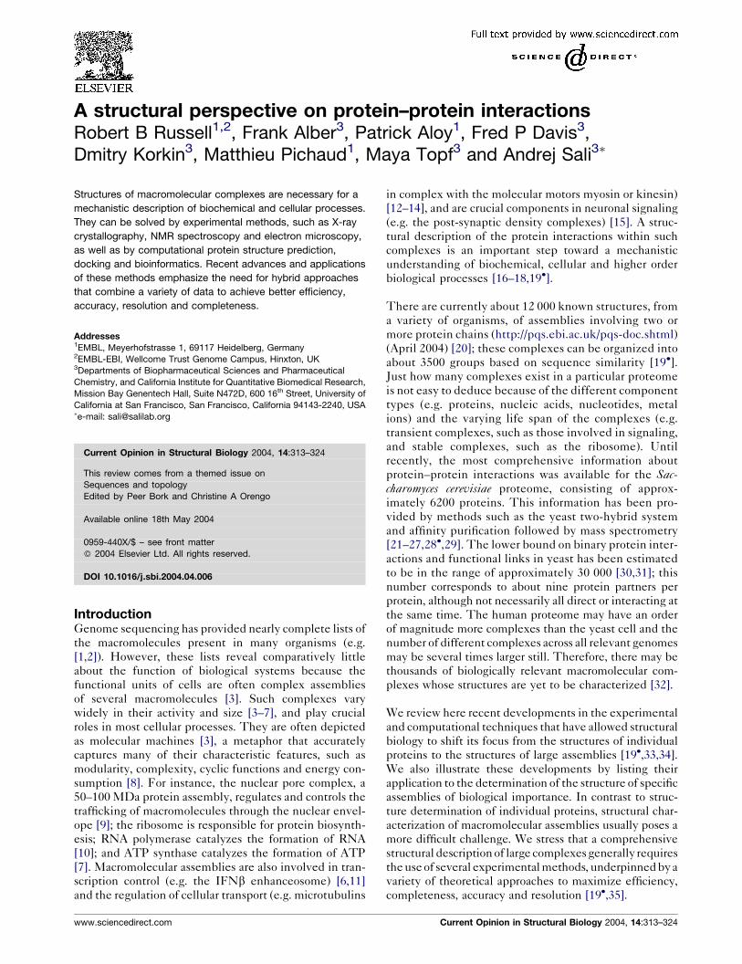
X-ray crystallography and NMRspectroscopyX-ray crystallography has been the most prolific techni-
que for the structural analysis of proteins and protein
complexes, and is still the ‘gold standard’ in terms of
accuracy and resolution (Figure 1a). Structures of several
macromolecular assemblies have recently been solved by
X-ray crystallography: RNA polymerase [36], the riboso-
mal subunits [37–41], the complete ribosome and its
functional complexes [42], the proteasome [43], the
GroEL chaperonin [44], various complexes of the cellular
transport machinery [12,13], the Arp2/3 complex [45],
photosystem I and the light-harvesting complex of photo-
system II [46,47], the SRP (signal recognition particle)
complex involved in nascent protein targeting [48�], and
various viral capsid and virion structures [49–51]. How-
ever, the number of structures of macromolecular assem-
blies solved by X-ray crystallography is still quite small
compared to that of the individual proteins and it will
probably be many years before we have a complete
repertoire of high-resolution structures for the hundreds
of complexes in a typical cell. This discrepancy is due
mainly to the difficult production of sufficient quantities
of the sample and its crystallization.
NMR spectroscopy allows the determination of atomic
structures of ever-larger subunits and even their com-
plexes [52–54]. Increasingly, it is used to identify residues
involved in protein interactions (Figure 1b) [55–58].
Recent technical advances have allowed its application
to systems as large as the 900 kDa GroEL–GroES com-
plex [52]. Also, it was recently used to describe structural
differences between interactions among different LIM
and SH3 domains [59].
Electron microscopy and electrontomographyThere are several variants of electron microscopy (EM),
including single-particle EM (Figure 1c) [60], electron
tomography (Figure 1d) [61] and electron crystallography
of regular two-dimensional arrays of the sample [62].
For particles with molecular weights greater than
200–500 kDa, single-particle cryo-EM can determine the
Figure 1
Methods for the structural characterization of macromolecular assemblies. (a) Electron diffraction map and three-dimensional structure of the
bacterial degradosome component PNPase (polyribonucleotide phosphorylase) determined by X-ray crystallography [183]. X-ray crystallography
integrates the diffraction patterns collected after bombarding a crystallized protein or complex with X-rays to construct its three-dimensional
structure. In principle, there is no size limit on the structures studied using this technique, although it is often difficult to obtain sufficient
material for crystallization. This technique provides atomic-resolution structures and thus molecular details of how the interactions between the
different components occur. (b) Three-dimensional structure and plot showing chemical shifts upon association of the human survival motor
neuron (SMN) tudor domain solved by NMR spectroscopy [184]. NMR spectroscopy extracts distances between atoms by measuring transitions
between different nuclear spin states within a magnetic field. These distances are then used as restraints to build three-dimensional structures.
NMR spectroscopy also provides atomic-resolution structures, but is generally limited to proteins of about 300 residues. It plays an increasingly
important role in studying interaction interfaces between structures determined independently. (c) EM micrograph and three-dimensional
reconstruction of adeno-associated virus type 2 empty capsids [185]. EM is based on the analysis of images of stained particles. Different
views and conformations of the complexes are trapped and thus thousands of images have to be averaged to reconstruct the three-dimensional
structure. Classical implementations were limited to a resolution of 20 A. More recently, single-particle cryo techniques, whereby samples are fast
frozen before study, have reached resolutions as high as approximately 6 A. EM provides information about the overall shape and symmetry of
macromolecules. (d) Slice images and rendered surface of a ribosome-decorated portion of endoplasmic reticulum [73]. In electron tomography,
the specimen studied is progressively tilted upon an axis perpendicular to the electron beam. A set of projection images is then recorded andused to build a three-dimensional model. This technique can tackle large organelles or even complete cells without perturbing their physiological
environment. It provides shape information at resolutions of approximately 30 A and promises to reach higher resolutions soon. (e) Yeast two-hybrid
array screen and small network of interacting proteins [124��,186]. Interaction discovery comprises many different methods whose objective
is to determine spatial proximity between proteins. These include techniques such as the two-hybrid system, affinity purification, FRET,
chemical cross-linking, footprinting and protein arrays. These methods provide very limited structural information and no molecular details.
Their strength is that they often give a quasi-comprehensive list of protein interactions and the networks they form.
314 Sequences and topology
Current Opinion in Structural Biology 2004, 14:313–324 www.sciencedirect.com
electron density of an assembly at resolutions as high as
approximately 5 A [63�,64–66,67�,68�,69,70�]. The full
three-dimensional structure of the particle is recon-
structed from many two-dimensional projections of the
specimen, each showing the object from a different angle.
Imaging by cryo-EM requires neither large quantities of
the sample nor the sample in a crystalline form. There-
fore, single-particle cryo-EM is a powerful tool to inves-
tigate the structure and dynamics of macromolecular
assemblies for which X-ray structure determination is
very difficult. Although it is generally impossible to build
atomic models solely from cryo-EM density maps, the
maps give valuable insights into the structure and
mechanism of large complexes (e.g. [63�]). They are
particularly useful when combined with atomic-resolu-
tion structures of the subunits, as reviewed in the section
on hybrid methods below.
One of the most exciting developments in structural
biology is the new generation of tomography methods
based on multiple tilted views of the same object [33,71].
Although electron tomography can be used to study the
structures of isolated macromolecular assemblies at a
relatively low resolution of a few nanometers, its true
potential lies in visualizing the assemblies in an unper-
turbed cellular context [72]. These data sets provide
fascinating three-dimensional images of entities as large
as a small cell at approximately 5 nm resolution [73]. To
widen the scope of cellular tomography, it is necessary to
improve the resolution of the tomographic images, as
well as identify the structures in these images [73–75].
Theoretical considerations [76] and ongoing improve-
ments in the instrumentation make a resolution as high
as 2 nm a realistic goal [77].
Low-resolution experimental methodsSeveral experimental techniques can provide structural
information about protein interactions at low resolution
(Figure 1e). This information may be used to infer the
configuration of the proteins in a complex. Methods
for the mapping of protein interactions may provide
contact or proximity restraints for pairs of proteins that
are useful in the modeling of higher order complexes.
Such methods include new implementations of the two-
hybrid system [78–81], tagged affinity chromatography
[82�,83] and the combination of phage display with other
techniques [84], such as synthesis of peptides on cellu-
lose membranes (SPOT) [85��]. Because of the low-
resolution nature of these biochemical characterizations,
care is needed in their interpretation. For example,
comparing biochemically derived interaction sets against
known three-dimensional structures of complexes
revealed potential sources of systematic errors in inter-
action discovery, such as indirect interactions in two-
hybrid systems, the obstruction of interfaces by mole-
cular labels and artificial promiscuity in the detected
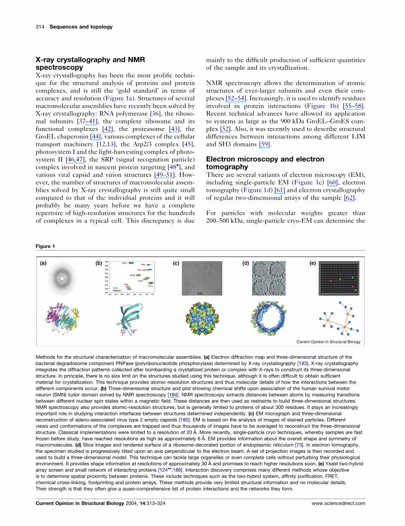
interactions (Figure 2) [86].
Biophysical, biochemical and molecular biology methods
can also be used to derive low-resolution information
about the relative position and orientation of domains
in a larger complex. These methods include: site-directed
mutagenesis, which can identify the residues that med-
iate the interaction [87]; various forms of footprinting,
such as hydrogen-deuterium exchange [88,89�] and
hydroxyl radical footprinting [90], which can identify
surfaces buried upon complex formation; chemical cross-
linking [91–93], which can identify interacting residues;
fluorescence resonance energy transfer (FRET) [94,95],
which can determine the distance between labeled
groups on the interacting proteins; and Fourier transform
IR spectroscopy (FTIR), which describes structural
changes upon complex formation [96]. Small angle
X-ray scattering (SAXS) is another biophysical method
that can provide low-resolution information about the
shape of a complex. For instance, SAXS has recently
been used to study the dynamics of conformational
change in Bruton tyrosine kinase [97,98].
Computational protein–protein dockingWhen atomic structures of the individual proteins
involved in an interaction are known, either by experi-
ment or by modeling, several computational methods are
available that suggest the structure of the interaction [99].
Figure 2
CKS
CDK2
N-t
C-t
N-t
Actin
Profilin
C-t
Current Opinion in Structural Biology
>18 Å
Cyclin A
(a) (b)
Examples of potential errors in biochemical interaction discovery
techniques, as revealed by a structure-based analysis [17]. (a) Indirectinteractions between cyclin-dependent kinase regulatory subunit (CKS)
and cyclin A detected by the yeast two-hybrid system. Several
interactions between CKS domains and cyclins were reported in
genome-scale two-hybrid studies [21,187]. However, analysis of three-
dimensional structures suggests that the endogenous cyclin-dependent
kinase 2 (CDK2) probably mediates the interaction, as combining the
CDK2–CKS and CDK2–cyclin A structures places the CKS and cyclin
domains 18 A apart [86]. (b) An example of an interaction that is not
detected by any screen, possibly because molecular labels (e.g. affinity
purification tags, or two-hybrid DNA binding or activation domains) are
interfering with the interaction. The X-ray structure of the actin–profilin
complex reveals that the actin C terminus (C-t) lies at the interaction
interface (the other N and C termini are also labeled).
Structural perspective on protein–protein interactions Russell et al. 315
www.sciencedirect.com Current Opinion in Structural Biology 2004, 14:313–324
Most of these docking methods aim to predict the atomic
model of a complex by maximizing the shape and che-
mical complementarity between a given pair of interact-
ing proteins [99–102]. Docking strategies usually rely on a
two-stage approach: they first generate a set of possible
orientations of the two docked proteins and then score
them in the hope that the native complex will be ranked
highly. Katchalski-Katzir, Vakser and co-workers [103]
have pioneered a fast Fourier transform (FFT)-based
method for rapidly searching through the space of possi-
ble docked configurations. Due to its computational
efficiency, it has also been incorporated into programs
such as FTDock and 3D-Dock [104–106], GRAMM
[107], DOT [108], ZDOCK [109] and HEX [110]. Other
docking programs include ICM [111] and ROSETTA
[112]. The searches may be restrained by other consid-
erations, such as the known binding site location. These
methods differ in protein representation, in the scoring of
different configurations and in the search for the best
solutions. Some methods boldly model the actual diffu-
sion/collision trajectories involved in the docking process
[113,114].
Although docking methods are not sufficiently accurate to
predict whether or not two proteins actually interact with
each other, they can sometimes correctly identify the
interacting surfaces between two structurally defined
subunits [115]. Docking methods are systematically
assessed through blind trials in the Critical Assessment
of PRedicted Interactions (CAPRI) [101,116�,117]. Pre-
dictions are made just before the structures are solved
experimentally, followed by the assessment of the models
at the CAPRI meetings. The best of the methods
assessed in the last CAPRI experiment correctly pre-
dicted three of the seven target complexes [116�].
Methods that are able to work with comparative protein
structure models [118] instead of experimentally deter-
mined subunit structures would extend the applicability
of docking to many more biological problems, but would
probably have poorer performance. Currently, docking is
often applied in concert with experimental techniques,
including site-directed mutagenesis [119], amide hydro-
gen-deuterium exchange [89�] and NMR spectroscopy
[120�,121], as well as solid-state binding and surface
plasmon resonance [122].
Inferring interactions by homologyProtein interactions can also be modeled by similarity
[123,124��,125]. If a complex of known structure compris-
ing homologs of a pair of interacting proteins is available,
it is usually possible to build a model by comparative
modeling [126]. There are now approximately 2000 dis-
tinct interaction types of known structure (i.e. whereby
interacting domains sharing 30% or greater sequence
identity are considered to be a single type; P Aloy, RB
Russell, unpublished).
Building a model of the interaction between a pair of
proteins based on the known structure of the complex
between interacting homologs raises the question of
whether or not homology of the subunits implies sim-
ilarity of interaction. It was found that interactions
between proteins of the same fold tend to be similar
when the sequence identity is above approximately 30%
[127�]. Below this cutoff, there is a twilight zone where
interactions may or may not be similar geometrically.
Given a template, it is possible to model an interaction
using standard comparative modeling techniques [126].
However, frequently there are multiple templates for the
same interaction type. In addition, a single interaction
template can be used to model many putative interactions
in a single organism. Therefore, it is important to assess
the likelihood of these potential interactions, particularly
in the absence of experimental validation [128��]. For
example, each of the dozens of fibroblast growth factors
(FGFs) interacts with one or more of seven receptors
with different affinities [129]. Two approaches have been
developed recently that attempt to predict specificity by
modeling interactions. The first approach, implemented
by InterPReTS [123,130] and ModBase [125], uses
empirical pair potentials derived from interfaces of
known structure to score how well a pair of homologous
proteins fits a known complex structure. The second
approach, MULTIPROSPECTOR, is similar, although
it attempts to study more distantly related protein
sequences by threading sequences onto a library of inter-
acting templates, followed by scoring how well the indi-
vidual sequences fit their proposed folds and the interface
between them [131]. Both approaches have since been
applied to study large collections of sequences and inter-
actions [124��,125,132��].
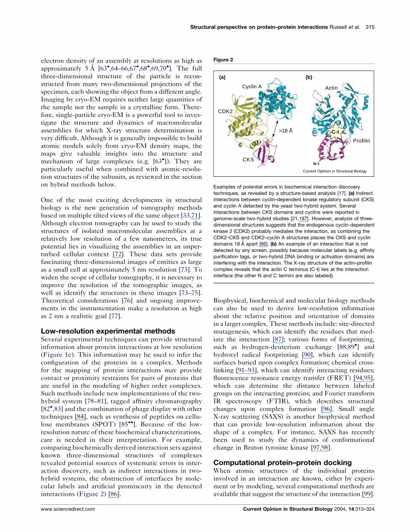
For some large complexes, the specificity of interactions
within a family of homologous subunits is an important
determinant of complex assembly. For instance, the
chaperonin CCT consists of eight homologous subunits
that are all similar to the single subunit comprising the
thermosome [133]. Thus, building CCT using the ther-
mosome requires the conversion of a seven-subunit ring
into an eight-subunit ring, and then choosing the correct
arrangement from the 5040 (8!/8) possibilities. It is pos-
sible to guide this process by experiment, such as the
detection of subcomplexes that reveal preferred interact-
ing pairs [134] or the application of the two-hybrid system
[135]. InterPReTS was also applied to select one of the
120 possible arrangements of six exosome subunits
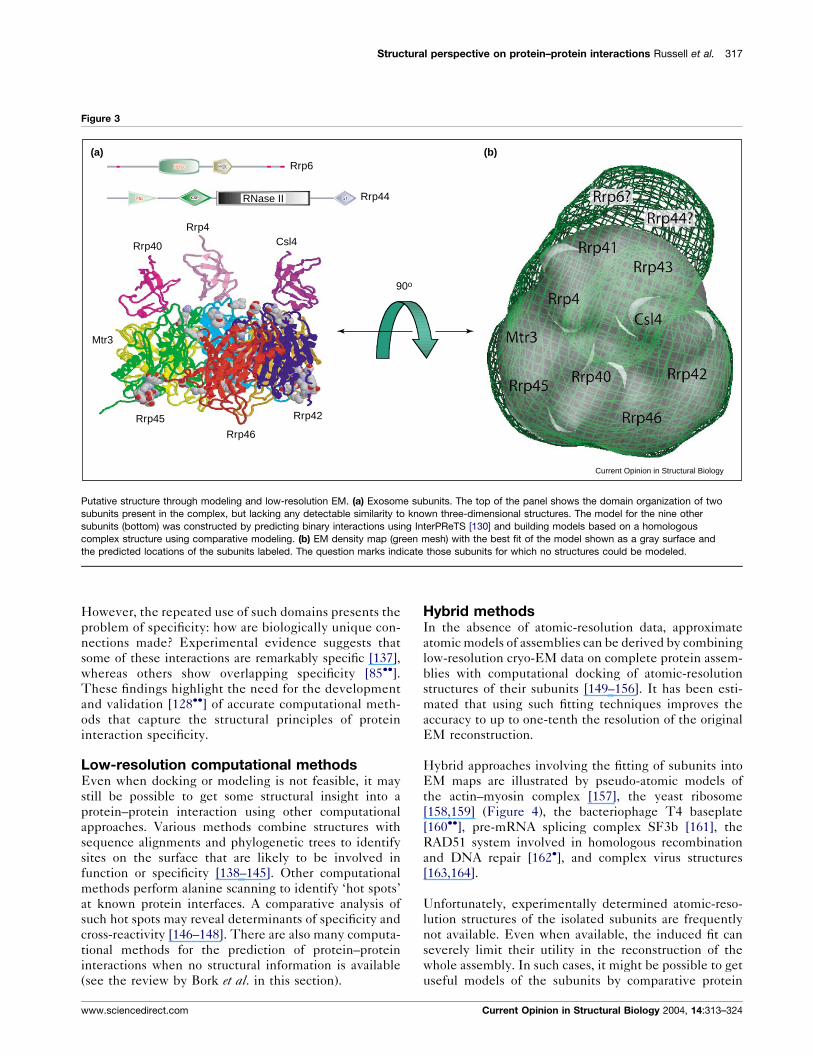
(Figure 3) [136], with mixed results.
In eukaryotes, many of the protein–protein interactions
in regulatory signaling networks are mediated by mod-
ular protein interaction domains. Such domains appear to
have been used in a modular fashion throughout evolu-
tion to generate novel connections between proteins.
316 Sequences and topology
Current Opinion in Structural Biology 2004, 14:313–324 www.sciencedirect.com
However, the repeated use of such domains presents the
problem of specificity: how are biologically unique con-
nections made? Experimental evidence suggests that
some of these interactions are remarkably specific [137],
whereas others show overlapping specificity [85��].These findings highlight the need for the development
and validation [128��] of accurate computational meth-
ods that capture the structural principles of protein
interaction specificity.
Low-resolution computational methodsEven when docking or modeling is not feasible, it may
still be possible to get some structural insight into a
protein–protein interaction using other computational
approaches. Various methods combine structures with
sequence alignments and phylogenetic trees to identify
sites on the surface that are likely to be involved in
function or specificity [138–145]. Other computational
methods perform alanine scanning to identify ‘hot spots’
at known protein interfaces. A comparative analysis of
such hot spots may reveal determinants of specificity and
cross-reactivity [146–148]. There are also many computa-
tional methods for the prediction of protein–protein
interactions when no structural information is available
(see the review by Bork et al. in this section).
Hybrid methodsIn the absence of atomic-resolution data, approximate
atomic models of assemblies can be derived by combining
low-resolution cryo-EM data on complete protein assem-
blies with computational docking of atomic-resolution
structures of their subunits [149–156]. It has been esti-
mated that using such fitting techniques improves the
accuracy to up to one-tenth the resolution of the original
EM reconstruction.
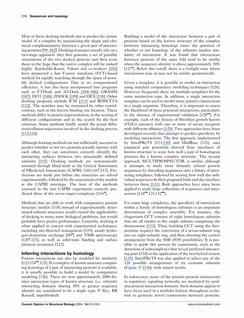
Hybrid approaches involving the fitting of subunits into
EM maps are illustrated by pseudo-atomic models of
the actin–myosin complex [157], the yeast ribosome
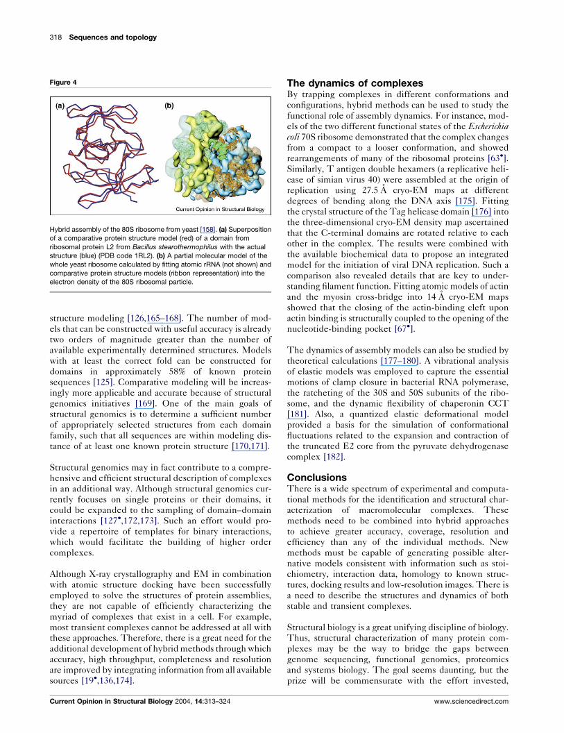
[158,159] (Figure 4), the bacteriophage T4 baseplate
[160��], pre-mRNA splicing complex SF3b [161], the
RAD51 system involved in homologous recombination
and DNA repair [162�], and complex virus structures
[163,164].
Unfortunately, experimentally determined atomic-reso-
lution structures of the isolated subunits are frequently
not available. Even when available, the induced fit can
severely limit their utility in the reconstruction of the
whole assembly. In such cases, it might be possible to get
useful models of the subunits by comparative protein
Figure 3
Csl4Rrp40
Rrp4
Rrp45
Mtr3
Rrp46
Rrp42
Rrp6
Rrp44
90o
RNase II
(a) (b)
Current Opinion in Structural Biology
Putative structure through modeling and low-resolution EM. (a) Exosome subunits. The top of the panel shows the domain organization of two
subunits present in the complex, but lacking any detectable similarity to known three-dimensional structures. The model for the nine other
subunits (bottom) was constructed by predicting binary interactions using InterPReTS [130] and building models based on a homologous
complex structure using comparative modeling. (b) EM density map (green mesh) with the best fit of the model shown as a gray surface and
the predicted locations of the subunits labeled. The question marks indicate those subunits for which no structures could be modeled.
Structural perspective on protein–protein interactions Russell et al. 317
www.sciencedirect.com Current Opinion in Structural Biology 2004, 14:313–324
structure modeling [126,165–168]. The number of mod-
els that can be constructed with useful accuracy is already
two orders of magnitude greater than the number of
available experimentally determined structures. Models
with at least the correct fold can be constructed for
domains in approximately 58% of known protein
sequences [125]. Comparative modeling will be increas-
ingly more applicable and accurate because of structural
genomics initiatives [169]. One of the main goals of
structural genomics is to determine a sufficient number
of appropriately selected structures from each domain
family, such that all sequences are within modeling dis-
tance of at least one known protein structure [170,171].
Structural genomics may in fact contribute to a compre-
hensive and efficient structural description of complexes
in an additional way. Although structural genomics cur-
rently focuses on single proteins or their domains, it
could be expanded to the sampling of domain–domain
interactions [127�,172,173]. Such an effort would pro-
vide a repertoire of templates for binary interactions,
which would facilitate the building of higher order
complexes.
Although X-ray crystallography and EM in combination
with atomic structure docking have been successfully
employed to solve the structures of protein assemblies,
they are not capable of efficiently characterizing the
myriad of complexes that exist in a cell. For example,
most transient complexes cannot be addressed at all with
these approaches. Therefore, there is a great need for the
additional development of hybrid methods through which
accuracy, high throughput, completeness and resolution
are improved by integrating information from all available
sources [19�,136,174].
The dynamics of complexesBy trapping complexes in different conformations and
configurations, hybrid methods can be used to study the
functional role of assembly dynamics. For instance, mod-
els of the two different functional states of the Escherichiacoli 70S ribosome demonstrated that the complex changes
from a compact to a looser conformation, and showed
rearrangements of many of the ribosomal proteins [63�].Similarly, T antigen double hexamers (a replicative heli-
case of simian virus 40) were assembled at the origin of
replication using 27.5 A cryo-EM maps at different
degrees of bending along the DNA axis [175]. Fitting
the crystal structure of the Tag helicase domain [176] into
the three-dimensional cryo-EM density map ascertained
that the C-terminal domains are rotated relative to each
other in the complex. The results were combined with
the available biochemical data to propose an integrated
model for the initiation of viral DNA replication. Such a
comparison also revealed details that are key to under-
standing filament function. Fitting atomic models of actin
and the myosin cross-bridge into 14 A cryo-EM maps
showed that the closing of the actin-binding cleft upon
actin binding is structurally coupled to the opening of the
nucleotide-binding pocket [67�].
The dynamics of assembly models can also be studied by
theoretical calculations [177–180]. A vibrational analysis
of elastic models was employed to capture the essential
motions of clamp closure in bacterial RNA polymerase,
the ratcheting of the 30S and 50S subunits of the ribo-
some, and the dynamic flexibility of chaperonin CCT
[181]. Also, a quantized elastic deformational model
provided a basis for the simulation of conformational
fluctuations related to the expansion and contraction of
the truncated E2 core from the pyruvate dehydrogenase
complex [182].
ConclusionsThere is a wide spectrum of experimental and computa-
tional methods for the identification and structural char-
acterization of macromolecular complexes. These
methods need to be combined into hybrid approaches
to achieve greater accuracy, coverage, resolution and
efficiency than any of the individual methods. New
methods must be capable of generating possible alter-
native models consistent with information such as stoi-
chiometry, interaction data, homology to known struc-
tures, docking results and low-resolution images. There is
a need to describe the structures and dynamics of both
stable and transient complexes.
Structural biology is a great unifying discipline of biology.
Thus, structural characterization of many protein com-
plexes may be the way to bridge the gaps between
genome sequencing, functional genomics, proteomics
and systems biology. The goal seems daunting, but the
prize will be commensurate with the effort invested,
Figure 4
Hybrid assembly of the 80S ribosome from yeast [158]. (a) Superposition
of a comparative protein structure model (red) of a domain from
ribosomal protein L2 from Bacillus stearothermophilus with the actual
structure (blue) (PDB code 1RL2). (b) A partial molecular model of the
whole yeast ribosome calculated by fitting atomic rRNA (not shown) and
comparative protein structure models (ribbon representation) into the
electron density of the 80S ribosomal particle.
318 Sequences and topology
Current Opinion in Structural Biology 2004, 14:313–324 www.sciencedirect.com
given the importance of molecular machines and func-
tional networks in biology and medicine.
AcknowledgementsWe are grateful to Tanja Kortemme, Damien Devos, MS Madhusudan,Narayanan Eswar, Mike Kim, Matt Baker, Wah Chiu, WolfgangBaumeister and David Agard for discussions about the modeling ofassembly structures. We also acknowledge the support of the NIH, NSF,HFSP, SUN, IBM, Intel and The Sandler Family SupportingFoundation (AS).
References and recommended readingPapers of particular interest, published within the annual period ofreview, have been highlighted as:
� of special interest��of outstanding interest
1. Lander ES, Linton LM, Birren B, Nusbaum C, Zody MC, Baldwin J,Devon K, Dewar K, Doyle M, FitzHugh W et al.: Initial sequencingand analysis of the human genome. Nature 2001, 409:860-921.
2. Venter JC, Adams MD, Myers EW, Li PW, Mural RJ, Sutton GG,Smith HO, Yandell M, Evans CA, Holt RA et al.: The sequence ofthe human genome. Science 2001, 291:1304-1351.
3. Alberts B: The cell as a collection of protein machines:preparing the next generation of molecular biologists.Cell 1998, 92:291-294.
4. Goto NK, Zor T, Martinez-Yamout M, Dyson HJ, Wright PE:Cooperativity in transcription factor binding to the coactivatorCREB-binding protein (CBP). The mixed lineage leukemiaprotein (MLL) activation domain binds to an allosteric siteon the KIX domain. J Biol Chem 2002, 277:43168-43174.
5. Grakoui A, Bromley SK, Sumen C, Davis MM, Shaw AS, Allen PM,Dustin ML: The immunological synapse: a molecular machinecontrolling T cell activation. Science 1999, 285:221-227.
6. Courey AJ: Cooperativity in transcriptional control.Curr Biol 2001, 11:R250-R252.
7. Noji H, Yoshida M: The rotary machine in the cell, ATP synthase.J Biol Chem 2001, 276:1665-1668.
8. Nogales E, Grigorieff N: Molecular machines: putting the piecestogether. J Cell Biol 2001, 152:F1-F10.
9. Rout MP, Aitchison JD, Suprapto A, Hjertaas K, Zhao Y, Chait BT:The yeast nuclear pore complex: composition, architecture,and transport mechanism. J Cell Biol 2000, 148:635-651.
10. Murakami KS, Darst SA: Bacterial RNA polymerases: the wholostory. Curr Opin Struct Biol 2003, 13:31-39.
11. Nogales E: Recent structural insights into transcriptionpreinitiation complexes. J Cell Sci 2000, 113:4391-4397.
12. Vale RD: The molecular motor toolbox for intracellulartransport. Cell 2003, 112:467-480.
13. Goldstein LS, Yang Z: Microtubule-based transport systemsin neurons: the roles of kinesins and dyneins. Annu RevNeurosci 2000, 23:39-71.
14. Vale RD, Milligan RA: The way things move: looking under thehood of molecular motor proteins. Science 2000, 288:88-95.
15. Kennedy MB: Signal-processing machines at the postsynapticdensity. Science 2000, 290:750-754.
16. Park J, Lappe M, Teichmann SA: Mapping protein familyinteractions: intramolecular and intermolecular protein familyinteraction repertoires in the PDB and yeast. J Mol Biol 2001,307:929-938.
17. Aloy P, Russell RB: Potential artefacts in protein-interactionnetworks. FEBS Lett 2002, 530:253-254.
18. Edwards A, Kus B, Jansen R, Greenbaum D, Greenblatt J,Gerstein M: Bridging structural biology and genomics:assessing protein-interaction data with known complexes.Trends Genet 2002, 18:529-536.
19.�
Sali A, Glaeser R, Earnest T, Baumeister W: From words toliterature in structural proteomics. Nature 2003, 422:216-225.
This review summarizes current efforts in structural proteomics. It pro-vides a plethora of references for the various experimental and theoreticalmethods used to obtain structural information about macromolecularassemblies.
20. Henrick K, Thornton JM: PQS: a protein quaternary structure fileserver. Trends Biochem Sci 1998, 23:358-361.
21. Uetz P, Giot L, Cagney G, Mansfield TA, Judson RS, Knight JR,Lockshon D, Narayan V, Srinivasan M, Pochart P et al.:A comprehensive analysis of protein-protein interactions inSaccharomyces cerevisiae. Nature 2000, 403:623-627.
22. Ito T, Chiba T, Yoshida M: Exploring the protein interactomeusing comprehensive two-hybrid projects. Trends Biotechnol2001, 19:S23-S27.
23. Rain JC, Selig L, De Reuse H, Battaglia V, Reverdy C, Simon S,Lenzen G, Petel F, Wojcik J, Schachter V et al.: The protein-proteininteraction map of Helicobacter pylori. Nature 2001,409:211-215.
24. Gavin AC, Bosche M, Krause R, Grandi P, Marzioch M, Bauer A,Schultz J, Rick JM, Michon AM, Cruciat CM et al.: Functionalorganization of the yeast proteome by systematic analysisof protein complexes. Nature 2002, 415:141-147.
25. Ho Y, Gruhler A, Heilbut A, Bader GD, Moore L, Adams SL, Millar A,Taylor P, Bennett K, Boutilier K et al.: Systematic identification ofprotein complexes in Saccharomyces cerevisiae by massspectrometry. Nature 2002, 415:180-183.
26. Giot L, Bader JS, Brouwer C, Chaudhuri A, Kuang B, Li Y, Hao YL,Ooi CE, Godwin B, Vitols E et al.: A protein interaction map ofDrosophila melanogaster. Science 2003, 302:1727-1736.
27. Phizicky E, Bastiaens PI, Zhu H, Snyder M, Fields S: Proteinanalysis on a proteomic scale. Nature 2003, 422:208-215.
28.�
Aebersold R, Mann M: Mass spectrometry-based proteomics.Nature 2003, 422:198-207.
This review provides a good overview of the role of mass-spectrometry-based proteomics in the study of protein–protein interactions, the map-ping of cell organelles and the generation of quantitative protein profilesfor a variety of species.
29. Andersen JS, Wilkinson CJ, Mayor T, Mortensen P, Nigg EA,Mann M: Proteomic characterization of the human centrosomeby protein correlation profiling. Nature 2003, 426:570-574.
30. Kumar A, Snyder M: Protein complexes take the bait.Nature 2002, 415:123-124.
31. von Mering C, Krause R, Snel B, Cornell M, Oliver SG, Fields S,Bork P: Comparative assessment of large-scale data sets ofprotein-protein interactions. Nature 2002, 417:399-403.
32. Abbott A: Proteomics: the society of proteins. Nature 2002,417:894-896.
33. Baumeister W: Electron tomography: towards visualizing themolecular organization of the cytoplasm. Curr Opin Struct Biol2002, 12:679-684.
34. Sali A, Kuriyan J: Challenges at the frontiers of structuralbiology. Trends Cell Biol 1999, 9:M20-M24.
35. Alber F, Eswar N, Sali A: Structure determination ofmacromolecular complexes by experiment and computation.In Nucleic Acids and Molecular Biology, Volume 15, PracticalBioinformatics. Edited by Bujnicki JM. : Springer-Verlag;2004:73-96.
36. Zhang G, Campbell EA, Minakhin L, Richter C, Severinov K,Darst SA: Crystal structure of Thermus aquaticus coreRNA polymerase at 3.3 A- resolution. Cell 1999, 98:811-824.
37. Ban N, Nissen P, Hansen J, Moore PB, Steitz TA:The complete atomic structure of the large ribosomalsubunit at 2.4 A- resolution. Science 2000, 289:905-920.
38. Carter AP, Clemons WM, Brodersen DE, Morgan-Warren RJ,Wimberly BT, Ramakrishnan V: Functional insights from thestructure of the 30S ribosomal subunit and its interactions withantibiotics. Nature 2000, 407:340-348.
Structural perspective on protein–protein interactions Russell et al. 319
www.sciencedirect.com Current Opinion in Structural Biology 2004, 14:313–324
39. Harms J, Schluenzen F, Zarivach R, Bashan A, Gat S, Agmon I,Bartels H, Franceschi F, Yonath A: High resolution structure ofthe large ribosomal subunit from a mesophilic eubacterium.Cell 2001, 107:679-688.
40. Wimberly BT, Brodersen DE, Morgan-Warren RJ, Carter AP,Vonrhein C, Hartsch T, Ramakrishnan V: Structure of the 30Sribosomal subunit. Nature 2000, 407:327-339.
41. Schluenzen F, Tocilj A, Zarivach R, Harms J, Gluehmann M,Janell D, Bashan A, Bartels H, Agmon I, Franceschi F et al.:Structure of functionally activated small ribosomal subunitat 3.3 angstroms resolution. Cell 2000, 102:615-623.
42. Yusupov MM, Yusupova GZ, Baucom A, Lieberman K, Earnest TN,Cate JH, Noller HF: Crystal structure of the ribosome at 5.5 A-
resolution. Science 2001, 292:883-896.
43. Lowe J, Stock D, Jap B, Zwickl P, Baumeister W, Huber R:Crystal structure of the 20S proteasome from the archaeonT. acidophilum at 3.4 A resolution. Science 1995,268:533-539.
44. Braig K, Otwinowski Z, Hegde R, Boisvert DC, Joachimiak A,Horwich AL, Sigler PB: The crystal structure of the bacterialchaperonin GroEL at 2.8 A. Nature 1994, 371:578-586.
45. Robinson RC, Turbedsky K, Kaiser DA, Marchand JB, Higgs HN,Choe S, Pollard TD: Crystal structure of Arp2/3 complex.Science 2001, 294:1679-1684.
46. Ben-Shem A, Frolow F, Nelson N: Crystal structure of plantphotosystem I. Nature 2003, 426:630-635.
47. Liu Z, Yan H, Wang K, Kuang T, Zhang J, Gui L, An X, Chang W:Crystal structure of spinach major light-harvesting complex at2.72 A resolution. Nature 2004, 428:287-292.
48.�
Egea PF, Shan SO, Napetschnig J, Savage DF, Walter P,Stroud RM: Substrate twinning activates the signal recognitionparticle and its receptor. Nature 2004, 427:215-221.
The 1.9 A X-ray structure of a complex formed by two GTPases, the signalrecognition particle (SRP) and its receptor (SR), demonstrates a novelmode of substrate twinning. The structure suggests a unique activationmechanism for the SRP family of GTPases.
49. Grimes J, Basak AK, Roy P, Stuart D: The crystal structure ofbluetongue virus VP7. Nature 1995, 373:167-170.
50. Oda Y, Saeki K, Takahashi Y, Maeda T, Naitow H, Tsukihara T,Fukuyama K: Crystal structure of tobacco necrosis virus at2.25 A- resolution. J Mol Biol 2000, 300:153-169.
51. Nakagawa A, Miyazaki N, Taka J, Naitow H, Ogawa A, Fujimoto Z,Mizuno H, Higashi T, Watanabe Y, Omura T et al.: The atomicstructure of rice dwarf virus reveals the self-assemblymechanism of component proteins. Structure 2003,11:1227-1238.
52. Fiaux J, Bertelsen EB, Horwich AL, Wuthrich K: NMR analysisof a 900K GroEL GroES complex. Nature 2002, 418:207-211.
53. Fushman D, Xu R, Cowburn D: Direct determination of changesof interdomain orientation on ligation: use of the orientationaldependence of 15N NMR relaxation in Abl SH(32).Biochemistry 1999, 38:10225-10230.
54. Nakanishi T, Miyazawa M, Sakakura M, Terasawa H, Takahashi H,Shimada I: Determination of the interface of a large proteincomplex by transferred cross-saturation measurements.J Mol Biol 2002, 318:245-249.
55. Zuiderweg ER: Mapping protein-protein interactions in solutionby NMR spectroscopy. Biochemistry 2002, 41:1-7.
56. Frickel EM, Riek R, Jelesarov I, Helenius A, Wuthrich K, Ellgaard L:TROSY-NMR reveals interaction between ERp57 and the tip ofthe calreticulin P-domain. Proc Natl Acad Sci USA 2002,99:1954-1959.
57. Pellecchia M, Sebbel P, Hermanns U, Wuthrich K, Glockshuber R:Pilus chaperone FimC-adhesin FimH interactions mapped byTROSY-NMR. Nat Struct Biol 1999, 6:336-339.
58. Fernandez C, Wider G: TROSY in NMR studies of the structureand function of large biological macromolecules. Curr OpinStruct Biol 2003, 13:570-580.
59. Velyvis A, Vaynberg J, Yang Y, Vinogradova O, Zhang Y, Wu C,Qin J: Structural and functional insights into PINCH LIM4domain-mediated integrin signaling. Nat Struct Biol 2003,10:558-564.
60. Frank J: Single-particle imaging of macromolecules by cryo-electron microscopy. Annu Rev Biophys Biomol Struct 2002,31:303-319.
61. Baumeister W, Grimm R, Walz J: Electron tomography ofmolecules and cells. Trends Cell Biol 1999, 9:81-85.
62. Nogales E, Wolf SG, Downing KH: Structure of the alpha betatubulin dimer by electron crystallography. Nature 1998,391:199-203.
63.�
Gao H, Sengupta J, Valle M, Korostelev A, Eswar N, Stagg SM,Roey PV, Agrawal RK, Harvey SC, Sali A et al.: Study of thestructural dynamics of the E. coli 70S ribosome using real-space refinement. Cell 2003, 113:789-801.
Forty-four atomic models built by comparative modeling were fit into thecryo-EM density map of the 70S unit of the E. coli ribosome (11.5 Aresolution) in two different functional states. The work demonstrates thatthe ribosome changes from a compact structure to a looser one, coupledwith the rearrangement of many of the proteins.
64. Halic M, Becker T, Pool MR, Spahn CM, Grassucci RA, Frank J,Beckmann R: Structure of the signal recognition particleinteracting with the elongation-arrested ribosome.Nature 2004, 427:808-814.
65. Davis JA, Takagi Y, Kornberg RD, Asturias FA: Structure of theyeast RNA polymerase II holoenzyme: Mediator conformationand polymerase interaction. Mol Cell 2002, 10:409-415.
66. Yonekura K, Maki-Yonekura S, Namba K: Complete atomic modelof the bacterial flagellar filament by electron cryomicroscopy.Nature 2003, 424:643-650.
67.�
Holmes KC, Angert I, Kull FJ, Jahn W, Schroder RR: Electroncryo-microscopy shows how strong binding of myosin toactin releases nucleotide. Nature 2003, 425:423-427.
The fitting of atomic models of actin and the myosin cross-bridge into14 A cryo-EM maps shows that the closing of the actin-binding cleft uponactin binding is structurally coupled to the opening of the nucleotide-binding pocket.
68.�
Jiang W, Li Z, Zhang Z, Baker ML, Prevelige PE Jr, Chiu W:Coat protein fold and maturation transition of bacteriophageP22 seen at subnanometer resolutions. Nat Struct Biol 2003,10:131-135.
Structural analysis of bacteriophage P22 in two different functional statesusing cryo-EM density at subnanometer resolution shows that a largeconformational change of the P22 capsid during maturation transitioninvolves both domain movement of individual subunits and refolding ofthe capsid protein.
69. Zhang X, Walker SB, Chipman PR, Nibert ML, Baker TS: Reoviruspolymerase lambda 3 localized by cryo-electron microscopyof virions at a resolution of 7.6 A- . Nat Struct Biol 2003,10:1011-1018.
70.�
Zhang W, Chipman PR, Corver J, Johnson PR, Zhang Y,Mukhopadhyay S, Baker TS, Strauss JH, Rossmann MG,Kuhn RJ: Visualization of membrane protein domains bycryo-electron microscopy of dengue virus. Nat Struct Biol2003, 10:907-912.
Three-dimensional cryo-EM reconstruction (9.5 A resolution) reveals sec-ondary structural features of 180 envelope and 180 membrane proteinsin the lipid envelope of mature dengue virus. This is one of only a fewdeterminations of the disposition of transmembrane proteins in situ and itshows that the nucleocapsid core and envelope proteins do not directlyinteract in the mature virus.
71. Grunewald K, Desai P, Winkler DC, Heymann JB, Belnap DM,Baumeister W, Steven AC: Three-dimensional structure ofherpes simplex virus from cryo-electron tomography.Science 2003, 302:1396-1398.
72. Grunewald K, Medalia O, Gross A, Steven AC, Baumeister W:Prospects of electron cryotomography to visualizemacromolecular complexes inside cellular compartments:implications of crowding. Biophys Chem 2003, 100:577-591.
73. Medalia O, Weber I, Frangakis AS, Nicastro D, Gerisch G,Baumeister W: Macromolecular architecture in eukaryotic cells
320 Sequences and topology
Current Opinion in Structural Biology 2004, 14:313–324 www.sciencedirect.com
visualized by cryoelectron tomography. Science 2002,298:1209-1213.
74. Bohm J, Frangakis AS, Hegerl R, Nickell S, Typke D, Baumeister W:Toward detecting and identifying macromolecules in a cellularcontext: template matching applied to electron tomograms.Proc Natl Acad Sci USA 2000, 97:14245-14250.
75. Frangakis AS, Bohm J, Forster F, Nickell S, Nicastro D, Typke D,Hegerl R, Baumeister W: Identification of macromolecularcomplexes in cryoelectron tomograms of phantom cells.Proc Natl Acad Sci USA 2002, 99:14153-14158.
76. Grimm R, Singh H, Rachel R, Typke D, Zillig W, Baumeister W:Electron tomography of ice-embedded prokaryotic cells.Biophys J 1998, 74:1031-1042.
77. Plitzko JM, Frangakis AS, Nickell S, Forster F, Gross A,Baumeister W: In vivo veritas: electron cryotomography ofcells. Trends Biotechnol 2002, 20:S40-S44.
78. Stagljar I, Fields S: Analysis of membrane protein interactionsusing yeast-based technologies. Trends Biochem Sci 2002,27:559-563.
79. Burchett SA, Flanary P, Aston C, Jiang L, Young KH, Uetz P,Fields S, Dohlman HG: Regulation of stress response signalingby the N-terminal dishevelled/EGL-10/pleckstrin domain ofSst2, a regulator of G protein signaling in Saccharomycescerevisiae. J Biol Chem 2002, 277:22156-22167.
80. Michnick SW: Exploring protein interactions by interaction-induced folding of proteins from complementary peptidefragments. Curr Opin Struct Biol 2001, 11:472-477.
81. Hu CD, Kerppola TK: Simultaneous visualization of multipleprotein interactions in living cells using multicolorfluorescence complementation analysis. Nat Biotechnol 2003,21:539-545.
82.�
Ranish JA, Yi EC, Leslie DM, Purvine SO, Goodlett DR, Eng J,Aebersold R: The study of macromolecular complexes byquantitative proteomics. Nat Genet 2003, 33:349-355.
This paper describes a new generic strategy for determining the specificcomposition, changes in composition and changes in the abundance ofprotein complexes using mass spectrometry. Two examples are studied:the identification of genuine components of the RNA polymerase II pre-initiation complex within a high background of co-purifying proteins, andthe detailed quantitative changes in the abundance and composition ofimmunopurified STE12 protein complexes from yeast cells exposed todifferent environmental conditions.
83. Himeda CL, Ranish JA, Angello JC, Maire P, Aebersold R,Hauschka SD: Quantitative proteomic identification of six4 asthe trex-binding factor in the muscle creatine kinase enhancer.Mol Cell Biol 2004, 24:2132-2143.
84. Tong AH, Drees B, Nardelli G, Bader GD, Brannetti B, Castagnoli L,Evangelista M, Ferracuti S, Nelson B, Paoluzi S et al.: A combinedexperimental and computational strategy to define proteininteraction networks for peptide recognition modules.Science 2002, 295:321-324.
85.��
Landgraf C, Panni S, Montecchi-Palazzi L, Castagnoli L,Schneider-Mergener J, Volkmer-Engert R, Cesareni G:Protein interaction networks by proteome peptide scanning.PLoS Biol 2004, 2:E14.
A variant of the work described in [84]. Here, candidate peptides in theyeast genome that match the consensus derived from phage display aresynthesized and fixed to cellulose membranes, and then probed by SH3domains. This reveals distinct classes of binding preferences, as well assome overlapping specificities. The strategy can be easily modified toprobe the entire proteome.
86. Aloy P, Russell RB: The third dimension for protein interactionsand complexes. Trends Biochem Sci 2002, 27:633-638.
87. Cunningham BC, Jhurani P, Ng P, Wells JA: Receptor andantibody epitopes in human growth hormone identifiedby homolog-scanning mutagenesis. Science 1989,243:1330-1336.
88. Lanman J, Lam TT, Barnes S, Sakalian M, Emmett MR,Marshall AG, Prevelige PE Jr: Identification of novel interactionsin HIV-1 capsid protein assembly by high-resolution massspectrometry. J Mol Biol 2003, 325:759-772.
89.�
Anand GS, Law D, Mandell JG, Snead AN, Tsigelny I, Taylor SS,Eyck LFT, Komives EA: Identification of the protein kinase Aregulatory RIalpha-catalytic subunit interface by amide H/2Hexchange and protein docking. Proc Natl Acad Sci USA 2003,100:13264-13269.
The authors identify the interface between the two subunits (catalytic andregulatory) of protein kinase A (PKA) by computational docking andsubsequent filtering of the solutions based on amide hydrogen-deuteriumexchange interface protection data.
90. Guan JQ, Almo SC, Reisler E, Chance MR: Structuralreorganization of proteins revealed by radiolysis and massspectrometry: G-actin solution structure is divalent cationdependent. Biochemistry 2003, 42:11992-12000.
91. Trester-Zedlitz M, Kamada K, Burley SK, Fenyo D, Chait BT,Muir TW: A modular cross-linking approach for exploringprotein interactions. J Am Chem Soc 2003, 125:2416-2425.
92. Back JW, de Jong L, Muijsers AO, de Koster CG: Chemicalcross-linking and mass spectrometry for protein structuralmodeling. J Mol Biol 2003, 331:303-313.
93. Serino G, Su H, Peng Z, Tsuge T, Wei N, Gu H, Deng XW:Characterization of the last subunit of the Arabidopsis COP9signalosome: implications for the overall structure and origin ofthe complex. Plant Cell 2003, 15:719-731.
94. Truong K, Ikura M: The use of FRET imaging microscopy todetect protein-protein interactions and protein conformationalchanges in vivo. Curr Opin Struct Biol 2001, 11:573-578.
95. Yan Y, Marriott G: Analysis of protein interactions usingfluorescence technologies. Curr Opin Chem Biol 2003,7:635-640.
96. Kariakin A, Davydov D, Peterson JA, Jung C: A new approach tothe study of protein-protein interaction by FTIR: complexformation between cytochrome P450BM-3 heme domain andFMN reductase domain. Biochemistry 2002, 41:13514-13525.
97. Marquez JA, Smith CI, Petoukhov MV, Lo Surdo P, Mattsson PT,Knekt M, Westlund A, Scheffzek K, Saraste M, Svergun DI:Conformation of full-length Bruton tyrosine kinase (Btk)from synchrotron X-ray solution scattering. EMBO J 2003,22:4616-4624.
98. Svergun DI, Aldag I, Sieck T, Altendorf K, Koch MH, Kane DJ,Kozin MB, Gruber G: A model of the quaternary structure of theEscherichia coli F1 ATPase from X-ray solution scattering andevidence for structural changes in the delta subunit during ATPhydrolysis. Biophys J 1998, 75:2212-2219.
99. Gray JJ, Moughon SE, Kortemme T, Schueler-Furman O,Misura KM, Morozov AV, Baker D: Protein-protein dockingpredictions for the CAPRI experiment. Proteins 2003,52:118-122.
100. Smith GR, Sternberg MJ: Prediction of protein-proteininteractions by docking methods. Curr Opin Struct Biol 2002,12:28-35.
101. Janin J, Henrick K, Moult J, Eyck LT, Sternberg MJ, Vajda S,Vakser I, Wodak SJ: CAPRI: a Critical Assessment ofPRedicted Interactions. Proteins 2003, 52:2-9.
102. Schneidman-Duhovny D, Inbar Y, Polak V, Shatsky M, Halperin I,Benyamini H, Barzilai A, Dror O, Haspel N, Nussinov R et al.: Takinggeometry to its edge: fast unbound rigid (and hinge-bent)docking. Proteins 2003, 52:107-112.
103. Katchalski-Katzir E, Shariv I, Eisenstein M, Friesem AA, Aflalo C,Vakser IA: Molecular surface recognition: determination ofgeometric fit between proteins and their ligands by correlationtechniques. Proc Natl Acad Sci USA 1992, 89:2195-2199.
104. Gabb HA, Jackson RM, Sternberg MJ: Modelling proteindocking using shape complementarity, electrostatics andbiochemical information. J Mol Biol 1997, 272:106-120.
105. Moont G, Sternberg MJ: Modeling protein-protein andprotein-DNA docking. Edited by Lengauer T. Weinheim:Wiley-VCH; 2001.
106. Jackson RM, Gabb HA, Sternberg MJ: Rapid refinement ofprotein interfaces incorporating solvation: application to thedocking problem. J Mol Biol 1998, 276:265-285.
Structural perspective on protein–protein interactions Russell et al. 321
www.sciencedirect.com Current Opinion in Structural Biology 2004, 14:313–324
107. Vakser IA: Protein docking for low-resolution structures.Protein Eng 1995, 8:371-377.
108. Mandell JG, Roberts VA, Pique ME, Kotlovyi V, Mitchell JC,Nelson E, Tsigelny I, Ten Eyck LF: Protein docking usingcontinuum electrostatics and geometric fit. Protein Eng 2001,14:105-113.
109. Chen R, Li L, Weng Z: ZDOCK: an initial-stage protein-dockingalgorithm. Proteins 2003, 52:80-87.
110. Ritchie DW, Kemp GJ: Protein docking using spherical polarFourier correlations. Proteins 2000, 39:178-194.
111. Fernandez-Recio J, Totrov M, Abagyan R: Soft protein-protein docking in internal coordinates. Protein Sci 2002,11:280-291.
112. Gray JJ, Moughon S, Wang C, Schueler-Furman O, Kuhlman B,Rohl CA, Baker D: Protein-protein docking with simultaneousoptimization of rigid-body displacement and side-chainconformations. J Mol Biol 2003, 331:281-299.
113. Gabdoulline RR, Wade RC: Protein-protein association:investigation of factors influencing association rates byBrownian dynamics simulations. J Mol Biol 2001,306:1139-1155.
114. Fitzjohn PW, Bates PA: Guided docking: first step to locatepotential binding sites. Proteins 2003, 52:28-32.
115. Strynadka NC, Eisenstein M, Katchalski-Katzir E, Shoichet BK,Kuntz ID, Abagyan R, Totrov M, Janin J, Cherfils J,Zimmerman F et al.: Molecular docking programs successfullypredict the binding of a beta-lactamase inhibitory proteinto TEM-1 beta-lactamase. Nat Struct Biol 1996, 3:233-239.
116.�
Mendez R, Leplae R, De Maria L, Wodak SJ: Assessment of blindpredictions of protein-protein interactions: current status ofdocking methods. Proteins 2003, 52:51-67.
This paper reviews the results from the first two rounds of the CAPRIcommunity-wide docking experiment, in which 19 groups attempted topredict the structures of seven protein–protein complexes. The targets,as well as the docking methods and protocols used in the predictions, aredescribed. In total, five of the seven target complexes were predicted toan acceptable accuracy.
117. Vajda S, Camacho CJ: Protein-protein docking: is the glasshalf-full or half-empty? Trends Biotechnol 2004, 22:110-116.
118. Tovchigrechko A, Wells CA, Vakser IA: Docking of proteinmodels. Protein Sci 2002, 11:1888-1896.
119. Morillas M, Gomez-Puertas P, Rubi B, Clotet J, Arino J, Valencia A,Hegardt FG, Serra D, Asins G: Structural model of a malonyl-CoA-binding site of carnitine octanoyltransferase and carnitinepalmitoyltransferase I: mutational analysis of a malonyl-CoAaffinity domain. J Biol Chem 2002, 277:11473-11480.
120.�
Dobrodumov A, Gronenborn AM: Filtering and selection ofstructural models: combining docking and NMR.Proteins 2003, 53:18-32.
The authors demonstrate an approach to protein complex structuredetermination that first generates structural models by computationaldocking of the subunits and then filters these using experimental NMRconstraints (residual dipolar couplings and chemical shift mapping).
121. Dominguez C, Boelens R, Bonvin AM: HADDOCK: a protein-protein docking approach based on biochemical or biophysicalinformation. J Am Chem Soc 2003, 125:1731-1737.
122. Romijn RA, Westein E, Bouma B, Schiphorst ME, Sixma JJ,Lenting PJ, Huizinga EG: Mapping the collagen-binding sitein the von Willebrand factor-A3 domain. J Biol Chem 2003,278:15035-15039.
123. Aloy P, Russell RB: Interrogating protein interaction networksthrough structural biology. Proc Natl Acad Sci USA 2002,99:5896-5901.
124.��
Aloy P, Bottcher B, Ceulemans H, Leutwein C, Mellwig C,Fischer S, Gavin AC, Bork P, Superti-Furga G, Serrano L et al.:Structure-based assembly of protein complexes in yeast.Science 2004, 303:2026-2029.
Large-scale structural analysis of complexes in yeast using bioinfor-matics and EM. As complete models as possible were built for over100 yeast complexes and all complex–complex interactions. For some,
EM maps could be used to confirm models or combine separatelymodeled subcomplexes.
125. Pieper U, Eswar N, Braberg H, Madhusudhan MS, Davis FP,Stuart AC, Mirkovic N, Rossi A, Marti-Renom MA, Fiser A et al.:MODBASE, a database of annotated comparative proteinstructure models, and associated resources. Nucleic Acids Res2004, 32:D217-D222.
126. Marti-Renom MA, Stuart AC, Fiser A, Sanchez R, Melo F,Sali A: Comparative protein structure modeling of genesand genomes. Annu Rev Biophys Biomol Struct 2000,29:291-325.
127.�
Aloy P, Ceulemans H, Stark A, Russell RB: The relationshipbetween sequence and interaction divergence in proteins.J Mol Biol 2003, 332:989-998.
This study finds that the structural binding modes of proteins are con-served at around 30% sequence identity. Below this cutoff, the bindingmode may or may not be similar.
128.��
Kortemme T, Joachimiak LA, Bullock AN, Schuler AD, Stoddard BL,Baker D: Computational redesign of protein-protein interactionspecificity. Nat Struct Mol Biol 2004, 11:371-379.
The authors develop a computational strategy to redesign specificity atprotein–protein interfaces and apply it to create new specifically inter-acting DNase–inhibitor protein pairs. In addition to in vitro and in vivobiochemical confirmation of the specificities, a crystal structure of one ofthe pairs confirms the computationally designed interface structure to animpressive 0.62 A all-atom rmsd.
129. Ornitz DM, Xu J, Colvin JS, McEwen DG, MacArthur CA, Coulier F,Gao G, Goldfarb M: Receptor specificity of the fibroblast growthfactor family. J Biol Chem 1996, 271:15292-15297.
130. Aloy P, Russell RB: InterPreTS: protein interaction predictionthrough tertiary structure. Bioinformatics 2003, 19:161-162.
131. Lu L, Lu H, Skolnick J: MULTIPROSPECTOR: an algorithm for theprediction of protein-protein interactions by multimericthreading. Proteins 2002, 49:350-364.
132.��
Lu L, Arakaki AK, Lu H, Skolnick J: Multimeric threading-basedprediction of protein-protein interactions on a genomic scale:application to the Saccharomyces cerevisiae proteome.Genome Res 2003 June, 13:1146-1154.
The authors predict the protein interaction network in S. cerevisiae byapplying their MULTIPROSPECTOR threading algorithm to the entireproteome.
133. Valpuesta JM, Martin-Benito J, Gomez-Puertas P, Carrascosa JL,Willison KR: Structure and function of a protein folding machine:the eukaryotic cytosolic chaperonin CCT. FEBS Lett 2002,529:11-16.
134. Liou AK, Willison KR: Elucidation of the subunit orientation inCCT (chaperonin containing TCP1) from the subunitcomposition of CCT micro-complexes. EMBO J 1997,16:4311-4316.
135. Raijmakers R, Noordman YE, van Venrooij WJ, Pruijn GJ:Protein-protein interactions of hCsl4p with other humanexosome subunits. J Mol Biol 2002, 315:809-818.
136. Aloy P, Ciccarelli FD, Leutwein C, Gavin AC, Superti-Furga G,Bork P, Boettcher B, Russell RB: A complex prediction:three-dimensional model of the yeast exosome. EMBO Rep2002, 3:628-635.
137. Zarrinpar A, Park SH, Lim WA: Optimization of specificity in acellular protein interaction network by negative selection.Nature 2003, 426:676-680.
138. Ma B, Elkayam T, Wolfson H, Nussinov R: Protein-proteininteractions: structurally conserved residues distinguishbetween binding sites and exposed protein surfaces.Proc Natl Acad Sci USA 2003, 100:5772-5777.
139. Yao H, Kristensen DM, Mihalek I, Sowa ME, Shaw C, Kimmel M,Kavraki L, Lichtarge O: An accurate, sensitive, and scalablemethod to identify functional sites in protein structures.J Mol Biol 2003, 326:255-261.
140. Fariselli P, Pazos F, Valencia A, Casadio R: Prediction ofprotein–protein interaction sites in heterocomplexes withneural networks. Eur J Biochem 2002, 269:1356-1361.
322 Sequences and topology
Current Opinion in Structural Biology 2004, 14:313–324 www.sciencedirect.com
141. del Sol Mesa A, Pazos F, Valencia A: Automatic methods forpredicting functionally important residues. J Mol Biol 2003,326:1289-1302.
142. Lichtarge O, Bourne HR, Cohen FE: An evolutionary trace methoddefines binding surfaces common to protein families.J Mol Biol 1996, 257:342-358.
143. Hannenhalli SS, Russell RB: Analysis and prediction offunctional sub-types from protein sequence alignments.J Mol Biol 2000, 303:61-76.
144. Aloy P, Querol E, Aviles FX, Sternberg MJE: Automated structure-based prediction of functional sites in proteins: applications toassessing the validity of inheriting protein function fromhomology in genome annotation and to protein docking.J Mol Biol 2001, 311:395-408.
145. Landgraf R, Xenarios I, Eisenberg D: Three-dimensional clusteranalysis identifies interfaces and functional residue clusters inproteins. J Mol Biol 2001, 307:1487-1502.
146. Kortemme T, Baker D: A simple physical model for bindingenergy hot spots in protein-protein complexes. Proc NatlAcad Sci USA 2002, 99:14116-14121.
147. Boulanger MJ, Bankovich AJ, Kortemme T, Baker D, Garcia KC:Convergent mechanisms for recognition of divergentcytokines by the shared signaling receptor gp130. Mol Cell2003, 12:577-589.
148. McFarland BJ, Kortemme T, Yu SF, Baker D, Strong RK:Symmetry recognizing asymmetry: analysis of the interactionsbetween the C-type lectin-like immunoreceptor NKG2D andMHC class I-like ligands. Structure 2003, 11:411-422.
149. Volkmann N, Hanein D, Ouyang G, Trybus KM, DeRosier DJ,Lowey S: Evidence for cleft closure in actomyosin uponADP release. Nat Struct Biol 2000, 7:1147-1155.
150. Roseman AM: Docking structures of domains into mapsfrom cryo-electron microscopy using local correlation.Acta Crystallogr D Biol Crystallogr 2000, 56:1332-1340.
151. Wriggers W, Birmanns S: Using situs for flexible and rigid-bodyfitting of multiresolution single-molecule data. J Struct Biol2001, 133:193-202.
152. Ceulemans H, Russell RB: Fast fitting of atomic structures tolower resolution electron density maps by surface overlapmaximization. J Mol Biol 2004, 338:783-793.
153. Volkmann N, Hanein D: Quantitative fitting of atomic modelsinto observed densities derived by electron microscopy.J Struct Biol 1999, 125:176-184.
154. Rossmann MG, Bernal R, Pletnev SV: Combining electronmicroscopic with x-ray crystallographic structures.J Struct Biol 2001, 136:190-200.
155. Chiu W, Baker ML, Jiang W, Zhou ZH: Deriving folds ofmacromolecular complexes through electron cryomicroscopyand bioinformatics approaches. Curr Opin Struct Biol 2002,12:263-269.
156. Chacon P, Wriggers W: Multi-resolution contour-based fitting ofmacromolecular structures. J Mol Biol 2002, 317:375-384.
157. Volkmann N, Ouyang G, Trybus KM, DeRosier DJ, Lowey S,Hanein D: Myosin isoforms show unique conformations inthe actin-bound state. Proc Natl Acad Sci USA 2003,100:3227-3232.
158. Spahn CM, Beckmann R, Eswar N, Penczek PA, Sali A, Blobel G,Frank J: Structure of the 80S ribosome from Saccharomycescerevisiae–tRNA- ribosome and subunit-subunit interactions.Cell 2001, 107:373-386.
159. Beckmann R, Spahn CM, Eswar N, Helmers J, Penczek PA, Sali A,Frank J, Blobel G: Architecture of the protein-conductingchannel associated with the translating 80S ribosome.Cell 2001, 107:361-372.
160.��
Kostyuchenko VA, Leiman PG, Chipman PR, Kanamaru S,van Raaij MJ, Arisaka F, Mesyanzhinov VV, Rossmann MG:Three-dimensional structure of bacteriophage T4 baseplate.Nat Struct Biol 2003, 10:688-693.
Three-dimensional cryo-EM reconstruction of the baseplate–tail tubecomplex of bacteriophage T4 at 12 A resolution. The X-ray structuresof six proteins were fitted into the electron density and the location of fourothers was determined with the help of biochemical data. The overallstructure suggests a mechanism of baseplate triggering and structuraltransition during the initial stages of T4 infection.
161. Golas MM, Sander B, Will CL, Luhrmann R, Stark H: Moleculararchitecture of the multiprotein splicing factor SF3b.Science 2003, 300:980-984.
162.�
Shin DS, Pellegrini L, Daniels DS, Yelent B, Craig L, Bates D,Yu DS, Shivji MK, Hitomi C, Arvai AS et al.: Full-length archaealRad51 structure and mutants: mechanisms for RAD51assembly and control by BRCA2. EMBO J 2003, 22:4566-4576.
The crystal structure of full-length RAD51 (2.85 A resolution), in combina-tion with SAXS, cryo-EM and mutagenesis, was used to characterize amacromolecular machine with a central role in repairing DNA double-strand breaks by homologous recombination. The study suggests howBRC repeats may disrupt RAD51 ring assembly and direct RAD51 to formfibers in response to cellular DNA damage.
163. Zhou ZH, Baker ML, Jiang W, Dougherty M, Jakana J, Dong G,Lu G, Chiu W: Electron cryomicroscopy and bioinformaticssuggest protein fold models for rice dwarf virus.Nat Struct Biol 2001, 8:868-873.
164. Baker ML, Jiang W, Bowman BR, Zhou ZH, Quiocho FA, Rixon FJ,Chiu W: Architecture of the herpes simplex virus majorcapsid protein derived from structural bioinformatics.J Mol Biol 2003, 331:447-456.
165. Blundell TL, Sibanda BL, Sternberg MJ, Thornton JM:Knowledge-based prediction of protein structures and thedesign of novel molecules. Nature 1987, 326:347-352.
166. Greer J: Comparative modeling methods: application tothe family of the mammalian serine proteases. Proteins 1990,7:317-334.
167. Sali A, Blundell TL: Comparative protein modelling bysatisfaction of spatial restraints. J Mol Biol 1993, 234:779-815.
168. Sauder JM, Dunbrack RL Jr: Genomic fold assignmentand rational modeling of proteins of biological interest.Proc Int Conf Intell Syst Mol Biol 2000, 8:296-306.
169. Burley SK, Almo SC, Bonanno JB, Capel M, Chance MR,Gaasterland T, Lin D, Sali A, Studier FW, Swaminathan S:Structural genomics: beyond the human genome project.Nat Genet 1999, 23:151-157.
170. Vitkup D, Melamud E, Moult J, Sander C: Completeness instructural genomics. Nat Struct Biol 2001, 8:559-566.
171. Baker D, Sali A: Protein structure prediction and structuralgenomics. Science 2001, 294:93-96.
172. Apic G, Gough J, Teichmann SA: Domain combinations inarchaeal, eubacterial and eukaryotic proteomes.J Mol Biol 2001, 310:311-325.
173. Sali A: NIH workshop on structural proteomics of biologicalcomplexes. Structure 2003, 11:1043-1047.
174. Malhotra A, Tan RK, Harvey SC: Prediction of the three-dimensional structure of Escherichia coli 30S ribosomalsubunit: a molecular mechanics approach. Proc Natl AcadSci USA 1990, 87:1950-1954.
175. Gomez-Lorenzo MG, Valle M, Frank J, Gruss C, Sorzano CO,Chen XS, Donate LE, Carazo JM: Large T antigen on the simianvirus 40 origin of replication: a 3D snapshot prior to DNAreplication. EMBO J 2003, 22:6205-6213.
176. Li D, Zhao R, Lilyestrom W, Gai D, Zhang R, DeCaprio JA,Fanning E, Jochimiak A, Szakonyi G, Chen XS: Structure of thereplicative helicase of the oncoprotein SV40 large tumourantigen. Nature 2003, 423:512-518.
177. Keskin O, Bahar I, Flatow D, Covell DG, Jernigan RL: Molecularmechanisms of chaperonin GroEL-GroES function.Biochemistry 2002, 41:491-501.
178. Keskin O, Durell SR, Bahar I, Jernigan RL, Covell DG: Relatingmolecular flexibility to function: a case study of tubulin.Biophys J 2002, 83:663-680.
Structural perspective on protein–protein interactions Russell et al. 323
www.sciencedirect.com Current Opinion in Structural Biology 2004, 14:313–324
179. Ming D, Kong Y, Wakil SJ, Brink J, Ma J: Domain movementsin human fatty acid synthase by quantized elasticdeformational model. Proc Natl Acad Sci USA 2002,99:7895-7899.
180. Tama F, Wriggers W, Brooks CL III: Exploring global distortionsof biological macromolecules and assemblies from low-resolution structural information and elastic network theory.J Mol Biol 2002, 321:297-305.
181. Chacon P, Tama F, Wriggers W: Mega-Dalton biomolecularmotion captured from electron microscopy reconstructions.J Mol Biol 2003, 326:485-492.
182. Kong Y, Ming D, Wu Y, Stoops JK, Zhou ZH, Ma J: Conformationalflexibility of pyruvate dehydrogenase complexes: acomputational analysis by quantized elastic deformationalmodel. J Mol Biol 2003, 330:129-135.
183. Symmons MF, Jones GH, Luisi BF: A duplicated fold is thestructural basis for polynucleotide phosphorylase catalyticactivity, processivity, and regulation. Structure Fold Des 2000,8:1215-1226.
184. Selenko P, Sprangers R, Stier G, Buhler D, Fischer U, Sattler M:SMN tudor domain structure and its interaction with the Smproteins. Nat Struct Biol 2001, 8:27-31.
185. Kronenberg S, Kleinschmidt JA, Bottcher B: Electron cryo-microscopy and image reconstruction of adeno-associatedvirus type 2 empty capsids. EMBO Rep 2001, 2:997-1002.
186. Uetz P: Two-hybrid arrays. Curr Opin Chem Biol 2002, 6:57-62.
187. Ito T, Chiba T, Ozawa R, Yoshida M, Hattori M, Sakaki Y:A comprehensive two-hybrid analysis to explore the yeastprotein interactome. Proc Natl Acad Sci USA 2001, 98:4569-4574.
324 Sequences and topology
Current Opinion in Structural Biology 2004, 14:313–324 www.sciencedirect.com