the fossil teeth of the peking man - nature
TRANSCRIPT
1SCIENtIfIC RePoRTS | (2018) 8:2066 | DOI:10.1038/s41598-018-20432-y
www.nature.com/scientificreports
The fossil teeth of the Peking ManSong Xing1, María Martinón-Torres2,3 & José María Bermúdez de Castro2,3
This study provides new original data, including the endostructure of most Zhoukoudian H. erectus teeth preserved to date, since the publication of Black in 1927 and Weidenreich in 1937. The new evidence ratifies the similarities of Zhoukoudian with other East Asian mid-Middle Pleistocene hominins such as Hexian and Yiyuan, and allows defining a dental pattern potentially characteristic of this population commonly referred to as classic H. erectus. Given the possible chronological overlaps of classic H. erectus with other archaic Homo, the characterization of this group becomes a key issue when deciphering the taxonomy and evolutionary scenario of the Middle Pleistocene hominins in East Asia. Internally, the most remarkable feature of Zhoukoudian teeth is the highly crenulated enamel-dentine junction (EDJ) and its imprint on the roof of the pulp cavity. So far, this “dendrite-like” EDJ has been found only in East Asia Middle Pleistocene hominins although a large group of samples were assessed, and it could be useful to dentally define classic H. erectus in China. The crenulated EDJ surface, together with the stout roots and the taurodontism could be a mechanism to withstand high biomechanical demand despite a general dentognathic reduction, particularly of the crowns, in these populations.
The “Peking Man” from Zhoukoudian Locality 1, Beijing, China, is one of the earliest and most emblematic homi-nins ever found in human history1. It refers to Sinanthropus pekinensis (now usually lumped into the Homo erectus taxon) named by Black in 19271. The evolutionary interpretation of the Zhoukoudian hominin materials has changed throughout the years. The primary description and comparison of the Zhoukoudian materials1–5 filled, at that time a perceived gap between ape and man, and they were crucial for the characterization of the species H. erectus6–8. Later studies suggested that Zhoukoudian had a less central role in our evolutionary story, representing no more than a side branch in our “family tree”9–11. Some researchers have even argued that Zhoukoudian sample may not be a good representative of the H. erectus taxon given the distinctive cranial differences they have when compared to the Java counterparts the species was named on12–14.
The problem is that, for many decades, the name H. erectus has been used as a blanket term to refer to almost any hominin found in Asia during the Pleistocene until the appearance of Homo sapiens. Recent fossil and genetic data suggested that the taxonomies of the Asian hominins may have been oversimplified. In particular, discover-ies like the Xuchang crania in North China15 or the reassessment of fossils samples such as Xujiayao16–18, Maba19 or Panxian Dadong20 reinforce the idea that other hominin lineages different from H. erectus may have lived in continental Asia during the same period. These fossils present some primitive features in common with other H. erectus samples: the Maba endocast is narrow at the frontal lobes and short and flattened in the parietal areas19; the Xuchang neurocrania is low and inferiorly broad15; the Xujiayao sample comprises a thick and strongly built cranium, and large and complex molars16,21 and the Panxian Dadong upper central incisor displays conspicuous finger-like projections at its lingual surface20. While displaying a group of ancestral features, these hominins also show derived features that approximate them to H. sapiens. In this aspect, it is noteworthy the neurocra-nial enlargement and gracilization in the Xuchang specimen15; a high and rounded temporal squama, simplified occlusal and smooth buccal surfaces of upper premolars, and a symmetrical crown outline with a pronounc-edly reduced lingual cusp of P3 in the Xujiayao sample16,21; derived positions of the frontal lobes in relation to the orbits and morphologies of frontal sinus and the frontal squama in Maba19; and modern human-like crown outline shape of upper and lower premolars and incisor-like lower canine in Panxian Dadong20. In addition, Xuchang and Xujiayao have some features that were classically found in the Neanderthal lineage. Xuchang dis-plays a suprainiac fovea and its nuchal torus and temporal labyrinth have been described as Neanderthal-like15. Similarly, the morphologies of Xujiayao’s temporal labyrinth and mandible also resemble those of Neanderthals22. In addition, fossils like Dali and Yunxian have been referred to as “archaic” or “post-erectus” hominins and possi-ble representatives of H. heidelbergensis taxon23–25. These, together with the Xujiayao or the Xuchang hypodigm,
1Key Laboratory of Vertebrate Evolution and Human Origins of Chinese Academy of Sciences, Institute of Vertebrate Paleontology and Paleoanthropology, Chinese Academy of Sciences, Beijing, 100044, China. 2National Research Center on Human Evolution (CENIEH), Paseo de la Sierra de Atapuerca 3, 09002, Burgos, Spain. 3University College London (UCL) Anthropology, 14 Taviton Street, London, WC1H 0BW, UK. Correspondence and requests for materials should be addressed to S.X. (email: [email protected])
Received: 15 May 2017
Accepted: 18 January 2018
Published: xx xx xxxx
OPEN
www.nature.com/scientificreports/
2SCIENtIfIC RePoRTS | (2018) 8:2066 | DOI:10.1038/s41598-018-20432-y
could be potential candidates to represent the phenotypically “elusive” Denisovans16,26. Given a taxonomically more diverse context for the Middle Pleistocene in Asia, the identification and definition of morphological fea-tures that can define H. erectus in China, become an issue of central importance to understand the evolutionary story of the genus Homo in continental Asia.
Unfortunately, the majority of the Zhoukoudian fossils unearthed before 1937 were lost during World War II. As a consequence, most of the studies and discussions about this paramount sample, in the last 80 years, have been solely based on casts and on the descriptions and drawings made by of Weidenreich in 1930 s and 1940 s2–5. This has prevented the applications of the latest technologies developed in the field of virtual anthropology, such as microtomography (micro-CT).
After World War II, three systematic excavations were developed in Zhoukoudian Locality 127–29. The excavations from 1949-1959 provided five isolated teeth and one mandible27,28. Another isolated tooth was found in 196629. These six teeth provide us the opportunity to restudy and characterize the dental features of the Zhoukoudian using original fossils, instead of casts and descriptions. Here, we provide new original data, including a detailed and comprehensive study of the endostructure of most Zhoukoudian teeth preserved to date through the application of microcomputed tomography (micro-CT). The teeth are compared against a large Homo sample from Europe, Asia and Africa including modern humans and some unpublished Middle Pleistocene fossils from Asia. The original Zhoukoudian sample presented here consists of 6 original fossil teeth, including I1 (PA66), P3 (PA67), P4 (PA68), P3 (PA110), M1 (PA69), and M2 (PA70). Our comparison will has a special focus on other H. erectus sensu lato (s.l.) from China, Java, Dmanisi and Africa. H. erectus s.l. is used here to refer to the Early and Middle Pleistocene Homo specimens of Africa/West Asia (also called H. ergaster, Telanthropus capensis, Homo leakeyi, Atlanthropus mauritanicus) and East Asia (often called “classic H. erectus”). In order to assess the Zhoukoudian’s affinities with other hominins, we performed morphological comparisons of both external (outer enamel surface or OES) and internal (enamel-dentine junction or EDJ and pulp cavity) features, as well as geometric morphometric analysis of the crown outline shape.
ResultsHere we will summarize the morphologies of the 6 Zhoukoudian fossil teeth and their comparisons with other H. erectus s.l. A detailed description and comparison of each tooth can be found in the Description of dental mor-phologies and Comparative dental morphology of the SI Text and SI Table 1 of the Supplementary Information.
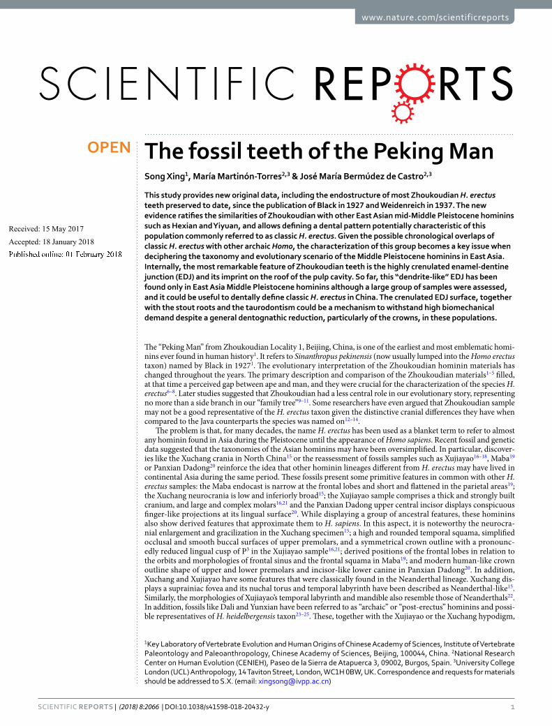
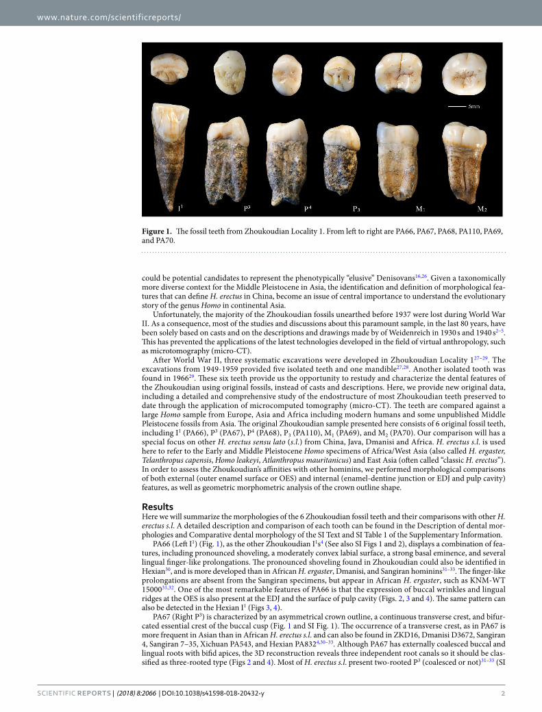
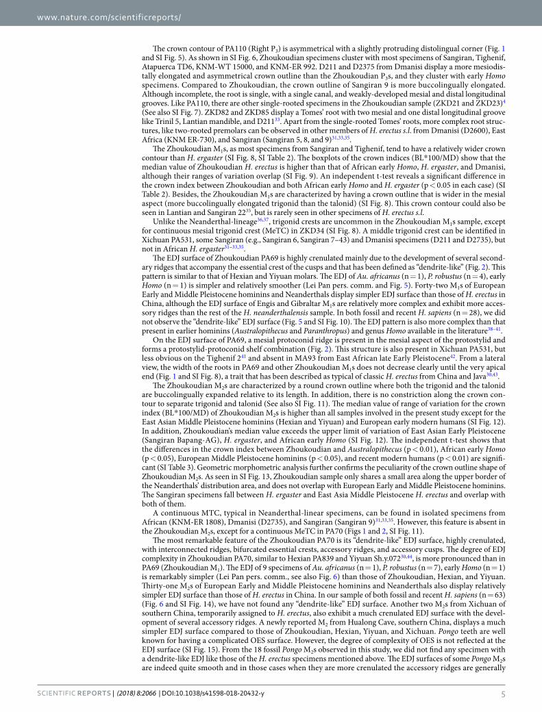
PA66 (Left I1) (Fig. 1), as the other Zhoukoudian I1s4 (See also SI Figs 1 and 2), displays a combination of fea-tures, including pronounced shoveling, a moderately convex labial surface, a strong basal eminence, and several lingual finger-like prolongations. The pronounced shoveling found in Zhoukoudian could also be identified in Hexian30, and is more developed than in African H. ergaster, Dmanisi, and Sangiran hominins31–33. The finger-like prolongations are absent from the Sangiran specimens, but appear in African H. ergaster, such as KNM-WT 1500031,32. One of the most remarkable features of PA66 is that the expression of buccal wrinkles and lingual ridges at the OES is also present at the EDJ and the surface of pulp cavity (Figs. 2, 3 and 4). The same pattern can also be detected in the Hexian I1 (Figs 3, 4).
PA67 (Right P3) is characterized by an asymmetrical crown outline, a continuous transverse crest, and bifur-cated essential crest of the buccal cusp (Fig. 1 and SI Fig. 1). The occurrence of a transverse crest, as in PA67 is more frequent in Asian than in African H. erectus s.l. and can also be found in ZKD16, Dmanisi D3672, Sangiran 4, Sangiran 7–35, Xichuan PA543, and Hexian PA8324,30–33. Although PA67 has externally coalesced buccal and lingual roots with bifid apices, the 3D reconstruction reveals three independent root canals so it should be clas-sified as three-rooted type (Figs 2 and 4). Most of H. erectus s.l. present two-rooted P3 (coalesced or not)31–33 (SI
Figure 1. The fossil teeth from Zhoukoudian Locality 1. From left to right are PA66, PA67, PA68, PA110, PA69, and PA70.

www.nature.com/scientificreports/
3SCIENtIfIC RePoRTS | (2018) 8:2066 | DOI:10.1038/s41598-018-20432-y
Figure 2. 3D virtual reconstructions of the Zhoukoudian fossil teeth. First row: occlusal view of the enamel surface; second row: occlusal view of the dentine surface; third row: buccal view of the whole tooth with the enamel and dentine being transparent and pulp cavity being opaque; forth row: mesial view of the whole tooth with the enamel and dentine being transparent and pulp cavity being opaque. From left to right are PA66, PA67, PA68, PA110, PA69, and PA70.
Figure 3. Comparisons of Zhoukoudian and Hexian I1s in the features of dentine surface. B: buccal; D: distal; M: mesial. Note the crenulations of the dentine at the labial surface.

www.nature.com/scientificreports/
4SCIENtIfIC RePoRTS | (2018) 8:2066 | DOI:10.1038/s41598-018-20432-y
Fig. 3). Three-rooted P3s like that of Zhoukoudian are only found in a few cases like Hexian PA832, Sangiran 7–35 and African H. ergaster KNM-ER 180830–32.
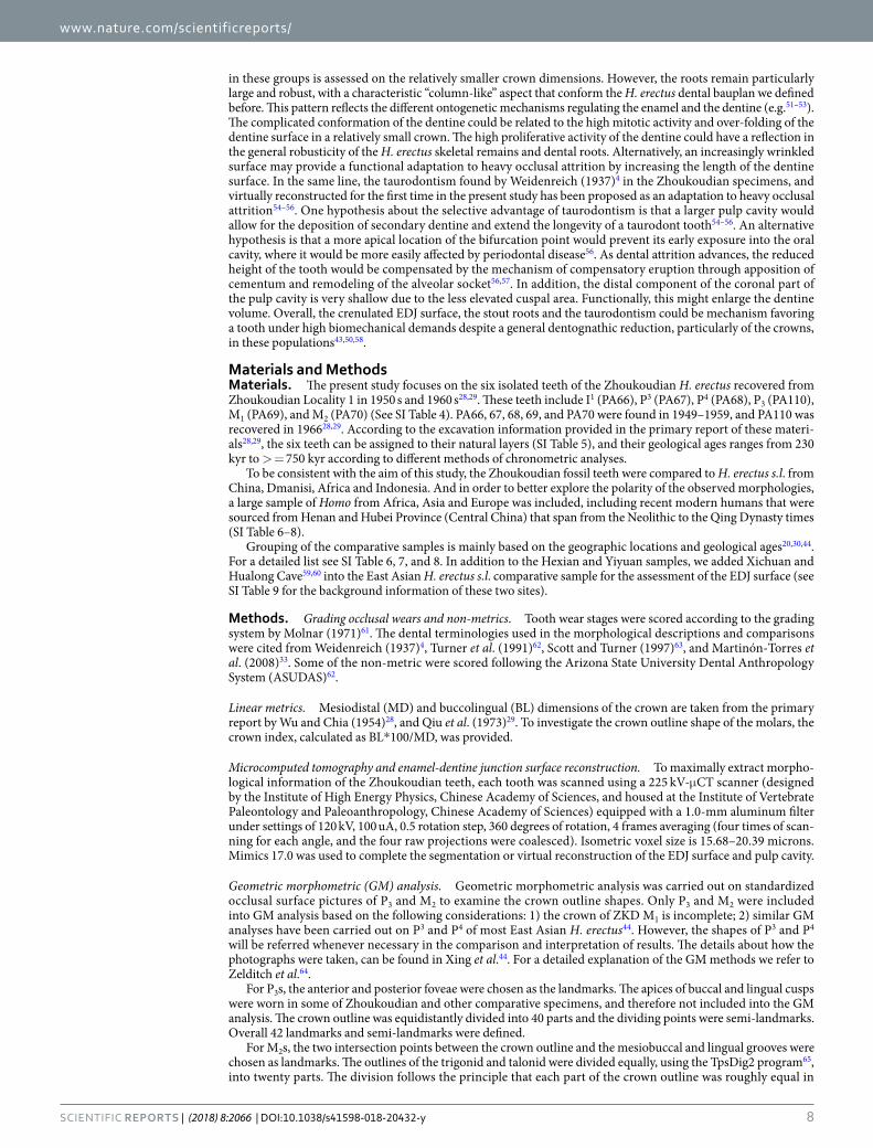
The crown contour of PA68 (Right P4) is ellipse-like and slightly asymmetrical, and the widths of the buccal and lingual cusps are roughly equal (Fig. 1 and SI Fig. 1). This type of crown outline shape is typical of other H. erectus s.l. specimens (SI Fig. 4)4,31,32. The presence of a continuous transverse crest, like that in PA68, also exists in other specimens of H. erectus s.l., such as ZKD 27, Sangiran 7–29, and KNM-ER 37334,31,32. The root is wide and comprises two radicals that coalesce along most of its length except for a strongly bifurcated tip. The number of premolar roots in H. erectus s.l. ranges from two to three roots, and in some cases the buccal and lingual roots are coalesced as in PA6831–33. An enlarged pulp cavity (hypertaurodont; term from Shaw [1928]34), as shown in the Zhoukoudian PA68, can also be observed in the East Asian Middle Pleistocene hominin from Yiyuan (Sh.y. 007) (Fig. 4).
Figure 4. Comparisons of Zhoukoudian, Hexian, Yiyuan, Xichuan, Hualong Cave I1s, P3, P4, and M2s in the morphologies of pulp cavity.
www.nature.com/scientificreports/
5SCIENtIfIC RePoRTS | (2018) 8:2066 | DOI:10.1038/s41598-018-20432-y
The crown contour of PA110 (Right P3) is asymmetrical with a slightly protruding distolingual corner (Fig. 1 and SI Fig. 5). As shown in SI Fig. 6, Zhoukoudian specimens cluster with most specimens of Sangiran, Tighenif, Atapuerca TD6, KNM-WT 15000, and KNM-ER 992. D211 and D2375 from Dmanisi display a more mesiodis-tally elongated and asymmetrical crown outline than the Zhoukoudian P3s, and they cluster with early Homo specimens. Compared to Zhoukoudian, the crown outline of Sangiran 9 is more buccolingually elongated. Although incomplete, the root is single, with a single canal, and weakly-developed mesial and distal longitudinal grooves. Like PA110, there are other single-rooted specimens in the Zhoukoudian sample (ZKD21 and ZKD23)4 (See also SI Fig. 7). ZKD82 and ZKD85 display a Tomes’ root with two mesial and one distal longitudinal groove like Trinil 5, Lantian mandible, and D21133. Apart from the single-rooted Tomes’ roots, more complex root struc-tures, like two-rooted premolars can be observed in other members of H. erectus s.l. from Dmanisi (D2600), East Africa (KNM ER-730), and Sangiran (Sangiran 5, 8, and 9)31,33,35.
The Zhoukoudian M1s, as most specimens from Sangiran and Tighenif, tend to have a relatively wider crown contour than H. ergaster (SI Fig. 8, SI Table 2). The boxplots of the crown indices (BL*100/MD) show that the median value of Zhoukoudian H. erectus is higher than that of African early Homo, H. ergaster, and Dmanisi, although their ranges of variation overlap (SI Fig. 9). An independent t-test reveals a significant difference in the crown index between Zhoukoudian and both African early Homo and H. ergaster (p < 0.05 in each case) (SI Table 2). Besides, the Zhoukoudian M1s are characterized by having a crown outline that is wider in the mesial aspect (more buccolingually elongated trigonid than the talonid) (SI Fig. 8). This crown contour could also be seen in Lantian and Sangiran 2235, but is rarely seen in other specimens of H. erectus s.l.
Unlike the Neanderthal-lineage36,37, trigonid crests are uncommon in the Zhoukoudian M1s sample, except for continuous mesial trigonid crest (MeTC) in ZKD34 (SI Fig. 8). A middle trigonid crest can be identified in Xichuan PA531, some Sangiran (e.g., Sangiran 6, Sangiran 7–43) and Dmanisi specimens (D211 and D2735), but not in African H. ergaster31–33,35.
The EDJ surface of Zhoukoudian PA69 is highly crenulated mainly due to the development of several second-ary ridges that accompany the essential crest of the cusps and that has been defined as “dendrite-like” (Fig. 2). This pattern is similar to that of Hexian and Yiyuan molars. The EDJ of Au. africanus (n = 1), P. robustus (n = 4), early Homo (n = 1) is simpler and relatively smoother (Lei Pan pers. comm. and Fig. 5). Forty-two M1s of European Early and Middle Pleistocene hominins and Neanderthals display simpler EDJ surface than those of H. erectus in China, although the EDJ surface of Engis and Gibraltar M1s are relatively more complex and exhibit more acces-sory ridges than the rest of the H. neanderthalensis sample. In both fossil and recent H. sapiens (n = 28), we did not observe the “dendrite-like” EDJ surface (Fig. 5 and SI Fig. 10). The EDJ pattern is also more complex than that present in earlier hominins (Australopithecus and Paranthropus) and genus Homo available in the literature38–41.
On the EDJ surface of PA69, a mesial protoconid ridge is present in the mesial aspect of the protostylid and forms a protostylid-protoconid shelf combination (Fig. 2). This structure is also present in Xichuan PA531, but less obvious on the Tighenif 241 and absent in MA93 from East African late Early Pleistocene42. From a lateral view, the width of the roots in PA69 and other Zhoukoudian M1s does not decrease clearly until the very apical end (Fig. 1 and SI Fig. 8), a trait that has been described as typical of classic H. erectus from China and Java30,43.
The Zhoukoudian M2s are characterized by a round crown outline where both the trigonid and the talonid are buccolingually expanded relative to its length. In addition, there is no constriction along the crown con-tour to separate trigonid and talonid (See also SI Fig. 11). The median value of range of variation for the crown index (BL*100/MD) of Zhoukoudian M2s is higher than all samples involved in the present study except for the East Asian Middle Pleistocene hominins (Hexian and Yiyuan) and European early modern humans (SI Fig. 12). In addition, Zhoukoudian’s median value exceeds the upper limit of variation of East Asian Early Pleistocene (Sangiran Bapang-AG), H. ergaster, and African early Homo (SI Fig. 12). The independent t-test shows that the differences in the crown index between Zhoukoudian and Australopithecus (p < 0.01), African early Homo (p < 0.05), European Middle Pleistocene hominins (p < 0.05), and recent modern humans (p < 0.01) are signifi-cant (SI Table 3). Geometric morphometric analysis further confirms the peculiarity of the crown outline shape of Zhoukoudian M2s. As seen in SI Fig. 13, Zhoukoudian sample only shares a small area along the upper border of the Neanderthals’ distribution area, and does not overlap with European Early and Middle Pleistocene hominins. The Sangiran specimens fall between H. ergaster and East Asia Middle Pleistocene H. erectus and overlap with both of them.
A continuous MTC, typical in Neanderthal-linear specimens, can be found in isolated specimens from African (KNM-ER 1808), Dmanisi (D2735), and Sangiran (Sangiran 9)31,33,35. However, this feature is absent in the Zhoukoudian M2s, except for a continuous MeTC in PA70 (Figs 1 and 2, SI Fig. 11).
The most remarkable feature of the Zhoukoudian PA70 is its “dendrite-like” EDJ surface, highly crenulated, with interconnected ridges, bifurcated essential crests, accessory ridges, and accessory cusps. The degree of EDJ complexity in Zhoukoudian PA70, similar to Hexian PA839 and Yiyuan Sh.y.07230,44, is more pronounced than in PA69 (Zhoukoudian M1). The EDJ of 9 specimens of Au. africanus (n = 1), P. robustus (n = 7), early Homo (n = 1) is remarkably simpler (Lei Pan pers. comm., see also Fig. 6) than those of Zhoukoudian, Hexian, and Yiyuan. Thirty-one M2s of European Early and Middle Pleistocene hominins and Neanderthals also display relatively simpler EDJ surface than those of H. erectus in China. In our sample of both fossil and recent H. sapiens (n = 63) (Fig. 6 and SI Fig. 14), we have not found any “dendrite-like” EDJ surface. Another two M2s from Xichuan of southern China, temporarily assigned to H. erectus, also exhibit a much crenulated EDJ surface with the devel-opment of several accessory ridges. A newly reported M2 from Hualong Cave, southern China, displays a much simpler EDJ surface compared to those of Zhoukoudian, Hexian, Yiyuan, and Xichuan. Pongo teeth are well known for having a complicated OES surface. However, the degree of complexity of OES is not reflected at the EDJ surface (SI Fig. 15). From the 18 fossil Pongo M2s observed in this study, we did not find any specimen with a dendrite-like EDJ like those of the H. erectus specimens mentioned above. The EDJ surfaces of some Pongo M2s are indeed quite smooth and in those cases when they are more crenulated the accessory ridges are generally

www.nature.com/scientificreports/
6SCIENtIfIC RePoRTS | (2018) 8:2066 | DOI:10.1038/s41598-018-20432-y
thinner and lower than those from Zhoukoudian, Hexian, Yiyuan, and Xichuan. None of the hominins available in the literature show this type of highly-crenulated EDJ38–41,45. In these specimens, the secondary grooves and ridges of both the enamel and the dentine surfaces are also reflected at the occlusal surface of the pulp cavity, being a peculiarity not recorded in any other hominin group so far (Fig. 4).
At the EDJ, the protostylid-protoconid shelf combination, as in PA69, also exist in PA70 and Hexian M2 (PA839)30. Comparatively, the protostylid is less elevated in the samples of North African Middle Pleistocene41 and specimens of Sangiran Bapang-AG assemblage40.The PA70 root consists of two radicals that coalesce along the whole length and that do not narrow until the tip (SI Fig. 5). The 3D virtual reconstruction of PA70 in the present study reveals an enlarged pulp cavity (Figs 2 and 4) compared to that of ZKD KI and GI4, Hexian PA831 (Fig. 4)46 and Tighenif specimens41. Furthermore, the distal component of the coronal part of the pulp cavity in Zhoukoudian PA70, Hexian PA831, and Yiyuan Sh.y.072 is shallow due to the less elevated cuspal area (Fig. 4). This pattern is different from that of Hualong Cave specimen, Tighenif 1 and 241 and NG92 D6 ZE 57 s/d 76 of Sangiran40, where the cuspal areas of the talonid are relatively sharper than those of Zhoukoudian, Hexian, and Yiyuan.
DiscussionThis is the first time, since the publications of the Zhoukoudian teeth by Black in 19271 and Weidenreich in 19374, that new original data, including a detailed and comprehensive study of the endostructure of most Zhoukoudian teeth preserved to date, are provided. This paper also presents the first direct comparisons of orig-inal Zhoukoudian sample with a large sample of Early and Middle Pleistocene teeth from Asia (e.g., Xichuan and Hualong Cave) and Europe (the Gran Dolina-TD6 and Sima de los Huesos fossils from Atapuerca). The new evidence confirms the similarities of the Zhoukoudian sample with other East Asian Middle Pleistocene homin-ins such as Hexian30,46, Yiyuan44, and Xichuan, and allow us to define a characteristic dental pattern for the pop-ulations that inhabited China during the Middle Pleistocene and that are usually classified as classic H. erectus.
Externally, Zhoukoudian teeth show the dental features that have been proposed in previous studies as typ-ical of East Asian Middle Pleistocene H. erectus30,44,46 such as i) moderately convex labial surfaces, tuberculum
Figure 5. Comparison of M1 dentine surfaces of Zhoukoudian and other comparative specimens. TD6 level of Atapuerca-Gran Dolina; SH: Atapuerca-Sima de los Huesos; RMH: Recent modern human. Some of the dental images were mirror-imaged to facilitate the comparison with the Zhoukoudian M1. CT data of Engis, Gibraltar, and Qafzeh are from European Synchrotron Radiation Facility (ESRF) (http://paleo.esrf.fr) and published in Smith et al.67. Data of Krapina is from NESPOS68. Picture of SKX 257 was revised after Pan69.

www.nature.com/scientificreports/
7SCIENtIfIC RePoRTS | (2018) 8:2066 | DOI:10.1038/s41598-018-20432-y
dentale in the shape of several finger-like prolongations and pronounced shoveling in upper central incisors, ii) bucco-lingually expanded crown outline in M2, iii) bucco-lingually expanded mesial cusps compared to the distal cusps in molars, iv) rare occurrence of middle trigonid crest, (v) robust “column-like” dental roots that only narrow at the tip, and (vi) shelf-like protostylid and mesial protoconid ridge at the EDJ. These features are also partially present in Java (this study and references32,35) except for the Bapang fossil reported by Zanolli (2013)47 which present remarkably more simplified external and internal morphology.
Thanks to the application of micro-CT scanning this paper presents some morphological features at the den-tine surface that have not been reported so far in any other hominin outside China and that could potentially represent unique characteristics of classic H. erectus in this region. Our study shows that the highly crenulated “dendrite-like” EDJ surface previously identified only in the Zhoukoudian, Yiyuan and Hexian M2s30,44,46, is also found in the M1 of Zhoukoudian, and the M2s of the Xichuan site from southern China. Surprisingly, the “dendrite-like” EDJ surface is also imprinted on the roof of the pulp cavity of these teeth. The crenulated labial surface of Zhoukoudian I1 might also be related to the complexity of the OES and EDJ of posterior teeth. To date, these features have been only described in Zhoukoudian, Yiyuan, Hexian and Xichuan hominins, and are absent in other hominin groups (Australopithecus, Paranthropus and Homo) analyzed by ourselves and/or available in the literature38–41,48,49. Unfortunately, no virtual reconstruction of the EDJ surface of Trinil teeth (holotype of H. erectus) is available (but see SI Fig. 16 for the relatively smooth EDJ lines of Trinil M3), so we cannot confirm whether this pattern is tentatively autopomorphic of the H. erectus taxon or a particularity of the H. erectus pop-ulations from China. In both cases, the morphological information provided here could be particularly useful to dentally define H. erectus from China and to distinguish them from other hominin lineages that may have poten-tially inhabited the continent at the same time.
Future studies and more data could shed light on the evolutionary meaning of this highly-crenulated EDJ surface. H. erectus from both continental and Southeast Asia has been characterized as displaying some degree dentognathic reduction in comparison to contemporaneous populations from Africa43,50. The dental reduction
Figure 6. Comparison of M2 dentine surfaces of Zhoukoudian and other comparative specimens. TD6 level of Atapuerca-Gran Dolina; SH: Atapuerca-Sima de los Huesos. RMH: Recent modern human. Some of the dental images were mirror-imaged to facilitate the comparison with the Zhoukoudian M2. CT data of Engis, Gibraltar, and Qafzeh are from European Synchrotron Radiation Facility (ESRF) (http://paleo.esrf.fr) and published in Smith et al.67. Data of Regourdou and Krapina is from NESPOS68. Picture of SK15 was revised after Pan69.
www.nature.com/scientificreports/
8SCIENtIfIC RePoRTS | (2018) 8:2066 | DOI:10.1038/s41598-018-20432-y
in these groups is assessed on the relatively smaller crown dimensions. However, the roots remain particularly large and robust, with a characteristic “column-like” aspect that conform the H. erectus dental bauplan we defined before. This pattern reflects the different ontogenetic mechanisms regulating the enamel and the dentine (e.g.51–53). The complicated conformation of the dentine could be related to the high mitotic activity and over-folding of the dentine surface in a relatively small crown. The high proliferative activity of the dentine could have a reflection in the general robusticity of the H. erectus skeletal remains and dental roots. Alternatively, an increasingly wrinkled surface may provide a functional adaptation to heavy occlusal attrition by increasing the length of the dentine surface. In the same line, the taurodontism found by Weidenreich (1937)4 in the Zhoukoudian specimens, and virtually reconstructed for the first time in the present study has been proposed as an adaptation to heavy occlusal attrition54–56. One hypothesis about the selective advantage of taurodontism is that a larger pulp cavity would allow for the deposition of secondary dentine and extend the longevity of a taurodont tooth54–56. An alternative hypothesis is that a more apical location of the bifurcation point would prevent its early exposure into the oral cavity, where it would be more easily affected by periodontal disease56. As dental attrition advances, the reduced height of the tooth would be compensated by the mechanism of compensatory eruption through apposition of cementum and remodeling of the alveolar socket56,57. In addition, the distal component of the coronal part of the pulp cavity is very shallow due to the less elevated cuspal area. Functionally, this might enlarge the dentine volume. Overall, the crenulated EDJ surface, the stout roots and the taurodontism could be mechanism favoring a tooth under high biomechanical demands despite a general dentognathic reduction, particularly of the crowns, in these populations43,50,58.
Materials and MethodsMaterials. The present study focuses on the six isolated teeth of the Zhoukoudian H. erectus recovered from Zhoukoudian Locality 1 in 1950 s and 1960 s28,29. These teeth include I1 (PA66), P3 (PA67), P4 (PA68), P3 (PA110), M1 (PA69), and M2 (PA70) (See SI Table 4). PA66, 67, 68, 69, and PA70 were found in 1949–1959, and PA110 was recovered in 196628,29. According to the excavation information provided in the primary report of these materi-als28,29, the six teeth can be assigned to their natural layers (SI Table 5), and their geological ages ranges from 230 kyr to > = 750 kyr according to different methods of chronometric analyses.
To be consistent with the aim of this study, the Zhoukoudian fossil teeth were compared to H. erectus s.l. from China, Dmanisi, Africa and Indonesia. And in order to better explore the polarity of the observed morphologies, a large sample of Homo from Africa, Asia and Europe was included, including recent modern humans that were sourced from Henan and Hubei Province (Central China) that span from the Neolithic to the Qing Dynasty times (SI Table 6–8).
Grouping of the comparative samples is mainly based on the geographic locations and geological ages20,30,44. For a detailed list see SI Table 6, 7, and 8. In addition to the Hexian and Yiyuan samples, we added Xichuan and Hualong Cave59,60 into the East Asian H. erectus s.l. comparative sample for the assessment of the EDJ surface (see SI Table 9 for the background information of these two sites).
Methods. Grading occlusal wears and non-metrics. Tooth wear stages were scored according to the grading system by Molnar (1971)61. The dental terminologies used in the morphological descriptions and comparisons were cited from Weidenreich (1937)4, Turner et al. (1991)62, Scott and Turner (1997)63, and Martinón-Torres et al. (2008)33. Some of the non-metric were scored following the Arizona State University Dental Anthropology System (ASUDAS)62.
Linear metrics. Mesiodistal (MD) and buccolingual (BL) dimensions of the crown are taken from the primary report by Wu and Chia (1954)28, and Qiu et al. (1973)29. To investigate the crown outline shape of the molars, the crown index, calculated as BL*100/MD, was provided.
Microcomputed tomography and enamel-dentine junction surface reconstruction. To maximally extract morpho-logical information of the Zhoukoudian teeth, each tooth was scanned using a 225 kV-μCT scanner (designed by the Institute of High Energy Physics, Chinese Academy of Sciences, and housed at the Institute of Vertebrate Paleontology and Paleoanthropology, Chinese Academy of Sciences) equipped with a 1.0-mm aluminum filter under settings of 120 kV, 100 uA, 0.5 rotation step, 360 degrees of rotation, 4 frames averaging (four times of scan-ning for each angle, and the four raw projections were coalesced). Isometric voxel size is 15.68–20.39 microns. Mimics 17.0 was used to complete the segmentation or virtual reconstruction of the EDJ surface and pulp cavity.
Geometric morphometric (GM) analysis. Geometric morphometric analysis was carried out on standardized occlusal surface pictures of P3 and M2 to examine the crown outline shapes. Only P3 and M2 were included into GM analysis based on the following considerations: 1) the crown of ZKD M1 is incomplete; 2) similar GM analyses have been carried out on P3 and P4 of most East Asian H. erectus44. However, the shapes of P3 and P4 will be referred whenever necessary in the comparison and interpretation of results. The details about how the photographs were taken, can be found in Xing et al.44. For a detailed explanation of the GM methods we refer to Zelditch et al.64.
For P3s, the anterior and posterior foveae were chosen as the landmarks. The apices of buccal and lingual cusps were worn in some of Zhoukoudian and other comparative specimens, and therefore not included into the GM analysis. The crown outline was equidistantly divided into 40 parts and the dividing points were semi-landmarks. Overall 42 landmarks and semi-landmarks were defined.
For M2s, the two intersection points between the crown outline and the mesiobuccal and lingual grooves were chosen as landmarks. The outlines of the trigonid and talonid were divided equally, using the TpsDig2 program65, into twenty parts. The division follows the principle that each part of the crown outline was roughly equal in
www.nature.com/scientificreports/
9SCIENtIfIC RePoRTS | (2018) 8:2066 | DOI:10.1038/s41598-018-20432-y
length. The dividing points were treated as semi-landmarks. In total 40 landmarks and semi-landmarks were defined.
The TpsDig2 program65 was employed to digitize landmarks and semi-landmarks. The TpsRelw program66 was used to undertake superimposition on the raw coordinate data and the relative warp analysis (or principal component analysis) of shape variables.
Availability of materials and data. All data generated or analyzed during this study are included in this published article (and its Supplementary Information files).
References 1. Black, D. On a lower molar hominid tooth from the Chou Kou Tien deposit. Palaeontologica Sinica, Series D 7, 1–29 (1927). 2. Weidenreich, F. The skull of Sinanthropus pekinensis: a comparative study on a primitive hominid skull. Palaeontologica Sinica, Series
D 5, 1–150 (1943). 3. Weidenreich, F. The mandibles of Sinanthropus pekinensis: a comparative study. Palaeontologica Sinica, Series D 4(7), 1–162 (1936). 4. Weidenreich, F. The dentition of Sinanthropus pekinensis: a comparative odontography of the hominids. Palaeontologica Sinica, New
Series D 1, 1–180 (1937). 5. Weidenreich, F. The extremity bones of Sinanthropus pekinensis: a comparative odontography of the hominids. Palaeontologica
Sinica, New Series D 5, 1–150 (1941). 6. Weidenreich, F. Some Problems dealing with ancient man. American Anthropologist 42, 375–383 (1940). 7. Mayr, E. On the concepts and terminology of vertical subspecies and species. National Research Council Committee on Common
Problems of Genetics, Paleontology, and Systematics Bulletin 2, 11–16 (1944). 8. Mayr, E. Taxonomic categories in fossil hominids. Cold Spring Harbor Symposia on Quantitative Biology 15, 109–118 (1950). 9. Groves, C. P. & Mazák, V. An approach to the taxonomy of the Hominidae: gracile Villafranchian hominids ofAfrica. Casopis pro
Mineralogii Geologii 20, 225–247 (1975). 10. Stringer, C. B. The definition of Homo erectus and the existence of the species in Africa and Europe. Courier Forschungs-Institut
Senckenberg 69, 131–143 (1984). 11. Andrews, P. An alternative interpretation of characters used to define Homo erectus. Courier Forschungs-Institut Senckenberg 69,
167–175 (1984). 12. Antón, S. C. Evolutionary significance of cranial variation in Asian Homo erectus. American Journal of Physical Anthropology 118,
301–323 (2002). 13. Kidder, J. H. & Durband, A. C. A re-evaluation of the metric diversity within Homo erectus. Journal of Human Evolution 46, 297–313
(2004). 14. Kaifu, Y. et al. Cranial morphology of Javanese Homo erectus: New evidence for continuous evolution, specialization, and terminal
extinction. Journal of Human Evolution 55, 551–580 (2008). 15. Li, Z.-Y. et al. Late Pleistocene archaic human crania from Xuchang, China. Science 355, 969–972 (2017). 16. Xing, S., Martinón-Torres, M., Bermúdez de Castro, J. M., Wu, X. & Liu, W. Hominin teeth from the early Late Pleistocene site of
Xujiayao, Northern China. American Journal of Physical Anthropology 156, 224–240 (2015). 17. Xing, S. et al. Perikymata distribution in Homo with special reference to the Xujiayao juvenile. American Journal of Physical
Anthropology 157, 684–693 (2015). 18. Wu, X. J. & Trinkaus, E. The Xujiayao 14 mandibular ramus and Pleistocene Homo mandibular variation. Comptes Rendus Palevol
13, 333–341 (2014). 19. Wu, X. & Bruner, E. The endocranial anatomy of Maba 1. American Journal of Physical Anthropology 160, 633–643 (2016). 20. Liu, W. et al. Late Middle Pleistocene hominin teeth from Panxian Dadong, South China. Journal of Human Evolution 64, 337–355
(2013). 21. Chia, L., Wei, Q. & Li, C. Report on the excavation of Hsuchiayao man site in 1976. Vertebrata Palasiatica 17, 277–293 (1979). 22. Wu, X. J. et al. The temporal labyrinths of Eastern Eurasian Pleistocene humans. Proceedings of the National Academy of Sciences of
the United States of America 111, 10509–10513 (2014). 23. Stringer, C. B. & Barnes, I. Decipehring the Denisovans. Proceedings of the National Academy of Sciences of the United States of
America 112, 15542–15543 (2015). 24. Buck, L. & Stringer, C. Homo heidelbergensis. Current Biology 24, R214–R215 (2014). 25. Stringer, C. The status of Homo heidelbergensis (Schoetensack 1908). Evolutionary Anthropology 21, 101–107 (2012). 26. Martinón-Torres, M., Xing, S., Liu, W. & Bermúdez de Castro, J. M. A. “source and sink” model for East Asia? Preliminary approach
through the dental evidence. Comptes Rendus Palevol, https://doi.org/10.1016/j.crpv.2015.09.011. In press. 27. Wu, J.-K. & Chao, T. K. New discovery of a Sinanthropus mandible in Choukoutien. Paleovertebrata et Paleoanthropologia 1, 155–158
(1959). 28. Wu, J.-K. & Chia, L.-P. New discovery of a Sinanthropus pekinensis in Choukoutien. Acta Palaeontologica Sinica 2, 267–288 (1954). 29. Qiu, Z., Gu, Y., Zhang, Y. & Zhang, S. Newly discovered Sinanthropus remains and stone artifacts at Zhoukoudian. Vertebrata
Palasiatica 11, 109–131 (1973). 30. Xing, S. et al. Middle Pleistocene Hominin Teeth from Longtan Cave, Hexian, China. PLOS ONE 9, e114265 (2015). 31. Wood, B. Hominid cranial remains. Koobi Fora research project, Vol. 4. (Clarendon Press, 1991). 32. Grine, F. & Franzen, J. Fossil hominid teeth from the Sangiran dome (Java, Indonesia). Courier Forschungs-Institut Senckenberg 171,
75–103 (1994). 33. Martinón-Torres, M. et al. Dental remains from Dmanisi (Republic of Georgia): Morphological analysis and comparative study.
Journal of Human Evolution 55, 249–273 (2008). 34. Shaw, J. C. Taurodont Teeth in South African Races. Journal of Anatomy 62, 476–498.1 (1928). 35. Kaifu, Y., Aziz, F. & Baba, H. Hominid Mandibular Remains from Sangiran: 1952–1986 Collection. American Journal of Physical
Anthropology 128, 497–519 (2005). 36. Bailey, S. E. A closer look at Neanderthal postcanine dental morphology: The mandibular dentition. The Anatomical Record 269,
148–156 (2002). 37. Martinón-Torres, M. et al. Morphological description and comparison of the dental remains from Atapuerca-Sima de los Huesos
site (Spain). Journal of Human Evolution 62, 7–58 (2012). 38. Skinner, M. M., Gunz, P., Wood, B. A. & Hublin, J.-J. Enamel-dentine junction (EDJ) morphology distinguishes the lower molars of
Australopithecus africanus and Paranthropus robustus. Journal of Human Evolution 55, 979–988 (2008). 39. Bailey, S. E., Skinner, M. M. & Hublin, J.-J. What lies beneath? An evaluation of lower molar trigonid crest patterns based on both
dentine and enamel expression. American Journal of Physical Anthropology 145, 505–518 (2011). 40. Zanolli, C. Molar crown inner structural organization in Javanese Homo erectus. American Journal of Physical Anthropology 156,
148–157 (2015). 41. Zanolli, C. & Mazurier, A. Endostructural characterization of the H. heidelbergensis dental remains from the early Middle
Pleistocene site of Tighenif, Algeria. Comptes Rendus Palevol 12, 293–304 (2013).

www.nature.com/scientificreports/
1 0SCIENtIfIC RePoRTS | (2018) 8:2066 | DOI:10.1038/s41598-018-20432-y
42. Zanolli, C. et al. The late Early Pleistocene human dental remains from Uadi Aalad and Mulhuli-Amo (Buia), Eritrean Danakil: Macromorphology and microstructure. Journal of Human Evolution 74, 96–113 (2014).
43. Kaifu, Y. Advanced dental reduction in Javanese Homo erectus. Anthropological Science 114, 35–43 (2006). 44. Xing, S. et al. Hominin teeth from the Middle Pleistocene site of Yiyuan, Eastern China. Journal of Human Evolution 95, 33–54
(2016). 45. Martínez de Pinillos, M. et al. Trigonid crests expression in Atapuerca-Sima de los Huesos lower molars: Internal and external
morphological expression and evolutionary inferences. Comptes Rendus Palevol 13, 205–221 (2014). 46. Liu, W. et al. A mandible from the Middle Pleistocene Hexian site and its significance in relation to the variability of Asian Homo
erectus. American Journal of Physical Anthropology 162, 715–731 (2017). 47. Zanolli, C. Additional Evidence for Morpho-Dimensional Tooth Crown Variation in a New Indonesian H. erectus Sample from the
Sangiran Dome (Central Java). PLoS ONE 8(7), e67233 (2013). 48. Skinner, M. M., Wood, B. A. & Hublin, J.-J. Protostylid expression at the enamel-dentine junction and enamel surface of mandibular
molars of Paranthropus robustus and Australopithecus africanus. Journal of Human Evolution 56, 76–85 (2009). 49. Martínez de Pinillos, M., Martinón-Torres, M., Martín-Francés, L., Arsuaga, J. L. & Bermúdez de Castro, J. M. Comparative analysis
of the trigonid crests patterns in Homo antecessor molars at the enamel and dentine surfaces. Quaternary International 433(Part A), 189–198 (2017).
50. Martinón-Torres, M. et al. Dental evidence on the hominin dispersals during the Pleistocene. Proceedings of the National Academy of Sciences of the United States of America 104, 13279–13282 (2007).
51. Alvesalo, L. In Anthropological perspectives on tooth morphology (ed G.R. Scott, Irish, J.D.) 92–107 (Cambridge University Press, 2013).
52. Schwartz, G. T. & Dean, M. C. Sexual Dimorphism in Modern Human Permanent Teeth. American Journal of Physical Anthropology 128, 312–317 (2005).
53. Schwartz, G. T., Reid, D. J. & Dean, C. Developmental aspects of sexual dimorphism in hominoid canines. International Journal of Primatology 22, 837–860 (2001).
54. Coon, C. S. The origin of races. (Alfred A. Knopf, Inc., 1962). 55. Hamner, J. E. III, Witkop, C. J. Jr. & Metro, P. S. Taurodontism. Oral Surgery, Oral Medicine, Oral Pathology 18, 409–418 (1964). 56. Blumberg, J. E., Hylander, W. L. & Goepp, R. A. Taurodontism: A biometric study. American Journal of Physical Anthropology 34,
243–255 (1971). 57. Martinon-Torres, M. et al. Early Pleistocene human mandible from Sima del Elefante (TE) cave site in Sierra de Atapuerca (Spain):
a palaeopathological study. Journal of Human Evolution 61, 1–11 (2011). 58. Kaifu, Y. et al. Taxonomic affinities and evolutionary history of the early Pleistocene hominids of Java: Dentognathic evidence.
American Journal of Physical Anthropology 128, 709–726 (2005). 59. Wu, R. & Wu, X. Human fossil from Xichuan, Henan. Vertebrata Palasiatica 20, 1–9 (1982). 60. Gong, X. et al. Human fossils found from Hualong Cave, Dongzhi County, Anhui Province. Acta Anthropologica Sinica 33, 427–436
(2014). 61. Molnar, S. Human tooth wear, tooth function and cultural variability. American Journal of Physical Anthropology 34, 175–189 (1971). 62. Turner, C. G. II., Nichol, C. R. & Scott, G. R. Scoring procedures for key morphological traits of the permanent dentition: The
Arizona State University Dental Anthropology System. In Advances in Dental Anthropology (eds M.; Kelley, M. & Larsen, C.) pp. 13–31 (Wiley-Liss, 1991).
63. Scott, G. R. & Turner, C. G II. The anthropology of modern human teeth: Dental morphology and its variation in recent human populations Vol. 20. (Cambridge University Press, 1997).
64. Zelditch, M. L., Swiderski, D. L. & Sheets, H. D. Geometric morphometrics for biologists: a primer. (Elsevier Academic Press, 2012). 65. Rohlf, F. J. TpsDig2. Ecology and Evolution, SUNY. Stony Brook, New York http://life.bio.sunysb.edu/morph/ (1998). 66. Rohlf, F. J. TpsRelw. Ecology and Evolution, SUNY. Stony Brook, New York http://life.bio.sunysb.edu/morph/ (1998). 67. Smith et al. Dental evidence for ontogenetic differences between modern humans and Neanderthals. Proceedings of the National
Academy of Sciences of the United States of America 107, 20913–20928 (2010). 68. NESPOS database, Neanderthal Studies Professional Online Service. http://www.nespos.org (2015). 69. Pan, L. The earliest members of the genus Homo in South Africa: evidence from dental inner structure of lower postcanine dentition:
Ph. D. Dissertation, Université de Toulouse 3. 2016.
AcknowledgementsThe authors thank all the colleagues and institutions that have helped us to accomplish this study. The authors would like to thank all the colleagues that have helped us to accomplish this study. Prof. Juan Luis Arsuaga and Prof. Eudald Carbonell allow the access to the Atapuerca fossils. We also express our gratitude to several people for providing access to the studied materials and their helpful assistance during observations: R. Clarke (University of the Witwatersrand, South Africa), J. Svoboda (Institute of Archaeology, Paleolithic and Paleoethnology Research Center, Dolní Vestonice, Czech Republic), I. Tattersall, K. Mowbray, and G. Sawyer (American Museum of Natural History, New York), John de Vos (Naturallis Museum, Leiden, The Netherlands), H. de Lumley, M.-A. de Lumley, and A. Vialet (Institut de Paléontologie Humaine, Paris, France), P. Tassy (Muséum National d’Histoire Naturelle, Paris, France), Dr. Yousuke Kaifu (National Science Museum, Tokyo), and Dr. Mi Zhou (Institute of Archeology and Cultural Relics of Hubei Province, Wuhan). Prof. Wolpoff provided the valuable data of dental metrics of fossil hominins from around the world. Thanks to Mark Sier from Oxford University for his valuable suggestions. Thanks to Dr. Lei Pan for her great help with the observation of EDJ surfaces of Australopithecus and African early Homo. This work was supported by the Chinese Academy of Sciences (XDPB05, 132311KYSB20160004), the National Natural Science Foundation of China (41630102, 41672020), Ministerio de Economía y Competitividad of Spain MINECO/FEDER (CGL2015-65387-C3-3-P), Acción Integrada España Francia (HF2007-0115), British Academy (International Partnership and Mobility Scheme PM160019) and the Leakey Foundation through the personal support of Dub Crook (2014, 2015, 2016) and Gordon Getty (2012) to one of the authors (M.M.-T.).
Author ContributionsS.X. and M.M.T. contributed to the data collection and analysis. S.X., M.M.T., J.M.B.C. wrote the paper.
Additional InformationSupplementary information accompanies this paper at https://doi.org/10.1038/s41598-018-20432-y.Competing Interests: The authors declare that they have no competing interests.
www.nature.com/scientificreports/
1 1SCIENtIfIC RePoRTS | (2018) 8:2066 | DOI:10.1038/s41598-018-20432-y
Publisher's note: Springer Nature remains neutral with regard to jurisdictional claims in published maps and institutional affiliations.
Open Access This article is licensed under a Creative Commons Attribution 4.0 International License, which permits use, sharing, adaptation, distribution and reproduction in any medium or
format, as long as you give appropriate credit to the original author(s) and the source, provide a link to the Cre-ative Commons license, and indicate if changes were made. The images or other third party material in this article are included in the article’s Creative Commons license, unless indicated otherwise in a credit line to the material. If material is not included in the article’s Creative Commons license and your intended use is not per-mitted by statutory regulation or exceeds the permitted use, you will need to obtain permission directly from the copyright holder. To view a copy of this license, visit http://creativecommons.org/licenses/by/4.0/. © The Author(s) 2018