the first selenoanhydride in the series of chlorophyll ... - mdpi
TRANSCRIPT
molecules
Article
The First Selenoanhydride in the Series of Chlorophyll aDerivatives, Its Stability and Photoinduced Cytotoxicity
Viktor Pogorilyy 1,* , Anna Plyutinskaya 2, Nikita Suvorov 1, Ekaterina Diachkova 3,4,* , Yuriy Vasil’ev 5,6,Andrei Pankratov 2, Andrey Mironov 1 and Mikhail Grin 1
�����������������
Citation: Pogorilyy, V.; Plyutinskaya,
A.; Suvorov, N.; Diachkova, E.;
Vasil’ev, Y.; Pankratov, A.; Mironov,
A.; Grin, M. The First
Selenoanhydride in the Series of
Chlorophyll a Derivatives, Its
Stability and Photoinduced
Cytotoxicity. Molecules 2021, 26, 7298.
https://doi.org/10.3390/
molecules26237298
Academic Editors: Carlos J.
P. Monteiro, Catarina I. V. Ramos and
M. Amparo F. Faustino
Received: 30 October 2021
Accepted: 29 November 2021
Published: 1 December 2021
Publisher’s Note: MDPI stays neutral
with regard to jurisdictional claims in
published maps and institutional affil-
iations.
Copyright: © 2021 by the authors.
Licensee MDPI, Basel, Switzerland.
This article is an open access article
distributed under the terms and
conditions of the Creative Commons
Attribution (CC BY) license (https://
creativecommons.org/licenses/by/
4.0/).
1 Department of Chemistry and Technology of Biologically Active Compounds, Medicinal and OrganicChemistry, Institute of Fine Chemical Technologies, MIREA-Russian Technological University, 86 VernadskyAvenue, 119571 Moscow, Russia; [email protected] (N.S.); [email protected] (A.M.);[email protected] (M.G.)
2 P. Hertsen Moscow Oncology Research Institute—Branch of the National Medical Research RadiologicalCentre of the Ministry of Health of the Russian Federation, 2nd Botkinsky pr., 3, 125284 Moscow, Russia;[email protected] (A.P.); [email protected] (A.P.)
3 Department of Oral Surgery of Borovsky Institute of Dentistry, I.M. Sechenov First Moscow State MedicalUniversity (Sechenov University), Trubetskaya St. bldg. 8\2, 119435 Moscow, Russia
4 Department of Fundamental Medical Disciplines, Medical Faculty, Moscow Region State University (MRSU),Str. Radio 10 Build 1, 105005 Moscow, Russia
5 Department of Operative Surgery and Topographic Anatomy, I.M. Sechenov First Moscow State MedicalUniversity (Sechenov University), Trubetskaya St. bldg. 8\2, 119435 Moscow, Russia; [email protected]
6 Department of Prosthetic Dentistry, Dental Faculty, Kazan State Medical University of the Ministry of Healthof Russia, Str. Butlerova 49, 420012 Kazan, Russia
* Correspondence: [email protected] (V.P.); [email protected] (E.D.)
Abstract: In this work, we obtained the first selenium-containing chlorin with a chalcogen atomin exlocycle E. It was shown that the spectral properties were preserved in the target compoundand the stability increased at two different pH values, in comparison with the starting purpurin-18. The derivatives have sufficiently high fluorescence and singlet oxygen quantum yields. Thephotoinduced cytotoxicity of sulfur- and selenium-anhydrides of chlorin p6 studied for the first timein vitro on the S37 cell line was found to be two times higher that of purpurin-18 and purpurinimidestudied previously. Moreover, the dark cytotoxicity increased four-fold in comparison with the lattercompounds. Apparently, the increase in the dark cytotoxicity is due to the interaction of the pigmentsstudied with sulfur- and selenium-containing endogenous intracellular compounds. Intracellulardistributions of thioanhydride and selenoanhydride chlorin p6 in S37 cells were shown in cytoplasmby diffusion distribution. The intracellular concentration of the sulfur derivative turned out to behigher and, as a consequence, its photoinduced cytotoxicity was higher as well.
Keywords: naturally occurring chlorins; chlorin p6; cyclic thioanhydrides; cyclic selenoanhydrides;photodynamic therapy (PDT); photosensitizers (PS)
1. Introduction
The physical and chemical properties of selenium, an element belonging to the chalco-gen group, are like those of sulfur, which makes it possible to compare not only theelements themselves but also their compounds [1–3]. For example, an analogy may bedrawn between cysteine and selenocysteine that are proteinogenic amino acids, with thedifference that the sulfur atom in the thiol group of cysteine is replaced by a seleniumatom in the selenol group of selenocysteine [4]. The latter is found in more than 25 types ofprotein molecules in mammal organisms [5–7]. Selenium is also a member of the groupof glutathione peroxidase (GPx) enzymes that are involved in the reduction of hydrogenperoxide to water [8,9].
Many body disorders, including cancer, atherosclerosis, arthritis, and diseases of thecardiovascular system, are attributed to the lack of selenium [10,11]. Compared to sulfur,
Molecules 2021, 26, 7298. https://doi.org/10.3390/molecules26237298 https://www.mdpi.com/journal/molecules
Molecules 2021, 26, 7298 2 of 14
selenium exhibits higher antioxidant properties due to its higher nuclear charge, atomicmass, ionic and covalent radii, and a smaller ionization energy [12–14].
The main interest in selenium and its compounds stems from their wide use asantitumor agents [15–18]. It is believed that organic compounds of selenium are generatorsof superoxide anion O2− , hydrogen peroxide, and hydroxyl radicals that play the role ofmediators of oxidative stress and apoptosis, leading to the death of tumor cells [19]. Tumorcells protect themselves by activating an antioxidant system that involves glutathione(GT), glutathione peroxidase (GPx), glutathione S-transferase (GST), glutaredoxin (Grx),thioredoxin (Trx), superoxide dismutase (SOD), and catalase (CAT) [20–22]. The latter areoxidized by selenium compounds, thus making a cell vulnerable to oxidative stress.
Selenophene is the simplest heterocyclic compound containing a selenium atom [23,24].Several inorganic compounds are used as selenating agents in organic synthesis, includ-ing phosphorus pentaselenide, selenium oxides, and hydrogen selenide, which cause thecyclization of linear organic molecules to give selenophenes and their derivatives [24–26].The latter are used as efficient antioxidants and extracting agents for the isolation and sepa-ration of metals, while some of them have high biological activity or find use as polymericmaterials (tetra-substituted selenophene derivatives) [27–29].
Cyclic selenium-containing molecules can affect DNA repair, which allows one touse them as cancer-treatment drugs [15,16,19,25]. Selenium derivatives can suppress theexpression of HLA-E in tumor cells, thereby making them susceptible to natural killer cells,which leads to tumor-cell death [30,31].
The successful development of photodynamic therapy (PDT) for cancer is closelyrelated to the development of highly efficient drugs, i.e., photosensitizers (PSs) that lacktheir own bioactivity but, upon irradiation with light of a certain wavelength, pass intoan excited state and react with oxygen to generate active oxygen forms, including singletoxygen and radical anions [32]. The latter possess a cytotoxic effect but do not damage thesurrounding normal tissues due to the limited lifetime and free path. An optimal PS shouldabsorb light in the red or near-infrared spectral range, since, in this case, the light penetratesmore deeply into a tissue. PSs should have high quantum yields for singlet oxygen andfluorescence to work as theranostics, and should also be amphiphilic compounds with hightropism toward tumors of various genesis [33,34]. Derivatives of natural chlorins meet allthe above requirements, taking into account the fact that the pathways of their metabolismand excretion are well-known [35,36].
Earlier, our research group obtained derivatives of natural chlorins containing variousheterocycles, annulated with the main macrocycle, as photosensitizers for photodynamictherapy of cancer [37–40].
The aim of this study was to synthesize a previously unknown chlorin p6 seleniumanhydride and to study its photoinduced cytotoxicity toward a culture of tumor cells ofmouse sarcoma S-37.
2. Results
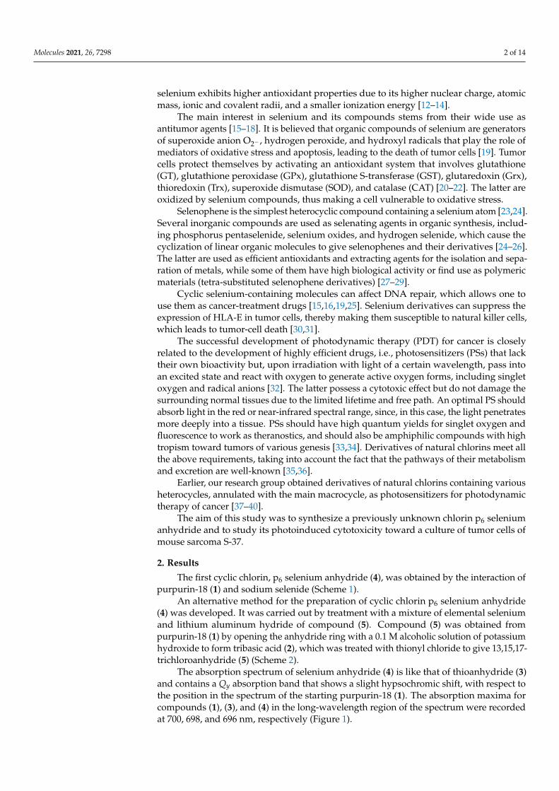
The first cyclic chlorin, p6 selenium anhydride (4), was obtained by the interaction ofpurpurin-18 (1) and sodium selenide (Scheme 1).
An alternative method for the preparation of cyclic chlorin p6 selenium anhydride(4) was developed. It was carried out by treatment with a mixture of elemental seleniumand lithium aluminum hydride of compound (5). Compound (5) was obtained frompurpurin-18 (1) by opening the anhydride ring with a 0.1 M alcoholic solution of potassiumhydroxide to form tribasic acid (2), which was treated with thionyl chloride to give 13,15,17-trichloroanhydride (5) (Scheme 2).
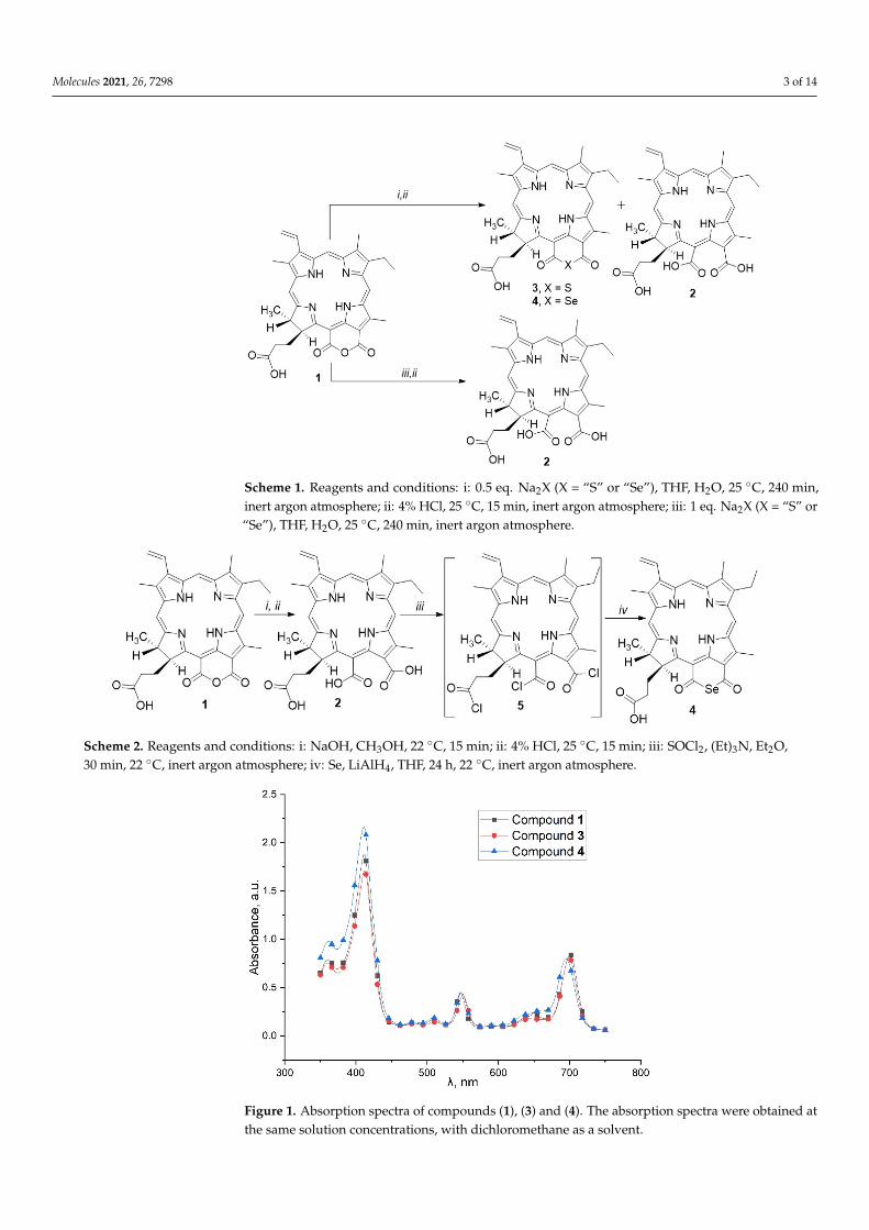
The absorption spectrum of selenium anhydride (4) is like that of thioanhydride (3)and contains a Qy absorption band that shows a slight hypsochromic shift, with respect tothe position in the spectrum of the starting purpurin-18 (1). The absorption maxima forcompounds (1), (3), and (4) in the long-wavelength region of the spectrum were recordedat 700, 698, and 696 nm, respectively (Figure 1).
Molecules 2021, 26, 7298 3 of 14Molecules 2021, 26, x FOR PEER REVIEW 3 of 15
Scheme 1. Reagents and conditions: i: 0.5 eq. Na2X (X = “S” or “Se”), THF, H2O, 25 °C, 240 min, inert argon atmosphere; ii: 4% HCl, 25 °C, 15 min, inert argon atmosphere; iii: 1 eq. Na2X (X = “S” or “Se”), THF, H2O, 25 °C, 240 min, inert argon atmosphere.
An alternative method for the preparation of cyclic chlorin p6 selenium anhydride (4) was developed. It was carried out by treatment with a mixture of elemental selenium and lithium aluminum hydride of compound (5). Compound (5) was obtained from purpurin-18 (1) by opening the anhydride ring with a 0.1 M alcoholic solution of potassium hydrox-ide to form tribasic acid (2), which was treated with thionyl chloride to give 13,15,17-tri-chloroanhydride (5) (Scheme 2).
Scheme 2. Reagents and conditions: i: NaOH, CH3OH, 22 °C, 15 min; ii: 4% HCl, 25 °C, 15 min; iii: SOCl2, (Et)3N, Et2O, 30 min, 22 °C, inert argon atmosphere; iv: Se, LiAlH4, THF, 24 h., 22 °C, inert argon atmosphere.
The absorption spectrum of selenium anhydride (4) is like that of thioanhydride (3) and contains a Qy absorption band that shows a slight hypsochromic shift, with respect to the position in the spectrum of the starting purpurin-18 (1). The absorption maxima for compounds (1), (3), and (4) in the long-wavelength region of the spectrum were recorded at 700, 698, and 696 nm, respectively (Figure 1).
Scheme 1. Reagents and conditions: i: 0.5 eq. Na2X (X = “S” or “Se”), THF, H2O, 25 ◦C, 240 min,inert argon atmosphere; ii: 4% HCl, 25 ◦C, 15 min, inert argon atmosphere; iii: 1 eq. Na2X (X = “S” or“Se”), THF, H2O, 25 ◦C, 240 min, inert argon atmosphere.
Molecules 2021, 26, x FOR PEER REVIEW 3 of 15
Scheme 1. Reagents and conditions: i: 0.5 eq. Na2X (X = “S” or “Se”), THF, H2O, 25 °C, 240 min, inert argon atmosphere; ii: 4% HCl, 25 °C, 15 min, inert argon atmosphere; iii: 1 eq. Na2X (X = “S” or “Se”), THF, H2O, 25 °C, 240 min, inert argon atmosphere.
An alternative method for the preparation of cyclic chlorin p6 selenium anhydride (4) was developed. It was carried out by treatment with a mixture of elemental selenium and lithium aluminum hydride of compound (5). Compound (5) was obtained from purpurin-18 (1) by opening the anhydride ring with a 0.1 M alcoholic solution of potassium hydrox-ide to form tribasic acid (2), which was treated with thionyl chloride to give 13,15,17-tri-chloroanhydride (5) (Scheme 2).
Scheme 2. Reagents and conditions: i: NaOH, CH3OH, 22 °C, 15 min; ii: 4% HCl, 25 °C, 15 min; iii: SOCl2, (Et)3N, Et2O, 30 min, 22 °C, inert argon atmosphere; iv: Se, LiAlH4, THF, 24 h., 22 °C, inert argon atmosphere.
The absorption spectrum of selenium anhydride (4) is like that of thioanhydride (3) and contains a Qy absorption band that shows a slight hypsochromic shift, with respect to the position in the spectrum of the starting purpurin-18 (1). The absorption maxima for compounds (1), (3), and (4) in the long-wavelength region of the spectrum were recorded at 700, 698, and 696 nm, respectively (Figure 1).
Scheme 2. Reagents and conditions: i: NaOH, CH3OH, 22 ◦C, 15 min; ii: 4% HCl, 25 ◦C, 15 min; iii: SOCl2, (Et)3N, Et2O,30 min, 22 ◦C, inert argon atmosphere; iv: Se, LiAlH4, THF, 24 h, 22 ◦C, inert argon atmosphere.
Molecules 2021, 26, x FOR PEER REVIEW 4 of 15
Figure 1. Absorption spectra of compounds (1), (3) and (4). The absorption spectra were obtained at the same solution concentrations, with dichloromethane as a solvent.
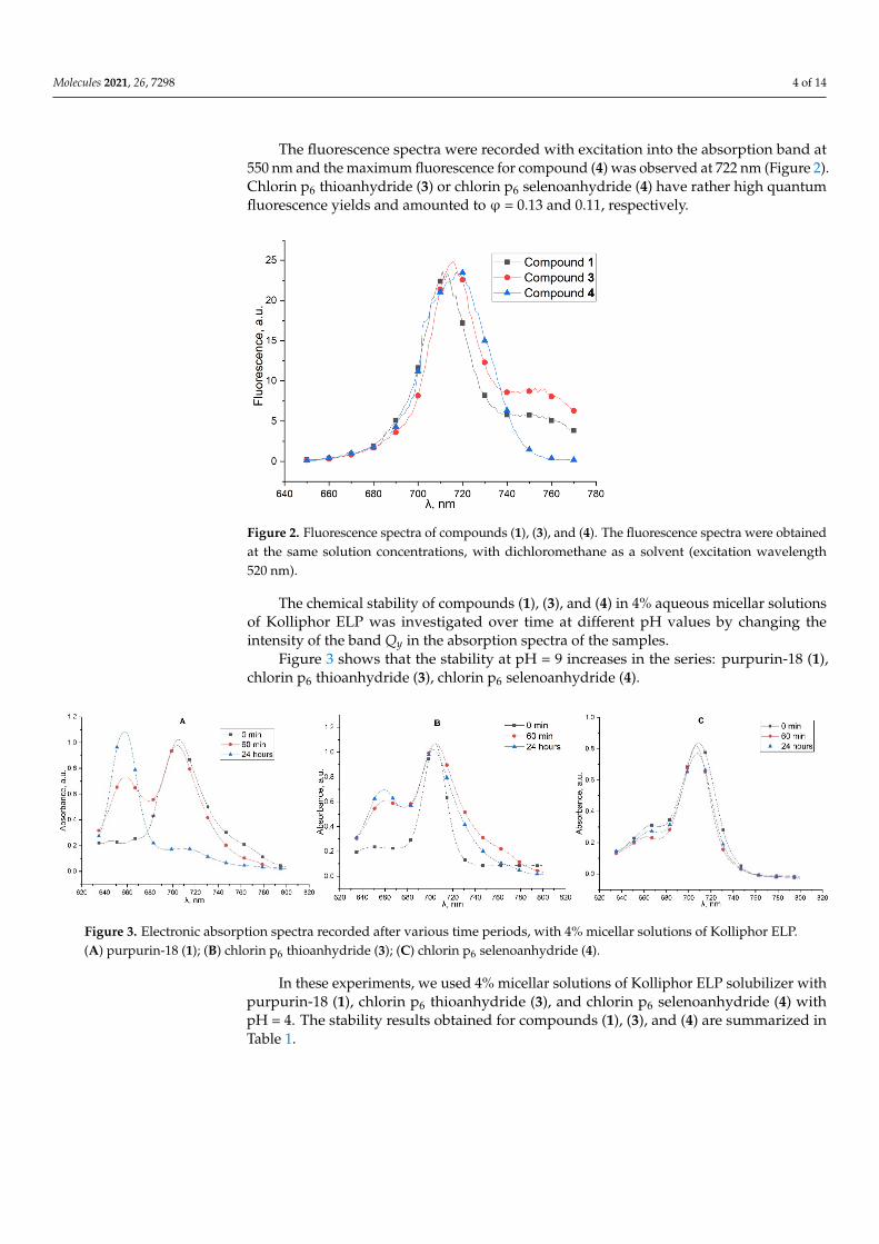
The fluorescence spectra were recorded with excitation into the absorption band at 550 nm and the maximum fluorescence for compound (4) was observed at 722 nm (Figure 2). Сhlorin p6 thioanhydride (3) or chlorin p6 selenoanhydride (4) have rather high quan-tum fluorescence yields and amounted to φ = 0.13 and 0.11, respectively.
Figure 2. Fluorescence spectra of compounds (1), (3), and (4). The fluorescence spectra were ob-tained at the same solution concentrations, with dichloromethane as a solvent (excitation wave-length 520 nm).
The chemical stability of compounds (1), (3), and (4) in 4% aqueous micellar solutions of Kolliphor ELP was investigated over time at different pH values by changing the in-tensity of the band Qy in the absorption spectra of the samples.
Figure 3 shows that the stability at pH = 9 increases in the series: purpurin-18 (1), chlorin p6 thioanhydride (3), chlorin p6 selenoanhydride (4).
Figure 1. Absorption spectra of compounds (1), (3) and (4). The absorption spectra were obtained atthe same solution concentrations, with dichloromethane as a solvent.
Molecules 2021, 26, 7298 4 of 14
The fluorescence spectra were recorded with excitation into the absorption band at550 nm and the maximum fluorescence for compound (4) was observed at 722 nm (Figure 2).Chlorin p6 thioanhydride (3) or chlorin p6 selenoanhydride (4) have rather high quantumfluorescence yields and amounted to ϕ = 0.13 and 0.11, respectively.
Molecules 2021, 26, x FOR PEER REVIEW 4 of 15
Figure 1. Absorption spectra of compounds (1), (3) and (4). The absorption spectra were obtained at the same solution concentrations, with dichloromethane as a solvent.
The fluorescence spectra were recorded with excitation into the absorption band at 550 nm and the maximum fluorescence for compound (4) was observed at 722 nm (Figure 2). Сhlorin p6 thioanhydride (3) or chlorin p6 selenoanhydride (4) have rather high quan-tum fluorescence yields and amounted to φ = 0.13 and 0.11, respectively.
Figure 2. Fluorescence spectra of compounds (1), (3), and (4). The fluorescence spectra were ob-tained at the same solution concentrations, with dichloromethane as a solvent (excitation wave-length 520 nm).
The chemical stability of compounds (1), (3), and (4) in 4% aqueous micellar solutions of Kolliphor ELP was investigated over time at different pH values by changing the in-tensity of the band Qy in the absorption spectra of the samples.
Figure 3 shows that the stability at pH = 9 increases in the series: purpurin-18 (1), chlorin p6 thioanhydride (3), chlorin p6 selenoanhydride (4).
Figure 2. Fluorescence spectra of compounds (1), (3), and (4). The fluorescence spectra were obtainedat the same solution concentrations, with dichloromethane as a solvent (excitation wavelength520 nm).
The chemical stability of compounds (1), (3), and (4) in 4% aqueous micellar solutionsof Kolliphor ELP was investigated over time at different pH values by changing theintensity of the band Qy in the absorption spectra of the samples.
Figure 3 shows that the stability at pH = 9 increases in the series: purpurin-18 (1),chlorin p6 thioanhydride (3), chlorin p6 selenoanhydride (4).
Molecules 2021, 26, x FOR PEER REVIEW 5 of 15
Figure 3. Electronic absorption spectra recorded after various time periods, with 4% micellar solutions of Kolliphor ELP. (A) purpurin-18 (1); (B) chlorin p6 thioanhydride (3); (C) chlorin p6 selenoanhydride (4).
In these experiments, we used 4% micellar solutions of Kolliphor ELP solubilizer with purpurin-18 (1), chlorin p6 thioanhydride (3), and chlorin p6 selenoanhydride (4) with pH = 4. The stability results obtained for compounds (1), (3), and (4) are summarized in Table 1.
Table 1. Intensity of the absorption maxima of compounds (1), (3), and (4) under acidic conditions at pH = 4.
Time, min. Purpurin-18 (1) Chlorin p6 Thioanhydride (3) Chlorin p6 Selenoanhydride (4) Intensity at 700 nm Content, % Intensity at 698 nm Content, % Intensity at 696 nm Content, % 0 0.99055 100 0.98381 100 0.99306 100
60 0.72335 73 0.74801 76 0.93902 94.5 1440 0.51956 52.5 0.69036 70 0.89705 90
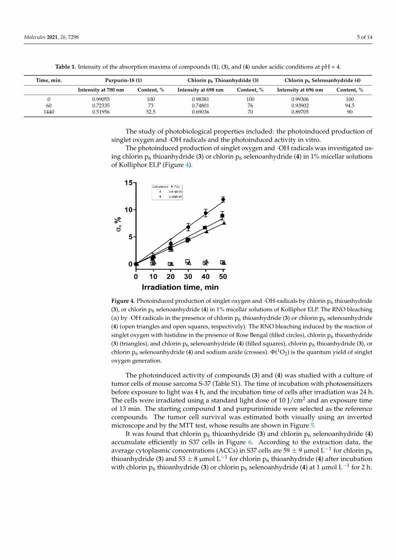
The study of photobiological properties included: the photoinduced production of singlet oxygen and ∙OH radicals and the photoinduced activity in vitro.
The photoinduced production of singlet oxygen and ∙OH radicals was investigated using chlorin p6 thioanhydride (3) or chlorin p6 selenoanhydride (4) in 1% micellar solu-tions of Kolliphor ELP (Figure 4).
Figure 4. Photoinduced production of singlet oxygen and ∙OH-radicals by chlorin p6 thioanhydride (3), or chlorin p6 selenoanhydride (4) in 1% micellar solutions of Kolliphor ELP. The RNO bleaching (α) by ∙OH radicals in the presence of chlorin p6 thioanhydride (3) or chlorin p6 selenoanhydride (4) (open triangles and open squares, respectively). The RNO bleaching induced by the reaction of sin-glet oxygen with histidine in the presence of Rose Bengal (filled circles), chlorin p6 thioanhydride (3) (triangles), and chlorin p6 selenoanhydride (4) (filled squares), chlorin p6 thioanhydride (3), or chlo-rin p6 selenoanhydride (4) and sodium azide (crosses). Φ(1O2) is the quantum yield of singlet oxygen generation.
Figure 3. Electronic absorption spectra recorded after various time periods, with 4% micellar solutions of Kolliphor ELP.(A) purpurin-18 (1); (B) chlorin p6 thioanhydride (3); (C) chlorin p6 selenoanhydride (4).
In these experiments, we used 4% micellar solutions of Kolliphor ELP solubilizer withpurpurin-18 (1), chlorin p6 thioanhydride (3), and chlorin p6 selenoanhydride (4) withpH = 4. The stability results obtained for compounds (1), (3), and (4) are summarized inTable 1.
Molecules 2021, 26, 7298 5 of 14
Table 1. Intensity of the absorption maxima of compounds (1), (3), and (4) under acidic conditions at pH = 4.
Time, min. Purpurin-18 (1) Chlorin p6 Thioanhydride (3) Chlorin p6 Selenoanhydride (4)
Intensity at 700 nm Content, % Intensity at 698 nm Content, % Intensity at 696 nm Content, %
0 0.99055 100 0.98381 100 0.99306 10060 0.72335 73 0.74801 76 0.93902 94.5
1440 0.51956 52.5 0.69036 70 0.89705 90
The study of photobiological properties included: the photoinduced production ofsinglet oxygen and ·OH radicals and the photoinduced activity in vitro.
The photoinduced production of singlet oxygen and ·OH radicals was investigated us-ing chlorin p6 thioanhydride (3) or chlorin p6 selenoanhydride (4) in 1% micellar solutionsof Kolliphor ELP (Figure 4).
Molecules 2021, 26, x FOR PEER REVIEW 5 of 15
Figure 3. Electronic absorption spectra recorded after various time periods, with 4% micellar solutions of Kolliphor ELP. (A) purpurin-18 (1); (B) chlorin p6 thioanhydride (3); (C) chlorin p6 selenoanhydride (4).
In these experiments, we used 4% micellar solutions of Kolliphor ELP solubilizer with purpurin-18 (1), chlorin p6 thioanhydride (3), and chlorin p6 selenoanhydride (4) with pH = 4. The stability results obtained for compounds (1), (3), and (4) are summarized in Table 1.
Table 1. Intensity of the absorption maxima of compounds (1), (3), and (4) under acidic conditions at pH = 4.
Time, min. Purpurin-18 (1) Chlorin p6 Thioanhydride (3) Chlorin p6 Selenoanhydride (4) Intensity at 700 nm Content, % Intensity at 698 nm Content, % Intensity at 696 nm Content, % 0 0.99055 100 0.98381 100 0.99306 100
60 0.72335 73 0.74801 76 0.93902 94.5 1440 0.51956 52.5 0.69036 70 0.89705 90
The study of photobiological properties included: the photoinduced production of singlet oxygen and ∙OH radicals and the photoinduced activity in vitro.
The photoinduced production of singlet oxygen and ∙OH radicals was investigated using chlorin p6 thioanhydride (3) or chlorin p6 selenoanhydride (4) in 1% micellar solu-tions of Kolliphor ELP (Figure 4).
Figure 4. Photoinduced production of singlet oxygen and ∙OH-radicals by chlorin p6 thioanhydride (3), or chlorin p6 selenoanhydride (4) in 1% micellar solutions of Kolliphor ELP. The RNO bleaching (α) by ∙OH radicals in the presence of chlorin p6 thioanhydride (3) or chlorin p6 selenoanhydride (4) (open triangles and open squares, respectively). The RNO bleaching induced by the reaction of sin-glet oxygen with histidine in the presence of Rose Bengal (filled circles), chlorin p6 thioanhydride (3) (triangles), and chlorin p6 selenoanhydride (4) (filled squares), chlorin p6 thioanhydride (3), or chlo-rin p6 selenoanhydride (4) and sodium azide (crosses). Φ(1O2) is the quantum yield of singlet oxygen generation.
Figure 4. Photoinduced production of singlet oxygen and ·OH-radicals by chlorin p6 thioanhydride(3), or chlorin p6 selenoanhydride (4) in 1% micellar solutions of Kolliphor ELP. The RNO bleaching(α) by ·OH radicals in the presence of chlorin p6 thioanhydride (3) or chlorin p6 selenoanhydride(4) (open triangles and open squares, respectively). The RNO bleaching induced by the reaction ofsinglet oxygen with histidine in the presence of Rose Bengal (filled circles), chlorin p6 thioanhydride(3) (triangles), and chlorin p6 selenoanhydride (4) (filled squares), chlorin p6 thioanhydride (3), orchlorin p6 selenoanhydride (4) and sodium azide (crosses). Φ(1O2) is the quantum yield of singletoxygen generation.
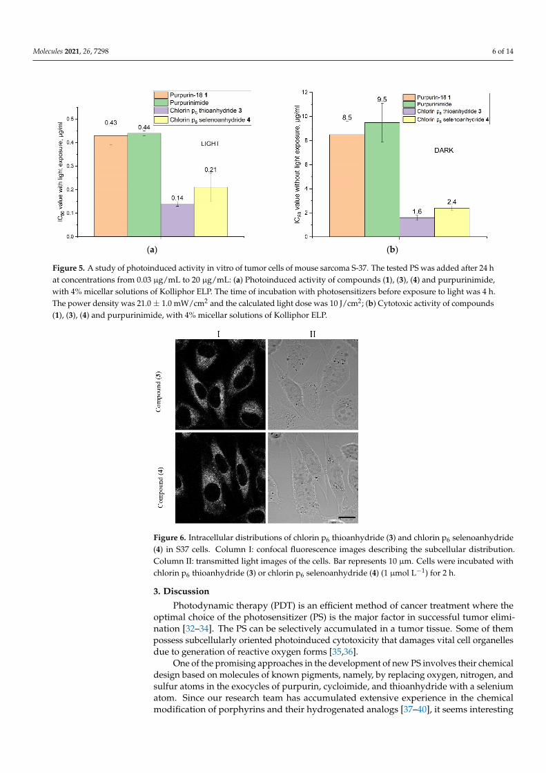
The photoinduced activity of compounds (3) and (4) was studied with a culture oftumor cells of mouse sarcoma S-37 (Table S1). The time of incubation with photosensitizersbefore exposure to light was 4 h, and the incubation time of cells after irradiation was 24 h.The cells were irradiated using a standard light dose of 10 J/cm2 and an exposure timeof 13 min. The starting compound 1 and purpurinimide were selected as the referencecompounds. The tumor cell survival was estimated both visually using an invertedmicroscope and by the MTT test, whose results are shown in Figure 5.
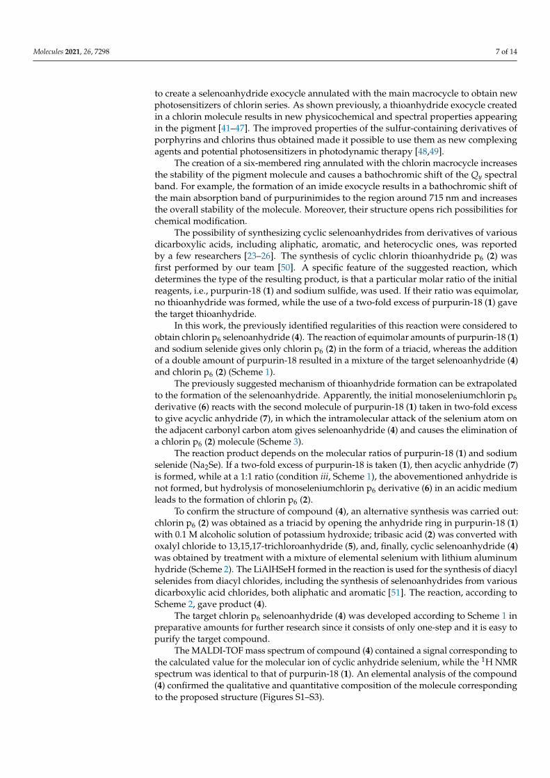
It was found that chlorin p6 thioanhydride (3) and chlorin p6 selenoanhydride (4)accumulate efficiently in S37 cells in Figure 6. According to the extraction data, theaverage cytoplasmic concentrations (ACCs) in S37 cells are 59 ± 9 µmol L−1 for chlorin p6thioanhydride (3) and 53 ± 8 µmol L−1 for chlorin p6 thioanhydride (4) after incubationwith chlorin p6 thioanhydride (3) or chlorin p6 selenoanhydride (4) at 1 µmol L−1 for 2 h.
Molecules 2021, 26, 7298 6 of 14
Molecules 2021, 26, x FOR PEER REVIEW 6 of 15
The photoinduced activity of compounds (3) and (4) was studied with a culture of tumor cells of mouse sarcoma S-37 (Table S1). The time of incubation with photosensitiz-ers before exposure to light was 4 h, and the incubation time of cells after irradiation was 24 h. The cells were irradiated using a standard light dose of 10 J/cm2 and an exposure time of 13 min. The starting compound 1 and purpurinimide were selected as the refer-ence compounds. The tumor cell survival was estimated both visually using an inverted microscope and by the MTT test, whose results are shown in Figure 5.
(a) (b)
Figure 5. A study of photoinduced activity in vitro of tumor cells of mouse sarcoma S-37. The tested PS was added after 24 h at concentrations from 0.03 μg/mL to 20 μg/mL: (a) Photoinduced activity of compounds (1), (3), (4) and purpu-rinimide, with 4% micellar solutions of Kolliphor ELP. The time of incubation with photosensitizers before exposure to light was 4 h. The power density was 21.0 ± 1.0 mW/cm2 and the calculated light dose was 10 J/cm2; (b) Cytotoxic activity of compounds (1), (3), (4) and purpurinimide, with 4% micellar solutions of Kolliphor ELP.
It was found that chlorin p6 thioanhydride (3) and chlorin p6 selenoanhydride (4) ac-cumulate efficiently in S37 cells in Figure 6. According to the extraction data, the average cytoplasmic concentrations (ACCs) in S37 cells are 59 ± 9 μmol L−1 for chlorin p6 thioanhy-dride (3) and 53 ± 8 μmol L−1 for chlorin p6 thioanhydride (4) after incubation with chlorin p6 thioanhydride (3) or chlorin p6 selenoanhydride (4) at 1 μmol L−1 for 2 h.
Figure 5. A study of photoinduced activity in vitro of tumor cells of mouse sarcoma S-37. The tested PS was added after 24 hat concentrations from 0.03 µg/mL to 20 µg/mL: (a) Photoinduced activity of compounds (1), (3), (4) and purpurinimide,with 4% micellar solutions of Kolliphor ELP. The time of incubation with photosensitizers before exposure to light was 4 h.The power density was 21.0± 1.0 mW/cm2 and the calculated light dose was 10 J/cm2; (b) Cytotoxic activity of compounds(1), (3), (4) and purpurinimide, with 4% micellar solutions of Kolliphor ELP.
Molecules 2021, 26, x FOR PEER REVIEW 6 of 15
The photoinduced activity of compounds (3) and (4) was studied with a culture of tumor cells of mouse sarcoma S-37 (Table S1). The time of incubation with photosensitiz-ers before exposure to light was 4 h, and the incubation time of cells after irradiation was 24 h. The cells were irradiated using a standard light dose of 10 J/cm2 and an exposure time of 13 min. The starting compound 1 and purpurinimide were selected as the refer-ence compounds. The tumor cell survival was estimated both visually using an inverted microscope and by the MTT test, whose results are shown in Figure 5.
(a) (b)
Figure 5. A study of photoinduced activity in vitro of tumor cells of mouse sarcoma S-37. The tested PS was added after 24 h at concentrations from 0.03 μg/mL to 20 μg/mL: (a) Photoinduced activity of compounds (1), (3), (4) and purpu-rinimide, with 4% micellar solutions of Kolliphor ELP. The time of incubation with photosensitizers before exposure to light was 4 h. The power density was 21.0 ± 1.0 mW/cm2 and the calculated light dose was 10 J/cm2; (b) Cytotoxic activity of compounds (1), (3), (4) and purpurinimide, with 4% micellar solutions of Kolliphor ELP.
It was found that chlorin p6 thioanhydride (3) and chlorin p6 selenoanhydride (4) ac-cumulate efficiently in S37 cells in Figure 6. According to the extraction data, the average cytoplasmic concentrations (ACCs) in S37 cells are 59 ± 9 μmol L−1 for chlorin p6 thioanhy-dride (3) and 53 ± 8 μmol L−1 for chlorin p6 thioanhydride (4) after incubation with chlorin p6 thioanhydride (3) or chlorin p6 selenoanhydride (4) at 1 μmol L−1 for 2 h.
Figure 6. Intracellular distributions of chlorin p6 thioanhydride (3) and chlorin p6 selenoanhydride(4) in S37 cells. Column I: confocal fluorescence images describing the subcellular distribution.Column II: transmitted light images of the cells. Bar represents 10 µm. Cells were incubated withchlorin p6 thioanhydride (3) or chlorin p6 selenoanhydride (4) (1 µmol L−1) for 2 h.
3. Discussion
Photodynamic therapy (PDT) is an efficient method of cancer treatment where theoptimal choice of the photosensitizer (PS) is the major factor in successful tumor elimi-nation [32–34]. The PS can be selectively accumulated in a tumor tissue. Some of thempossess subcellularly oriented photoinduced cytotoxicity that damages vital cell organellesdue to generation of reactive oxygen forms [35,36].
One of the promising approaches in the development of new PS involves their chemicaldesign based on molecules of known pigments, namely, by replacing oxygen, nitrogen, andsulfur atoms in the exocycles of purpurin, cycloimide, and thioanhydride with a seleniumatom. Since our research team has accumulated extensive experience in the chemicalmodification of porphyrins and their hydrogenated analogs [37–40], it seems interesting
Molecules 2021, 26, 7298 7 of 14
to create a selenoanhydride exocycle annulated with the main macrocycle to obtain newphotosensitizers of chlorin series. As shown previously, a thioanhydride exocycle createdin a chlorin molecule results in new physicochemical and spectral properties appearingin the pigment [41–47]. The improved properties of the sulfur-containing derivatives ofporphyrins and chlorins thus obtained made it possible to use them as new complexingagents and potential photosensitizers in photodynamic therapy [48,49].
The creation of a six-membered ring annulated with the chlorin macrocycle increasesthe stability of the pigment molecule and causes a bathochromic shift of the Qy spectralband. For example, the formation of an imide exocycle results in a bathochromic shift ofthe main absorption band of purpurinimides to the region around 715 nm and increasesthe overall stability of the molecule. Moreover, their structure opens rich possibilities forchemical modification.
The possibility of synthesizing cyclic selenoanhydrides from derivatives of variousdicarboxylic acids, including aliphatic, aromatic, and heterocyclic ones, was reportedby a few researchers [23–26]. The synthesis of cyclic chlorin thioanhydride p6 (2) wasfirst performed by our team [50]. A specific feature of the suggested reaction, whichdetermines the type of the resulting product, is that a particular molar ratio of the initialreagents, i.e., purpurin-18 (1) and sodium sulfide, was used. If their ratio was equimolar,no thioanhydride was formed, while the use of a two-fold excess of purpurin-18 (1) gavethe target thioanhydride.
In this work, the previously identified regularities of this reaction were considered toobtain chlorin p6 selenoanhydride (4). The reaction of equimolar amounts of purpurin-18 (1)and sodium selenide gives only chlorin p6 (2) in the form of a triacid, whereas the additionof a double amount of purpurin-18 resulted in a mixture of the target selenoanhydride (4)and chlorin p6 (2) (Scheme 1).
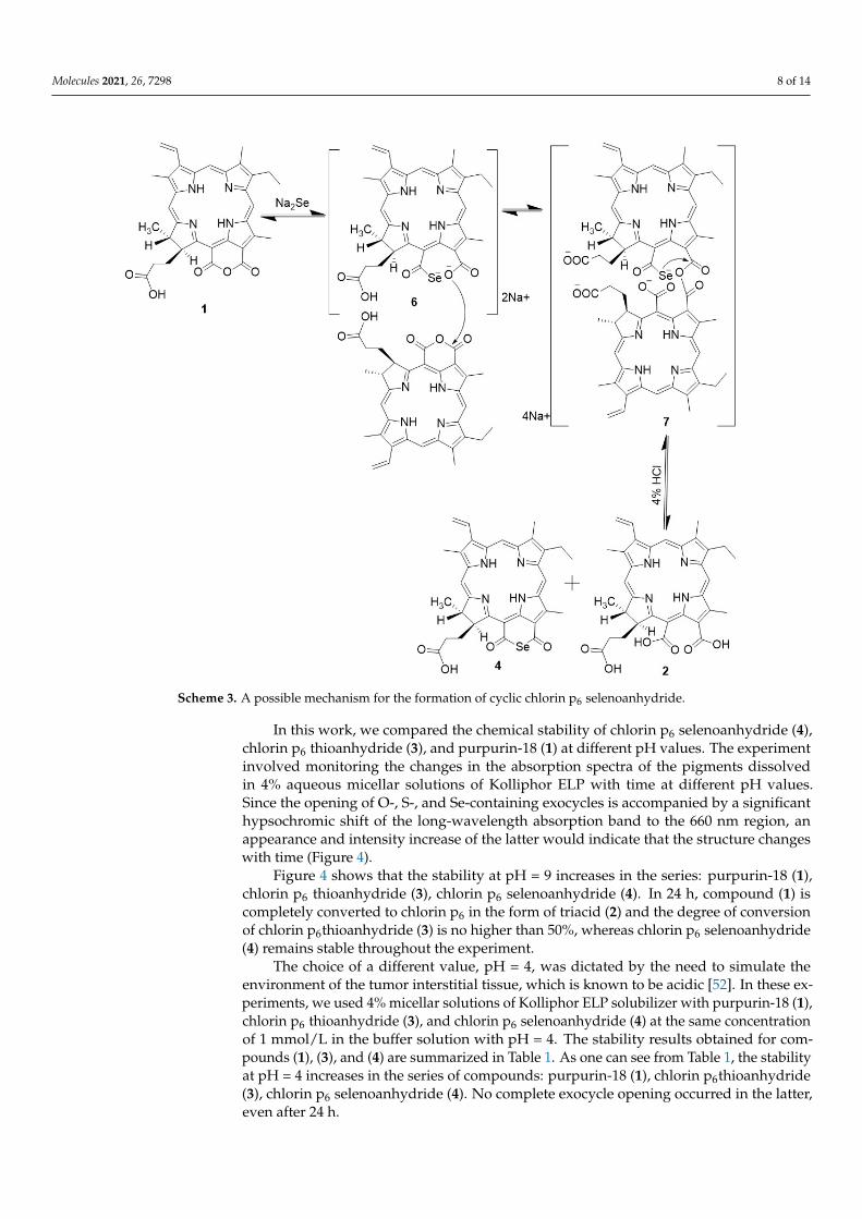
The previously suggested mechanism of thioanhydride formation can be extrapolatedto the formation of the selenoanhydride. Apparently, the initial monoseleniumchlorin p6derivative (6) reacts with the second molecule of purpurin-18 (1) taken in two-fold excessto give acyclic anhydride (7), in which the intramolecular attack of the selenium atom onthe adjacent carbonyl carbon atom gives selenoanhydride (4) and causes the elimination ofa chlorin p6 (2) molecule (Scheme 3).
The reaction product depends on the molecular ratios of purpurin-18 (1) and sodiumselenide (Na2Se). If a two-fold excess of purpurin-18 is taken (1), then acyclic anhydride (7)is formed, while at a 1:1 ratio (condition iii, Scheme 1), the abovementioned anhydride isnot formed, but hydrolysis of monoseleniumchlorin p6 derivative (6) in an acidic mediumleads to the formation of chlorin p6 (2).
To confirm the structure of compound (4), an alternative synthesis was carried out:chlorin p6 (2) was obtained as a triacid by opening the anhydride ring in purpurin-18 (1)with 0.1 M alcoholic solution of potassium hydroxide; tribasic acid (2) was converted withoxalyl chloride to 13,15,17-trichloroanhydride (5), and, finally, cyclic selenoanhydride (4)was obtained by treatment with a mixture of elemental selenium with lithium aluminumhydride (Scheme 2). The LiAlHSeH formed in the reaction is used for the synthesis of diacylselenides from diacyl chlorides, including the synthesis of selenoanhydrides from variousdicarboxylic acid chlorides, both aliphatic and aromatic [51]. The reaction, according toScheme 2, gave product (4).
The target chlorin p6 selenoanhydride (4) was developed according to Scheme 1 inpreparative amounts for further research since it consists of only one-step and it is easy topurify the target compound.
The MALDI-TOF mass spectrum of compound (4) contained a signal corresponding tothe calculated value for the molecular ion of cyclic anhydride selenium, while the 1H NMRspectrum was identical to that of purpurin-18 (1). An elemental analysis of the compound(4) confirmed the qualitative and quantitative composition of the molecule correspondingto the proposed structure (Figures S1–S3).
Molecules 2021, 26, 7298 8 of 14Molecules 2021, 26, x FOR PEER REVIEW 8 of 15
Scheme 3. A possible mechanism for the formation of cyclic chlorin p6 selenoanhydride.
To confirm the structure of compound (4), an alternative synthesis was carried out: chlorin p6 (2) was obtained as a triacid by opening the anhydride ring in purpurin-18 (1) with 0.1 M alcoholic solution of potassium hydroxide; tribasic acid (2) was converted with oxalyl chloride to 13,15,17-trichloroanhydride (5), and, finally, cyclic selenoanhydride (4) was obtained by treatment with a mixture of elemental selenium with lithium aluminum hydride (Scheme 2). The LiAlHSеH formed in the reaction is used for the synthesis of diacyl selenides from diacyl chlorides, including the synthesis of selenoanhydrides from various dicarboxylic acid chlorides, both aliphatic and aromatic [51]. The reaction, accord-ing to Scheme 2, gave product (4).
The target chlorin p6 selenoanhydride (4) was developed according to Scheme 1 in preparative amounts for further research since it consists of only one-step and it is easy to purify the target compound.
The MALDI-TOF mass spectrum of compound (4) contained a signal corresponding to the calculated value for the molecular ion of cyclic anhydride selenium, while the 1H NMR spectrum was identical to that of purpurin-18 (1). An elemental analysis of the com-pound (4) confirmed the qualitative and quantitative composition of the molecule corre-sponding to the proposed structure (Figures S1–S3).
In this work, we compared the chemical stability of chlorin p6 selenoanhydride (4), chlorin p6 thioanhydride (3), and purpurin-18 (1) at different pH values. The experiment involved monitoring the changes in the absorption spectra of the pigments dissolved in 4% aqueous micellar solutions of Kolliphor ELP with time at different pH values. Since
Scheme 3. A possible mechanism for the formation of cyclic chlorin p6 selenoanhydride.
In this work, we compared the chemical stability of chlorin p6 selenoanhydride (4),chlorin p6 thioanhydride (3), and purpurin-18 (1) at different pH values. The experimentinvolved monitoring the changes in the absorption spectra of the pigments dissolvedin 4% aqueous micellar solutions of Kolliphor ELP with time at different pH values.Since the opening of O-, S-, and Se-containing exocycles is accompanied by a significanthypsochromic shift of the long-wavelength absorption band to the 660 nm region, anappearance and intensity increase of the latter would indicate that the structure changeswith time (Figure 4).
Figure 4 shows that the stability at pH = 9 increases in the series: purpurin-18 (1),chlorin p6 thioanhydride (3), chlorin p6 selenoanhydride (4). In 24 h, compound (1) iscompletely converted to chlorin p6 in the form of triacid (2) and the degree of conversionof chlorin p6thioanhydride (3) is no higher than 50%, whereas chlorin p6 selenoanhydride(4) remains stable throughout the experiment.
The choice of a different value, pH = 4, was dictated by the need to simulate theenvironment of the tumor interstitial tissue, which is known to be acidic [52]. In these ex-periments, we used 4% micellar solutions of Kolliphor ELP solubilizer with purpurin-18 (1),chlorin p6 thioanhydride (3), and chlorin p6 selenoanhydride (4) at the same concentrationof 1 mmol/L in the buffer solution with pH = 4. The stability results obtained for com-pounds (1), (3), and (4) are summarized in Table 1. As one can see from Table 1, the stabilityat pH = 4 increases in the series of compounds: purpurin-18 (1), chlorin p6thioanhydride(3), chlorin p6 selenoanhydride (4). No complete exocycle opening occurred in the latter,even after 24 h.
Molecules 2021, 26, 7298 9 of 14
The more electronegative heteroatoms (O, N, S, Se) in the exocycle, the higher the reac-tivity of the adjacent carbonyl carbon atom in nucleophilic addition/substitution reactions.Apparently, this is the reason for the greater stability of the selenium anhydride ring.
The main indicator of PS efficiency is the singlet oxygen quantum yield, which wasdetermined for two compounds. Chlorin p6 thioanhydride (3) or chlorin p6 selenoanhy-dride (4) have rather high quantum yields of photoinduced singlet oxygen generation andamounted to 0.47 ± 0.03 and 0.58 ± 0.04, respectively. Sodium azide, a well-known specificsinglet oxygen quencher, totally suppressed the photoinduced chlorin p6 thioanhydride (3)or chlorin p6 selenoanhydride (4)-mediated bleaching of RNO. Chlorin p6 thioanhydride(3) and chlorin p6 selenoanhydride (4) do not produce ·OH radicals under illumination.
The photoinduced activity of compounds (3) and (4) was studied with a culture oftumor cells of mouse sarcoma S-37 (Table S1). The values presented in Figure 5a show atwo-fold increase in photoinduced cytotoxicity in Se-containing derivatives (4) comparedto the pigments containing an anhydride or imide exocycle. Moreover, the dark cytotoxicityincreased four-fold, in comparison with the latter compounds (Figure 5b). Apparently,the increase in the dark cytotoxicity is due to the interaction of the pigments studied withsulfur- and selenium-containing endogenous intracellular compounds. Such interactionscan cause a violation of the redox balance in tumor cells, for example, by blocking theglutathione antioxidant system [19] or suppressing DNA repair [15]. The photoinducedcytotoxicity of compounds (3) and (4) is an order of magnitude higher than the darkcytotoxicity, which is comparable to the previously studied chlorophyll a derivatives andcommercial PS [53].
Intracellular distributions of chlorin p6 thioanhydride (3) and chlorin p6 selenoanhy-dride (4) in S37 cells were shown (Figure 6). In cytoplasm, chlorin p6 thioanhydride (3) andchlorin p6 selenoanhydride (4) demonstrate similar diffuse distribution. A consequence ofthe higher accumulation of compound (3) in S37 cells in comparison with compound (4) isthe higher photoinduced cytotoxicity shown by us, above (Figure 5).
4. Materials and Methods4.1. Materials and Instruments
All the solvents used in this work were prepared according to common techniques asdescribed elsewhere. Column chromatography was performed on silica gel 40/60 (Merck,Darmstadt, Germany). Preparative thin-layer chromatography (TLC) was performedon glass plates with silica gel 60 (Merck, Darmstadt, Germany). Analytical TLC wasperformed on aluminum plates with Silica gel 60 F254 with a fluorescent probe (Merck,Darmstadt, Germany).
Absorption and fluorescence spectra were recorded on Shimadzu UV1800 UV/VIS(Shimadzu, Duisburg, Germany) and Shimadzu RF-5301 (Shimadzu, Duisburg, Germany)spectrophotometers in quartz cuvettes (0.4 × 1.0 cm) with an optical path length of 1 cm(spectral slit width 1 nm), with dichloromethane as a solvent. The absorption spectra wererecorded in the range of 350–750 nm. Fluorescence spectra were recorded in the range650–770 nm (excitation wavelength 520 nm), with dichloromethane as a solvent. The fluo-rescence quantum yield was determined by the relative method. A solution of purpurin-18in toluene was used as a standard [54]. 1H and 13C NMR spectra were registered on BrukerDPX 300 (Bruker, Alzenau, Germany). NMR samples were prepared in CDCl3. Matrix-assisted laser desorption/ionization (MALDI) mass spectra were recorded on a BrukerUltraflex TOF/TOF spectrometer (Bruker, Alzenau, Germany), with 2,5-dihydroxybenzoicacid as the matrix. Elemental analysis was performed on EA 1112, Thermo Finnigan,Thermo Scientific, CE Instruments (Thermo Fisher Scientific, Waltham, MA, USA).
Extraction of chlorophyll a from Spirulina platensis, synthesis of purpurin-18, andsynthesis of cyclic thioanhydride of chlorin p6 were carried out as described previously [50].
Molecules 2021, 26, 7298 10 of 14
4.2. Synthesis
Cyclic chlorin p6 selenoanhydride (4).Method 1:Purpurin-18 (1) (177 µmol, 100 mg) was dissolved in tetrahydrofuran (3 mL), and 0.9 M
aqueous sodium selenide (1 mL) was added. The reaction mixture was stirred at 25 ◦Cfor 240 min in an inert argon atmosphere. The product was extracted with 4% aqueoushydrochloric acid/chloroform (125/25, v/v) and then purified by column chromatographyin dichloromethane/methanol (15/1, v/v). The yield of the target compound (4) was27 mg (27%).
Method 2:Chlorin p6 (2). Compound (1) (177 µmol, 100 mg) was dissolved in methanol (5 mL),
and 1 M sodium hydroxide (2 mL) was added [50]. The reaction mixture was stirred at 22 ◦Cfor 15 min. The product was extracted with 4% aqueous hydrochloric acid/chloroform(50/5, v/v). The solution was concentrated in a vacuum, and the product was purified bycolumn chromatography (CH2Cl2/CH3OH, 1/1, v/v). The yield of the target compound 2was 75 mg (75%).
Thionyl chloride (134 µmol, 26 µL) and triethylamine (10 µmol, 1.5 µL) were addedto the solution of compound (2) (67 µmol) in diethyl ether (3 mL), and the reaction wasperformed at 22 ◦C for 30 min in argon atmosphere. After this, the LiAlHSeH reagentwas added to product 7. The reagent was prepared beforehand by the addition of lithiumaluminum hydride (2.6 mg) to a solution of selenium (2.2 mg) in tetrahydrofuran (250 µL).The resulting solution was incubated in an inert argon atmosphere for 24 h at 22 ◦C, andthe compound (4) was extracted with chloroform (15 mL) and repeatedly washed withwater (150 mL). The solution was concentrated in a vacuum, and the product was purifiedby preparative TLC (CH2Cl2/CH3OH, 60/1, v/v). The yield of the target compound (4)was 17 mg (42.5%).
UV/VIS (CH2Cl2) λ max, nm (ε, M−1sm−1): 412 (9800), 481 (22354), 550 (546), 696(10506). MALDI-TOF MS m/z [M+H]+ calculated for C33H32N4O4Se+H, 627.61 found:627.67. 1H NMR (300 MHz, CDCl3, δ, ppm): 9.58 (1H, s, 5H), 9.37 (1H, s, 10H), 8.59 (1H,s, 20H), 7.90 (1H, dd, J = 17.9, 11.4 Hz, 31-H), 6.31 (1H, dd, J = 17.9, 1.3 Hz, 32-Ha), 6.21(1H, dd, J = 11.5, 1.3 Hz, 32-Hb), 5.21 (1H, m, 17-H), 4.39 (1H, q, J = 6 Hz, 18-H), 3.79 (3H,s, 12-CH3), 3.63 (2H, m, 81-CH2), 3.36 (3H, s, 7-CH3), 3.17 (3H, s, 2-CH3), 2.76 (1H, m,172-CH2
b), 2.49 (1H, m, 171-CH2b), 2.48 (1H, m, 171-CH2
a), 2.02 (1H, m, 171-CH2a), 1.76
(3H, d, J = 7.3 Hz, 18-CH3), 1.67 (3H, t, J = 7.6 Hz, 82-CH3), 0.24 (1H, s, NH), and −0.06 (1H,s, NH). 13C NMR (75 MHz, CDCl3, δ, ppm): 177.40, 176.46, 173.60, 165.21, 164.10, 156.15,149.94, 145.84, 144.01, 139.22, 137.68, 136.48, 133.68, 131.62, 129.65, 128.28, 123.65, 107.45,102.99, 94.91,62.67, 54.92, 51.56, 49.14, 32.48, 31.21, 29.63, 23.78, 19.20, 17.30, 12.19, 11.86,and 10.93. Elemental analysis for CHN, calculated for C33H32N4O4Se (%): C, 63.15; H, 5.14;N, 8.93; found (%): C, 63.12; H, 5.16; N, 8.91.
4.3. A Study of Stability at Different pH Values
The stability was estimated by the variation in the intensity of the Qy absorption bandin the electronic spectra of the samples.
A total of 4% Micellar solutions of Kolliphor ELP with purpurin-18 (1), chlorin thioan-hydride p6 (3), and chlorin p6 selenoanhydride (4) were added to buffer solutions preparedaccording to standard methods, so that the final concentration of the pigments in thesolution was 1 mmol/L and the pH was 4 or 9. The changes in the absorption spectra ofthe resulting solutions were monitored for 24 h of incubation.
4.4. A Study of Photoinduced Production of Singlet Oxygen and ·OH-Radicals
The ability of chlorin p6 thioanhydride (3) and chlorin p6 selenoanhydride (4) forthe photoinduced generation of singlet oxygen was estimated using a 4-nitroso-N, N-dimethylaniline–histidine (RNO) assay [55], as in [56], with minor modifications. RoseBengal was used as a reference compound with a known quantum yield of singlet oxygen

Molecules 2021, 26, 7298 11 of 14
generation (Φ(1O2) = 0.75) [57]. The RNO-based assay, in the absence of histidine, wasused under the same experimental conditions for the detection of ·OH radicals [58].
4.5. A Study of Photoinduced Activity In Vitro
The cells were cultivated under standard conditions at 37 ◦C in a humidified atmo-sphere with 5% CO2, in a DMEM environment with the addition of L-glutamine (2 mM)and fetal calf serum (10%, PanEco, Moscow, Russia). The photoinduced efficiency wasestimated as follows: S37 cells were seeded in a 96-well flat-bottomed plate for microtitra-tion (Costar, New York, NY, USA). The tested PS was added after 24 h at concentrationsranging from 0.03 µg/mL to 20 µg/mL. The time of the incubation with photosensitizersbefore exposure to light was 4 h. After that, the cells were irradiated with a halogen lampthrough a KS-19 broadband filter that passed light with wavelengths above 720 nm, anda water filter 5 cm thick, equipped with a liquid circulation system (λ ≥ 1000 nm). Thepower density was 21.0 ± 1.0 mW/cm2 and the calculated light dose was 10 J/cm2. Thestarting compound 1 and purpurinimide were selected as the reference compounds. Afterirradiation, the cells were incubated for 24 h under standard conditions. To analyze thePS cytotoxicity and the cytotoxicity (without irradiation), cells were kept for 24 h in thedark. Survival was estimated by visual inspection and colorimetrically using the MTT test.Cell-growth inhibitions of more than 50% were considered biologically significant. Thisvalue was calculated as the average of three independent tests.
4.6. A Study Intracellular Accumulation
To study intracellular accumulation of chlorin p6 thioanhydride (3) and chlorin p6selenoanhydride (4), cells were seeded on cover glasses in 24-well plates (seeding densityof 5 × 10⁴ cells per well) a day before the experiment. Confocal fluorescent imageswere recorded using a laser scanning confocal microscope Leica TCS SP2 (Leica, Wetzlar,Germany) with a 63× water-immersion lens (numerical aperture 1.2). Lateral and axialresolutions were 0.2 and 1 µm, respectively. Fluorescence of chlorin p6 thioanhydride (3)and chlorin p6 selenoanhydride (4) was excited at the 561 nm wavelength and recorded inthe 680–750 nm range.
4.7. Assessment of Intracellular Accumulation
To estimate the intracellular accumulation of chlorin p6 thioanhydride (3) and chlorinp6 selenoanhydride (4) with an extraction technique, 3.5 × 10⁶ cells were incubated withchlorin p6 thioanhydride (3) or chlorin p6 selenoanhydride (4) (1 µmol L−1) in 5.0 mL of acomplete medium for 2 h, washed, detached from a flask, pelleted, and lysed with 0.25%Triton X-100 for 30 min. The concentration of chlorin p6 thioanhydride (3) and chlorin p6selenoanhydride (4) in cellular extract (Cex) was measured using fluorescence spectroscopywith reference solutions of chlorin p6 thioanhydride (3) and chlorin p6 selenoanhydride (4)of known concentrations in 0.25% Triton X-100. The average cytoplasmic concentration(ACC) of chlorin p6 thioanhydride (3) and chlorin p6 selenoanhydride (4) was calculated as
ACC = Cex × Vex/(Nc × Vcyt)
where Vex, Nc, and Vcyt are an extract volume, a cell number, and an approximate volumeof cytoplasm of a cell ((1.2 ± 0.2) × 10−12 L), respectively. A total cell volume was assumedto be equal to (1.7 ± 0.2) × 10−12 L.
5. Conclusions
A selenium-containing derivative of purpurin-18, namely, chlorin p6 selenoanhydride,was obtained in this work. The chemical synthesis was performed using two approaches.The first involved a direct reaction of purpurin-18 with sodium selenide, accompanied bya replacement of oxygen in exocycle E with a selenium atom. The second involved thereaction of chlorin p6 13,15,17-trichloroanhydride with elemental selenium in the presenceof lithium aluminum hydride.
Molecules 2021, 26, 7298 12 of 14
The target compound was characterized by spectral and physicochemical methods.Stability studies showed that the selenoanhydride was more stable under acid (pH = 4)and basic conditions (pH = 9) than the thioanhydride and the starting purpurin-18. Thederivatives have sufficiently high fluorescence and singlet oxygen quantum yields. In vitrobiological studies of chlorin p6 selenoanhydride on the S37 cell line showed a two-foldincrease in the photoinduced cytotoxic effect, compared to the previously obtained pur-purinimide and the original purpurin-18, while the dark cytotoxicity increased four-fold, incomparison with the latter. We have shown that the chlorin p6 thioanhydride accumulatesbetter in S37 cells and has a higher photoinduced cytotoxicity.
Supplementary Materials: The following are available online. Figure S1: Mass spectrum of com-pound 4; Figure S2: 1H NMR spectrum of compound 4; Figure S3: 13C NMR spectrum of compound4; Table S1: Photoinduced activity of compounds 1, 3, 4, and purpurinimide.
Author Contributions: Conceptualization, V.P., M.G. and E.D.; methodology, A.M., A.P. (AndreiPankratov) and M.G.; validation, V.P., N.S., A.P. (Anna Plyutinskaya) and M.G.; data curation,V.P., N.S., A.P. (Andrei Pankratov) and Y.V.; writing—original draft preparation, V.P., A.P. (AnnaPlyutinskaya), N.S., M.G., Y.V. and E.D.; writing—review and editing, A.P. (Andrei Pankratov), A.M.and M.G.; visualization, V.P., N.S. and A.P. (Anna Plyutinskaya); funding acquisition, A.M. and M.G.All authors have read and agreed to the published version of the manuscript.
Funding: The synthesis of chlorin p6 selenoanhydride and chlorin p6 thioanhydride and theircharacterization was supported by the Russian Foundation for Basic Research (No 20-33-90289). Thestudy of stability at different pH values and spectral properties was supported by the Russian ScienceFoundation (No 21-13-00078). The study of photoinduced activity in vitro was supported by theMinistry of Science and Higher Education of the Russian Federation (0706-2020-0019).
Data Availability Statement: The data presented in this study are available from the authors.
Acknowledgments: This work was performed using the equipment of the Shared Science andTraining Center for Collective Use RTU MIREA and supported by the Ministry of Science and HigherEducation of the Russian Federation. The authors would like to thank Ignatova A.A., Shemyakin andOvchinnikov Institute of Bioorganic Chemistry, RAS, Lab. of Optical Microscopy and Spectroscopyof Biomolecules.
Conflicts of Interest: The authors declare no conflict of interest.
Sample Availability: Samples of the compounds are available from the authors upon special requestand the agreement of the Russian Foundation for Basic Research and the Russian Science Foundation.
References1. Furdyna, J.K.; Dong, S.N.; Lee, S.; Liu, X.; Dobrowolska, M. The ubiquitous nature of chalcogenides in science and technology. In
Chalcogenide; Woodhead Publishing: Duxford, UK, 2020; pp. 1–30. [CrossRef]2. Ho, P.C.; Wang, J.Z.; Meloni, F.; Vargas-Baca, I. Chalcogen bonding in materials chemistry. Coord. Chem. Rev. 2020, 422, 213464.
[CrossRef]3. Wessjohann, L.A.; Schneider, A.; Abbas, M.; Brandt, W. Selenium in chemistry and biochemistry in comparison to sulfur. Biol.
Chem. 2007, 338, 997–1006. [CrossRef]4. Bhagavan, N.V.; Ha, C.-E. Chapter 3—Amino Acids. In Essentials of Medical Biochemistry, 2nd ed.; Academic Press: Cambridge,
MA, USA, 2015; pp. 21–29. [CrossRef]5. Jacob, C.; Giles, G.I.; Giles, N.M.; Sies, H. Sulfur and selenium: The role of oxidation state in protein structure and function.
Angew. Chem. Int. Ed. 2003, 42, 4742–4758. [CrossRef]6. Gladyshev, V.N.; Hatfield, D.L. Selenocysteine-containing proteins in mammals. J. Biomed. Sci. 1999, 6, 151–160. [CrossRef]7. Clark, D.P.; Pazdernik, N.J.; McGehee, M.R. Chapter 13—Protein Synthesis. In Molecular Biology, 3rd ed.; Elsevier: Amsterdam,
The Netherlands, 2019; pp. 397–444.8. Singh, V.P.; Poon, J.F.; Butcher, R.J.; Engman, L. Pyridoxine-derived organoselenium compounds with glutathione peroxidase-like
and chain-breaking antioxidant activity. Chem. Eur. J. 2014, 20, 12563–12571. [CrossRef]9. Brigelius-Flohé, R.; Maiorino, M. Glutathione peroxidases. Biochim. Biophys. Acta Gen. Subj. 2013, 1830, 3289–3303. [CrossRef]10. Rayman, M.P. The importance of selenium to human health. Lancet 2000, 356, 233–241. [CrossRef]11. Rayman, M.P. Selenium and human health. Lancet 2012, 379, 1256–1268. [CrossRef]12. Levander, O.A. Selenium and sulfur in antioxidant protective systems: Relationships with vitamin E and malaria. Proc. Soc. Exp.
Biol. Med. 1992, 200, 255–259. [CrossRef] [PubMed]

Molecules 2021, 26, 7298 13 of 14
13. Battin, E.E.; Brumaghim, J.L. Antioxidant activity of sulfur and selenium: A review of reactive oxygen species scavenging,glutathione peroxidase, and metal-binding antioxidant mechanisms. Cell Biochem. Biophys. 2009, 55, 1–23. [CrossRef] [PubMed]
14. Cheng, B.; Lian, H.-F.; Liu, Y.-Y.; Yu, X.-H.; Sun, Y.-L.; Sun, X.-D.; Shi, Q.-H.; Liu, S.-Q. Effects of selenium and sulfur onantioxidants and physiological parameters of garlic plants during senescence. J. Integr. Agric. 2016, 15, 566–572. [CrossRef]
15. Kamal, A.; Iqbal, M.A.; Bhatti, H.N. Therapeutic applications of selenium-derived compounds. Rev. Inorg. Chem. 2018, 38, 49–76.[CrossRef]
16. Gandin, V.; Khalkar, P.; Braude, J.; Fernandes, A.P. Organic selenium compounds as potential chemotherapeutic agents forimproved cancer treatment. Free Radic. Biol. Med. 2018, 127, 80–97. [CrossRef] [PubMed]
17. Radomska, D.; Czarnomysy, R.; Radomski, D.; Bielawski, K. Selenium Compounds as Novel Potential Anticancer Agents. Int. J.Mol. Sci. 2021, 22, 1009. [CrossRef]
18. Li, T.; Xu, H. Selenium-containing nanomaterials for cancer treatment. Cell Rep. Phys. Sci. 2020, 1, 100111. [CrossRef]19. Plano, D.; Baquedano, Y.; Ibáñez, E.; Jiménez, I.; Palop, J.A.; Spallholz, J.E.; Sanmartín, C. Antioxidant-prooxidant properties of a
new organoselenium compound library. Molecules 2010, 15, 7292–7312. [CrossRef] [PubMed]20. George, S.; Abrahamse, H. Redox potential of antioxidants in cancer progression and prevention. Antioxidants 2020, 9, 1156.
[CrossRef] [PubMed]21. Zalewska-Ziob, M.; Adamek, B.; Kasperczyk, J.; Romuk, E.; Hudziec, E.; Chwalinska, E.; Dobija-Kubica, K.; Rogozinski, P.;
Brulinski, K. Activity of antioxidant enzymes in the tumor and adjacent noncancerous tissues of non-small-cell lung cancer.Oxidative Med. Cell. Longev. 2019, 2019, 2901840. [CrossRef]
22. Marengo, B.; Nitti, M.; Furfaro, A.L.; Colla, R.; De Ciucis, C.; Marinari, U.M.; Pronzato, M.A.; Traverso, N.; Domenicotti, C. Redoxhomeostasis and cellular antioxidant systems: Crucial players in cancer growth and therapy. Oxid. Med. Cell. Longev. 2016, 2016,6235641. [CrossRef]
23. Eicher, T.; Hauptmann, S.; Speicher, A. The Chemistry of Heterocycles: Structures, Reactions, Synthesis, and Applications; John Wiley &Sons: Hoboken, NJ, USA, 2013; pp. 69–70.
24. Pelkey, E.T. 3.13—Selenophenes. In Comprehensive Heterocyclic Chemistry; Elsevier: Amsterdam, The Netherlands, 2008; Volume 3,pp. 975–1006. [CrossRef]
25. Mugesh, G.; du Mont, W.W.; Sies, H. Chemistry of biologically important synthetic organoselenium compounds. Chem. Rev. 2001,101, 2125–2180. [CrossRef] [PubMed]
26. Makhal, P.N.; Nandi, A.; Kaki, V.R. Insights into the Recent Synthetic Advances of Organoselenium Compounds. ChemistrySelect2021, 6, 663–679. [CrossRef]
27. Kalyanam, N.; Majeed, M. Selenium compounds in medicine and nutrition. Chim. Oggi 2007, 25, 36–38.28. Kanari, N.; Allain, E.; Shallari, S.; Diot, F.; Diliberto, S.; Patisson, F.; Yvon, J. Thermochemical route for extraction and recycling of
critical, strategic and high value elements from by-products and end-of-life materials, Part I: Treatment of a Copper By-Product inAir Atmosphere. Materials 2019, 12, 1625. [CrossRef]
29. Patra, A.; Bendikov, M.; Chand, S. Poly(3,4-ethylenedioxyselenophene) and its derivatives: Novel organic electronic materials.Acc. Chem. Res. 2014, 47, 1465–1474. [CrossRef] [PubMed]
30. Sun, C.; Ji, S.; Li, F.; Xu, H. Diselenide-containing hyperbranched polymer with light-induced cytotoxicity. ACS Appl. Mater.Interfaces 2017, 9, 12924–12929. [CrossRef] [PubMed]
31. Li, T.; Pan, S.; Gao, S.; Xiang, W.; Sun, C.; Cao, W.; Xu, H. Diselenide–Pemetrexed Assemblies for Combined Cancer Immuno-,Radio-, and Chemotherapies. Angew. Chem. Int. Ed. 2020, 59, 2700–2704. [CrossRef]
32. Kwiatkowski, S.; Knap, B.; Przystupski, D.; Saczko, J.; Kedzierska, E.; Knap-Czop, K.; Kulbacka, J. Photodynamic therapy—Mechanisms, photosensitizers and combinations. Biomed. Pharmacother. 2018, 106, 1098–1107. [CrossRef]
33. Pucelik, B.; Sułek, A.; Dabrowski, J.M. Bacteriochlorins and their metal complexes as NIR-absorbing photosensitizers: Properties,mechanisms, and applications. Coord. Chem. Rev. 2020, 416, 213340. [CrossRef]
34. Koifman, O.; Ageeva, T.; Beletskaya, I.; Averin, A.; Yakushev, A.; Tomilova, L.; Dubinina, T.; Tsivadze, A.; Gorbunova, Y.;Martynov, A.; et al. Macroheterocyclic Compounds—A Key Building Block in New Functional Materials and Molecular Devices.Macroheterocyclic 2020, 13, 316–447. [CrossRef]
35. Lam, M.; Oleinick, N.L.; Nieminen, A.L. Photodynamic therapy-induced apoptosis in epidermoid carcinoma cells: Reactiveoxygen species and mitochondrial inner membrane permeabilization. J. Biol. Chem. 2001, 276, 47379–47386. [CrossRef]
36. Semyonov, D.Y.; Vasil’ev, Y.L.; Dydykin, S.S.; Stranadko, E.F.; Shubin, V.K.; Bogomazov, Y.K.; Morokhotov, V.A.; Shcherbyuk,A.N.; Morozov, S.V.; Zakharov, Y.I. Antimicrobial and antimycotic photodynamic therapy (review of literature). Biomed. Photonics2021, 10, 25–31. [CrossRef]
37. Grin, M.A.; Tikhonov, S.I.; Petrova, A.S.; Pogorilyy, V.A.; Noev, A.N.; Tatarskiy, V.V.; Shpakovsky, D.B.; Milaeva, E.R.; Kalinina,E.V.; Chernov, N.N.; et al. New Derivatives of Bacteriopurpurin with Thiolated Au (I) Complexes: Dual Darkand Light ActivatedAntitumor Potency. Anti Cancer Agents Med. Chem. 2020, 20, 49–58. [CrossRef]
38. Suvorov, N.V.; Machulkin, A.E.; Ivanova, A.V.; Popkov, A.M.; Bondareva, E.A.; Plotnikova, E.A.; Yakubovskaya, R.I.; Majouga,A.G.; Mironov, A.F.; Grin, M.A. Synthesis of PSMA-targeted 131- and 152-substituted chlorin e6 derivatives and their biologicalproperties. J. Porphyr. Phthalocyanines 2018, 22, 1030–1038. [CrossRef]
Molecules 2021, 26, 7298 14 of 14
39. Mironov, A.F.; Grin, M.A.; Pantushenko, I.V.; Ostroverkhov, P.V.; Ivanenkov, Y.A.; Filkov, G.I.; Plotnikova, E.A.; Karmakova,T.A.; Starovoitova, A.V.; Burmistrova, N.; et al. Synthesis and Investigation of photophysical and biological properties of novels-containing bacteriopurpurinimides. J. Med. Chem. 2017, 60, 10220–10230. [CrossRef]
40. Mironov, A.F.; Ostroverkhov, P.V.; Tikhonov, S.I.; Pogorilyy, V.A.; Kirin, N.S.; Chudakova, O.O.; Tsygankov, A.A.; Grin, M.A.Amino acid derivatives of natural chlorins as a platform for the creation of targeted photosensitizers in oncology. Fine Chem.Technol. 2021, 15, 16–33. [CrossRef]
41. Latos-Grazynski, L.; Pacholska, E.; Chmielewski, P.J.; Olmstead, M.M.; Balch, A.L.; Latos-Grazynski, L. Alteration of the Reactivityof a Tellurophene Within a Core-Modified Porphyrin Environment: Synthesis and Oxidation of 21-Telluraporphyrin. Angew.Chem. Int. Ed. Engl. 1995, 34, 2252–2254. [CrossRef]
42. Kobayashi, N.; Kadish, K.M.; Smith, K.M.; Guilard, R. The Porphyrin Handbook; Academic Press: San Diego, CA, USA, 2000;Volume 2, pp. 301–360.
43. Srinivasan, A.; Mahajan, S.; Pushpan, S.K.; Ravikumar, M.; Chandrashekar, T.K. Synthesis of meso-substituted core modifiedexpanded porphyrins; effect of acid catalysts on the cyclization. Tetrahedron Lett. 1998, 39, 1961–1964. [CrossRef]
44. Latos-Grazynski, L.; Lisowski, J.; Olmstead, M.M.; Balch, A.L. Five-coordinate complexes of 21-thiaporphyrin. Preparations,spectra, and structures of iron(II), nickel(II), and copper(II) complexes. Inorg. Chem. 1989, 28, 1183–1188. [CrossRef]
45. Latos-Grazynski, L.; Olmstead, M.M.; Balch, A.L. The first structural characterization of a nickel(I) macrocyclic system: Structureof nickel(I) diphenyldi-p-toyl-21-thiaporphyrin. Inorg. Chem. 1989, 28, 4065–4066. [CrossRef]
46. Gebauer, A.; Schmidt, J.A.; Arnold, J. Synthesis, characterization, and properties of a lithium 21-thiaporphyrin complex. Inorg.Chem. 2000, 39, 3424–3427. [CrossRef] [PubMed]
47. Smith, K.M. Porphyrins and Metalloporphyrins; Elsevier: Amsterdam, The Netherlands, 1975; Volume 9, pp. 3–27.48. Latos-Grazynski, L.; Lisowski, J.; Olmstead, M.M.; Balch, A.L. 21-Thiatetra-p-tolylporphyrin and its copper(II) bicarbonate
complex. Structural effects of copper-thiophene binding. J. Am. Chem. Soc. 1987, 109, 4428–4429. [CrossRef]49. Symonowicz, K.; Ziolkowski, P.; Chmielewski, P.; Latos-Grazynski, L.; Rabczynski, J.; Osiecka, B.J.; Milach, J. Tumor histopathol-
ogy following new sensitizers: Dithiaporphyrin- and sulfoxaporphyrin-mediated photodynamic therapy. Anticancer Res. 1999, 19,5385–5391.
50. Pogorilyy, V.; Kirin, N.; Mironov, A.; Grin, M. A new cyclic thioanhydride derived from chlorophyll a and its aurophilic properties.Dye Pigments 2021, 184, 108858. [CrossRef]
51. Ishihara, H.; Koketsu, M.; Fukuta, Y.; Nada, F. Reaction of lithium aluminum hydride with elemental selenium: Its application asa selenating reagent into organic molecules. J. Am. Chem. Soc. 2001, 123, 8408–8409. [CrossRef]
52. Zhang, X.; Lin, Y.; Gillies, R.J. Tumor pH and its measurement. J. Nucl. Med. 2010, 51, 1167–1170. [CrossRef] [PubMed]53. Pylina, Y.I.; Shadrin, D.M.; Shevchenko, O.G.; Startseva, O.M.; Velegzhaninov, I.O.; Belykh, D.V.; Velegzhaninov, I.O. Dark and
photoinduced cytotoxic activity of the new chlorophyll-a derivatives with oligoethylene glycol substituents on the periphery oftheir macrocycles. Int. J. Mol. Sci. 2017, 18, 103. [CrossRef] [PubMed]
54. Zenkevich, E.; Sagun, E.; Knyukshto, V.; Shulga, A.; Mironov, A.; Efremova, O.; Bonnett, R.; Songca, S.P.; Kassem, M. Photophysicaland photochemical properties of potential porphyrin and chlorin photosensitizers for PDT. J. Photochem. Photobiol. B Biol. 1996,33, 171–180. [CrossRef]
55. Blum, A.; Grossweiner, L.I. Singlet oxygen generation by hematoporphyrin IX, uroporphyrin I and hematoporphyrin derivativeat 546 nm in phosphate buffer and in the presence of egg phosphatidylcholine liposomes. Photochem. Photobiol. Sci. 1985, 41,27–32. [CrossRef] [PubMed]
56. Efremenko, A.V.; Ignatova, A.A.; Borsheva, A.A.; Grin, M.A.; Bregadze, V.I.; Sivaev, I.B.; Sivaev, I.B.; Mironov, A.F.; Feofanov,A.V. Cobalt bis(dicarbollide) versus closo-dodecaborate in boronated chlorin e6 conjugates: Implications for photodynamic andboron-neutron capture therapy. Photochem. Photobiol. Sci. 2012, 11, 645–652. [CrossRef]
57. Gandin, E.; Lion, Y.; Van de Vorst, A. Quantum yield of singlet oxygen production by xanthene derivatives. Photochem. Photobiol.1983, 37, 271–278. [CrossRef]
58. Bors, W.; Saran, M.; Lengfelder, E.; Michel, C.; Fuchs, C.; Frenzel, C. Detection of oxygen radicals in biological reactions. Photochem.Photobiol. 1978, 28, 629–637. [CrossRef] [PubMed]