the etiology and clinical diagnosis of cholecystitis and
TRANSCRIPT

University of Nebraska Medical Center University of Nebraska Medical Center
DigitalCommons@UNMC DigitalCommons@UNMC
MD Theses Special Collections
5-1-1936
The etiology and clinical diagnosis of cholecystitis and The etiology and clinical diagnosis of cholecystitis and
cholelithiasis cholelithiasis
Robert R. Livingston University of Nebraska Medical Center
This manuscript is historical in nature and may not reflect current medical research and
practice. Search PubMed for current research.
Follow this and additional works at: https://digitalcommons.unmc.edu/mdtheses
Part of the Medical Education Commons
Recommended Citation Recommended Citation Livingston, Robert R., "The etiology and clinical diagnosis of cholecystitis and cholelithiasis" (1936). MD Theses. 448. https://digitalcommons.unmc.edu/mdtheses/448
This Thesis is brought to you for free and open access by the Special Collections at DigitalCommons@UNMC. It has been accepted for inclusion in MD Theses by an authorized administrator of DigitalCommons@UNMC. For more information, please contact [email protected].
,-
THE ETIOLOGY AND CLINICAL DIAGNOSIS
Of
CHOLECYSTITIS AND OHOLELITHIASIS
Robert Ramsay Livingston
. seni or The si s
University of Nebraska
CQllege of Medicine
April, 1936

480796
-,
1.
INTRODUCTION
As mants knowledge of anatomy has advanced, so
has his skill in the diagnosis of pathological conditions
of the body. This was particularly true in relation to
morbid processes of the gastro-intestinal tract, for,
since the diseased organ could neither be seen or palpated,
the diagnosis had to be a symptomatic one. As the medical
profession gradually freed itself of the superstitions of
the early centuries, and in addition beoame ourious enough
to perform post-mortem examinations to find the cause of
death, and not simply as an opportunity to do dissection,
more acourate oonoeption of intra-abdominal disease pro
oesses resulted.
This same insatiable desire has persisted to the
present day, and whenever the physioian is oonfronted with
a patient who has oomplaints referable to the intestinal
traot, he prooeeds to obtain as exact information about
that complaint as possible. If it seems at all indioated,
the patient is sent to the roentgenologist, in order that
the suspected ulcer orater may be seen, to demonstrate the
lack of function of the gall bladder by its failure to oon
.oentrate, to visualize the kidney stone, or to see the fil
ling defect of the neoplasm. This scientific branch of
medicine is a highly neoessar~ one, and should be made
available to every patient, but the easy aooe ssibili ty of
its information may be abu·sed. The physioian, in many in
stances is all too liable to allow the roentgenolOgist to
2.
make the diagnosis, rather than himself. This same tenden
oy is evident also in the temptation to make many laboratory
tests, and rely too strongly on the fallible evidence of
chemistry, rather than developing his own reasoning and de
ducti ve powers.
This paper, therefor, is an effort to present
oholecystitis and cholelithiasis and the diagnosis of these
related conditions, with disregard to the laboratory and
X-ray findings, hoping to encourage the more extensive use
of the physician's own tools and aids: his eyes, ears, hands
and mind.
--
3.
The theory has been advanoed that oholeoYstitis
and oholelithiasis were unknown to the early Egyptian be
oause of a differenoe in the diet of those days, yet an
Egyptian mummy of about 2000 B. C. has been found, whose
ga,ll bladder was paoked with stones. (22) The symptom of
jaundioe assooiated with abdominal oolio is also mentioned
by Aristotle in 360 B. 0., and sinoe he found oonstipation
with this oondition, we may assume that it was due to ohole
oystitis. Galen in 16S A. D. mentions oolic, with nausea
and vomiting of bile and oonstipation. He asserted that
jaundioe is the result of absorption of bile into the blood;
when the body is full of bile he disoovered that it was sel
dom found in the stools. (4S, 35) Paulus Aeginetae (53),
in his sixth book mentions that jaundioe is a diffusion of
bile over the shole body. When there is a sense of heaviness
in the right hypoohondrium it indioates that the duots are
obstruoted. If, with this jaundioe there is fever and the
alvine disobarges are white an affeotion of the gall bladder
or its ducts is indioated. For tbis oondition he mentions
the use of cholegogue cathartios, suoh as hellebore. The
first mention of gall stones by a European writer seems to
be that by Antonius Benivenius .10 died in 1592. (46) ·There
was severe excruciating pain in the bowels, the cause of
which was not easily understood; this preceded the passing
of a stone in the excreta, which made the diagnosis." Ap
parently the patient died for he mentions taking one hundred
twenty stones from the gall bladder of this woman Which were
various Sizes, hard, dark in color, cuboid in shape, and on

-
4.
breaking them open, showed laminated appearance. He also
bas descr1bed finding a gall stone in the gall bladder,
black and the s1ze of a large dry ohestunt. (35)
Following the desor1pt10n of the gall stones not
ed by Antonius Benivenius, Fernel1us (35) 1n l5~ gave a
desor1pt10n not only of gall stones but of their assoc1ated
symptoms. He observed that obstruotion of the oommon duct
may lead to a swelling of the gall bladder. A white d1scol
oration of the feoes, the passage of dark urine and of
jaund1ce. EttmUller 1n h1s d1ssertation ent1tled "De ictero
flavo, n1gro at albo" (Oper. med., Tom. II, page 1, Oolleg.
praot., Sect. VII, Oap. IV, page 442) (25 P. 173), gives a
remarkably aoourate c11nical p1cture of bi11ary tract dis
ease. He speaks of paln in the precordial region whioh ap
pears with ioterus and is acoompanied by nausea, difficult
respiration and a reddish color of the urine. He states
that fever is often present and in many oases pain in the
right bypochondrium, which may either be readily removed or
be very difficult to oure, or, lastly, may be removable but
shows a tendency to recur. In the latter type, he said,
stones were commonly found in the gall bladdeD. He also
noted that colio and icterus are sometimes seen after child
birth and that ioterus frequently follows the attaok of colic
and that it 1s frequently the result of an obstruotion to
the flow of bile into the intestine or of insuffioient se
cretion of bile by the liver as is seen from the fact that
1cterus may oocur without an obstruotion in the bile pas
sages following fever, the bite of wild animals, abuse of
-
blood-letting, etc. Be observed further that icterus does
not necessarily always occur in cases of gall stones. Be
waa convinced that the gall bladder could be extirpated
without endangering the life of the animal and be quotes
the important experiment of one of the students in Leyden
who removed the gall bladder from a dog without observing
any bad results. He considered that icterus was due to
obstruction of the common bile-duct which caused a regur
gitation of bile into the blood. Be also states that
there is no remedy for gall stone a.
The following quotation is probably one of the
earliest of the fairly accurate anatomical descriptions
of the gall bladder: "low to speake of the Gal, or the
chest of the Gal:-----and it is as a purse or a pannicular
vesike in the holownease of the Lyver, about the middle
perle Ie or lobe, ordeyned to receyve tbe Cholerlke super
fluities which are ingendered in the Lyver. The which
purse or bagge hath three holes or neckes, by the fyrste
he drawetb to him from the Lyver the choler, by the
second necke he sendeth to the bottom of the stomache
choler to further the digestion." .(64)
Korgagni in his work "de sedibus et cauaibua
morborum lf (25 P. 143)' t makea an excellent presentation of
cholelithiasis. According to bim, occlusion of the bl1e
passages is a result of a contraction of the ducts and a
thickening of the mucous lining. He observed that if the
oystlc duct alone is occluded icterus is not observed.
--
6.
He mentions as predisposing causes, the age of the subject,
sedentary habits and other factor s.
Haller in his great pathological work "Disputations
ad Morborum l1 presents two cases of post mortem examinations.
(30)
Oase 1. This patient had a turgid gall bladder
containing many stones. On cutting, it
felt like cartilage and contained forty
five stones, but no bile. There was a
large stone impacted in the common duct
at its junction With the cystic duct.
The hepatio pores were double their or
dinary capaoi ty. The patient did not
have an ioterus; was only muddy.
Oase 2. A woman aged fifty-two who for six years
had had many pains in the right hypo
chondrium. In the year 1699 after much
pain she passed two stones by anus. Ex
amination of thi s woman showed a gall
bladder adherent to the liver and to the
duodenum, it having communication with
the "bladder. "Pain in the right hypo
chondrium, under the diaphragm, in the
area over where the coledochus enters
the inferior duodenum; greater and les
ser, now graye, now lancinating, con
tinuing indefinitely--these signs are
-.
7.
definitely diagnostic of gall bladder"
stones." "Iausea and vomiting occur
later. The patient becomes jaundiced
and the bowels white." He mentions
that the prognosis depends on the size
of the stone blook1ng, and this can be
told by the state of the jaundice. If
it continues to deepen this means com
plete blockage, fever, delirium and
death. (21)
Duprat in lSO, was of the opinion that gall
stones were formed during an acute hepatitis, and then
block the choledochus, so that there is more pain and ~n
eral enlargement of the duct system. "--------But only,
sometimes, short and fleeting pains, repeated jaundices,
etce , these maladies are not yet known because ~. the state
of anatomical research."
RosemeU1ler in lSl6 (56) was one of the first to
. venture an opinion as to the funot ion of the gall bladder.
He mentioned the fact that the bile flows from the liver
to the gall bladder and is condensed and held there until
digestion, when it is forced out through the ductus chole
dochus into the duodenum.
It is interesting to note that Baillie in 1920
(5) was not able· to give a much better description of the
pathology of the gall bladder than was made 100 to 150 years
previously. He found that it was very common, on dissection,
--
s.
to see adhesions around the gall bladder which, he stated,
were a cOllsequence of previous inflammation. He found
gall stones very frequently. The gall bladder was some-
time s much enlarged with very thick ooats and full of stones.
If only one stone was found it was usually quite large; he
describes one the size of a hent s egg. He found that the
composition of the stones varied, very few stones oontain
ing bile, and that they were usually laminated wi th a rad
iated oenter. He stated that there was no jaundice if the
cystic duot was obstruoted, but only inflammation and 00-
oasionally suppuration. In discussing the di seasee of the
gall bladder he states that if the stones stay in the blad
der there are seldom any symptoms, but if one of the stones
enters the duotus oholedoohus, exoruoiating pain results,
with languor, siokness and vomiting with jaundioe.
Abernethy about the same time (1828) admitted that
he was ignorant of the functions of the liver and of the
bile. He mentions taking fifteen hundred stone s from one
gall bladder.' Goupil in lS3l (24) describes a syndrome
whioh he calls bilious fever and attributes to hepatitiS.
This oonsisted of thirst, anorexia, fever, jaundice and
pain in the right hypoohondrium.
In lS55 Diokson (20) described icterus associated
with gall stones and stated that the symptom of pain is
oaused by stone passing through the ducts. Bis description
of suoh an attaok does not vary much from that of previous
writers. It is that of great pain in the epigastrium soon

-
9.
extending to the right hypochondrium and associated with
nausea, frequent retchings and vomiting.
Niemeyer in 1965 (4~) was one of the early path
ologists to advance a plausible theory of the formation of
gall stones. Briefly, he thought that the calculus was
formed around a nuoleus of muoous membrane and was the re
sult of too muoh oaloium in the bile, or a deorease in bile
acid, thus causing a preoipitation of oholester1n and bile
pigments. He observed that gall stones occurred more fre
quently in women than in men and that the greater percen
tage were cholesterin stones. He also was of the opinion
that the pai n in the right hypoohondrium attributed by so
many of the early authors to hypotitis was really the phen
omenum of inflammation and uloeration of the gallbladder
and bile passages. Ooats (13) believed that stones were
a result of stagnation a: bile from the gall bladder and
oaused oholeoysti tiS, and Oohnbein (4) in 1990 al so thought
that stones were caused by stagnation plus some change in
the chem1stry of the bile, cause unknown. He also believed
that inflammation of the gall bladder is oaused by stones.
In embryos of 2.5 mm. there is seen a sacculation
from the ventral floor of the fore-gut just cranial to the
yolk-stalk. As this hepatio d1 vertlculus pookets outward,
it plunges direotly into the splanohnio mesoderm of the ventral
mesentery. Oontinued growth spreads the mesentery, its
mesoderm forming all the oonnective tissue and smooth muscle
associated with the liver and biliary system, the mucosa
-
10.
being entodermal in origin. The gall bladder and its cystio
duct represent a special offshoot of this early diverticulum.
Until the sixth or seventh embryonic weeks the gallbladder
is solid. (3)
ANATOKY AND PHYSIOLOGY
OF THE
BILIARY TRAOT
The gall bladder is an oblong, pear shaped sac,
which is fastened by loose connective tissue in the fossa
v8ssicae felleae of the li var. When the bladder is full
its broad, blind and rounded end projects somewhat beyond
the anterior abdominal wall just median from the anterior
extremity of the ninth costal cartilege of the right side.
From the fundus the body of the bladder extends upward,
backward and somewhat to the left and narrowing suddenly
goes over at the right side of the porta hepatis into the
ductus cysticus. The gall bladder comes into contact be
low with the transverse colon and behind with the descen
ding portion of the duodenum. The fundus and the posterior
inferior surface possess a peritoneal coat. The wall con
sists of a delicate layer of mostly circular muscle fibres
and a muoous membrane from which pro~ect numerous ridges,
the plioae, arranged in the form of a network. The duotus
crsticus leaves the bladder with a marked downward bend,
extends downward and to the left and after a short course
joins the duotus bepaticus; its muoous membrane presents a
series of folds, whioh are arranged in the form of a spiral,
11.
the 8ptral valves of Heister, and whioh oorrespond to grooves
on the external surfaoe. The duotus oholedoohus runs down
ward and in the direotion of the duotlB hepatious at first in
the hepatoduodenal ligament to the right of the portal vein,
then in the head of the panoreas at the junction of the left
and posterior walls of the desoending duodenum, into which it
opens near the panoreatio duot or in oommon with it.
The blood supply is by a small artery, the oystio
artery, whioh is a branoh of the right hepatio artery, a de
rivative of the oeliao. The venous drainage is by the oystio
vein whioh opens either into the stem or the right branoh of
the portal vein.
The nerves of the gall bladder and extra hepatio
duots are derived from the same souroe as those whioh go to
the liver, arising OOiefl1' in the hepatio plexus. In the
oiroular fibres of the gall ·oladder the nerves form a ple xus
similar to Auerbach's plexus in the intestine. Branohes
from this plexus give rise to a seoondary plexus in the
muoosa. (59)
The large lymphatio vessels running over the gall
bladder bring lymph from the liver and the ooats of the
gall bladder; they follow the inner side of the oystio duot
lind end in the mesenterio lymph glands. There is a very
dense network of lymph ohannels in the subserous layer into
whioh entry the sub-muoous sets of lymphatios. There are
normally no solitary lymph follioles in the gall bladder
similar to those in the appendix. One of the most important

12.
pOints about the lympbatics of the gall bladder is that
they have a very intimate anastomosis with those of the
liver. (63)
Kodama (35), by the injection of trypan blue
into the subserosa of the first portion of the duodenum,
found that the dye passed through the lymphatic vessele
along the common duct and entered the wall of the gall
bladder. When the solution was injected into the lymphatic
vessels immediately beneath the serosa of the gall bladder -"
the dye was carried to the upper part of the duodenum.
Graham bas shown that the interlobular lymphatics
of the liver connect with the gall bladder and ramify over
the head of the pancreas. With injections of Prussian blue
into the wall of the gall bladder the dye was seen to pass
through the lymphatics into the liver and along the common
duct. (25) As the result of studies wi tb experimental
cholecystitis in the dog, he has come to the conclusion
that infection of the gall bladder can occur through the
lymphatics as the result of hepatitis. (54)
The gall bladder has three types of activity, those
of absorption, secretion and motor activity. The gall blad
der absorbs mainly water so that the hepatic bile, as a.
result, is concentrated four to ten tines. Oonsequently
of this in the fasting human, the gall bladder may contain
the entire twenty-four hour output of the liver. The acutely
inflamed mucosa. does not concentrate or evacuate. However,
if the inflammation is patchy or in the muscular or serous:
coats the gall bladder will concentrate. If fibrosis occurs
as a result of the inflammation, concentration, if any, is
light. Oholesterosis does not interfrere with conoentration
or emptying unless it is associated with a moderate or severe
oholecystitis. The gall bladder normally seoretes a muooid
fluid at a ra.te of a"bout twenty oc. per 24 hours. One is
the rytbmio tonus ohange whioh is equal to about 1 to 3 cm.
o'! bile pressure. The other is the tonio oontraction of the
musculature at a whole and is equal to 20 to 30 om. of bile
pressure. The power of the normal gall bladder is not equal
to the seoretory pressure of the liver. The ohief stimulus
of oontraction is oholeoystokinin Whioh is seoreted by the
duodenal mucosa. The produotion of this substance is sti~
ulated mainly by acids and fats in the upper intestine,
especially egg yolks and oream. Atropine relaxes the gall
bladder but does not abolish the effeot of oholecystokinin.
The Sphincter of Oddi. Siooe the sphinoter can
resist up to 75 cm. of bile pressure it is obvious that it
must relax for the gall bladder to empty itself. Ivy has
shown that when the gall bladder oontraots, the sphincter
relaxe s, and state s the follow! ng findings,
1. Increasing duedenal muscular tone inhibits
the flow of bile into the duodenum.
2. Deoreasing the duedno musoular tone inoreases
the fiow of the bi le into the duodenum.
3. Ohemioal irritation of the duodenum delays
gall bladder evaouation.

14.
4. Atropine favors the flow of bile, pilocarpine
stops it.
5. Morphine inhibits the flow of Oile.
,. Magnesium, sulphate favors the flow of bile.
Ivy has found that there are three types of evacuation of
the ga.ll bladder in response to a fat meal:
1. The primary relaxation of the gall bladder
with closure of the sphincter, 1Ibich is onl,.
temporary. This condition is rare.
2. Oontraction and evacuation start quickly and
evacuation occurs rapidly. This type prevails
in 95(~ of the cases.
3. Evacuation starts and then stops, filling of
the gall bladder then occurring. Tbis iffol
lowed by further evacuation. (32)
Winkelstein (6g) Van Meter (31) and Mann (36) be
lieve that the gall bladder is emptied by the pressure of
adjaoent, distended organs, by the milking action of duodenal
peristalt~c waves, and by the intra-abdominal pressure of
respiration, when the papilla of vater is open.
Boyd (9) and Mentzer (40) have shown that the
cholesterol found in the epithelial oells of the mucosa is
due to the absorption and not excu:etion. The former, in the
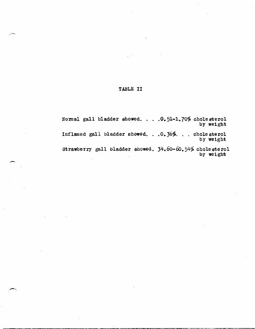
chemical analYsiS of a number of gall bladders found that
the normal gall bladder showed from O.51--1.70~ cholesterol
by weight.
-
o HOLEOY STITI S
The term o~oleoystitis is often loosely used to
oomprise a number of disturbanoes of the gall bladder, of
which some are true inflammations and others perhaps dev
iations from a normal function or metabolism of the gall
bladder. It has been suggested that the term "oho1eoysto
pathy" be used instead of the more prevalent oholeoystitis.
Sinoe the olinioal diagnosis of cholecystitis and oho1e-
1i thissi sis arrived at by a oon s1deration of symptoms and
physioal findings which oou1d be caused by either of the
two conditions this topic will be considered separately.
THE ETIOLOGY OF OHOLEOYSTITIS
Inoidente. Mentzer (43) in a study of 49,659 new
patients at the Mayo Olinic in 1922 found that 5% complained
of gall bladder di sease and that of 612 neoropsy specimens,
62% showed gross le sions of the gall bladder. The Prudential
Life Insurance Oompany found that 33 out of every million
died of gall bladder disease in 1919. In 110 women who had
been pregnant, 64~ showed cholesterosis of the gall bladder
and g2~ had gall bladder lesions. In 34 patients weighing
over 210 pounds 70'fo showed oho1esterosis. Oonstant (15) in
his series of ten thousand of female patients over a period
of 5 years iound 930 cases of oholeoYstitis, 60% of Whioh
showed oa1culi. The symptoms of gall bladder disease ooour
most frequently during the fourth deoade or later. Blalook
(26) found that 2a% of all his cases gave a history of a
preceding typhoid infeC?tion, the average time between the
infection and the appearance of gall bladder symptoms being

-
16.
five years. The mode of infection of the gall bladder by
bacteria, and the type of bacteria found in the gall blad
der has been the o'bject of a great deal of experimental
work. Bacteriological investigations of the gall bladder
have been made in three ways, by cultures of the contained
bile or other fluid content, by cultures of the ground up
wall of the organ and by the injection of bacteria in ex
perimental animals. Rosenow (57) stressed the desirability
of making cultures from the wall rather than from the con
tents of the org~n. His cultures from the wall showed
streptococci more frequently than any other type but his
most valuatae contribution was that the injection of the
streptococci into experimental animals produced gall blad
der lesions in 79fq. In four of the human cases in which
he found pure cholesterin stones there were very few
bacteria in the wall of the gall bladder and none in the
stone. Williams (67) using cultures obtained from macer
ated gall bladder walls, obtained streptococci in l6~ of
his 106 oases of cholecystitis and Bacoli in 20%. Be
found streptococci in 24% and found that these organisms
had very slight if any localizing power. .Brown (12)
found streptocooci in 30% (in his series of cases) of the
gall bladdere studied which had slight lesions and in 75~
of the grossly diseased gall bladders. Be found that cul
tures made from tonsillar pus and injected intravenously
into rabbits produced no lasting bacteremia, no septicemia
and lesions only in the gall bladder, finding no lesions
17.
in the stomach, spleen, kidneys, appendix, lungs or heart.
Mentzer et al (41) obtained positive oultures in whioh
streptocci predominated in 9S~ of their 193 cases wben the
cultures were made from the wall of the gall bladder.
The importance of biliary stasis in the gall
bladder as a factor in the production of cholecystitis has
been much discussed but at present the question seems far
from being settled.
There is no doubt that stasis of bile in the
gall bladder for a long period of time Will produce con
centration of that bile, but whether this stasis is the
cause or the result of cholecystitis has not been definitely
proven. The type of individual in which a biliary stasis
is most pronounced upon cholecystographic examination, the
visceroptotic, is not the type in whioh biliary di sease is
most frequent.
The reoent work of Aronsohn and Andrews (4)
should be mentioned here, for it opens a new field of spec
ulation as to the etiology of gall bladder disease, espec
ially those cases in which few if any bacteria could be
demonstrated. Their findings are as follows: after tbe
injeotion of 6% crystalline pure egg albumen in Ringers
solution into the gall bladders of 15 dogs, 1; showed
evidence of reoent aotive inflammatory processes in the
wall in 48 hours, (edema, fibrinous exudate and hemorrhage).
After the dog had been de-sensitized to this material, the
result was the same. It is the impression of the authors
that man may give a similar reaotion, and that it is in the
nature of an anaphylactic condition. They quote Alvarez
and Croutch, who had presented cases of gall bladder pain
which were caused by ohicken, milk, and eggs, as an ana
phylaotic reaotion, and whioh never reourred after the
elimination of these foods from the diet.
The authors also sensitized three dogs to the
crystalline egg solution, and then injeoted the gall blad
ders as before. The same reaotion as before was obtained,
but more marked, and occurring in one-half hour after the
injeotion.
PathoS!nesis, From a study of the anatomy of
the gall bladder it oan readily be seen there are four pos
sibilities of a mode of infeotion of the gall bladder.
1. A desoending infeotion frma the liver, the
baoteria being oarried by the bile.
2. Asoending infeoti ons from the duodenum, by
way of the common bile duot.
3. Hematogenous or lymphotogenous infeotions.
4. Infeotion spreading from inflamed contiguous
organs.
Of these four possibilities it is only the last two whioh
take into the consideration the actual infection of the
gall bladder wall, the first two possibilities assuming
contact tnflammation through the muoosa. A brief survey
of ourrent literature on the baoterial content of bile and
of the gall bladder shows a high percentage of sterile bile
19.
cultures. Keyer (41+) et al after the intravenous inj'eo
tion of B. typhosus (g,OOO to 24,000 million) in 500 ra.b
bits found in microscopio evidence of infection of the
gall bladder in only one-third of the animals, and they
believe that infeotion of the wall is due to spread from
the mucosa, which has been infeoted by the bile.
Alvarez, et al (2), after making cultures of the
walls and bile of gall bladders removed at operation found
the wall infected in 63'% and the l)ile in 29%.
Denton (19) in hi s study of many pathologic gall
bladders found a lar~ percentage that were sterile. In
600 neoropsie s he deomonstrated baoteria in 4 out of 94
acute cases. Brown (12) made cultures of various media
from 70 gall bladders. His results are tabulated in Table
1. Meyer, Nielson and Feusier (44) state "It is ~nerally
known from the studies of Cushing and from our own, that
typhoid bacilli introduced direotly into the cystio duot
of dogs, disappear rapidly, and that c holeoysti ti soan
only be produced by considerable injury of the wall, or by
plaoing a foreign 'body in the gall bladder.~Karzer)"
Graham and Peterman (25) in experiments wi th 10 dogs in
which large amounts of pathogenic colon baoilli were in-
j eoted into the normal gall bladder found that no chole
oystitis was produced sufficient to be demonstrable by
gross appearanoe unless the outflow from the gall bladder
was interfered with by ligation of the cystic duot or by
injury to the blood supply, produced by ligation of the
20.
oystio artery. One would expeot to find a large peroentage
of oases of oholecysti tis in cases of baoteremia if the
gall bladder was infected by means of the bile beoause of
the faot that the liver is qlite ineffioient as a baotia
filter as shown by the experiments show~ by Patey and Whitby
(52). Osler (51) states that oholecystitis ooourred in
only 19 of his series of 1500 oases of typhoid fever, a
disease in which baoteremia is an almost constant oocurrence.
In general there are only two routes available
for the transmission of organisms to the wall of the gall
bladder. One of these is the blood stream and the other
is the lymph stream. A great deal of work has been done in
experimental animals in attempting to produoe cholecystitis
by the intravenous injection of baoteria, and with varying
results. Rosenow (57) found that with the intravenous in
jection of large numbers of B. ooli and strep. viridans
that he was able to produce lesions of the gall bladder
traot in 7~/o of hi s dogs.
Brown (12) with the int ravenous injeotion of oul
tures made from tonsillar pus and oontaining prinoipally
strep. vividans produoed no lasting baoteremia, no septi
oemia, and lesions only in the gall bladder. There oan be no
doubt, apparently, that oholeoystitis is not infrequently
hematogenous in origin espeoially with typhoid fever, but
in many cases of oholecysti tis there is no evidenoe of a
pre-existing bacteremia. If the gall bladder is to be
oonsidered as being infeoted by bacteria ooming from other
21.
abdominal organs this infeotion must arise either as a
retrograde thrombus or a pylephlebitis of the oystio vein,
which drains into the portal vein.
The most ~nerally accepted evidence on gall
bladder infeotion pOints to the fact that the oholeoYstitis
represents in a large majority of oases a direct extension
to the wall of the gall bladder from a liver whioh is al
ready infeoted.
Peterman et al (54) found that in the dog experi
mental choleoystitis is regularly aooompanied by a hepatitis
of the same type as fo~nd in man. This is most marked in
the right lobe of the liver, espeoially near the gall bladder.
If the infeotion had oome to the liver by the portal vein a
more even distribution of the hepatitis would be expeoted.
llann and Willson (37) in 57 cases of aoute cholecystitis
found associated areas of looal chronic inflammatory hepa
titis radiating from the gall bladder area. Examination of
this microsoopically showed connective tissue in the peri
lobular spaoes with subsequent atrophy of the enclosed
lobules. Graham (26) showed, in a series of cases in Which
small pieces of the liver adjoining the gall bladder were
removed at operation for cholecystitis, that microscopic
evidence of hepatitis existed in every oase. Graham to
gether with Peterman (27) found that in a majority of cases,
oholecystitis was a direot extension from an already inflamed
liver. They found that the hepatitis usually begins and is
most marked in the interlobular or periportal spaces, and. is
apparently due to infection brought to the liver by the
·22.
portal vein. They believe that this hepatitis extends to
the wall of the gall bladder by means of the lymphatics.
Judd (34) agrees with this opinion of Graham's. Patey and
Whitby (52) disagree with the work of Graham and believe
that the choleoystitis precedes the hepatitis. Referenoe
has been made to the work of Sudler and Kodama who have
shown the very intimate lymphatic connection between the
liver and the gall bladder. The frequent occurrence of
gall bladder infeotion with disease of the abdominal organs
drained by the portal system and particularly the appendix
and duodenum has been noted by many authors, among whom
are Hargis et a1 (31), Mann (37), Mayo (30), Graham (2S),
Davis (17), Constant (15), Walton (65) and Mentzer (43).
The latter found that in gO<% of his 612 oases of 91111 blad
der disease there was associated disease in the appendix,
stomach or duodenum. Graham (25), page 125, perforued an
experiment which oonsisted of the produotion of hepatitis,
cholecystitis and oholedoohitis by portal vein injeotions.
The types of lesion found agreed With those Whioh ooour in
spontaneous oases in man. It was possible al so after the
injeotion for them to demonstrate the infeoting organisms
in the liver and the wall of the gall bladder. He found
that the hepatitis always followed these portal vein injeo
tions and it in turn was followed by infeotions of the gall
bladder. Be suggests the fact that frequently, perha.ps, a
vioious oirole between the gall bladder and liver is
e stabli shed whereby each may reinfeot the other.
-
var1et1es of CholecystitisL There is nothing
specific about the pathological changes in the gall bladder
in ordinary inflammation. Acute cholecystitis occurs in
many forms. In the simplest of these the organ 1s reddened,
the wall thickened and edematous and the mucous membrane
inflamed and congested. Bancroft ("), desoribing the
pathology of this condition, says that the mucosa and vil11
are relatively nofmal but there is thickening of the sub
mucosa and muscularis. This condition of acute cholecystitis
may pass on to the suppurative stage, empyema of the gall
bladder, in which case the organ is g~atly swollen and tense
and shows black areas if gangrene has set in. The gall blad
der in acute cholecystitis usually has patches of cream
colored or yellowish-gray fibrin on its external surfaoe.
These may result in the formation of thiok, fibrinous adhes
ions causing the gall bladder to be adherent to adjacent
organs. Acute oholeoysti ti s nearly always is caused by ob
struction of the oystio duot, usually by stone.
Denton (19) states that the changes in the gall
bladder usually due to impaction of stone in the cystic duct, are
intramural edema, whioh is followed by venous distention, and
then intra.'Ilura.l hemorrhage or hematoma. The amount of damage
may range from edema in the submucosa to extensi va sub-muoous
hemorrhage with destruction of a large amount of the muoosa.
However, tags of entirely normal mucosa could be. found in the
cavity. He found that the intramural edema and hematoma tend
to be replaoed by scar tissue. He demonstrated bacteria in

-
24.
the wall in 4 out of 94 acute oases. Upon opening the acute
gall bladder, pus and stone s are most commonly fOUnd. Per
foration may re"sult, e specially when the infecti on is due to
typhoid. bacilli. Micoscropic examination of the wall of the
gall bladder in cases of acute cholecystitis shows usually
the whole wall invaded by leucocytes, and areas of hemorrhage
are common. In many cases the deeper layers, especially the
sub-serous, are the sites of the most severe inflammatory
changes. In the more severe grades the epithelium may be
destroyed in places, but it is a striking fact that it rarely
if ever is completely destroyed by inflammati on. Boyd (10)
141ller (45)
Ohronic OholecYstitis. Acute inflammation of the
gall bladder very often passes into a chronic inflammation,
clinical observation showing that a great majority of the
gall bladders that become infected remain infected. In. chronic
cholecystitis tbe wall is thickened and graYish because of
the presence of fibrous tissue, In advanced cases the wall
may be a cm, or more thick. There may be many fibrous adhes
ions to other organs. When the gall bladder is opened the
mucosa in some cases will seem to be papilliform but inmost
cases it will be found to be unusually flattened, The villi
are coarsely shaped and often show ulceration of the tips.
There is much scar tissue formation, and round cell infiltra
tion of the muscularis, stones are frequently present. If
bile is present it may be either dark and tarry or golden
and very thin, closely resembling hepatic bile. This latter

25.
condition probably indicates that the concentrating power has
been destroyed by damage to the gall bladder wall Bancroft «(P).
Microscopic examination shows the wall to be invaded with
lymphocytes and plasma cells, many specimens showing poly
morphonuclear cells. If calculi are present the cellular
infiltration is heaviest just under the mucous membrane. Boyd
(10) •
strawberry Gall Bladder. This condition was first
described by Moynihan in 1909. (~oted by Graham (2S). Boyd
(11) has devoted much study to this type of lesion of the gall
bladder. He has the opinion that the yellowish material found
in the mucosa is an ester of cholesterol. He believes that
this substance is normally absorbed from the bile by the gall
bladder and that if something interferes with the normal ab
sorption, strawberry gall bladder results. His ta.bulated re
sults are shown in Table 2. This is also the opinion of
Graham (28), Bancroft ("), Denton (Jq), Mentzer (40) (42).
Boyd believes that the most probable factor in preventing the
normal absorption is chronic infection, and probably results
from the extensive fibrous tissue changes in the wall of the
gall bladder.
OHOLELITHIASI S
The incidence of the occurrence of gall stones found
in routine post mortem examinations varies considerably.
Mentzer (42) in 693 consecutive necropsies at the Mayo Olinic
found that 21.67% of adults had stones in the gall bladder.
Mayo (~q) states that 7010 of gall bladder cases have stones.
26.
stanton <-flD) found stones in 77% of 282 gall bladder opera
tions. Deaver and Bortz (lS) in their series of 903 oases
of gall bladder disease fou~d stones in 50%. Oholelithiasis
is of muoh more frequent ooourrence in women than in men, the
ratio being from 2 to 5 times more frequent. The inoidenoe
of gall stones inoreases as age advanoes. Mentzer (43) found
no stones in persons under 21 in his series of oases, stones
oocurring most frequently between the ages of 40 and 60.
walton (65) Rossi (58)
Ohemical Oonstituents.
1. Bile Aoids. These usually appear as sodium salts.
Gall bladder bile oontains from 5 to 10 parts of bile salts,
whioh are of speoial significenoe in the formation of gall
stones, because they are one of the most important substanoes
in keeping the chole sterol of the bile in solution. .Bile salts,
if found in stones, are present in minute amounts.
2. Bilirubin and Eiliverdin. These alB the ohief
·oile pigments. These substances are al so held in solution by
the bile salts. Bilirubin does not combine readily with the
oaloium of normal bile, but when the oonoent ration of bile
salts becomes law it preCipitates out of solution and combines
with the oaloium to form an insoluble substanoe, bilirubin
caloium.
3. Oalcium of the Bile. This substanoe is seoreted
in the greater part, by the liver, and, possibly, to some ex
tent by the bile duots. It is important in that it forms
compounds with the bile pigments.
-.
Olassification of B1l1ary Oalculi, The usual classification
of biliary calculi divides them, essent1ally, into two types,
cholesterin stones and b11irub1n calo1um stones.
1. Oholesterol stones. The pure cholesterol stone,
conta1ning about 98% cholesterol, is of very 1nf~quent oc
currence. It is oval or round in shape, has a crystallin
struoture and shows no s1gns of stratif1cation. The laminated
stone, whioh is of Slightly more f~quent ocourrence, contains
from 75 to 90% cholesterol and, in add1tion, small amounts of
calo1um pigment, these two substances being arranged in al ter
nate layers. By far the most common type of stone 1 s that
which is composed of oholesterol and bile pigment. They have
no gross crystallin structure, are stratified and have a soft
nucleus. They are of var10us shapes, seldom occur singly,
and for this reason are ~nerally faceted.
2. Bilirub1n Oalcium stones. These are usually
small, ranging from the size of a grain of sand to a pea.
They consist chiefly of b1l1rubin calc1um mixed with muous
and rarely oonta1n cholesterol. These stones are frequently
of intra-hepatic origin.
3. Rare. Imperfeotly orystal11zed oholesterol
stones and stones formed of ·calo1um carbonate are found, but
very rarely, in man.
The shape of bil1ary oalouli is subjeot to great
variation. In general 1t depends on the type of stone, 1ts
looat1on in the b11iary tract, and the number of stones
present. Single stones are round or ovo1d, multiple stones
are faoeted due to pressure and friot10n upon eaoh other.
There are a number of predisposing oauses of
biliary oalouli. The presenoe of a nuoleus, upon whioh
substanoes, whioh orystallize out of the biliary solution
oan form, is essential. This may be a mass of bile pigment
but is usually formed of desquamated oells. Banoroft (6),
blood, Denton (19), bacteria, Oliver (50), muous or preoip
itated proteins. Biliary stasis also seems to be an es
sential predisposing oondition. Clinioal ooservation shows
that sedentery habits, obesity, pregnanoy, laok of exeroise,
and organio obstruotion dispose to stagnation of bile in
the gall bladder, whioh favors the multiplication of bac
teria, if present, in the bile. The growth of these bac
teria, with a resultant ohange in the reaotion of the bile,
favors stone formation. Diet seems to play some part in the
formation of gall stones. The rate of seoretion of bile by
the liver, and the emptying of the gall bladder, is influ
enoed by diet. Long intervals between meals and a low fat
diet would thus favor stagnation of bile in the gall bladder.
Foods whioh are rioh in oholesterol may tend to produoe a
hyperoholesterolemia although this has never been definitely
proven. It has been estimated that 80 to 90% of women with
gall stones have had one or more ohildren. (191~) Mentzer
(J1'3) found gall stones in 82%> of 110 women who had been preg
nant.
Theories of the Formation of Biliary Calculi.
1. Stasi s Theory. In addition to what has already
been said about the part whioh stagnation may play in etiology
-
29.
of gall stones, the work of Whitaker (66) is of interest. He
produced stones experimentally by altering the normal mechan
ism of filling and emptying of the gall bladder so that stasis
and over-concentration of bile resulted.
2. Infection Theory. Normal bile, coming from a
normal biliary tract, is sterile. However, as we have seen,
it is ea,sy for bacteria to enter the biliary tract from the
liver. Infection theory was first suggested by Galippe in
1886 and accepted by Naunyn in 1992 (quoted in (2!». Bac
teria are found in the gall bladder wall in the majority of
cases of gall stones. (19), (50), (4:;), (57), (2!), (65),
(62), (47). The presence of bacteria in the wall do not only
alter the motility and concentrating activity of t,he gall blad
der (28) but also change the composition of the bl1e.
3. Cholesterol Theory. The role of cholesterol in
the formation of gall stones is still indefinite, although
the greater maj ori ty of gall stone s contain chole sterol. A
great deal of work has been done upon the part this substance
plays by Moynihan, Boyd, Mentzer and a number of others.
Oholesterol is present in a higher concentration in the gall
bladder bile than in the hepatic bile. Wbether this is a
relative increase due to the physiological concentration of
the bile or an actual increase, the cholesterol being excreted
into the bile from the blood by the gall bladder mucosa, has
never been definitely decided.
The strawber~y gall bladder, which, as stated prev
iously, is generally believed to be a condition of cholester-
30.
os1s, is the preoursor in a great number of instanoes of gall
stones. A number of men have attempted to show oorrelation
between hyperoholesterolemia and an inorease in biliary ohol
esterol, beoause of the high number of oases of oholesterosis
and oholesterol stones found in women who have previously
been pregnant, hyperoholesterolemia being known to acoompany
pregnanoy. Jen •• · (33) taking the average normal oholesterin
in the blood serum as 1.4a gm./1000 00. found a hyperoholest
erolemia in all oases in whioh oholesterol stones were found
in operation. Table~.
The work of Graham (2g) as shown in Table 3 shows
the definite inorease in oholesterol oonoentration of the bile
after sojourn in the gall bladder (as a result of ligation of
the oystio duot). These results would tend to show that ohol
esterol is exoreted and not absorbed by the gall bladder
muoosa sinoe if it were absorbed one would expeot a relative
deorease·· in oholesterol.
THE SYMPTOMS AND OLINICAL DIAGNOSIS
OF CHOLELITHIASIS AND CHOLECYSTITIS
Inflammation of the gall bladder shows itself in many
forms, the olinioal pioture, therefore, being varied. Because
of the fact that the symptoms and physical findings of many
oases of ~ll bladder stones are similar to those of aoute
or ohronio inflammation of the gall bladder, the se oondi tions
will be oonsidered together. Pain is one of the most oonstant
oomplaint s -of patient s with gall bladder di sease, ocourring
in 96% of Blalook's series (7) of ggg cases, rable 5, with
31.
the next most consta,nt complaints being indigestion, especially
for fatty foods, eructation of gas, acid stomach, regurgitation,
bloa.ting, constipation, headaches, jaundice, vomiting and loss
of weight.
Acute Cholecystitis. Of alta cases of gall bladder
disaase at Barnes Hospital, 7~6~ were acute cholecystitis. (25)
Ev1dences of per1tonitis occur early, caus1ng pa1n ma1nly 1n
the upper right quadrant, right rectus rig1dity and tenderness,
vomiting, leucocytos1~ and fever. The pain is usually of a
cont1nuous, aching oharacter, and may often be cramp-like with
severe paroxysms, especially if stones are present. The pa1n
is often referred to the back, especially to the angle of the
scapula and the lower dorsal spine. Th1s pain usually subsides
in 24 to 4a hours and in 7 to 10 days the acute symptoms are
usually gone. Chills, if they occur, are usually not severe
and the fever is not, as a rule, high (102°) unless there is a
fairly extensive involvement of the liver.
If stones are not present there is usually no ja~
dice. If jaundice is present it ind~cates an obstruction of
the hepatic bile, due either to stones in the hepatiC or common
ducts, or to an inflammatory obstruction of the intrahepatic
ducts by edema, and cellular 1nfiltration. Leucocytosis is
generally from 12 to 18000. Respirat10ns, espeCially deep res
pirations, are painful and usually restricted on the right
s1de.
In acute cholecystitis with stone impacted in the
cyst1c duct, noth1ng flows in or out of the gall bladder, but

-
the mucous membrane continues secreting mucus as long as pos
sible. The subsequent rapid di stentlon of the gall bla.dder
causes excruciating pain in the upper right quadrant. This
pain is paroxysmal and severe, rather than continuous, and
ceases abruptly if the stone drops back into the gall bladder
or is passed.
Physical examination, aside from upper right rectus
rigidity, is rather unsatisfactory •. Some enlargement of the
liver ms.y be present, and if palpable it is found to be abnor
mally tender along its entire edge.
Diagnosi sis usually easy in the typical case and can
be made in most instances by the history. However, almost any
other acute intra-abdominal condition may have the same symptoms,
especially acute appendicitis or perforated peptic ulcer. The
latter may particularly be confusing if the attack of acute
cholecystitis is an exa.cerbation of a chronic cholecystitis
which has been giving gastric symptoms, as the history of
chronic dyspepsia may be considered that of a peptic ulcer.
Postponement of operation because of mistaken diagnosis can be
dangerous. The peritonitis resulting from an acute appendicitis
or a perforated peptic ulcer is usually more severe than that
occurring with acute cholecystitis, the location of maximal
tenderness is different, and, in the case of appendioi tis, the
age of the patient is important. Appendicitis is most frequent
in young adults, while cholecystitis is most frequent in middle
age, in women, and in the obese. With acute pyelitis and renal
coliC, pus or blood is found in urine.

-
-
--------.----
33.
serious oardiao oonditions (coronary thromboSiS,
infarotion, or angina) may give symptoms very similar to
aoute oholecYstitis even to tenderness and rigidity in the
upper right quadrant. This latter condition is due to the
sudden engorgement of the liver with blood. The results of
a oomparison of the symptoms and physioal findings of ohole
lithiasis and ooronary ocolusion made by Faulkner et al (23)
are tabulated in Tables 6 and 7. Pneumonia and pleurisy on the right side must also
be differentiated, espeoially diaphragmatio pleurisy. With
these two conditions, of oourse, there are the usual lung
findings of a pneumonia prooess.
OHRONIC OHOLECYSTITIS
Ohronio oholeoystitis is one of the most frequent
ailments of adult humanity. All oases of this oondition may
not be definitely infeotious, i. e., the strawberry Gall Blad
der, whioh, however, oauses symptoms analogous to true ohronio
oholeoystitis. Perhaps a better term for this group of gall
bladder diseases would be ohronic oholecystopathy.
Chronic oholeoysti ti sis chiefly a di sease of middle
a~J with its origins in early life, probably an attaok of
aoute oholeoystitis. This disease has a muoh higher inoidenoe
of ooourrenoe in women than in men, espeoially in the obese
and those who have had multiple pregnanoies. A phrase used
by many authors to desoribe the female patient who is most
liable to have a ohronio inflammatory condition of the gall
bladder is "female, fat and forty. II

-
The symptoms of chronic cholecystitis are extremely
varied, ranging from recurring attacks of severe biliary colic,
which is sometimes followed by jaundice, to vague complaints
of "stomach trouble." Those patients who complain of reour
rent colic most likely have stones. Graham (25) found that
55% of 3g0 gall bladders removed at Barnes Hospital with a
post-operative diagnosis of chronic cholecystitis, contained
stones. Some patients give a history of spells of vague sore
ness or discomfort in the upper abdomen, a little more marked
on the right side. Still others may have only dyspeptic symp
toms, or perhaps di sturbances in other part s of the body, as
the joints, caused by the gall bladder as the focus of infeo
tion. Many patients complain of a qualitative food dyspepsia,
especially for greases and greasy foods, raw apples, cabbage
and other foods of slow digestion; eruotations or a oonstant
sensation of gas in the stomach is a very frequent symptom.
The gastrio sYIDptom.s may occur before meals, two to three
hours after meals or late at night. Oonstipation, sometimes
extending over several years, may be oomplained of and many
patients have the "laxative habit." The dyspepsia occurring
with chronio cholecystitis has never been explained. A number
of men have attributed it to an alteration of the gastriC
seoretion but reports on this are not oonolusive. Bonar (g)
in his series of cases of chronic oholeoystitis found that
49~ had aohlorhydria, 23% had hyperchlorhydria, and 28% had a
normal reaction. Griffiths (29) found 90% of his cases to have
a hyperchlorhydria. Piersol and Bockus (55) studied the pan
creatic enzymes in 40 cases of chrOnic oholecYstitis, and found

,-
35.
them to be diminished in g5% of the cases. Blalock (7) is of
the opinion that the dyspepsia is probably due to vague reflex
action resulting in disturbed function of the stomach or in
testine and leading to eructation, hyperaoidity, hypermotility
and spastio const ipation.
The diagnosis of ohronio oholecystitis is diffioult
beoause of the variety of symptoms, and hence a painstaking
history extending baok to the age of 20 must be taken. In a
typioal history the patient will remember an attaok of jaundice
or indigestion in the seoond decade, associated with mild, 80-
called bilious atta.ck8, headaches, constipation and possibly
flatulence. Then develop the "toxemic" symptoms such as per
iodic headaches with or withoutnausea, these headaches may
resemble migraine; the patient complains that at this time and
possibly up until the present he has a practically oonstant
feeling of lassitude; later the patient develops the reflex
digestive symptoms of fullness, belching, sour eructation or
regurgitation, a foul taste in the mouth and.a qualitative
dyspepsia. During the third and fourth decades the history
becomes more typical. The patient now begins to complain of
pain, ranging from severe, sharp, shooting pain in the upper
right quadrant to a dull aching sensation in the right hypo
chondrium, which is referred usually around the bottom of the
thoracic cage to the right scapular region. Occasionally the
paln may be higher in the shoulder. Swalm (61). Cope (16)
states that gall stones and gall bladder disease are less com
monly the cause of phreniC shoulder pain than are perforated
pylorio or duodeno ulcer; that nel ther oholeoystl ti s or stone
in the oystio duot oaused pain on top of the shoulder unless
there is looal peritonitis; that sone impaoted in the oommon
duot does not oause pain on top of the shoulder until edema
.and oongestion of adjaoent parts ooours. Phystoal examination
of ohronio oholeoystitis is usually uncertain, definite'
physioal signs not being present in all oases. .Murphy's sign
(the hand is plaoed deeply under the infraoostal margin on the
right side, and the patient asked to take a deep breath. As
soon as the liver oomes downwards on the examining palm an im
mediate catoh in breathing occurs. This Should be checked
with the opposite side.) is quite suggestive if present. Wolf
(69) finds a definite area of tenderness at the tip of the 9th
costal cartilege in many oases. He also believes that finding
evidences of focal infeotion such as painful joints or a myo
oarditis is suggestive. Riesman's ulnar oonoussion test may
be elioited in some oases. In doing this the patient holds a
full breath ana the examiner alternately strikes the upper right
quadrant s with the ulnar surfa,oe of the hand. This te st ma.y
oause pain in the upper right quadrant if gall bladder disease
is present. Slight enlargement of the liver may be deteoted
and in many oases there is a palpable oeoum and a oontraoted
desoending oolon.
There are a number of fairly oommon pathologioal oon
ditions whioh must be oonsidered in the differential diagnosis
of ohronio gall bladder disease. The patient with spastic
oonstipation often oomplains of severe oramping pain in the
37.
right upper quadrant and may even have nausea and vomiting. If
tenderness is present it is usually more diffuse than that which
occurs in gall bladder disease and extends sometimes over most
of the transverse and descending colon.
Intestinal allergy may give symptoms which imitate re
current attacks of cholecystitis. The physician should be very
careful in making his diagnosis, if the patient has hay fever or
asthma.
Ohronic appendicitis symptoms may closely resemble
those of chronic cholecystitis, e specially in a high appendix.
The definite tenderness in McBurney's point may help to dif
ferentiate this condition, however the fact must not be lost
sight of that ,chronic appendioitis is not infrequently assoc
iated with chronic cholecystitis.
Various types of Cirrhosis of the liver may cause
conf~sion in diagnosis. As the liver pathology inoreases, of
course, asoites and other signs of portal obstruction become
evident. Biliary cirrhosis, however, is associated with and
probably due to long standing infection of the gall bla,dder
and biliary ducts.
In lesions of the spine, particularly in Potts Dis
ease of the lower thoracic vertebrae, the pain may be referred
to the upper right quadrant and :th~re" may even be a spasticity
of the muscles in this area. However, in thi s disease there
are definite spinal signs such as limited dorsiflexion, pain
in the back and spinal weakness.
Intrathoracic inflaDnatory lesions on the right side,
such as chronic pleurisies, may give pain symptoms suggestive
of chronic gall bladder disease but the definite history of
chest infection plus acareful examination of the thorax will
identify this condition.
The most difficult problem in differential diagnosis
is that of lesions of the stomach and duodenum, especially
chronic peptic ulcer. Unless there is a typical history of
fOOd, pain, food, relief, the only way in which this condition
may be differentiated is by X-ray examination.
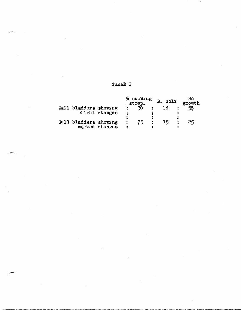
TABLE I
% shoWing B. coli No strep. growth
Gall bladders showing 30 • 18 5S • slight changes • • : • ;
: , : • Gall bladder s shon ng · 75 • 15 : 25 • •
marked changes • : •
-
-
TABLE II
Normal gall bladder showed. • • .0.51-1.70% chole sterol by weight
Inflamed gall bladder showed •.• 0.36~ ... cholesterol by weight
Strawberry gall bladder showed. 34.60-60.54% oholesterol by weight
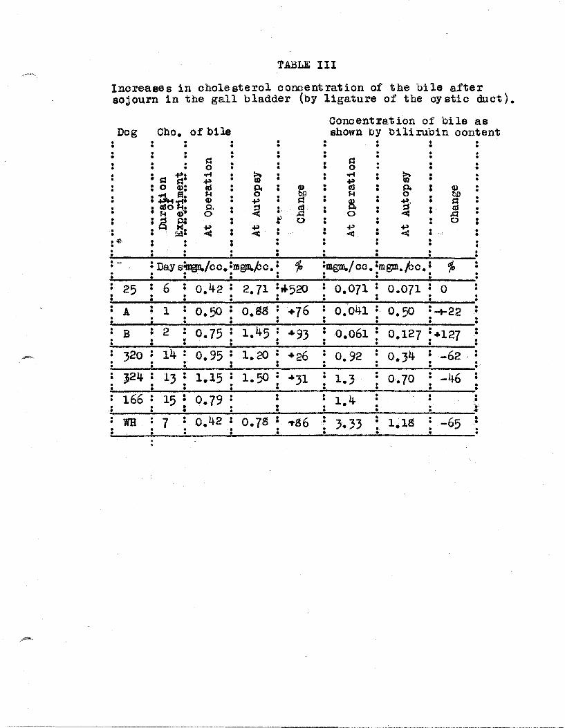
TABLE III
Increases in cho1e sterol concent ration of the bile after sojourn in the gall bladder (by ligature of the cy stic duct) •
Ooncentra.t ion of bile as Dog Cho. of bile shown by bilirubin content
• • • • • • • • • • • • • • • • • • • : • • • : • • • • • s:: • • ~ • • • • 0 • • • : • • • • • 0 • • 0 • • • • ~: ....t • t; • • ..... • t- o • • • s:I .p • • • ':d • • 0
• : 0 Q): cd • .·Pt • Q) • • p. : ~ • • • • • • • • ..... s. ~ • 0 • bO • ~ • 0 • • • • ..-..elf. Q) • .p • s:: • R • ~' • s:: • • : «so iI p.
" ~ • jJ • • 0 cd • 0 ~ Q) 0 0 • • 0 • Cal • a • • :g ~ 0 :f 0 0 • • • • • • • • • ., ¥ • .p • • ..j.:) • .p • 0 · . f:4: « « « « • • • • • • · . • .- : • : 0 • • • • • • 0 • • • • • • · 0 • • • • • • • • • • • • · - °D • } • p • fo :mgm./ oc. :mgm.. /cc. : 10· .. 0 • ay s~ co.·m@1D. c.' • · : . . . .0.
t • • • • • • : 25 : 6 • 0.42 • 2.71 :"'520 • 0.071 • 0.071 : 0 : • • • • : : • • • • • • • 0 0 • • • • • 0
A • 1 • 0.50 • O.Sg • +76 • 0.041 • 0.50 :+22 0 • • • • • • • • • • • • : • • • : • • • • • • • • B i 2 • 0.75 • 1 45 : ~93 • 0.061 • 0.127 • • • • • • 0 :~127 • • • • • • • • • 0 • • • • • • • • 0
· 320 • 14 : 0.95 : 1.20 • +26 • 0.92 • 0.34 • -62 • • • • • • • • • ~ • : • · • • • . . • • • • •
• J24 • 13 • 1.15 • 1.50 • • • -46 • • • • • • +31 1.3 • 0.70 • • · • • • • • • • • • • • • • • • • • 166 • 15 • 0.79 · • • 1.4 • • • • • • • • • • •
• • • • • • • • :. ·0 • • • • • • • • WH 7 • 0.42 • 0.7S : ... S6 • • 1.1S · -65 • • • • ~ 3.33 • • • • • • · • • .. • • • • • • • • • • •

TABLE IV
Average normal ot' cholesterin in blood serum
is 1.48 gm./1OOOcc.
Oase Oholesterol
6 5. 2 Gallstones at operation
10 3. 22 tI " " 11 5.55 It It " 12 5.57 " " " 13 9.49 " 11 "
Hypercholesterinemia persists after ope ration
15 4.77 3 days post-operative. stone s found.
16 2.13 4 " tI tI " .. 17 2.10 5 ft rt tf " " 18 3.77 5 ft If It " " 19 3.36 6 If " ,t " " 20 3.96 19 " tI " " "
-
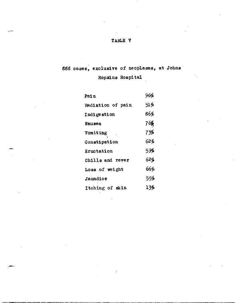
TABLE V
ggg oases, exolus1ve of neoplasms, at Johns
Hopains Hospital
Pain 96%
Rad1ation of pa1n 51%
Ind1ge stion g6~
Nausea 7~ Vomiting 7310
Oonst1pation 62%
Eructation 59%
Oh11ls and rever 62%
Loss of weight 66~
Jaundioe 55%
Itching of sk1n l3~

Symptom
Nausea
Vomiting
Pa.lpitation
Dyspnea
synoope
Fa.intness
Vertigo
Weakness
Tenderness in r. u. q.
TABLE VI,
Coronary Ooolusion
5
7
:3
17 6
5
3 4
5
Chole li t hi asi s
12
16
0
0
0
0
0
2
2;

TABLE VII
Position of Pain
Preoordial
substernal
Chest
Preoord. & Epigastrio
Epigastrio
Lower Abdomen
Diffuse over abd.
R. U. Q.
R. U. Q. & Ep1gast.
Lower rt. thorax
Left breast
Both flanks
Angle of Rt. soapula
Coronary Cholelithiasis Ooolusion
2
2
1
2
~
2
1
o o o o o o
o o o o
15 o o
9
1
2
1
1
1

. -
BIBLIOGRAPHY
Abernethy, J. Lectures on Anatomy, surgery and Pathology. Perkins 00. Boston, 1828. v. 2, p. 138
2. Alvarez, W. C., Keyer, K. F., Rusk. G. Y., Taylor, F. B., Easton, J. Transactions of the section on Gastroenterology and Proctology of the Am. Med. Assn. 1923
4.
5.
6.
8.
p. 35
!rey, L. B. Developmental Anatomy. Philadelphia, 1931. p. 182
W. B. saunders 00.,
Aronsobn1 . H. G., Andrews, E. stud.1e s on Non-bacterial Cholecys~itls. Proced. soc. Exp. Biol. and Med. 32:1629. 1935
Bail11e, :M., Morbid Anatomy of the Human Body. & Hazzard Philadelphia, 1820
Hiclanan
Bancroft, F. W., Ohronic Oholecystitis Without Stone. Ann. of surge 78:608 1923
Blalock, W., A. Olinical study of ~11iary Tract Disease. Jour. Am. Med. Assn., 83:2057, 192~
Bonar, T. Q., Gastric secretion in Appendicitis and Cholelithiasis. Guy's Hospital Report, 72:400, 1922.
9. Boyd, W., Studies in Gall-bladder Pathology. Brit. Jour. surg., 10:337, 1923.
10. Boyd, W., surgical Pathology. delphi a, l~ 5. p. 349
W. B. Saunders Co., Phila
11. Boyd, W., Gall-bladder Problems.
12. Brown, R. 0., A Study on the Etiology of Oholecystitis and its Production by the Injection of streptococci. Arch. Int. Med., 23:185, 1919.
13.
14.
15.
16.
17.
Coats, Joseph, A. Manual of Pathology. & 00., Philadelphia., 18g3.
H. O. Lea's Son
Cohnheim, J., Lectures on General pathology. McXee, A. B. New sydenham Soc., London, 1990.
Tr. by v. 3.
Constant, A. B., The Physio-pathology of the Gall Bladder. Clin. Med. & Surg., 37:877
Oope, Z., A. Clinical Study of Phrenio Should er Pal n: with Speoial Bearing on the Diagnosi s of Aoute Abdominal Disease. Brit. Jour. surge 10:192, 192 2-23 •
Davis, B. B~, The Diseased Gall Bladder. Jour., 18:86, 1933.
Nebr. state Med.
lS. Deaver, J. B., Bortz, E. L., Gall Bladder Disease: A Review of 903 Oases. Jour. Am. Med. Assn., 88:619, 1927.
19. Denton, J., The Mode of Origin of Gall-bladder Lesions. Arch. of SUrge 14:1 part 1, 1927
20. Dickson, S. H., Elements of Medicine. Blanchard & Lea, Philadelphia, 1855.
21. Duprat, Cours DIEtudes Medicales. Letellier et Cie., Paris, 1803. Tome second.
22. Ebers, Georg, The Ebers Papyrus. Tr. from German By Bryan, C. P., Geoffrey Bles, London, 1930.
23. Faulkner, J. M., Marble, H. 0., White, p. D., The Differential Diagnosis of Ooronary Occlusion and of Oholeli thiasi s. Jour. Am. Med. Assn., 83: 2080, 1924.-
24. Goupil, J. M. A.I
The New Medical Doctrine. Tr. by Nott, J. C. Columbia, 831.
25. Graham, E. A., colet W. H., Oopher, G. H., Moore, S. Diseases of the Gal Bladder and Bile Illcts. Lea & Febiger, Philadelphia, 1928.
26. Graham, E. A., Hepatitis: A Constant Accgmpaniment of Cholecystitis, Surg., Gynec. and Obst., 20:521,1918.
27. Graham, E. A., Peterman, M. G., Further Observations on the Lymphatic Origin of Cholecystitis, Oholedochitis and the Associated Pancreatitis. Arch. of surg., 23. 1922.
fa. Graham, E. A., The Olinical Application of SOme Recent Knowledge of the Biliary Tract. The Harvey Lectures 1933-34.
29. Griffiths, H. E., The Relationship of the Diseases of the Gall Bladder to the Secretory Function of the Stomach and Pancreas. Lancet, 2:203, 1924.
30.
32.
Hallerus, Albertus, Disputationes ad Morborum. MarciMichael, Lausanae, 1757. -v. 3.
Hargis, E. H., Harer, W. B., VanMeter, V. 0., studies of the Function of the Gall-bladder. Surg., Gynac. and Obsta )4:307, 1922
IvY, A. C., Bergh, G. S., The Applied Physiology of the Extra-Hepatic Biliary tract. Jour. Am. Med. Assn., 103: 1500, Nov. 17, 1934.
;4.
;5.
;7.
;9.
Jenes, E., The Value of the Determination of the Chole sterin Content of the Blood in th~ Diagnosis of Cholelithiasis. Jour. Am. Med. Assn., 0;:146,1912
Judd, E. S., Relatio n of the Liver and Pancreas to Infection of the Gall-bladder. Jour. Am. Med. Assn., 77: 197, 1921
Kodama,S., The Lymphatics of the Ex-.ra:-hepatic Biliary Passages. SUrg., Gynec. and Obst., 43:140,1926 •
• ann, F. C., A Physiologic Consideratin of the Gallbladder. Jour. Am. Med. Assn., 83:g29, 1924
Mann, F. C., Chronic Gall Bladder Disease in the Young Adult. Jour. Am. Med. Assn., 8;:981, 1924
Mayo, W. ""J., "Innocent" Gall-stones a Myth. Assn., 50:1021, 1911.
Jour. Am. Med.
Jlayo, C. H., Gallstones and Diseases of the Gall Bladder. Am. surge Assn. t 81: 955, 1925.
40. Mentzer, S. H., Cholesterosis of the Gall-bladder. Am. Jour. Path., 1:;83, 1925.
41. Mentzer, S. H., Judd, E. S., Parkhill, E., A Bacterial Study of Gall-bladders removed at Operation. Am. Jour. Med. Sci., 173:16, 1927.
42.
44.
Mentzer, S., H.,. Pathogene si s of Biliary calculi. of surg., 14: lLt, 1927.
Arch.
Mentzer, S. H., A Clinical Study of Cholecystitis and Choleli thiasi s. surg., Gynec. and Obst., 42: 7g2, 1926
Meyer, K. F., Neilson, N. 14., Feusier, M. L., The Mecha.nism of Gall Bl adder Infections in Laboratory Animals. Jour. Infect. Dis. 28:456, 1921.
45. Miller, R. H., Acute CholecJ~titis. Ann. Surg., 92:644, 1930.
46. Korgagni, Joannis Baptistae, Adversaria Anatomia Adversaria, 171S;
Moynihan, B., SOme Aspects of Cholelithiasis. Jour., 1:39;, 1925.
Brit. Med.
48. Neuberger, M., History of Medicine. Tr. by Palyfair, E. Oxford University Press, 1910. v. 1.
49. ~iemeyer, PatholOgie Interne. Tr. par Oulman et sengel. Bailliere, paris, 1965.
.-.•
50.
53.
55.
Oliver, S. F., The Etiology of Gallstones. Lab. and Clin. Med., 8:242, 1922.
Jour. of
Osler, Wil11am, The Pr1noiples and Praot10e of Med10ine. Edited by MoCrae, T. D. Appleton and Co. New York, 1926. P. 23.
Patey, D. H., Whitby, L. E. H., The Paths of Gall Bladder Infeotion: An Exper1mental Study. Brit. Jour. Surg., 20: 5S0, 1932-33
Paulus Aeg1neta, The seven Books of. Tr. by Adams, F. Sydennam Soo., London, 1947. v. 1 & 2.
The
Peterman, M. G., Priest, W. S., Graham, E. A., The Assoc1at10n of Hepat1tis with Exper1mental Cholecystitis and its Bear1ng on the patho~nes1s of Cholecyst1t1s in the Human. Aroh. Surg., 2:92, 1~21
Piersol, G. M., Bockus, H. L., The Panoreat1c Enzymes 1n Choleoyst1 ti s. Arch. Int. Med., 35: 20~, 1925.
56. Rosenmuller, I. 0., Compend1um Anatomicum. Leipsig, 1816. p. 285.
57.
59.
60.
61 • •
62.
64-.
Rosenow, E. C., The Etiology of Oholeoy sti ti s and Gallstones, and their production by the Intravenous Injection of Bacter1a. Jour. Infeot. Dis., Ifh 527. 1916
Rossi, A., The Atonl0 Gall Bladder and the Strawberry Gall Bladder. Am. Jour. Roent., 2]: 205, 19}2
Spalteholz, W., Hand-atlas of Human Anatomy. Tr. by Barker, L. F.,J. B. Lipplnoott 00., Philadelphia, v. 2 & 3
stanton, E. ",M., The Stoneless Gall Bladder. Assn., 87:2bO, 1926.
Jour. Am. Med.
SWalm, W. A., Diagnosis of Gall Traot Disease. Med., 29:247, 1930
Northwest
sweet4
J. E., The Formation of Gallstones. Ann. surg., 99:36 , 1934
Sudler, M. T., The Arohitecture of the Gall Bladder. Johns Hopkins Hosp., 12:126, 1901.
Vloary, Thomas., The Anatomie of the Bodle of Man. (Edit1on of 1548). N. Trubner & Co., London, lS8S.
Bull.
Walton, A. J., Some Modern Aspects of Choleoyst1tis and Cholellthiasis. Lanoet 1:334, 19;0.
66.
67.
Will taker, L. R., The Meohani em of the Gall Bladder and its Relation to Choleoystitis. Jour. Am. Med. Assn., gS: 1542, 1927.
Williams, B., MoLaohlon, D. G. S., The Aetiology of CholecYstltis, Lanoet 2:342, 1930.
6S.- Wlnkelsteln, A., Motor Meoh~nism of the Gall-bladder. Jour. Am. Med. Assn., gO:174a , 1923
69. Wolf. A. E. M.1. Some Affeotions of the Gall Bladder. Praotioner, 12~:61J, 1930