the dna-bending protein hmgb1 is a cellular cofactor of sleeping beauty transposition
TRANSCRIPT
The DNA-bending protein HMGB1 is a cellularcofactor of Sleeping Beauty transpositionHatem Zayed1, Zsuzsanna IzsvaÂk1,2, Dheeraj Khare1, Udo Heinemann1,3 and ZoltaÂn Ivics1,*
1Max DelbruÈck Center for Molecular Medicine, Robert RoÈssle Strasse 10, D-13092 Berlin, Germany, 2Institute ofBiochemistry, Biological Research Center of the Hungarian Academy of Sciences, Szeged, Hungary and 3Instituteof Chemistry/Chrystallography, Free University of Berlin, Germany
Received February 3, 2003; Revised and Accepted March 3, 2003
ABSTRACT
Sleeping Beauty (SB) is the most active Tc1/mariner-type transposon in vertebrates. SB containstwo transposase-binding sites (DRs) at the end ofeach terminal inverted repeat (IR), a feature termedthe IR/DR structure. We investigated the involve-ment of cellular proteins in the regulation of SBtransposition. Here, we establish that the DNA-bending, high-mobility group protein, HMGB1 isa host-encoded cofactor of SB transposition.Transposition was severely reduced in mouse cellsde®cient in HMGB1. This effect was rescued bytransient over-expression of HMGB1, and waspartially complemented by HMGB2, but not with theHMGA1 protein. Over-expression of HMGB1 in wild-type mouse cells enhanced transposition, indicatingthat HMGB1 can be a limiting factor of transposition.SB transposase was found to interact with HMGB1in vivo, suggesting that the transposase may recruitHMGB1 to transposon DNA. HMGB1 stimulatedpreferential binding of the transposase to the DRfurther from the cleavage site, and promoted bend-ing of DNA fragments containing the transposon IR.We propose that the role of HMGB1 is to ensure thattransposase±transposon complexes are ®rst formedat the internal DRs, and subsequently to promotejuxtaposition of functional sites in transposonDNA, thereby assisting the formation of synapticcomplexes.
INTRODUCTION
The use of transposable elements as genetic tools contributedsigni®cantly to our understanding of biological systems. Invertebrates, such tools could be applied to both research andtherapeutics. Tc1/mariner elements are probably the mostwidespread transposons in nature (1). These elements are ableto transpose in species other than their hosts, and are thereforeemerging tools for functional genomics in several organisms(1). However, the vast majority of naturally occurring Tc1/mariner-like transposons are non-functional due to inactivat-ing mutations. In vertebrates, not a single active element has
been found. Based on a comparative phylogenetic approach,we have reconstructed an active Tc1-like transposon from bitsand pieces of inactive elements found in the genomes ofteleost ®sh, and named this transposon Sleeping Beauty (SB)(2).
SB is ¯anked by ~230 bp terminal inverted repeats (IRs),which contain binding sites for the enzymatic factor oftransposition, the transposase. The transposase binding sites(DRs) of SB elements are repeated twice per IR in a directorientation (2). This special organization of IR, termed IR/DR,is an evolutionarily conserved feature of a group of Tc1-liketransposons, but not that of the Tc1 element itself (1,3). Inaddition to the DRs, the left IR of SB contains a transpositionalenhancer-like sequence, termed HDR (4). Speci®c binding tothe DRs is mediated by an N-terminal, paired-like DNA-binding domain of the transposase (2,4,5). The catalyticdomain of the transposase, responsible for the DNA cleavageand joining reactions, is characterized by a conserved aminoacid triad, the DDE motif, which is found in a large group ofrecombinases (6), including retroviral integrases and theRAG1 V(D)J recombinase involved in immunoglobulin generearrangements (1).
SB transposes via a DNA intermediate, through a cut-and-paste mechanism. The transposition process can arbitrarily bedivided into at least four major steps: (i) binding of thetransposase to its sites within the transposon IRs; (ii)formation of a synaptic complex in which the two ends ofthe elements are paired and held together by transposasesubunits; (iii) excision from the donor site; (iv) reintegration ata target site. On the molecular level, mobility of DNA-basedtransposable elements can be regulated by imposing con-straints on transposition. One important form of transposi-tional control is represented by regulatory `checkpoints', atwhich certain molecular requirements have to be ful®lled forthe transpositional reaction to proceed. These requirementscan operate at any of the four different stages of transpositionlisted above, and can be brought about by both element- andhost-encoded factors.
Several DNA recombination reactions are stimulated byDNA-bending proteins. For example, the transposase bind-ing sites of bacteriophage Mu are brought together by thebending action of the Escherichia coli HU protein (7). Hinrecombinase-mediated recombination and bacteriophage lintegration are strongly stimulated by HU (8) and integrationhost factor (IHF) (9), respectively. The eukaryotic high
*To whom correspondence should be addressed. Tel: +49 30 9406 2546; Fax: +49 30 9406 2547; Email: [email protected]
Nucleic Acids Research, 2003, Vol. 31, No. 9 2313±2322DOI: 10.1093/nar/gkg341
Nucleic Acids Research, Vol. 31 No. 9 ã Oxford University Press 2003; all rights reserved
mobility group (HMG) proteins can functionally replace HUand IHF in some recombination reactions, indicating somelevel of exchangeability between these DNA-bending proteins(10). All of these DNA-bending proteins are believed to assistrecombinational mechanisms by facilitating the formation ofactive recombinase±DNA complexes (11,12).
HMG proteins are classi®ed into three subfamilies,HMGB1/2 (formerly known as HMG1/2), HMGA1a/b (for-merly known as HMGI/Y) and HMGN1/2 (formerly known asHMGB14/17, that share many physical characteristics, butdiffer in their main functional domains (13). Both the HMGBand HMGA1 group proteins are known to bind A/T-rich DNAthrough interactions with the minor groove of the DNA helix(12). HMGB1 is an abundant (~106 molecules/cell), non-histone, nuclear protein associated with eukaryotic chromatin(12). Through its DNA-binding domain, termed the HMG-box, HMGB1 binds DNA in a sequence-independent manner,but with preference for certain DNA structures including four-way junctions and severely undertwisted DNA (13±16).HMGB1 has low af®nity to B-form DNA, and is thought tobe recruited by other DNA-binding proteins through protein±protein interactions, and induce a local distortion of the DNAupon binding. The ability of HMGB1/2 proteins to bend DNAwas demonstrated in vitro (13). These proteins facilitate self-ligation of short DNA fragments (17,18), and can bridge linearDNA fragments thereby enhancing multimerization of longerDNAs (19). Together with the closely related HMGB2protein, HMGB1 has been implicated in a number ofeukaryotic cellular processes including gene regulation,DNA replication and recombination (12,20). HMGB1/2directly interact with a number of proteins, including someHOX (21) and POU domain (22) transcription factors and theTATA-binding protein (23), and facilitate their bindingthrough protein±protein interactions. HMGB1/2 were shownto enhance immunoglobulin V(D)J recombination by enfor-cing speci®c DNA recognition (24) through their interactionwith the RAG1/2 recombinase complex (25), and facilitatingcleavage (24). In addition, HMGB1 was found to promote Repprotein-mediated site-speci®c cleavage of adeno-associatedvirus DNA (26). The production of retroviral cDNA does notrequire an excision step, but the downstream events ofretroviral integration are highly similar to other transpositionalreactions (27). Interestingly, HMGA1 family members, butnot HMGB1/2, are required for retroviral cDNA integration(28,29). Both V(D)J recombination and retroviral integrationhave common features with SB transposition. RAG-mediatedcleavage at the ends of recombination signal sequences (RSSs)in V(D)J recombination is probably analogous to the excisionstep of transposition, whereas the biochemical steps leading toinsertion of signal molecules, retrovirus integration and DNAtransposition are essentially the same (27).
SB mediates transposition in a variety of vertebrate species(30), and is more active than other members of the Tc1/mariner family (31). Because there is substantial interest indeveloping transposon technology for gene therapy (32) andgene discovery (31), it is of importance to dissect themolecular mechanisms involved in transposition and itsregulation. In particular, differential interactions between thetransposon and host-encoded factors may result in limitationof host range. In this work, we evaluated HMG proteins ascellular host factors of SB transposition in mammalian cells.
We have found that HMGB1 is required for ef®cient SBtransposition. SB transposition was signi®cantly reduced inHMGB1-de®cient mouse cells. This effect was fully comple-mented by expressing HMGB1, partially by expressingHMGB2, but not with HMGA1. Interestingly, transientover-expression of HMGB1 in wild-type mouse cellsenhanced transposition, indicating that HMGB1 is a limitingfactor of transposition. SB transposase was found to interactwith HMGB1 in vivo, suggesting that the transposase mayactively recruit HMGB1 to transposon DNA via protein±protein interactions. HMGB1 enhanced preferential binding ofthe SB transposase to the internal transposase binding siteswithin the transposon IRs, and promoted bending of DNAfragments comprising the transposon IRs. These data areconsistent with a role of HMGB1 in synaptic complexformation in transposition.
MATERIALS AND METHODS
Protein expression, puri®cation and electrophoreticmobility shift assay (EMSA)
Production of N123 was done as described (2). A plasmidexpressing a hexahistidine-tagged version of HMGB1 wasdescribed in Aidinis et al. (25), and was kindly provided by M.Bianchi, Milan, Italy. Protein expression was induced by theaddition of 0.4 mM IPTG in E.coli BL21 cells. Puri®cationwas done using a nickel resin (Qiagen), according to themanufucturer's protocol. The puri®ed protein was dialyzedagainst 25 mM HEPES pH 7.4, 10% glycerol, 1 M NaCl and2 mM b-mercaptoethanol, and its concentration determined bysodium dodecyl sulphate (SDS)±polyacrylamide gel electro-phoresis.
The plasmid expressing a maltose-binding protein(MBP)±SB transposase fusion protein was made by cloningthe SB transposase gene into the XmnI/EcoRI sites of pAML-c2X (NEB). The plasmid was transformed into the BL21-CodonPlus-RIL E.coli strain (Stratagene). Protein puri®cationprotocol was as described by the manufacturer of the amyloseresin (NEB). A 1 l bacterial culture was grown to OD (A600)~0.5, IPTG was added to a ®nal concentration of 0.3 mM, andfurther incubated at 37°C for 2 h. Cells were harvested andresuspended in 30 ml of column buffer (CB = 20 mM TrispH 7.4, 200 mM NaCl, 1 mM EDTA and 1 mM DTT). Beforecell lysis, 0.6 mg DNase I and 0.5% (v/v) polyethyleneiminewas added. Cell lysis was done by French press at 1200 p.s.i.,and the pellet obtained after centrifugation was resuspended in50 ml CB containing 750 mM NaCl. In the higher ionicstrength buffer, MBP±SB was dissolved, but nucleic acids andsome other proteins remained in the pellet. The supernatantwas diluted 1:5 with CB and loaded on an amylose resincolumn (12 ml of resin equilibrated with column buffer) with a¯ow rate not exceeding 1 ml/min. Washing was done with12 column vol of wash buffer (CB with 750 mM NaCl). Thefusion protein was eluted with elution buffer (20 mM Tris pH7.4, 750 mM NaCl, 1 mM EDTA, 1 mM DTT and 10 mMmaltose), 25 fractions of 2 ml each were collected; thefractions having the fusion protein were pooled and concen-trated to 0.4 mg/ml.
2314 Nucleic Acids Research, 2003, Vol. 31, No. 9
An EcoRI fragment comprising the left IR of the SB elementcontaining both transposase binding sites, an A¯II/HindIIIfragment containing only the inner DR (IDR), and an EcoRI/HindIII fragment of a modi®ed SB element lacking the IDR,and thus containing only the outer DR (ODR) (30), were end-labeled using [a32P]dATP and Klenow. Equal amounts ofDNA fragments were used for labeling. Nucleoprotein com-plexes were formed in 20 mM HEPES (pH 7.5), 0.1 mMEDTA, 0.1 mg/ml BSA, 150 mM NaCl, 1 mM DTT in a totalvolume of 10 ml. Reactions contained ~0.08 nM DNA probe,1 mg poly(dI)(dC), 75 nM HMGB1 and 0.2, 0.1 or 0.05 ngN123. Reactions with MBP±SB transposase fusion contained~0.1 nM of left IR probe (EcoRI fragment) and either 2 nM or20 nM of puri®ed MBP±SB with or without 0.1 or 1 mM ofpuri®ed HMGB1. After a 15 min incubation on ice, 5 ml ofloading dye containing 50% glycerol and bromophenol bluewas added, and the samples loaded onto a 5% polyacrylamidegel. Radioactive bands were quanti®ed using a MolecularDynamics PhosphorImager System.
Ligase-mediated circularization assay
A 32P-labeled left IR fragment (~0.04 nM) with cohesiveEcoRI ends was pre-incubated with 6 nM HMGB1 on ice for20 min in T4 DNA ligase buffer, in a ®nal volume of 50 ml.The ligation reaction was initiated by the addition of 0.025 Uof T4 DNA ligase (NEB), and incubated at 16°C. Aliquots of9 ml of the reaction mixture were withdrawn at different timepoints (0, 5, 15, 30 and 60 min), and were added to 41 ml ofstop solution (0.5% SDS, 10 mM EDTA, 1 mg/ml ProteinaseK). The reactions were incubated at 50°C for 2 h, extractedwith phenol/chloroform/isoamyl alcohol, and then withchloroform/isoamyl alcohol. Aliquots of 30 ml of the 60 minsample were taken, and digested with 100 U of ExonucleaseIII (ExoIII) (NEB) at 37°C for 30 min. Aliquots of 25 ml ofeach extracted sample were run on a 4% non-denaturingpolyacrylamide gel, gels were dried and autoradiographed at±80°C.
For bacterial transformations, ~0.1 nM linearized tran-sposon DNA was pre-incubated with 12 nM HMGB1, then1 U/reaction of the T4 DNA ligase was added, and the reactionallowed to proceed for 0, 15, 30 and 60 min. Reactions wereterminated by the addition of stop solution, the DNA wasprecipitated, resuspended in TE, and electroporated intoDH10B E.coli cells.
Immunoprecipitation
Nuclear extracts from ~2.0 3 107 IRES-SB and IRES-K cellswere prepared essentially as described previously (30). Thenuclear extract was diluted to contain 100 mM NaCl withbinding buffer [25 mm HEPES±KOH pH 7.9, 1.5 mM MgCl2,10 mm KCl, 0.5 mM EDTA, 0.2 mM DTT, 0.1 mM NP-40,12% glycerol, 13 complete protease inhibitors (Roche)]. Forthe DNase I treatment, MgCl2 concentration was increased to6 mM and 6 U of DNase I (Ambion) was added. The extractwas incubated at 23°C for 30 min, followed by the addition of2 ml of 500 mM EDTA. The extract was precleared in twosteps, ®rst by adding 1 mg mouse IgG and 20 ml ProteinG-Sepharose (50% slurry in PBS) for 60 min at 4°C, and thenby adding 50 ml Protein G-Sepharose followed by an overnightincubation at 4°C. Immunoprecipitation was performed using1 mg HMGB1 antibody (Santa Cruz Biotech.), or actin
antibody (clone ACTN05, Neomarkers), or p15 antibody (R-20, Santa Cruz Biotech.), or goat preimmune serum (Sigma)and 15 ml Protein G-Sepharose (50% slurry in PBS). The tubeswere rotated overnight at 4°C. The beads were washed fourtimes in PBS, resuspended in SDS sample buffer, andsubjected to western hybridization with a rabbit polyclonalantibody against the SB transposase.
Co-immunoprecipitation was done using puri®ed HMGB1(1 mM) and puri®ed MBP±SB (0.2 mM), either alone ortogether in 20 ml ®nal volume in binding buffer containing25 mM HEPES (pH 7.5), 10 mM MgCl2, 1 mM DTT, 2%glycerol, 0.25 mg BSA, 50 mM KCl, 0.01% NP-40 for 20 minat 4°C. Then ~1 nM of a 32P-labeled transposon fragmentcontaining the left IR (~120 000 c.p.m.) was added, and thereaction continued for a further 45 min. One microgram ofeither anti-HMGB1 or anti-SB was added, and the incubationcontinued for 2 h at 4°C with rotation. Protein A- and ProteinG-Sepharoses were pretreated with the binding buffer con-taining 500 mg/ml of herring sperm DNA overnight. Analiquot of 50 ml pretreated Protein A-Sepharose was added tothe anti-SB samples, 50 ml of pretreated Protein G-Sepharosewas added to the anti-HMGB1 samples, and incubationcontinued for 3 h at 4°C. The immunoprecipitate was washedthree times with the binding buffer. Radioactivity of the DNAthat remained bound to the beads was quanti®ed with a liquidscintillation counter.
Cell culture and in vivo transposition assay
Cell lines were grown in DMEM supplemented with 10% fetalbovine serum. Wild-type (VA1) and HMGB1-de®cient (C1)mouse embryonic ®broblast (MEF) cell lines have beendescribed previously (33), and were kindly provided byM. Bianchi, Milan, Italy. All DNA transfections were donewith FUGENE6 transfection reagent (Roche, Germany). Cellswere cotransfected with 90 ng each of pCMV-SB (2) andpT/zeo, (30) and 500 ng plasmid expressing HMGB1 (25),HMGB2 (25), HMGA1 (34) or b-galactosidase. 105 trans-fected cells were plated out for selection, using 100 mg/mlzeocin (Invitrogen). After 3 weeks of selection, colonies werestained and counted as described previously (2).Transfectability of the C1 cell line is lower than that of theVA1 line, which was determined by transfection of a GFP-expressing plasmid. Therefore, transpositional ef®ciencies inthe two cell lines presented in Figure 1B have been normalizedto transfection ef®ciencies.
RESULTS
HMGB1 is required for ef®cient SB transposition inmouse cells
We assessed the importance of HMGB1 for SB transpositionby applying an in vivo transposition assay (2) on wild-type andHMGB1-de®cient mouse cells (Fig. 1). The assay is based oncotransfection of a donor plasmid carrying a zeocin resistancegene (zeo)-marked transposon and a transposase-expressinghelper plasmid into cultured cells (Fig. 1A). In controlexperiments, a plasmid expressing b-galactosidase (CMVb)substitutes for the transposase helper plasmid. Cells are placedunder antibiotic selection, and the numbers of resistantcolonies counted. The ratio between numbers obtained in the
Nucleic Acids Research, 2003, Vol. 31, No. 9 2315
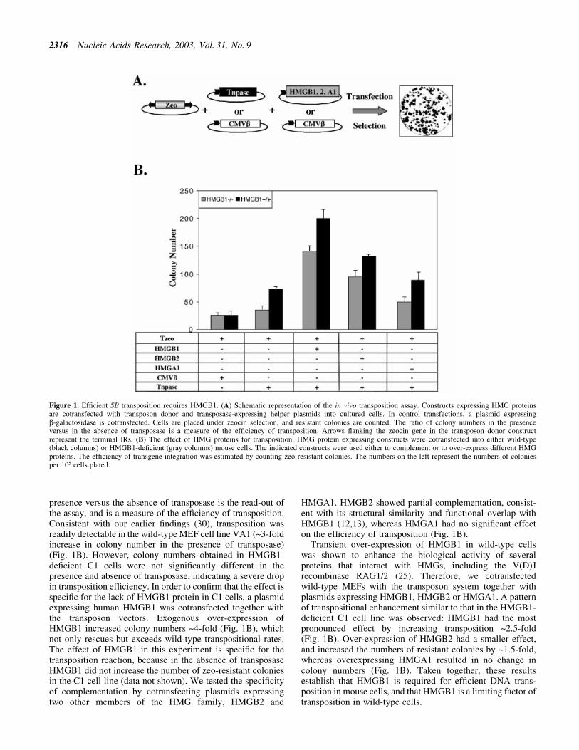
presence versus the absence of transposase is the read-out ofthe assay, and is a measure of the ef®ciency of transposition.Consistent with our earlier ®ndings (30), transposition wasreadily detectable in the wild-type MEF cell line VA1 (~3-foldincrease in colony number in the presence of transposase)(Fig. 1B). However, colony numbers obtained in HMGB1-de®cient C1 cells were not signi®cantly different in thepresence and absence of transposase, indicating a severe dropin transposition ef®ciency. In order to con®rm that the effect isspeci®c for the lack of HMGB1 protein in C1 cells, a plasmidexpressing human HMGB1 was cotransfected together withthe transposon vectors. Exogenous over-expression ofHMGB1 increased colony numbers ~4-fold (Fig. 1B), whichnot only rescues but exceeds wild-type transpositional rates.The effect of HMGB1 in this experiment is speci®c for thetransposition reaction, because in the absence of transposaseHMGB1 did not increase the number of zeo-resistant coloniesin the C1 cell line (data not shown). We tested the speci®cityof complementation by cotransfecting plasmids expressingtwo other members of the HMG family, HMGB2 and
HMGA1. HMGB2 showed partial complementation, consist-ent with its structural similarity and functional overlap withHMGB1 (12,13), whereas HMGA1 had no signi®cant effecton the ef®ciency of transposition (Fig. 1B).
Transient over-expression of HMGB1 in wild-type cellswas shown to enhance the biological activity of severalproteins that interact with HMGs, including the V(D)Jrecombinase RAG1/2 (25). Therefore, we cotransfectedwild-type MEFs with the transposon system together withplasmids expressing HMGB1, HMGB2 or HMGA1. A patternof transpositional enhancement similar to that in the HMGB1-de®cient C1 cell line was observed: HMGB1 had the mostpronounced effect by increasing transposition ~2.5-fold(Fig. 1B). Over-expression of HMGB2 had a smaller effect,and increased the numbers of resistant colonies by ~1.5-fold,whereas overexpressing HMGA1 resulted in no change incolony numbers (Fig. 1B). Taken together, these resultsestablish that HMGB1 is required for ef®cient DNA trans-position in mouse cells, and that HMGB1 is a limiting factor oftransposition in wild-type cells.
Figure 1. Ef®cient SB transposition requires HMGB1. (A) Schematic representation of the in vivo transposition assay. Constructs expressing HMG proteinsare cotransfected with transposon donor and transposase-expressing helper plasmids into cultured cells. In control transfections, a plasmid expressingb-galactosidase is cotransfected. Cells are placed under zeocin selection, and resistant colonies are counted. The ratio of colony numbers in the presenceversus in the absence of transposase is a measure of the ef®ciency of transposition. Arrows ¯anking the zeocin gene in the transposon donor constructrepresent the terminal IRs. (B) The effect of HMG proteins for transposition. HMG protein expressing constructs were cotransfected into either wild-type(black columns) or HMGB1-de®cient (gray columns) mouse cells. The indicated constructs were used either to complement or to over-express different HMGproteins. The ef®ciency of transgene integration was estimated by counting zeo-resistant colonies. The numbers on the left represent the numbers of coloniesper 105 cells plated.
2316 Nucleic Acids Research, 2003, Vol. 31, No. 9
HMGB1 enhances bending of the SB transposonterminal IR and the full length transposon
Upon binding to DNA, HMG proteins induce conformationalchanges in the DNA, thereby facilitating juxtaposition ofdistantly bound proteins and assembly of multiprotein com-plexes (11,12). SB has two transposase-binding sites perterminal IR, separated by ~160 bp spacer regions. Wehypothesized that the bending activity of HMGB1 couldcontribute to bringing the DRs and/or the complete IRs closerin space, thereby assisting the formation and/or stabilization ofa synaptic complex.
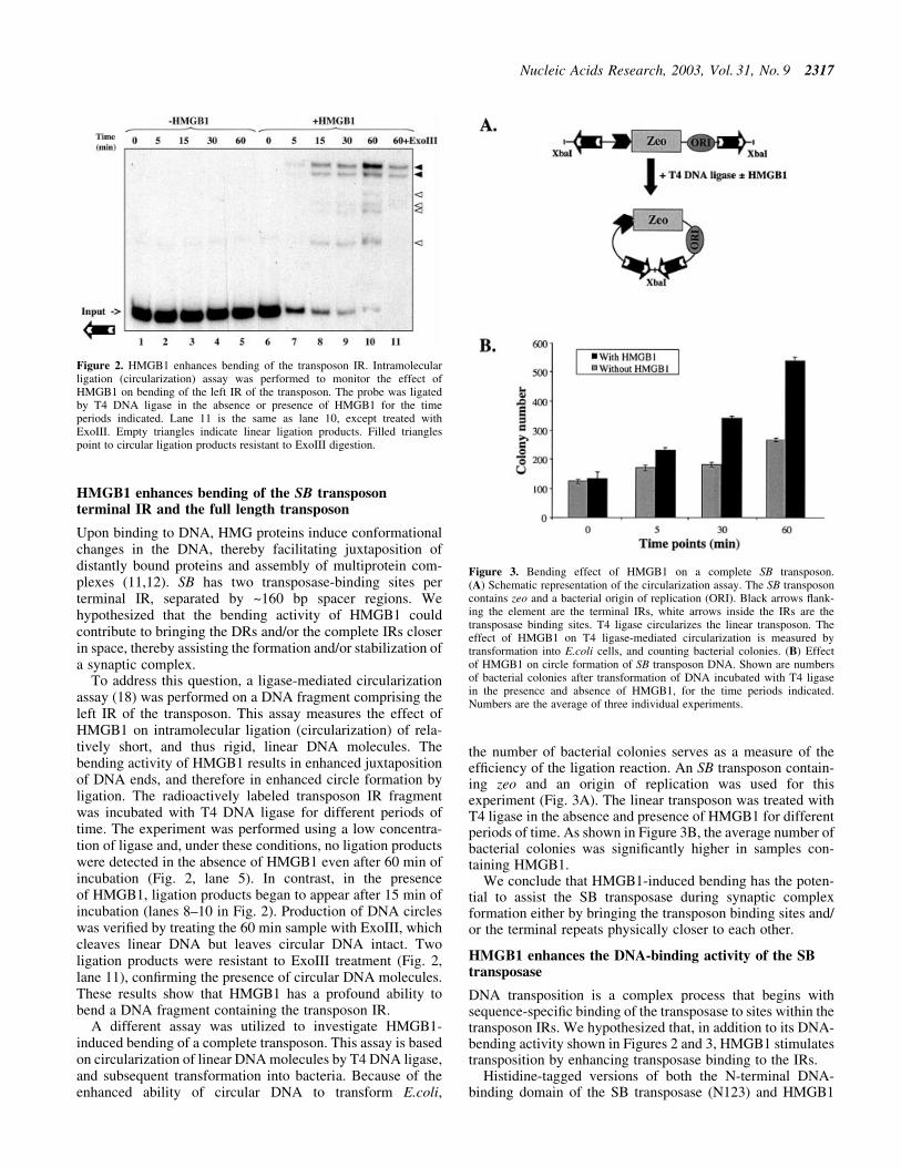
To address this question, a ligase-mediated circularizationassay (18) was performed on a DNA fragment comprising theleft IR of the transposon. This assay measures the effect ofHMGB1 on intramolecular ligation (circularization) of rela-tively short, and thus rigid, linear DNA molecules. Thebending activity of HMGB1 results in enhanced juxtapositionof DNA ends, and therefore in enhanced circle formation byligation. The radioactively labeled transposon IR fragmentwas incubated with T4 DNA ligase for different periods oftime. The experiment was performed using a low concentra-tion of ligase and, under these conditions, no ligation productswere detected in the absence of HMGB1 even after 60 min ofincubation (Fig. 2, lane 5). In contrast, in the presenceof HMGB1, ligation products began to appear after 15 min ofincubation (lanes 8±10 in Fig. 2). Production of DNA circleswas veri®ed by treating the 60 min sample with ExoIII, whichcleaves linear DNA but leaves circular DNA intact. Twoligation products were resistant to ExoIII treatment (Fig. 2,lane 11), con®rming the presence of circular DNA molecules.These results show that HMGB1 has a profound ability tobend a DNA fragment containing the transposon IR.
A different assay was utilized to investigate HMGB1-induced bending of a complete transposon. This assay is basedon circularization of linear DNA molecules by T4 DNA ligase,and subsequent transformation into bacteria. Because of theenhanced ability of circular DNA to transform E.coli,
the number of bacterial colonies serves as a measure of theef®ciency of the ligation reaction. An SB transposon contain-ing zeo and an origin of replication was used for thisexperiment (Fig. 3A). The linear transposon was treated withT4 ligase in the absence and presence of HMGB1 for differentperiods of time. As shown in Figure 3B, the average number ofbacterial colonies was signi®cantly higher in samples con-taining HMGB1.
We conclude that HMGB1-induced bending has the poten-tial to assist the SB transposase during synaptic complexformation either by bringing the transposon binding sites and/or the terminal repeats physically closer to each other.
HMGB1 enhances the DNA-binding activity of the SBtransposase
DNA transposition is a complex process that begins withsequence-speci®c binding of the transposase to sites within thetransposon IRs. We hypothesized that, in addition to its DNA-bending activity shown in Figures 2 and 3, HMGB1 stimulatestransposition by enhancing transposase binding to the IRs.
Histidine-tagged versions of both the N-terminal DNA-binding domain of the SB transposase (N123) and HMGB1
Figure 2. HMGB1 enhances bending of the transposon IR. Intramolecularligation (circularization) assay was performed to monitor the effect ofHMGB1 on bending of the left IR of the transposon. The probe was ligatedby T4 DNA ligase in the absence or presence of HMGB1 for the timeperiods indicated. Lane 11 is the same as lane 10, except treated withExoIII. Empty triangles indicate linear ligation products. Filled trianglespoint to circular ligation products resistant to ExoIII digestion.
Figure 3. Bending effect of HMGB1 on a complete SB transposon.(A) Schematic representation of the circularization assay. The SB transposoncontains zeo and a bacterial origin of replication (ORI). Black arrows ¯ank-ing the element are the terminal IRs, white arrows inside the IRs are thetransposase binding sites. T4 ligase circularizes the linear transposon. Theeffect of HMGB1 on T4 ligase-mediated circularization is measured bytransformation into E.coli cells, and counting bacterial colonies. (B) Effectof HMGB1 on circle formation of SB transposon DNA. Shown are numbersof bacterial colonies after transformation of DNA incubated with T4 ligasein the presence and absence of HMGB1, for the time periods indicated.Numbers are the average of three individual experiments.
Nucleic Acids Research, 2003, Vol. 31, No. 9 2317
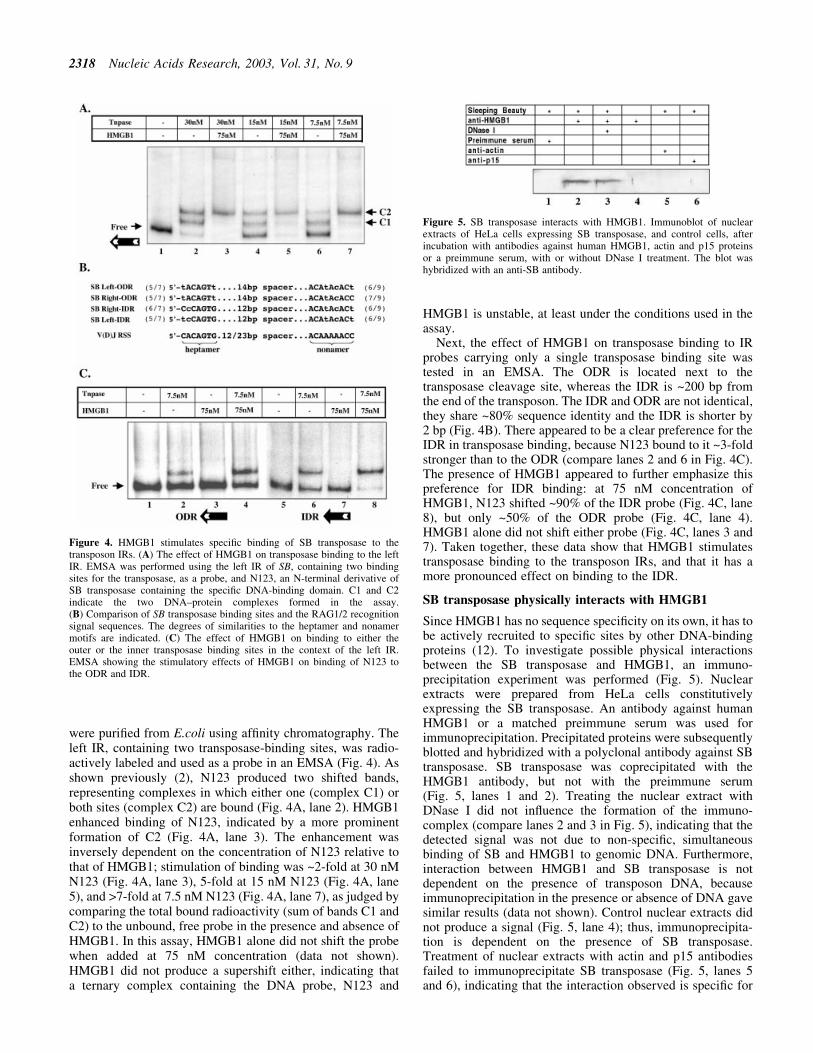
were puri®ed from E.coli using af®nity chromatography. Theleft IR, containing two transposase-binding sites, was radio-actively labeled and used as a probe in an EMSA (Fig. 4). Asshown previously (2), N123 produced two shifted bands,representing complexes in which either one (complex C1) orboth sites (complex C2) are bound (Fig. 4A, lane 2). HMGB1enhanced binding of N123, indicated by a more prominentformation of C2 (Fig. 4A, lane 3). The enhancement wasinversely dependent on the concentration of N123 relative tothat of HMGB1; stimulation of binding was ~2-fold at 30 nMN123 (Fig. 4A, lane 3), 5-fold at 15 nM N123 (Fig. 4A, lane5), and >7-fold at 7.5 nM N123 (Fig. 4A, lane 7), as judged bycomparing the total bound radioactivity (sum of bands C1 andC2) to the unbound, free probe in the presence and absence ofHMGB1. In this assay, HMGB1 alone did not shift the probewhen added at 75 nM concentration (data not shown).HMGB1 did not produce a supershift either, indicating thata ternary complex containing the DNA probe, N123 and
HMGB1 is unstable, at least under the conditions used in theassay.
Next, the effect of HMGB1 on transposase binding to IRprobes carrying only a single transposase binding site wastested in an EMSA. The ODR is located next to thetransposase cleavage site, whereas the IDR is ~200 bp fromthe end of the transposon. The IDR and ODR are not identical,they share ~80% sequence identity and the IDR is shorter by2 bp (Fig. 4B). There appeared to be a clear preference for theIDR in transposase binding, because N123 bound to it ~3-foldstronger than to the ODR (compare lanes 2 and 6 in Fig. 4C).The presence of HMGB1 appeared to further emphasize thispreference for IDR binding: at 75 nM concentration ofHMGB1, N123 shifted ~90% of the IDR probe (Fig. 4C, lane8), but only ~50% of the ODR probe (Fig. 4C, lane 4).HMGB1 alone did not shift either probe (Fig. 4C, lanes 3 and7). Taken together, these data show that HMGB1 stimulatestransposase binding to the transposon IRs, and that it has amore pronounced effect on binding to the IDR.
SB transposase physically interacts with HMGB1
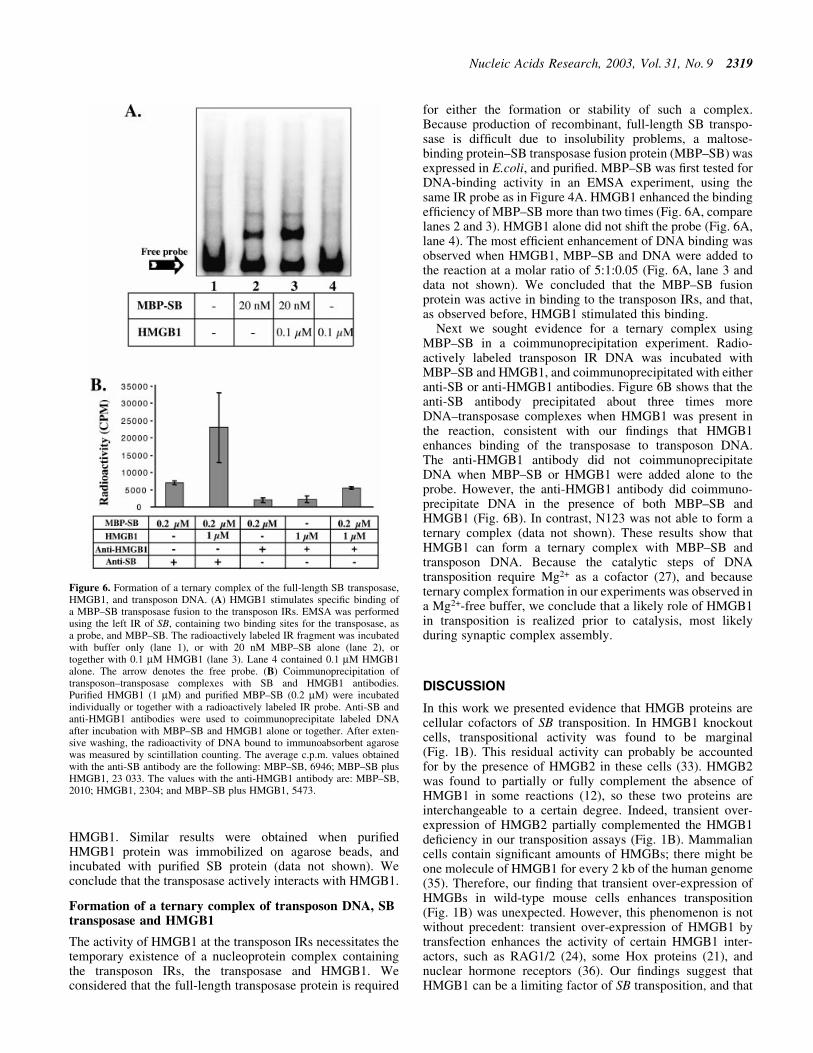
Since HMGB1 has no sequence speci®city on its own, it has tobe actively recruited to speci®c sites by other DNA-bindingproteins (12). To investigate possible physical interactionsbetween the SB transposase and HMGB1, an immuno-precipitation experiment was performed (Fig. 5). Nuclearextracts were prepared from HeLa cells constitutivelyexpressing the SB transposase. An antibody against humanHMGB1 or a matched preimmune serum was used forimmunoprecipitation. Precipitated proteins were subsequentlyblotted and hybridized with a polyclonal antibody against SBtransposase. SB transposase was coprecipitated with theHMGB1 antibody, but not with the preimmune serum(Fig. 5, lanes 1 and 2). Treating the nuclear extract withDNase I did not in¯uence the formation of the immuno-complex (compare lanes 2 and 3 in Fig. 5), indicating that thedetected signal was not due to non-speci®c, simultaneousbinding of SB and HMGB1 to genomic DNA. Furthermore,interaction between HMGB1 and SB transposase is notdependent on the presence of transposon DNA, becauseimmunoprecipitation in the presence or absence of DNA gavesimilar results (data not shown). Control nuclear extracts didnot produce a signal (Fig. 5, lane 4); thus, immunoprecipita-tion is dependent on the presence of SB transposase.Treatment of nuclear extracts with actin and p15 antibodiesfailed to immunoprecipitate SB transposase (Fig. 5, lanes 5and 6), indicating that the interaction observed is speci®c for
Figure 4. HMGB1 stimulates speci®c binding of SB transposase to thetransposon IRs. (A) The effect of HMGB1 on transposase binding to the leftIR. EMSA was performed using the left IR of SB, containing two bindingsites for the transposase, as a probe, and N123, an N-terminal derivative ofSB transposase containing the speci®c DNA-binding domain. C1 and C2indicate the two DNA±protein complexes formed in the assay.(B) Comparison of SB transposase binding sites and the RAG1/2 recognitionsignal sequences. The degrees of similarities to the heptamer and nonamermotifs are indicated. (C) The effect of HMGB1 on binding to either theouter or the inner transposase binding sites in the context of the left IR.EMSA showing the stimulatory effects of HMGB1 on binding of N123 tothe ODR and IDR.
Figure 5. SB transposase interacts with HMGB1. Immunoblot of nuclearextracts of HeLa cells expressing SB transposase, and control cells, afterincubation with antibodies against human HMGB1, actin and p15 proteinsor a preimmune serum, with or without DNase I treatment. The blot washybridized with an anti-SB antibody.
2318 Nucleic Acids Research, 2003, Vol. 31, No. 9
HMGB1. Similar results were obtained when puri®edHMGB1 protein was immobilized on agarose beads, andincubated with puri®ed SB protein (data not shown). Weconclude that the transposase actively interacts with HMGB1.
Formation of a ternary complex of transposon DNA, SBtransposase and HMGB1
The activity of HMGB1 at the transposon IRs necessitates thetemporary existence of a nucleoprotein complex containingthe transposon IRs, the transposase and HMGB1. Weconsidered that the full-length transposase protein is required
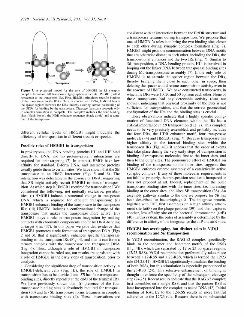
for either the formation or stability of such a complex.Because production of recombinant, full-length SB transpo-sase is dif®cult due to insolubility problems, a maltose-binding protein±SB transposase fusion protein (MBP±SB) wasexpressed in E.coli, and puri®ed. MBP±SB was ®rst tested forDNA-binding activity in an EMSA experiment, using thesame IR probe as in Figure 4A. HMGB1 enhanced the bindingef®ciency of MBP±SB more than two times (Fig. 6A, comparelanes 2 and 3). HMGB1 alone did not shift the probe (Fig. 6A,lane 4). The most ef®cient enhancement of DNA binding wasobserved when HMGB1, MBP±SB and DNA were added tothe reaction at a molar ratio of 5:1:0.05 (Fig. 6A, lane 3 anddata not shown). We concluded that the MBP±SB fusionprotein was active in binding to the transposon IRs, and that,as observed before, HMGB1 stimulated this binding.
Next we sought evidence for a ternary complex usingMBP±SB in a coimmunoprecipitation experiment. Radio-actively labeled transposon IR DNA was incubated withMBP±SB and HMGB1, and coimmunoprecipitated with eitheranti-SB or anti-HMGB1 antibodies. Figure 6B shows that theanti-SB antibody precipitated about three times moreDNA±transposase complexes when HMGB1 was present inthe reaction, consistent with our ®ndings that HMGB1enhances binding of the transposase to transposon DNA.The anti-HMGB1 antibody did not coimmunoprecipitateDNA when MBP±SB or HMGB1 were added alone to theprobe. However, the anti-HMGB1 antibody did coimmuno-precipitate DNA in the presence of both MBP±SB andHMGB1 (Fig. 6B). In contrast, N123 was not able to form aternary complex (data not shown). These results show thatHMGB1 can form a ternary complex with MBP±SB andtransposon DNA. Because the catalytic steps of DNAtransposition require Mg2+ as a cofactor (27), and becauseternary complex formation in our experiments was observed ina Mg2+-free buffer, we conclude that a likely role of HMGB1in transposition is realized prior to catalysis, most likelyduring synaptic complex assembly.
DISCUSSION
In this work we presented evidence that HMGB proteins arecellular cofactors of SB transposition. In HMGB1 knockoutcells, transpositional activity was found to be marginal(Fig. 1B). This residual activity can probably be accountedfor by the presence of HMGB2 in these cells (33). HMGB2was found to partially or fully complement the absence ofHMGB1 in some reactions (12), so these two proteins areinterchangeable to a certain degree. Indeed, transient over-expression of HMGB2 partially complemented the HMGB1de®ciency in our transposition assays (Fig. 1B). Mammaliancells contain signi®cant amounts of HMGBs; there might beone molecule of HMGB1 for every 2 kb of the human genome(35). Therefore, our ®nding that transient over-expression ofHMGBs in wild-type mouse cells enhances transposition(Fig. 1B) was unexpected. However, this phenomenon is notwithout precedent: transient over-expression of HMGB1 bytransfection enhances the activity of certain HMGB1 inter-actors, such as RAG1/2 (24), some Hox proteins (21), andnuclear hormone receptors (36). Our ®ndings suggest thatHMGB1 can be a limiting factor of SB transposition, and that
Figure 6. Formation of a ternary complex of the full-length SB transposase,HMGB1, and transposon DNA. (A) HMGB1 stimulates speci®c binding ofa MBP±SB transposase fusion to the transposon IRs. EMSA was performedusing the left IR of SB, containing two binding sites for the transposase, asa probe, and MBP±SB. The radioactively labeled IR fragment was incubatedwith buffer only (lane 1), or with 20 nM MBP±SB alone (lane 2), ortogether with 0.1 mM HMGB1 (lane 3). Lane 4 contained 0.1 mM HMGB1alone. The arrow denotes the free probe. (B) Coimmunoprecipitation oftransposon±transposase complexes with SB and HMGB1 antibodies.Puri®ed HMGB1 (1 mM) and puri®ed MBP±SB (0.2 mM) were incubatedindividually or together with a radioactively labeled IR probe. Anti-SB andanti-HMGB1 antibodies were used to coimmunoprecipitate labeled DNAafter incubation with MBP±SB and HMGB1 alone or together. After exten-sive washing, the radioactivity of DNA bound to immunoabsorbent agarosewas measured by scintillation counting. The average c.p.m. values obtainedwith the anti-SB antibody are the following: MBP±SB, 6946; MBP±SB plusHMGB1, 23 033. The values with the anti-HMGB1 antibody are: MBP±SB,2010; HMGB1, 2304; and MBP±SB plus HMGB1, 5473.
Nucleic Acids Research, 2003, Vol. 31, No. 9 2319
different cellular levels of HMGB1 might modulate theef®ciency of transposition in different tissues or species.
Possible roles of HMGB1 in transposition
In prokaryotes, the DNA-bending proteins HU and IHF binddirectly to DNA, and no protein±protein interactions arerequired for their targeting (7). In contrast, HMGs have lowaf®nity for standard, B-form DNA, and interactor proteinsusually guide them to certain sites. We have shown that the SBtransposase is an HMG interactor (Figs 5 and 6). Theinteraction was detectable in the absence of DNA, suggestingthat SB might actively recruit HMGB1 to sites of transpos-ition. At which step is HMGB1 required for transposition? Weconsidered the following, not mutually exclusive, possibil-ities: (i) HMGB1 induces a structural change in transposonDNA, which is required for ef®cient transposition; (ii)HMGB1 enhances binding of the transposase to the transposonIRs; (iii) HMGB1 induces a conformational change of thetransposase that makes the transposase more active; (iv)HMGB1 plays a role in transposon integration by makingcontacts with chromatin components and/or by DNA-bendingat target sites (37). In this paper we provided evidence thatHMGB1 promotes circle formation of transposon DNA (Figs2 and 3), that it signi®cantly enhances speci®c transposasebinding to the transposon IRs (Fig. 4), and that it can form aternary complex with the transposase and transposon DNA(Fig. 6). Thus, although a role of HMGB1 in transposonintegration cannot be ruled out, our results are consistent witha role of HMGB1 in the early steps of transposition, prior tocatalysis.
Considering the signi®cant drop of transposition activity inHMGB1-de®cient cells (Fig. 1B), the role of HMGB1 intransposition has to be a critical one. SB has four transposase-binding sites, directly repeated at the ends of the terminal IRs.We have previously shown that: (i) presence of the fourtransposase binding sites is absolutely required for transpos-ition (30) and (ii) SB transposase forms tetramers in complexwith transposase-binding sites (4). These observations are
consistent with an interaction between the IR/DR structure anda transposase tetramer during transposition. We propose thatone of HMGB1's roles is to bring the two binding sites closerto each other during synaptic complex formation (Fig. 7).HMGB1 might promote communication between DNA motifsthat are otherwise distant to each other, including the DRs, thetranspositional enhancer and the two IRs (Fig. 7). Similar toSB transposition, a DNA-bending protein, HU, is involved inlooping out the linker DNA between transposase binding sitesduring Mu-transpososome assembly (7). If the only role ofHMGB1 is to extrude the spacer region between the DRs,thereby bringing them close to each other in space, thendeleting the spacer would rescue transposition activity even inthe absence of HMGB1. We have constructed transposons, inwhich the DRs were 10, 20 and 50 bp from each other. None ofthese transposons had any detectable activity (data notshown), indicating that physical proximity of the DRs is notsuf®cient for transposition, and that the correct geometricalcon®guration of the IRs and the binding sites is crucial.
These observations indicate that a highly speci®c con®g-uration of functional DNA elements within the IRs has acritical importance in SB transposition (Fig. 7). This complexneeds to be very precisely assembled, and probably includesthe four DRs, the HDR enhancer motif, four transposasemolecules (4) and HMGB1 (Fig. 7). Because transposase hashigher af®nity to the internal binding sites within thetransposon IRs (Fig. 4C), it appears that the order of eventsthat take place during the very early steps of transposition isbinding of transposase molecules ®rst to the inner sites, andthen to the outer sites. The pronounced effect of HMGB1 onbinding of the transposase to the inner sites suggests thatHMGB1 enforces ordered assembly of a catalytically activesynaptic complex. If any of these molecular requirements isnot ful®lled properly, the transposition reaction is hampered ordoes not proceed at all. Indeed, replacement of the outertransposase binding sites with the inner sites, i.e. increasingbinding at the outer sites, abolishes SB transposition (38). Anassembly pathway similar to the one we propose for SB hasbeen described for bacteriophage l. The integrase protein,together with IHF, ®rst assembles on a high af®nity attach-ment site (attP) on the phage genome (39), and then capturesanother, low af®nity site on the bacterial chromosome (attB)(40). In this system, the order of assembly is determined by thedifference in af®nity of the integrase for the attP and attB sites.
HMGB1 has overlapping, but distinct roles in V(D)Jrecombination and SB transposition
In V(D)J recombination, the RAG1/2 complex speci®callybinds to the nonamer and heptamer motifs of the RSSs(Fig. 4B), which are separated by 12 or 23 bp spacer regions(12/23-RSS). V(D)J recombination preferentially takes placebetween a 12-RSS and a 23-RSS, which is termed the 12/23rule (24,25,41). HMGB1/2 signi®cantly stimulates the bindingof both RSSs, but this stimulation is especially pronounced atthe 23-RSS (24). This selective enhancement of binding isthought to enforce the speci®city of the subsequent cleavagestep (24,25). Recent results indicate that the RAG1/2 complex®rst assembles on a single RSS, and that the partner RSS islater incorporated into the complex as naked DNA (42). Initialbinding of RAG1/2 to the 12-RSS results in more faithfuladherence to the 12/23 rule. Because there is no substantial
Figure 7. A proposed model for the role of HMGB1 in SB synapticcomplex formation. SB transposase (gray spheres) recruits HMGB1 (dottedhexagons) to the transposon IRs. First, HMGB1 stimulates speci®c bindingof the transposase to the IDRs. Once in contact with DNA, HMGB1 bendsthe spacer regions between the DRs, thereby assuring correct positioning ofthe ODRs for binding by the transposase. Cleavage (scissors) proceeds onlyif complex formation is complete. The complex includes the four bindingsites (black boxes), the HDR enhancer sequence (black circle) and a tetra-mer of the transposase.
2320 Nucleic Acids Research, 2003, Vol. 31, No. 9
difference in the binding af®nity of RAG1/2 for naked 12- and23-RSSs in the presence of HMGB proteins, it has beensuggested that chromatin structure may in¯uence whetherRAG1/2 binds ®rst to a 12- or a 23-RSS in vivo (42).
The transposase-binding sites of SB resemble the RSSs intheir sequence (Fig. 4B). Similarly to the RSSs, the spacingbetween the nonamer and heptamer-like motifs within thetransposase-binding sites is different, 12 and 14 bps, in theinternal and external DRs, respectively. We have found thatSB transposase preferentially binds the IDR (12DR) (Fig. 4C).The 2 bp difference in spacer length between 12DR and 14DRmight not be suf®cient for HMGB1 to assert its DNA-bendingactivity to promote transposase binding. More likely, thehelical phasing of the heptamer- and nonamer-like sequencesin 14DR might be less favorable for transposase binding. Incontrast to V(D)J recombination, the original preference of theSB transposase for binding to the 12DR is not altered, but evenfurther emphasized in the presence of HMGB1 (Fig. 4C). Inconclusion, HMGB1 seems to have overlapping, but distinctroles in SB transposition and in V(D)J recombination.
The IR/DR-type organization of IRs is an evolutionarilyconserved feature of many transposons in the Tc1 family (1),but its function in transposition has been enigmatic. Ourresults suggest that the IR/DR introduces a higher levelregulation into the transposition process: the repeatedtransposase binding sites, their dissimilar af®nity for thetransposase, and the effect of HMGB1 to differentiallyenhance transposase binding to the inner sites are all importantfor a geometrically and timely orchestrated formation ofsynaptic complexes, which is a strict requirement for thesubsequent catalytic steps of transposition.
ACKNOWLEDGEMENTS
We thank D. Fiedler, E. StuÈwe and A. Katzer for theirtechnical assistance, and members of the Transposition Groupat the MDC for their helpful comments on the manuscript.Plasmids expressing HMGB1 and HMGB2 and the VA1 andC1 cell lines were kindly supplied by M. Bianchi. The plasmidexpressing HMGA1 was obtained from A. Fusco. D.K. issupported by the graduate college `Model Studies of Structure,Properties and Recognition of Biological Macromolecules'and a research fellowship from DFG (GRK 8013). This workwas supported by EU grant QLG2-CT-2000-00821.
REFERENCES
1. Plasterk,R.H., IzsvaÂk,Z. and Ivics,Z. (1999) Resident aliens: the Tc1/mariner superfamily of transposable elements. Trends Genet., 15,326±332.
2. Ivics,Z., Hackett,P.B., Plasterk,R.H. and IzsvaÂk,Z. (1997) Molecularreconstruction of Sleeping Beauty, a Tc1-like transposon from ®sh and itstransposition in human cells. Cell, 91, 501±510.
3. IzsvaÂk,Z., Ivics,Z. and Hackett,P.B. (1995) Characterization of a Tc1-liketransposable element in zebra®sh (Danio rerio). Mol. Gen. Genet., 247,312±322.
4. IzsvaÂk,Z., Khare,D., Behlke,J., Heinemann,U., Plasterk,R.H. and Ivics,Z.(2002) Involvement of a bifunctional, paired-like DNA-binding domainand a transpositional enhancer in Sleeping Beauty transposition. J. Biol.Chem., 277, 34581±34588.
5. Ivics,Z., IzsvaÂk,Z., Minter,A. and Hackett,P.B. (1996) Identi®cation offunctional domains and evolution of Tc1-like transposable elements.Proc. Natl Acad. Sci. USA, 93, 5008±5013.
6. Doak,T.G., Doerder,F.P., Jahn,C.L. and Herrick,G. (1994) A proposedsuperfamily of transposase genes: transposon-like elements in ciliatedprotozoa and a common `D35E' motif. Proc. Natl Acad. Sci. USA, 91,942±946.
7. Lavoie,B.D. and Chaconas,G. (1990) Immunoelectron microscopicanalysis of the A, B and HU protein content of bacteriophage Mutranspososomes. J. Biol. Chem., 265, 1623±1627.
8. Haykinson,M.J. and Johnson,R.C. (1993) DNA looping and the helicalrepeat in vitro and in vivo: effect of HU protein and enhancer location onHin invertasome assembly. EMBO J., 12, 2503±2512.
9. Goodman,S.D. and Nash,H.A. (1989) Functional replacement of aprotein-induced bend in a DNA recombination site. Nature, 341,251±254.
10. Segall,A.M., Goodman,S.D. and Nash,H.A. (1994) Architecturalelements in nucleoprotein complexes: interchangeability of speci®c andnon-speci®c DNA binding proteins. EMBO J., 13, 4536±4548.
11. Paull,T.T., Haykinson,M.J. and Johnson,R.C. (1993) The nonspeci®cDNA-binding and -bending proteins HMG1 and HMG2 promote theassembly of complex nucleoprotein structures. Genes Dev., 7,1521±1534.
12. Bustin,M. (1999) Regulation of DNA-dependent activities by thefunctional motifs of the high-mobility-group chromosomal proteins.Mol. Cell. Biol., 19, 5237±5246.
13. Thomas,J.O. and Travers,A.A. (2001) HMG1 and 2 and related`architectural' DNA-binding proteins. Trends Biochem. Sci., 26,167±174.
14. Bianchi,M.E., Beltrame,M. and Paonessa,G. (1989) Speci®c recognitionof cruciform DNA by nuclear protein HMG1. Science, 243, 1056±1059.
15. Bianchi,M.E., Falciola,L., Ferrari,S. and Lilley,D.M. (1992) The DNAbinding site of HMG1 protein is composed of two similar segments(HMG boxes), both of which have counterparts in other eukaryoticregulatory proteins. EMBO J., 11, 1055±1063.
16. Pohler,J.R., Norman,D.G., Bramham,J., Bianchi,M.E. and Lilley,D.M.(1998) HMG box proteins bind to four-way DNA junctions in their openconformation. EMBO J., 17, 817±826.
17. Pil,P.M., Chow,C.S. and Lippard,S.J. (1993) High-mobility-group 1protein mediates DNA bending as determined by ring closures. Proc.Natl Acad. Sci. USA, 90, 9465±9469.
18. Stros,M. (1998) DNA bending by the chromosomal protein HMG1 andits high mobility group box domains. Effect of ¯anking sequences. J. Biol.Chem., 273, 10355±10361.
19. Stros,M., Cherny,D. and Jovin,T.M. (2000) HMG1 protein stimulatesDNA end joining by promoting association of DNA molecules via theirends. Eur. J. Biochem., 267, 4088±4097.
20. Muller,S., Scaf®di,P., Degryse,B., Bonaldi,T., Ronfani,L., Agresti,A.,Beltrame,M. and Bianchi,M.E. (2001) The double life of HMGB1chromatin protein: architectural factor and extracellular signal. EMBO J.,20, 4337±4340.
21. Zappavigna,V., Falciola,L., Helmer-Citterich,M., Mavilio,F. andBianchi,M.E. (1996) HMG1 interacts with HOX proteins and enhancestheir DNA binding and transcriptional activation. EMBO J., 15,4981±4991.
22. Zwilling,S., Konig,H. and Wirth,T. (1995) High mobility group protein 2functionally interacts with the POU domains of octamer transcriptionfactors. EMBO J., 14, 1198±1208.
23. Sutrias-Grau,M., Bianchi,M.E. and Bernues,J. (1999) High mobilitygroup protein 1 interacts speci®cally with the core domain of humanTATA box-binding protein and interferes with transcription factor IIBwithin the pre-initiation complex. J. Biol. Chem., 274, 1628±1634.
24. van Gent,D.C., Hiom,K., Paull,T.T. and Gellert,M. (1997) Stimulation ofV(D)J cleavage by high mobility group proteins. EMBO J., 16,2665±2670.
25. Aidinis,V., Bonaldi,T., Beltrame,M., Santagata,S., Bianchi,M.E. andSpanopoulou,E. (1999) The RAG1 homeodomain recruits HMG1 andHMG2 to facilitate recombination signal sequence binding and toenhance the intrinsic DNA-bending activity of RAG1-RAG2. Mol. Cell.Biol., 19, 6532±6542.
26. Costello,E., Saudan,P., Winocour,E., Pizer,L. and Beard,P. (1997) Highmobility group chromosomal protein 1 binds to the adeno-associatedvirus replication protein (Rep) and promotes Rep-mediated site-speci®ccleavage of DNA, ATPase activity and transcriptional repression. EMBOJ., 17, 5943±5954.
27. Craig,N.L. (1995) Unity in transposition reactions. Science, 270,253±254.
Nucleic Acids Research, 2003, Vol. 31, No. 9 2321
28. Li,L., Yoder,K., Hansen,M.S., Olvera,J., Miller,M.D. and Bushman,F.D.(2000) Retroviral cDNA integration: stimulation by HMG I familyproteins. J. Virol., 74, 10965±10974.
29. Hindmarsh,P., Ridky,T., Reeves,R. Andrake,M., Skalka,A.M. and Leis,J.(1999) HMG protein family members stimulate humanimmunode®ciency virus type 1 and avian sarcoma virus concerted DNAintegration in vitro. J. Virol., 73, 2994±3003.
30. IzsvaÂk,Z., Ivics,Z. and Plasterk,R.H. (2000) Sleeping Beauty, a widehost-range transposon vector for genetic transformation in vertebrates.J. Mol. Biol., 302, 93±102.
31. Fischer,S.E., Wienholds,E. and Plasterk,R.H. (2001) Regulatedtransposition of a ®sh transposon in the mouse germ line. Proc. NatlAcad. Sci. USA, 98, 6759±6764.
32. Yant,S.R., Meuse,L., Chiu,W., Ivics,Z., IzsvaÂk,Z. and Kay,M.A. (2000)Somatic integration and long-term transgene expression in normal andhaemophilic mice using a DNA transposon system. Nature Genet., 25,35±41.
33. Calogero,S., Grassi,F., Aguzzi,A., Voigtlander,T., Ferrier,P., Ferrari,S.and Bianchi,M.E. (1999) The lack of chromosomal protein Hmg1 doesnot disrupt cell growth but causes lethal hypoglycaemia in newborn mice.Nature Genet., 22, 276±280.
34. Melillo,R.M., Pierantoni,G.M., Scala,S., Battista,S., Fedele,M., Stella,A.,De Biasio,M.C., Chiappetta,G., Fidanza,V., Condorelli,G., Santoro,M.,Croce,C.M., Viglietto,G. and Fusco,A. (2001) Critical role of theHMGI(Y) proteins in adipocytic cell growth and differentiation. Mol.Cell. Biol., 21, 2485±2495.
35. Bianchi,M.E. and Beltrame,M. (2000) Upwardly mobile proteins. Therole of HMG proteins in chromatin structure, gene expression andneoplasia. EMBO Rep., 1, 109±114.
36. Boonyaratanakornkit,V., Melvin,V., Prendergast,P., Altmann,M.,Ronfani,L., Bianchi,M.E., Taraseviciene,L., Nordeen,S.K.,Allegretto,E.A. and Edwards,D.P. (1998) High-mobility group chromatinproteins 1 and 2 functionally interact with steroid hormone receptors toenhance their DNA binding in vitro and transcriptional activity inmammalian cells. Mol. Cell. Biol., 18, 4471±4487.
37. Vigdal,T.J., Kaufman,C.D., IzsvaÂk,Z., Voytas,D.F. and Ivics,Z. (2002)Common physical properties of DNA affecting target site selection ofSleeping Beauty and other Tc1/mariner transposable elements. J. Mol.Biol., 323, 441±452.
38. Cui,Z., Geurts,A.M., Liu,G., Kaufman,C.D. and Hackett,P.B. (2002)Structure±function analysis of the inverted terminal repeats of theSleeping Beauty transposon. J. Mol. Biol., 318, 1221±1235.
39. Richet,E., Abcarian,P. and Nash,H. (1986) Synapsis of attachment sitesduring lambda integrative recombination involves capture of a nakedDNA by a protein±DNA complex. Cell, 46, 1011±1021.
40. Patsey,R.L. and Bruist,M.F. (1995) Characterization of the interactionbetween the lambda intasome and attB. J. Mol. Biol., 252, 47±58.
41. Hiom,K. and Gellert,M. (1998) Assembly of a 12/23 paired signalcomplex: a critical control point in V(D)J recombination. Mol. Cell.Biol., 1, 1011±1019.
42. Jones,J.M. and Gellert,M. (2002) Ordered assembly of the V(D)Jsynaptic complex ensures accurate recombination. EMBO J., 21,4162±4171.
2322 Nucleic Acids Research, 2003, Vol. 31, No. 9