the calcineurin-nfat pathway controls activity-dependent circadian gene expression in slow skeletal...
TRANSCRIPT

Original article
The calcineurin-NFAT pathway controlsactivity-dependent circadian gene expressionin slow skeletal muscle
Kenneth A. Dyar 1,*,7, Stefano Ciciliot 1, Guidantonio Malagoli Tagliazucchi 2,6, Giorgia Pallafacchina 1,4,Jana Tothova 1, Carla Argentini 1, Lisa Agatea 1, Reimar Abraham 1, Miika Ahdesmäki 5, Mattia Forcato 2,Silvio Bicciato 2, Stefano Schiaffino 1,4, Bert Blaauw 1,3,**
ABSTRACT
Objective: Physical activity and circadian rhythms are well-established determinants of human health and disease, but the relationship betweenmuscle activity and the circadian regulation of muscle genes is a relatively new area of research. It is unknown whether muscle activity andmuscle clock rhythms are coupled together, nor whether activity rhythms can drive circadian gene expression in skeletal muscle.Methods: We compared the circadian transcriptomes of two mouse hindlimb muscles with vastly different circadian activity patterns, thecontinuously active slow soleus and the sporadically active fast tibialis anterior, in the presence or absence of a functional skeletal muscle clock(skeletal muscle-specific Bmal1 KO). In addition, we compared the effect of denervation on muscle circadian gene expression.Results: We found that different skeletal muscles exhibit major differences in their circadian transcriptomes, yet core clock gene oscillationswere essentially identical in fast and slow muscles. Furthermore, denervation caused relatively minor changes in circadian expression of mostcore clock genes, yet major differences in expression level, phase and amplitude of many muscle circadian genes.Conclusions: We report that activity controls the oscillation of around 15% of skeletal muscle circadian genes independently of the core muscleclock, and we have identified the Ca2þ-dependent calcineurin-NFAT pathway as an important mediator of activity-dependent circadian geneexpression, showing that circadian locomotor activity rhythms drive circadian rhythms of NFAT nuclear translocation and target gene expression.
� 2015 The Authors. Published by Elsevier GmbH. This is an open access article under the CC BY-NC-ND license (http://creativecommons.org/licenses/by-nc-nd/4.0/).
Keywords Skeletal muscle; Circadian; Activity; NFAT
1. INTRODUCTION
The mammalian circadian clock is an evolutionarily conserved, adaptivemeans of coordinating internal physiology in anticipation of environ-mental conditions [34], and is composed of a central pacemaker in thesuprachiasmatic nucleus (SCN) of the hypothalamus and autonomousmolecular oscillators in cells of peripheral tissues. Timing informationregarding the daily light/dark cycle is transmitted directly from the retinato the SCN, which in turn synchronizes clocks in the periphery byregulating various metabolic and behavioral outputs, including loco-motor activity, feeding behavior and body temperature [13].At the cellular level, circadian time is maintained by a rhythm gener-ating molecular clock composed of two interlocking regulatory feed-back loops [17]. In the primary loop, the bHLH-PAS domain-containing
1Venetian Institute of Molecular Medicine (VIMM), Padova, Italy 2Center for GenomeModena, Italy 3Department of Biomedical Sciences, University of Padova, Italy 4Institutefor Medical Informatics, Statistics and Epidemiology, University of Leipzig, Germany 6Is20122 Milan, Italy
7 Present address: Molecular Endocrinology, Institute for Diabetes and Obesity, Helm
*Corresponding author. Institute for Diabetes and Obesity, Helmholtz Zentrum Münmuenchen.de (K.A. Dyar).
**Corresponding author. Venetian Institute of Molecular Medicine (VIMM), Via Orus 2,
Received September 4, 2015 � Accepted September 15, 2015 � Available online 25 S
http://dx.doi.org/10.1016/j.molmet.2015.09.004
MOLECULAR METABOLISM 4 (2015) 823e833 � 2015The Authors. Published by Elsevier GmbH. This is an openwww.molecularmetabolism.com
transcription factors CLOCK and BMAL1 form heterodimers in thecytoplasm and translocate to the nucleus where they bind to E-boxsequences of target genes, including Period (Per) and Cryptochrome(Cry) transcription factors. PER and CRY proteins form heterodimers inthe cytoplasm and translocate to the nucleus, repressing activation bythe CLOCK-BMAL1 complex. In a second regulatory loop, CLOCK-BMAL1 activation leads to transcription and translation of Rev-Erba,which represses Bmal1 transcription by inhibiting the ROR enhancerelement. In addition, all tissues contain a large number of 24-houroscillating genes, most of which are tissue-specific [31,36,49,59]and some of which are directly regulated by extrinsic circadian sig-nals independent of this core molecular clock [25].From a circadian perspective, skeletal muscle is unique among pe-ripheral tissues, as muscle fibers contain local circadian clocks and are
Research, Department of Life Sciences, University of Modena and Reggio Emilia,of Neurosciences, Consiglio Nazionale delle Ricerche (CNR), Padova, Italy 5Institutetituto Nazionale Genetica Molecolare ‘Romeo ed Enrica Invernizzi’, Via F. Sforza 35,
holtz Zentrum München, Germany.
chen, Parkring 13, 85748 Garching, Germany. E-mail: kenneth.dyar@helmholtz-
35129 Padova, Italy. E-mail: [email protected] (B. Blaauw).
eptember 2015
accessarticle under the CCBY-NC-ND license (http://creativecommons.org/licenses/by-nc-nd/4.0/). 823
Original article
targets of blood-born circadian signals like other cell types. Yet musclefibers also respond to rhythmic motor neuron activity, which is likewisecontrolled by the SCN pacemaker [39]. Motor activity has traditionallybeen used as a read-out of the circadian timing system, but it is unclearwhether muscle activity and muscle clock gene rhythms are coupled,and whether activity affects the entrainment of the muscle clock. Whilefeeding time has emerged as a dominant entrainment factor for mostperipheral tissues studied to date [12,48], including skeletal muscle[5,55] time-restricted feeding also alters circadian locomotor activity[55] and body temperature rhythms [12]. Similarly, various exerciseprotocols, including voluntary and involuntary running, have beenshown to influence entrainment of the core muscle clock [55], butlikewise affect the central pacemaker [15,30] circadian hormonerelease [54] and body temperature rhythms [46]. As such, a clearunderstanding of the relationship between activity rhythms and musclecircadian genes remained elusive. Muscle activity reflects nerve activity,and a role for neural signals entraining the muscle clock has beensuggested by parabiosis experiments between intact and SCN-lesionedmice, showing that non-neural signals are able to maintain the circadianrhythm of clock gene expression in liver but not in skeletal muscle [18].However, other studies have pointed to the dissociation between clockgene expression in skeletal muscle and rhythmic locomotor activity, astransplantation of a functional SCN into SCN-lesioned Syrian hamstersfailed to reinstate normal gene oscillation in skeletal muscle, despiterestoring locomotor activity rhythms with the period of the donor [19].Nakao and colleagues recently reported that sciatic nerve denervationdisrupts circadian expression of some skeletal muscle clock geneswithout affecting circadian rhythms of body temperature or plasmacorticosterone levels [35], but whether the effects on muscle clockgenes were directly dependent upon the loss of nerve activity or anindirect result of muscle remodeling could not be determined.To investigate the relative roles of intrinsic versus extrinsic circadiansignals in the regulation of circadian muscle gene expression, wepreviously generated mice with muscle-specific inactivation of theBmal1 gene (Bmal1 mKO), thus disrupting local muscle clock function[14]. We have shown that these mice display an essentially normalphenotype [14], in contrast to global knockout of Bmal1, which leads tosevere muscle atrophy, reduced body weight and shorter life span [23].In agreement with results obtained from other tissue-specific Bmal1knockout mice [25], we found a significant proportion of cycling genesmaintain their circadian oscillation in muscle cells even with a dis-rupted core oscillator, and thus must respond to extrinsic circadiansignals. To determine whether circadian activity rhythms drive theoscillation of these clock-independent genes, we examined circadiangene expression in fast and slow muscles with vastly different circa-dian activity patterns, and compared them to contralateral denervatedmuscles with complete loss of activity. Importantly, these comparisonswere made in the same animals, thus avoiding systemic effects thatcomplicate the interpretation of studies on the role of exercise oncircadian rhythms. We found the oscillation pattern of core clock genesis essentially identical in fast and slow muscles, despite major dif-ferences in circadian activity levels. Furthermore, core clock geneexpression shows relatively minor changes after denervation, incontrast to the striking changes induced by time-restricted feeding.Furthermore, we report that nerve activity controls the oscillation ofaround 15% of skeletal muscle circadian genes independently of thecore muscle clock, and we have identified the Ca2þ-dependentcalcineurin-NFAT pathway as an important mediator of activity-dependent circadian gene expression, showing that circadian loco-motor activity rhythms drive circadian rhythms of NFAT nucleartranslocation and target gene expression.
824 MOLECULAR METABOLISM 4 (2015) 823e833 � 2015 The Authors. Published by Elsevier Gmb
2. MATERIALS AND METHODS
2.1. AnimalsAll experiments were performed on 2e4 month old adult male mice,with standard chow (Mucedola, Settimo Milanese, Italy) and waterprovided ad libitum. Mice with skeletal muscle-specific inactivation ofBmal1 (Bmal1 mKO) were generated as previously described [14]. Inbrief, a mouse line with floxed Bmal1 gene [50] was crossed with micecarrying a Cre recombinase transgene under control of the Mlc1fpromoter (Mlc1f-Cre), which is exclusively active in skeletal muscle [4].Cre-negative littermates were used as wildtype controls (Ctrl). Exper-imental protocols were reviewed and approved by the local AnimalCare Committee, University of Padova. Prior to all experiments, animalswere acclimated for at least 4 weeks in an isolated temperaturecontrolled room (22 �C) under a 12-h lightedark regimen, with lightson at Zeitgeber time 0 (ZT0; 6 am), lights off at ZT12 (6 pm). Tibialisanterior (TA) and soleus (SOL) muscles, and in some experiments alsoliver and heart, were collected immediately after cervical dislocation at4-h intervals across the day/night cycle. Tissues were snap frozen inliquid nitrogen and stored at �80 �C until subsequent use. For time-restricted feeding, food access was restricted to the light phase (ZT0-12) for 14 days. For denervation experiments, mice were anesthetizedby i.p. injection of a mixture of Zoletil 100� (a combination of Zola-zapam and Tiletamine, 1:1, 10 mg/kg, Labotatoire Virbac) andRompun� (Xilazine 2%, 0.06 ml/kg, Bayer) and a 1e2 mm segment ofthe sciatic nerve was removed high in the thigh. Muscles fromdenervated and sham-operated contralateral leg used as control wereremoved after 7 days.
2.2. RNA isolation and qPCRTotal RNA was isolated using TRIzol (Invitrogen) followed by cleanupwith the RNeasy Mini Kit (Qiagen). RNA integrity was evaluated with theAgilent 2100 Bioanalyzer (Agilent Technologies, Palo Alto, CA) andquantified with a NanoVue spectrophotometer (GE Healthcare LifeSciences, Baie d’Urfe, QC). Complementary DNA from each samplewas generated from 0.8 mg of RNA reverse-transcribed with InvitrogenSuperscript III reverse transcriptase. Primer sets were designed usingPrimer-BLAST (NCBI) and were validated prior to use by gradient PCRand gel analysis to test for optimal annealing temperature, reactionefficiency and specificity. Duplicates of sample cDNA were thenamplified on the 7900HT Fast Real-Time PCR System (Applied Bio-systems) using the Fast SYBR Green RT-PCR kit (Applied Biosystems).Specificity of gene amplification was confirmed by analyzing thedissociation curve with SDS 2.3 software (Applied Biosystems).Analysis was performed using the standard curve method and all datawere normalized relative to 36B4 expression.
2.3. Gene expression profiling and analysesGene expression profiling for the denervation experiment was carriedout using 250 ng of pooled RNA from 6 biological replicates for eachtime point hybridized to Mouse Genome 430 2.0 Arrays (Affymetrix).The gene expression data were normalized and summarized with RMAusing custom CDF files (Version 14.1.0; EntrezG) to remap the probeson the arrays to the genome and transcriptome library data [11].Differentially expressed genes were determined using a default R2threshold of 0.6 and a cubic regression model in maSigPro (microarraySignificant Profiles), a two-step regression-based method to identifygenes with significant temporal expression changes and significantdifferences between experimental groups in time series microarrayexperiments [10]. Rhythmic genes that cycle with a 24-hour periodwere identified using a BenjaminieHochberg Q-value < 0.2 in the
H. This is an open accessarticle under the CCBY-NC-ND license (http://creativecommons.org/licenses/by-nc-nd/4.0/).www.molecularmetabolism.com

non-parametric algorithm JTK_Cycle [21]. 24-hour cycling genesidentified with this cut-off all had adjusted p < 0.01 (Dataset S1).Over-representation analysis was performed using Gene Set Enrich-ment Analysis software (GSEA; http://www.broadinstitute.org/gsea/index.jsp) [51] and genesets from the TRANSFAC database. Gene-sets were defined as significantly enriched if the False Discovery Rate(FDR) was <0.2 and p < 0.05 when using Signal 2 Noise as metricand 10,000 permutations of geneset labels.
2.4. Plasmids and in vivo electroporationThe NFATc1-GFP construct, a gift from Rhonda Bassel-Duby, con-sists of a pEGFPeN1 plasmid (Clontech) containing NFATc1 linked toEGFP [9]. The NFATc3-GFP construct was a gift from ShoichiroMiyatake [3]. The histone 2B red fluorescent protein constructconsists of histone 2B linked to RFP (H2B-RFP) and was a gift fromManuela Zaccolo. Transfection of plasmid DNA was performed aspreviously described [53]. Prior to surgery, mice were anesthetizedby i.p. injection of a mixture of Zoletil 100� (a combination ofZolazapam and Tiletamine, 1:1, 10 mg/kg, Laboratoire Virbac) andRompun� (Xilazine 2%, 0.06 ml/kg, Bayer). The soleus and tibialisanterior muscles were surgically exposed and isolated by a smallhindlimb incision. Plasmid injection was followed by electroporationwith stainless steel spatula electrodes connected to a ECM830 BTXporator (Genetronics, San Diego, CA) as described [32]. Transversecryosections (10 mm) of muscles were used for detection andquantification of transfected NFAT-GFP fusion proteins. NFATc3-GFPlabeling was enhanced with polyclonal rabbit anti-GFP (1:200, Mo-lecular Probes) and Cy2-conjugated anti-rabbit IgG (1:150, JacksonImmunoresearch). Images for NFATc1-GFP, NFATc3-GFP and nu-clear H2B-RFP were collected with a BioRad Laboratories confocalmicroscope (Radiance 2100 MP equipped with an argon laser for488 nm excitation of GFP fluorescence, a Nikon 60x/1.4 Plan Apoobjective, a 500 DCLPXR beamsplitter and HQ515/30 emission filter;all filters were from Chroma Technology Corp.) using Lasersharp2000 software (BioRad). All images were analyzed using Image-Jsoftware (NIH image) and mean nuclear/cytoplasmic fluorescencewas quantified based on the ratio of nuclear to cytoplasmic fluo-rescence for each muscle fiber measured (see Figure S3). NFAT-dependent transcriptional activity was monitored as previouslydescribed [32] by an NFAT reporter construct consisting of 9 tandemNFAT-binding sites from the interleukin 4 gene fused to a basal a-MyHC promoter and linked to firefly luciferase [9]. The NFAT reporterwas co-injected with a Renilla luciferase expression vector (pRL-TK,Promega) to normalize for transfection efficiency and muscles werecollected 7 days after cotransfection, at ZT0, 4, 8, 12, 16, and 20.Frozen muscles were minced with pestle and mortar in liquid ni-trogen and resuspended in 2.5 mL/mg passive lysis buffer (Prom-ega). After centrifugation at 12,000 g for 30 min, luciferase activityof supernatant was measured with a previously calibrated FD-20/20Luminometer (Turner Designs, Sunnyvale, CA). Protein concentrationof the supernatant was determined using the Bradford method withbovine serum albumin as standard for the calibration curve. Resultsare expressed as the mean ratio of firefly luciferase to Renillaluciferase � SEM.
3. RESULTS
3.1. Circadian genes are differentially expressed in slow and fastskeletal musclesTo investigate the role of activity on skeletal muscle circadian geneexpression, we first compared the circadian expression of core clock
MOLECULAR METABOLISM 4 (2015) 823e833 � 2015The Authors. Published by Elsevier GmbH. This is an openwww.molecularmetabolism.com
genes and some of their canonical targets in the slow, continuouslyactive soleus (SOL) and the fast, sporadically active tibialis anterior(TA), two muscles with completely different fiber type and motor unitcomposition [45]. Slow soleus (SOL) is composed of around 90% type1 slow and 2A fibers (Figure S1), whereas fast tibialis anterior (TA) andextensor digitorum longus (EDL) are composed of around 90% fast 2Band 2X fibers. The activity profiles of the corresponding motor units arenot known in the mouse, but firing pattern analyses of rat motor units,based on continuous electromyographic recording for extended pe-riods, indicate that slow motor units in rat soleus are active for morethan 30% of the time over 24 h, whereas the predominant motor unitsin EDL, a fast muscle similar to TA, display sporadic short bursts ofactivity, with total time occupied by bursts being less than 0.5% of thetime over 24 h for most units [20]. Consistent with this are continuouselectromyographic analyses showing that the rat soleus is activemostly during the night [16].To identify circadian genes in SOL and TA muscles we used the algo-rithm JTK_CYCLE [21] and found 1359 (SOL) and 684 (TA) 24-h cyclinggenes (Figure 1A). Applying False Discovery Rate (FDR) cut-offs ofvarying stringencies had little impact on the relative abundance of 24-hour cycling genes found in SOL and TA, and we consistently foundaround 2e3 times more cycling genes in SOL than TA (Figure S4).Interestingly, peak expression of these 24-hour cycling genes displayeda bimodal distribution, with larger clusters of genes peaking around thelightedark and darkelight transitions in both SOL and TA, coincidingwith rest/activity and fasting/feeding phase transitions (Figure 1B).Comparing circadian genes between muscles, we further identified asubset of 334 cycling genes common to both SOL and TA (Figure 1C;Dataset S1). In contrast to major differences in the number of circadiangenes between SOL and TA, circadian expression of the core clockgenes was essentially the same, as verified by qPCR (Figure 1D), withonly a few core clock-associated genes showing significant differencesbetween SOL and TA. Specifically, Dbp amplitude was higher in SOL,whereas expression levels of Rora and Dec1 were higher in TA.On the other hand, the majority of 24-hour cycling transcripts werespecific to each muscle (Figure 1C; Dataset S1), with 75% and 51% ofcircadian genes cycling only in SOL or TA, respectively. These differ-ences underscore the specificity of circadian regulation betweendifferent muscles, and likely reflect known differences in activity levels,metabolism and functional properties. Selected SOL- and TA-specificcircadian genes are shown in Figure 1E and F, while a comprehen-sive list of muscle type-specific circadian genes can be found inDataset S1.
3.2. Skeletal muscles show Bmal1-dependent and -independentcircadian genesTo identify muscle circadian genes regulated by the core muscle clock,we compared the list of 24-hour cycling genes found in SOL and TAmuscles of wildtype mice (Figure 2A) with the lists of genes differ-entially regulated genes in SOL and TA muscles of their muscle-specific Bmal1 KO littermates [14]. Accordingly, we identified a sub-set of 132 skeletal muscle clock-dependent circadian genes commonto both SOL and TA (Dataset S2), again including most of the knowncore clock and clock-dependent genes, in addition to novel musclecircadian genes (see Figure 2B for selected genes; see Dataset S2 forthe complete list).These newly identified muscle clock-dependent circadian genes codefor a range of structural proteins, receptor subunits, signaling mole-cules, metabolic enzymes and transcription factors, and many alreadyhave a well-defined physiological relevance, including Adrb2, encodingthe beta2-adrenergic receptor, Acvr1b, encoding the activin receptor
accessarticle under the CCBY-NC-ND license (http://creativecommons.org/licenses/by-nc-nd/4.0/). 825

Figure 1: Circadian genes in different skeletal muscles. A. Phase map of circadian genes in slow/oxidative soleus (SOL) and fast/glycolytic tibialis anterior (TA) skeletalmuscles identified by JTK_Cycle. Muscles (n ¼ 3/group/time point) were collected at 4 h intervals over 24 h, throughout the light (white box) and dark phase (black box). B.Temporal distribution of circadian genes according to peak expression. Note bimodal distribution, with increased numbers of genes peaking around the transitions from light/resting/fasting and dark/active/feeding phases, particularly in the highly active SOL. C. Comparison of circadian genes between SOL and TA. D. Diurnal expression profiles of coreclock genes and clock-associated genes in SOL and TA muscles determined by qPCR and expressed relative to 36b4 (arbitrary units; mean � SEM; n ¼ 6/time point; *p ¼ 0.05,***p ¼ 0.001, 2-way ANOVA with Bonferroni correction). EeF. Diurnal expression profiles of selected SOL-specific (E) and TA-specific (F) circadian genes determined bymicroarray and plotted as mean absolute expression levels (n ¼ 3/time point; mean � SEM *p ¼ 0.05, **p ¼ 0.01, ***p ¼ 0.001, 2-way ANOVA with Bonferroni correction). SeeDataset S1 for complete list.
Original article
1b (also called Alk4), a component of the myostatin receptor complex,and Glul, coding for the enzyme responsible for glutamine synthesis.Other less well-known clock-dependent muscle circadian genesinclude Mylk4, coding for a highly expressed myosin light chain kinaseof unknown physiological relevance.Despite significant changes in circadian expression of several hundredgenes due to muscle clock disruption [14], we found the majority ofcircadian genes in both SOL and TA were not significantly altered(Figure 2A), suggesting they are regulated by extrinsic circadian sig-nals, and not by the core skeletal muscle clock. Accordingly, around
826 MOLECULAR METABOLISM 4 (2015) 823e833 � 2015 The Authors. Published by Elsevier Gmb
70% of SOL circadian genes (942 of 1359) and about 60% of TAcircadian genes (414 of 684) remained unchanged in mKO musclesaccording to differential expression analysis using maSigPro. We found121 of these clock-independent circadian genes common to both SOLand TA, including important insulin-responsive metabolic regulators(Figure 2C) such as Forkhead box protein O1 (Foxo1), encoding atranscription factor involved in muscle mass regulation, and G(0)/G(1)switch gene 2 (G0s2) which is induced by insulin in human skeletalmuscle [37] and codes for an inhibitor of adipose triglyceride lipase(ATGL) thus blocking lipid droplet degradation [57].
H. This is an open accessarticle under the CCBY-NC-ND license (http://creativecommons.org/licenses/by-nc-nd/4.0/).www.molecularmetabolism.com
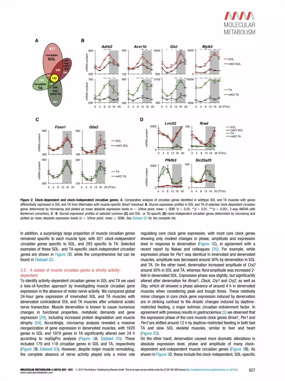
Figure 2: Clock-dependent and clock-independent circadian genes. A. Comparative analysis of circadian genes identified in wildtype SOL and TA muscles with genesdifferentially expressed in SOL and TA from littermates with muscle-specific Bmal1 knockout. B. Diurnal expression profiles in SOL and TA of selected clock-dependent circadiangenes determined by microarray and plotted as mean absolute expression levels (n ¼ 3/time point; mean � SEM *p ¼ 0.05, **p ¼ 0.01, ***p ¼ 0.001, 2-way ANOVA withBonferroni correction). CeD. Diurnal expression profiles of selected common (C) and SOL- or TA-specific (D) clock-independent circadian genes determined by microarray andplotted as mean absolute expression levels (n ¼ 3/time point; mean � SEM). See Dataset S2 for the complete list.
In addition, a surprisingly large proportion of muscle circadian genesremained specific to each muscle type, with 821 clock-independentcircadian genes specific to SOL, and 293 specific to TA. Selectedexamples of these SOL- and TA-specific clock-independent circadiangenes are shown in Figure 2D, while the comprehensive list can befound in Dataset S2.
3.3. A subset of muscle circadian genes is strictly activity-dependentTo identify activity-dependent circadian genes in SOL and TA we useda loss-of-function approach by investigating muscle circadian geneexpression in the absence of motor nerve activity. We compared global24-hour gene expression of innervated SOL and TA muscles withdenervated contralateral SOL and TA muscles after unilateral sciaticnerve transection. Muscle denervation is known to cause numerouschanges in functional properties, metabolic demands and geneexpression [29], including increased protein degradation and muscleatrophy [44]. Accordingly, microarray analysis revealed a massivereorganization of gene expression in denervated muscles, with 1629genes in SOL and 1979 genes in TA significantly altered over 24 haccording to maSigPro analysis (Figure 3A; Dataset S3). Theseincluded 179 and 118 circadian genes in SOL and TA, respectively(Figure 3B; Dataset S3). However, despite major muscle remodeling,the complete absence of nerve activity played only a minor role
MOLECULAR METABOLISM 4 (2015) 823e833 � 2015The Authors. Published by Elsevier GmbH. This is an openwww.molecularmetabolism.com
regulating core clock gene expression, with most core clock genesshowing only modest changes in phase, amplitude and expressionlevel in response to denervation (Figure 3C), in agreement with arecent report by Nakao and colleagues [35]. For example, whileexpression phase for Per1 was identical in innervated and denervatedmuscles, amplitude was decreased around 30% by denervation in SOLand TA. On the other hand, denervation increased amplitude of Cry2around 30% in SOL and TA, whereas Rora amplitude was increased 2-fold in denervated SOL. Expression phase was slightly, but significantlyaltered after denervation for Bmal1, Clock, Cry1 and Cry2, as well asDbp, which all showed a phase advance of around 4 h in denervatedmuscles when considering peak and trough times. These relativelyminor changes in core clock gene expression induced by denervationare in striking contrast to the drastic changes induced by daytime-restricted feeding, a major extrinsic circadian entrainment factor. Inagreement with previous results in gastrocnemius [5] we observed thatthe expression phase of the core muscle clock genes Bmal1, Per1 andPer2 are shifted around 12 h by daytime-restricted feeding in both fastTA and slow SOL skeletal muscles, similar to liver and heart(Figure S2).On the other hand, denervation caused more dramatic alterations inabsolute expression level, phase and amplitude of many clock-dependent and-independent muscle circadian genes (Figure 3B). Asshown in Figure 3D, these include the clock-independent, SOL-specific
accessarticle under the CCBY-NC-ND license (http://creativecommons.org/licenses/by-nc-nd/4.0/). 827

Figure 3: Activity-dependent circadian genes. A. Phase map of differentially expressed genes in control SOL and TA muscles and denervated contralateral muscles over 24-hidentified by maSigPro. Muscles (n ¼ 6/group/time point) were collected at 4-h intervals over 24 h, with RNA pooled and hybridized to Affymetrix arrays as described in themethods. B. Comparative analysis of circadian genes identified in wildtype SOL and TA muscles with genes differentially expressed in response to denervation. C. Diurnalexpression profiles of core clock genes and clock-associated genes in innervated and contralateral denervated SOL and TA muscles determined by qPCR and expressed relative to36b4 (arbitrary units; mean � SEM; n ¼ 6/group/time point; *p ¼ 0.05, **p ¼ 0.01, ***p ¼ 0.001, 2-way ANOVA with Bonferroni correction). D. Diurnal expression profiles ofselected activity-dependent genes in innervated and contralateral denervated SOL and TA muscles determined by qPCR and expressed relative to 36b4 (arbitrary units;mean � SEM; n ¼ 6/group/time point).
Original article
circadian gene Frizzled9 (Fzd9), coding for a 7-transmembrane domainWnt receptor. Interestingly, Fzd9 was expressed antiphase to the Wntinhibitor Dickkopf 2 (Dkk2), and expression levels for Fzd9 and Dkk2were both severely reduced by denervation, with Fzd9 expressionreduced 92% in denervated SOL, and Dkk2 expression reduced 85%.Similar changes were observed for circadian genes predominantlyexpressed in TA, such as the clock-dependent circadian geneKyphoscoliosis peptidase (Ky), coding for a titin- and filamin C-bindingcytoskeletal protein, which showed 88% reduced expression and 30%reduced amplitude in denervated TA, and the clock-independentcircadian gene Pfkfb3, which codes for a homodimeric bifunctionalenzyme that catalyzes the synthesis and degradation of fructose 2,6-bisphosphate, and which showed 80% reduced expression and 70%reduced amplitude in denervated TA.
828 MOLECULAR METABOLISM 4 (2015) 823e833 � 2015 The Authors. Published by Elsevier Gmb
3.4. Activity-dependent circadian pathways in the highly activesoleusTo gain biological insight into the specificity of circadian geneexpression potentially regulated by muscle activity, we focused ourattention on the highly active soleus. We performed pathway enrich-ment analysis of the 1025 SOL-specific circadian genes using thecomputational software Enrichr [8] and genesets from the BioCartadatabase. We found 15 significantly enriched pathways which, inessence, are all pathways involving either p38 mitogen-activatedprotein kinase (MAPK) and activation of downstream transcriptionfactors AP1 and/or CREB, or calcium-dependent pathways involvingcalcineurin and activation of NFAT transcription factors (Figure 4A).Both p38 MAPK and calcineurin are nerve activity-dependent regula-tors of muscle gene expression, and play important roles in skeletal
H. This is an open accessarticle under the CCBY-NC-ND license (http://creativecommons.org/licenses/by-nc-nd/4.0/).www.molecularmetabolism.com

Figure 4: Activity-dependent circadian pathways identified by enrichmentanalysis. A. BioCarta pathway enrichment analysis performed on wildtype SOL-specificcircadian genes. All significant pathways involve p38 MAPK and activation of down-stream transcription factors AP1 and/or CREB, or calcineurin and activation of NFATtranscription factors. B. Activity-dependent transcriptional mediators identified by GeneSet Enrichment Analysis using the TRANSFAC database.
muscle adaptation in response to exercise [1] and maintenance of theslow fiber phenotype [47].To better define transcriptional mediators of activity-dependentcircadian gene expression, we performed Gene Set EnrichmentAnalysis (GSEA [51]), interrogating genesets from the TRANSFACdatabase with SOL-specific circadian genes found to be differentiallyexpressed after denervation. The TRANSFAC geneset collection cal-culates enrichment scores for groups of genes that share specifictranscription factor binding motifs within promoter regions, þ/�2 kbfrom their transcription start sites. Only 5 transcription factors weresignificantly associated with changes in activity-dependent circadiangene expression in SOL (Figure 4B): AP1, Foxo4, Ets-2, NFAT, andLEF1. NFAT and AP1 are particularly interesting candidates, as they areknown to interact and cooperate at promoters or enhancers to activateor repress target genes in a context-dependent manner, despite beingactivated by distinct upstream signaling pathways [27].
3.5. NFAT transcription factors regulate activity-dependentcircadian gene regulationTo better define how activity-dependent signaling pathways regulatecircadian gene expression, we focused our attention on calcineurin-NFAT. We previously showed that NFATc1, a member of the NFATfamily of transcription factors, is a slow-type nerve activity sensorin vivo [32,53]. In fast skeletal muscles like TA, NFATc1 is normallyphosphorylated and found localized in the cytoplasm, but slow-typeelectrical stimulation causes calcineurin-dependent NFATc1 dephos-phorylation and nuclear translocation, as revealed by the nuclear/
MOLECULAR METABOLISM 4 (2015) 823e833 � 2015The Authors. Published by Elsevier GmbH. This is an openwww.molecularmetabolism.com
cytoplasmic distribution of an NFATc1-GFP fusion protein [26,53].Conversely, NFATc1-GFP displays a greater nuclear localization in SOL,but nuclear export is rapidly induced by denervation [53].To monitor 24-hour calcineurin-NFAT signaling in vivo, we transfectedmouse SOL and TA muscles with plasmids coding for NFATc1-GFP orNFATc3-GFP fusion proteins, and measured NFAT nuclear/cytoplasmiclocalization over 24 h. In SOL muscles, as shown in Figure 5A and B,NFATc1 and NFATc3 were both predominantly cytoplasmic in themiddle of the light/resting phase (ZT4), yet displayed a robust nuclearlocalization during the dark/active phase (ZT16). Quantification ofnuclear to cytoplasmic GFP fluorescence (Figure S3) confirmed 24-hour nucleocytoplasmic shuttling, with accumulation of both NFATc1and NFATc3 in myonuclei of the soleus starting around the beginningof the dark phase (ZT12), with a clear peak at ZT16 (Figure 5A&B). This24-hour NFAT nuclear translocation was absent in TA muscles fromthe same mice, with maximum nuclear localization in the TA remainingaround the basal levels detected during the light/resting phase in SOL.To investigate whether 24-hr rhythms of NFAT nuclear translocationare nerve activity-dependent, we performed unilateral transection ofthe sciatic nerve and collected NFATc1- or NFATc3-GFP transfectedsoleus muscles 12 h later, at the previously established peak (ZT16) ortrough (ZT4) time points for NFAT nuclear localization. As shown inFigure 5C, the normal peak of nuclear NFATc1 and NFATc3 accumu-lation seen during the dark phase (ZT16) in the innervated SOL (inn) isabolished in the contralateral denervated SOL (den).To assess whether the circadian nuclear accumulation of NFATc1 andNFATc3 correlates with transcriptional activation of NFAT targets, wetransfected SOL and TA muscles with an NFAT reporter consisting of aconcatemer of 9 tandem NFAT sites linked to luciferase [7]. Weobserved a 24-hour variation in luciferase activity that closely followedthe cycle of nuclear NFAT accumulation, but with a 4e8 h delay. Areliable endogenous calcineurin-NFAT reporter is Rcan1.4 expression,since it codes for the Rcan1 isoform selectively activated bycalcineurin-NFAT [42] due to a dense cluster of 15 consensus NFATbinding sites within the promoter [56]. As shown in Figure 5E, Rcan1.4is a SOL-specific and clock-independent circadian gene in skeletalmuscle (left graph). Furthermore, endogenous Rcan1.4 transcriptsdisplay a robust activity-dependent circadian oscillation specifically ininnervated SOL muscles, (Figure 5E, right graph) with increasedexpression starting at around ZT16, the peak of NFAT nuclear locali-zation (Figure 5A and B), and with peak expression between ZT20-ZT0,coinciding with the peak of the NFAT-luciferase reporter (Figure. 5D).Importantly, denervation caused a 70% reduction in mean 24-hourRcan1.4 expression and 30% reduced amplitude (Figure 5E, rightgraph). These changes are likely due to the lack of activity per se,rather than secondary changes in gene expression accompanyingdenervation, as we detected 90% reduced Rcan1.4 expression at ZT0already 12hr after performing denervation (Figure 5F).
4. DISCUSSION
The first clock gene, later identified as period (per), was discovered inDrosophila by studying mutations that altered free-running circadianlocomotor activity rhythms [24]. Activity has since been widely used asa behavioral readout of circadian timing in both flies and mammals, butthe relationship between rhythmic activity and circadian gene rhythmsin skeletal muscle, and whether changes in activity can directly affectthe intrinsic muscle clock, remain largely unknown. Clock gene mu-tations often disrupt rhythmic locomotor activity, and the SCN clock isboth necessary and sufficient to drive circadian activity rhythmswithout participation of other circadian clocks [22,52]. On the other
accessarticle under the CCBY-NC-ND license (http://creativecommons.org/licenses/by-nc-nd/4.0/). 829

Figure 5: Activity-dependent circadian rhythms of NFAT nucleocytoplasmic shuttling and transcriptional activity. AeB. Nucleocytoplasmic localization of NFATc1- andNFATc3-GFP in transverse sections of transfected SOL muscles during the light (ZT4) and dark phases (ZT16). Transfected myofibers were identified by co-transfection with aconstitutively nuclear Histone 2B-RFP (H2B-RFP) construct. Right panels show quantification of circadian nucleocytoplasmic shuttling of NFATc1- and NFATc3-GFP in transfectedSOL and TA muscles (mean nuclear/cytoplasmic fluorescence � SEM; n > 100 transfected fibers per time point; see Figure S3 for procedure). C. Diurnal rhythm of NFAT nucleartranslocation is nerve activity-dependent: unilateral transection of the sciatic nerve 12 h prior to tissue collection reduced the previously observed peak of nuclear localization atnight (ZT16) to mere basal levels observed during the day (ZT4). Nuclear localization was quantified as the ratio of mean nuclear/cytoplasmic fluorescence � SEM; n ¼ 4 muscles/group/time point, with n > 100 transfected fibers analyzed for each group/time point (***p < 0.001 Student’s t-test). D. Diurnal NFAT transcriptional activity in SOL and TA musclestransfected with an NFAT-dependent luciferase reporter. Nuclear localization of NFATc1-GFP (dashed line) has been superimposed to demonstrate its correlation with NFAT reporteractivity. Note the w8 h delay between NFAT nuclear localization and reporter activity in SOL (mean � SEM; n ¼ 5 per time point). E. Diurnal expression profiles of endogenousNFAT sensor Rcan1.4 in SOL and TA muscles from control and Bmal1 mKO littermates (left panel), and denervated (den) muscles compared to contralateral innervated muscles
Original article
830 MOLECULAR METABOLISM 4 (2015) 823e833 � 2015 The Authors. Published by Elsevier GmbH. This is an open accessarticle under the CCBY-NC-ND license (http://creativecommons.org/licenses/by-nc-nd/4.0/).www.molecularmetabolism.com

SCN
Activity Feeding
Muscle clock
Muscle circadian
genes
Figure 6: Intrinsic and extrinsic signals regulate skeletal muscle circadian geneexpression. The hypothalamic suprachiasmatic nucleus (SCN) controls circadianrhythms of locomotor activity and feeding, which in turn drive the circadian rhythm ofthe muscle clock and of all other muscle oscillating genes, many of which are alsoregulated by the clock. Additional extrinsic factors related to activity and feedingrhythms, such as temperature, hormone release or autonomic innervation are notindicated.
hand, changes in activity have been shown to exert feedback controlover the central pacemaker.While exercise has recently been shown to regulate core clock geneexpression in skeletal muscle [55], exercise also produces a variety ofrelevant systemic physiological responses, including hormonalchanges and increased body temperature, thus affecting manydifferent tissues, including the central pacemaker. As such, it has beendifficult to discern whether activity plays a direct role regulating themuscle clock, or whether some correlate of activity is responsible forthe observed changes. A few studies have addressed a direct effect ofmuscle activity on the core skeletal muscle clock by ruling out systemiceffects of exercise. Acute one-leg exercise in humans was found tomodulate the expression of some core clock genes in the exercised butnot in the contralateral leg [58]. However, only two time points wereexamined and so it could not be determined whether exercise causes aphase advance or a phase delay in core clock gene expression, and theeccentric exercise protocol used in this experiment is known to lead tomuscle damage and inflammation [28]. More recently, Nakao andcolleagues monitored circadian gene expression in the fast gastroc-nemius muscle at 7 and 28 days after unilateral sciatic nerve tran-section [35]. In agreement with our results, they reported variousgene-specific alterations in core clock gene expression in responseto denervation.Comparing muscles with different activity levels, we show that coreclock gene expression is essentially identical in two muscles withcompletely different circadian activity patterns: the continuously activeslow SOL and the sporadically active fast TA. Furthermore, completeloss of muscle activity induced by denervation does not abrogate thediurnal cycling of core clock genes, and, although it causes somesignificant changes to their circadian expression pattern, the effect is
(right panel) determined by qPCR relative to 36b4 expression (mean � SEM; n ¼ 3/time pSOL than in TA, peaks during the dark/active phase (shaded area) and is markedly reducecollection similarly reduced peak Rcan1.4 expression seen in contralateral SOL at ZT0, astest).
MOLECULAR METABOLISM 4 (2015) 823e833 � 2015The Authors. Published by Elsevier GmbH. This is an openwww.molecularmetabolism.com
much less striking than the complete phase shift induced by restrictingfood access to the light phase [5,55]. Indeed, in light of the fact thatcore clock genes are only slightly affected by denervation in the midstof such a massive global reorganization of gene expression (Figure 3A),our data suggest that activity plays only a minor, if any, direct role inregulating the muscle clock. While Per1 expression maintained anidentical phase, and showed only reduced amplitude after denervation,Cry2 showed increased amplitude. Since both are CLOCK/BMAL1targets, they must receive additional activity-dependent inputs. Manyof the observed changes due to denervation, including the modestphase advance of Bmal1, Clock, Cry1, Cry2, and Dbp, will requirefurther investigation to clarify whether they are indeed a result ofaltered activity-dependent signaling to clock genes, or whether theyare secondary effects of denervation.An important finding of this study is the demonstration that musclecircadian genes differ greatly among different skeletal muscles, withthe order of magnitude of these differences similar to what is foundwhen comparing circadian transcriptomes among different organs[59]. Our results thus underline the complexity of circadian regulationamong different muscle types, and the importance of taking fibercomposition, metabolism and activity levels into account when con-ducting circadian experiments. While most skeletal muscles of miceare composed of 90% or more type 2 fast muscle fibers, humanmuscles are composed of 40% or greater type I slow fibers. Our studytherefore provides a novel perspective relevant for human physiology,as previous skeletal muscle circadian transcriptome studies havefocused predominantly on fast muscles like gastrocnemius[2,5,31,35]. We also identify a set of muscle clock-dependent circa-dian genes common to different skeletal muscles, giving insight intothe general regulation of skeletal muscle physiology and function bythe intrinsic muscle clock.Another major result of this study is the identification of circadianmuscle genes that are dependent on activity and independent of thecore oscillator. To better define the role of activity, we examined theeffect of denervation, which allowed us to compare normally active andparalyzed muscles from the same animals. Even though denervationcompletely blocks muscle activity, there are some limitations to thedenervation model that must be considered. For example, blood flow isreduced in denervated muscles, and so availability of circulating fac-tors may be impaired in denervated muscle. In addition, denervationcauses skeletal muscle remodeling, with downregulation of genesencoding contractile and mitochondrial proteins and up-regulation ofgenes involved in the proteasome and lysosome pathways of proteindegradation [38]. Thus, as pointed out by Nakao and colleagues [35], itis possible that some of the observed changes are an indirectconsequence of these transcriptional changes rather than direct loss ofnerve activity. However, the finding that nerve section has an acuteeffect on the expression of circadian genes that are not dependent onthe core muscle clock, like Rcan1.4, supports a direct role of nerve-dependent muscle activity on circadian gene expression in skeletalmuscle. Finally, section of the sciatic nerve, which completely blocksmotor nerve activity, could also alter sympathetic innervation of legmuscles; however, a large proportion of sympathetic nerves thatinnervate mouse hindlimb muscles are contained in the femoral andobturator nerves, and not exclusively within the sciatic nerve [33].
oint in left panel, n ¼ 6/time point in right panel). Note Rcan1.4 expression is higher ind by denervation. F. Acute unilateral transection of the sciatic nerve 12 h prior to tissuedetermined by qPCR relative to 36b4 (mean � SEM; n ¼ 5; ***p < 0.001 Student’s t-
accessarticle under the CCBY-NC-ND license (http://creativecommons.org/licenses/by-nc-nd/4.0/). 831

Original article
How can activity drive the oscillation of skeletal muscle circadiangenes? Unbiased enrichment analyses of SOL-specific and activity-dependent circadian genes indicate the Ca2þ-dependent calcineurin-NFAT pathway is one important mediator of activity-dependent circa-dian gene expression in slow muscles. Upon dephosphorylation by thephosphatase calcineurin, NFAT proteins translocate to the nucleus andactivate or repress target genes [40,41]. We report here that NFATresponds to circadian activity rhythms, as shown by increased nuclearimport of NFATc1 and NFATc3 during the active/dark phase, followedby increased transcriptional activity of an NFAT-dependent reporter.Similar circadian changes in calcineurin-NFAT activity were recentlydescribed in the heart [43]. Accordingly, a well established NFATtarget, Rcan1 (DSCR1/MCIP1) is a 24-hour oscillating gene in liver andheart [6,49], and Rcan1.4 transcripts have been shown to displaycircadian oscillations in mouse heart similar to what we observed inmouse SOL, with peak expression around ZT0 and a trough aroundZT12, immediately preceding the peak and trough of RCAN1.4 proteinlevels [43]. We show that circadian oscillation of Rcan1.4 is abolishedby denervation, yet unaffected by disruption of the core muscle clock(Figure 5E).In summary, our work gives regulatory context to the hundreds ofgenes cycling day and night in different skeletal muscles by identifyingspecific signaling pathways driving their oscillation. We show howvarious extrinsic and intrinsic circadian factors, including activity andthe core muscle clock, can be integrated to organize circadian geneexpression, and ultimately skeletal muscle physiology and function.Our work highlights the coordinated control of muscle circadian genesby both activity and feeding-dependent mechanisms, either actingdirectly on gene expression, or indirectly through the core muscle clock(Figure 6).
ACKNOWLEDGMENTS
This work was supported by grants from the University of Padova (CPDA 114898/11)
(PRAT to B.B.), European Commission (FP7 Integrated Project MYOAGE to S.S.) and
the Italian Space Agency (ASI, project OSMA to S.S.). The support of the Alexander
von Humboldt Foundation to M.A. is gratefully acknowledged.
CONFLICT OF INTEREST
None declared.
APPENDIX A. SUPPLEMENTARY DATA
Supplementary data related to this article can be found at http://dx.doi.org/10.1016/j.
molmet.2015.09.004.
REFERENCES
[1] Akimoto, T., Pohnert, S.C., Li, P., Zhang, M., Gumbs, C., Rosenberg, P.B.,
et al., 2005. Exercise stimulates Pgc-1alpha transcription in skeletal muscle
through activation of the p38 MAPK pathway. The Journal of Biological
Chemistry 280:19587e19593.
[2] Almon, R.R., Yang, E., Lai, W., Androulakis, I.P., Ghimbovschi, S.,
Hoffman, E.P., et al., 2008. Relationships between circadian rhythms and
modulation of gene expression by glucocorticoids in skeletal muscle. American
Journal of PhysiologyeRegulatory Integrative and Comparative Physiology 295:
R1031eR1047.
[3] Amasaki, Y., Adachi, S., Ishida, Y., Iwata, M., Arai, N., Arai, K., et al., 2002.
A constitutively nuclear form of NFATx shows efficient transactivation activity
832 MOLECULAR METABOLISM 4 (2015) 823e833 � 2015 The Authors. Published by Elsevier Gmb
and induces differentiation of CD4(þ)CD8(þ) T cells. Journal of Biological
Chemistry 277:25640e25648.
[4] Bothe, G.W., Haspel, J.A., Smith, C.L., Wiener, H.H., Burden, S.J., 2000.
Selective expression of Cre recombinase in skeletal muscle fibers. Genesis 26:
165e166.
[5] Bray, M.S., Ratcliffe, W.F., Grenett, M.H., Brewer, R.A., Gamble, K.L.,
Young, M.E., 2013. Quantitative analysis of light-phase restricted feeding
reveals metabolic dyssynchrony in mice. International Journal of Obesity
(London) 37:843e852.
[6] Bray, M.S., Shaw, C.A., Moore, M.W., Garcia, R.A., Zanquetta, M.M.,
Durgan, D.J., et al., 2008. Disruption of the circadian clock within the car-
diomyocyte influences myocardial contractile function, metabolism, and gene
expression. American Journal of Physiology e Heart and Circulatory Physiology
294:H1036eH1047.
[7] Braz, J.C., Bueno, O.F., Liang, Q., Wilkins, B.J., Dai, Y.S., Parsons, S., et al.,
2003. Targeted inhibition of p38 MAPK promotes hypertrophic cardiomyopathy
through upregulation of calcineurin-NFAT signaling. Journal of Clinical Inves-
tigation 111:1475e1486.
[8] Chen, E.Y., Tan, C.M., Kou, Y., Duan, Q., Wang, Z., Meirelles, G.V., et al., 2013.
Enrichr: interactive and collaborative HTML5 gene list enrichment analysis tool.
BMC Bioinformatics 14:128.
[9] Chin, E.R., Olson, E.N., Richardson, J.A., Yang, Q., Humphries, C.,
Shelton, J.M., et al., 1998. A calcineurin-dependent transcriptional pathway
controls skeletal muscle fiber type. Genes & Development 12:2499e2509.
[10] Conesa, A., Nueda, M.J., Ferrer, A., maSigPro, Talon M., 2006. a method to
identify significantly differential expression profiles in time-course microarray
experiments. Bioinformatics 22:1096e1102.
[11] Dai, M., Wang, P., Boyd, A.D., Kostov, G., Athey, B., Jones, E.G., et al., 2005.
Evolving gene/transcript definitions significantly alter the interpretation of
GeneChip data. Nucleic Acids Research 33:e175.
[12] Damiola, F., Le Minh, N., Preitner, N., Kornmann, B., Fleury-Olela, F.,
Schibler, U., 2000. Restricted feeding uncouples circadian oscillators in pe-
ripheral tissues from the central pacemaker in the suprachiasmatic nucleus.
Genes & Development 14:2950e2961.
[13] Dibner, C., Schibler, U., Albrecht, U., 2010. The mammalian circadian timing
system: organization and coordination of central and peripheral clocks. Annual
Review of Physiology 72:517e549.
[14] Dyar, K.A., Ciciliot, S., Wright, L.E., Bienso, R.S., Tagliazucchi, G.M.,
Patel, V.R., et al., 2014. Muscle insulin sensitivity and glucose metabolism are
controlled by the intrinsic muscle clock. Molecular Metabolism 3:29e41.
[15] Edgar, D.M., Dement, W.C., 1991. Regularly scheduled voluntary exercise
synchronizes the mouse circadian clock. American Journal of Physiology 261:
R928eR933.
[16] Elder, G.C., Toner, L.V., 1998. Muscle shortening induced by tenotomy does
not reduce activity levels in rat soleus. Journal of Physiology 512(Pt 1):
251e265.
[17] Green, C.B., Takahashi, J.S., Bass, J., 2008. The meter of metabolism. Cell
134:728e742.
[18] Guo, H., Brewer, J.M., Champhekar, A., Harris, R.B., Bittman, E.L., 2005.
Differential control of peripheral circadian rhythms by suprachiasmatic-
dependent neural signals. Proceedings of the National Academy of Sciences
of the United States of America 102:3111e3116.
[19] Guo, H., Brewer, J.M., Lehman, M.N., Bittman, E.L., 2006. Suprachiasmatic
regulation of circadian rhythms of gene expression in hamster peripheral or-
gans: effects of transplanting the pacemaker. Journal of Neuroscience 26:
6406e6412.
[20] Hennig, R., Lomo, T., 1985. Firing patterns of motor units in normal rats.
Nature 314:164e166.
[21] Hughes, M.E., Hogenesch, J.B., Kornacker, K., 2010. JTK_CYCLE: an efficient
nonparametric algorithm for detecting rhythmic components in genome-scale
data sets. Journal of Biological Rhythms 25:372e380.
H. This is an open accessarticle under the CCBY-NC-ND license (http://creativecommons.org/licenses/by-nc-nd/4.0/).www.molecularmetabolism.com

[22] Hughes, M.E., Hong, H.K., Chong, J.L., Indacochea, A.A., Lee, S.S., Han, M.,
et al., 2012. Brain-specific rescue of clock reveals system-driven transcrip-
tional rhythms in peripheral tissue. PLoS Genetics 8:e1002835.
[23] Kondratov, R.V., Kondratova, A.A., Gorbacheva, V.Y., Vykhovanets, O.V.,
Antoch, M.P., 2006. Early aging and age-related pathologies in mice deficient
in BMAL1, the core component of the circadian clock. Genes & Development
20:1868e1873.
[24] Konopka, R.J., Benzer, S., 1971. Clock mutants of Drosophila melanogaster.Proceedings of the National Academy of Sciences of the United States of
America 68:2112e2116.
[25] Kornmann, B., Schaad, O., Bujard, H., Takahashi, J.S., Schibler, U., 2007.
System-driven and oscillator-dependent circadian transcription in mice with a
conditionally active liver clock. PLoS Biology 5:e34.
[26] Liu, Y., Cseresnyes, Z., Randall, W.R., Schneider, M.F., 2001. Activity-
dependent nuclear translocation and intranuclear distribution of NFATc in adult
skeletal muscle fibers. Journal of Cell Biology 155:27e39.
[27] Macian, F., Lopez-Rodriguez, C., Rao, A., 2001. Partners in transcription: NFAT
and AP-1. Oncogene 20:2476e2489.
[28] MacNeil, L.G., Melov, S., Hubbard, A.E., Baker, S.K., Tarnopolsky, M.A., 2010.
Eccentric exercise activates novel transcriptional regulation of hypertrophic
signaling pathways not affected by hormone changes. PLoS One 5:e10695.
[29] Magnusson, C., Svensson, A., Christerson, U., Tagerud, S., 2005. Denervation-
induced alterations in gene expression in mouse skeletal muscle. European
Journal of Neuroscience 21:577e580.
[30] Marchant, E.G., Mistlberger, R.E., 1996. Entrainment and phase shifting of
circadian rhythms in mice by forced treadmill running. Physiology Behaviour
60:657e663.
[31] McCarthy, J.J., Andrews, J.L., McDearmon, E.L., Campbell, K.S., Barber, B.K.,
Miller, B.H., et al., 2007. Identification of the circadian transcriptome in adult
mouse skeletal muscle. Physiological Genomics 31:86e95.
[32] McCullagh, K.J., Calabria, E., Pallafacchina, G., Ciciliot, S., Serrano, A.L.,
Argentini, C., et al., 2004. NFAT is a nerve activity sensor in skeletal muscle
and controls activity-dependent myosin switching. Proceedings of the National
Academy of Sciences of the United States of America 101:10590e10595.
[33] Minokoshi, Y., Kim, Y.B., Peroni, O.D., Fryer, L.G., Muller, C., Carling, D., et al.,
2002. Leptin stimulates fatty-acid oxidation by activating AMP-activated pro-
tein kinase. Nature 415:339e343.
[34] Mohawk, J.A., Green, C.B., Takahashi, J.S., 2012. Central and peripheral
circadian clocks in mammals. Annual Reviews of Neuroscience 35:445e462.
[35] Nakao, R., Yamamoto, S., Horikawa, K., Yasumoto, Y., Nikawa, T., Mukai, C.,
et al., 2015. Atypical expression of circadian clock genes in denervated mouse
skeletal muscle. Chronobiology International 32:486e496.
[36] Panda, S., Antoch, M.P., Miller, B.H., Su, A.I., Schook, A.B., Straume, M.,
et al., 2002. Coordinated transcription of key pathways in the mouse by the
circadian clock. Cell 109:307e320.
[37] Parikh, H., Carlsson, E., Chutkow, W.A., Johansson, L.E., Storgaard, H.,
Poulsen, P., et al., 2007. TXNIP regulates peripheral glucose metabolism in
humans. PLoS Medicine 4:e158.
[38] Raffaello, A., Laveder, P., Romualdi, C., Bean, C., Toniolo, L., Germinario, E., et al.,
2006. Denervation inmurine fast-twitchmuscle: short-term physiological changes
and temporal expression profiling. Physiology Genomics 25:60e74.
[39] Ralph, M.R., Foster, R.G., Davis, F.C., Menaker, M., 1990. Transplanted su-
prachiasmatic nucleus determines circadian period. Science 247:975e978.
[40] Rana, Z.A., Gundersen, K., Buonanno, A., 2008. Activity-dependent repression
of muscle genes by NFAT. Proceedings of the National Academy of Sciences of
the United States of America 105:5921e5926.
[41] Rana, Z.A., Gundersen, K., Buonanno, A., 2009. The ups and downs of gene
regulation by electrical activity in skeletal muscles. Journal of Muscle
Research and Cell Motility 30:255e260.
[42] Rotter, D., Grinsfelder, D.B., Parra, V., Pedrozo, Z., Singh, S., Sachan, N., et al.,
2014. Calcineurin and its regulator, RCAN1, confer time-of-day changes in
MOLECULAR METABOLISM 4 (2015) 823e833 � 2015The Authors. Published by Elsevier GmbH. This is an openwww.molecularmetabolism.com
susceptibility of the heart to ischemia/reperfusion. Journal of Molecular and
Cellular Cardiology 74:103e111.
[43] Sachan, N., Dey, A., Rotter, D., Grinsfelder, D.B., Battiprolu, P.K., Sikder, D.,
et al., 2011. Sustained hemodynamic stress disrupts normal circadian rhythms
in calcineurin-dependent signaling and protein phosphorylation in the heart.
Circulation Research 108:437e445.
[44] Schiaffino, S., Dyar, K.A., Ciciliot, S., Blaauw, B., Sandri, M., 2013. Mechanisms
regulating skeletalmuscle growthand atrophy. TheFEBSJournal 280:4294e4314.
[45] Schiaffino, S., Reggiani, C., 2011. Fiber types in mammalian skeletal muscles.
Physiological Reviews 91:1447e1531.
[46] Schroeder, A.M., Truong, D., Loh, D.H., Jordan, M.C., Roos, K.P., Colwell, C.S.,
2012. Voluntary scheduled exercise alters diurnal rhythms of behaviour,
physiology and gene expression in wild-type and vasoactive intestinal peptide-
deficient mice. The Journal of Physiology 590:6213e6226.
[47] Serrano, A.L., Murgia, M., Pallafacchina, G., Calabria, E., Coniglio, P.,
Lomo, T., et al., 2001. Calcineurin controls nerve activity-dependent speci-
fication of slow skeletal muscle fibers but not muscle growth. Proceedings of
the National Academy of Sciences of the United States of America 98:
13108e13113.
[48] Stokkan, K.A., Yamazaki, S., Tei, H., Sakaki, Y., Menaker,M., 2001. Entrainment of
the circadian clock in the liver by feeding. Science 291:490e493.
[49] Storch, K.F., Lipan, O., Leykin, I., Viswanathan, N., Davis, F.C., Wong, W.H.,
et al., 2002. Extensive and divergent circadian gene expression in liver and
heart. Nature 417:78e83.
[50] Storch, K.F., Paz, C., Signorovitch, J., Raviola, E., Pawlyk, B., Li, T., et al., 2007.
Physiological importance of a circadian clock outside the suprachiasmatic nucleus.
Cold Spring Harbor Symposia on Quantitative Biology 72:307e318.
[51] Subramanian, A., Tamayo, P., Mootha, V.K., Mukherjee, S., Ebert, B.L.,
Gillette, M.A., et al., 2005. Gene set enrichment analysis: a knowledge-based
approach for interpreting genome-wide expression profiles. Proceedings of
the National Academy of Sciences of the United States of America 102:
15545e15550.
[52] Sujino, M., Masumoto, K.H., Yamaguchi, S., van der Horst, G.T.,
Okamura, H., Inouye, S.T., 2003. Suprachiasmatic nucleus grafts restore
circadian behavioral rhythms of genetically arrhythmic mice. Current
Biology 13:664e668.
[53] Tothova, J., Blaauw, B., Pallafacchina, G., Rudolf, R., Argentini, C.,
Reggiani, C., et al., 2006. NFATc1 nucleocytoplasmic shuttling is
controlled by nerve activity in skeletal muscle. Journal of Cell Science
119:1604e1611.
[54] Van Reeth, O., Sturis, J., Byrne, M.M., Blackman, J.D., L’Hermite-
Baleriaux, M., Leproult, R., et al., 1994. Nocturnal exercise phase delays
circadian rhythms of melatonin and thyrotropin secretion in normal men.
American Journal of Physiology 266:E964eE974.
[55] Wolff, G., Esser, K.A., 2012. Scheduled exercise phase shifts the circadian
clock in skeletal muscle. Medicine & Science in Sports & Exercise 44:
1663e1670.
[56] Yang, J., Rothermel, B., Vega, R.B., Frey, N., McKinsey, T.A., Olson, E.N.,
et al., 2000. Independent signals control expression of the calcineurin inhib-
itory proteins MCIP1 and MCIP2 in striated muscles. Circulation Research 87:
E61eE68.
[57] Yang, X., Lu, X., Lombes, M., Rha, G.B., Chi, Y.I., Guerin, T.M., et al., 2010.
The G(0)/G(1) switch gene 2 regulates adipose lipolysis through association
with adipose triglyceride lipase. Cell Metabolism 11:194e205.
[58] Zambon, A.C., McDearmon, E.L., Salomonis, N., Vranizan, K.M.,
Johansen, K.L., Adey, D., et al., 2003. Time- and exercise-dependent gene
regulation in human skeletal muscle. Genome Biology 4:R61.
[59] Zhang, R., Lahens, N.F., Ballance, H.I., Hughes, M.E., Hogenesch, J.B., 2014.
A circadian gene expression atlas in mammals: implications for biology and
medicine. Proceedings of the National Academy of Sciences of the United
States of America 111:16219e16224.
accessarticle under the CCBY-NC-ND license (http://creativecommons.org/licenses/by-nc-nd/4.0/). 833