the active conformation of avilamycin a is conferred by avix12, a radical adomet enzyme
TRANSCRIPT

1
The active Conformation of Avilamycin A is Confered by AviX12, a Radical SAM Enzyme*
Raija Boll‡, Carsten Hofmann‡, Björn Heitmann¶, Gerd Hauser¶, Steffen Glaser¶, Thorsten Koslowski§, Thorsten Friedrich║ and Andreas Bechthold‡**
From the ‡Institut für Pharmazeutische Wissenschaften, Pharmazeutische Biologie und Biotechnologie, Albert-Ludwigs-Universität Freiburg, Stefan-Meier-Strasse 19, D-79104 Freiburg, Germany, ¶Institut für Organische Chemie und Biochemie, Technische Universität München, Lichtenbergstraße 4, D-85747 Garching, Germany, §Institut für Physikalische Chemie, Albert-Ludwigs-Universität Freiburg, Albertstr. 23a, D-79104 Freiburg, Germany and ║Institut für Organische Chemie und Biochemie, Albert-Ludwigs-Universität Freiburg, Albertstr. 21, D-79104 Freiburg, Germany
Running title: Active Conformation of Avilamycin A Address correspondence to: Andreas Bechthold, Institut für Pharmazeutische Wissenschaften, Pharmazeutische Biologie und Biotechnologie, Albert-Ludwigs-Universität Freiburg, Stefan-Meier-Strasse 19, D-79104 Freiburg, Germany
The antibiotic avilamycin A is
produced by Streptomyces viridochromogenes Tü57. Avilamycin belongs to the family of orthosomycins with a linear heptasaccacharide chain linked to a terminal dichloroisoeverninic acid as aglycone. The gene cluster for avilamycin biosynthesis contains 54 open reading frames. Inactivation of one of these genes, namely aviX12, led to the formation of a novel avilamycin derivative named gavibamycin N1. The structure of the new metabolite was confirmed by MS and NMR analysis. It harbors glucose as a component of the heptasaccharide chain instead of a mannose moiety in avilamycin A. Antibacterial activity tests against a spectrum of Gram-positive organisms showed that the new derivative possesses drastically decreased biological activity in comparison to avilamycin A. Thus, AviX12 seems to be implicated in converting avilamycin to its bioactive conformation by catalysing an unusual epimerisation reaction. Sequence comparisons grouped AviX12 in the radical S-adenosyl-L-methionine protein family. AviX12 engineered with a His-tag was overexpressed in Escherichia coli and purified by affinity chromatography. The iron sulfur cluster (Fe/S cluster) present in radical SAM enzymes was detected in purified AviX12 by means of electron paramagnetic resonance (EPR) spectroscopy.
Avilamycins are oligosaccharide antibiotics isolated from Streptomyces viridochromogenes Tü57 (S. viridochromogenes Tü57) 1. Along with everninomycins, curamycins and flambamycins they belong to the orthosomycin group of antibiotics (1). Structural features of the avilamycins are a terminal dichloroisoeverninic acid moiety (residue A in Fig. 1) and a heptasaccharide chain consisting of D-olivose (residues B and C), 2-deoxy-D-evalose (D), 4-O-methyl-D-fucose (E), 2,6-di-O-methyl-D-mannose (F), L-lyxose (G) and eurekanate (H). Avilamycin A, the main compound produced by S. viridochromogenes Tü57 was shown to be active against many Gram-positive bacteria, including emerging problem organisms such as vancomycin-resistant enterococci, methicillin-resistant staphylococci and penicillin-resistant pneumococci (2). Evernimicin (Ziracin), which is structurally very similar to avilamycin, was under investigation for approval by Schering-Plough. Due to side effects and its poor water solubility the further development was stopped in 2000 (3). Both avilamycin and evernimicin were shown to inhibit protein biosynthesis by binding to the 50S ribosomal subunit of the bacterial ribosomes (4-6). Recently, we reported that methylation of G2535 and U2479 in domain V of the 23S rRNA confers resistance to avilamycin by preventing the antibiotic from binding to the ribosome (7). This was in accordance with results obtained by footprinting avilamycin on Escherichia coli ribosomal subunits (8). Based on these data, it is suggested that avilamycin interacts with the
http://www.jbc.org/cgi/doi/10.1074/jbc.M601508200The latest version is at JBC Papers in Press. Published on March 14, 2006 as Manuscript M601508200
Copyright 2006 by The American Society for Biochemistry and Molecular Biology, Inc.
by guest on January 27, 2016http://w
ww
.jbc.org/D
ownloaded from
2
ribosomal A-site and interferes with initiation factor IF2 and tRNA binding.
The complete avilamycin biosynthetic gene cluster containing 54 open reading frames was sequenced (9). The corresponding genes were named avi. Based upon sequence similarities of the deduced proteins to enzymes of known function in the database, a putative biosynthetic pathway for avilamycin has been proposed (9). Gene disruption experiments with putative methyltransferase genes have led to new avilamycin derivatives with enhanced water solubility, named gavibamycins (2). After deletion of aviB1 and aviO2, components of an incomplete pyruvate-dehydrogenase-complex, an avilamycin derivative was obtained lacking the terminal acetyl residue at position C4 of the eurekanate moiety of avilamycin A (10).
It is most likely that the biosynthesis of avilamycin starts with the formation of the unusual pentose L-lyxose. Next plausible steps toward the formation of the heptasaccharide chain might be the unusual C1-C1 linkage between lyxose and mannose and subsequently the attachment of the eurekanate to the L-lyxose moiety. A knockout mutation in aviE2, the gene encoding an Uridin diphosphate-glucuronic acid decarboxylase (UDP-glucuronic acid decarboxylase) involved in biosynthesis of lyxose from glucose, led to the breakdown of avilamycin A biosynthesis, confirming the presumption of the start of avilamycin biosynthesis in the coupling of lyxose and mannose (11). Inactivation of aviGT4, a putative glycoslytransferase, led to the formation of a new avilamycin derivative lacking the terminal eurekanate residue (11).
There still are genes with unknown function in the avilamycin biosynthetic gene cluster. One of these genes is aviX12, positioned in the centre of the avilamycin biosynthetic gene cluster in neighbourhood to methyltransferase and sugar biosynthetic genes (9). AviX12 shows no significant similarities to proteins of known function but it contains a sequence motif typical for the radical S-adenosyl-L-methionine (SAM) protein family. Members of this family are among others involved in oxidative processes and making AviX12 a good candidate for oxidative reactions in the avilamycin biosynthesis like the building of the methylene bridge at the terminal eurekanate moiety or the orthoester linkages. In order to gain insight into the function of AviX12, aviX12 was inactivated
and the structure of a new avilamycin derivative that accumulated in this mutant was determinated. Our results indicated the involvement of AviX12 in the formation of the biological active conformation of avilamycin A by catalysing an unusual epimerisation reaction. Furthermore, aviX12 was modified with a His-tag and overexpressed in E. coli. The protein was purified by affinity chromatography and the bound Fe/S cluster was characterized by means of EPR spectroscopy.
Experimental Procedures
Bacterial Strains, Plasmids and Culture Condition - DNA manipulation was carried out using E. coli XL-1 Blue MRF` (Stratagene) as the host strain. Before transforming S. viridochromogenes strains, plasmids were propagated in E. coli ET 12567 (dam-, dcm-, hsdS, CmR) (12) to obtain unmethylated DNA. E. coli BL21 (DE3)pLysS (Stratagene) was used for heterologous protein expression experiment. E. coli strains were grown on Luria-Bertani (LB) agar or liquid medium containing the appropriate antibiotic. S. viridochromogenes GW4 is a derivative of S. viridochromogenes Tü57, deficient in aviG4, a methyltransferase gene, leading to production of gavibamycin A1 with a free phenolic hydroxy group (9) (Fig. 1). Plasmids pBluescript SK- (pBSK-) (Stratagene) and pUC18 (New England Biolabs) were used for cloning. Plasmid pSP1 (13), conferring erythromycin resistance, was a kind gift of Dr. S. Pelzer, and pSET152 (14), conferring apramycin resistance, was obtained from Eli Lilly & Co. (Indianapolis, IN). The construction of pSET-1cerm has been described elsewhere (15). General Genetic Manipulation, PCR and Sequence Analysis - Routine methods were performed as described (16). Isolation of E. coli plasmid DNA, DNA restriction, DNA modification and Southern hybridisation were performed following the manufacturers directions (Amersham Biosciences, Roche Diagnostics, Promega, Stratagene). Streptomyces protoplast formation, transformation and protoplast regeneration were performed as described (17). PCR was carried out using a GeneAmp PCR System 9700 (Applied Biosystems). Oligonucleotide primers were purchased at Qiagen-Operon GmbH. Computer-aided sequence analysis was
by guest on January 27, 2016http://w
ww
.jbc.org/D
ownloaded from

3
done with the DNAsis software package (version 2.1, 1995; Hitachi Software Engineering). Database searches were performed with the BLAST 2.0 program (18) on the server of the National Center for Biotechnology Information, Bethesda, MD.
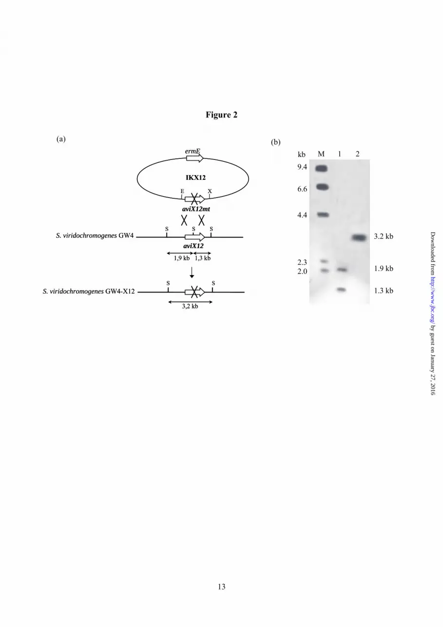
Gene Inactivation of aviX12 - AviX12 was PCR amplified using oligonucleotides 5´-CTACCTGGAATTCCTGCTGACC-3´ (aviX12F) and 5´-CAGCGCCGTCTAGAATCCGTAG-3´ (aviX12R) containing engineered EcoRI and XbaI restriction sites (underlined), respectively. The resulting fragment was isolated with the Nucleospin® Extract kit (Macherey Nagel), digested with EcoRI and XbaI, and ligated into pUC18 previously digested with the same enzymes, generating plasmid pUC-aviX12. The product was confirmed by DNA sequencing (4base lab). A unique SacII restriction site was altered by SacII restriction, subsequent treatment with T4 DNA polymerase and religation. DNA sequencing confirmed the expected 2 bp deletion in aviX12, leading to frameshift mutation. After restriction with EcoRI and XbaI, the insert was transferred to plasmid pSP1 to generate IKX12. The inactivation construct was used to transform protoplasts of S. viridochromogenes GW4. Transformants were propagated in HA medium without erythromycin selection for 16 generations. Selection of erythromycin-sensitive colonies gave the double crossover mutant S. viridochromogenes GW4-X12. The deletion within the gene was confirmed by PCR. PCR fragment obtained from mutant S. viridochromogenes GW4-X12 using primers 5´-GCGCGAGCCGGAGAAGCCGGAGA-3´ (aviX12P-F) and 5´- TCTTGCGCAGCCCCTCGGCAACCA-3´ (aviX12P-R) could not be cleaved by SacII (Fig. 2a) whereas the PCR fragment obtained from S. viridochromogenes GW4 could be digested by SacII. Southern Hybridisation - For Southern hybridisation genomic DNA from mutant and wild type strain was completely digested with SacII, fractionated by agarose gel electrophoresis, and transferred to a positively charged nylon membrane (Hybond N+, Amersham Biosciences). The probe was a 1302 bp EcoRI/XbaI fragment from IKX12 labeled with digoxigenin-dUTP by the random priming method. Hybridisation of the probe
with DNA fragments on the nylon membrane was detected by the chromogenic method using procedures described by Roche Diagnostics.
Complementation of mutant S. viridochromogenes GW4-X12 - To determine clearly that the mutation event affected only the desired gene and not other genes, aviX12 was ligated behind the ermE* promoter of pSET-1cerm and the resulting complementation construct pSETerm-aviX12 was introduced by protoplast transformation into the ∆aviX12-mutant. The complementation led to restored gavibamycin production.
Construction of pRSET-X12 Expressing AviX12 - The aviX12 gene was PCR amplified using primers 5´-AGCCGGAGCTGCAGGCCATATGACCCAG-3´ (X12ProtF) and 5´-CGTCTCGAATTCGTAGGTCTTGCGCAG-3´ (X12ProtR) containing engineered PstI and EcoRI restriction sites (underlined), respectively. The product was isolated with the Nucleospin® Extract kit (Macherey Nagel), digested with PstI and EcoRI, and ligated into the T7 expression vector pRSETb (Invitrogen) previously digested with the same enzymes. The product pRSET-X12 was confirmed by DNA sequencing (4base lab).
Expression and Purification of AviX12 (N-His6-tagged) - For purification of expressed protein, E. coli strain BL21 (DE3)pLysS cells (Stratagene), carrying either pRSET-X12 with aviX12 or the pRSETb vector alone, were grown in LB broth containing 50 µg/ml Carbenicillin and 30 µg/ml Chloramphenicol to OD600 = 0.6. Protein expression was induced by the addition of 1 mM isopropyl-β-D-thiogalactopyranoside, and growth was continued at 37 °C for 4 h. Cells were harvested by centrifugation and stored at -20 °C. The cell pellet from 100 ml culture was resuspended in 4 ml lysis buffer consisting of 50 mM NaH2PO4, 300 mM NaCl, 10 mM imidazole and 5 mM dithiothreitol (DTT). Cells were broken by a single pass through a French pressure cell (Thermo Spectronic) at 700 psi. After centrifugation the supernatant fraction was used for purification procedure. Protein was bound to nickel nitrilotriacetic acid (Ni-NTA) agarose (Qiagen) and loaded onto a column. The column was washed twice with washing buffer containing 50 mM NaH2PO4,
by guest on January 27, 2016http://w
ww
.jbc.org/D
ownloaded from
4
300 mM NaCl, 20 mM imidazole and 5 mM DTT. The protein was recovered with elution buffer (50 mM NaH2PO4, 300 mM NaCl, 250 mM imidazole and 5 mM DTT). The imidazole was removed by passing the protein over a PD-10 (SephadexTM G-25, Amersham Biosciences) desalting column equilibrated in 10 mM Tris, pH 8.0, and 5 mM DTT. Expression and purification of AviX12 was monitored by SDS-Polyacrylamide gel electrophoresis (SDS-PAGE) (4 % stacking gel and 12 % resolving gel) followed by Coomassie Blue staining. The concentration of purified protein was estimated by the Bradford dye-binding method (19). The molecular mass of the purified protein was determined by means of LC/ESI-MS.
Isolation of Avilamycin Derivatives - Strains were grown in SG medium containing 2 % glucose, 1 % soy peptone, 0.1 % CaCO3, 20 mM L-Valine and 1 ml of 0.1 % CoCl2 solution, for 72 hours at 28 °C. Cultures were filtered, the filtrate was extracted twice by ethyl acetate and evaporated to dryness. The mycelium was broken with acetone and filtered again. After evaporation of acetone, it was also extracted with ethyl acetate and evaporated to dryness. Crude extracts from filtrate and mycelium were combined and applied to a solid-phase extraction cartridge (SepPak C18, Waters Associates). The cartridge was eluted with 50 % and 80 % methanol. The 80 % fraction contained about 90 % of the avilamycin derivatives. The evaporated and lyophilisized fractions were redissolved in acetonitrile and water. Further isolation was performed on an Agilent 1100 system using a semipreparative column (Zorbax SB-C18, 5 µm, 9.4 µm × 150 mm). For elution, the following gradient profile was used: solvent A: 5 mM ammoniumacetate in water; solvent B: acetonitrile, nonlinear gradient, 30 % - 50 % B within 20 min at flow rate of 3.5 ml/min. A mass-based fraction collector was used to isolate the avilamycin derivatives. The final isolation step was performed using a gelpermeation column (Plgel, 5 µm; 100 Å: 300 × 7.5 µm; MW < 4000) As solvent acetonitrile at a flow rate of 1 ml/min was used. Again, a mass-based fraction collector was used to isolate the avilamycin derivatives.
Biological Properties - The antimicrobial activity of the new derivative was determined by the agar plate diffusion assay using Bacillus subtilis as the test strain. Susceptibility of
Staphylococci, Streptococci and Enterococci to different derivatives was determined by the microdilution test according to NCCLS guidelines. Vancomycin was used as a standard to ensure the reliability of the determined minimum inhibitory concentrations.
Structural Characterization of the new Gavibamycin derivative accumulated by S. viridochromogenes GW4-X12 - HPLC-MS: High performance liquid chromatography/ electrospray ionisation-mass spectrometry (HPLC/ESI-MS) was performed on an Agilent 1100 Series system with an electrospray chamber and a quadrupole detector. HPLC analysis was carried out on a Zorbax SB-C18 column (5 µm, 4.6 × 150 mm Agilent) with a Zorbax SB-C18 precolumn (5 µm, 4.6 × 12.5 mm; Agilent). A nonlinear gradient from 20 % to 70 % acetonitrile in 0.5 % acetic acid over 22 min at a flow rate of 0.5 ml/min was used. The column temperature was 23 °C and the UV detection wavelengths were 254 and 300 nm. The chamber settings were as following: drying gas flow 12 l/min, nebulize pressure 50 psig, drying gas temperature 300°C. The samples were analysed in positive and negative scan mode with a mass range of 700-1500 Da. NMR Analysis: NMR spectra were recorded on samples dissolved in 330µl DMSO-d6 in SHIGEMI® NMR (Tokyo, Japan) tubes at a temperature of 295 K on a Bruker DMX 750 MHz NMR spectrometer of the Bavaria NMR Center in Garching. The following spectra have been used for the structure elucidation: 13C-1D, 13C-filtered nuclear Overhauser enhancement spectroscopy (NOESY) (20), correlated spectroscopy (COSY) (21), heteronuclear single quantum coherence (HSQC) (22), and heteronuclear multiple bond coherence (HMBC) (23). The spectra were analysed using XWINNMR 3.6 (Bruker) and SPARKY 3 (24).
Spectroscopic Procedures - UV/vis-spectroscopy: A TIDAS-UVI/1001-1 diode array spectrometer with 512 diodes (J&M, Aalen, Germany) was used for UV/vis-spectroscopy. Spectra were recorded in the range from 200 to 600 nm in a buffer of 10 mM Tris, pH 8.0, and 5 mM DTT. The spectrum of a 1 mL aliqout of the preparation as isolated was corresponded as spectrum of the oxidised protein. The sample was mixed in a stirred optical cell with a few grains of
by guest on January 27, 2016http://w
ww
.jbc.org/D
ownloaded from

5
sodium dithionite at room temperature while spectra were continously recorded every 2 seconds. When there was no more spectral shift, the spectrum corresponded to the spectrum of the reduced protein. Spectra were processed using the ‘Spectrachrom’ software package (J&M, Aalen, Germany). EPR-spectroscopy: EPR spectra of the air-oxidized and dithionite-reduced preparation were recorded with an X-band spectrometer EMX 6/1 (Bruker) equipped with a helium-flow cryostat (Oxford). Spectra were recorded at 9.46 GHz microwave frequency, a modulation amplitude of 0.6 mT, time constant of 0.164 s, and scan rate of 17.9 mT/min. The optimal signal was measured at a temperature of 10 K and 1 mW microwave power. Computer simulations of the spectra were performed using the programme SimFonia (Bruker), assuming no hyperfine interaction and Gaussian lineshape.
Calculation of the avilamycin conformation - Possible conformers of avilamycin were identified in a two-step molecular modelling process. A low-energy conformation was constructed and optimized within the ArgusLab program package (25, 26) using a the classical unified force field (UFF, (27)). In the relaxed molecule, by visual ionspection five C–O single bonds were identified that permit rotations of major fragments of the molecule around in steps of 120 degrees. The corresponding 35 = 743 rotamers have been generated and checked for a significant atomic overlap using the repulsive part of the Lennard-Jones potential applying standard van-der-Waals radii. The geometry of each of the conformers has been optimized by a combination of BFGS and steepest descent techniques. The energy at the nine conformational minima has again been calculated using the UFF force field referenced above. GenBank accession number – The GenBank accession number for the DNA sequence reported in this paper is AAK83189.1.
RESULTS Inactivation of aviX12 - The inactivation of aviX12 was achieved by insertion of a frameshift mutation at a singular SacII restriction site central in aviX12 (Fig. 2a). As
wildtype strain for the inactivation experiment S. viridochromogenes GW4 was chosen, a mutant carrying a deletion in the methyltransferase gene aviG4 leading to the production of gavibamycin A1 and A3 (Fig. 1). Protoplasts of S. viridochromogenes GW4 transformed with inactivation construct IKX12 were screened for a erythromycin-resistant phenotype. The strains selected were propagated without erythromycin selection. Two erythromycin-sensitive strains were examined by PCR, one of these proved the deletion of aviX12. This was verified by Southern hybridisation (Fig. 2b). The mutant strain was designated S. viridochromogenes GW4-X12. HPLC analysis showed the production of new avilamycin derivatives in comparison to the wild type strain. Identification of new Avilamycin Derivatives in S. viridochromogenes GW4-X12 - For analysis of secondary metabolite formation, both wild type and ∆aviX12 mutant were cultivated in production medium as described in Experimental Procedures. Ethyl acetate extracts of the culture supernatants were analysed by HPLC-UV and HPLC/ESI-MS. Gavibamycin A1 (1388 u) and gavibamycin A3 (1390 u) were detected in extract of S. viridochromogenes GW4. In contrast, the ∆aviX12-mutant accumulated four new compounds with the masses M = 1376 u, M = 1374 u (main compound), M = 1332 u and M = 1262 u, respectively. The mass of the main compounds are indicating the loss of a methyl group in comparison to the main compounds of the wild type gavibamycin A1 (M = 1388) and gavibamycin A3 (M = 1390). The new compounds were named gavibamycin N1 (M = 1374) and gavibamycin N3 (M = 1376). The other two compounds could be related to a derivative missing in addition to the methyl group the acetate moiety at the eurekante (1332 u) and a derivative missing additionally the isobutyryl group at the lyxose moiety (1262 u), respectively.
Structure Elucidation of the Gavibamycin A1 and A3 Derivatives - To elucidate the structure of the main compound of mutant S. viridochromogenes GW4-X12, this product was isolated as described in Experimental Procedures. The NMR analysis of the mutants of this study is based on the completed 1H and 13C resonance assignment of avilamycin A and avilamycin C (2). The samples under
by guest on January 27, 2016http://w
ww
.jbc.org/D
ownloaded from

6
investigation were prepared by feeding the bacteria 13C-labeled L-methionine resulting in a partial 13C-labeling of the products. For gavibamycin N1 carbons C27, C34, C43, and C61 showed enhanced signal intensities due to the labeling (the nomenclature is given in Fig.1). The unambiguous identification of these carbons was done by the analysis of the 13C-filtered NOESY and the HMBC spectra. In figure 3 the resonances of atoms in the vicinity of C37 (C2 position in ring F) are shown in a section of the HSQC spectra of gavibamycin N1 and avilamycin A. In comparison to avilamycin A, two methyl groups (C7 and C41) are missing in gavibamycin N1. Surprisingly this is the same labeling pattern found in gavibamycin I1 (2). Due to the fact that it is very unlikely for an organism to have two enzymes with the same function a further structural analysis was carried out. Careful HPLC-UV analysis revealed that both compounds showed different retention times under the conditions described in the Experimental procedures. As a first step all 1H- and 13C-chemical shifts of gavibamycins I1 and N1 were compared. No significant difference was observed except for the atoms in the vicinity of C37. In a second step the 3JH,H-couplings, which are directly linked to the dihedral angle of two hydrogens by the Karplus equation, were analyzed at the site of C37. For the pair H36/H37 (the hydrogens bound to carbons C36 and C37, respectively) a 3J-coupling of 8±0.5 Hz was found for gavibamycin N1. This value is characteristic for an axial/axial orientation of H36 and H37 with respect to the ring. In contrast, for avilamycin A, a value of 3.5 ±0.5 Hz was found for the 3JH36,H37-coupling, corresponding to an axial/equatorial orientation. This result indicated that a glucose is incorporated in gavibamycin N1 instead of a mannose in wildtype gavibamycin A1. Similar studies were carried out on the X12 mutation derivative with a molecular weight of 1332 amu. The conformation of H36/H37 was found to be axial/axial as for gavibamycin N1. Compared to gavibamycin N1 and avilamycin A this compound is lacking the acetyl moiety in ring H which is replaced by a hydrogen. This result is analogous to the changes reported for the mutations ITB1 and ITO2 (10).
Antibacterial Activity of Gavibamycin A1 and Gavibamycin N1 - Extracts of wild type and S.
viridochromogenes GW4-X12 were pretested against Bacillus subtilis using the agar diffusion test. The extract of S. viridochromogenes GW4-X12 showed still antibiotic activity but the in comparison to the wild type extract it was lower. Furthermore, the gavibamycin N1 of S. viridochromogenes GW4-X12 and gavibamycin A1 of the wild type strain were tested against a panel of pathogenic Gram-positive organisms, including two vancomycin-resistant strains, using the microdilution assay. The antibacterial activity of each derivative against the clinical isolates is presented in table 1. All isolates were susceptible to gavibamycin A1 (MIC range < 0.5 to 8 µg/ml). The changing of the mannose against a glucose in gavibamycin N1 drastically affected the activity against all tested organisms. The conformation of avilamycin A - Of the nine conformers computed (see experimental procedures, calculation of the avilamycin conformation), seven lie within an energy interval of 50 kJ/mol around the absolute minimum. They either correspond to a linear shape of the molecule (two conformers) or create the general impression of a U-shape (five conformers including the two lowest in energy). A typical example for the latter is shown in figure 4. Although the number of possible rotamers has been reduced considerably by the application of a molecular modelling procedure, only its combination with other criteria like the chemical accessibility of functional groups may help to identify a unique, biologically active conformation.
Sequence Analysis of AviX12 - The amino acid sequence of AviX12 comprises 396 amino acids. When we first published the sequence BLAST searches did not reveal meaningful similarities to proteins with known function in database. Recently, Sofia et al. have identified the radical SAM protein superfamily by bioinformatic techniques (28). Despite low overall sequence similarity all Radical SAM enzymes contain an unconventional [4Fe/4S] cluster coordinated by three closely-spaced cysteine residues, creating the defining CxxxCxxC motif of this family (28). AviX12 contains this characteristic cysteine motif in its amino acid sequence precisely from amino acids C156 to C163 indicating that AviX12 belongs to the radical SAM family. The fourth
by guest on January 27, 2016http://w
ww
.jbc.org/D
ownloaded from

7
Fe of the cluster is coordinated by SAM. The function of radical SAM enzymes is to generate catalytic 5’-deoxyadenosyl radicals.
Expression and Purification of the N-His6-tagged AviX12 - To demonstrate that AviX12 contains an Fe/S cluster, we cloned aviX12 into the expression vector pRSETb behind the T7 RNA polymerase promoter to generate an N-terminal hexahistidine fusion protein. The protein was overexpressed in E. coli BL21 (DE3)pLysS. A protein of the predicted molecular mass of AviX12 (48,995 Da) was observed by SDS-PAGE in extracts of isopropyl-β-D-thiogalactopyranoside-induced cells. AviX12 was purified by nickel-chelation chromatography. The preparation was desalted and subjected to LC/ESI-MS. The molecular mass of the preparation was determined to 49,011 Da. The difference of 16 u might be due to oxidation of methionine.
Spectroscopic Characterisation of the Iron-Sulfur-Cluster - The UV/vis spectrum of the AviX12 preparation as isolated showed beside the peak of the aromatic amino acids at 280 nm a broad structureless absorbance from 300 to 550 nm indicating the presence of a non-protein cofactor (Fig. 5). This signal was bleached by addition of dithionite leading to a broad negative peak around 450 nm in the reduced-minus-oxidised difference spectrum (Fig. 5). This is a typical spectral feature of protein bound Fe/S clusters (29). EPR spectra were recorded to determine the type of Fe/S cluster present in the AviX12 preparation. No EPR signals were detectable with the sample reduced by dithionite in the temperature range from 5 to 100 K. However, the oxidised protein showed a signal at temperatures below 20 K which was clearly seen at 10 K (Fig. 5). The signal exhibited an axial symmetry with a slight rhombic distortion and is typical for an [3Fe/4S] cluster. It was simulated with the following parameters: gx = 2.007, gy = 2.018, and gz = 2.025; Lx = 1.65 mT; Ly = 1.1 mT, and Lz = 1.4 mT (Fig. 5). From spin quantiations the presence of approximately one [3Fe/4S] cluster per AviX12 was calculated. Preliminary attempts to reconstitute an [4Fe/4S] cluster in the preparation under anaerobic conditions and in the presence of SAM were not successful, so far.
DISCUSSION
The biosynthesis of saccharide containing polyketides usually starts with the formation of the polyketide moiety. The sugar is attached to the polyketide at a later stage. In contrast, avilamycin biosynthesis starts with the formation of a disaccharide and the polyketide moiety is attached to the hexasaccharide at the very end of the biosynthetic route. D-Mannose was discussed to be one component of the disaccharide starter molecule (11). Our data now indicate that the disaccharide is synthesized from D-glucose instead of D-mannose and that AviX12 is involved in C2-epimerisation of the glucose moiety after the whole avilamycin molecule is generated.
The reactions of most epimerases take place at a chiral carbon adjacent to an activating moiety such as a carbonyl group and the catalysis typically involves a simple deprotonation/reprotonation mechanism. Here epimerization takes place at an unactivated center. Most probably AviX12 and SAM generate a 5´deoxyadenosyl radical, which abstracts a hydrogen atom a position C-2 of the mannose moiety to form a radical intermediate. This intermediate undergoes epimerization by an unknown mechanism.
The antibiotic activity of gavibamycin N1, main product of the S. viridochromogenes GW4-X12, is very low. This strongly reduced activity might be explained by a conformational change of the whole molecule leading to a inability to bind to the ribosome. AviX12 activity is believed to induce an important conformational change of the molecule resulting in its active form.
The calculation of the lowest energetic conformation of avilamycin A suggests an U-form of the whole molecule. This conformation might be stabilised by hydrogen bonds between evalose and mannose on one hand and evalose and fucose on the other hand, as these sugars are located in the region of the angle of the molecule.
The transition of the low active gavibamycin N1 to the active avilamycin A is mediated by AviX12. AviX12 was identified as a member of the radical SAM family. The presence of an Fe/S cluster in the preparation of AviX12 being typical for radical SAM enzymes was proven by spectroscopic methods. The EPR spectra clearly show that AviX12 contains an [3Fe/4S]-cluster that is stable during the isolation of the protein. Different types of Fe/S clusters have been described for radical SAM enzymes. It was
by guest on January 27, 2016http://w
ww
.jbc.org/D
ownloaded from

8
unequivocally shown that a [4Fe/4S] cluster is always present in the active enzymes (30;31). However, only the lysine 2,3 aminomutase seems to contain this cluster type after isolation [32]. The other members of the family of radical SAM enzymes have been isolated either as apo-enzymes or containing [2Fe/2S] or [3Fe/4S] clusters or some combinations of these components. Some preparations were reported to contain a [4Fe/4S] cluster as a minor component. In most cases, the active [4Fe/4S] cluster is obtained by reducing the preparations under unaerobic conditions. Our preparation did not contain any detectable traces of an [4Fe/4S] cluster. Radical SAM enzymes share structural features important for building the active sites of the proteins which mostly contain the [4Fe/4S]-cluster, the SAM cofactor and the substrate shielding the radical intermediates from the surrounding medium. So far, we were
not able to reconstitute the functional [4Fe/4S] cluster by addition of iron, SAM, and the substrate or any combinations thereof under anaerobic conditions. This is in agreement with the fact that our preparation shows no enzymatic activity. It remains an open question whether the purification by means of affinity chromatography has irreversibly damaged the protein or whether another cellular factor not present in our assay is needed for reconstitution of the cluster and enzymatic activity.
It would be very interesting to learn whether the large avilamycin molecule is located in the active site of AviX12 and whether the conformational switch of the molecule after epimerisation also takes places in the proteins active site. To our best knowledge AviX12 is the first example of a protein belonging to the radical SAM family being involved in the epimerisation of a sugar.
REFERENCES
1. Wright, E. D. (1979) Tetrahedron 35, 1207-1237
2. Weitnauer, G., Hauser, G., Hofmann, C., Linder, U., Boll, R., Pelz, K., Glaser, S. J., and Bechthold, A. (2004) Chem.Biol. 11, 1403-1411
3. Schering-Plough (2000) http://www.sch-plough.com/news/2000/research/20000505.html.
4. Belova, L., Tenson, T., Xiong, L., McNicholas, P. M., and Mankin, A. S. (2001) Proc.Natl.Acad.Sci.USA 98, 3726-3731
5. McNicholas, P. M., Najarian, D. J., Mann, P. A., Hesk, D., Hare, R. S., Shaw, K. J., and Black, T. A. (2000) Antimicrob.Agents Chemother. 44, 1121-1126
6. McNicholas, P. M., Mann, P. A., Najarian, D. J., Miesel, L., Hare, R. S., and Black, T. A. (2001) Antimicrob.Agents Chemother. 45, 79-83
7. Treede, I., Jakobsen, L., Kirpekar, F., Vester, B., Weitnauer, G., Bechthold, A., and Douthwaite, S. (2003) Mol.Microbiol. 49, 309-318
8. Kofoed, C. B. and Vester, B. (2002) Antimicrob.Agents Chemother. 46, 3339-3342
9. Weitnauer, G., Mühlenweg, A., Trefzer, A., Hoffmeister, D., Süssmuth, R. D., Jung, G., Welzel, K., Vente, A., Girreser, U., and Bechthold, A. (2001) Chem.Biol. 8, 569-581
10. Treede, I., Hauser, G., Muhlenweg, A., Hofmann, C., Schmidt, M., Weitnauer, G., Glaser, S., and Bechthold, A. (2005) Applied and Environmental Microbiology 71, 400-406
11. Hofmann, C., Boll, R., Heitmann, B., Hauser, G., Duerr, C., Frerich, A., Weitnauer, G., Glaser, S. J., and Bechthold, A. (2005) Chem.Biol. 12, 1137-1143
12. Macneil, D. J., Gewain, K. M., Ruby, C. L., Dezeny, G., Gibbons, P. H., and Macneil, T. (1992) Gene 111, 61-68
by guest on January 27, 2016http://w
ww
.jbc.org/D
ownloaded from

9
13. Pelzer, S., Reichert, W., Huppert, M., Heckmann, D., and Wohlleben, W. (1997) J.Biotechnol. 56, 115-128
14. Bierman, M., Logan, R., O'Brien, K., Seno, E. T., Rao, R. N., and Schoner, B. E. (1992) Gene 116, 43-49
15. Hoffmeister, D., Ichinose, K., and Bechthold, A. (2001) Chem.Biol. 8, 557-567
16. Sambrook, J., Fritsch, E. F., and Maniatis, T. (1989), Cold Spring Harbour Laboratory Press
17. Hopwood, D. A., Bibb, M. J., Chater, K. F., Kieser, T., Bruton, C. J., Kieser, H. M., Lydiate, D. J., Smith, C. P., Ward, J. M., and Schrempf, H. (1985), The John Innes Foundation, Norwich, U.K.
18. Altschul, S. F., Madden, T. L., Schaffer, A. A., Zhang, J. H., Zhang, Z., Miller, W., and Lipman, D. J. (1997) Nuc.Acids Res. 25, 3389-3402
19. Bradford, M. M. (1976) Analytical Biochemistry 72, 248-254
20. Otting, G., Senn, H., Wagner, G., and Wüthrich, K. (1986) J.Magn.Reson. 70, 500-505
21. Rance, M., Soerensen, O. W., Bodenhausen, G., Wagner, G., Ernst, R. R., and Wüthrich, K. (1983) Biochem.Biophys.Res.Commun. 117, 479-485
22. Bodenhausen, G. and Ruben, D. J. (1980) Chem.Phys.Lett. 69, 185-189
23. Summers, M. F., Marzilli, L. G., and Bax, A. (1986) J.Am.Chem.Soc. 108, 4285-4294
24. Goddard, T. D. and Kneller, D. G. (2004) University of California, San Francisco
25. Thompson, M. A., and Zerner M. C. (1991) J. Am. Chem. Soc., 113, 8210
26. Thompson, M.A., Glendening, D., Feller, D., J. (1994) Phys. Chem. 98, 10465-10476.
27. Casewit, C. J., Colwell, K. S., Rappé, A. K. (1992) JACS 114, 10035.
28. Sofia, H. J., Chen, G., Hetzler, B. G., Reyes-Spindola, J. F., and Miller, N. E. (2001) Nuc.Acids Res. 29, 1097-1106
29. Rasmussen, T., Scheide, D., Brors, B., Kintscher, L., Weiss, H., Friedrich, T. (2001) Biochemistry. 22, 6124-31
30. Duin, E. C., Lafferty, M. E., Crouse, B. R., Allen, R. M., Sanyal, I., Flint, D. H., and Johnson, M. K. (1997) Biochemistry 36, 11811-11820
31. Liu, A. and Gräslund, A. (2000) J.Biol.Chem. 275, 12367-12373
32. Wu, W., Booker, S., Lieder, K.W., Bandarian, V. Reed, G.H. and Frey, P.A. (2000) Biochemistry 39, 9561-9570
footnotes * This work was supported by the DFG (Deutsche Forschungsgemeinschaft), grant BE1389/5-2 and a project in the Graduiertenkolleg “Biochemie der Enzyme” both to A. Bechthold. 1 The abbreviations used are: SAM, S-adenosyl-L-methionine; E. coli, Escherichia coli; S. viridochromogenes, Streptomyces viridochromogenes; Uridin diphosphate-glucuronic acid
by guest on January 27, 2016http://w
ww
.jbc.org/D
ownloaded from

10
decarboxylase, UDP-glucuronic acid decarboxylase; HPLC/ESI-MS, high performance liquid chromatography/ electrospray ionisation-mass spectrometry; NOESY, nuclear Overhauser enhancement spectroscopy; COSY, correlated spectroscopy; HSQC, heteronuclear single quantum coherence; HMBC, heteronuclear multiple bond coherence; EPR, electron paramagnetic resonance; Fe/S cluster, iron-sulfur cluster; MIC, minimum inhibitory concentration; DTT, dithiothreitol.
FIGURE LEGENDS
Fig. 1. Structures of avilamycins and gavibamycins investigated during this study. Fig. 2. a, diagram illustrating the construction of S. viridochromogenes GW4-X12 by disrupting aviX12 by insertion of a frameshift mutation. S, SacII; E, EcoRI; X, XbaI. b, Southern hybridisation of S. viridochromogenes GW4 and S. viridochromogenes GW4-X12 genomic DNA digested with SacII. Lane M, digoxigenin-labeled molecular weight standard; lane 1, S. viridochromogenes GW4 genomic DNA; lane 2, S. viridochromogenes GW4-X12 genomic DNA. Fig. 3. Section of the 13C-HSQC spectra of avilamycin A (solid contour lines) and gavibamycin N1 (dashed contour lines). Resonances for C37, C38, C39, C40 and C42 are lettered. Arrows are indicating the changes in chemical shifts for the resonances of the two compounds. For C36 (not shown) a change in chemical shift of 0.31 ppm and 1.6 ppm is observed for 1H and 13C, respectively. Fig. 4. U-shape conformation of avilamycin A. C-atoms are shown in grey, H-atoms are shown in white, O-atoms are shown in red and Cl-atoms are shown in green. Fig. 5. Spectra of the purified AviX12. A) UV/vis spectrum of the oxidised and reduced preparation. The inset shows the reduced-minus-oxidised difference spectrum. B) EPR spectra of the reduced preparation at 40K (a), the oxidized preparation at 10 K (b), the reduced preparation at 10 K (c), and the simulated spectrum (d). by guest on January 27, 2016
http://ww
w.jbc.org/
Dow
nloaded from

11
Table 1. Minimum inhibitory concentration (MIC) of avilamycin derivatives determined by microdilution according to NCCLS guidelines Vancomycin Gavibamycin A1 Gavibamycin N1 Staphylococcus aureus ATCC 25923
1 8 16
MRSA RV 5/98 < 0.5 4 16 Staphylococcus epidermidis DSM 1798
1 4 32
Streptococcus pyogenes E12449/98
< 0.25 2 8
Streptococcus pneumoniae E2919/94
< 0.25 < 0.5 4
Enterococcus faecalis ATCC 19212
< 0.25 2 16
Enterococcus faecalis H10513/99
> 128 1 8
Enterococcus faecium Vanco-H8914/00
> 128 8 16
by guest on January 27, 2016http://w
ww
.jbc.org/D
ownloaded from

12
Figure 1
strain/mutant R1 R2 R3 main compound wild type CH3 COCH3 CH3 avilamycin A wild type CH3 CH(OH)CH3 CH3 avilamycin C GW4 H COCH3 CH3 gavibamycin A1 GW4 H CH(OH)CH3 CH3 gavibamycin A3 GW4-GW6 H COCH3 H gavibamycin I1
GW4-X12 H COCH3 H (OH group equatorial)
gavibamycin N1
GW4-X12 H CH(OH)CH3 H (OH group equatorial)
gavibamycin N3
OCH3
ClOH
ClO
O
CH3
OHO
OCH3
OHO
O
OCH3
CH3
OOR1
OO
O
CH3
CH3
O
OH
O CH3
OR3
OOCH3
OH
OCH3
O
O
O O CH3 R2
OHO
O
15
39
12
14
10
20
18 16
27
28
2231
56
585753
61
4544
36
4341
46
4748
4950
51
52
38
39 4042
2932
35
34
24
26
2115
2 4
68
A
B CD
E FG
H
OCH3
ClOH
ClO
O
CH3
OHO
OCH3
OHO
O
OCH3
CH3
OOR1
OO
O
CH3
CH3
O
OH
O CH3
OR3
OOCH3
OH
OCH3
O
O
O O CH3 R2
OHO
O
15
39
12
14
10
20
18 16
27
28
2231
56
585753
61
4544
36
4341
46
4748
4950
51
52
38
39 4042
2932
35
34
24
26
2115
2 4
68
A
B CD
E FG
H
by guest on January 27, 2016http://w
ww
.jbc.org/D
ownloaded from
13
Figure 2
1 2kb9.4
6.6
4.4
2.32.0
M
3.2 kb
1.9 kb
1.3 kb
(b)
S S S
aviX12S. viridochromogenes GW4
S SS. viridochromogenes GW4-X12
1,9 kb 1,3 kb
3,2 kb
E X
ermE
IKX12
aviX12mt
S S S
aviX12S. viridochromogenes GW4
S SS. viridochromogenes GW4-X12
1,9 kb 1,3 kb
3,2 kb
E X
ermE
IKX12
aviX12mt
(a)
by guest on January 27, 2016http://w
ww
.jbc.org/D
ownloaded from


Koslowski, Thorsten Friedrich and Andreas BechtholdRaija Boll, Carsten Hofmann, Björn Heitmann, Gerd Hauser, Steffen Glaser, Thorsten
enzymeThe active conformation of avilamycin A is confered by AviX12, a radical SAM
published online March 14, 2006J. Biol. Chem.
10.1074/jbc.M601508200Access the most updated version of this article at doi:
Alerts:
When a correction for this article is posted•
When this article is cited•
to choose from all of JBC's e-mail alertsClick here
http://www.jbc.org/content/early/2006/03/14/jbc.M601508200.citation.full.html#ref-list-1
This article cites 0 references, 0 of which can be accessed free at
by guest on January 27, 2016http://w
ww
.jbc.org/D
ownloaded from