technetium99m- 1,2-bis(bis(2-ethoxyethyl) phosphino)ethane: human biodistribution, dosimetry and...
TRANSCRIPT
1993;34:30-38.J Nucl Med. Graham, David Hinge, Joyce Davidson and Avijit LahiriBrian Higley, Francis W. Smith, Terry Smith, Howard G. Gemmell, Prabir Das Gupta, Dragouten V. Gvozdanovic, David Dosimetry and Safety of a New Myocardial Perfusion Imaging AgentTechnetium-99m-1,2-bis[bis(2-Ethoxyethyl) Phosphino]Ethane: Human Biodistribution,
http://jnm.snmjournals.org/content/34/1/30This article and updated information are available at:
http://jnm.snmjournals.org/site/subscriptions/online.xhtml
Information about subscriptions to JNM can be found at:
http://jnm.snmjournals.org/site/misc/permission.xhtmlInformation about reproducing figures, tables, or other portions of this article can be found online at:
(Print ISSN: 0161-5505, Online ISSN: 2159-662X)1850 Samuel Morse Drive, Reston, VA 20190.SNMMI | Society of Nuclear Medicine and Molecular Imaging
is published monthly.The Journal of Nuclear Medicine
© Copyright 1993 SNMMI; all rights reserved.
by on October 7, 2014. For personal use only. jnm.snmjournals.org Downloaded from by on October 7, 2014. For personal use only. jnm.snmjournals.org Downloaded from
suitable photons (140 keY) for gamma camera imagingand, due to its shorter half-life (6.03 hr), reduced patientradiation dose for a higher injected activity. It has beenclaimed that a technetium-based agent could significantlyimprove the image quality compared with that obtainablewith 201Tldue to a twofold increase in resolution and anincreased count rate resulting from a fourfold increase inadministered activity (2).
Technetium-99m-1,2-bis[bis(2-ethoxyethyl)phosphino]ethane (tetrofosmin or P53) is a novel lipophilic technetium phosphine dioxo cation (@mTc(tetrofosmin)2O2]+).The ligand (tetrofosmin, Fig. 1) has been formulated intoa freeze-dried kit which yields an injection containing99mTctetrofosmin on reconstitution with pertechnetate(3). Preclinical studies in the rat, guinea pig and minipighave shown that @mTc@tetrofosminhas good heart uptakeand retention, together with rapid clearance from liver,lung and blood. The formulation has been shown to havea very high safety margin in both single and repeated doseintravenous toxicology studies. The ligand has been demonstrated not to exhibit mutagenic potential in a range ofin vitro and in vivo mutagenicity studies.
Previously, a limited human volunteer study was performed using a different formulation which yielded 99mTc..
tetrofosmin. This study in three healthy male volunteers indicated the potential ofthis novel technetium complex (4).
Unlike 201Tl,the myocardial distribution of 99mTc@tetro@fosmin does not alter rapidly with time and hence diagnosis of reversible myocardial ischemia could not beachieved with a single injection of the reconstituted kit.Consequently, in clinical practice it will be necessary toadminister two doses of the agent during a completediagnostic protocol.
The present study was designed to establish the biodistribution, dosimetry and safety ofthis new 99mTccomplex,produced using a freeze-dried formulation, in volunteerssubmitted to physical conditions similar to those requiredfor a diagnostic study.
A novel @‘“Tccomplex(1,2-bis[bis(2-ethoxyethyl)phosphino]ethane, @“Tc-tetrofosmin)has been developed to replace20111 in myocardial perfusion imaging. Biodistribution, safety
and dosimetryof @“Tc-tetrofosminwere studiedin 12 malevolunteers,each at rest and duringexercise.Safety parameters measured to 48 hr postinjection revealed no clinicallysignificant long-term drug-relatedchanges. Biodistributionwas studiedby acquiringwhole-bodyor serialstatic imagesupto 48 hrpostinjection.Technetium-99m-tetrofosminshowsgood heart uptake (1.2%) with retention.Clearanceis excellent from blood (<5% by 10 mm), liver (<4.5% by 60 mm)andlung.Sequestrationof activityby skeletalmuscleisenhanceddurlng exercise. Radiationdosimetry calculations indicate thatthe effectivedose, assuminga 3.5 hr bladdervoidingperiod,is 32.9 x 10-@red/mCi (8.9 x 10@ mSv/MBq) at rest and26.7 x 1O@rad/mCi (7.1 x 1O@mSv/MBq) after exercise.Technetium-99m-tetrofosmincan producehighqualitymyocardial images from 5 mm to several hours postinjection.
JNucIMed1993;34:30-38
xercise myocardial scintigraphy is an establishedmethod for diagnosing and assessing the severity of coronary artery disease (CAD) (1). Thallium-201 is currentlythe most widely used radiopharmaceutical for myocardialperfusion scintigraphy. However, 201'flis not an ideal agentbecause of a low gamma photopeak energy (69—83keV)that limits resolution and a relatively long half-life (73 hr)that limits the administered activity.
Technetium-99m-labeled radiopharmaceuticals promise certain advantages over 201fl. These include extemporaneous reconstitution from a kit when required, more
ReceivedMay 26, 1992; revisionaccepted Aug. 17. 1992.For correspondenceor reprintscontact: BrianHigley,PharmaceuticalsR&D,Amersham Intl. plc, Whfte Uon Rd., Amersham, Buckinghamshire. HP7. 9LL,UK.
30 The Journalof NuclearMedicine•Vol. 34 •No. 1 •January1993
Technetium-99m- 1,2-bis[bis(2-Ethoxyethyl)Phosphino]Ethane: Human Biodistribution,Dosimetry and Safety of a New MyocardialPerfusion Imaging AgentBrian Higley, Francis W. Smith, Terry Smith, Howard G. Gemmell, Prabir Das Gupta,
Dragouten V. Gvozdanovic, David Graham, David Hinge, Joyce Davidson and Avijit Lahiri
Pharmaceuticals R&D, Amersham Internationalpic, Amersham, Buckinghamshire, UK; Department ofNuclear Medicine,Aberdeen Royal Infirmary, Forester/till, Aberdeen, UK; and Department ofCardiology, Northwick Park Hospital and SectionofMedical Physics, Clinical Research Centre, Harrow, Middlesex, UK
by on October 7, 2014. For personal use only. jnm.snmjournals.org Downloaded from
each volunteerrangedfrom 40% to 82%(mean 61% ±11%) ofa single vial at rest and from 40% to 87% (mean 59% ±16%) ofa single vial during exercise. The total cumulative chemical dose(sum of rest and exercise injection) ranged from 88% to 162%(mean 120%±29%)of the total contentsof a singlevial.
Following an overnight fast, injections were performed both atrest, each subject being injected intravenously with the agentwhile lying supine, and within the next 7—14days during a periodof sub-maximal graded exercise (limited by achieving 85% agepredicted maximum heart rate, fatigue or dyspnea). Exercise wasundertaken either supine using a bicycle (Northwick Park) orerect using a treadmill (Aberdeen). After injection during exercise,the subject was encouraged to continue exercising for a furtherminute.
Subjects were allowed a light meal after approximately 4 hr inthe study.
Safety ParametersVital signs, including pulse, temperature and blood pressure,
were measured within 30 mm prior to injection, and remeasuredat 5, 10, 15, 30 mm, 1, 2, 4, 6, 24 and 48 hr postinjection. A 12-lead electrocardiogram (ECG) was recorded 30 mm prior toinjection and at 24 and 48 hr postinjection. ECG was monitoreddiscontinuously for the first 6 hr postinjection, using limb leads,in orderto facilitatemonitoringduringimaging.
Baseline blood samples for hematology (including total anddifferential white blood cell, red blood cell and platelet count,hemoglobin and hematocrit) and biochemistry (including electrolytes, creatinine, urea, total protein, albumin, gamma-glutamyltransferase, alkaline phosphatase, aspartate transaminase, alaninetransaminase, creatine phosphokinase and lactate dehydrogenase)were taken within 5 mm prior to drug administration and repeated at 30 mm, 6, 12, 24 and 48 hr postinjection. The bloodsamples were taken via a cannula placed in the opposite arm tothat in which the drug was administered. Urine samples (midstream) were also obtained and analyzed by dipstick methods forprotein, glucose, blood cells, bilirubin and ketones.
ClearanceStudiesAnti-coagulated blood was collected, from the opposite arm to
that in whichthe injectionwasmade,at the followingtimes:—5(baseline), 2, 5, 10,20, 30 mm and 1,2, 4, 12and 24 hr. Duplicatewhole blood samples (0.5 ml) were processed for counting. Afterremoval ofthe whole blood aliquot, the remainder ofthe samplewas centrifuged and duplicate 0.5 ml plasma samples processedin a similar way. Data were corrected for background and decay.Whole blood and plasma activity were calculated in terms ofpercentage of administered activity in the estimated total blood(or plasma) volume.
Urine was collected, in plastic bottles containing preservative,for 48 hr after injection. This was divided into collections frombaseline (pre-injection) and where possible, 0—2,2—4,4—8,8—12,12—24,and 24—48hr. Radioactivity in these samples was determined by counting in a fixed, reproducible geometry systemmidway between opposing Nal detectors and was calculated interms of percentage of administered activity per sample. Feceswere collected for 48 hr postinjection and radioactivity in thesesamples was determined in a similar way to that used for determination of urinary activity.
BiodistributionStudiesDifferent techniques for imaging the biodistribution of @mTc@
tetrofosmin were used at the two centers.
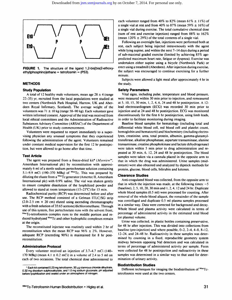
P P
FIGURE1. Thestructureof the ligand1,2-bis[bis(2-ethoxyethyl)phosphino]ethane= tetrofosmin= (P53).
METHODS
StudyPopulationA total of 12healthymalevolunteers,meanage28 ±4 (range
22—35)yr. recruited from the local populationswerestudied attwo centers (Northwick Park Hospital, Harrow, UK and Aberdeen Royal Infirmary, Scotland). The average weight of thevolunteers was 7 1 ±10 kg (range 58—90 kg). Each volunteer gave
written informed consent. Approval ofthe trial was received fromlocal ethical committeesand the Administration of RadioactiveSubstances Advisory Committee (ARSAC) ofthe Department ofHealth (UK) prior to study commencement.
Volunteers were requested to report immediately to a supervising physician any unusual symptoms that they experiencedfollowing the administration of the agent. Volunteers remainedunder constant medical supervision for the first 12 hr postinjection, but were allowed to go home after that time.
TestArticleThe agent was prepared from a freeze-dried kit* (MyovieWTM.
Amersham International plc) by reconstitution with approximately 6 ml ofa sterile sodium pertechnetate solution containing5. 1—9.9mCi (190—370 MBq) of @mTc.This was prepared by
diluting the eluate from a 99mTcgenerator (Amertec II, AmershamInternational plc) with 0.9% saline. The vial was shaken gentlyto ensure complete dissolution of the lyophilized powder andallowed to stand at room temperature (15—25°C)for 15 mm.
Radiochemical purity (RCP) was determined before and afteruse. The RCP method consisted of a Gelman ITLC/SG strip(2.0—2.5cm X 20 cm) elutedusingascendingchromatographywith a fresh solution of35:65 acetone/dichloromethane. Throughuse of this system, free pertechnetate runs with the solvent front,99mTctetrofosmin complex runs to the middle portion and reduced hydrolyzed 99mTcand other hydrophilic complexes remainat the origin.
The reconstituted injectate was routinely used within 2 hr ofreconstitution when the mean RCP was 96% ±2%. However,adequate RCP (exceeding 90%) is maintained for 8 hr postreconstitution.
AdministrationProtocolEvery volunteer received an injection of 3.7—4.7mCi (140—
170MBq) (mean4.1 ±0.2 mCi) in a volume of2.4 to 5 ml oneach of two occasions.The total chemicaldose administeredto
.@ contained 0.23 mg tetrofosmin, 30 @gstannous chloride dihydrate,
0.32mgdisodiumsuiphosalicylate,and1.0mgsodiumgluconate,pHadjustedbefore lyophilizationand sealed under an atmosphereof nitrogen.
99mTcTetrofosminHuman Biodistnbution •Higley et al. 31
EtO@@
EtO\@ \@jOEt
by on October 7, 2014. For personal use only. jnm.snmjournals.org Downloaded from
Whole-body images were obtained at Northwick Park usingan IGE400ATgammacamera (400mm FOV)fittedwith a lowenergy, diverging parallel-hole collimator to increase the lateralviewing aspect. Both anterior and posterior views were obtained.The overalltime from startingthe anterior viewto finishingtheposterior view was about 18 mm.
At Aberdeen, an IGE 500A Maxicamera (500 mm FOV) wasused with a parallel-hole, low-energy, general-purpose collimator.The imagingtechnique consistedof nine static images,six anterior coveringthe whole length of the body and three posteriorwith imaging ofthe legs omitted. There was considerable overlapbetween adjacent views. The complete sequence took 20—26mm.Imaging was started at 5, 30 mm, 1, 2, 4, 8, 24 and 48 hrpostinjection.
QualitativeAnalysisof ImagesAll imageswere reviewedto determine the subjectivebiodis
tribution and determine which organs and tissueswere clearlyvisible and warranted quantitative assessment. Additionally, allanterior whole-body images and static chest views were reviewedas unprocessed hardcopies by two blinded readers. Readers wereasked to assess the general image quality and whether diagnostically useful myocardial images had been obtained.
Quantitative Analysis of ImagesTotal body and individual organ retention of @mTcat the
variousimagingtimes wereestimatedusingthe geometricmeanof anterior and posterior images, corrected for physical decay of99mTc during the period from the time of injection to the midtime ofthat scan.
Whole-Body Images. At Northwick Park, total counts and
number ofpixels withina regionof interest(ROI)drawn aroundthe entire body image were recorded. Total counts were correctedusingan averagebackgroundcount per pixelobtainedfrom fourseparate regions outside the body contour. At Aberdeen, totalcounts in the body image were estimated by summation ofconsecutive regions after allowance was made for overlappingareas of adjacent static views, and corrected for background asabove.
Retention in Organs and Tissues. A number ofspecific organsexhibitedsignificantuptake of @mTc@tetrofosmin,at least in theearly stages of the study. These were identified as heart, lungs,liver, gallbladder, kidneys, salivary glands, thyroid, urinary bladder and gastrointestinaltract. Skeletalmusclealsoshowedsignificant uptake, but it was felt inappropriate to attempt to quantifythis since it was difficult to determine a representative sample,particularly during the exercise study. For each scanning session,ROIs were drawn around each organ and the ROl counts andpixel number wererecorded.A correctionfor body backgroundactivity was made using the counts-to-pixel ratio obtained froman appropriate ROl positioned close to the organ. For organslying within the abdominal region, corrections were often appliedbecause of overlap. For example, because of difficulty in distinguishing the right kidney due to the proximity of liver andgallbladder,the uptake in both kidneyswasdeterminedas twicethat in the left kidney.The latter was more readilymeasuredinthe initial stagesbut, later on, the presence of activity in thegastrointestinal tract made kidney activity difficult to measure.Whereoverlapoforgan activityoccurred,theactivityin the organof lowerconcentrationwas determined from the full organ sizeand the estimatedcounts/pixel from a nonoverlappedsite. This
method was used, for example, to determine the rapidly diminishing activity in the liver in the presence of gallbladder activity.In these conditions,gallbladderactivitywasdeterminedby subtracting an adjacent liver background from counts in the gallbladderROl.
Organ Ratios. Heart-to-lung and heart-to-liver ratios werecalculated on a counts/pixel basis using the average count densityfor the whole organ. However, background clearance was rapidsuch that, in all individuals, at later time points, heart-to-background ratios became meaninglessly large. At each time point,data are presented only for individuals in whom adequate background activity could be determined. Hence, the mean datapresented tend to significantly underestimate the true mean atlater time points.
DosimetryCakulation of Biokinetic Data. Because of the time span
involved in performing a single anterior and posterior scanningsession, the mid-time of such a session was used for plottingwhole-body and organ retention times. The geometric mean ofthe first whole-bodyanterior and posterior scans was taken torepresent 100% of administered activity and all the organ geometric means and whole-body geometric means obtained fromsubsequentsessionswererelatedto this value.Total body retention was corrected for the estimated activity in the gastrointestinaltract and urinary bladder to give retention for “total-bodylesscontents ofgastrointestinal tract and bladder.―In some cases, thisvalue was ascertained using estimated total excreted activity.These values and retention values in all the selected organs(including whole blood) were fitted empirically to multiexponential equations to facilitate the calculation of residence times (5)for dosimetry purposes.
From the estimatesof total urine activityand total fecalplusgastrointestinalactivityat 48 hr, valuesfor f@and fF,the relativefractions of excreted activity passing via the urinary and fecalroute respectively, where f,,+ fF= 1, were obtained separately forthe rest and exercise studies. These values were required toimplement the bladder (6,7) and gastrointestinal (8) dosimetrymodels. The gastrointestinal tract model allowing for input to thesmall intestine was used to estimate residence times in sectionsofthe gut (9).
Dose Cakulation. Dose estimates were based on the standardMIRD formalism(5). S,,..,valueswereavailableforall the sourceorgans (10) identified in this study, except salivary glands. The Svalue for salivary glands as both source and target organ wascalculated specially. Dose to salivary glands from activity in otherorgans, and vice versa, was ignored.
Computer software(MIRDOSE2 (revised),Oak RidgeAssociated Universities) was used with residence times obtained inthe present study to give doses to 25 target organs per unitadministered activity (mGy/MBq; rad/mCi). MIRDOSE 2, ifrequired, calculates the bladder residence time directly from theexponential whole-body retention parameters and the value of f@,and this method was used to vary the bladder voiding period toobserve its effect on dose calculations.
Cakulation of Effective Dose. Based on the results of doseestimates to various target organs (Table 3), a value for theeffective dose was calculated for both the rest and exercise studies(11).In thecalculationsof effectivedose,theprocedureadoptedby ICRP 53 (6), of using the mean value of male and femalegonad doses, was used.
32 The Journal of Nuclear Medicine •Vol. 34 •No. 1 •January 1993
by on October 7, 2014. For personal use only. jnm.snmjournals.org Downloaded from
Data EvaluationData were analyzed to give group mean values and standard
deviations. Statistical significance of differences was determinedusing Student's t- or paired t-test where appropriate, with p <0.05 beingconsideredsignificant.
A comparison of data obtained at the two centers and underthe two physiological conditions generally indicated only minordifferences between the two centers and hence data are presentedseparately for rest and exercise studies but are pooled for the twocenters.
All safety data, in the form of vital signs, hematology andblood biochemistry, were compared on an individual volunteerbasis separately. A comparison was made of pre- and postinjection values for each parameter monitored and changes betweennormal and abnormal ranges were determined. Data were analyzed to determine whether clinically significant drug-relatedtrends could be detected.
RESULTS
Safety DataNo serious adverse reactions were reported. Only one
volunteer reported any symptoms. This individual reported feeling slightly warm immediately after the initial(resting) injection (80% of complete vial) and was mildlynauseous between 12 and 24 hr postinjection. However,his temperature did not deviate from normal at any time
postinjection. He also reported a “metallic―taste immediately after the second (exercise) injection (62% of cornplete vial). On neither occasion was this the highest doseadministered to a volunteer. This “metallic―taste is aneffect that has previously been reported following theadministration of 99mTc cations (12,13) but with a morefrequent incidence than seen in this study.
Vital Signs. No ECG abnormalities were reported following either first or second injection of the agent. Therewas no consistent drug-related effect on blood pressure ortemperature.
In the rest study, a number of the volunteers (7/12)displayed a slight drop in pulse rate immediately (5 mm)following injection, ranging from 3 to 12 bpm, but thiswas followed by a slight increase in pulse rate at 10 mmpostinjection (ranging from 1 to 12 bpm). It is probablethat the reported initial decreases in pulse rate may reflectan elevated heart rate due to anxiety prior to injectionwhich resolved soon after injection. There did not appearto be a consistent drug-related effect on pulse rate.
Hematology/Biochemistry. White blood cell (WBC)count elevation (within the normal range) was a commonfeature noted between 6 and 24 hr postinjection. Theaverage percentage increase was reasonably consistent atabout 50% after both rest and exercise.
Pre-injection WBC counts were similar for individualvolunteers when entering both the resting and exercisearms ofthe protocol. It was concluded therefore that theremight be a small drug-related effect on WBC counts, butthat it was transient and of little clinical significance.
There were no clinically significant drug-related changes
in any of the other hematology, blood biochemistry orurinalysis parameters determined.
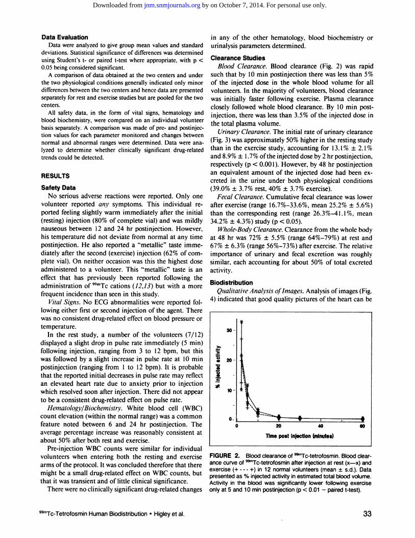
ClearanceStudiesBlood Clearance. Blood clearance (Fig. 2) was rapid
such that by 10 mm postinjection there was less than 5%of the injected dose in the whole blood volume for allvolunteers. In the majority of volunteers, blood clearancewas initially faster following exercise. Plasma clearanceclosely followed whole blood clearance. By 10 mm postinjection, there was less than 3.5% of the injected dose inthe total plasma volume.
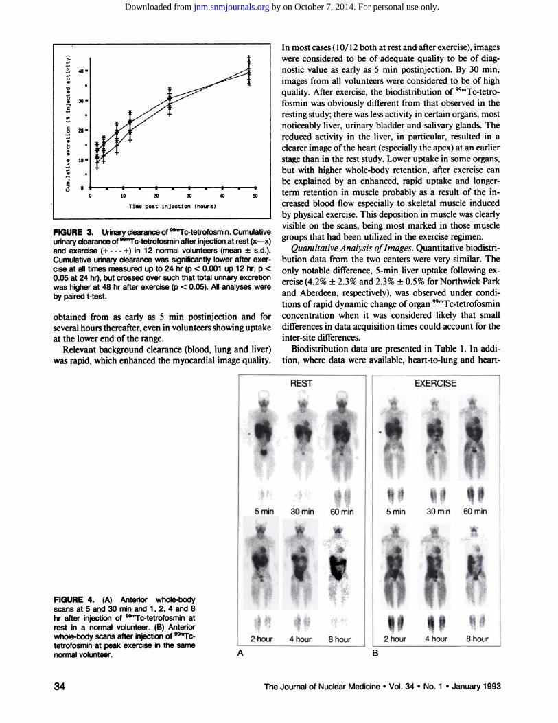
Urinarv Clearance. The initial rate of urinary clearance(Fig. 3) was approximately 50% higher in the resting studythan in the exercise study, accounting for 13. 1% ±2. 1%and 8.9% ±1.7% ofthe injected dose by 2 hr postinjection,respectively (p < 0.001). However, by 48 hr postinjectionan equivalent amount of the injected dose had been cxcreted in the urine under both physiological conditions(39.0%±3.7% rest,40% ±3.7%exercise).
Fecal Clearance. Cumulative fecal clearance was lowerafter exercise (range l6.7%—33.6%,mean 25.2% ±5.6%)than the corresponding rest (range 26.3%—41. 1%, mean34.2% ±4.3%) study (p < 0.05).
Whole-Body Clearance. Clearance from the whole bodyat 48 hr was 72% ±5.5% (range 64%—79%) at rest and67%±6.3%(range56%—73%)afterexercise.Therelativeimportance of urinary and fecal excretion was roughlysimilar, each accounting for about 50% of total excretedactivity.
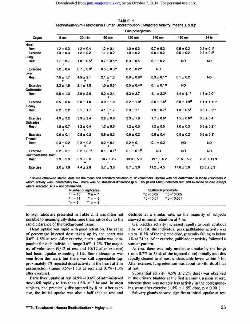
BiodistributionQualitative Analysis oflmages. Analysis of images (Fig.
4) indicated that good quality pictures of the heart can be
FIGURE2. Bloodclearanceof @“Tc-tetrofosmin.Bloodclearancecurve of @“Tc-tetrofosminafter injectionat rest (x—x)andexercise(+ - - - +) in 12 normal volunteers(mean±s.d.). Datapresentedas% injectedactivityinestimatedtotalbloodvolume.Activity in the blood was significantly lower following exerciseonly at 5 and 10 mmpostinjection(p < 0.01 —pairedt-test).
30
U@20
.@
2
0.20 40 SO
TimepostInjection@mlnutes)
99mTcTetrofosminHuman Biodistribution •Higley et al. 33
by on October 7, 2014. For personal use only. jnm.snmjournals.org Downloaded from
In most cases (10/12 both at rest and after exercise), imageswere considered to be of adequate quality to be of diagnostic value as early as 5 mm postinjection. By 30 mm,images from all volunteers were considered to be of highquality. After exercise, the biodistribution of 99mTc@tetro@fosmin was obviously different from that observed in theresting study; there was less activity in certain organs, mostnoticeably liver, urinary bladder and salivary glands. Thereduced activity in the liver, in particular, resulted in aclearer image ofthe heart (especially the apex) at an earlierstage than in the rest study. Lower uptake in some organs,but with higher whole-body retention, after exercise canbe explained by an enhanced, rapid uptake and longerterm retention in muscle probably as a result of the increased blood flow especially to skeletal muscle inducedby physical exercise. This deposition in muscle was clearlyvisible on the scans, being most marked in those musclegroups that had been utilized in the exercise regimen.
Quantitative Analysis oflmages. Quantitative biodistribution data from the two centers were very similar. Theonly notable difference, 5-mm liver uptake following exercise (4.2% ±2.3% and 2.3% ±0.5% for Northwick Parkand Aberdeen, respectively), was observed under conditions of rapid dynamic change of organ @mTc@tetrofosminconcentration when it was considered likely that smalldifferences in data acquisition times could account for theinter-site differences.
Biodistnbution data are presented in Table 1. In addition, where data were available, heart-to-lung and heart
REST
4,>
4,C)
V
4,00
C
C0
4,0
0x0
0>
4,0
E
0
4O@
20•
to
0 10 20 30 40 50
Time post injection (hours)
FIGURE3. Urinaryclearanceof @“Tc-tetrofosmin.Cumulativeurinaryclearanceof@―Tc-tetrofosminafter injectionat rest (x—x)and exercise (+ - - - +) in 12 normal volunteers (mean ±s.d.).Cumulativeurinaryclearancewas significantlylowerafterexerelse at all times measured up to 24 hr (p < 0.001 up 12 hr, p <0.05at24hr),butcrossedoversuchthattotalunnaryexcretionwas higherat 48 hr afterexercise(p < 0.05). Allanalyseswereby pairedt-test.
obtained from as early as 5 mm postinjection and forseveral hours thereafter, even in volunteers showing uptakeat the lower end of the range.
Relevant background clearance (blood, lung and liver)was rapid, which enhanced the myocardial image quality.
EXERCISE
,$@5 mm 30 mm 60 mm
@ * A@* @r
ft “2hour 4hour 8hour
FIGURE4. (A) Anteriorwhole-bodyscansat 5 and30 mmand1, 2, 4 and8hr after injection of @°“Tc-tetrofosminatrest in a normalvolunteer.(B) Anteriorwhole-bodyscansafterinjectionof @‘Tctetrofosminat peak exercise in the samenormalvolunteer. BA
34 The Journal of Nuclear Medicine •Vol. 34 •No. 1 â€J̃anuary 1993
‘jJ5
mm30mm60mm@%
5<.@:
@!‘@*@t,o4.@v@€@2
hour4 hour8 hour
by on October 7, 2014. For personal use only. jnm.snmjournals.org Downloaded from
Timepostinjection
Organ 5 mm 30 mm 60 mm 120 mm 240mm 480mm 24 hr
4 Unless otherwise stated, data are the mean and standard deviation of 1 2 volunteers. Uptake was not determined in those volunteers in
whomactivitywas undetectablylow.Therewas no statisticaldifference(p > 0.05 pairedt-test)betweenrest andexercisestudiesexceptwhereindicated.ND= not determined.
Numberof replicates StatisticalprobabilitytnlO 55n=7 “pcz0.05 ttp<0.0055n=11 **n=6 tp<0.01 ttp.(0.001‘n=9 ***n3
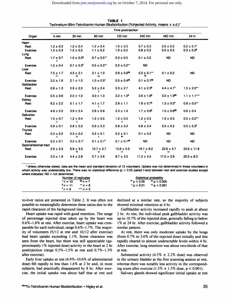
TABLE ITechnetium-99m-Tetrofosmin Human Biodistnbution (%Injected Activity, means ±s.d.)t
HeartRest 1.2±0.3 1.2±0.4 1.2±0.4 1.0±0.3 0.7±0.3 0.5±0.2 0.2±O.l@Exercise 1.3 ±0.3 1.2 ±0.2 1.1 ±0.2 1.0 ±0.2 0.8±0.2 0.5±0.2 0.3±O.2@
LungRest 1.7 ±0.7 1.0 ±O.5@ 0.7 ±O.5'@ 0.3 ±0.5 0.1 ±0.2
ExerciseThyroid
Rest
Exercise
Exercise 1.2 ±0.4 0.7 ±0.3' 0.5 ±0.34* 0.2 ±@ NDLiver
Rest
ExerciseGallbladder
Rest
ExerciseKidney
Rest
ExerciseSalivaries
Rest
7.5 ±1.74.5± 2.12.1± 1.00.9 ±0.6k0.3±0.1@tt5*U‘I3.2
±1.92.1± 1.01.0± [email protected] ±0.4k0.1± 0.1k
NDND0.1
±0.2ND
ND
5.3 ±2.7 4.1 ±2.3@ 4.4 ±4.1' 1.5±2.0*4
3.2 ±i.0@ 2.6 ±1.6@ 3.0±1.9@ 1.1±1.1's'
2.9 ±1.1 1.9±07@ 1.3±0.5' 0.8±@
2.3±1.0 1.7±0.6@ 1.5±0.6's 0.6±0.4
1.2±0.5 1.2±0.5 1.2±0.5
0.8 ±1.32.9 ±2.05.2 ±2.40.5
±0.62.0 ±1.03.0 ±1.06.2
±2.25.1 ±1.74.1 ±1.74.9
±2.23.9 ±2.42.8 ±0.91.5
±0.7S
0.9 ±0.11.2±0.4S
0.8 ±0.20.9 ±0.30.8 ±0.20.8 ±0.41.0±
0.3
0.5 ±0.20.5±
0.2k
0.5±0.3'0.3
±0.2S
0.2 ±0.10.3
±0.2tt
0.2±[email protected]
±0.1S
0.1 ±0.1'0.2
±0.111
0.1 ±[email protected]
±0.2
NDND NDNDND2.9
±2.3
2.0±1.96.9
±3.0@1
4.4±2.810.7
±2.7tt
5.7±3.613.8
±5.0tt
8.7±3.019.1
±6.2S
11.2±4.522.6
±5.7tt
17.0±3.923.6
±11.9
20.5±8.3
GastrointestinaltractRest
Exercise
to-liver ratios are presented in Table 2. It was often notpossible to meaningfully determine these ratios due to therapid clearance of the background tissue.
Heart uptake was rapid with good retention. The rangeof percentage injected dose taken up by the heart wasO.6%—l.8%at rest. After exercise, heart uptake was cornparable for each individual, range O.6%—l.7%. The majority of volunteers (9/12 at rest and 10/12 after exercise)had heart uptake exceeding 1.1%. Some clearance wasseen from the heart, but there was still appreciable (approximately 1% injected dose) activity in the heart at 2 hrpostinjection (range 0.5%—l.5% at rest and 0.7%—l.3%after exercise).
Early liver uptake at rest (4.9%—lO.6% of administereddose) fell rapidly to less than 1.6% at 2 hr and, in mostsubjects, had practically disappeared by 8 hr. After exercisc, the initial uptake was about half that at rest and
declined at a similar rate, so the majority of subjectsshowed minimal retention at 4 hr.
Gallbladder activity increased rapidly to peak at about2 hr. At rest, the individual peak gallbladder activity wasup to 10.7% ofthe injected dose, generally falling to below1% at 24 hr. After exercise, gallbladder activity followed asimilar pattern.
At rest, there was only moderate uptake by the lungs(from 0.7% to 3.0% of the injected dose) initially and thisrapidly cleared to almost undetectable levels within 4 hr.After exercise, lung retention was about two-thirds of thatat rest.
Substantial activity (4.5% ±2.2% dose) was observedin the urinary bladder at the first scanning session at rest,whereas there was notably less activity in the corresponding scans after exercise (1.5% ±1.5% dose, p < 0.001).
Salivary glands showed significant initial uptake at rest
@“Tc-TetrofosminHuman Biodistribution •Higleyet al. 35
I @Trrwr@mq@ riumari DIUUISLIILJUIIUII ni@iey et ai.
by on October 7, 2014. For personal use only. jnm.snmjournals.org Downloaded from
PatientAgeMIno.M/FM (yr)(V/N)
EF(%)# Vess.Dis. EKG G/F
TABLE IClinical Characteristics
CABG(V/N)
123456789
F 48 V V 18M 51 V V 14M 46 V V 10M 52 V N 10M 41 N N 4F 62 N N 23M 34 N N 13F 33 N N 14F 56 N N 14
323310001
Ant.MI/LVH/RBBBAnt.MIAnt.MI/RBBBLVHLVHLBBBAnt.MIAnt./Sept.MI/LVHLBBB
FGFGGFGFG
Ant.= anterior;CABG= coronaryarterybypassgrafting;EF= ejectionfraction;F = fasted;G = glucoseloaded;lnf. = inferior;LBBB=left bundlebranchblock;LVH= left ventricularhypertrophy;Ml = myocardialinfarction;RBBB= rightbundlebranchblock;Sept.= septal;Vess.Dis.= @75%stenosisin anyepicardialcoronaryartery.
positioning to obtain the tomographic transmission and emissionscans. The collimators were set at 65 mm giving a full width athalf maximum (FWHM)resolutionof 16 mm. Three transmission planes were acquired for 10 mm (two direct planes, onecross-plane) for attenuation correction. Ten millicuries (370MBq) of ‘3NH3were administered intravenously and data wereacquired for 10 mm after 3 mm of equilibration. Immediatelyfollowing the perfusion study acquisition, 10 mCi (370 MBq) ofISFT'JO were injected intravenously. After a 30 mm period for
accumulation, emission data were acquired for 10 mm. Patientswerestudiedin the fastingstate (l2 hr fasting)(n = 4) or afterreceiving 50 g of oral glucose (Glucola) 30 mm prior to theinjection of ‘8FDG(n = 5).
PET Image AnalysisPerfusionand metabolic PET imageswere reconstructed,at
tenuation corrected, and transferred onto a SUN III workstationwhere a circumferentialregion of interest (ROI) count densityprofilewasconstructedusingan interactiveprogram.The initialROI(containing21 pixels)wasassignedto theintersectionof theright ventricular free wall, the interventricular septum and theleft ventricular anterior wall at the mid ventricular level (Fig.lA). Contiguous ROIs of the same size were then placed in aclockwise fashion around the circumference of the perfusion andmetabolic PET images. By assigning the first ROI as 0 degrees,the radial anglewasautomaticallycomputed by the programforthe linejoining the geometriccenter of the left ventricularcavitywith the center of the ROl. This method of analysis allowedpaired data of mean count density of all pixels within a givenROlandradialangleto begenerated.Thecircumferentialcountdensity profile for each ROI was then normalized for the ROlwith the highest mean count density. In addition, the PET imageswereinterpretedby two experiencedobserverswithoutbenefitofclinical information. The interpretation of either ISCM orNISCMwasbasedon the uniformityof tracerdistributionin themyocardium.
PathologyThe hearts were removedat the time of transplantation and
placed in phosphate-bufferedformalin for approximately48 hrto allowfixation.A physicianfrom the PET laboratorywith thestudy pathologistoriented the heart in its anatomic position.While viewing the transaxial PET images, they selected a mid
ventricular transaxial slice corresponding to a PET imaging level.This slice was then divided into eight or nine cross sections whichwere processed for routine histology. Two paraffin embeddedsections were prepared from each tissue block; one was stainedwith hematoxylin and eosin, the other with a Masson trichromestain, and the circumferential transmural extent of the infarctedtissue was determined (Figs. lB—C).Beginning at the same starting point of 0 degrees established in the PET ROl analysis, thepercent of noninfarcted tissue was determined by planimetry ineach 10-degree sector (Fig. lB). Histologic myocardial infarctionwas defined as dense, confluent areas of fibrosis with loss ofnormal myocardial architecture.
Data AnalysisPair-wisecorrelation,using Spearman rank order correlation
coefficients (for nonparametric data), was used to correlate normalized 18pj@jand ‘3NH3ROI count density with percent noninfarcted tissue at similar radial locations (±5degrees) aroundthe circumferenceof the myocardium.
RESULTS
There were four patients in the 15CM group and fivepatients in the NISCM group. The average age of the15CMgroup was 49 yr (range 46—52)and for the NISCMgroup 45 yr (range 33—62).All patients in the 15CM grouphad a history of one or more documented myocardialinfarctions and three of four patients had EKG criteria (Qwaves) of myocardial damage. The mean EF was 13% forboth the 15CM and NISCM groups (Table 1). No historyofprior myocardial infarctions were present in the patientswith NISCM, although two patients had anterior precordial Q-waves. All patients with 15CM had significant coronary artery disease pathologically (>75% stenosis in atleast one epicardial coronary artery). None of the fivepatients with NISCM had angiographic evidence of significant (>75%) coronary artery disease, but two patients hadpathologic evidence of significant one-vessel coronary artery disease. Neither of these patients had a totally occluded vessel.
Pathologic examination of all hearts with NISCM revealed no gross or histologic evidence of myocardial in
by on October 7, 2014. For personal use only. jnm.snmjournals.org Downloaded from
Time postinjection
Organ 5 mm 30 mm 60 mm i 20 mm 240mm 480mm 24 hr
TABLE 1Technetium-99m-Tetrofosmin Human Biodistribution (%Injected Activity, means ±s.d.)t
HeartRest1.2±0.31.2 ±0.41.2± 0.41.0 ±0.30.7 ±0.30.5 ±0.20.2 ±O.VExercise1.3±0.31.2±0.21.1±0.21.0±0.20.8±0.20.5±0.20.3±0.2k
LungRest 1.7±0.7 1.0±0.5@ 0.7 ±0.54* 0.3 ±0.5 0.1±0.2
S
1.2 ±0.4 0.7 ±0.3' 0.5 ±0.34* 0.2 ±0.2**
4 Unless otherwise stated, data are the mean and standard deviation of 1 2 volunteers. Uptake was not determined in those volunteers in
whomactivitywasundetectablylow.Therewasnostatisticaldifference(p> 0.05 pairedt-test)betweenrestandexercisestudiesexceptwhereindicated.ND= notdetermined.
Numberof replicates StatisticalprobabilitytnlO @5n=7 “p<0.05 ttp<0.0055n=11 ttn=6 tp<0.01 ttp<O.OOl‘n=9 44tn3
NDND0.1±0.2ND
Exercise NDLiver
Rest 7.5 ±1.7 4.5 ±2.1 2.1 ±1.0 0.9 ±0.6w 0.3±0.1@tt St 5* 11
Exercise 3.2 ±1.9 2.1 ±1.0 1.0±O.5@ 0.5 ±0.4w 0.1±0.1k NDGallbladder
Rest 0.8 ±1.3 2.9 ±2.0 5.2 ±2.4 5.3 ±2.7 4.1 ±2.3@ 4.4±4.1' 1.5±2.0**
Exercise 0.5 ±0.6 2.0±1.0 3.0 ±1.0 3.2 ±1.0@ 2.6±1.6@ 3.0±1.9k@@Kidney
Rest 6.2 ±2.2 5.1±1.7 4.1 ±1.7 2.9 ±1.1 1.9±0.7@ 1.3±0.5' 0.8±0.54*
Exercise 4.9 ±2.2 3.9±2.4 2.8 ±0.9 2.3 ±1.0 1.7±0.6@ 1.5±O.6@ 0.6±0.4Salivanes
Rest 1.5 ±0.7 1.2±0.4 1.2 ±0.5 1.2 ±0.5 1.2 ±0.5 1.0±0.3 0.5±0.24*S S S S
Exercise 0.9 ±0.1 0.8 ±0.2 0.9 ±0.3 0.8 ±0.2 0.8 ±0.4 0.5±0.2 0.5±0.3'Thyroid
Rest 0.3 ±0.2 0.3±0.2 0.2 ±0.1 0.2 ±0.1 0.1 ±0.2 ND NDS tt S 11
Exercise 0.2 ±0.1 0.2 ±O.1@ 0.1 ±0.1' 0.1 ±0.1k ND ND NDGastrointestinaltract
Rest 2.9 ±2.3 6.9 ±3.0 10.7±2.7 13.8±5.0 19.1±6.2 22.6±5.7 23.6±11.9¶1 tt tt S tt
Exercise 2.0 ±1.9 4.4 ±2.8 5.7 ±3.6 8.7 ±3.0 11.2±4.5 17.0±3.9 20.5±8.3
to-liver ratios are presented in Table 2. It was often notpossible to meaningfully determine these ratios due to therapid clearance of the background tissue.
Heart uptake was rapid with good retention. The rangeof percentage injected dose taken up by the heart was0.6%—l.8%atrest.Afterexercise,heartuptakewascornparable for each individual, range 0.6%—l.7%. The majority of volunteers (9/12 at rest and 10/12 after exercise)had heart uptake exceeding 1.1%. Some clearance wasseen from the heart, but there was still appreciable (approximately 1% injected dose) activity in the heart at 2 hrpostinjection (range 0.5%—l.5% at rest and 0.7%—l.3%after exercise).
Early liver uptake at rest (4.9%—lO.6%of administereddose) fell rapidly to less than 1.6% at 2 hr and, in mostsubjects, had practically disappeared by 8 hr. After exercise, the initial uptake was about half that at rest and
declined at a similar rate, so the majority of subjectsshowed minimal retention at 4 hr.
Gallbladder activity increased rapidly to peak at about2 hr. At rest, the individual peak gallbladder activity was
up to 10.7% ofthe injected dose, generally falling to below1% at 24 hr. After exercise, gallbladder activity followed asimilar pattern.
At rest, there was only moderate uptake by the lungs(from 0.7% to 3.0% of the injected dose) initially and thisrapidly cleared to almost undetectable levels within 4 hr.After exercise, lung retention was about two-thirds of thatat rest.
Substantial activity (4.5% ±2.2% dose) was observedin the urinary bladder at the first scanning session at rest,whereas there was notably less activity in the corresponding scans after exercise (1 .5% ±1.5% dose, p < 0.001).
Salivary glands showed significant initial uptake at rest
@“Tc-TetrofosminHuman Biodistribution •Higleyet al. 35
by on October 7, 2014. For personal use only. jnm.snmjournals.org Downloaded from
Timeafterinjection(mm)5
3060 120
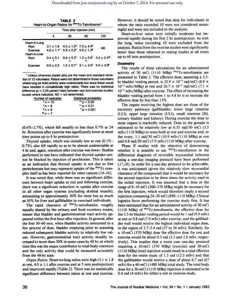
However, it should be noted that data for individuals inwhom the ratio exceeded 10 were not considered meaningful and were not included in the analysis.
Heart-to-liver ratios were initially moderate but improved rapidly during the first 2 hr postinjection. As withthe lung, ratios exceeding 10 were excluded from the
ND analysis. Ratios from the exercisestudies were significantlybetter than those obtained in resting studies at all timesup to 60 mm postinjection.
TABLE2Heart-to-Organ Ratios for@
Heart-to-LungRestExercise
Heart-to-LiverRest
3.1±1.8 4.5 ±1•5*7.3±[email protected]±l.l@ 5.9 ±[email protected]±1.3k
0.4±0.1 0.6 ±[email protected]±[email protected] ±@I ** tt
4 Unless otherwise stated data are the mean and standard devia
tionof 12volunteers.Ratioswerenotdeterminedinthosevolunteerswherelung(orliver)activitywereundetectablylowsincethesewouldhaveresultedin unrealisticallyhighratios.Therewas no statisticaldifference(p> 0.05pairedt-test)betweenrestandexercisestudiesexceptwhereindicated.ND= notdetermined.
Numberof replicates StatisticalprobabilitytnlO @p<0.05n=8 ttp<0.01@n=6 ‘p<0.001
5* n = 7
(0.6%—2.7%), which fell steadily to less than 0.7% at 24hr. Retention after exercise was significantly lower at mosttime points up to 8 hr postinjection.
Thyroid uptake, which was initially low at rest (0.1%—0.7%), also fell rapidly so as to be almost undetectable at4 hr and, again, retention after exercise was lower. Studiesperformed in rats have indicated that thyroid uptake cannot be blocked by injection of perchlorate. This is takenas an indication that thyroid uptake is not due to freepertechnetate but may represent uptake of the 99mTccornplex itself as has been reported for other cations (14—16).
It was noted that, while there was no significant difference between heart uptake at rest and following exercise,there was a significant reduction in uptake after exercisein all other organ systems (excluding skeletal muscle),amounting to approximately 30% for most but as muchas 50% for liver and gallbladder in exercised individuals.
The rapid clearance of 99mTc@tetrofosmin,roughlyequally shared by the urinary and fecal excretory routes,meant that bladder and gastrointestinal tract activity appeared within the first hour after injection. In general, afterthe first 30—60mm, when bladder activity amounted to afew percent of dose, bladder emptying prior to scanningreduced subsequent bladder activity to relatively low values. However, gastrointestinal tract activity steadily increased to more than 30% in some cases by 48 hr, at whichtime this was the major contributor to total-body retentionand the only activity that could be measured accuratelyfrom the 48-hr scan.
Organ Ratios. Heart-to-lung ratios were high (3. 1 ±1.8at rest, 4.0 ±1.1 after exercise and at 5 mm postinjection)and improved rapidly (Table 2). There was no statisticallysignificant difference between ratios at rest and exercise.
DosimetryThe results of these calculations for an administered
activity of 30 mCi (1 110 MBq) 99mTc@tetrofosmin arepresented in Table 3. The effective dose, assuming a 3.5-hr bladder voiding period, is 32.9 x l0@ rad/mCi (8.9 xl0'@mSv/MBq) at restand 26.7 x l0@ rad/mCi (7.1 xl0'@mSv/MBq)afterexercise.Theeffectof increasingthebladder voiding period from 1 to 4.8 hr is to increase theeffective dose by less than 13%.
The organs receiving the highest dose are those of theexcretory pathways (gallbladder, lower large intestine(LLI), upper large intestine (ULI), small intestine (SI),urinary bladder and kidney). During exercise the dose tomost organs is markedly reduced. Dose to the gonads isestimated to be relatively low at 0.35 rad/30 mCi (3.5mSv/l 110 MBq) to testis both at rest and exercise and, tothe ovary, 1. 1 rad/30 mCi ( 10.9 mSv/ 1110 MBq) at restand 0.8 rad/30 mCi (7.9 mSv/l 110 MBq) after exercise.
Phase II studies with the objective of determiningwhether it is possible to use 99mTc@tetrofosminin thedifferential diagnosis of reversible myocardial ischemiausing a one-clay imaging protocol have been performed(1 7,18). In order for a one-day protocol to be achievable,it was anticipated (given the relatively slow myocardialclearance of the compound) that it would be necessary forthe second injection to be three times the activity used inthe initial injection. It was assumed that a dose in therange of8—lOmCi (300—370MBq) might be necessary forthe first injection, which would therefore imply a secondinjection containing 24—30mCi (890—1110 MBq). Clinicallogistics favor performing the exercise study first. It hasbeen estimated that for an administered activity of 30 mCi(1 110 MBq) of 99mTc..tetrofosmifl the effective dose forthe 3.5-hr bladder voiding period would be 1 rad (9.9 mSv)at rest or 0.8 rad (7.9 mSv) after exercise, and the gallbladder wall would receive the highest individual organ dose,in the region of 3.7—5.4rad (37 to 54 mSv). Similarly, fora 10-mCi (370 MBq) dose the effective dose for rest andexercise would be about 0.3 rad (3.3 and 2.6 mSv, respectively). This implies that a worst case one-day protocolrequiring a 10-mCi (370 MBq) (exercise) and 30-mCi(1 110 MBq) (rest) injection would result in a total effectivedose for the entire study of 1.3 rad (12.5 mSv) and thatthe gallbladder would receive a dose of about 6.7 rad (67mSv) for a 40-mCi ( 1480 MBq) total study. The total bodydose for a 30-mCi ( 1110 MBq) injection is estimated to be0.4 rad(4 mSv)foreithera restor exercisestudy.
Exercise 0.8±0.3 1.2 ±[email protected] ±[email protected] ±3.0@
36 The Journal of Nuclear Medicine•Vol.34 •No. 1 •January 1993
by on October 7, 2014. For personal use only. jnm.snmjournals.org Downloaded from
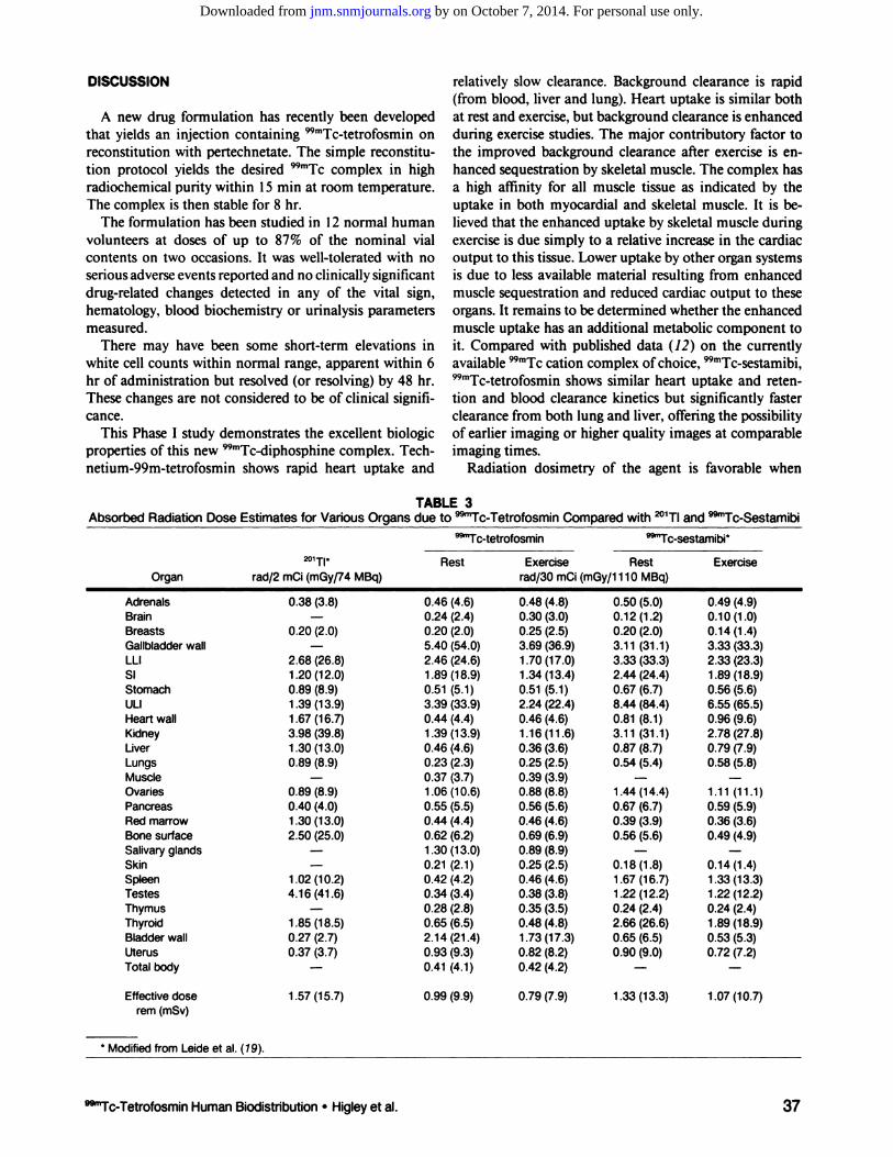
Absorbed Radiation Dose Estimates for Various OrganTABLE3
s due to @“Tc-TetrofosminComparedwith @°1TIand@“Tc-Sestamibi201T1'@“Tc-tetrofosmin
@“Tc-sestamibiRest
Exercise RestExerciseOrganrad/2mCi(mGy/74MBq)rad/30 mCi(mGy/1110MBq)Adrenals
0.38(3.8)0.46(4.6) 0.48(4.8) 0.50(5.0) 0.49(4.9)Brain—0.24(2.4) 0.30(3.0) 0.12(1.2)0.10(1.0)Breasts
0.20(2.0)0.20(2.0) 0.25(2.5) 0.20(2.0) 0.14(1.4)Gallbladderwall —5.40 (54.0) 3.69(36.9) 3.11(31.1)3.33(33.3)LLI
2.68(26.8)2.46(24.6) 1.70(17.0) 3.33(33.3) 2.33(23.3)SI1.20(12.0)1 .89(18.9) 1.34(13.4) 2.44(24.4)1.89(18.9)Stomach0.89(8.9)0.51 (5.1) 0.51(5.1) 0.67(6.7) 0.56(5.6)ULI1.39(13.9)3.39 (33.9) 2.24(22.4) 8.44(84.4) 6.55(65.5)Heart
wall 1.67 (16.7)0.44 (4.4) 0.46(4.6) 0.81(8.1)0.96(9.6)Kidney3.98(39.8)1.39 (13.9) 1.16(11.6) 3.11(31.1)2.78(27.8)Liver1.30(13.0)0.46 (4.6) 0.36(3.6) 0.87(8.7)0.79(7.9)Lungs0.89(8.9)0.23(2.3) 0.25(2.5) 0.54(5.4) 0.58(5.8)Muscle
—0.37(3.7) 0.39(3.9) ——Ovaries0.89(8.9)1 .06(10.6) 0.88(8.8) 1.44(14.4) 1.11 (11.1)Pancreas0.40(4.0)0.55(5.5) 0.56(5.6) 0.67(6.7) 0.59(5.9)Red
marrow 1.30(13.0)0.44 (4.4) 0.46(4.6) 0.39(3.9)0.36(3.6)Bonesurface2.50(25.0)0.62(6.2) 0.69(6.9) 0.56(5.6) 0.49(4.9)Salivary
glands —1 .30(13.0) 0.89(8.9) ——Skin—0.21 (2.1) 0.25(2.5) 0.18(1.8) 0.14(1.4)Spleen
1.02(10.2)0.42 (4.2) 0.46(4.6) 1.67(16.7) 1.33(13.3)Testes4.16(41.6)0.34(3.4) 0.38(3.8) 1.22(12.2) 1.22(12.2)Thymus
—0.28 (2.8) 0.35(3.5) 0.24(2.4)0.24(2.4)Thyroid1.85(18.5)0.65(6.5) 0.48(4.8) 2.66(26.6) 1.89(18.9)Bladderwall0.27(2.7)2.14(21.4) 1.73(17.3) 0.65(6.5) 0.53(5.3)Uterus0.37(3.7)0.93(9.3) 0.82(8.2) 0.90(9.0)0.72(7.2)Total
body —0.41 (4.1) 0.42(4.2) ——Effective
dose 1.57(15.7)0.99 (9.9) 0.79(7.9) 1.33(13.3) 1.07(10.7)rem(mSv)*
Modified from Leide et al. (19).
DISCUSSION
A new drug formulation has recently been developedthat yields an injection containing 99mTc..tetrofosminonreconstitution with pertechnetate. The simple reconstitution protocol yields the desired 99mTccomplex in highradiochemical purity within 15 mm at room temperature.The complex is then stable for 8 hr.
The formulation has been studied in 12 normal humanvolunteers at doses of up to 87% of the nominal vialcontents on two occasions. It was well-tolerated with noserious adverse events reported and no clinically significantdrug-related changes detected in any of the vital sign,hematology, blood biochemistry or urinalysis parametersmeasured.
There may have been some short-term elevations inwhite cell counts within normal range, apparent within 6hr of administration but resolved (or resolving) by 48 hr.These changes are not considered to be of clinical significance.
This Phase I study demonstrates the excellent biologicproperties of this new 99mTc..@Jipho5phiflecomplex. Technetium-99m-tetrofosmin shows rapid heart uptake and
relatively slow clearance. Background clearance is rapid(from blood, liver and lung). Heart uptake is similar bothat rest and exercise, but background clearance is enhancedduring exercise studies. The major contributory factor tothe improved background clearance after exercise is enhanced sequestration by skeletal muscle. The complex hasa high affinity for all muscle tissue as indicated by theuptake in both myocardial and skeletal muscle. It is believed that the enhanced uptake by skeletal muscle duringexercise is due simply to a relative increase in the cardiacoutput to this tissue. Lower uptake by other organ systemsis due to less available material resulting from enhancedmuscle sequestration and reduced cardiac output to theseorgans. It remains to be determined whether the enhancedmuscle uptake has an additional metabolic component toit. Compared with published data (12) on the currentlyavailable 99mTccation complex ofchoice, 99mTc@sestamibi,99mTctetrofosmifl shows similar heart uptake and retention and blood clearance kinetics but significantly fasterclearance from both lung and liver, offering the possibilityof earlier imaging or higher quality images at comparableimaging times.
Radiation dosimetry of the agent is favorable when
@“Tc-TetrofosminHumanBiodistnbution•Higleyet al. 37
by on October 7, 2014. For personal use only. jnm.snmjournals.org Downloaded from
4. Lahiri A, Higley B, Kelly JD, et al. Myocardial perfusion imaging in manusing new ttmTc functionalised diphosphine complexes. Eur J Nuci Med1989;15:425.
5. Loevinger R, Berman M. A revised schema for calculating the absorbeddose from biologically distributed radionuclides. MIRD pamphlet no. 1,revised. New York: Society ofNuclear Medicine; 1976.
6. International Commission on Radiological Protection. Limits for intakes
ofradionuclidesbyworkers.JCRPPublication30:Part 1.Oxford:Pergamon; 1979.
7. Cloutier RL, Smith SA, Watson EE, Snyder WS, Warner 0G. Dose to thefetus from radionuclides in the bladder. Health Phys 1973;25:147—161.
8. International Commission on Radiological Protection. Radiation dose topatientsfrom radiopharmaceuticals.JCRPPublication 53.Ann ICRPI8(1—4). Oxford: Pergamon; 1988.
9. SmithT, LahiriA, GemmellHG, et al. Dosimetryof@@mTc@tetrofosmin,anew myocardial perfusion imaging agent. In: wauon EE, Schlafke-StelsonAT, eds. Proceedings of 5th international radiopharmaceutical dosimetrysymposium. Oak Ridge Associated Universities, CONF 910529. 1992:467—481.
10. Snyder WS, Ford MR, warner OG, Watson SB. “5―absorbed dose perunit cumulated activity for selected radionuclides and organs. MIRDpamphlet no. 11. New York: Society of Nuclear Medicine; 1975.
1 1. International Commission on Radiological Protection. Recommendations
ofthe ICRP.ICRP Publication60.AnnJCRP2J(I-3). Oxford:Pergamon;1991.
12. Wackers FiT, Berman DS, Maddahi J, et al. Technetium-99m hexakis 2-methoxyisobutyl isonitrile: human biodistribution, dosimetry, safety andpreliminary comparison to thallium-201 for myocardial perfusion imaging.JNuclMed 1989;30:301—311.
13. Zanelli GD, Lahiri A, Patel N, et al. Animal and human studies ofa new‘@Tc-1abelledphosphine-isocyanide complex with possible applications toradionuclide ventriculography. Eur J Nucl Med 1987;l3:12—17.
14. Oster ZH, Strauss HW, Harrison K, Burns HD, Pitt B. Thallium-201distribution in the thyroid: relationship to thyroidal trapping function.Radiology 1978;l26:733—734.
15. Ramanathan P. Patel RB, Subrahmanyam N, Nayak UN, Sachdev SS,Ramamoorthy N. visuali@tion ofsuppressed thyroid tissue by technetium99m-tertiary butyl isonitrile: an alternative to post-TSH stimulation scanning.JNuclMed 1990;3l:1163—1165.
16. Civelek AC, Durski K, Shafique I, Matsumura K, et al. Failure of perchlorate to inhibit Tc-99m isonitrile binding by the thyroid during myocardialperfusion studies. Clin NuciMed 1991;16:358—361.
17. Sridhara as, Braat 5, Itti R, Rigo P. Cload P. Lahiri A. Early and latemyocardial imaging with a new technetium-99m diphosphine (PPN1O1 1)in coronary artery disease[Abstract]. J Am Coil Cardiol 1992:in press.
18. Rigo P, Braat 5, Itti R, et al. Myocardial imaging with technetium P53.Comparison with thallium in suspected coronary artery disease [Abstract].JAm CoilCardiol1992:inpress.
19. Leide 5, Diemer H, Ahlgren L, Mattsson S. In vivo distribution anddosimetry of Tc-99m mibi in man. In: Watson EE, Schlafke-Stelson AT,eds. Proceedings of5th international radiopharmaceutical dosimetry symposium. Oak Ridge Associated Universities, CONF 910529. 1992:483—497.
compared with that reported for 201Tl(6), allowing in theworst case scenario for 99mTctetrofosmifl (i.e. at rest) theadministration of up to 20 times the thallium activity fora similar effective dose (Table 3). The more rapid excretioncompared with @mTc@sestamibi(12) results in a differentand in many respects more favorable dosimetry patternfor [email protected] doses to gallbladder, brain, redmarrow and urinary bladder are elevated as are the dosesto breast, bone surface, skin, thymus and uterus afterexercise. The doses to all other organs, including ovaries,testes, SI, ULI, LLI, liver, lungs, kidneys, spleen andthyroid, are significantly decreased for 99mTc@tetrofosmincompared with those reported for 99mTc@sestamibi(19).
This study indicates that the agent is capable of producing high quality diagnostic images of the heart. It wasconcluded that the formulation warranted further investigation in patients to determine its value in the diagnosisof ischemic heart disease. The agent has now successfullyundergone a Phase II trial to compare one- and two-dayimaging protocols for the differential diagnosis of infarctfrom reversible ischemia in patients with ischemic heartdisease (1 7,18). It is currently in Phase III clinical trials,which compare SPECT and planar imaging for a similarindication.
38 The Journalof NuclearMedicine•Vol. 34 •No. 1 •January1993
ACKNOWLEDGMENTSWe wish to thank A.M. Forster, C.M. Archer, R.D. Pickett,
J.A. Marriott, B. Edwards, K.R. Nagle, P.S. Weisner, E.A. Platts,H.K. Gill and numerous other colleagues at Amersham International, as wellas technical and clinicalcolleaguesat NorthwickPark Hospital and Aberdeen Royal Infirmary, without whoseefforts these studies would not have been possible.
REFERENCES1. Kaul 5. A look at 15 years of planar thallium imaging. Am Heart J 1989;
I 18:581—601.2. StraussHW,PittB.Thallium-201asamyocardialimagingagent.Semin
Nuc/Med 1977;7:3—7.3. KellyJD, ForsterAM,HigleyB,et al. Technetium-99mphosphino-ether
complexes: technetium-99m tetrofosmin as a new radiopharmaceutical formyocardialperfusionimaging.J NuciMed 1992:inpress.
by on October 7, 2014. For personal use only. jnm.snmjournals.org Downloaded from