targeting sensory axon regeneration in adult spinal cord
TRANSCRIPT

Development/Plasticity/Repair
Targeting Sensory Axon Regeneration in Adult Spinal Cord
Xiao-Qing Tang, Paula Heron, Charles Mashburn, and George M. SmithDepartment of Physiology, Spinal Cord and Brain Injury Research Center, University of Kentucky Chandler Medical Center, Lexington, Kentucky 40536-0298
Extensive regeneration of sensory axons into the spinal cord can be achieved experimentally after dorsal root injury, but no effort hasbeen made to target regenerating axons and restore a normal lamina-specific projection pattern. Ectopic axon growth is potentiallyassociated with functional disorders such as chronic pain and autonomic dysreflexia. This study was designed to target regeneratingaxons to normal synaptic locations in the spinal cord by combining positive and negative guidance molecules. Previously, we observedthat, after dorsal rhizotomy, overexpression of NGF leads to robust regeneration and sprouting of calcitonin gene-related peptide(CGRP)-positive nociceptive axons throughout dorsal horn and ventral horns. To restrict these axons within superficial laminas, adeno-virus expressing semaphorin 3A was injected into the ventral spinal cord 3 d after NGF virus injection. Semaphorin 3A expression wasobserved in deep dorsal and ventral cord regions and limited axon growth to laminas I and II, shaping axonal regeneration toward thenormal distribution pattern. NGF and semaphorin 3A treatment also targeted the regeneration of substance P-positive nociceptive axonsbut had no effect on injured isolectin B4-binding nociceptive axons. Axon regeneration led to functional restoration of nociception inboth NGF- and NGF/semaphorin 3A-treated rats. Although no significant difference in behavior was found between these two groups,confocal microscopy illustrated ectopic synaptic formations in deeper laminas in NGF/green fluorescent protein-treated rats. The resultssuggested that antagonistic guidance cues can be used to induce and refine regeneration within the CNS, which is important for long-term, optimal functional recovery.
Key words: semaphorin 3A; nerve growth factor; primary nociceptive axons; regeneration; axon guidance; lamina specific
IntroductionParamount to functional recovery is reestablishment of synapticconnections to the most appropriate target region. In many re-gions of the CNS, laminar specificity is an important determinantof synaptic specificity (Sanes and Yamagata, 1999). Reestablish-ment of laminar specificity should allow higher-order plasticityto modify circuits and correct slight targeting errors. With this inmind, can regenerating axons in the adult be targeted to laminar-specific regions and will this increase synaptic density within thatlamina? Studies showed that receptors of some developmentallyimportant guidance cues continue to operate in adulthood; thus,it may be possible to exogenously provide essential guidance sup-port after adult CNS injury. Conversely, difficulty has remainedfor introducing guidance molecules in a temporal and spatialmanner conducive to restoring the exquisite organization of spe-cific neuronal pathways. To this end, we incorporated a develop-mental repulsive guidance mechanism (Kolodkin, 1996) into ourregeneration strategy (Romero et al., 2001; Tang et al., 2004a) toaid the patterning of connections during sensory axon regenera-tion. The refined strategy uses both positive and negative cues to
generate a dorsal gradient to induce axon regeneration that di-minishes ventrally into a chemorepulsive gradient to limit regen-eration to the upper dorsal lamina of the spinal cord.
Developmental growth and guidance of sensory axonsthrough the dorsal roots and into the spinal cord has been studiedextensively. These axons enter the spinal cord from the root andterminate at distinct laminas in spinal cord gray matter. Differentsubtypes of sensory axons show prominent lamina-specific con-nectivity: calcitonin gene-related peptide (CGRP)- and substanceP (SP)-positive (�) nociceptors restrict their terminals to laminaI and the outer aspect of lamina II (IIo), isolectin B4 (IB4)-binding nociceptors to the inner aspect of lamina II (IIi), andTrkC-positive proprioceptors to Clark’s column and the ventralhorn. After dorsal root injury in adult animals, sensory axonsregenerate within the dorsal root but will stop abruptly after con-tact with the surface of the spinal cord. Features of the dorsal rootentry zone (DREZ) (a specialized transitional zone between thespinal cord and the dorsal root) generate a nonpermissive barrierfor regenerating sensory axons analogous to regenerative obsta-cles present after other CNS injuries (Pindzola et al., 1993; Alds-kogius and Kozlova, 2002; Priestley et al., 2002). These featuresmake dorsal root injury a suitable and simplified model to studyCNS regeneration and restoration of specific projection patterns.Previous studies show that combined zymosan and chondroiti-nase ABC treatment or the application of neurotrophins facili-tates functional regeneration of sensory axon through the DREZand into the spinal cord (Ramer et al., 2000; Romero et al., 2001;Steinmetz et al., 2005). The best regeneration has been observedwith nociceptive C-fiber axons (CGRP- and SP-containing) in
Received Oct. 24, 2006; accepted April 25, 2007.This work was supported by National Institute of Neurological Disorders and Stroke Grant NS 38126 and the
Kentucky Spinal Cord and Head Injury Trust. We thank Aileen P. Lim and Jun Yang for technical assistance and imagequantification.
Correspondence should be addressed to Dr. George M. Smith, Department of Physiology, Spinal Cord and BrainInjury Research Center, University of Kentucky Chandler Medical Center, Lexington, KY 40536-0298. E-mail:[email protected].
DOI:10.1523/JNEUROSCI.1442-07.2007Copyright © 2007 Society for Neuroscience 0270-6474/07/276068-11$15.00/0
6068 • The Journal of Neuroscience, May 30, 2007 • 27(22):6068 – 6078
the presence of nerve growth factor (NGF). Notably, these regen-erating C-fibers fail to terminate at their normal targets (laminasI and IIo) and regenerate throughout the entire dorsal horn (Ro-mero et al., 2001; Tang et al., 2004a). Because adult C-fibersrespond to semaphorin 3A (Sema3A), which is thought to pre-vent their growth into deeper dorsal laminas during develop-ment, selective coexpression of NGF and sema3A was used totarget a lamina-restricted connection pattern for injured CGRP�
and SP� nociceptive afferents.
Materials and MethodsAnimals. Fifty-four adult (250 –350 g) female Sprague Dawley rats (Har-lan Sprague Dawley, Indianapolis, IN) were used in this study. All surgi-cal procedures and animal maintenance complied with the National In-stitutes of Health guidelines regarding the care and use of experimentalanimals and were approved by the Institutional Animal Care and Re-search Advisory Committee.
Adenoviral vectors. Replication-defective recombinant adenoviruses(Ad) expressing nerve growth factor (NGF/Ad), green fluorescent pro-tein (GFP/Ad, control virus) and semaphorin 3A (Sema3A/Ad) wereconstructed as described previously (Romero et al., 2000; Tang et al.,2004a). In addition to standard deletions in E1 and E3 coding regions, allviruses also encoded the ts125 mutation in the E2a, DNA binding pro-tein, to further reduce potential toxicity after administration in vivo (Ro-mero and Smith, 1998). All plaque-purified adenoviruses were examinedfor replication-competent adenoviruses via PCR. Viruses were then am-plified and purified by double cesium chloride gradient ultracentrifuga-tion. The physical number of viral particles was determined by opticalabsorbency. The number of infectious particles was estimated usingAdeno-X Rapid titer kit (Clontech, Palo Alto, CA) or directly countinggreen fluorescent cells 48 h after transduction of human embryonic kid-ney 293 cells. The quality of these viruses was determined by examiningthe ratio of infectious to total (live and dead) number of virus, which was�1:50 pfu to total virus particles.
Surgical procedures. Deeply anesthetized animals [ketamine (67 mg/kg,i.p.)/xylazine (6.7 mg/kg, i.p.)] underwent a hemilaminectomy at theL1–L2 vertebral segments to expose the lumbar dorsal roots. With the useof #5 Dummond forceps, triple-crush lesions of 10 s each were inflicted attwo sites separated by 3 mm along the L4, L5 afferents at �5– 8 mm fromthe DREZ. The dorsal roots immediately rostral (L3) and caudal (L6) tothe injured site were double crushed and tightly ligated to create a zone ofdennervation immediately rostral and caudal L4/L5. All lesions wereperformed unilaterally on the right side.
Two weeks after dorsal root injury, all animals received a second hemi-laminectomy at the T13–L1 vertebral segments to expose the lumbarspinal cord. For injections of GFP/Ad, NGF�GFP/Ad, orNGF�Sema3A/Ad, the needle was inserted through the intact dura andinto the spinal cord along the L4, L5 DREZ. For the rats receiving acombination of two virus treatments, the second virus (GFP or Sema3A/Ad) was given 3 d after NGF/Ad treatment. For these animals, afterNGF/Ad administration, the spinal cord above the dura was covered witha small piece of stretched Parafilm and a piece of 50% dextran sulfate-soaked Gelfoam to reduce scar formation (Wujek et al., 1991) and facil-itate injections of the second virus. Each virus administration consists ofeight injections (0.3 �l/each, 0.5 mm apart, 0.6 mm deep for NGF/Adand 1.2 mm deep for GFP or Sema3A/Ad) of adenoviral vectors (NGF/Ad, 5 � 10 4 pfu/�l; GFP or Sema3A/Ad, 1.25 � 10 6 pfu/�l) along theDREZ. All injections were performed using a beveled glass micropipettepulled to an external diameter of between 30 and 50 �m. A total of 300 nlwere injected at each site using a nano-injector (World Precision Instru-ments, Sarasota, FL) at a precise depth from the spinal cord dorsal surfaceusing the coordinates on a M3301 fine micromanipulator (Narishige viaWorld Precision Instruments). Viral doses were determined from previ-ous studies and pilot experiments. Before and the next day after eachvirus administration, rats received 100 �g intraperitoneally of a 1:1 com-bined solution of CD-4 (W3/25) and CD-45 (MRC OX-22) antisera totransiently suppress the immune response.
For relesion experiments, right L4 and L5 dorsal roots were reexposed
4 weeks after adenovirus injections (6 weeks after injury). The roots wererelesioned by transection, and the proximal stump was ligated to preventany axonal regeneration.
Western blots. One week after injections, the L4/L5 segment of sham-injured rats injected with NGF, GFP, or Sema3a Adt were processed forWestern blot analysis (n � 2 per group) as described previously (Tang etal., 2004b). Briefly, a 4 mm segment of the dorsal spinal cord containingthe injected region was dissected and immediately frozen. The tissue wasthen homogenized manually with a Dounce in 200 �l of 1% SDS inTris–EDTA buffer with proteinase inhibitors (10 �g/ml aprotinin, 1�g/ml leupeptin, and 1 mM PMSF) and sonicated using a Branson soni-fier 450 (VWR Scientific, West Chester, PA). After centrifuging at 14,000rpm, supernatant was assayed for protein concentration using a BCA kit(Pierce, Rockford, IL) and diluted with 3� Laemmli’s buffer, and 50 �gof protein was loaded for each sample. After running the sample in 14%(for NGF) or 7.5% (for Sema3A) SDS-polyacrylamide gel, proteins weretransferred to nitrocellulose membranes. Membranes were blocked us-ing Odyssey (LI-COR Biosciences, Lincoln, NE) blocking buffer. NGFwas identified by rabbit anti-NGF antibody (1:500; Accurate Chemicals,Westbury, NY), and adenovirus-mediated Sema3A expression was de-tected by mouse anti-myc (9E10, 1:2000; Upstate, Charlottesville, VA),and semaphorin 3A was detected using a rabbit anti-Sema3A (SP1221,1:500; ECM Bioscience, Versailles, KY). After a 2 h incubation in primaryantibody, the membranes were washed five times for 10 min in Tris-buffered saline with 0.1% Tween 20 and incubated in goat anti-rabbit(for NGF and Sema3A) or mouse (for c-myc) IgG (1:7500; Invitrogen,Carlsbad, CA) conjugated with AlexaFluor 680 for 1 h. Membranes werewashed as above, and images were captured using an Odyssey infraredimaging system (LI-COR Biosciences) at 700 nM.
Retrograde tracing. To evaluate neuronal populations regeneratinginto the dorsal horn, two fluorescent tracers were used for the retrogradetracing experiments. All tracer injections were made on the right side,through a beveled glass micropipette connected to a Nanoliter injector.Immediately after L4, L5 double-crush injury, 4 �l of 5% fluorogold (FG)(in 20% DMSO, Biotium, Hayward, CA) was injected into the crush siteto label all lesioned neurons in corresponding DRGs. The animals thenreceived injections of adenoviral vectors 2 weeks after injury, as describedabove. Three weeks after virus injections, the L4, L5 spinal cord wasreexposed, and eight injections (0.15 �l/each, 0.5 mm apart, 0.7 mmdeep) of 5% dextran–Texas Red (TR) (in 20% DMSO; Invitrogen, Carls-bad, CA) were made into the cord. For all dye injections, the needle wasinserted into the dorsal midline at a 30° angle to avoid dye injection intothe DREZ. The wound was closed, and the rat was allowed to survive for1 week before it was killed. All spinal cord sections were examined for dyeinjection to verify accuracy of injection into the upper dorsal horn (sup-plemental Fig. 1, available at www.jneurosci.org as supplementalmaterial).
Histology. Four or 6 weeks (for the relesion experiment) after adeno-virus injections, animals were anesthetized using sodium pentobarbital(Nembutal, 90 mg/kg), perfused transcardially with 0.9% saline, fol-lowed by 4% paraformaldehyde in 0.1 M phosphate buffer, pH 7.5. Lum-bar spinal cord and DRGs were taken out and incubated in the samefixative for �24 h. The tissue was then cryoprotected with 30% sucrose in0.1 M phosphate buffer at 4°C for another 2 d. Thirty-micrometer slide(for DRGs) or floating (for spinal cord) sections were cut on a cryostat.All fluorescent (expressing GFP) tissue sections was handled and storedin the dark.
For immunoperoxidase staining, spinal cord sections were incubatedwith polyclonal antiserum against rat CGRP (1:20,000; Sigma, St. Louis,MO). Visualization was achieved by incubating tissue in biotinylatedsecondary antibodies followed by the Vectastain Elite ABC reagents(Vector Laboratories, Burlingame, CA), and developed using the perox-idase substrate, 3,3-diaminobenzidine to generate a brown color.
For single- or double-fluorescence staining, spinal cord or DRG sec-tions were incubated in anti-CGRP (1:500 to 1:1000), substance P (1:500;Research Biochemicals, Natick, MA), myc (9E10, ascites, 1:100; Ameri-can Type Culture Collection, Manassas, VA), synaptophysin (1:400;Sigma), or a biotinylated IB4 solution (1:1000; Sigma). For double stain-ing, two antibodies were added to the incubation simultaneously. Red
Tang et al. • Targeting Regeneration in Adult Spinal Cord J. Neurosci., May 30, 2007 • 27(22):6068 – 6078 • 6069

and green fluorescence labeling were achieved by additional incubationin species-specific Texas Red- and FITC-conjugated secondary antibod-ies, respectively (Jackson ImmunoResearch, West Grove, PA). Blue flu-orescence labeling was achieved by direct incubation in 7-amino-4-methylcoumarin-3-acetic acid-conjugated streptavidin (for IB4staining) or incubation in a biotinylated secondary antibody, followed byAMPA–streptavidin incubation (Jackson ImmunoResearch). Afterstaining, sections were coverslipped with 5% propyl-gallate in glycerolfor fluorescence microscopy.
Image analysis. To quantify axon growth in different laminas of thespinal cord, three sections within L4, L5 spinal cord were randomly se-lected for each animal. Outlines of laminas I to VI were drawn on arepresentative spinal cord image (see Fig. 2 A). Using MetaMorph Imag-ing System 5.0 (Universal Imaging, West Chester, PA), the outlines wereset as a template and applied to appropriate locations in different spinalcord images by a technician blinded to the experimental conditions.CGRP axon-occupying area was quantified within the outline of eachlamina using a macro that applied a standardized optical density thresh-old to each image and measured the area of staining equal to or greaterthan threshold. The program was calibrated for automatic conversion ofarea measurements into micrometers.
To count the number of fluorescent-labeled neurons in DRGs, thetissue was cut at 30 �m thickness in complete section series through theDRG. Every third section (10 per DRG) was used for counting labeledDRG neurons. The sections were observed under 10� objective of anE-800 epifluorescent microscope (Nikon, Tokyo, Japan). Texas Red-labeled neurons were visualized using filter for Texas Red (excitationwavelength, 540 –580 nm), and fluorogold-labeled neurons were visual-ized with UV-2A filter (excitation wavelength, 330 –380 nm). Digitalimages of randomly selected areas from sections were taken using Meta-Morph Imaging software. Fluorogold-labeled, Texas Red-labeled, andcolabeled neurons with clear cell bodies and visible nuclei were countedmanually from the pictures, and the number was compared betweendifferent groups. All image analysis was done under blinded conditions.
Behavioral analysis. The latency of paw withdrawal from a radiant heatsource was used to measure the response time to noxious thermal stim-uli, as described previously (Hargreaves et al., 1988). All animals weretested before the surgery to establish baseline latencies, after which, test-ing was performed once every week. Briefly, we placed the rats into aninverted clear plastic chamber on a glass floor. After a 5 min acclimationperiod, the plantar surface of the hindpaw was exposed to a beam ofcalibrated radiant heat applied through the glass floor. Paw withdrawallatency (PWL) was detected automatically by a photocell and taken as abehavioral index of the nociceptive threshold. Therefore, a score that wasincreased significantly over baseline represented analgesia (antinocicep-tion). A 22 s maximum was used to ensure that no tissue damage oc-curred to the paw. Individuals conducting these experiments wereblinded to the treatment. PWL readings were always taken in duplicate at�10 min intervals.
Confocal microscope analysis. Single optical sections through the dorsalhorn were obtained with a Leica (Nussloch, Germany) TCS SP laserscanning inverted confocal microscope equipped with a 100� oil objec-tive, argon/krypton, helium/neon, and multiphoton lasers. Fluorescentimages were acquired in the near red (tetramethylrhodamine isothiocya-nate, 600 – 680 nm) and UV (410 – 470 nm) range. Sequential scanningwas performed with the resolution set to 1024 � 1024 pixels, and singleoptical sections �1 �m thick were captured. All focal planes containinglabeled CGRP fibers and synaptophysin puncta were stacked. Thestacked sections obtained with each laser were overlaid to identify colo-calization between the blue CGRP fibers and the red synaptophysinpuncta, which appear purple. Brightness and contrast were adjusted inPhotoshop 5.5 (Adobe Systems, San Jose, CA) for better clarity of theblue-colored staining.
Statistical analysis. The number of DRG neurons were comparedamong different groups using one-way ANOVA, followed by Tukey’spost hoc test. Two-way ANOVA was used to compare the axon growththrough different laminas and between the different groups, as well as thechanges in PWL over time after different treatments. All data are pre-
sented as the mean � SD. p values �0.05 were considered as statisticallysignificant.
ResultsCombination of NGF and Sema3A overexpression in adultspinal cord restored normal projection patterns of CGRP �
sensory axons after dorsal root injuryCGRP-positive neurons in the dorsal root ganglion represent asubset of nociceptive neurons that express TrkA receptor tyrosinekinase and require the neurotrophin NGF for survival duringdevelopment. They send out thinly myelinated or unmyelinatedaxons peripherally and centrally, and the central axons enter thespinal cord, arborize, and terminate to form synapses within lam-ina I and outer region of laminar II (IIo) (Snider and McMahon,1998). Therefore, the distribution of CGRP� nociceptive axonsin normal spinal cord is restricted to superficial dorsal horn (Fig.1B–D, left side). In these experiments, dorsal root lesions com-pletely transected all of the sensory axons in the root, causingdegeneration of the axons distal to the crush site. The spinal cordbecomes denervated, and there is no spontaneous regenerationback into the cord after injury and injection of control GFP en-coding virus (Fig. 1B) (Romero et al., 2001; Tang et al., 2004a,b).As shown previously (Fig. 1C) (Romero et al., 2001; Tang et al.,2004a,b), NGF acts as a potent growth-promoting and attractivemolecule for CGRP� nociceptive axons in vivo. The distributionof the regenerating axon projections, however, is remarkably dif-ferent from that of their normal pattern. It is concentratedaround the injection site and lacks a lamina-specific pattern, oc-cupying all of the laminas in the dorsal horn, even extending intothe ventral horn. Lamina specificity of neuronal connections to alarge extent determines functional specificity in many parts of thevertebrate CNS (Sanes and Yamagata, 1999). Aberrant growth ofthese sensory axons into deeper dorsal and ventral cord is corre-lated with several pathological conditions (Wong et al., 2000;Tang et al., 2004a,b; Cameron et al., 2006). It is important, there-fore, to instruct regenerating CGRP� axons to terminate withintheir normal synaptic locations, that is, the superficial dorsal lam-inas. To achieve this by NGF alone is difficult because axonswould be attracted toward and throughout the expression area,and we have been unable to induce laminar-specific NGFexpression.
To limit regeneration, Sema3A, a well characterized repulsiveguidance molecule, may serve as a good candidate because (1)expression of Sema3A in ventral spinal cord helps to establish thelamina-specific projection of primary sensory axons during de-velopment (Messersmith et al., 1995; Fu et al., 2000), and (2)adult sensory neurons express Sema3A receptor neuropilin-1 andplexin-A1 retaining responsiveness to Sema3A (Tanelian et al.,1997; Gavazzi, 2001; Tang et al., 2004b). From previous studies,we knew that simultaneous dorsal expression of Sema3A andNGF could inhibit the sprouting of nociceptive axons in the pres-ence of NGF in non-injured cords (Tang et al., 2004b) or createsan exclusion zone that disrupts the NGF-mediated growth ofthese fibers during development (Pasterkamp et al., 2000). Inpreliminary experiments, we found that ventral expression ofsemaphorin worked best 3 d after NGF injections. Initially, sev-eral injection parameters were tried in which simultaneous injec-tions of NGF and Sema3A encoding adenovirus either (1) fail tosupport axon regeneration when the Sema3A concentration wastoo high or (2) did not restrict regeneration to upper dorsal hornwhen too low (supplemental Fig. 2, available at www.jneurosci.org as supplemental material). Specific temporal and spatial ex-pression of these two antagonistically acting molecules seems im-
6070 • J. Neurosci., May 30, 2007 • 27(22):6068 – 6078 Tang et al. • Targeting Regeneration in Adult Spinal Cord

portant in shaping the distribution of CGRP� fiber regeneration(Fig. 1A).
For the remaining experiments 2 weeks after dorsal root in-jury, NGF-expressing virus (5 � 10 4 pfu/�l) was injected 0.6 mmdeep (superficial) into the spinal cord. This 2 week waiting periodallows for injured axons to regenerate up to the surface of thecord and examination of preregenerative nociceptive latencies.Three days after NGF injections, Sema3A-expressing virus(1.25 � 10 6 pfu/�l) was injected into the same area, only 0.5 mmdeeper (deep) than the NGF injection site. Axonal regenerationinto the spinal cord was examined 4 weeks after Sema3A/Ad in-jections. Regenerating CGRP� axons terminated within the su-perficial dorsal horn, resembling the distribution of projectionsof CGRP-positive axons within the normal spinal cord (Fig. 1D).Only with a temporal delay in semaphorin expression within theventral horn at the high dose were regenerating fibers restricted tothe upper dorsal horn. The delay in Sema3A expression couldactivate NGF-mediated responses to elicit regeneration beforethe establishment of the inhibitory gradient, and, as these axonsenter the more ventrally organized semaphorin gradient, theirgrowth is restricted. Thus, the growth pattern was not only af-fected by the dosage and spatial organization but also the tempo-ral expression of the two opposing gradients. Current axon guid-
ance studies indicate that the growth coneresponse to guidance cues depends on thenature, dosage, presentation of the cue,and the internal state of the neuron. Ourin vivo findings are consistent with thosestudies and demonstrate that regeneratingaxons require similar precision in the ex-pression of guidance cues for proper tar-geting of regenerating axons within theadult spinal cord.
To further study the lamina-specificregeneration induced by NGF/Sema3Atreatment, CGRP� axon growth in each ofthe relative spinal cord laminas (I–VI) wasquantified (Fig. 2A). The distribution pat-tern was analyzed in normal spinal cordand spinal cords after injury receiving dif-ferent treatments (Fig. 2B). A two-wayANOVA with repeated measures showed asignificant main effect for lamina-specificinnervation among treatment groups(F(3,96) � 36.27; p � 0.01). In normal spi-nal cord, the majority of CGRP� axonsare located in laminas I and II (lamina I,19%; II, 52%; III, 18%; IV, 10%; V, 1%;n � 6). Dorsal root lesions and injection ofGFP/Ad into superficial and deep spinalcord regions resulted in no quantifiableaxonal regeneration into the spinal cord.Superficial NGF and deep GFP (NGF/GFP) treatment induced copious amountof regeneration into all six dorsal laminas(lamina I, 5%; II, 17%; III, 25%; IV, 24%;V, 20%; VI, 9%; n � 6). This distributionwas significantly different from normal(F(1,10) � 72.6; p � 0.01). In contrast,NGF/Sema3A-induced regeneration wasmore confined to laminas I and II (laminaI, 12%; II, 36%; III, 27%; IV, 11%; V, 9%;VI, 5%; n � 5). This effect was signifi-
cantly different from the NGF/GFP treatment group (F(1,9) �63.4; p � 0.01) but not from normal spinal cord (F(1,9) � 0.1; p �0.1). These data show that Sema3A expression reduced NGF-induced axon growth into deeper laminas, and the combinationof NGF/Sema3A treatment restored a normal-like lamina-specific distribution of CGRP� axons in spinal cord.
Sema3A overexpression within the spinal cord delineates anexclusion zone for regenerating CGRP � axonsTo directly identify the relationship between Sema3A overex-pression and the establishment of lamina-specific CGRP� axonalregeneration, a myc antibody was used to visualize virus-mediated Sema3A expression. Spinal cord sections from NGF/Sema3A-treated rats were double stained by myc and CGRP im-munohistochemistry (Fig. 3A,B). Sema3A overexpression wasprimarily concentrated to the ventral and intermediate part of thespinal cord but extended into the dorsal cord (Fig. 3B). Overlaidimages in Figure 3, C and D, show CGRP� axons terminating atthe dorsal extent of the Sema3A expression zone. The Sema3Aappeared to form an “inhibitory boundary” for adult regenerat-ing CGRP� axons, reminiscent of that observed by Fu et al.(2000). Therefore, virus-induced Sema3A expression within thedeeper dorsal laminas restricts NGF-induced regeneration to su-
Figure 1. After dorsal root injury, combined NGF and Sema3A treatment induces and targets CGRP � sensory axon regenera-tion to the superficial dorsal horn. Rat underwent a double-crush injury on right L4, L5 dorsal roots, and the adjacent L3, L6 dorsalroots were crushed and tied to prevent collateral sprouting from these segments (A). Two weeks after the injury, individual orcombined injections of adenoviruses encoding GFP alone (GFP/Ad, 1.25 � 10 6 pfu/�l, injection depth of 0.6 and 1.2 mm),NGF/GFP (NGF/Ad, 5 � 10 4 pfu/�l, injection depth of 0.6 mm; GFP/Ad, 1.25 � 10 6 pfu/�l, injection depth of 1.2 mm), orNGF/Sema3A (NGF/Ad, 5 � 10 4 pfu/�l, injection depth of 0.6 mm; Sema3A/Ad, 1.25 � 10 6 pfu/�l, injection depth of 1.2 mm)were made dorsally into the right L4, L5 spinal cord. In animals that received NGF plus GFP or Sema3A/Ad injections, the secondvirus (GFP or Sema3A/Ad) was given 3 d after NGF/Ad injection. Four weeks after virus injection, rats were killed, and thedistribution of CGRP-containing sensory axons were examined within the spinal cord by immunohistochemistry (B–D; the left sideshows sham control, whereas the right is the experimental side). After dorsal root injury, CGRP � axons are absent from this regionof the spinal cord, except for a small number of collateral fiber in Lissauer’s tract, as seen in the GFP/Ad injection group (B). Nospontaneous regeneration into the spinal cord occurred for up to 6 weeks after the injury. NGF overexpression in dorsal cord-induced extensive regeneration of CGRP � axons into the injection area (C), which occupied all dorsal laminas and extended intoventral spinal cord. NGF and Sema3A/Ad were used together to introduce attractive (A, blue color) and repulsive (A, green color)gradients into different regions of the spinal cord. They work coordinately to induce and target CGRP � axon regeneration into thesuperficial dorsal horn (D), which shows a similar distribution to that of the normal spinal cord (left side). Scale bar, 0.5 mm
Tang et al. • Targeting Regeneration in Adult Spinal Cord J. Neurosci., May 30, 2007 • 27(22):6068 – 6078 • 6071

perficial laminas and, thus, more accu-rately directs reconstruction of a normal-like termination pattern for theseregenerating axons.
NGF and Sema3A treatment regulatesregeneration of substance-P �
nociceptive axons in spinal cordIn adult dorsal root ganglion, at least twoclasses of nociceptive neurons exist(Snider and McMahon, 1998). One ex-presses neuropeptide CGRP and/or SP,NGF, and receptor tyrosine kinase TrkAand projects to laminas I and IIo of spinalcord (accounting for �40 – 45% of DRGneurons); another expresses enzymesFRAP and TMP, glial cell line-derivedneurotrophic factor (GDNF) receptorc-Ret, binds lectin IB4, and projects tolamina IIi (accounting for �30 –35% ofDRG neurons) (Molliver et al., 1997). Weshowed previously that intact SP� noci-ceptive axons are sensitive to NGF andSema3A treatment (Tang et al., 2004b). Toexamine whether injured axons from dif-ferent subpopulations of nociceptive neu-rons react to NGF and Sema3A treatment,we stained the spinal cord for SP-containing and IB4-binding nociceptiveaxons after different treatments (Fig. 4).We found that injured SP� fibers respondsimilarly to NGF and Sema3A overexpres-sion as CGRP� fibers (Fig. 4G). From pre-vious studies, we know that NGF inducesextensive regeneration of SP� fibers intothe NGF/Ad injection area, and here weshow that the combination of NGF/Sema3A also restricted these axons to thesuperficial dorsal horn. However, injuredIB4-binding nociceptive axons were unaf-fected by NGF or Sema3A treatment andremained absent from the spinal cord aftertreatment (Fig. 4F,H).
NGF/GFP and NGF/Sema3A treatmentinduced sensory axon regeneration intothe spinal cord, as well as axonsprouting from high lumbar regionsPrevious studies suggested that NGF-induced regrowth of nociceptive axons in the spinal cord could befrom two sources: regeneration of injured axons or sprouting ofintact axons from upper or lower spinal segments and possiblythe pia (Romero et al., 2001; Tang et al., 2004a). In this study, weused two retrograde tracers to confirm sensory axon regenerationafter NGF and NGF/Sema3A treatment. First, immediately afterL4, L5 dorsal root injury, FG was injected into the lesion site tolabel all of the injured sensory neurons in L4 and L5 DRGs. Twoweeks later, the animals then received GFP/Ad, NGF�GFP/Ad,or NGF�Sema3A/Ad injections. Three weeks after virus injec-tion (and 1 week before the animals were killed), a small amountof a second tracer, dextran–TR, was injected into the L4 and L5region of the spinal cord. This should label axons that have eitherregenerated into the spinal cord from the lesioned dorsal root or
sprouted from non-injured axons from the dorsolateral tract ofLissauer or from the pia. The pia is highly innervated with periph-eral nociceptive axons that might sprout into the spinal cord inresponse to NGF. The needle was also inserted at the midline at anangle to minimize tracer leakage past the DREZ and into thedorsal root. In the first experiment to assess the extent of sprout-ing, dorsal roots L3–L6 were cut and tied to prevent regenerationinto these nerves, whereas L2 was left intact (Table 1). After theanimals were killed, L2, L3, L4, and L5 DRGs were extracted andsectioned, and the number of labeled neurons were counted ineach DRG. The logic of this scheme was to assess the relativecontribution of nociceptive axons sprouting from the L2 spinalcord region or peripheral pial axons sprouting from L4 and L5DRG neurons versus regenerating axons. Counting Texas Red-
Figure 2. NGF/Sema3A treatment restored lamina-specific projections of CGRP � axons after dorsal root injury. To quantify thelaminar distribution of regenerated axons, a mask of six consecutive boxes were drawn on the representative image of normalspinal cords, outlining spinal cord laminas I–VI (A). This was used as a template and applied to the appropriate position of thespinal cord. CGRP axon-occupying area was quantified in each lamina. B, In normal spinal cord, CGRP � axons are mostly locatedin superficial laminas I and II. Dorsal root injury almost completely abolished CGRP fibers in all laminas. NGF treatment inducedrobust regeneration of axons in all laminas, which is significantly different from the normal distribution. In contrast, NGF/Sema3A-induced regeneration is more selectively localized to superficial laminas I and II, which closely patterned the normal distribution.Scale bar, 0.5 mm. Values represent mean� SD; n �3– 6. *p �0.05, **p �0.01 compared with normal spinal cord; ##p �0.01compared with NGF�GFP group, analyzed by two-way ANOVA.
Figure 3. Expression of endogenous and induced expression of NGF and semaphorin 3A within the spinal cord. In NGF/Sema3A-treated rats, regenerating CGRP � axons (A, right side) are limited to the upper dorsal laminas similar to normal control (A, leftside). Virus-derived Sema3A expression was visualized by anti-myc antibody (B, blue) and extended throughout laminas III–VI.Double staining for CGRP (red) and Sema3A (C, D) clearly illustrate that Sema3A inhibited CGRP � axon growth into the expressionarea and helped restrict axon regeneration within superficial laminas. Scale bars: A–C, 0.5 mm; D, 100 �m. Westerns blots ofspinal cords isolated 7 d after injection of GFP/Ad (C1, C2), NGF/Ad (N1, N2), or Sema3A/Ad (S1, S2) into the spinal cord 2 weeksafter dorsal root lesions. Blots stained for NGF show good expression only in cords injected with NGF/Ad. Likewise, very littlesemaphorin 3A is identified within any treatment groups using a rat-specific semaphorin 3A antibody (Sema) but is clearlyexpressed in rats treated with Sema3A/Ad and stained with antibodies that recognize the c-myc epitope tag (c-myc). Blots on leftshow staining in positive controls, purified NGF (N), or semaphorin 3A (S) and embryonic day 14 spinal cord (E14).
6072 • J. Neurosci., May 30, 2007 • 27(22):6068 – 6078 Tang et al. • Targeting Regeneration in Adult Spinal Cord

labeled neurons within the L2–L5 DRGs correspond to axonsprouting from non-injured axons within Lissauer’s tract andpial sprouting from peripheral nerves, respectively (Table 1). Aspreviously hypothesized, the vast majority of sprouting axonswithin treated spinal cords arose from non-injured axons in Lis-sauer’s tract, and very few were derived from the pia at all levels(Table 1).
To examine regenerating axons from L4 and L5 DRGs, a sec-ond series of experiments were conducted. For these experi-ments, the percentage of neurons with regenerating axons wascalculated by dividing the number FG/TR-colabeled neurons bythe number of FG-labeled neurons (Table 2). Because regenerat-
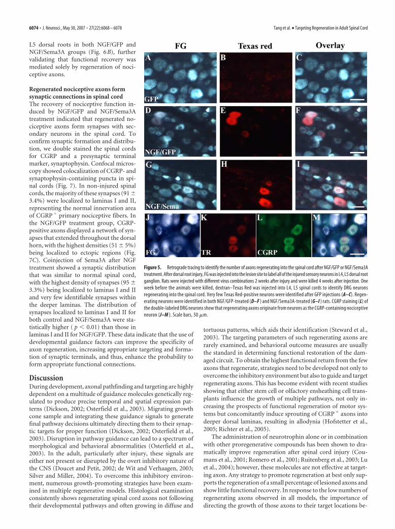
ing axons stop at the DREZ and no axonsregenerate into the spinal cord afterGFP/Ad treatment, the colabeled DRGneurons in this group should represent thenonregenerated neurons taking up tracerleaking past the DREZ. This turned out tobe very few neurons (Table 2, Fig. 5A–C).There were many more colabeled neuronsin both NGF/GFP (448 � 218) and NGF/Sema3A (240 � 80) groups (Table 2, Fig.5D–I). This suggested that NGF or NGF/Sema3A treatments induced a consider-able proportion of DRG neurons to regen-erate into the spinal cord. Thus, CGRP�
or SP� axons observed within the spinalcord for these treatment groups canmostly be attributed to regenerated fibers.Furthermore, costaining these DRG sec-tions using CGRP antibodies confirmedthe vast majority (�90) of regeneratingaxons to be from CGRP-positive neurons(Fig. 5 J,K)
Targeted regeneration of nociceptiveaxons induced by NGF/Sema3Aoverexpression led to recovery ofnociceptive functionThe majority of sensory information fromthe rat hindlimb is transferred to the spi-nal cord by the ipsilateral dorsal roots L2–L6, in which CGRP� and SP� axons me-diate cutaneous nociceptive sensation.
Nociception of the hindlimb can be tested by measuring PWL inresponse to radiant heat applied to the plantar surface of the rathindlimb. Previously, we demonstrated that lesioning of thesedorsal roots caused a complete loss of paw withdrawal responseon the ipsilateral side, and NGF-induced regeneration of noci-ceptive axons into the spinal cord resulted in recovery of nocicep-tive function and a reduction in PWL (Romero et al., 2001).However, the extent of recovery was dependent of the number ofregenerating axons (Tang et al., 2004a). In this study, we exam-ined whether NGF/Sema3A-induced and targeted axonal regen-eration caused nociceptive functional recovery, and whether dif-ferences in the distribution of nociceptive axons resulted indifferent nociceptive responses. PWL was measured before andonce a week after injury and treatment (Fig. 6A). Analysis usingtwo-way ANOVA with repeated measures showed a significantmain effect in latencies between treatment groups (F(2,91) �21.74; p � 0.01). In all groups, ipsilateral PWL rose to the cutoffvalue after injury because of the absence of a withdrawal re-sponse. No recovery of PWL was observed in the GFP/Ad groupthroughout the experimental period. When compared with con-trols, both NGF�GFP/Ad (F(1,9) � 40.4; p � 0.001) andNGF�Sema3A/Ad (F(1,8) � 28.0; p � 0.01) injected animalsshowed a decrease in PWL starting 1 week after injection andrecovered the PWL to near-normal level 3– 4 weeks after injec-tions (n � 5), indicating an almost complete functional recovery.There was no statistical difference in PWL response times be-tween NGF/GFP and NGF/Sema3A groups (F(1,9) � 1.5; p �0.05). No significant thermal hyperalgesia or mechanical allo-dynia developed in the NGF/GFP group (data not shown) withinthe observation period of this experiment. In addition, PWL re-turned to the pretreatment levels after relesioning of only L4 and
Table 2. Number and percentage of FG/Texas red-colabeled neurons in L4, L5 DRGs
Cell number in L4, L5 DRG
Group FG TR Colabeled (Colabeled:FG)%
GFP 2150 � 666 5 � 2 5 � 2 0.2 � 0.1NGF � GFP 2574 � 1242 448 � 228 407 � 218 15.3 � 2.8*NGF � Sema 2673 � 717 240 � 80 236 � 77 8.9 � 1.4*,**
Values represent mean � SD, n � 3. *p � 0.01 compared with GFP group; **p � 0.05 compared with NGF � GFPgroup, analyzed by one-way ANOVA.
Figure 4. NGF/Sema3A overexpression targeted regeneration of injured SP � nociceptive axons to superficial dorsal horn.Sections were colabeled using SP and IB4 immunohistochemistry to identify two classes of nociceptive axons. SP-containing axonsnormally terminate at laminas I and II (left side in A, C, E, G) and disappeared after dorsal root injury (C). NGF/GFP overexpressioninduced extensive regeneration of SP axons into the dorsal horn (E), and NGF/Sema3A overexpression reduced axon growth intodeeper laminas, shaping regenerating axons toward a normal distribution (G). IB4-binding nociceptive axons normally terminateat inner aspect of lamina II (left side in B, D, F, H ), and NGF/GFP (F ) or NGF/Sema3A (H) treatment has no effect on this class ofaxons after injury. Scale bar, 0.5 mm.
Table 1. Number of Texas Red-labeled DRG neurons from axons that have sproutedinto the L4/L5 region of the spinal cord from L2 or meninges 4 weeks aftertreatment
Number of TR-labeled cell in DRG
Group L2 L3 L4, L5a
NGF � GFP 218 � 132 21 � 8 44 � 39NGF � Sema ND 9 � 9 ND
Values represent mean � SD, n � 3.aValues indicate peripheral axon regeneration after cutting and tying dorsal roots L3–L6 to prevent regeneration.
Tang et al. • Targeting Regeneration in Adult Spinal Cord J. Neurosci., May 30, 2007 • 27(22):6068 – 6078 • 6073
L5 dorsal roots in both NGF/GFP andNGF/Sema3A groups (Fig. 6B), furthervalidating that functional recovery wasmediated solely by regeneration of noci-ceptive axons.
Regenerated nociceptive axons formsynaptic connections in spinal cordThe recovery of nociceptive function in-duced by NGF/GFP and NGF/Sema3Atreatment indicated that regenerated no-ciceptive axons form synapses with sec-ondary neurons in the spinal cord. Toconfirm synaptic formation and distribu-tion, we double stained the spinal cordsfor CGRP and a presynaptic terminalmarker, synaptophysin. Confocal micros-copy showed colocalization of CGRP- andsynaptophysin-containing puncta in spi-nal cords (Fig. 7). In non-injured spinalcords, the majority of these synapses (91 �3.4%) were localized to laminas I and II,representing the normal innervation areaof CGRP� primary nociceptive fibers. Inthe NGF/GFP treatment group, CGRP-positive axons displayed a network of syn-apses that extended throughout the dorsalhorn, with the highest densities (51 � 5%)being localized to ectopic regions (Fig.7C). Coinjection of Sema3A after NGFtreatment showed a synaptic distributionthat was similar to normal spinal cord,with the highest density of synapses (95 �3.3%) being localized to laminas I and IIand very few identifiable synapses withinthe deeper laminas. The distribution ofsynapses localized to laminas I and II forboth control and NGF/Sema3A were sta-tistically higher ( p � 0.01) than those inlaminas I and II for NGF/GFP. These data indicate that the use ofdevelopmental guidance factors can improve the specificity ofaxon regeneration, increasing appropriate targeting and forma-tion of synaptic terminals, and thus, enhance the probability toform appropriate functional connections.
DiscussionDuring development, axonal pathfinding and targeting are highlydependent on a multitude of guidance molecules genetically reg-ulated to produce precise temporal and spatial expression pat-terns (Dickson, 2002; Osterfield et al., 2003). Migrating growthcone sample and integrating these guidance signals to generatefinal pathway decisions ultimately directing them to their synap-tic targets for proper function (Dickson, 2002; Osterfield et al.,2003). Disruption in pathway guidance can lead to a spectrum ofmorphological and behavioral abnormalities (Osterfield et al.,2003). In the adult, particularly after injury, these signals areeither not present or disrupted by the overt inhibitory nature ofthe CNS (Doucet and Petit, 2002; de Wit and Verhaagen, 2003;Silver and Miller, 2004). To overcome this inhibitory environ-ment, numerous growth-promoting strategies have been exam-ined in multiple regenerative models. Histological examinationconsistently shows regenerating spinal cord axons not followingtheir developmental pathways and often growing in diffuse and
tortuous patterns, which aids their identification (Steward et al.,2003). The targeting parameters of such regenerating axons arerarely examined, and behavioral outcome measures are usuallythe standard in determining functional restoration of the dam-aged circuit. To obtain the highest functional return from the fewaxons that regenerate, strategies need to be developed not only toovercome the inhibitory environment but also to guide and targetregenerating axons. This has become evident with recent studiesshowing that either stem cell or olfactory ensheathing cell trans-plants influence the growth of multiple pathways, not only in-creasing the prospects of functional regeneration of motor sys-tems but concomitantly induce sprouting of CGRP� axons intodeeper dorsal laminas, resulting in allodynia (Hofstetter et al.,2005; Richter et al., 2005).
The administration of neurotrophin alone or in combinationwith other proregenerative compounds has been shown to dra-matically improve regeneration after spinal cord injury (Cou-mans et al., 2001; Romero et al., 2001; Ruitenberg et al., 2003; Luet al., 2004); however, these molecules are not effective at target-ing axon. Any strategy to promote regeneration at best only sup-ports the regeneration of a small percentage of lesioned axons andshow little functional recovery. In response to the low numbers ofregenerating axons observed in all models, the importance ofdirecting the growth of those axons to their target locations be-
Figure 5. Retrograde tracing to identify the number of axons regenerating into the spinal cord after NGF/GFP or NGF/Sema3Atreatment. After dorsal root injury, FG was injected into the lesion site to label all of the injured sensory neurons in L4, L5 dorsal rootganglion. Rats were injected with different virus combinations 2 weeks after injury and were killed 4 weeks after injection. Oneweek before the animals were killed, dextran–Texas Red was injected into L4, L5 spinal cords to identify DRG neuronsregenerating into the spinal cord. Very few Texas Red-positive neurons were identified after GFP injections (A–C). Regen-erating neurons were identified in both NGF/GFP-treated (D–F ) and NGF/Sema3A-treated (G–I ) rats. CGRP staining (L) ofthe double-labeled DRG neurons show that regenerating axons originate from neurons as the CGRP-containing nociceptiveneuron (J–M ). Scale bars, 50 �m.
6074 • J. Neurosci., May 30, 2007 • 27(22):6068 – 6078 Tang et al. • Targeting Regeneration in Adult Spinal Cord

comes paramount. In addition, the use of growth-promotingagents such as neurotrophins increase the prospect of axons re-generating into generalized regions with less targeting capability,because regeneration is dependent on regions in which thegrowth factor is present (Blesch et al., 2002). Very few studieshave examined synaptic connections let alone the specificity ofthe targets after axonal regeneration, and much of the recoveryassociated with regenerating axons may be attributable to su-praspinal reorganization of circuits or contralateral sprouting toestablish novel functional circuits (Fouad et al., 2001; Kim et al.,2006). Equally as important to targeting and enhancing the effi-ciency of regenerating circuits is the elimination of aberrant con-nections, which could either cause detrimental function (e.g.,
pain and autonomic dysreflexia) (Romero et al., 2000; Cameronet al., 2006) or diverge to coactivate both motor inhibitory andexcitatory circuits to reduce overall functional recovery.
During spinal cord development, sensory axons bypass inap-propriate lamina and project directly to their target lamina(Ozaki and Snider, 1997; Sharma and Frank, 1998) in which theyestablish lamina-restricted synaptic terminals (Sanes and Yama-gata, 1999). For TrkA-positive (peptidergic nociceptive) axons,entry into the spinal cord and projection into laminas I and IIcorrelate with the ventral migration of a chemorepulsive gradientestablished by semaphorin 3A expression (Fitzgerald et al., 1993;Messersmith et al., 1995; Wright and Snider, 1995; Puschel et al.,1996; Fu et al., 2000). In the embryo, as fibers enter and begin toform arbors, semaphorin 3A expression extends high into thedorsal horn. During this period, the addition of exogenous NGFhas little effect on their ventral-mediated growth. As sensory axongrowth continues, the semaphorin 3A gradient migrates ven-trally, and, at these early postnatal time points, exogenous NGFincreases growth over dorsal as well as ventral regions (Redmondet al., 1997). This alteration in NGF sensitivity to elicit axongrowth throughout the dorsal and ventral horns correlates withthe dorsal reduction in semaphorin 3A expression, which in theadult persists only in motor neurons (Puschel et al., 1996; Pas-terkamp et al., 2000). Thus, semaphorin 3A has the ability tomodulate NGF-induced axon growth even during development.
In the present study, the production of opposing guidance cuegradients were used to target the regeneration of primary peptidergicnociceptive axons to their dorsal lamina. Dorsal expression of NGFwas used to induce regeneration of nociceptive axons into the spinalcord; however, as demonstrated in previous studies, these axons failto terminate in their normal targets and continue to extend through-out the entire dorsal horn and, in some cases, extend into the ventralhorn as well (Romero et al., 2001). To restrict these axons fromregenerating into the deeper dorsal or ventral horns, semaphorin 3Awas expressed within the ventral horn. It is proposed that this mol-ecule produces a gradient for repulsion to commence (Kolodkin1996; Nakamura et al., 2000). Although the extent of the gradientwas difficult to quantify by immunofluorescence, the geneticmethod for cellular expression of these factors would provide a lo-calized production source, and the secretion of either NGF orSema3A would necessitate the production of a gradient extendingoutward from the local source of production (Cao and Shoichet,2003; Heron et al., 2006; Taylor et al., 2006). Our results with NGF/Sema3A-treated rats suggest that regenerating axons encountered apermissive environment (provided by NGF) within the superficiallayers of the dorsal horn and then stop in contact with Sema3A, inwhich the overall signal becomes inhibitory. The pattern of adultCGRP-positive axons terminating growth at the dorsal aspect of theSema3A gradient appear almost identical to the developmental pat-terning of TrkA-positive axons encountering Sema3A in the chickembryo (Shepherd et al., 1997; Fu et al., 2000). Likewise, this pattern-ing led to a regional distribution of synapses for regenerating noci-ceptive axons that were primarily confined to laminas I and II. Thismatched the synaptic distribution of these axons in the normal cord,although at a significantly lower density. Expression of NGF aloneled to a dramatically different synaptic patterning that was moreevenly distributed throughout all laminas within the dorsal horn.Retrograde track tracing experiments also demonstrated that co-expression of semaphorin resulted in a �40% reduction in thetotal number of regenerating nociceptive axons when comparedwith NGF but no reduction in functional recovery. This is incontrast to our previous studies that show that similar reductionsin nontargeted regenerating nociceptive axons result in a sub-
Figure 6. Both NGF/GFP- and NGF/Sema3A-induced axon regeneration leads to near com-plete functional recovery of thermal nociception. PWL was measured on both right and lefthindlimbs before and once a week after injury and treatment. The ratios of right (experimental)and left (control) hindpaw latencies were compared to examine the influence of different treat-ments on nociceptive functions. A, L4, L5 dorsal root injury caused an immediate loss of pawwithdrawal response, raising the ipsilateral PWL to the cutoff value. No spontaneous recovery ofPWL was found in GFP/Ad-treated rats. NGF/GFP and NGF/Sema3A treatments induced recov-ery of nociceptive function and decreased PWL to near-normal levels by the end of the study.There is no significant difference in PWL between these two groups throughout the observationtime. PWL recovery disappeared after relesioning the L4 and L5 dorsal roots in eitherNGF�GFP/Ad- or NGF�Sema3A/Ad-treated rats (B), indicating that the recovery was attrib-uted to regenerating sensory axons and not axon sprouting. Values represent mean � SD; n �5– 6. *p � 0.05, **p � 0.01, analyzed by two-way ANOVA.
Tang et al. • Targeting Regeneration in Adult Spinal Cord J. Neurosci., May 30, 2007 • 27(22):6068 – 6078 • 6075

stantial reduction in functional recovery(Tang et al., 2004a), indicating that, al-though coexpression of sema3A resultedin fewer regenerating axons, more preciselaminar targeting of synaptic connectionincreased the overall efficacy of the recon-structed circuits to enhance functionalreturn.
Our previous studies indicate thatNGF-mediated ectopic sprouting of non-injured CGRP-positive sensory axons intodeeper spinal cord laminas are involved inthe onset of chronic pain and autonomicdysreflexia (Romero et al., 2000; Cameronet al., 2006). In the present study, indica-tors of chronic pain behavior such as ther-mal hyperalgesia and mechanical allo-dynia were not manifested in animalstreated with either NGF/GFP or NGF/Sema3A, and the incidences of autonomydiminished in both groups. The failure ofregenerating axons to form functionalsynapses is an unlikely reason for this un-usual behavior for two reasons. First, rees-tablishment of normal protective pain, in-dicative of paw withdrawal to heat, stronglysuggests that regenerating axons make func-tional synapses onto neurons that form thissynaptic reflex. Second, analysis of synapseformation by double labeling CGRP-positive axons for synaptophysin demon-strated extensive ectopic synapses in thedeeper dorsal laminas after treatment withNGF/GFP but not NGF/Sema3A. So why doectopic synapses from regenerating axonsfail to elicit chronic pain? One possibility isthat other sensory pathways contribute tothe progression of chronic pain, and the re-generation of a single pathway or axon pop-ulation is insufficient (Neumann et al., 1996;Vulchanova et al., 2001; Braz et al., 2005). Inthe present study, regeneration of the IB4-positive C-fiber and A-� myelinated axonswere not observed. These axons are thoughtto regulate pain response, and their absencemay prevent convergent signals that induceneuropathic pain. Another possibility is thenumber of regenerating axons provided aninsufficient driving force to elicit chronicpain. In both NGF treatment groups, only afraction (�25–50%) of the total number ofC-fibers regenerated, and receptor field sig-naling to neurokinin-1 neurons might notbe sufficient to trigger chronic pain re-sponses. A final possibility is that the 4 weektime window for posttreatment behavioralanalysis was too short to support sufficientmaturation of normal or ectopic synapses todisplay pathological symptoms and thatthese behavioral outcomes will increase overtime. These possibilities and several othersare presently being addressed.
Figure 7. Confocal microscopy images of synaptic-like structures associated with regeneration of CGRP � nociceptive axons in thespinal cord. The spinal cord was double stained for CGRP (blue) and presynaptic protein synaptophysin (red). Colabeling for CGRP andsynaptophysin (violet) showed the presence of synaptic clusters associated with regenerated CGRP � nociceptive axons within the dorsalhorn. A, Colocalization of CGRP and synaptophysin staining in normal (control) dorsal horn or after treatment with either NGF/GFP orNGF/Sema3A. Synaptic profiles from different laminas are restricted to the superficial dorsal laminas after NGF/Sema3A treatment similarto that of the control. Scale bar, 100 �m. Higher magnification within laminas I, III, and V confirm lower density of synaptic profiles inlaminas III and V after NGF/Sema3A treatment but not NGF/GFP treatment. Scale bar, 10�m. B, High-resolution confocal microscopy wasused to visualize individual CGRP–synaptophysin vesicle clusters (purple). Scale bar, 5 �m. C, Graph showing the distribution of synapticprofiles within laminas I/II, III, and V. Counts represent colabeled synaptic puncta �1 �m 2 from regions 100 � 100 �m within laminarepresented in photographs in A. The vast majority of synaptic profiles were localized to laminas I/II for both normal and NGF/Sema3A-treated cord, whereas synapses were more evenly distributed throughout all laminas with NGF/GFP treatment. Values represent mean�SD; n � 3. *p � 0.001 compared with lamina I/II of normal spinal cord; #p � 0.01 compared with laminas I/II of NGF/Sema3A-treatedcords; ##p � 0.01 compared with lamina III of NGF/Sema3A cords, analyzed by two-way ANOVA.
6076 • J. Neurosci., May 30, 2007 • 27(22):6068 – 6078 Tang et al. • Targeting Regeneration in Adult Spinal Cord

In summary, the combination of NGF and Sema3A in thisstudy induced targeted axon regeneration and synaptic forma-tion into the denervated spinal cord, which led to successful no-ciceptive functional recovery. It provides insights of using devel-opmental guidance cues to reestablish specific neuronalconnections in adult CNS, which is essential for optimal long-term functional recovery.
ReferencesAldskogius H, Kozlova EN (2002) Strategies for repair of the deafferented
spinal cord. Brain Res Brain Res Rev 40:301–308.Blesch A, Lu P, Tuszynski MH (2002) Neurotrophic factors, gene ther-
apy, and neural stem cells for spinal cord repair. Brain Res Bull57:833– 838.
Braz JM, Nassar MA, Wood JN, Basbaum AI (2005) Parallel “pain” path-ways arise from subpopulations of primary afferent nociceptor. Neuron47:787–793.
Cameron AA, Smith GM, Randall DC, Brown DR, Rabchevsky AG (2006)Genetic manipulation of intraspinal plasticity after spinal cord injuryalters the severity of autonomic dysreflexia. J Neurosci 26:2923–2932.
Cao X, Shoichet MS (2003) Investigating the synergistic effect of combinedneurotrophic factor concentration gradient to guide axonal growth. Neu-roscience 122:381–389.
Coumans JV, Lin TT, Dai HN, MacArthur L, McAtee M, Nash C, Bregman BS(2001) Axonal regeneration and functional recovery after complete spi-nal cord transaction in rats by delayed treatment with transplant andneurotrophins. J Neurosci 21:9334 –9344.
de Wit J, Verhaagen J (2003) Role of semaphorins in the adult nervoussystem. Prog Neurobiol 71:249 –267.
Dickson BJ (2002) Molecular mechanisms of axon guidance. Science298:1959 –1964.
Doucet G, Petit A (2002) Seeking axon guidance molecules in the adult ratCNS. Prog Brain Res 137:453– 465.
Fitzgerald M, Kwiat GC, Middleton J, Pini A (1993) Ventral spinal cordinhibition of neurite outgrowth from embryonic rat dorsal root ganglia.Development 117:1377–1384.
Fouad K, Pedersen V, Schwab ME, Brosamle C (2001) Cervical sprouting ofcorticospinal fibers after thoracic spinal cord injury accompanies shifts inevoked motor responses. Curr Biol 11:1766 –1770.
Fu SY, Sharma K, Luo Y, Raper JA, Frank E (2000) SEMA3A regulates de-veloping sensory projections in the chicken spinal cord. J Neurobiol45:227–236.
Gavazzi I (2001) Semaphorin-neuropilin-1 interactions in plasticity and re-generation of adult neurons. Cell Tissue Res 305:275–284.
Hargreaves K, Dubner R, Brown F, Flores C, Joris J (1988) A new and sen-sitive method for measuring thermal nociception in cutaneous hyperal-gesia. Pain 32:77– 88.
Hofstetter CP, Holmstrom NA, Lilja JA, Schweinhardt P, Hao J, Spenger C,Wiesenfeld-Hallin Z, Kurpad SN, Frisen J, Olson L (2005) Allodynialimits the usefulness of intraspinal neural stem cell grafts; directed differ-entiation improves outcome. Nat Neurosci 8:346 –353.
Kim BG, Dai HN, McAtee M, Vicini S, Bregman BS (2006) Remodeling ofsynaptic structures in the motor cortex following spinal cord injury. ExpNeurol 198:401– 415.
Kolodkin AL (1996) Semaphorins: mediators of repulsive growth coneguidance. Trends Cell Biol 6:15–22.
Lu P, Yang H, Jones LL, Filbin MT, Tuszynski MH (2004) Combinatorialtherapy with neurotrophins and cAMP promotes axonal regenerationbeyond sites of spinal cord injury. J Neurosci 24:6402– 6409.
Messersmith EK, Leonardo ED, Shatz CJ, Tessier-Lavigne M, Goodman CS,Kolodkin AL (1995) Semaphorin III can function as a selective che-morepellent to pattern sensory projections in the spinal cord. Neuron14:949 –959.
Molliver DC, Wright DE, Leitner ML, Parsadanian AS, Doster K, Wen D, YanQ, Snider WD (1997) IB4-binding DRG neurons switch from NGF toGDNF dependence in early postnatal life. Neuron 19:849 – 861.
Nakamura F, Kalb RG, Strittmatter SM (2000) Molecular basis ofsemaphorin-mediated axon guidance. J Neurobiol 44:219 –229.
Neumann S, Doubell TP, Leslie T, Woolf CJ (1996) Inflammatory pain hy-persensitivity mediated by phenotypic switch in myelinated primary sen-sory neurons. Nature 384:360 –364.
Osterfield M, Kirschner MW, Flanagan JG (2003) Graded positional infor-mation: interpretation for both fate and guidance. Cell 113:425– 428.
Ozaki S, Snider WD (1997) Initial trajectories of sensory axons toward lam-inar targets in the developing mouse spinal cord. J Comp Neurol380:215–229.
Pasterkamp RJ, Giger RJ, Baker RE, Hermens WT, Verhaagen J (2000) Ec-topic adenoviral vector-directed expression of Sema3A in organotypicspinal cord explants inhibits growth of primary sensory afferents. DevBiol 220:129 –141.
Pindzola RR, Doller C, Silver J (1993) Putative inhibitory extracellularmatrix molecules at the dorsal root entry zone of the spinal cordduring development and after root and sciatic nerve lesions. Dev Biol156:34 – 48.
Priestley JV, Ramer MS, King VR, McMahon SB, Brown RA (2002) Stimu-lating regeneration in the damaged spinal cord. J Physiol (Paris)96:123–133.
Puschel AW, Adams RH, Betz H (1996) The sensory innervation of themouse spinal cord may be patterned by differential expression of anddifferential responsiveness to semaphorins. Mol Cell Neurosci7:419 – 431.
Ramer MS, Priestley JV, McMahon SB (2000) Functional regeneration ofsensory axons into the adult spinal cord. Nature 403:312–316.
Redmond L, Xie H, Ziskind-Conhaim L, Hockfield S (1997) Cues intrinsicto the spinal cord determine the pattern and timing of primary afferentgrowth. Dev Biol 182:205–218.
Richter MW, Fletcher PA, Liu J, Tetzlaff W, Roskams AJ (2005) Laminapropria and olfactory bulb ensheathing cells exhibit differential integra-tion and migration and promote differential axon sprouting in the le-sioned spinal cord. J Neurosci 25:10700 –10711.
Romero MI, Smith GM (1998) Adenoviral gene transfer into the normaland injured spinal cord: enhanced transgene stability by combined ad-ministration of temperature-sensitive virus and transient immune block-ade. Gene Ther 5:1612–1621.
Romero MI, Rangappa N, Li L, Lightfoot E, Garry MG, Smith GM (2000)Extensive sprouting of sensory afferents and hyperalgesia induced by con-ditional expression of nerve growth factor in the adult spinal cord. J Neu-rosci 20:4435– 4445.
Romero MI, Rangappa N, Garry MG, Smith GM (2001) Functional regen-eration of chronically injured sensory afferents into adult spinal cord afterneurotrophin gene therapy. J Neurosci 21:8408 – 8416.
Ruitenberg MJ, Plant GW, Hamers FP, Wortel J, Blits B, Dijkhuizen PA,Gispen WH, Boer GJ, Verhaagen J (2003) Ex vivo adenoviral vector-mediated neurotrophin gene transfer to olfactory ensheathing glia:effects on rubrospinal tract regeneration, lesion size, and functionalrecovery after implantation in the injured rat spinal cord. J Neurosci23:7045–7058.
Sanes JR, Yamagata M (1999) Formation of lamina-specific synaptic con-nections. Curr Opin Neurobiol 9:79 – 87.
Sharma K, Frank E (1998) Sensory axons are guided by local cues in thedeveloping dorsal spinal cord. Development 125:635– 643.
Shepherd IT, Luo Y, Lefcort F, Reichardt LF, Raper JA (1997) A sensoryaxon repellent secreted from ventral spinal cord explants is neutralized byantibodies raised against collapsin-1. Development 124:1377–1385.
Silver J, Miller JH (2004) Regeneration beyond the glial scar. Nat Rev Neu-rosci 5:146 –156.
Snider WD, McMahon SB (1998) Tackling pain at the source: new ideasabout nociceptors. Neuron 20:629 – 632.
Steinmetz MP, Horn KP, Tom VJ, Miller JH, Busch SA, Nair D, Silver DJ,Silver J (2005) Chronic enhancement of the intrinsic growth capacityof sensory neurons combined with the degradation of inhibitory pro-teoglycans allows functional regeneration of sensory axons throughthe dorsal root entry zone in the mammalian spinal cord. J Neurosci25:8066 – 8076.
Steward O, Zheng B, Tessier-Lavigne M (2003) False resurrections: distin-guishing regenerated from spared axons in the injured central nervoussystem. J Comp Neurol 459:1– 8.
Tanelian DL, Barry MA, Johnston SA, Le T, Smith GM (1997) Semaphorin
Tang et al. • Targeting Regeneration in Adult Spinal Cord J. Neurosci., May 30, 2007 • 27(22):6068 – 6078 • 6077

III can repulse and inhibit adult sensory afferents in vivo. Nat Med3:1398 –1401.
Tang XQ, Cai J, Nelson KD, Peng XJ, Smith GM (2004a) Functional repairafter dorsal root rhizotomy using nerve conduits and neurotrophic mol-ecules. Eur J Neurosci 20:1211–1218.
Tang XQ, Tanelian DL, Smith GM (2004b) Semaphorin3A inhibits nervegrowth factor-induced sprouting of nociceptive afferents in adult rat spi-nal cord. J Neurosci 24:819 – 827.
Taylor L, Jones L, Tuszynski MH, Blesch A (2006) Neurotrophin-3 gradi-ents established by lentiviral gene delivery promote short-distance axonalbridging beyond cellular grafts in the injured spinal cord. J Neurosci26:9713–9721.
Wright DE, Snider WD (1995) Neurotrophin receptor mRNA expressiondefines distinct populations of neurons in rat dorsal root ganglia. J CompNeurol 351:329 –338.
Wong ST, Atkinson BA, Weaver LC (2000) Confocal microscopic analysisreveals sprouting of primary afferent fibres in rat dorsal horn after spinalcord injury. Neurosci Lett 296:65– 68.
Wujek JR, Ahmad S, Harel A, Maier KH, Roufa D, Silver J (1991) A carbo-hydrate polymer that effectively prevents epidural fibrosis at laminectomysites in the rat. Exp Neurol 114:237–245.
Vulchanova L, Olson TH, StoneLS, Riedl MS, Elde R, Honda CN (2001)Cytotoxic targeting of isolectin IB4-binding sensory neurons. Neuro-science 108:143–155.
6078 • J. Neurosci., May 30, 2007 • 27(22):6068 – 6078 Tang et al. • Targeting Regeneration in Adult Spinal Cord