targeting lyn kinase in chorea-acanthocytosis - mdpi
TRANSCRIPT
Journal of
Personalized
Medicine
Article
Targeting Lyn Kinase in Chorea-Acanthocytosis: ATranslational Treatment Approach in a Rare Disease
Kevin Peikert 1,2 , Hannes Glaß 1,2, Enrica Federti 3, Alessandro Matte 3, Lisann Pelzl 4,5, Katja Akgün 1,Tjalf Ziemssen 1 , Rainer Ordemann 6, Florian Lang 4, The Network for Translational Research forNeuroacanthocytosis Patients †, Lucia De Franceschi 3,*,‡ and Andreas Hermann 1,2,7,8,9,*,‡
�����������������
Citation: Peikert, K.; Glaß, H.;
Federti, E.; Matte, A.; Pelzl, L.; Akgün,
K.; Ziemssen, T.; Ordemann, R.; Lang,
F.; et al. Targeting Lyn Kinase in
Chorea-Acanthocytosis: A
Translational Treatment Approach in
a Rare Disease. J. Pers. Med. 2021, 11,
392. https://doi.org/10.3390/
jpm11050392
Academic Editor: Felix Javier Jiménez
Jiménez
Received: 14 April 2021
Accepted: 6 May 2021
Published: 10 May 2021
Publisher’s Note: MDPI stays neutral
with regard to jurisdictional claims in
published maps and institutional affil-
iations.
Copyright: © 2021 by the authors.
Licensee MDPI, Basel, Switzerland.
This article is an open access article
distributed under the terms and
conditions of the Creative Commons
Attribution (CC BY) license (https://
creativecommons.org/licenses/by/
4.0/).
1 Department of Neurology, University Hospital Carl Gustav Carus, Technische Universität Dresden,01307 Dresden, Germany; [email protected] (K.P.); [email protected] (H.G.);[email protected] (K.A.); [email protected] (T.Z.)
2 Translational Neurodegeneration Section “Albrecht Kossel”, Department of Neurology, University MedicalCenter Rostock, 18147 Rostock, Germany
3 Department of Medicine, University of Verona, 37134 Verona, Italy; [email protected] (E.F.);[email protected] (A.M.)
4 Department of Physiology I, University of Tübingen, 72076 Tübingen, Germany;[email protected] (L.P.); [email protected] (F.L.)
5 Transfusion Medicine, Medical Faculty, Eberhard Karl University, 72076 Tübingen, Germany6 Medical Department I, University Hospital Carl Gustav Carus, Technische Universität Dresden,
01069 Dresden, Germany; [email protected] Center for Regenerative Therapies Dresden (CRTD), Technische Universität Dresden,
01307 Dresden, Germany8 DZNE, German Center for Neurodegenerative Diseases, Research Sites Dresden and Rostock/Greifswald,
18051 Rostock, Germany9 Center for Transdisciplinary Neurosciences Rostock (CTNR), University Medical Center Rostock, University
of Rostock, 18147 Rostock, Germany* Correspondence: [email protected] (L.D.F.); [email protected] (A.H.);
Tel.: +39-045-812-4401 (L.D.F.); +49-(0)381-494-9511 (A.H.); Fax: +39-045-802-7473 (L.D.F.)† Members of The Network for Translational Research for Neuroacanthocytosis Patients, see Appendix A.‡ Contributed equally to this work.
Abstract: Background: Chorea-acanthocytosis (ChAc) is a neurodegenerative disease caused bymutations in the VPS13A gene. It is characterized by several neurological symptoms and theappearance of acanthocytes. Elevated tyrosine kinase Lyn activity has been recently identified asone of the key pathophysiological mechanisms in this disease, and therefore represents a promisingdrug target. Methods: We evaluated an individual off-label treatment with the tyrosine kinaseinhibitor dasatinib (100 mg/d, 25.8–50.4 weeks) of three ChAc patients. Alongside thorough safetymonitoring, we assessed motor and non-motor scales (e.g., MDS-UPDRS, UHDRS, quality of life)as well as routine and experimental laboratory parameters (e.g., serum neurofilament, Lyn kinaseactivity, actin cytoskeleton in red blood cells). Results: Dasatinib appeared to be reasonably safe.The clinical parameters remained stable without significant improvement or deterioration. Regainof deep tendon reflexes was observed in one patient. Creatine kinase, serum neurofilament levels,and acanthocyte count did not reveal consistent effects. However, a reduction of initially elevatedLyn kinase activity and accumulated autophagy markers, as well as a partial restoration of the actincytoskeleton, was found in red blood cells. Conclusions: We report on the first treatment approachwith disease-modifying intention in ChAc. The experimental parameters indicate target engagementin red blood cells, while clinical effects on the central nervous system could not be proven withina rather short treatment time. Limited knowledge on the natural history of ChAc and the lack ofappropriate biomarkers remain major barriers for “clinical trial readiness”. We suggest a panel ofoutcome parameters for future clinical trials in ChAc.
Keywords: ChAc; neuroacanthocytosis; off-label; dasatinib; TKI
J. Pers. Med. 2021, 11, 392. https://doi.org/10.3390/jpm11050392 https://www.mdpi.com/journal/jpm
J. Pers. Med. 2021, 11, 392 2 of 14
1. Introduction
Chorea-acanthocytosis (ChAc) is a rare neurodegenerative disease of the early adult-hood which is characterized by a large spectrum of neurological symptoms and the presenceof acanthocytes [1–4]. The autosomal-recessive condition is caused by mutations in theVPS13A gene leading to loss of function of the respective encoded protein “chorein” [5–8].A disease-modifying therapy is not available yet. Hence, treatment options of the devastat-ing disease remain purely symptomatic [9], even though it causes considerable morbidity,markedly reduced life-span, and severely affects self-determined living.
The clinical phenotype of ChAc is highly heterogeneous. As patients often presentwith movement disorders like chorea, Parkinsonism, and/or dystonia [2,10,11], ChAcbelongs to the group of Huntington’s disease phenocopies [12]. Furthermore, dysarthriaand dysphagia, peripheral neuropathy, epilepsy, or cognitive impairment may occur,while tongue and lip biting, self-mutilating behavior, feeding dystonia, or head drops aremore specific signs of ChAc [1,2,10,11]. Red blood cell (RBC) acanthocytosis, elevatedcreatine kinase (CK), and serum neurofilament (sNfL) levels are common laboratory find-ings [1,2,10,13]. In correlation with the clinical manifestations, loss of striatal mediumspiny neurons and distinct cortical neurodegeneration represent the main histopatholog-ical characteristics [14,15]. Epidemiological estimations suggest a prevalence of around1000–5000 cases worldwide [1].
While there is growing evidence that members of the VPS13 protein family are in-volved in the non-vesicular transport of phospholipids [16–19], the precise function ofthese proteins in humans remains incompletely understood. It is most likely that impairedlipid transfer, and consequently, disturbed organelle lipid homeostasis, contributes toneuronal dysfunction in this disease [20]. So far, VPS13A has been implicated in a varietyof important cellular processes, e.g., regulation of cytoskeletal architecture, exocytosis,autophagy, Na+/K+ pump capacity, and Ca2+ homeostasis, and therefore, overall cell sur-vival [4,21–26]. Hence, lack of functional VPS13A leads to impaired cellular homeostasis,particularly resulting in acanthocytosis and neurodegeneration. We recently identified twomechanisms that are considered to be key drivers of ChAc pathophysiology due to VPS13Adeficiency: decreased phosphoinositide-3-kinase signaling and increased activity of Srcfamily tyrosine kinase Lyn (for review, see [4]).
In previous studies, we found that ChAc RBCs and induced pluripotent stem cell(iPSC)-derived nerve cells are characterized by the accumulation of active Lyn: hyperactiveLyn kinase hyperphosphorylates membrane proteins in RBCs, e.g., band 3, which are in-volved in anchoring the membrane to the cytoskeletal network. This results in mechanicalinstability of the membrane [22,27]. Accumulation of active Lyn was also found to be re-lated to impairment of autophagy in erythrocytes [22]. Consistent with that, perturbationsof autophagy processes have been reported in in vitro cell models that are defective forVPS13A [17,28]. In a previous study, we showed that in vitro treatment of ChAc RBCs withSrc family kinase inhibitors reduces Lyn kinase activity, improves autophagy, and restoresthe morphological phenotype [22]. Moreover, Src family kinase inhibition also attenuatedpathologically enhanced synaptic transmission in striatal medium spiny neurons derivedfrom patient-specific iPSCs [29]. These findings strongly implicate that Lyn kinase shouldbe considered as a promising potential druggable target in ChAc. FDA-approved specificinhibitors of Src family kinases with a reasonable benefit–risk profile, such as the tyrosinekinase inhibitor (TKI) dasatinib, were successfully established in the treatment of chronicmyeloid leukemia [30]. Dasatinib was previously shown to cross the blood–brain barrier inhumans [31]. Therefore, these inhibitors are ideal candidates for “repurposing” strategiesin this context. TKIs have been evaluated in a variety of other neurodegenerative dis-eases, e.g., Parkinson’s disease (nilotinib), Alzheimer’s disease (e.g., nilotinib, saracatinib),or Amyotrophic Lateral Sclerosis (masitinib), each with a different pathophysiologicalrationale [30,32–36].
Here, we evaluate a translational off-label treatment with dasatinib (100 mg/d) inthree ChAc patients. To our knowledge, this is the first implemented potentially disease-
J. Pers. Med. 2021, 11, 392 3 of 14
modifying approach in this ultra-rare disease. Alongside thorough safety monitoring,we regularly performed assessments of treatment efficacy. Therefore, we designed anovel ChAc-related panel of read out parameters, also challenging current concepts ofdisease markers.
2. Materials and Methods2.1. Patients and Dasatinib Treatment
We treated three ChAc patients (P1–3) off-label with the FDA-approved tyrosinekinase inhibitor (TKI) dasatinib (DRKS00023177). Patient characteristics are shown inTable 1 and reflect the variety of ChAc phenotypes. Diagnosis was based on clinicalmanifestations, the absence of chorein in Western blot analysis, and genetic testing [37]. Allpatients gave their informed consent for off-label treatment with dasatinib, including therisk of possible life-threatening adverse reactions, as well as for video documentation andpublication of the data. Patients and healthy control blood donors were enrolled in ongoingstudies on the pathogenesis and natural history of neurodegenerative diseases approvedby the institutional review board of the Technische Universität Dresden, Germany (EK45022009, EK 78022015, EK517122019). The standard dose of 100 mg dasatinib per day wasadministered orally.
Table 1. Demographic characteristics, main clinical observations, and adverse reactions/events in chorea-acanthocytosispatients treated with dasatinib.
P1 P2 P3
Sex male male female
Age at beginning of treatment(Y) 27 32 49
Age at onset (Y)– symptoms – 14 – 23 – 21– diagnosis – 18 – 23 – 39
Current clinical presentation• refractory epilepsy• cognitive impairment• mild chorea
• refractory epilepsy• muscle atrophy/areflexia• mild chorea• cognitive impairment
• marked parkinsonism• epilepsy• cognitive impairment• dystonia• areflexia
Laboratory findings• elevated levels of: CK, AST,
ALT, cTroponin T, LDH• acanthocytosis
• elevated levels of: CK, AST,ALT, cTroponin T, LDH
• acanthocytosis
• elevated levels of: CK,cTroponin T, LDH
• lowered level ofhaptoglobin
• acanthocytosis
Time on dasatinib treatment(weeks) 50.4 48.7 25.8
Comedication
• Lacosamide 2 × 300 mg• Zonisamide 2 × 250 mg• Mirtazapine 1 × 15 mg• Vitamin D and Calcium• PRN:
Lorazepam/Midazolam
• Lacosamide 2 × 300 mg• Zonisamide 2 × 200 mg• Vitamin D• PRN:
Lorazepam/Midazolam
• Levetiracetam 2 × 2000 mg• Valproate 2 × 1000 mg• Clobazam 2 × 5 mg• Zonisamide 2 × 100 mg• Vitamin D
Main clinical observationsduring dasatinib treatment
• improvement of short-termmemory reported by thecaregivers
• variation of seizurefrequency as known/usualbefore treatment
• reappearance of deeptendon reflexes
• variation of seizurefrequency as known/usualbefore treatment
• variation of seizurefrequency as known/usualbefore treatment
J. Pers. Med. 2021, 11, 392 4 of 14
Table 1. Cont.
P1 P2 P3
Adverse reactions or events • mild intermittent diarrheaand abdominal pain
• mild intermittent diarrheaand abdominal pain
• Acne• discrete alopecia• increased frequency of
defecation
PRN—pro re nata; Y—years.
2.2. Evaluation of Dasatinib Treatment
Evaluation of the off-label treatment primarily included monitoring of potential ad-verse reactions or events, as well as outcome assessments in the context of routine careadjusted to the clinical phenotype (UHDRS, MDS-UPDRS, seizure frequency, blood acan-thocyte, and CK level). Evaluation was performed initially every 2 weeks, after 8 weeksof treatment monthly, and after 6 months of treatment every 2 months. At every visit,each patient was assessed with a clinical examination and medical history (including thecaregivers’ observations), the Unified Huntington’s Disease Rating Scale Total Motor Score(UHDRS-TMS), Total Functional Capacity (UHDRS-TFC), and Functional Assessment Scale(UHDRS-FA), as well as the Movement Disorders Society Unified Parkinson Disease RatingScale (MDS-UPDRS) parts I-III and the Clinical Global Impression (GCI) scale by a specialistexperienced in the care of patients with ChAc and other movement disorders. Patients wereasked to complete the McGill Quality of Life Single-Item Scale (McGill-QoL; range 0–10)and the Schedule for the Evaluation of Individual Quality of Life-Direct Weighting (SEIQoL)questionnaires. Routine electroencephalography was performed. Seizure frequency wasdefined as number of seizures within the preceding month. At baseline, after 2 months, andas indicated, patients underwent electrocardiography, echocardiography, and abdominalultrasound for safety monitoring. Routine laboratory chemistry/hematology tests werealso performed on blood samples obtained by venipuncture.
2.3. Serum Neurofilament Light Chain Quantification
Serum samples were stored at −20 ◦C directly after collection, since the neurofilamentlight chain is stable during freezing process [38,39]. The sNfl measurement was performedusing the Advantage NF-Light singleplex Kit and prepared as defined in the manufac-turer’s instructions (Quartered, Lexington, MA, Datasheet Quanterix: SimoaTM NF-Light®
Advantage Kit) as previously described [40,41] with the single molecule array (SIMOA)analysis. Both the mean intra-assay coefficient of variation of duplicates and the meaninter-assay coefficient of variation were <10%.
2.4. Immunoblot Analysis
Additional EDTA-blood samples (ChAc patients and healthy control donors) wereshipped to Verona, Italy at 4 ◦C and processed immediately after arrival. Lyn activitywas determined by Western blot analysis using an anti-phospho-Lyn (Y396) antibody, asdescribed previously [27]. We evaluated the amount of ULK1 and p62, known markers ofautophagy in RBCs, as reported previously [22,42,43].
2.5. Immunofluorescence
Additional EDTA-blood samples (ChAc patients and healthy control donors) wereshipped to Tübingen, Germany at 4 ◦C and processed immediately after arrival. The ery-throcytes were stained with an anti-ß-Actin-FITC-conjugated antibody (1:50; biorbyt) andPhalloidin-eFluor660 (1:100, eBioscience) to detect filamentous actin (F-actin) as describedpreviously [25,44]. Confocal microscopy was performed with a Zeiss LSM 5 EXCITERconfocal laser-scanning module (Carl Zeiss). The images were analyzed with the softwareof the instrument.
J. Pers. Med. 2021, 11, 392 5 of 14
2.6. Osmotic Fragility of Red Blood Cells
In previous reports, we showed that red cell osmotic fragility is increased in patientswith ChAc compared to healthy controls [22,27,45,46]. We evaluated osmotic fragility inEDTA blood using a single osmotic point at 158 mOsm. Erythrocytes from healthy controlswere always analyzed in the same experiments with ChAc RBCs.
2.7. Statistical Analysis
Data were analyzed with GraphPad Prism 5 software. Statistical analysis was madeby analysis of variance (ANOVA), and p < 0.05 was considered as statistically significant.
3. Results3.1. Dasatinib Treatment Was Safe
Overall, dasatinib treatment was safe in all three patients (Table 1). No severe adversereactions or events were reported. Because of discrete acne and mild alopecia as well asincreased defecation frequency, P3 and her caregivers asked for the discontinuation oftreatment after 25.8 weeks. Otherwise, P1 and P2 reported irregular defecation includingepisodes of mild diarrhea and abdominal pain, but they both stayed on dasatinib treatmentfor 48.7 and 50.4 weeks, respectively.
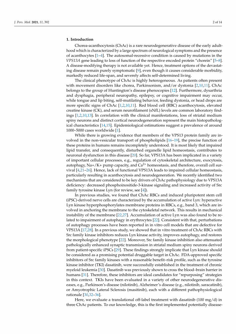
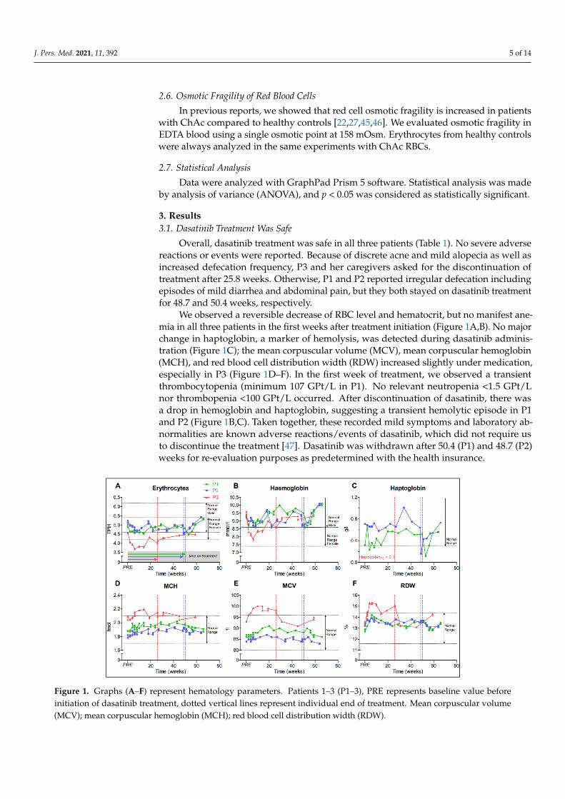
We observed a reversible decrease of RBC level and hematocrit, but no manifest ane-mia in all three patients in the first weeks after treatment initiation (Figure 1A,B). No majorchange in haptoglobin, a marker of hemolysis, was detected during dasatinib adminis-tration (Figure 1C); the mean corpuscular volume (MCV), mean corpuscular hemoglobin(MCH), and red blood cell distribution width (RDW) increased slightly under medication,especially in P3 (Figure 1D–F). In the first week of treatment, we observed a transientthrombocytopenia (minimum 107 GPt/L in P1). No relevant neutropenia <1.5 GPt/Lnor thrombopenia <100 GPt/L occurred. After discontinuation of dasatinib, there wasa drop in hemoglobin and haptoglobin, suggesting a transient hemolytic episode in P1and P2 (Figure 1B,C). Taken together, these recorded mild symptoms and laboratory ab-normalities are known adverse reactions/events of dasatinib, which did not require usto discontinue the treatment [47]. Dasatinib was withdrawn after 50.4 (P1) and 48.7 (P2)weeks for re-evaluation purposes as predetermined with the health insurance.J. Pers. Med. 2021, 11, x FOR PEER REVIEW 6 of 15
Figure 1. Graphs A-F represent hematology parameters. Patients 1–3 (P1–3), PRE represents baseline value before initia-tion of dasatinib treatment, dotted vertical lines represent individual end of treatment. Mean corpuscular volume (MCV); mean corpuscular hemoglobin (MCH); red blood cell distribution width (RDW).
Commented [M4]: Correct legend Figure 1. Graphs (A–F) represent hematology parameters. Patients 1–3 (P1–3), PRE represents baseline value beforeinitiation of dasatinib treatment, dotted vertical lines represent individual end of treatment. Mean corpuscular volume(MCV); mean corpuscular hemoglobin (MCH); red blood cell distribution width (RDW).
J. Pers. Med. 2021, 11, 392 6 of 14
3.2. Disease Progression Remained Stable during Dasatinib Treatment in ChAc Patients
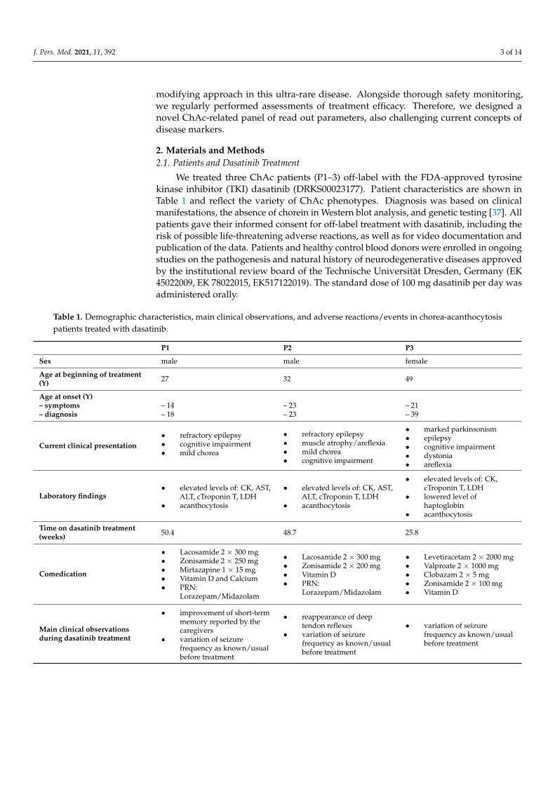
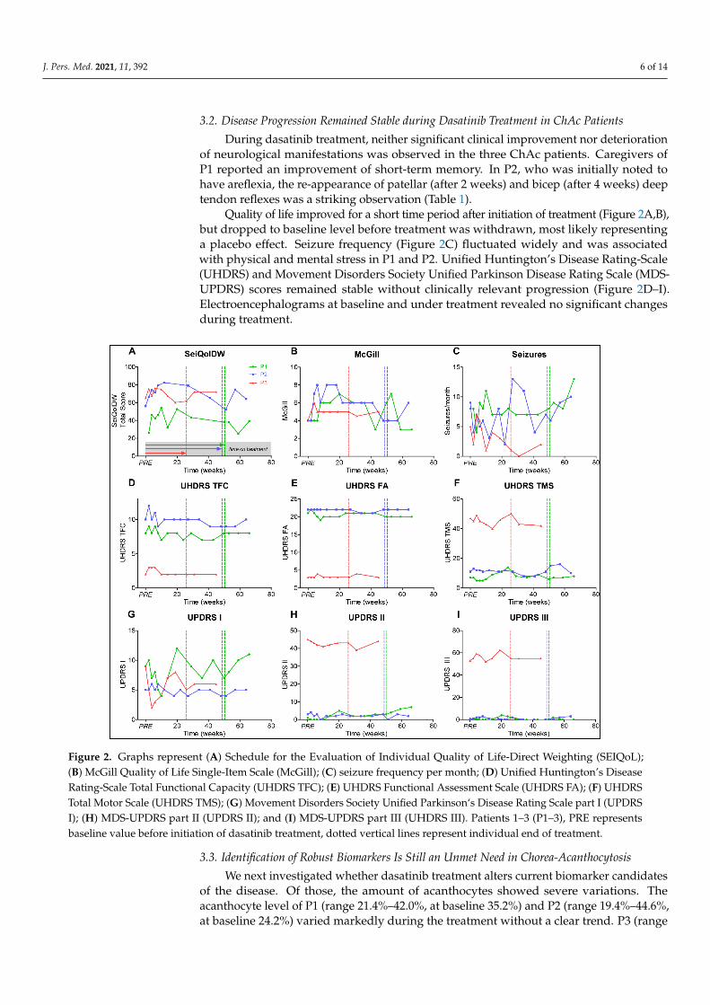
During dasatinib treatment, neither significant clinical improvement nor deteriorationof neurological manifestations was observed in the three ChAc patients. Caregivers ofP1 reported an improvement of short-term memory. In P2, who was initially noted tohave areflexia, the re-appearance of patellar (after 2 weeks) and bicep (after 4 weeks) deeptendon reflexes was a striking observation (Table 1).
Quality of life improved for a short time period after initiation of treatment (Figure 2A,B),but dropped to baseline level before treatment was withdrawn, most likely representinga placebo effect. Seizure frequency (Figure 2C) fluctuated widely and was associatedwith physical and mental stress in P1 and P2. Unified Huntington’s Disease Rating-Scale(UHDRS) and Movement Disorders Society Unified Parkinson Disease Rating Scale (MDS-UPDRS) scores remained stable without clinically relevant progression (Figure 2D–I).Electroencephalograms at baseline and under treatment revealed no significant changesduring treatment.
J. Pers. Med. 2021, 11, x FOR PEER REVIEW 6 of 14
3.2. Disease Progression Remained Stable during Dasatinib Treatment in ChAc Patients
During dasatinib treatment, neither significant clinical improvement nor deteriora-
tion of neurological manifestations was observed in the three ChAc patients. Caregivers
of P1 reported an improvement of short-term memory. In P2, who was initially noted to
have areflexia, the re-appearance of patellar (after 2 weeks) and bicep (after 4 weeks) deep
tendon reflexes was a striking observation (Table 1).
Quality of life improved for a short time period after initiation of treatment (Figure
2A,B), but dropped to baseline level before treatment was withdrawn, most likely repre-
senting a placebo effect. Seizure frequency (Figure 2C) fluctuated widely and was associ-
ated with physical and mental stress in P1 and P2. Unified Huntington’s Disease Rating-
Scale (UHDRS) and Movement Disorders Society Unified Parkinson Disease Rating Scale
(MDS-UPDRS) scores remained stable without clinically relevant progression (Figure 2D–
I). Electroencephalograms at baseline and under treatment revealed no significant
changes during treatment.
Figure 2. Graphs represent (A) Schedule for the Evaluation of Individual Quality of Life-Direct Weighting (SEIQoL); (B)
McGill Quality of Life Single-Item Scale (McGill); (C) seizure frequency per month; (D) Unified Huntington’s Disease
Rating-Scale Total Functional Capacity (UHDRS TFC); (E) UHDRS Functional Assessment Scale (UHDRS FA); (F) UHDRS
Total Motor Scale (UHDRS TMS); (G) Movement Disorders Society Unified Parkinson‘s Disease Rating Scale part I (UP-
DRS I); (H) MDS-UPDRS part II (UPDRS II); and (I) MDS-UPDRS part III (UHDRS III). Patients 1–3 (P1–3), PRE represents
baseline value before initiation of dasatinib treatment, dotted vertical lines represent individual end of treatment.
3.3. Identification of Robust Biomarkers Is Still an Unmet Need in Chorea-Acanthocytosis
We next investigated whether dasatinib treatment alters current biomarker candi-
dates of the disease. Of those, the amount of acanthocytes showed severe variations. The
Figure 2. Graphs represent (A) Schedule for the Evaluation of Individual Quality of Life-Direct Weighting (SEIQoL);(B) McGill Quality of Life Single-Item Scale (McGill); (C) seizure frequency per month; (D) Unified Huntington’s DiseaseRating-Scale Total Functional Capacity (UHDRS TFC); (E) UHDRS Functional Assessment Scale (UHDRS FA); (F) UHDRSTotal Motor Scale (UHDRS TMS); (G) Movement Disorders Society Unified Parkinson‘s Disease Rating Scale part I (UPDRSI); (H) MDS-UPDRS part II (UPDRS II); and (I) MDS-UPDRS part III (UHDRS III). Patients 1–3 (P1–3), PRE representsbaseline value before initiation of dasatinib treatment, dotted vertical lines represent individual end of treatment.
3.3. Identification of Robust Biomarkers Is Still an Unmet Need in Chorea-Acanthocytosis
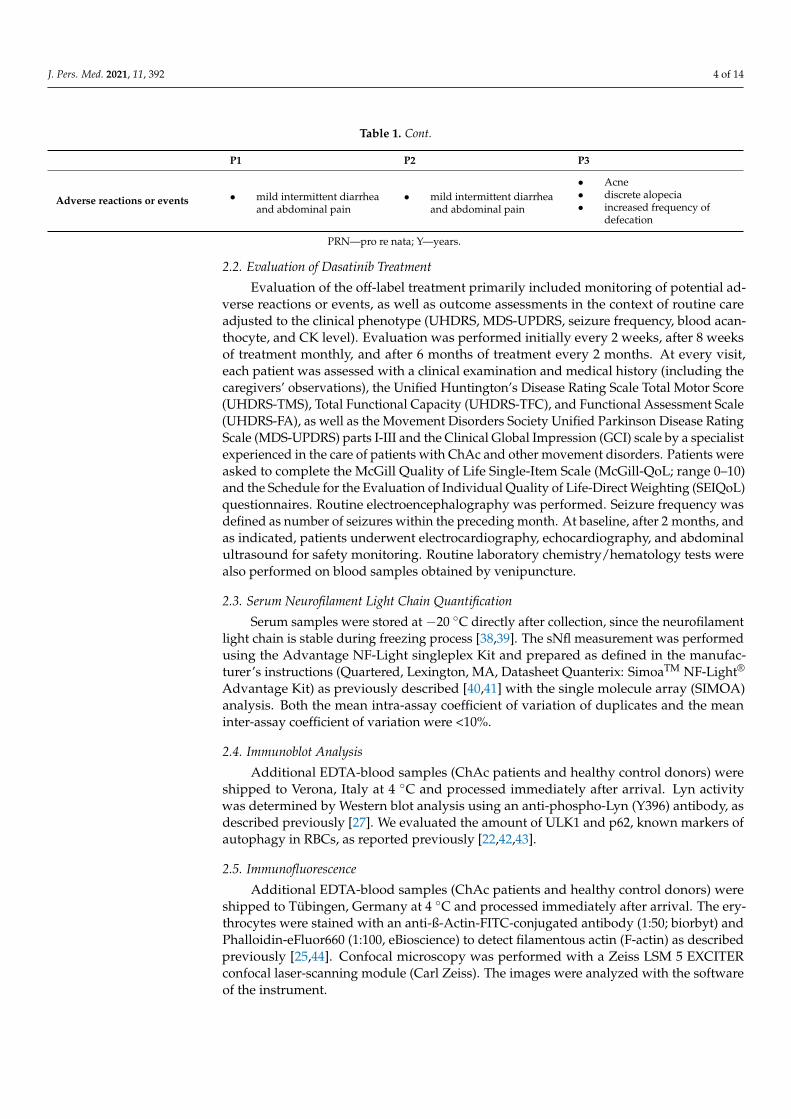
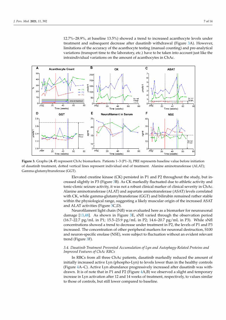
We next investigated whether dasatinib treatment alters current biomarker candidatesof the disease. Of those, the amount of acanthocytes showed severe variations. Theacanthocyte level of P1 (range 21.4%–42.0%, at baseline 35.2%) and P2 (range 19.4%–44.6%,at baseline 24.2%) varied markedly during the treatment without a clear trend. P3 (range
J. Pers. Med. 2021, 11, 392 7 of 14
12.7%–28.9%, at baseline 13.5%) showed a trend to increased acanthocyte levels undertreatment and subsequent decrease after dasatinib withdrawal (Figure 3A). However,limitations of the accuracy of the acanthocyte testing (manual counting) and pre-analyticalvariations (transport time to the laboratory, etc.) have to be taken into account just like theintraindividual variations on the amount of acanthocytes in ChAc.
J. Pers. Med. 2021, 11, x FOR PEER REVIEW 7 of 14
acanthocyte level of P1 (range 21.4%–42.0%, at baseline 35.2%) and P2 (range 19.4%–
44.6%, at baseline 24.2%) varied markedly during the treatment without a clear trend. P3
(range 12.7%–28.9%, at baseline 13.5%) showed a trend to increased acanthocyte levels
under treatment and subsequent decrease after dasatinib withdrawal (Figure 3A). How-
ever, limitations of the accuracy of the acanthocyte testing (manual counting) and pre-
analytical variations (transport time to the laboratory, etc.) have to be taken into account
just like the intraindividual variations on the amount of acanthocytes in ChAc.
Figure 3. Graphs A-F represent ChAc biomarkers. Patients 1–3 (P1–3), PRE represents baseline value before initiation of
dasatinib treatment, dotted vertical lines represent individual end of treatment. Alanine aminotransferase (ALAT);
Gamma-glutamyltransferase (GGT).
Elevated creatine kinase (CK) persisted in P1 and P2 throughout the study, but in-
creased slightly in P3 (Figure 3B). As CK markedly fluctuated due to athletic activity and
tonic-clonic seizure activity, it was not a robust clinical marker of clinical severity in ChAc.
Alanine aminotransferase (ALAT) and aspartate aminotransferase (ASAT) levels corre-
lated with CK, while gamma-glutamyltransferase (GGT) and bilirubin remained rather
stable within the physiological range, suggesting a likely muscular origin of the increased
ASAT and ALAT activities (Figure 3C,D).
Neurofilament light chain (Nfl) was evaluated here as a biomarker for neuroaxonal
damage [13,48]. As shown in Figure 3E, sNfl varied through the observation period (16.7–
22.7 pg/mL in P1; 15.5–23.9 pg/mL in P2; 14.6–20.7 pg/mL in P3). While sNfl concentra-
tions showed a trend to decrease under treatment in P2, the levels of P1 and P3 increased.
The concentration of other peripheral markers for neuronal destruction, S100 and neuron-
specific enolase (NSE), were subject to fluctuation without an evident relevant trend (Fig-
ure 3F).
3.4. Dasatinib Treatment Prevented Accumulation of Lyn and Autophagy-Related Proteins and
Improved Features of ChAc RBCs
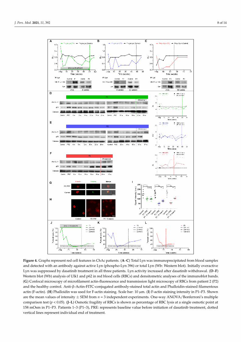
In RBCs from all three ChAc patients, dasatinib markedly reduced the amount of
initially increased active Lyn (phospho-Lyn) to levels lower than in the healthy controls
(Figure 4A–C). Active Lyn abundance progressively increased after dasatinib was with-
drawn. It is of note that in P1 and P2 (Figure 4A,B) we observed a slight and temporary
Figure 3. Graphs (A–F) represent ChAc biomarkers. Patients 1–3 (P1–3), PRE represents baseline value before initiationof dasatinib treatment, dotted vertical lines represent individual end of treatment. Alanine aminotransferase (ALAT);Gamma-glutamyltransferase (GGT).
Elevated creatine kinase (CK) persisted in P1 and P2 throughout the study, but in-creased slightly in P3 (Figure 3B). As CK markedly fluctuated due to athletic activity andtonic-clonic seizure activity, it was not a robust clinical marker of clinical severity in ChAc.Alanine aminotransferase (ALAT) and aspartate aminotransferase (ASAT) levels correlatedwith CK, while gamma-glutamyltransferase (GGT) and bilirubin remained rather stablewithin the physiological range, suggesting a likely muscular origin of the increased ASATand ALAT activities (Figure 3C,D).
Neurofilament light chain (Nfl) was evaluated here as a biomarker for neuroaxonaldamage [13,48]. As shown in Figure 3E, sNfl varied through the observation period(16.7–22.7 pg/mL in P1; 15.5–23.9 pg/mL in P2; 14.6–20.7 pg/mL in P3). While sNflconcentrations showed a trend to decrease under treatment in P2, the levels of P1 and P3increased. The concentration of other peripheral markers for neuronal destruction, S100and neuron-specific enolase (NSE), were subject to fluctuation without an evident relevanttrend (Figure 3F).
3.4. Dasatinib Treatment Prevented Accumulation of Lyn and Autophagy-Related Proteins andImproved Features of ChAc RBCs
In RBCs from all three ChAc patients, dasatinib markedly reduced the amount ofinitially increased active Lyn (phospho-Lyn) to levels lower than in the healthy controls(Figure 4A–C). Active Lyn abundance progressively increased after dasatinib was with-drawn. It is of note that in P1 and P2 (Figure 4A,B) we observed a slight and temporaryincrease in Lyn activation after 12 and 14 weeks of treatment, respectively, to values similarto those of controls, but still lower compared to baseline.
J. Pers. Med. 2021, 11, 392 8 of 14
J. Pers. Med. 2021, 11, x FOR PEER REVIEW 8 of 14
increase in Lyn activation after 12 and 14 weeks of treatment, respectively, to values sim-
ilar to those of controls, but still lower compared to baseline.
Figure 4. Graphs represent red cell features in ChAc patients. (A–C) Total Lyn was immunoprecipitated from blood samplesand detected with an antibody against active Lyn (phospho-Lyn 396) or total Lyn (Wb: Western blot). Initially overactiveLyn was suppressed by dasatinib treatment in all three patients. Lyn activity increased after dasatinib withdrawal. (D–F)Western blot (Wb) analysis of Ulk1 and p62 in red blood cells (RBCs) and densitometric analyses of the immunoblot bands.(G) Confocal microscopy of microfilament actin-fluorescence and transmission light microscopy of RBCs from patient 2 (P2)and the healthy control. Anti-β-Actin-FITC-conjugated antibody-stained total actin and Phalloidin-stained filamentousactin (F-actin). (H) Phalloidin was used for F-actin staining. Scale bar: 10 µm. (I) F-actin staining intensity in P1–P3. Shownare the mean values of intensity ± SEM from n = 3 independent experiments. One-way ANOVA/Bonferroni’s multiplecomparison test (p < 0.05). (J–L) Osmotic fragility of RBCs is shown as percentage of RBC lysis at a single osmotic point at158 mOsm in P1–P3. Patients 1–3 (P1–3), PRE: represents baseline value before initiation of dasatinib treatment, dottedvertical lines represent individual end of treatment.
J. Pers. Med. 2021, 11, 392 9 of 14
Autophagy initiator ULK1 markedly decreased after 4 weeks of treatment in allpatients (Figure 4D–F) and increased after dasatinib withdrawal. The accumulation ofthe late-phase autophagy marker p62 markedly decreased between 14 and 24 weeks oftreatment in P1 (Figure 4D), and between 12 and 22 weeks of dasatinib in P2 (Figure 4E).In contrast, p62 accumulation in P3 RBCs was only temporarily reduced versus baseline(Figure 4F) and re-accumulation along with active Lyn before the scheduled interruptionof dasatinib treatment was consistent with possibly uncompliant dosing.
We revealed a severe loss of cortical actin in RBCs from P2 and P3 as compared tothe healthy controls at baseline, whereas the cortical actin network was less impaired inP1 (Figure 4G,H). Consistent with the findings described above, we observed a slight butsignificant increase of F-actin staining in RBCs from P2 and P3 (Figure 4H,I) associatedwith improvement/normalization of osmotic fragility (Figure 4J–L). After withdrawal ofdasatinib, osmotic fragility again progressively deteriorated (Figure 4J–L).
4. Discussion
Chorea-acanthocytosis is a devastating neurodegenerative multi-system disorderwithout any established disease-modifying treatment. In our previous work, we showedthat elevated Lyn kinase activity represents a pathophysiological hallmark of the dis-ease [22,27,29]. FDA-approved TKIs like dasatinib with specificity to the Lyn kinase aretherefore promising candidates for a potential disease-modifying treatment. Herein, wereport on three phenotypically different ChAc patients who were prescribed dasatinib andmonitored for potential adverse reactions or events [47] and therapy response.
Dasatinib successfully engaged the target Lyn kinase with subsequent normalizationof ChAc RBC features, but neither showed positive nor negative effects on central nervoussystem parameters in the three ChAc patients. Oral administration of 100 mg dasatinibonce a day appeared to be reasonably safe in subjects with ChAc. Only rather mild adversereactions including gastrointestinal and cutaneous problems occurred, and hematologicaladverse reactions were transient and well tolerated.
Dasatinib sufficiently inhibited Lyn kinase at least in RBCs. In all patients, the phospho-Lyn/total Lyn ratio in erythrocytes was pathologically increased at baseline, consistent withour previous findings [22,27]. The ratio markedly decreased under dasatinib treatment.Moreover, Lyn kinase inhibition improved autophagy as supported by the reduction ofaccumulated autophagy-related proteins [22], and led to a rearrangement of the initiallyseverely impaired cortical filamentous actin network. Together with the reduction of theosmotic fragility, these findings implicate improved mechanical properties of ChAc RBCmembranes in dasatinib-treated subjects.
On the other hand, clinical motor and non-motor parameters could not show a cleartreatment effect. Considering the rather slow progression rate of ChAc, however, theabsence of an immediate short-term improvement with such a disease-modifying approachis not surprising. Since limited knowledge regarding the natural history and the het-erogeneity of the disease course complicates the interpretation of these data, it remainsuncertain if the lack of evident clinical progression in all three patients can be attributed topositive treatment effects. Most notable was the regain of deep tendon reflexes in P2, whichmight point to a neuromuscular (peripheral) treatment response. However, the variation ofdeep tendon reflexes in principle limits the significance of this finding and more objectiveparameters such as nerve conduction velocity studies (including H-reflex) should be per-formed in future studies. sNfl levels in cerebrospinal fluid or in serum reflects neuroaxonaldamage both of the central and the peripheral nervous system [48] and is known to beelevated in ChAc [13]. Under dasatinib treatment, there was a trend towards decreasedsNfl concentration in the patient who regained deep tendon reflexes; however, there wasan increase in the other two patients. It is not clear if this course is due to the therapy, thenatural history of the disease or if it reflects intraindividual variations. Therefore, sNfland other brain damage markers needs to be further established as biomarker for ChAc bylong-term profiling of the individual levels.
J. Pers. Med. 2021, 11, 392 10 of 14
One main finding of the study is that currently believed evident read-out parametersfor ChAc (e.g., acanthocyte count, elevated CK, seizure frequency) which were hypothe-sized to show possible short time effects all failed to be useful in a stringent longitudinalfollow-up of these patients. Particularly CK turned out to be subject of pronounced fluc-tuations due to athletic activity and bilateral convulsive seizures. Also, the amount ofacanthocytes could not be used as parameter to evaluate the drug effect due to limita-tions of the accuracy of the acanthocyte testing and significant intraindividual variationson the amount of acanthocytes in ChAc. Thus, all these parameters are not as robustas expected, which—combined with the slow progression rate of the disease— makes itcurrently difficult to perform clinical studies in this population of patients. With respect tothe limited knowledge of the natural history, defining appropriate read-out parametersremains challenging. This is still, however, a classical translational roadblock in ultra-rarediseases in general. Larger observational studies, such as worldwide case registers likethe Neuroacanthocytosis database, a submodule of the European Huntington’s DiseaseNetwork (https://www.euro-hd.net/html/na/submodule), need to be further supportedand enlarged to establish robust biomarkers and to enable “clinical trial readiness”. Ourstudy represents an additional contribution to achieve this objective, as we suggest a novelpanel of outcome parameters for future clinical studies and for cohort characterizationin ChAc.
Further open questions relate to the duration and time point of the treatment. It can beassumed that disease modification is more effective in the early stages of disease. However,all of our patients already experienced the first symptoms 9 to 28 years prior to dasatinibtherapy, which might limit its effects. Considering the rather low progression rate of ChAc,longer treatment periods might be necessary to achieve further results.
5. Conclusions
In summary, this is the first report on a potentially disease-modifying treatmentapproach in ChAc. Our observations during an off-label treatment suggest that dasatinib issafe and may successfully target pathologically accumulated Lyn kinase, at least in RBCs.However, only a very small number of patients were treated over a rather short time period,which are two major limitations of this study. Nevertheless, our observations warrantmoving forward with the evaluation of Lyn kinase inhibitor effects in larger studies. Thisstudy also reveals the need for further investigations of the natural history and robustbiomarkers of the disease.
Author Contributions: Conceptualization, K.P., R.O., L.D.F., and A.H.; Data curation, K.P. and H.G.;Formal analysis, K.P., H.G., E.F., A.M., L.P., and K.A.; Funding acquisition, A.H.; Investigation, K.P.,H.G., E.F., A.M., L.P., K.A., T.Z., L.D.F., and A.H.; Methodology, R.O., F.L., L.D.F., and A.H.; Projectadministration, A.H.; Resources, L.D.F. and A.H.; Supervision: A.H.; Validation, L.D.F. and A.H.;Writing—original draft, K.P.; Writing—review and editing, H.G., E.F., A.M., L.P., K.A., T.Z., R.O., F.L.,L.D.F., and A.H. All authors have read and agreed to the published version of the manuscript.
Funding: This study was supported by the Centre for Regenerative Therapies Dresden (CRTD),the German Center for Neurodegenerative Diseases (DZNE), research site Dresden, the HelmholtzVirtual Institute (VH-VI-510), the Else Kröner Clinician Scientist Program (TU Dresden, Germany)and the Rostock Academy for Clinician Scientists (RACS, University of Rostock, Germany).
Institutional Review Board Statement: Patients and healthy control blood donors were enrolled inongoing studies on the pathogenesis and natural history of neurodegenerative diseases approved bythe institutional review board of the Technische Universität Dresden, Germany (EK 45022009, EK78022015, EK517122019).
Informed Consent Statement: All patients gave their informed consent for off-label treatment withdasatinib, including the risk of possible life-threatening adverse reactions, as well as for videodocumentation and publication of the data.
Data Availability Statement: The data presented in this study are available within this article.
J. Pers. Med. 2021, 11, 392 11 of 14
Acknowledgments: The authors thank all patients and their families for giving consent for pub-lication of the data and the healthy control subjects for participation in this study. Expenses forthe off-label dasatinib prescription were gratefully covered in part by the Center for RegenerativeTherapies Dresden (CRTD, TU Dresden, Germany) and the public healthcare insurance of the pa-tients. We are especially grateful to Glenn (†) and Ginger Irvine as the founders of the Advocacy forNeuroacanthocytosis Patients (www.naadvocacy.org) and to Susan Wagner and Joy Willard-Willifordas representatives of the NA Advocacy USA (www.naadvocacyusa.org). Diagnostic Western blotanalysis for chorein was performed by G. Kwiatkowski and Benedikt Bader with the financial supportof the Advocacy for Neuroacanthocytosis Patients and of the ERA-net E-Rare consortium EMINA(European Multidisciplinary Initiative on Neuroacanthocytosis; BMBF01GM1003) in the labs ofProfessors Hans Kretzschmar/Armin Giese (Neuropathology) and Adrian Danek (Neurology) atLudwig-Maximilians-Universität Munich, Germany. K.P. was supported by the Else Kröner clinicianscientist program and the MeDDrive program (TU Dresden, Germany), as well as by the RostockAcademy for Clinician Scientists (RACS) and the FORUN program (University of Rostock, Germany).A.H. was supported by the “Hermann und Lilly Schilling-Stiftung für medizinische Forschungim Stifterverband”.
Conflicts of Interest: The authors declare that they have no competing interest related to themanuscript. Financial Disclosures of all authors (for the preceding 12 months): K.P. has receivedfunding from the Else Kröner Clinician Scientist Program (TU Dresden, Germany), the RostockAcademy for Clinician Scientists (RACS, University of Rostock, Germany), the MeDDrive grantof the Technische Universität Dresden, and the FORUN grant of the University of Rostock. K.A.received honoraria for presentation and consulting service from Biogen Idec, Merck, Sanofi, andRoche. T.Z. received honoraria for presentation and consulting service from Biogen, Bayer, Celgene,Novartis, Roche, Sanofi, and Teva. He received additional financial support for research activitiesfrom BAT, Biogen, Novartis, Roche, Teva, and Sanofi Aventis. L.D.F. has received research fundingfrom Agios. A.H. has received funding from the Federal Ministry of Education and Research (BMBF),the Helmholtz-Association, the Hermann und Lilly Schilling-Stiftung für medizinsiche Forschungim Stifterverband, the “Innovationsfond des Gemeinsamen Bundenausschusses”. He has receivedhonoraria for presentations, advisory boards, consultations from BIOGEN and Desitin. He hasreceived royalties from Elsevier Press. He serves as an editorial board member of BMC Neurology.H.G., A.M., E.F., L.P., R.O., and F.L. no funding to declare.
Abbreviations
Chorea-acanthocytosis (ChAc); Red blood cell (RBC); Creatine kinase (CK); Serum neurofilament(sNfL); Induced pluripotent stem cell (iPSC); Tyrosine kinase inhibitor (TKI); Patients 1–3 (P1–3); Uni-fied Huntington’s Disease Rating Scale Total Motor Score (UHDRS-TMS), Total Functional Capacity(UHDRS-TFC) and Functional Assessment Scale (UHDRS-FA); Movement Disorders Society UnifiedParkinson Disease Rating Scale (MDS-UPDRS); Clinical Global Impression (GCI); McGill Quality ofLife Single Item Scale (McGill-QoL); Schedule for the Evaluation of Individual Quality of Life-DirectWeighting (SEIQoL); Single molecule array (SIMOA); Mean corpuscular volume (MCV); Mean cor-puscular hemoglobin (MCH); Red blood cell distribution width (RDW); Alanine aminotransferase(ALAT); Aspartate aminotransferase (ASAT); Gamma-glutamyltransferase (GGT); Neurofilamentlight chain (Nfl); Neuron-specific enolase (NSE).
Appendix A
Members of The Network for Translational Research for Neuroacanthocytosis Patients(coordinator: Andreas Hermann):
Ezio Bonifacio (Center for Regenerative Therapies Dresden (CRTD), Technische Uni-versität Dresden, Dresden, Germany), Giel Bosman (Department of Biochemistry, RadboudUniversity Medical Center, Nijmegen, The Netherlands), Adrian Danek (NeurologischeKlinik, Klinikum der Universität, Ludwig-Maximilians-Universität München, Munich,Germany), Peter Claus (Institute of Neuroanatomy and Cell Biology, Hannover MedicalSchool, Hannover, Germany and Center for Systems Neuroscience (ZSN) Hannover, Han-nover, Germany), Jochen Guck (Biotechnology Center, Technische Universität Dresden,Dresden, Germany), Ulrich Salzer (Center for Medical Biochemistry, Max Perutz Labs,Medical University of Vienna, Austria).
J. Pers. Med. 2021, 11, 392 12 of 14
References1. Jung, H.H.; Danek, A.; Walker, R.H. Neuroacanthocytosis syndromes. Orphanet J. Rare Dis. 2011, 6, 68. [CrossRef] [PubMed]2. Velayos Baeza, A.; Dobson-Stone, C.; Rampoldi, L.; Bader, B.; Walker, R.H.; Danek, A.; Monaco, A.P. Chorea-Acanthocytosis. In
GeneReviews; Adam, M.P., Ardinger, H.H., Pagon, R.A., Wallace, S.E., Bean, L.J.H., Stephens, K., Amemiya, A., Eds.; University ofWashington: Seattle, WA, USA, 1993.
3. Walker, R.H.; Danek, A. “Neuroacanthocytosis”—Overdue for a Taxonomic Update. Tremor Other Hyperkinet. Mov. (N. Y.) 2021,11, 1. [CrossRef]
4. Peikert, K.; Danek, A.; Hermann, A. Current state of knowledge in Chorea-Acanthocytosis as core Neuroacanthocytosis syndrome.Eur. J. Med. Genet. 2018, 61, 699–705. [CrossRef]
5. Danek, A.; Bader, B.; Velayos-Baeza, A.; Walker, R.H. Autosomal recessive transmission of chorea-acanthocytosis confirmed. ActaNeuropathol. 2012, 123, 905–906. [CrossRef]
6. Dobson-Stone, C.; Danek, A.; Rampoldi, L.; Hardie, R.J.; Chalmers, R.M.; Wood, N.W.; Bohlega, S.; Dotti, M.T.; Federico, A.;Shizuka, M.; et al. Mutational spectrum of the CHAC gene in patients with chorea-acanthocytosis. Eur. J. Hum. Genet. EJHG 2002,10, 773–781. [CrossRef] [PubMed]
7. Rampoldi, L.; Dobson-Stone, C.; Rubio, J.P.; Danek, A.; Chalmers, R.M.; Wood, N.W.; Verellen, C.; Ferrer, X.; Malandrini, A.;Fabrizi, G.M.; et al. A conserved sorting-associated protein is mutant in chorea-acanthocytosis. Nat. Genet. 2001, 28, 119.[CrossRef] [PubMed]
8. Ueno, S.-I.; Maruki, Y.; Nakamura, M.; Tomemori, Y.; Kamae, K.; Tanabe, H.; Yamashita, Y.; Matsuda, S.; Kaneko, S.; Sano, A. Thegene encoding a newly discovered protein, chorein, is mutated in chorea-acanthocytosis. Nat. Genet. 2001, 28, 121. [CrossRef]
9. Walker, R.H. Management of Neuroacanthocytosis Syndromes. Tremor Other Hyperkinet. Mov. (N. Y.) 2015, 5, 346. [CrossRef]10. Rampoldi, L.; Danek, A.; Monaco, A.P. Clinical features and molecular bases of neuroacanthocytosis. J. Mol. Med. (Berl. Ger.)
2002, 80, 475–491. [CrossRef]11. Estevez-Fraga, C.; Lopez-Sendon Moreno, J.L.; Martinez-Castrillo, J.C. Phenomenology and disease progression of chorea-
acanthocytosis patients in Spain. Parkinsonism Relat. Disord. 2018, 49, 17–21. [CrossRef]12. Hermann, A.; Walker, R.H. Diagnosis and treatment of chorea syndromes. Curr. Neurol. Neurosci. Rep. 2015, 15, 514. [CrossRef]
[PubMed]13. Peikert, K.; Akgün, K.; Beste, C.; Ziemssen, T.; Buhmann, C.; Danek, A.; Hermann, A. Neurofilament light chain in serum is
significantly increased in chorea-acanthocytosis. Parkinsonism Relat. Disord. 2020, 80, 28–31. [CrossRef] [PubMed]14. Liu, J.; Heinsen, H.; Grinberg, L.T.; Alho, E.; Amaro, E., Jr.; Pasqualucci, C.A.; Rub, U.; den Dunnen, W.; Arzberger, T.; Schmitz, C.;
et al. Subcortical neurodegeneration in chorea: Similarities and differences between chorea-acanthocytosis and Huntington’sdisease. Parkinsonism Relat. Disord. 2018, 49, 54–59. [CrossRef]
15. Liu, J.; Heinsen, H.; Grinberg, L.T.; Alho, E.; Amaro, E., Jr.; Pasqualucci, C.A.; Rub, U.; Seidel, K.; den Dunnen, W.; Arzberger, T.;et al. Pathoarchitectonics of the cerebral cortex in chorea-acanthocytosis and HD. Neuropathol. Appl. Neurobiol. 2018, 45, 230–243.[CrossRef] [PubMed]
16. Gao, M.; Yang, H. VPS13: A lipid transfer protein making contacts at multiple cellular locations. J. Cell Biol. 2018, 217, 3322–3324.[CrossRef]
17. Kumar, N.; Leonzino, M.; Hancock-Cerutti, W.; Horenkamp, F.A.; Li, P.; Lees, J.A.; Wheeler, H.; Reinisch, K.M.; De Camilli, P.VPS13A and VPS13C are lipid transport proteins differentially localized at ER contact sites. J. Cell Biol. 2018, 217, 3625–3639.[CrossRef] [PubMed]
18. Yeshaw, W.M.; van der Zwaag, M.; Pinto, F.; Lahaye, L.L.; Faber, A.I.; Gomez-Sanchez, R.; Dolga, A.M.; Poland, C.; Monaco, A.P.;van IJzendoorn1, S.C.; et al. Human VPS13A is associated with multiple organelles and influences mitochondrial morphologyand lipid droplet motility. eLife 2019, 8, e43561. [CrossRef] [PubMed]
19. Park, J.S.; Neiman, A.M. XK is a partner for VPS13A: A molecular link between Chorea-Acanthocytosis and McLeod Syndrome.Mol. Biol. Cell 2020, 31, 2425–2436. [CrossRef]
20. Dziurdzik, S.K.; Conibear, E. The Vps13 Family of Lipid Transporters and Its Role at Membrane Contact Sites. Int. J. Mol. Sci.2021, 22, 2905. [CrossRef]
21. Lang, F.; Pelzl, L.; Schols, L.; Hermann, A.; Foller, M.; Schaffer, T.E.; Stournaras, C. Neurons, Erythrocytes and Beyond—TheDiverse Functions of Chorein. Neuro-Signals 2017, 25, 117–126. [CrossRef]
22. Lupo, F.; Tibaldi, E.; Matte, A.; Sharma, A.K.; Brunati, A.M.; Alper, S.L.; Zancanaro, C.; Benati, D.; Siciliano, A.; Bertoldi, M.;et al. A new molecular link between defective autophagy and erythroid abnormalities in chorea-acanthocytosis. Blood 2016, 128,2976–2987. [CrossRef]
23. Munoz-Braceras, S.; Calvo, R.; Escalante, R. TipC and the chorea-acanthocytosis protein VPS13A regulate autophagy in Dic-tyostelium and human HeLa cells. Autophagy 2015, 11, 918–927. [CrossRef]
24. Pelzl, L.; Elsir, B.; Sahu, I.; Bissinger, R.; Singh, Y.; Sukkar, B.; Honisch, S.; Schoels, L.; Jemaa, M.; Lang, E.; et al. Lithium Sensitivityof Store Operated Ca2+ Entry and Survival of Fibroblasts Isolated from Chorea-Acanthocytosis Patients. Cell. Physiol. Biochem.Int. J. Exp. Cell. Physiol. Biochem. Pharmacol. 2017, 42, 2066–2077. [CrossRef] [PubMed]
25. Foller, M.; Hermann, A.; Gu, S.; Alesutan, I.; Qadri, S.M.; Borst, O.; Schmidt, E.M.; Schiele, F.; vom Hagen, J.M.; Saft, C.; et al.Chorein-sensitive polymerization of cortical actin and suicidal cell death in chorea-acanthocytosis. FASEB J. Off. Publ. Fed. Am.Soc. Exp. Biol. 2012, 26, 1526–1534. [CrossRef]
J. Pers. Med. 2021, 11, 392 13 of 14
26. Hosseinzadeh, Z.; Hauser, S.; Singh, Y.; Pelzl, L.; Schuster, S.; Sharma, Y.; Höflinger, P.; Zacharopoulou, N.; Stournaras, C.;Rathbun, D.L.; et al. Decreased Na+/K+ ATPase Expression and Depolarized Cell Membrane in Neurons Differentiated fromChorea-Acanthocytosis Patients. Sci. Rep. 2020, 10, 8391. [CrossRef] [PubMed]
27. De Franceschi, L.; Tomelleri, C.; Matte, A.; Brunati, A.M.; Bovee-Geurts, P.H.; Bertoldi, M.; Lasonder, E.; Tibaldi, E.; Danek, A.;Walker, R.H.; et al. Erythrocyte membrane changes of chorea-acanthocytosis are the result of altered Lyn kinase activity. Blood2011, 118, 5652–5663. [CrossRef] [PubMed]
28. Munoz-Braceras, S.; Tornero-Ecija, A.R.; Vincent, O.; Escalante, R. VPS13A is closely associated with mitochondria and is requiredfor efficient lysosomal degradation. Dis. Models Mech. 2019, 12. [CrossRef]
29. Stanslowsky, N.; Reinhardt, P.; Glass, H.; Kalmbach, N.; Naujock, M.; Hensel, N.; Lubben, V.; Pal, A.; Venneri, A.; Lupo, F.; et al.Neuronal Dysfunction in iPSC-Derived Medium Spiny Neurons from Chorea-Acanthocytosis Patients Is Reversed by Src KinaseInhibition and F-Actin Stabilization. J. Neurosci. Off. J. Soc. Neurosci. 2016, 36, 12027–12043. [CrossRef]
30. Roskoski, R., Jr. Src protein-tyrosine kinase structure, mechanism, and small molecule inhibitors. Pharmacol. Res. 2015, 94, 9–25.[CrossRef]
31. Porkka, K.; Koskenvesa, P.; Lundan, T.; Rimpilainen, J.; Mustjoki, S.; Smykla, R.; Wild, R.; Luo, R.; Arnan, M.; Brethon, B.; et al.Dasatinib crosses the blood-brain barrier and is an efficient therapy for central nervous system Philadelphia chromosome-positiveleukemia. Blood 2008, 112, 1005–1012. [CrossRef] [PubMed]
32. Khairoalsindi, O.A.; Abuzinadah, A.R. Maximizing the Survival of Amyotrophic Lateral Sclerosis Patients: Current Perspectives.Neurol. Res. Int. 2018, 2018, 6534150. [CrossRef] [PubMed]
33. Trias, E.; Ibarburu, S.; Barreto-Nunez, R.; Babdor, J.; Maciel, T.T.; Guillo, M.; Gros, L.; Dubreuil, P.; Diaz-Amarilla, P.; Cassina, P.;et al. Post-paralysis tyrosine kinase inhibition with masitinib abrogates neuroinflammation and slows disease progression ininherited amyotrophic lateral sclerosis. J. Neuroinflamm. 2016, 13, 177. [CrossRef] [PubMed]
34. Turner, R.S.; Hebron, M.L.; Lawler, A.; Mundel, E.E.; Yusuf, N.; Starr, J.N.; Anjum, M.; Pagan, F.; Torres-Yaghi, Y.; Shi, W.; et al.Nilotinib Effects on Safety, Tolerability, and Biomarkers in Alzheimer’s Disease. Ann. Neurol. 2020, 88, 183–194. [CrossRef][PubMed]
35. Pagan, F.L.; Hebron, M.L.; Wilmarth, B.; Torres-Yaghi, Y.; Lawler, A.; Mundel, E.E.; Yusuf, N.; Starr, N.J.; Anjum, M.; Arellano, J.;et al. Nilotinib Effects on Safety, Tolerability, and Potential Biomarkers in Parkinson Disease: A Phase 2 Randomized ClinicalTrial. JAMA Neurol. 2020, 77, 309–317. [CrossRef]
36. Simuni, T.; Fiske, B.; Merchant, K.; Coffey, C.S.; Klingner, E.; Caspell-Garcia, C.; Lafontant, D.E.; Matthews, H.; Wyse, R.K.;Brundin, P.; et al. Efficacy of Nilotinib in Patients With Moderately Advanced Parkinson Disease: A Randomized Clinical Trial.JAMA Neurol. 2020, 78, 312–320. [CrossRef]
37. Dobson-Stone, C.; Velayos-Baeza, A.; Filippone, L.A.; Westbury, S.; Storch, A.; Erdmann, T.; Wroe, S.J.; Leenders, K.L.; Lang,A.E.; Dotti, M.T.; et al. Chorein detection for the diagnosis of chorea-acanthocytosis. Ann. Neurol. 2004, 56, 299–302. [CrossRef][PubMed]
38. Kuhle, J.; Plattner, K.; Bestwick, J.P.; Lindberg, R.L.; Ramagopalan, S.V.; Norgren, N.; Nissim, A.; Malaspina, A.; Leppert, D.;Giovannoni, G.; et al. A comparative study of CSF neurofilament light and heavy chain protein in MS. Mult. Scler. J. 2013, 19,1597–1603. [CrossRef] [PubMed]
39. Keshavan, A.; Heslegrave, A.; Zetterberg, H.; Schott, J.M. Stability of blood-based biomarkers of Alzheimer’s disease overmultiple freeze-thaw cycles. Alzheimer’s Dement. (Amst.) 2018, 10, 448–451. [CrossRef]
40. Beste, C.; Stock, A.K.; Zink, N.; Ocklenburg, S.; Akgun, K.; Ziemssen, T. How minimal variations in neuronal cytoskeletal integritymodulate cognitive control. NeuroImage 2019, 185, 129–139. [CrossRef]
41. Akgün, K.; Kretschmann, N.; Haase, R.; Proschmann, U.; Kitzler, H.H.; Reichmann, H.; Ziemssen, T. Profiling individualclinical responses by high-frequency serum neurofilament assessment in MS. Neurol. Neuroimmunol. Neuroinflamm. 2019, 6, e555.[CrossRef] [PubMed]
42. Matte, A.; De Falco, L.; Federti, E.; Cozzi, A.; Iolascon, A.; Levi, S.; Mohandas, N.; Zamo, A.; Bruno, M.; Lebouef, C.; et al.Peroxiredoxin-2: A Novel Regulator of Iron Homeostasis in Ineffective Erythropoiesis. Antioxid. Redox Signal. 2018, 28, 1–14.[CrossRef]
43. Beneduce, E.; Matte, A.; De Falco, L.; Mbiandjeu, S.; Chiabrando, D.; Tolosano, E.; Federti, E.; Petrillo, S.; Mohandas, N.; Siciliano,A.; et al. Fyn kinase is a novel modulator of erythropoietin signaling and stress erythropoiesis. Am. J. Hematol. 2019, 94, 10–20.[CrossRef] [PubMed]
44. Honisch, S.; Gu, S.; Vom Hagen, J.M.; Alkahtani, S.; Al Kahtane, A.A.; Tsapara, A.; Hermann, A.; Storch, A.; Schols, L.; Lang,F.; et al. Chorein Sensitive Arrangement of Cytoskeletal Architecture. Cell. Physiol. Biochem. Int. J. Exp. Cell. Physiol. Biochem.Pharmacol. 2015, 37, 399–408. [CrossRef]
45. Olivieri, O.; De Franceschi, L.; Bordin, L.; Manfredi, M.; Miraglia del Giudice, E.; Perrotta, S.; De Vivo, M.; Guarini, P.; Corrocher,R. Increased membrane protein phosphorylation and anion transport activity in chorea-acanthocytosis. Haematologica 1997, 82,648–653.
46. De Franceschi, L.; Fumagalli, L.; Olivieri, O.; Corrocher, R.; Lowell, C.A.; Berton, G. Deficiency of Src family kinases Fgr and Hckresults in activation of erythrocyte K/Cl cotransport. J. Clin. Investig. 1997, 99, 220–227. [CrossRef] [PubMed]

J. Pers. Med. 2021, 11, 392 14 of 14
47. Steegmann, J.L.; Baccarani, M.; Breccia, M.; Casado, L.F.; Garcia-Gutierrez, V.; Hochhaus, A.; Kim, D.W.; Kim, T.D.; Khoury, H.J.;Le Coutre, P.; et al. European LeukemiaNet recommendations for the management and avoidance of adverse events of treatmentin chronic myeloid leukaemia. Leukemia 2016, 30, 1648–1671. [CrossRef] [PubMed]
48. Khalil, M.; Teunissen, C.E.; Otto, M.; Piehl, F.; Sormani, M.P.; Gattringer, T.; Barro, C.; Kappos, L.; Comabella, M.; Fazekas, F.; et al.Neurofilaments as biomarkers in neurological disorders. Nat. Rev. Neurol. 2018, 14, 577–589. [CrossRef] [PubMed]