synthesis, characterisation, and antineoplastic cytotoxicity of hybrid naphthohydroquinone–nucleic...
TRANSCRIPT

ORI GINAL RESEARCH
Synthesis, characterisation, and antineoplasticcytotoxicity of hybrid naphthohydroquinone–nucleicbase mimic derivatives
Aurora Molinari Æ Claudia Ojeda Æ Alfonso Oliva ÆJose M. Miguel del Corral Æ M. Angeles Castro ÆPablo A. Garcıa Æ Carmen Cuevas ÆArturo San Feliciano
Received: 13 September 2007 / Accepted: 14 April 2008 / Published online: 13 May 2008
� Birkhauser Boston 2008
Abstract From a partially degraded Diels–Alder adduct of a-myrcene and 1,4-
benzoquinone, several model compounds belonging to a new series of 1,4-naph-
thohydroquinone derivatives have been prepared. Phenyl, pyridyl, imidazolyl and
some nucleic base mimic heterocycles have been attached to the naphthohydro-
quinone system through linkers of different size and type, leading to potentially
antineoplastic hybrid structures. The new compounds have been evaluated in vitro
for their cytotoxicity against cultured human cancer cells of A-549 lung carcinoma,
HT-29 colon adenocarcinoma and MDA-MB-231 breast carcinoma. GI50 values
ranged in the lM level.
Keywords Naphthohydroquinone � Hybrid structures � Antineoplastic cytotoxicity
Introduction
It is commonly accepted that simple or complex phenols, quinones and hydroqui-
nones are biologically active. Many of them are often cytotoxic for neoplastic cells
and a certain number are registered as anticancer drugs (O’Brien, 1991; Brunmark
and Cadenas, 1989; Powis, 1989). Their antitumour properties and mechanism of
action are attributed to a one-electron redox process of the quinone/hydroquinone
A. Molinari (&) � C. Ojeda � A. Oliva
Instituto de Quımica, Pontificia Universidad Catolica de Valparaıso, Casilla 4059 Valparaiso, Chile
e-mail: [email protected]
J. M. Miguel del Corral � M. A. Castro � P. A. Garcıa � A. San Feliciano
Departamento de Quımica Farmaceutica, Facultad de Farmacia, Universidad de Salamanca,
Salamanca, Spain
C. Cuevas
PharmaMar S.A., Avda de los Reyes, P.I. La Mina Norte, 28770-Colmenar Viejo, Madrid, Spain
Med Chem Res (2009) 18:59–69
DOI 10.1007/s00044-008-9108-1
MEDICINALCHEMISTRYRESEARCH
moiety, involving a semi-quinonic radical, which inhibits mitochondrial electron
transport and also decoupling oxidative phosphorylation (Esposti et al., 1984; Appel
and Powis, 1980; Konji et al., 1990). Furthermore they can act as topoisomerase
inhibitors, via DNA intercalation and reduction of the quinone moiety by oxido-
reductases (DT-diaphorase) (Cheng et al., 1996; Cheng, 1998; Chang et al., 1999).
A wide variety of prenylquinones/hydroquinones, their derivatives and related
compounds reported by us showed activity against several types of neoplastic cells.
They were usually prepared via Diels–Alder cyclocondensation between a-myrcene
and 1,2- or 1,4-benzoquinone, 2-acetyl-1,4-benzoquinone or 2-chloro-1,4-benzo-
quinone followed by further chemical transformations of the terpenyl side chain,
leading to several series of derivatives, displaying cytotoxicity IC50 values in the
lM range against several neoplastic cell lines. Some of the compounds tested
showed a moderated selectivity against P-388, Mel 28, SKBR3, K562 and PANC1
cell lines and resulted, in general, more potent than avarol and avarone (3–6 lM),
which were taken as reference standards in our research (Castro et al., 1996, 1998,
2002a, b, 2005a, b, c; Aguilera et al., 2000; Broughton et al., 2001; Araya et al.,2004).
It is also well known that many antineoplastic, antiviral and anti-infectious drugs
contain structures that mimic those of nucleic bases or nucleosides. As examples,
the common fluorouracil, azauridine and mercaptopurine are useful clinical drugs
which contain modified or false pyrimidine and purine nucleic bases, while the
acyclovir family, didanosine and zidovudine, among many other drugs, contain
modifications in the glycidic moiety of nucleosides (Lemke and Williams, 2002).
The common strategy of associating or hybridising two different bioactive
molecules or drugs with complementary pharmacophoric functions or with different
mechanisms of action, as a method for creating more efficacious, synergistic and
more potent drugs, has often been exploited with very good results. The pairs
sulphamethoxazole–trimethoprim, amoxicillin–clavulanic acid and imipenem–cila-
statin, are classic examples of antibacterial drugs associations. Benorilate is
probably the simplest example of covalent hybridizing of the two most common and
simple anti-inflammatories, acetylsalicylic acid and paracetamol (Korolkovas,
1988).The pyridine ring, either alone or fused to other planar homocyclic or
heterocyclic aromatic systems is widely present in a large and very significant
number of biologically active compounds and therapeutically useful drugs. This fact
makes the synthesis of pyridine and related derivatives an active research area in
new drug development. It is also believed that the presence of planar polycyclic
aromatic systems in active compounds is essential for their bioactivity. They usually
act as noncovalent DNA binders via intercalation induction of conformational
changes, unwinding the DNA at the binding site and interferences with the function
of DNA binding enzymes (Graves and Velea, 2000; Brana et al., 2001; Hurley,
2002; Chen et al., 2006). Vitamins B6 and P, isoniazid, chloroquine, ofloxacin,
quinine, aminacrine, etc. constitute a short list of the classic representative pyridine
bioactive compounds. Pyridine- and hydropyridine-containing compounds, such as
those of the camptothecin family, have shown their usefulness for treating several
types of cancer. Recently, other pyridine compounds such as several
60 Med Chem Res (2009) 18:59–69

pyridinylanthranilic acid derivatives (Cocco et al., 2004), 9-anilinoacridine mus-
tards (Bacherikov et al., 2004), new hexacyclic camptothecin derivatives (Chen
et al., 2005), diphenylpyridines (Bailly et al., 2005) and pyridine derivatives of
nitrobenzosuberone (Abdel-Hafez et al., 2006), among others, have been screened
against a wide variety of human neoplastic cell lines. In some cases, IC50 values in
the nM scale have been reported (Bacherikov et al., 2004).
Considering the mentioned antecedents, as part of our continuing research on
quinone/hydroquinone antineoplastics, we planned to carry out the association of
naphthoquinone/hydroquinone fragments from lead compounds previously reported
by us, with several types of nucleic base mimic fragments. As far as the authors know,
no previous description of cytotoxicity evaluation for such a type of hybrid molecules
has been reported in the literature. Thus, we describe in this paper the preparation of
hybrid molecules derived from naphthohydroquinonic compounds with the aim of
prospecting on their antineoplastic potentiality. The new compounds are hybrids
constituted by the naphthohydroquinone system and a nitrogen-containing heterocy-
clic fragment that mimics the nucleic base. The two units are linked by an alkyl or an
aminoalkyl chain of varying size. The compounds prepared were evaluated in order to
analyse the influence of the association of both systems on the antineoplastic
cytotoxicity and also the effect of the size of the linker on cytotoxicity.
Results and discussion
Chemistry
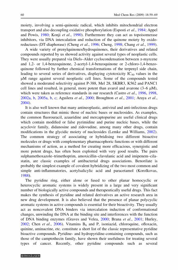
The new aminopyridyl and related derivatives of alkylnaphthohydroquinones were
prepared using the aldehyde 1 as the starting substrate. It was conveniently obtained
from the Diels–Alder cycloadduct formed with a-myrcene and p-benzoquinone,
followed by successive aromatisation with 2,3-dichloro-5,6-dicyano-1,4-benzoqui-
none (DDQ), epoxidation with m-chloroperbenzoic acid (MCPBA) and oxidative
degradation with periodic acid, according to the previously reported procedure (Castro
et al., 1998). Chemical transformations of the aldehyde 1 are summarized in
Scheme 1. The 3-(4-methoxyphenylamino)propyl compound 2 and the 3-(pyridin-3-
ylamino)propyl compound 3 were prepared by a direct solvent-free reductive
amination reaction of the aldehyde 1 with 4-methoxyaniline or 3-aminopyridine and
boric-acid-activated sodium borohydride. The reaction was carried out by grinding the
mixture with an agate mortar and pestle at room temperature in air (Cho and Kang,
2005). To obtain the 3-(2-aminopyridin-3-ylamino)propyl derivative 4, the imine
from 1 and 2,3-diaminopyridine was prepared in situ, either by refluxing with MgSO4
in benzene or by microwave (MW) irradiation of the mixture adsorbed on
montmorillonite K-10. The imine reduction was also performed with boric-acid-
activated sodium borohydride in an agate mortar. The 3-(1H-imidazo[4,5-b]pyridin-1-
yl)propyl compound 5 was prepared by refluxing derivative 4 with triethyl
orthoformiate. This procedure allowed the cyclisation of this reagent with the amino
groups of 4 by a substitution–elimination reaction (Grierson and Llegraverend, 2006).
The 2-(1H-imidazo[4,5-b]pyridin-2-yl)ethyl compound 6 and 2-(5,6-dichloro-1H-
Med Chem Res (2009) 18:59–69 61
benzo[d]imidazol-2-yl)ethyl compound 7 were prepared by an additional oxidation–
cyclisation sequence. Thus, the aldehyde 1 was refluxed with 2,3-diaminopyridine in
nitrobenzene or with 4,5-dichloro-1,2-phenylenediamine and p-benzoquinone in
ethanol to afford compounds 6 and 7, respectively. The main 1H and 13C data for these
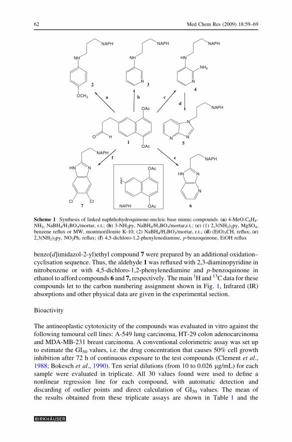
compounds let to the carbon numbering assignment shown in Fig. 1, Infrared (IR)
absorptions and other physical data are given in the experimental section.
Bioactivity
The antineoplastic cytotoxicity of the compounds was evaluated in vitro against the
following tumoural cell lines: A-549 lung carcinoma, HT-29 colon adenocarcinoma
and MDA-MB-231 breast carcinoma. A conventional colorimetric assay was set up
to estimate the GI50 values, i.e. the drug concentration that causes 50% cell growth
inhibition after 72 h of continuous exposure to the test compounds (Clement et al.,1988; Bokesch et al., 1990). Ten serial dilutions (from 10 to 0.026 lg/mL) for each
sample were evaluated in triplicate. All 30 values found were used to define a
nonlinear regression line for each compound, with automatic detection and
discarding of outlier points and direct calculation of GI50 values. The mean of
the results obtained from these triplicate assays are shown in Table 1 and the
HO
OAc
OAc1
NAPH
NH
OCH3
NAPH
NH
N32
OAc
OAc
NAPH
HN
N
NH2
4
NAPH
5
NAPH
6
NAPH
HN N
ClCl
HN N
N
NAPH7
a b cd
ef
N
NN
Scheme 1 Synthesis of linked naphthohydroquinone-nucleic base mimic compounds. (a) 4-MeO-C6H4-NH2, NaBH4/H3BO3/mortar, r.t.; (b) 3-NH2py, NaBH4/H3BO3/mortar,r.t.; (c) (1) 2,3(NH2)2py, MgSO4,benzene reflux or MW, montmorillonite K-10; (2) NaBH4/H3BO3/mortar, r.t.; (d) (EtO)3CH, reflux; (e)2,3(NH2)2py, NO2Ph, reflux; (f) 4,5-dichloro-1,2-phenylenediamine, p-benzoquinone, EtOH reflux
62 Med Chem Res (2009) 18:59–69
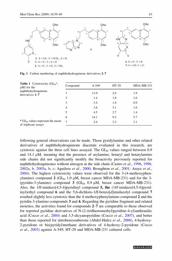
following general observations can be made. Those pyridylamino and other related
derivatives of naphthohydroquinone diacetate evaluated in this research, are
cytotoxic against the three cell lines assayed. The GI50 values ranged between 0.9
and 14.1 lM, meaning that the presence of arylamino, hetaryl and hetarylamino
side chains did not significantly modify the bioactivity previously reported for
naphthohydroquinones without nitrogen at the side chain (Castro et al., 1996, 1998,
2002a, b, 2005a, b, c; Aguilera et al., 2000; Broughton et al., 2001; Araya et al.,2004). The highest cytotoxicity values were observed for the 3-(4-methoxyphen-
ylamino) compound 2 (GI50 1.0 lM, breast cancer MDA-MB-231) and for the 3-
(pyridin-3-ylamino) compound 3 (GI50 0.9 lM, breast cancer MDA-MB-231).
Also, the 1H-imidazo[4,5-b]pyridinyl compound 5, the (1H-imidazo[4,5-b]pyrid-
inyl)ethyl compound 6 and the 5,6-dichloro-1H-benzo[d]imidazolyl compound 7resulted slightly less cytotoxic than the 4-methoxyphenylamino compound 2 and the
pyridin-3-ylamino compounds 3 and 4. Regarding the pyridine fragment and related
moieties, the activities found for compounds 2–7 are comparable to those observed
for reported pyridine derivatives of N-(2-(trifluoromethyl)pyridine-4-yl)anthranilic
acid (Cocco et al., 2004) and 3,5-dicyanopyridine (Cocco et al., 2007), and better
than those reported for nitrobenzosuberone (Abdel-Hafez et al., 2006), 4-hydroxy-
2-pyridone or bis(pyridyl)methane derivatives of 4-hydroxy-2-pyridone (Cocco
et al., 2003) against A-549, HT-29 and MDA-MB-231 cultured cells.
HN
X
OAc
OAc
11
9
6
88a
1
34a
3'
1´5'
5
OAc
OAc
9
6
8a1
34a
2 : X = CH ; Y = OCH3 ; Z = H
3 : X = N ; Y = Z = H 6: X = N ; Y = H
7: X = CH; Y = Cl
HN N
X
10
1'2'
3a'
5'
7a'
OAc
OAc
9
6
8a1
34a
5
10
N
NN
1´
2'
´3a'
6´
7a'
Y Y
Y4 : X = N ; Y = H ; Z = NH2
Fig. 1 Carbon numbering of naphthohydroquinone derivatives 2–7
Table 1 Cytotoxicity (GI50*,
lM) for the
naphthohydroquinone
derivatives 1–7
* GI50 values represent the mean
of triplicate assays
Compound A-549 HT-29 MDA-MB-231
1 11.0 2.6 1.9
2 1.4 1.8 1.0
3 3.4 1.6 0.9
4 3.6 3.1 1.6
5 4.5 2.7 1.4
6 14.1 9.2 5.7
7 2.6 3.3 3.1
Med Chem Res (2009) 18:59–69 63
While the spacing between the naphthalene fragment and the closest nitrogen is
identical in all of the compounds, the separation between the two cyclic systems, the
naphthalene-1,4-diyldiacetate (NAPH) and the hetaryl moieties, changes from two
(compounds 6, 7), to three (compound 5) to four (compounds 2–4) spacing units of the
linker. In this respect it should be noted that cytotoxicity increases with separation.
Additionally, in spite of the small number of compounds evaluated, it can be observed
that a higher number of nitrogen atoms in the hetarylamino fragment could negatively
influence the potency of these compounds, as the presence of two (compounds 4 and 7)
or more (compounds 5 and 6) nitrogen atoms in the hetaryl fragment leads to a
progressively diminished potency. These compounds could act either as nucleic base
antimetabolites or as redox modulators of the neoplastic cell metabolism, but a
definitive proposal about the actual mechanism would be too speculative at this stage
of the research. All these observations will serve to orient future research on this series
of naphthohydroquinone derivatives.
To summarise, we have prepared seven new arylaminated and hetarylaminated
derivatives of alkylnaphthohydroquinone diacetate, containing 4-metoxyphenyla-
mino, pyridin-3-ylamino, 1H-imidazo[4,5-b]pyridinyl and 5,6-dichloro-1H-
benzo[d]imidazolyl substituents, that essentially retain the activity of the parent
compound and whose GI50 values are in the lM range. The results of this research
will serve as an experimental base and the starting point for progressing towards the
development of new series of naphthohydroquinone derivatives. Natural nucleic
bases, their analogues, nucleosidyl and nucleotidyl substituents will be attached
through the side chain to evaluate the influence of this type of hybridisation on the
cytotoxicity of naphthohydroquinone diacetate.
Experimental
General
IR spectra were recorded on a Nicolet (Impact 410) spectrophotometer in NaCl film.
Nuclear magnetic resonance (NMR) spectra were recorded at 200 MHz for 1H and
50.3 MHz for 13C, in CDCl3, using tetramethylsilane (TMS) as internal reference,
on a Bruker AC 200. Chemical shift d values are expressed in ppm, followed by
multiplicity and coupling constants (J) in Hz. Column chromatography (CC) was
performed on silica gel (Merck N� 9385), and thin-layer chromatography (TLC) was
carried out on silica gel 60 F254 (Merck, 0.25 mm thick) aluminium sheets. High
resolution mass spectra (HRMS) were run on a VGTS-250 spectrometer working at
70 eV. Solvents and reagents were purified by standard procedures as necessary.
Chemistry
General procedure for reductive amination of aldehyde (1) and synthesisof compounds 2–4
Method A. Direct solvent-free reductive amination: Aldehyde 1 (0.30 mmol) was
ground with the aromatic amine (0.30 mmol) for 5 min in an agate mortar at room
64 Med Chem Res (2009) 18:59–69

temperature under solvent-free conditions. To the resulting mixture was added
sodium borohydride (0.30 mmol) and boric acid (0.30 mmol) and the mixture was
ground under identical conditions until TLC showed complete disappearance of the
starting aldehyde. The reaction mixture was quenched with saturated aqueous
NaHCO3 (1 9 10 mL) and extracted with dichloromethane or ethyl acetate
(3 9 15 mL). The combined extract was dried over anhydrous Na2SO4, filtered
and evaporated. The crude product obtained was further purified by a flash column
chromatography on silica gel using suitable mixtures of solvents as eluents.
Method B. Indirect reductive amination (imine reduction): (a) Aldehyde 1(0.30 mmol) and 2,3-diaminopyridine (0.30 mmol) were dissolved in dichloro-
methane (5 mL) and adsorbed on montmorillonite K-10 (258 mg). The mixture was
microwave irradiated (350 W) during 40 s, then extracted with ethyl acetate and
filtered over celite. The solution was dried over Na2SO4, filtered and evaporated to
dryness to afford the crude imine derivative. (b) Aldehyde 1 (0.30 mmol), 2,3-
diaminopyridine (0.30 mmol) and anhydrous MgSO4 (320 mg), in 6 mL benzene
were refluxed for 2 h. The mixture was filtered over celite, washed with ethyl
acetate and then evaporated to dryness to afford the crude imine derivative. The
crude imine (0.30 mmol), resulting from either procedure (a) or (b) of method B,
was reduced under solvent-free conditions as described in method A.
6-[3-(4-methoxyphenylamino)propyl]naphthalene-1,4-diyl diacetate (2)
Following method A, treatment of 1 with 4-methoxyaniline gave 56 mg of 2 after
column chromatography (eluent to hexane/dichloromethane/diethyl ether, 3:6:1), %
Yield 44, oil; IR (cm-1): 3392 (N-H), 1761 (C=O); 1H NMR (CDCl3, 200 MHz, dppm): 2.01 (m, 2H, C10-2H), 2.43 (s, 3H, CH3, OAc), 2.46 (s, 3H, CH3, OAc), 3.13
(t, J = 6.9 Hz, 2H, C11-2H), 3.76 (s, 3H, O-CH3), 6.53 (d, J = 6.6 Hz, 2H, C2’-H,
C4’-H), 6.80 (d, J = 6.6 Hz, 2H, C1’-H, C5’-H), 7.22 (s, 2H, C2-H, C3-H), 7.42 (dd,
J = 8.8 Hz, J = 1,7 Hz, 1H, C7-H), 7.65 (d, J = 1,7 Hz, 1H, C5-H), 7.81 (d,
J = 8.8 Hz, C8-H); 13C NMR (50.3 MHz, d ppm): 21.0 (CH3, 2 OAc), 31.1 (C10),
33.7 (C9), 44.3 (C11), 55.8 (O-CH3), 114.2 (C1’, C5’), 114.9 (C2’, C4’), 117.9 (C2),
120.2 (C5), 121.9 (C7), 126.3 (C4a), 127.8 (C8a), 128.4 (C8), 140.8 (C6), 142.6 (C3’),
144.0 (C4), 144.4 (C1), 152.9 C6’), 169.4 (C=O, OAc), 169.5 (C=O, OAc); HRMS
(FAB-POSI. M + 1) calcd. for C24H25 NO5: 408.1812, found: 408.1818.
6-[3-(3-pyridin-3-ylamino)propyl]naphthalene-1,4-diyl diacetate (3)
Following method A, treatment of 1 with 3-aminopyridine gave 32 mg of 3 after
column chromatography (eluent to dichloromethane/ethyl acetate, 3:7), % Yield 28,
oil; IR (cm-1): 3396 (N-H), 1761 (C=O); 1H NMR (CDCl3, 200 MHz, d ppm): 1.98
(m, 2H, C10-2H), 2.41 (s, 3H, CH3, OAc), 2.44 (s, 3H, CH3, OAc), 3.11 (t,
J = 6.2 Hz, 2H, C11-2H), 6.80 (dd, J = 8.0 Hz, J = 1.8 Hz, 1H, C4’-H), 7.06 (m,
1H, C2’-H), 7.20 (s, 2H, C2-H, C3-H), 7.38 (dd, J = 8.4 Hz, J = 1.7 Hz, 1H, C7-H),
7.60 (d, J = 1.7 Hz, 1H, C5-H), 8.01 (d, J = 8.4 Hz, 1H, C8-H); 13C NMR
(50.3 MHz, d ppm): 21.1 (CH3, 2 OAc), 30.7 (C10), 33.5 (C9), 42.8 (C11), 118.0 (C2,
Med Chem Res (2009) 18:59–69 65

C4’), 120.3 (C5’), 122.1 (C5), 123.9 (C7), 126.4 (C4a), 127.9 (C8a), 128.3 (C8), 136.0
(C6, C2’), 138.5 (C4, C6’), 144.3 (C3’), 144.4 (C1), 169.5 (C=O, 2 OAc); HRMS
(FAB-POSI. M + 1) calcd. for C22H22 N2O4: 408.1812, found: 408.1818.
6-[3-(2-aminopyridin-3-ylamino)propyl]naphthalene-1,4-diyl diacetate (4)
Following method B, treatment of 1 with 2,3-diaminopyridine gave 19 mg of 4 after
column chromatography (eluent to dichloromethane/methanol 96:4), % Yield 16,
oil; IR: 3362 (N-H), 3250 (N-H), 1760 (C=O) cm-1; 1H NMR (CDCl3, 200 MHz, dppm): 2.03 (m, 2H, C10-2H), 2.46 (s, 3H, CH3, OAc), 2.86 (s, 3H, CH3, OAc), 3.08
(t, J = 6.2 Hz, 2H, C11-2H), 6.69 (dd, J = 8.4 Hz, J = 1.6 Hz, C4’-H), 7.19 (s, 2H,
C2-H, C3-H), 7.39 (dd, J = 8.8 Hz, J = 1.6 Hz, 1H, C7-H), 7.61 (d, J = 1.6 Hz,
1H, C5-H), 7.79 (d, J = 8.8 Hz, 1H, C8-H); 13C NMR (50.3 MHz, d ppm): 21.1
(CH3, 2 OAc), 30.4 (C10), 33.7 (C9), 43.1 (C11), 116.8 (C2), 117.1 (C5’), 118.0 (C4’),
120.3 (C5), 122.0 (C7), 126.4 (C4a), 127.9 (C8a), 128.3 (C8), 132.4 (C6), 133.9 (C6’),
140.5 (C3’), 144.0 (C4), 144.4 (C1), 148.6 (C2’), 169.5 (C=O, 2 OAc); HRMS (FAB-
POSI. M + 1) calcd. for C22H23 N3O4: 394.1768, found: 394.1761.
Synthesis of heteroaryl derivatives (5–7)
6-[3-(1H-imidazo[4,5-b]pyridin-1-yl)propyl]naphthalene-1,4-diyl diacetate (5)
Pyridilamino derivative 4 (0.25 mmol) was refluxed with 2 mL triethyl orthoform-
iate for 18 h. Dilution with ethyl acetate was followed by washing with saturated
NaCl solution (1 9 30 mL) and the organic phase was dried over anhydrous
Na2SO4. After filtration, the solution was evaporated to dryness to afford the crude
reaction product, from which 19 mg of compound 5 were obtained after purification
by flash chromatography on silica gel (eluent to dichloromethane/ethanol, 96:4), %
Yield 16, oil; IR (cm-1): 1761 (C=O); 1H NMR (CDCl3, 200 MHz, d ppm): 2.45
(m, 2H, C10-2H), 2.46 (s, 3H, CH3, OAc), 2.47 (s, 3H, CH3, OAc), 4.19 (t,
J = 6.8 Hz, 2H, C11-2H), 7.23 (s, 2H, C2-H, C3-H), 7.34 (dd, J = 8.0 Hz,
J = 1.5 Hz, 1H, C7-H), 7.64 (d, J = 1.5 Hz, 1H, C5-H), 7.82 (d, J = 8.8 Hz, 1H,
C8-H), 8.09 (dd, J = 8.0 Hz, J = 1.5 Hz, 1H, C7’-H), 8.11 (s, 1H, C2’-H); 13C
NMR (50.3 MHz, d ppm): 21.1 (CH3, 2 OAc), 30.6 (C10), 38.8 (C9), 44.5 (C11),
117.5 (C6’), 118.2 (C2), 120.6 (C7’), 122.5 (C5), 126.1 (C8a), 127.0 (C4a), 127.7
(C7a’), 127.8 (C7, C8), 138.6 (C6), 144.0 (C4), 144.4 (C1), 145.1 (C2’, C5’), 156.4
(C3a’), 169.4 (C=O, 2 OAc); HRMS (FAB-POSI. M + 1) calcd. for C23H21 N3O4:
426.1431, found: 426.1424.
6-[2-(1H-imidazo[4,5-b]pyridin-2-yl)ethyl]naphthalene-1,4-diyl diacetate (6)
Aldehyde 1 (0.20 mmol) and 2,3-diaminopyridine (0.20 mmol) were refluxed with
5 mL nitrobenzene for 68 h under N2 atmosphere. The reaction mixture was diluted
with dichloromethane, filtered off over celite and vacuum centrifuged to remove
solvents, obtaining 18 mg of pure 6, % Yield 23, oil; IR(cm-1): 3390; 1H NMR
(CDCl3, 200 MHz, d ppm): 2.73 (s, 6H, 2 CH3, OAc), 3.41 (t, J = 7.6 Hz, 2H, C10-
66 Med Chem Res (2009) 18:59–69
2H), 7.22 (m, 1H, C7-H), 7.25 (s, 2H, C2-H, C3-H), 7.97 (d, J = 1.5 Hz, 1H, C5-H),
7.98 (d, J = 8.0 Hz, 1H, C8-H), 8.43 (m, 1H, C7’-H); 13C NMR (50.3 MHz, d ppm):
21.1 (CH3, 2 OAc), 29.7 (C9), 32.0 (C10), 115.8 (C6’), 118.4 (C2), 123.6 (C8a), 126.2
(C7’), 127.7 (C5), 128.9 (C7), 129.4 (C8), 134.6 (C4a), 135.5 (C7a’), 139.5 (C6),
143.5 (C5’), 143.8 (C3a’), 148.7 (C1), 149.6 (C4), 153.6 (C2’), 169.5 (C=O, 2 OAc);
HRMS (FAB-POSI. M + 1) calcd. for C22H19 N3O4: 412.1274, found: 412.1268.
6-[2-(5,6-dichloro-1H-benzo[d]imidazol-2-yl)ethyl]naphthalene-1,4-diyl diacetate(7)
Aldehyde 1 (0.20 mmol), 4,5-dichlorophenylene-1,2-diamine (0,20 mmol) and
p-benzoquinone (0,20 mmoles) were refluxed with 5 mL ethanol for 6 h. The
mixture was concentrated to dryness to afford 120 mg of crude product, which gave
86 mg of compound 7 after column chromatography (eluent to hexane/ethyl acetate,
1:1), % Yield 94, oil; IR (cm-1): 3315 (N-H), 1762 (C=O); 1H NMR (CDCl3,
200 MHz, d ppm): 2.28 (s, 3H, CH3, OAc), 2.41 (s, 3H, CH3, OAc), 3.05 (t,
J = 7.3 Hz, 2H, C10-2H), 7.07 (s, 6H, C2-H, C3-H, C4’-H, C7’-H), 7.09 (dd,
J = 8.8 Hz, J = 1.5 Hz, 1H, C7-H), 7.37 (d, J = 1.5 Hz, 1H, C5-H), 7.62 (d,
J = 8.8 Hz, 1H, C8-H); 13C NMR (50.3 MHz, d ppm): 20.8 (CH3, 2 OAc), 30.5
(C10), 34.1 (C9), 117.3 (C2), 118.0 (C4’, C7’), 120.3 (C5), 122.1 (C7), 126.0 (C5’,
C6’), 126.1 (C8a), 126.3 (C4a, C8), 128.1 (C6), 139.4 (C3a’, C7a’), 144.0 (C1), 144.3
(C4), 156.2 (C2’), 169.3 (C=O, 2 OAc); HRMS (FAB-POSI. M + 1) calcd. for
C23H18Cl2 N2 O4 457.0724, found: 457.0716.
Biological screening and in vitro cytotoxicity screening
A colorimetric assay using sulphorhodamine B (SRB) was adapted for a quantitative
measurement of cell growth and viability, following a previously described method
(Clement et al., 1988; Bokesch et al., 1990). Cells were seeded in 96-well
microtiter plates, at 5 9 103 cells per well in aliquots of 195 lL of Roswell Park
Memorial Institute (RPMI) medium, and they were allowed to attach to the plate
surface by growing in a drug-free medium for 18 h. Afterwards, samples were
added in aliquots of 5 lL (dissolved in dimethylsulfoxide/H2O, 3:7). After 72 h
exposure, the in vitro cytotoxicity was measured by the SRB methodology: cells
were fixed by adding 50 lL cold 50% (wt/vol) trichloroacetic acid (TCA) and
incubating for 60 min at 4�C. Plates were washed with deionised water and dried;
100 lL SRB solution (0.4% wt/vol in 1% acetic acid) was added to each microtiter
well and incubated for 10 min at room temperature. Unbound SRB was removed by
washing with 1% acetic acid. Plates were air-dried and bound stain was solubilised
with Tris buffer. Optical densities were read on an automated spectrophotometer
plate reader at a single wavelength of 490 nm. Data analyses were generated
automatically by the Laboratory Information Management System (LIMS) imple-
mentation. Using control optical density (OD) values (C), test OD values (T) and
time zero OD values (To), the drug concentration that caused a 50% growth
inhibition (GI50 value) was calculated from the equation: 100 9 [(T – T0)/(C –
T0)] = 50.
Med Chem Res (2009) 18:59–69 67
Acknowledgements The authors acknowledge the financial support from the Comision Nacional de
Investigacion Cientıfica y Tecnologica, CONICYT (Proyecto FONDECYT 1060447), Direccion de
Investigacion de la Vicerrectorıa de Investigacion y Estudios Avanzados de la Pontificia Universidad
Catolica de Valparaıso, Chile (Projects DI. 125.795-05 and 125.796-06), and Junta de Castilla y Leon,
Spain (Project SA114A06). C. Ojeda acknowledges financial support from the Programa Mecesup,
Project UCH 0408 for a research stay at the University of Salamanca, Spain. This research was performed
under the auspices of the ‘‘Programa Iberoamericano CYTED - Subprograma X’’.
References
Abdel-Hafez NA, Amr AE, Hammam AEG, Mohamed AM, Mohamed SF (2006) Anticancer activities of
some newly synthesized pyridine, pyrane, and pyrimidine derivatives. Bioorg Med Chem 14:5481–
5488
Aguilera N, Castro MA, Garcıa-Gravalos MD, Gordaliza M, Miguel del Corral JM, Molinari A, Oliva A,
San Feliciano A (2000) New antineoplastic prenylhydroquinones. Synthesis and evaluation. Bioorg
Med Chem 8:1027–1032
Appel PL, Powis G (1980) Relationship of the single-electron reduction potential of quinones to their
reduction by flavoproteins. Biochem Pharmacol 29:2567–2572
Araya C, Castro MA, Garcıa-Gravalos MD, Miguel del Corral JM, Molinari A, Oliva A, San Feliciano A
(2004) Cytotoxic-antineoplastic activity of acetyl derivatives of prenylnaphthohydroquinone. Il
Farmaco 59:651–656
Bacherikov VA, Chen CH, Chou TC, Dong HJ, Lin YW, Tsai TJ, Su TL (2004) Potent antitumor N-
mustard derivatives of 9-anilinoacridine, synthesis and antitumor evaluation. Bioorg Med Chem Lett
14:4719–4722
Bailly C, Dias N, Goossens JF, Jacquemard U, Lansiaux A, Routier S, Merour JY (2005) Synthesis of 2,5-
and 3,5-diphenylpyridine derivatives for DNA recognition and cytotoxicity. Eur J Med Chem
40:1087–1095
Bokesch H, Boyd MRJ, Kenney S, McMahon J, Monks A, Scudiero D, Skenan P, Storeng R, Vistica D,
Waren JT (1990) New colorimetric cytotoxicity assay for anticancer-drug screening. J Natl Cancer
Inst 82:1107–1112
Brana MF, Cacho M, De Pacual T, Gradillas B, Ramos BA (2001) Intercalators as anticancer drugs. Curr
Pharm Des 7:1745–1780
Broughton HB, Castro MA, Chamorro P, Garcıa-Gravalos MD, Gordaliza M, Mahiques MM, Miguel del
Corral JM, Molinari A, San Feliciano A (2001) New selective cytotoxic diterpenylquinones and
diterpenylhydroquinones. J Med Chem 44:1257–1267
Brunmark A, Cadenas E (1989) Oxidation of quinone by hydrogen peroxide, formation of epoxy and
hydroquinone adducts and electronically excited states. Basic Life Sci 49:81–86
Castro MA, Garcıa-Gravalos MD, Gordaliza M, Mahiques MM, Miguel del Corral JM, San Feliciano A
(1996) Synthesis and bioactivity of new antineoplastic terpenylquinones. Bioorg Med Chem Lett
6:1859–1864
Castro MA, Garcıa-Gravalos MD, Gordaliza M, Mahiques MM, Miguel del Corral JM, San Feliciano A
(1998) Further antineoplastic terpenylquinones and terpenylhydroquinones. Bioorg Med Chem
6:31–41
Castro MA, Garcıa-Gravalos MD, Gordaliza M, Gualberto SA, Martin ML, Miguel del Corral JM,
Oliveira AB, San Feliciano A (2002a) Synthesis and biological evaluation of cytotoxic 6(7)-alkyl-2-
hydroxy-1,4-naphthoquinones. Arch Pharm Pharm Med Chem 9:427–437
Castro MA, Gordaliza M, Gupta M, Miguel del Corral JM, Molinari A, Oliva A, Reinoso P, Solıs P, San
Feliciano A (2002b) Cytotoxic-antineoplastic activity of hydroquinone derivatives. Eur J Med Chem
37:177–182
Castro MA, Cuevas C, Gamito AM, Gordaliza M, Gualberto A, Martin ML, Miguel del Corral JM, San
Feliciano A (2005a) Synthesis and cytotoxicity of new aminoterpenylquinones. Bioorg Med Chem
13:631–644
Castro MA, Cuevas C, Miguel del Corral JM, Molinari A, Ojeda C, Oliva A, San Feliciano A (2005b)
New cytotoxic-antineoplastic prenyl-1,2-naphthohydroquinone derivatives. Bioorg Med Chem
13:6645–6650
68 Med Chem Res (2009) 18:59–69
Castro MA, Cuevas C, Escobar J, Gallardo C, Miguel del Corral JM, Molinari A, Ojeda C, Oliva A, San
Feliciano A (2005c) Synthesis, characterization and cytotoxicity of chloro derivatives of
prenylnaphthohydroquinone. Bioorg Med Chem 13:3841–3846
Chang HX, Cheng CC, Chou TC, Liu LF, Savaraj N, Yu C (1999) Design of antineoplastic agents based
on the ‘‘2-phenylnaphthalene-type’’ structural pattern. 4. Synthesis and biological activity of 2-
chloro-3-(substituted phenoxy)-1,4-naphthoquinones and related 5,8-dihydroxy-1,4-naphthoqui-
nones. J Med Chem 42:405–408
Chen Y, Gao H, Li C, Lu W, Pang T, Shen H, Sun J, Xu C, Zhang X (2005) Action of a novel pyrrolo[1,2-
c][1.3]benzodiazepine on the viability of Jurkat and neuronal/glial cells. Bioorg Med Chem Lett
15:3233–3236
Chen Y, Gu LQ, Huang ZS, Tan JH, Wang XD, Wu JY, Zhang QX (2006) Synthesis, DNA binding and
cytotoxicity of new pyrazole emodin derivatives. Eur J Med Chem 41:1041–1047
Cheng CC (1998) Structural aspects of antineoplastic agents. In: Ellis GP, West GB (eds) A new approach
in progress in medicinal chemistry, vol 25. Elsevier, Amsterdam, pp 35–83
Cheng CC, Chou TC, Luo IL (1996) Design of antineoplastic agents on the basis of the 2-
phenylnaphthalene-type structural pattern. 3. Synthesis and biological activity evaluation of 5H-
benzo[b]naphtho[2,3-d]pyrrole-6,11-dione derivatives. J Heterocycl Chem 33:113–117
Cho BT, Kang SK (2005) Direct and indirect reductive amination of aldehydes and ketones with solid
acid-activated sodium borohydride under solvent-free conditions. Tetrahedron 61:5725–5734
Clement JJ, Faircloth GT, Stewart DJ (1988) A simple screening for the quantitative measurement of
cytotoxicity, to resting primary lymphocyte culture. Tissue Cult Methods 11:201–205
Cocco MT, Congiu C, Onnis V (2003) New bis(pyridyl)methane derivatives from 4-hydroxy-2-
pyridones: synthesis and antitumoral activity. Eur J Med Chem 38:37–47
Cocco MT, Congiu C, Lilliu V, Onnis V (2004), Synthesis of new N-(2-(trifluoromethyl)pyridin-4-yl)
anthranilic acid derivatives and their evaluation as anticancer agents. Bioorg Med Chem Lett
14:5787–5791
Cocco MT, Congiu C, Lilliu V, Onnis V (2007) Synthesis and in vitro antitumoral activity of new 3,5-
dicyanopyridine derivatives. Bioorg Med Chem 5:1859–1867
Esposti MD, Lenaz G, Rotillo G (1984) Effects of dibromothymoquinone on the structure and function of
the mitochondrial bc1 complex. Biochem Biophys Acta 767:10–20
Graves DE, Velea LM (2000) Intercalative binding of small molecules to nucleic acids. Curr Org Chem
4:915–929
Grierson DS, Legraverend MJ (2006) The purines: potent and versatile small molecule inhibitors and
modulators of key biological targets. Bioorg Med Chem 14:3987–4006
Hurley HL (2002) DNA and its associated processes as targets for cancer therapy. Nat Rev Cancer 2:188–
200
Konji VN, Makawity DM, Olowookere JO (1990) Interaction of benzoquinones with mitochondria
interferes with oxidative phosphorylation characteristics. FEBS Lett 266:26–28
Korolkovas A (1988) Essentials of medicinal chemistry, 2nd edn. Wiley, New York
Lemke TL, Williams DA (2002) Foye’s principles of medicinal chemistry, 5th edn. Lippincot Williams &
Wilkims, Baltimore
O’Brien MD (1991) Molecular mechanism of quinone cytotoxicity. Chem Bio Interact 80:1–41
Powis G (1989) Free radical formation by antitumor quinones. Free Radic Biol Med 6:63–101
Med Chem Res (2009) 18:59–69 69