synthesis and characterization of the "pocket" porphyrins
TRANSCRIPT
3038
redox orbital model, which predicts the existence of three spatially isolated (intrinsically C,) portions of the molecular structure with, at most, minimal interaction. These conclusions are further substantiated by Heath’s observation of the intervalence charge-transfer transitions in the reduction products of [Ru-
of the vibronic coupling model of Schatz. Thus, a principal question remaining to be answered for the single ring model (both redox and optical orbitals) concerns the origin of the minimal
J. Am. Chem. SOC. 1983, 105, 3038-3052
interaction.
Acknowledgment. This research was supported by NSF Grant
Registry No. [ R ~ ( M e ~ - p h e n ) ~ ] ( P F ~ ) ~ , 85185-55-3; [Ru(Me2-
(Me,-bpy),](PF6),, 85185-57-5; FMP+, 85185-58-6; F M P , 85185-59-7; FMB+, 71619-84-6; RMB+, 65605-26-7; RMBO, 83605-52-1; RMB-, 83605-53-2; RMP+, 85185-60-0; R M P , 85185-61-1; R M P , 85185-62-2.
CHE-8 1-19702.
(bPY)312’, and both results consistent with the predictions’ bpy),](PF,),, 83605-44-1; [Fe(Mez-phen),](PF6),, 85202-3 1-9; [Fe-
Synthesis and Characterization of the “Pocket” Porphyrins’”
James P. Collman,*’b John I. Brauman,*lb Terrence J. Collins,lb Brent L. Iverson,lb George Lang,*lC Roger B. Pettman,lb Jonathan L. Sessler,lb and Marc A. Waltersld Contribution from the Department of Chemistry, Stanford University, Stanford, California 94305, the Department of Physics, Pennsylvania State University, University Park, Pennsylvania 16802, and the Department of Chemistry, Princeton University, Princeton, New Jersey 08540. Received July 23, 1982
Abstract: T h e synthesis of the “pocket” series is described. The iron(I1) complexes have been extensively characterized by ‘H NMR, MCD, Mossbauer, direct axial ligand titrations, and magnetic susceptibility determinations. T h e results of these experiments indicate tha t in toluene, regimes of axial base concentration can be found in which iron(I1) derivatives of the “pocket” porphyrins remain predominantly five-coordinate in the absence of gaseous ligand. Further, these five-coordinate complexes form stable and reversible oxygen adducts. The resonance Raman spectra of fivecoordinate iron(I1) “pocket” porphyrins are discussed as well as studies based on interactions with CO.
Recent attention has focused on developing synthetic iron(I1) porphyrin complexes which are capable of reversibly binding 02” in a manner analogous to hemoglobin (Hb)’ and myoglobin (Mb) .
(1) (a) Abstracted from the Ph.D. thesis of J.L.S., Stanford University, 1982. (b) Stanford University. (c) Pennsylvania State University. (d) Princeton University.
(2) (a) Collman, J. P. Acc. Chem. Res. 1977, 10, 265-272. (b) Collman, J. P.; Halbert, T. R.; Suslick, K. S. In “Metal Ion Activation of Dioxygen”; Spiro, T. G., Ed.; Wiley: New York, 1980; Chapter 1.
(3) Jones, R. D.; Summerville, D. A.; Basolo, F. Chem. Rev. 1979, 79, 139-1 79.
(4) Wang, J.-H. Acc. Chem. Res. 1970, 3, 90-97. (5) James, B. R. In “The Porphyrins”; Dolphin, D., Ed.; Academic Press:
New York, 1978; Vol. V, pp. 206-301. (6) Traylor, T. G. Acc. Chem. Res. 1981, 14, 102-109. (7) Abbreviations: Hb(R), Hb(T) = relaxed and tense hemoglobin (hu-
man), respectively; Mb = myoglobin (sperm whale); S7FeMb = apomyoglobin reconstituted with ”Fe-enriched hemin. Fe-N, refers to the Fe-histidine F8 bond in Hb, and to the analogous Fe-imidazole (or py) bond in the models. KB and KBB refer to the equilibria constants for the binding of a single axial base to either the unligated or ligated iron(I1) porphyrin complex. B = axial base; L = O2 or CO; P = porphyrinato ligand; Im = imidazole; 1-MeIm = 1-methylimidazole; 2-MeIm = 2-methylimidazole; 1 ,2-Me21m = 1,2-di- methylimidazole; py = pyridine; H-Im-d, and H-Im-d, = triply and quadruply deuterated imidazole; py-dS = perdeuterated pyridine; l-MeIm-d6 = per- deuterated 1-methylimidazole; TPivP = “picket fence”, meso-tetrakis(a,a,- a,a-o-pivalamidopheny1)porphyrinato; Piv,SCIm = ‘tailed” “picket fence’’ = meso-tris(a,a,a-o-pivalamidophenyl)-~-o-5-(N-imidazolyl)valeramido- phenylporphyrinato; Piv34CIm = “tailed” “picket fence” = meso-tris(a,a,a- o-pivalamidophenyl)-~-o-4-(N-imidazolyl)butyramidophenylporphyrinato; PF3Cu(py) = “pyridine-tailed” ”picket fence” = meso-tris(a,a,a-o-pival- amidophenyl)-~-o-3-(3-pyridyl)propylureidophenylporphyrinato; C,cap = “capped” = 5,10,15,2O-[pyromellitoyl(tetrakis(o-oxyethoxyphenyl))]- porphyrinato; TPP = meso-tetraphenylporphyrinato; Fe-Cu-4, see ref 17; “chelated protoheme” = protoheme-N-[3-( 1-imidazoyl)propyl]amide; PPIX = protoporphyrinato IX; PocPiv = “small” ‘pocket” = 5,10,15-((1,3,5- benzenetriyltriacetyl)tris(~,a,a-o-aminophenyl))-2O-(a-o-pivalamido- phenyl)porphyrinato, I; MedPoc = ‘medium” ‘pocket” = 5,10,15-( 1,3,5- tris( benzenetripropionyl)tris(~,a,a-o-aminophenyl))-2O-(a-o-pivalamido- phenyl)porphyrinato, 11; TalPoc = “tall” ‘pocket” = 5,10,15-(1,3,5-tris(ben- zenebutyryl)tris( a,a,a-o-aminophenyl))-20-(a-o-pivalamidophenyl)- porphyrinato, 111; “doming“ is the name given to the configuration in a five- coordinate metalloporphyrin in which the mean plane of the pyrrole nitrogens is different from that of the porphyrin; ‘ruffling” refers to a distortion of the porphyrin macrocycle toward D2,, geometry; cf. reg 36b.
0002-7863/83/1505-3038$01.50/0
Many such models have been prepared8-20 and their interactions with various ligands To date, our own contributions
(8) Chang, C. K.; Traylor, T. G. J . Am. Chem. SOC. 1973.95, 581C-5811. (9) (a) Collman, J. P.; Gagne, R. R.; Halbert, T. R.; Marchon, J. C.; Reed,
C. A. J . Am. Chem. Soc. 1973,95,7868-7870. (b) Collman, J. P.; Brauman, J. I.; Doxsee, K. M.; Halbert, T. R.; Bunnenberg, E.; Linder, R. E.; LaMar, G. N.; DelGaudio, J.; Lang, G.; Spartalian, K. Ibid. 1980, 102, 4182-4192.
(10) (a) Baldwin, J. E.; Almog, J.; Dyer, R. L.; Peters, M. J . Am. Chem. SOC. 1975, 97, 226-227. (b) Almog, J.; Baldwin, J. E.; Huff, J. Zbid. 1975,
(1 1) (a) Battersby, A. R.; Buckley, D. G.; Hartley, S. G.; Turnbull, M. D. J . Chem. SOC., Chem. Commun. 1976, 879-881. (b) Battersby, A. R.; Hamilton, A. D. Ibid. 1980, 117-1 19.
(12) Baldwin, J. E.; Klose, T.; Peters, M. J . Chem. SOC., Chem. Commun.
(13) (a) Momenteau, M.; Rougee, M.; Loock, B. Eur. J . Biochem. 1976, 71,63-76. (b) Momenteau, M.; Loock, B.; Mispelter, J.; Bisagni, E. Nouu. J . Chim. 1979, 3, 77-79.
(14) Momenteau, M.; Lavalette, D. J . Chem. SOC., Chem. Commun. 1982, 341-343.
(15) (a) Travlor. T. G.: Stvnes. D. V. J . Am. Chem. SOC. 1980. 102.
97,227-228.
1976, 881-883.
593’8-5939. (by Traylor, T. G:; Campbell, D.; Tsuchiya, S.; Mitchell, M.; Stynes, D. V. Ibid. 1980, 5939-5941.
(16) Traylor, T. G.; Mitchell, M. J.; Tsuchiya, S.; Campbell, D. H.; Stynes, D. V.; Koga, N. J . Am. Chem. SOC. 1981, 103, 5234-5236.
(17) Ward, B.; Wang, C.-B.; Chang, C. K. J . Am. Chem. SOC. 1981, 103, 5236-5238.
(18) (a) Ellis, P. E., Jr.; Linard, J. E.; Szymanski, T.; Jones, R. D.; Budge, J. R.; Basolo, F. J . Am. Chem. SOC. 1980, 102, 1889-1896. (b) Linard, J . E.; Ellis, P. E., Jr.; Budge, J. R.; Jones, R. D.; Basolo, F. Ibid. 1980, 102,
(19) Hashimoto, T.; Dyer, R. L.; Crossley, M. J.; Baldwin, J. E.; Basolo, F. J . Am. Chem. SOC. 1982, 104, 2101-2109.
(20) (a) A portion of this present work has appeared in preliminary form. Collman, J. P.; Brauman, J. I.; Collins, T. J.; Iverson, B.; Sessler, J. L. J . Am. Chem. SOC. 1981, 103, 2450-2452. (b) Results of gaseous ligand binding studies have been submitted for publication along with the present work. See: Collman, J. P.; Brauman, J. I.; Iverson, B. L.; Sessler, J. L.; Morris, R. M.; Gibson, Q. H., submitted for publication.
(21) (a) Brault, D.; Rougee, M. Biochem. Biophys. Res. Commun. 1974, 57,654-659. (b) Brault, D.; Rougee, M. Biochemistry 1974, 13,4591-4597. (c) Rougee, M.; Brault, D. Ibid. 1975, 14, 4100-4106.
(22) Collman, J. P.; Brauman, J. I.; Doxsee, K. M. Proc. Natl. Acad. Sci. W.S.A. 1979, 76, 6035-6039.
1896-1904.
0 1983 American Chemica l Society
Synthesis and Characterization of “Pocket” Porphyrins
Scheme I
b””‘ m
\ (+l,CEr n \ ,.*
m
in this area have been primarily concerned with the s y n t h e ~ i s , ~ * ~ ~ structural characterization,26-2s and gaseous ligand-binding properties22*23~29~30 of the “picket fence” and “tailed” “picket fence” prophyrins. Dilute toluene solutions of iron(I1) derivatives of the “picket fence” series have been shown capable of mimicking the O2 binding behavior of both the high affinity, relaxed (Hb(R)) and low affinity, tense (Hb(T)) forms of hemoglobin.,, By contrast, the “picket fence” complexes were found to bind CO with ca. 100-fold greater affinity than the natural systems.23 This was considered as being consistent with the p r o p o ~ a l ~ ’ - ~ ~ that steric effects within the heme cavity serve to lower selectively the CO binding affinities in hemoproteins. Structural studies are consistent with this proposal. Whereas 0, has been found to bind in an intrinsically bent fashion in both model^^'^^^ and hemoprotein^,^^ the CO ligand binds in a linear fashion only in simple iron(I1) porphyrin complexes.36 The FeCO unit is forced to adopt a nonlinear binding geometry in hemoprotein^^^ by virtue of in-
(23) Collman, J. P.; Brauman, J. I.; Doxsee, K. M.; Halbert, T. R.; Suslick, K. S. Proc. Natl. Acad. Sci. U.S.A. 1978, 75, 564-568.
(24) Romberg, R. W.; Kassner, R. J. Biochemistry 1979, 27, 5287-5392. (25) (a) Chang, C. K.; Traylor, T. G. Proc. Natl. Acad. Sci. U.S.A. 1975,
72, 1166-1170. (b) Geibel, J.; Cannon, J.; Campbell, D.; Traylor, T. G. J . Am. Chem. SOC. 1978, 100, 3575-3585. (c) Traylor, T. G.; Campbell, D.; Sharma, V . ; Geibel, J. Ibid. 1979, 101, 5376-5383. (d) Traylor, T. G.; Chang, C. K.; Geibel, J.; Berzinis, A,; Mincey, T.; Cannon, J. Ibid. 1979, 101, 5376-5383. (e) Traylor, T. G.; Chang, C. K.; Geibel, J.; Berzinis, A.; Mincey, T.; Cannon, J. Ibid. 1979, 101, 6716-6731. (f) Traylor, T. G.; Berzinis, A. D. Proc. Natl. Acad. Sci. U.S.A. 1980, 77, 3171-3175. (g) Traylor, T. G.; White, D. K.; Campbell, D. H.; Berzinis, A. P. J. Am. Chem. SOC. 1981, 103,
(26) Collman, J. P.; Gagne, R. R.; Reed, C. A,; Halbert, T. R.; Lang, G.; Robinson, W. T. J . Am. Chem. SOC. 1975, 97, 1427-1439.
(27) Jameson, G. B.; Rodley, G. A.; Robinson, W. T.; Gagne, R. R.; Reed, C. A.; Collman, J. P. Inorg. Chem. 1978, 17, 85C-857.
(28) Jameson, G. B.; Molinaro, F. S.; Ibers, J. A,; Collman, J. P.; Brau- man, J. I.; Rose, E.; Suslick, K. S. J . Am. Chem. SOC. 1980,102, 3224-3237.
(29) Collman, J. P.; Brauman, J. I.; Suslick, K. S. J . Am. Chem. Soc. 1975,
(30) Collman, J. P.; Brauman, J. I.; Suslick, K. S . Proc. Natl. Acad. Sci.
(31) Collman, J. P.; Brauman, J. I.; Halbert, T. R.; Suslick, K. S. Proc. Natl. Acad. Sci. U.S.A. 1976, 73, 3333-3337.
(32) Tucker, P. W.; Phillips, S. E. V.; Perutz, M. F.; Houtchens, R.; Caughey, W. S. Proc. Natl. Acad. Sci. U.S.A. 1978, 75, 1076-1080.
(33) (a) Wallace, W. J.; Volpe, J. A.; Maxwell, J. C.; Caughey, W. S.; Charache, S. Biochem. Biophys. Res. Commun. 1976, 68, 1379-1386. (b) Caughey, W. S. Ann. N.Y. Acad. Sci. 1970, 174, 148-153.
(34) Moffat, K.; Deatherage, J. F.; Seybert, D. W. Science (Washington,
(35) (a) Phillips, S. E. V. Nature (London) 1978, 273, 247-248. (b) Phillips, S. E. V.; Schoenborn, B. R. Ibid. 1981, 292, 81-82. (c) Shaanan, B. Ibid. 1982, 296, 683-684.
(36) (a) Peng, S.; Ibers, J. A. J . Am. Chem. SOC. 1976, 98, 8032-8036. (b) Hoard, J. L. In “Porphyrins and Metalloporphyrins”; Smith, K. M., Ed.; Elsevier: New York, 1975; Chapter 8.
4932-4936.
97, 7185-7186.
U.S.A. 1978, 75, 1052-1055.
D.C.) 1979, 206, 1035-1042.
J . Am. Chem. Soc., Vol. 105, No. 10, 1983 3039
HOAC -
R I C l C C l
z E-NH~
teractions with various distal side residues, particularly histidine,37a
This paper reports the synthesis and characterization of a new series of hemoprotein models, the iron( 11) “pocket” porphyrins. These compounds were specifically designed so as to investigate20122 more fully the influence that nonbonded steric effects may have on O2 and CO binding in well-characterized model systems. Throughout this series of congurent models, the “pockets” are expected to accommodate the formation of unhindered, bent FeO, units, while providing steric hindrance which should, to various extents, interfere with the normally linear binding of CO. It might therefore be expected that the 0, affinities of the “pocket” por- phyrin complexes should be similar to those of the “picket fence” complexes while the CO affinities should be reduced. The fol- lowing paper20b consists of a detailed comparison between the 0, and CO binding behavior of the iron(I1) “pocket” and “picket fence” porphyrin models. The results described therein suggest that steric encumbrance may indeed reduce CO affinities relative to those of 02. Other groups have also synthesized sterically encumbered models11Js-17*’9 with the objective of studying the effect of distal-side steric interactions on ligand affinities. The results obtained in, and conclusions drawn from, these studies are not necessarily in agreement with ours. Several reasons for the apparent disparities are discussed in the following paper.20b
Only model iron(I1) porphyrin complexes which remain five- coordinate about iron in the absence of gaseous ligands and which form stable 0, complexes can be used to measure directly both 0, and CO affinities under equilibrium conditions.2b It is im- portant to verify that these two criteria are satisfied by the “pocket” porphyrins. We have found that as dilute toluene solutions, stable oxygen adducts are formed from the iron(I1) “pocket” porphyrins Ib, IIb, and IIIb (Scheme I), provided sufficiently high concen- trations of coordinating base (1-MeIm or 1,2-Me21m) are present. Unfortunately, when 0, is admitted to solutions of the “tailed” “pocket” porphyrins IVb or Vb, rapid oxidation to the pox0 dimer occurs. This is in contrast to previous findings with the “tailed” “pickel fence” systems which form stable 0, c o m p l e ~ e s . ~ ~ , ~ ~ . ~ ~ In light of the limited utility of IVb and Vb as models, the bulk of our attention has focused on the more encumbered systems Ib and IIb. Since the “pocket” porphyrins Ib-IIIb requires large excess concentrations of axial base to stabilize the formation of the 0, adducts, we felt it important to characterize thoroughly the co- ordination chemistry of the iron(I1) “pocket” porphyrin complexes in the absence of gaseous ligand, so as to be assured that five-
leucine,37c or isoleucine.37d
(37) (a) Heidner, E. J.; Ladner, R. C.; Perutz, M. F. J . Mol. B i d . 1976, 104, 707-722. (b) Norvell, J. C.; Nunes, A. C.; Schoenborn, B. P. Science (Washington, D.,C.) 1975, 190, 568-570. (c) Huber, R.; Epp, 0.; Formanek, H. J . Mol. Biol. 1970, 52, 349-354. (d) Padlan, E. A,; Love, W. E. J . B i d . Chem. 1975, 249, 4067-4078.
3040 J . Am. Chem. SOC., Vol. 105, No. 10, 1983
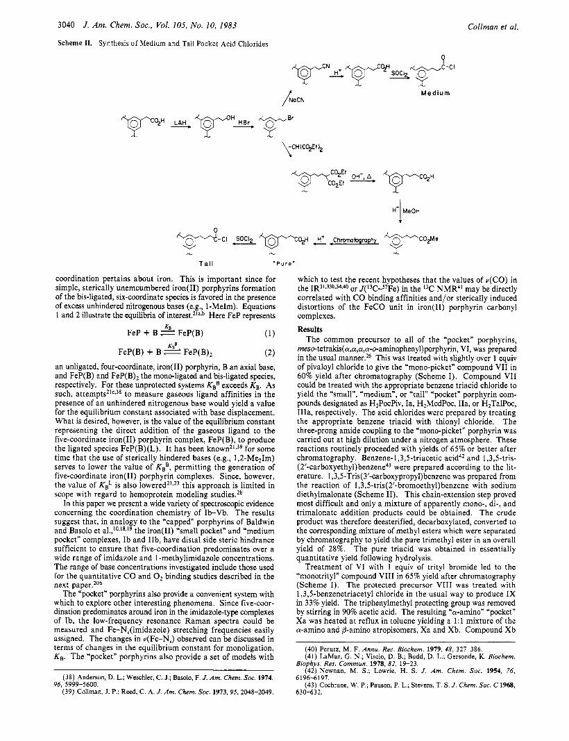
Scheme 11. Synthesis of Medium and Tall Pocket Acid Chlorides
Collman et al.
Q
/NoCN Medium
coordination pertains about iron. This is important since for simple, sterically unemcumbered iron(I1) porphyrins formation of the bis-ligated, six-coordinate species is favored in the presence of excess unhindered nitrogenous bases (e.g., 1-MeIm). Equations 1 and 2 illustrate the equilibria of interest.21a*b Here FeP represents
(1)
FeP(B) + B FeP(B), (2)
FeP + B 2 FeP(B)
an unligated, four-coordinate, iron(I1) porphyrin, B an axial base, and FeP(B) and FeP(B)* the mono-ligated and bis-ligated species, respectively. For these unprotected systems KBB exceeds KB. As such, attempt^^'^^^* to measure gaseous ligand affinities in the presence of an unhindered nitrogenous base would yield a value for the equilibrium constant associated with base displacement. What is desired, however, is the value of the equilibrium constant representing the direct addition of the gaseous ligand to the five-coordinate iron(I1) porphyrin complex, FeP(B), to produce the ligated species FeP(B)(L). It has been known21,39 for some time that the use of sterically hindered bases (e.g., 1,2-Me21m) serves to lower the value of KBB, permitting the generation of five-coordinate iron(I1) porphyrin complexes. Since, however, the value of KBL is also 1 0 w e r e d ~ ~ z ~ ~ this approach is limited in scope with regard to hemoprotein modeling studies.2b
In this paper we present a wide variety of spectroscopic evidence concerning the coordination chemistry of Ib-Vb. The results suggest that, in analogy to the “capped” porphyrins of Baldwin and Basolo et al.,10J8J9 the iron(I1) “small pocket” and “medium pocket” complexes, Ib and IIb, have distal side steric hindrance sufficient to ensure that five-coordination predominates over a wide range of imidazole and 1 -methylimidazole concentrations. The range of base concentrations investigated include those used for the quantitative CO and O2 binding studies described in the next paper.20b
The “pocket” porphyrins also provide a convenient system with which to explore other interesting phenomena. Since five-coor- dination predominates around iron in the imidazole-type complexes of Ib, the low-frequency resonance Raman spectra could be measured and Fe-N,(imidazole) stretching frequencies easily assigned. The changes in u(Fe-N,) observed can be discussed in terms of changes in the equilibrium constant for monoligation, KB. The “pocket” porphyrins also provide a set of models with
(38) Anderson, D. L.; Weschler, C. J.; Basolo, F. J. Am. Chem. Soc. 1974,
(39) Collman, J. P.; Reed, C. A. J. Am. Chem. Soc. 1973,95,2048-2049. 96, 5999-5600.
which to test the recent hypotheses that the values of u(C0) in the IR3’333b,34*40 or J(13C-57Fe) in the 13C NMR4’ may be directly correlated with CO binding affinities and/or sterically induced distortions of the FeCO unit in iron(I1) porphyrin carbonyl complexes.
Results The common precursor to all of the “pocket” porphyrins,
meso-tetrakis(a,a,a,a-o-aminophenyl)porphyrin, VI, was prepared in the usual manner.26 This was treated with slightly over 1 equiv of pivaloyl chloride to give the “mono-picket” compound VI1 in 60% yield after chromatography (Scheme I). Compound VI1 could be treated with the appropriate benzene triacid chloride to yield the “small”, “medium”, or “tail” “pocket” porphyrin com- pounds designated as H2PocPiv, Ia, H2MedPoc, IIa, or H,TalPoc, IIIa, respectively. The acid chlorides were prepared by treating the appropriate benzene triacid with thionyl chloride. The three-prong amide coupling to the “mono-picket” porphyrin was carried out at high dilution under a nitrogen atmosphere. These reactions routinely proceeded with yields of 65% or better after chromatography. Benzene-1,3,5-triacetic acid42 and 1,3,5-tris- (2’-carboxyethyl)ben~ene~~ were prepared according to the lit- erature. 1,3,5-Tris(3’-carboxypropyl)benzene was prepared from the reaction of 1,3,5-tris(2’-bromoethyl)benzene with sodium diethylmalonate (Scheme 11). This chain-extension step proved most difficult and only a mixture of apparently mono-, di-, and trimalonate addition products could be obtained. The crude product was therefore deesterified, decarboxylated, converted to the corresponding mixture of methyl esters which were separated by chromatography to yield the pure trimethyl ester in an overall yield of 28%. The pure triacid was obtained in essentially quantitative yield following hydrolysis.
Treatment of VI with 1 equiv of trityl bromide led to the “monotrityl” compound VI11 in 65% yield after chromatography (Scheme I). The protected precursor VI11 was treated with 1,3,5-benzenetriacetyl chloride in the usual way to produce IX in 33% yield. The triphenylmethyl protecting group was removed by stirring in 90% acetic acid. The resulting “a-amino” “pocket” Xa was heated at reflux in toluene yielding a 1 : 1 mixture of the a-amino and @-amino atropisomers, Xa and Xb. Compound Xb
(40) Perutz, M. F. Annu. Rev. Biochem. 1979, 48, 321-386. (41) LaMar, G. N.; Viscio, D. B.; Budd, D. L.; Gersonde, K. Biochem.
(42) Newnan, M. S.; Lowrie, H. S. J. Am. Chem. SOC. 1954, 76,
(43) Cochrane, W. P.; Pauson, P. L.; Stevens, T. S. J. Chem. SOC. C 1968,
Biophys. Res. Commun. 1978, 82, 19-23.
6 196-6 191.
630-63 2.
Synthesis and Characterization of “Pocket” Porphyrins
T
40’
3 0 -
b I
8 6 4 2 0 -2 PPM
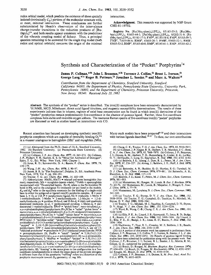
Figure 1. 100-MHz ‘ H N M R spectra in CDCll of H2PocPiv, Ia (spec- trum a) and a specifically deuterated analogue (spectrum b).
was isolated by chromatography and the recovered Xa was ree- quilibrated and rechromatographed to increase the overall yield of conversion to Xb. Solutions of Xb were exposed to phosgene gas and then either 3 4 1-imida~olyl)propylamine~~ or 343- pyridyl)pr~pylamine~~ was added to give the “tailed” “pocket” compounds Va or IVb in good yield. Because analogous “tailed“ compounds bearing appended imidazole units are sensitive to light and oxygen,9b the phosgene reactions and subsequent purification steps were carried out under N z and in the dark.
The metal-free porphyrins Ia-Va were characterized by ‘H NMR and mass spectroscopy. Elemental analysis was performed on Ia-IIIa. The ‘H N M R spectra proved especially informative for the simpler members of the “pocket” porphyrin series. In the IH N M R spectra of compounds Ia, IVa, and Va, it was possible to identify the signals arising from the porphyrin skeleton, the pivaloyl “picket”, the benzene cyclophane, and the methylene bridges. The methyl bridge signals are particularly diagnostic and could be assigned on the basis of spin-decoupling experiments. For instance, in compounds derived from benzene triacetic acid (e.g., Ia, Va, IVa) a characteristic doublet of doublets was observed at ca. 0.8 and 2.2 ppm. Irradiation of one of the doublets caused the other to collapse to a singlet. This AB system derives from the diastereotopic protons of the two equivalent bridging me- thylenes. In order to support this assignment, benzenetriacetic acid specifically deuterated in the a positions was used to prepare an analogue of Ia in which the six protons of methylene bridges were replaced by deuterons. Figure 1 shows the ‘H NMR spectra of both Ia and the specifically deuterated analogue. As expected, the signals assigned to the methylene bridges were not observed in the deuterated complex.
The iron(I1) derivatives (Ib, IIb, IIIb, IVb, Vb) were prepared under Nz from the metal-free porphyrins by means of the pre- viously reported direct iron(I1) insertion reaction using FeBr2.9b When possible, characterization studies were carried out imme- diately following insertion of Fe(I1). If necessary, the iron(I1) porphyrin complexes were stored as either the four- or five-co- ordinate solids. These were obtained by the slow addition of heptane to toluene solutions containing, when necessary, excess of the appropriate axial base. The crystalline solids were washed with heptane then dried in vacuo.
The iron(I1) complexes of the “pocket” series of porphyrins Ib-Vb and particularly their adducts with externally provided ligands (e.g., 1-MeIm, 1,2-Me21m, 02, CO) have been charac- terized by a wide variety of spectroscopic techniques (optical spectroscopy, ‘H NMR, Mossbauer, magnetic susceptibility, resonance Raman spectroscopy, IR, I3C NMR). Importantly, similar solution conditions were used for these spectroscopic characterizations as were used for the gaseous ligand-binding studies discussed in the following paper.20b Since we found that IVb, Vb, and pyridine complexes of Ib-IIIb oxidized in the presence of Oz, useful gaseous ligand-binding studies on these complexes could not be performed under equilibrium conditions.
(44) Schwan, T. J. J . Heterocycl. Chem. 1967, 4 , 633-634. (45) Hawes, E. M.; Davis, H. L. J . Heterocycl. Chem. 1973, 10, 39-42.
J . Am. Chem. SOC., Vola 105, No. 10, 1983 3041
, ___ 320 360 400 440 480
WAVELENGTH (nm)
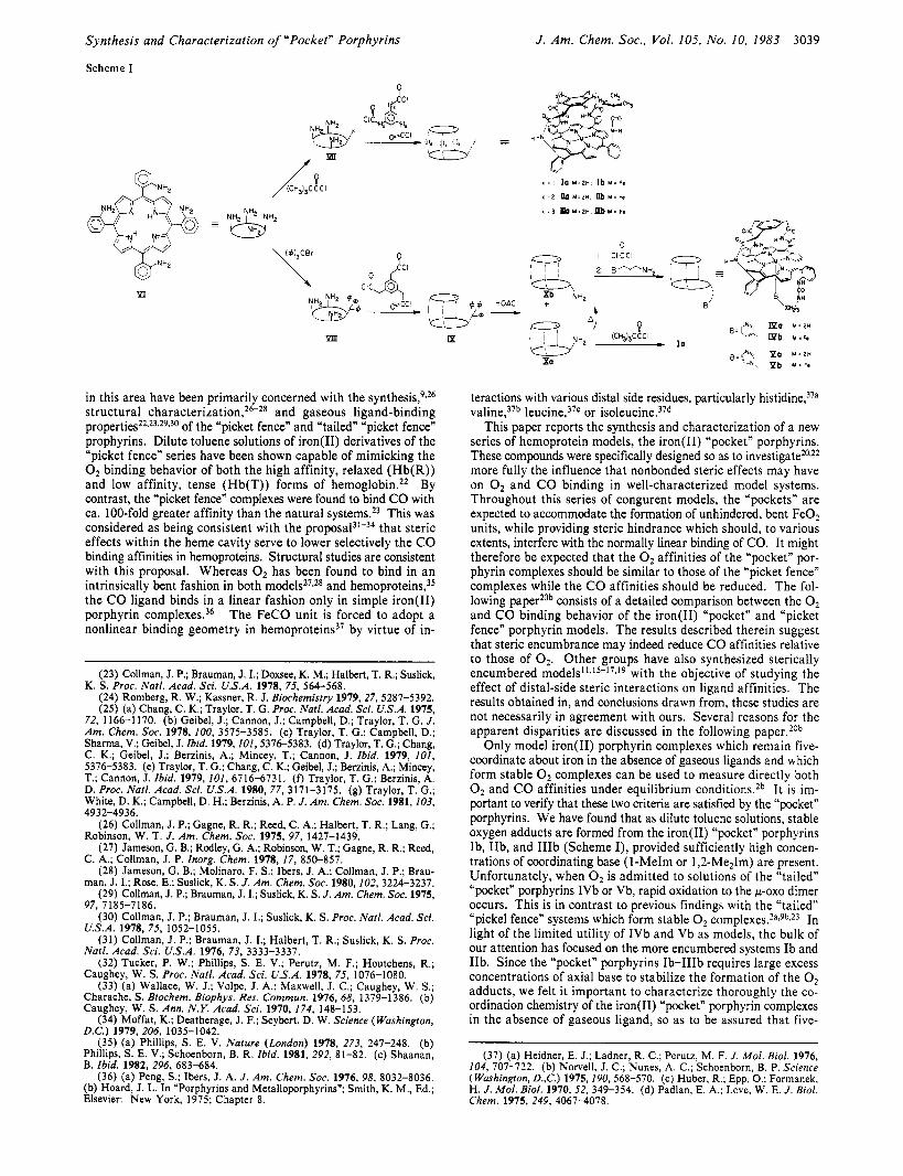
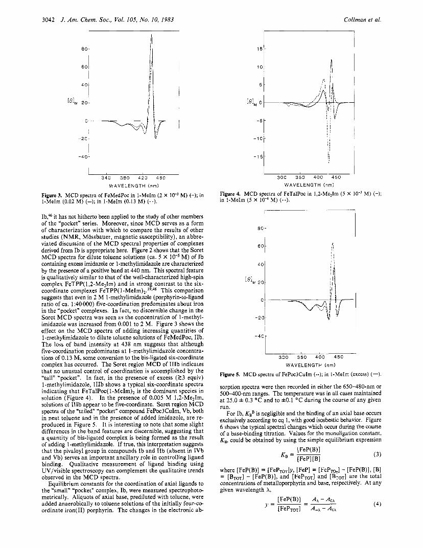
Figure 2. MCD spectra of FePocPiv in 1-MeIm (2.0 M) (-); in Im (0.03 M) (--).
Therefore, the spectroscopic characterization studies focused on complexes of Ib-IIIb with various imidazole ligands. The results from each of the characterizations are considered separately in the ensuing Discussion section.
Discussion Optical Spectroscopy. Both the electronic absorption and
magnetic circular dichroism spectra of all new iron(I1) “pocket” porphyrin complexes were routinely recorded and compared with those of standard iron(I1) tetraarylporphyrins. Recent work from our laboratory has served to establish a standard set of both UV/visible absorption and Soret MCD spectral data for iron(I1) tetraarylporphyrins in various spin states and coordination ge- o m e t r i e ~ . ~ , ~ ~ Several general trends have emerged. Four-coor- dinate iron(I1) tetraarylporphyrins have two bands of nearly equal intensity in the Soret region (350-500 nm) of the electronic absorption spectra, as well as a broad peck at roughly 540 nm in the visible region (500-700 nm). Addition of nitrogenous base to a four-coordinate iron(I1) porphyrin produces high-spin five- or low-spin six-coordinate complexes (eq 1 and 2), both of which exhibit spectra in the visible region with major bands at ca. 535 and 565 nm. In the Soret region, the absorption spectra of both the mono- and bis-ligated species appear qualitatively similar with A,,, for six-coordinate complexes appearing 5-10 nm further to the blue. Since these differences are slight, in situations where a possible mixture of five- or six-coordinate complexes may exist, we have preferred not to rely on electronic spectra in assigning coordination geometries or spin states for the various “pocket” porphyrin complexes. Once the coordination geometries are es- tablished by other means (vide infra), then the absorption spectra can serve as a useful “fingerprint” for the various complexes. Electronic spectra are therefore given for representative complexes of Ib, IIb, and IIIb in the Experimental Section.
In contrast to the electronic absorption spectra, we have found that the shape and sign (but not the intensity) of the MCD bands in the Soret region are a sensitive qualitative probe for spin state (and hence, coordination geometry) in complexes of iron( 11) porphyrin^.^^ In complexes derived from iron(I1) tetraaryl- porphyrins and various imidazole ligands, dramatic differences are observed in the Soret MCD between the five-coordinate, high-spin form and the bis-ligated, low-spin form. The Soret region of the five-coordinate complexes shows a strong positive band at ca. 440 nm, while the MCD spectra of the six-coordinate bis-ligated species show an inverted bell-shaped form at ca. 425 nm.
While MCD has previously been used to explore the coordi- nation characteristics of the “small” “pocket” complex, FePocPiv,
(46) Collman, J. P.; Basolo, F.; Bunnenberg, E.; Collins, T. J.; Dawson, J. H.; Ellis, P. E., Jr.; Marrocco, M. L.; Moscowitz, A.; Sessler, J. L.; Szy- manski, T. J . Am. Chem. Soc. 1981, 103, 5636-5648.
3042 J . Am. Chem. SOC., Vol. 105, No. 10, 1983 Collman et al.
, 80 i
60 I I ll I
I
I i i ,
I I
- 2 o t v
-40.
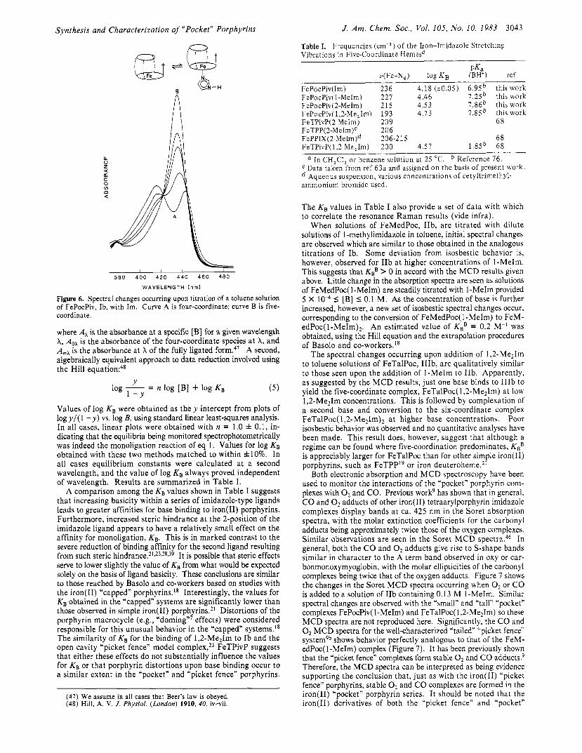
340 380 420 460 WAVELENGTH (nm)
Figure 3. MCD spectra of FeMedPoc in 1-MeIm (2 X lo-’ M) (-); in 1-MeIm (0.02 M) (-); in 1-MeIm (0.13 M) (--).
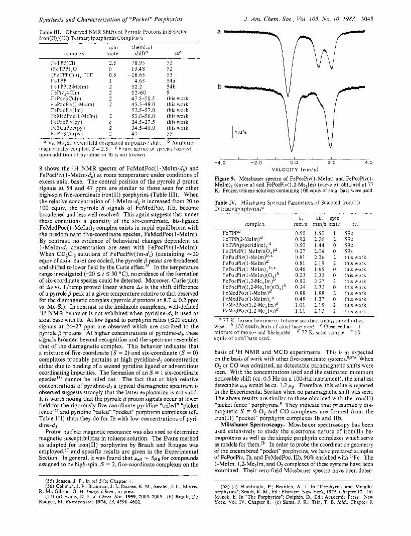
Ib,& it has not hitherto been applied to the study of other members of the “pocket” series. Moreover, since MCD serves as a form of characterization with which to compare the results of other studies (NMR, Mossbauer, magnetic susceptibility), an abbre- viated discussion of the MCD spectral properties of complexes derived from Ib is appropriate here. Figure 2 shows that the Soret MCD spectra for dilute toluene solutions (ca. 5 X M) of Ib containing excess imidazole or 1-methylimidazole are characterized by the presence of a positive band at 440 nm. This spectral feature is qualitatively similar to that of the well-characterized high-spin complex FeTPP( 1 ,2-Me21m) and in strong contrast to the six- coordinate complexes FeTPP( 1 -MeIm)2.’9s46 This comparison suggests that even in 2 M 1-methylimidazole (porphyrin-to-ligand ratio of ca. 1 :40 000) five-coordination predominates about iron in the “pocket” complexes. In fact, no discernible change in the Soret MCD spectra was seen as the concentration of l-methyl- imidazole was increased from 0.001 to 2 M. Figure 3 shows the effect on the MCD spectra of adding increasing quantities of 1 -methylimidazole to dilute toluene solutions of FeMedPoc, IIb. The loss of band intensity at 438 nm suggests that although five-coordination predominates a t 1 -methylimidazole concentra- tions of 0.13 M, some conversion to the bis-ligated six-coordinate complex has occurred. The Soret region MCD of IIIb indicates that no unusual control of coordination is accomplished by the “tall” “pocket”. In fact, in the presence of excess (23 equiv) 1-methylimidazole, IIIb shows a typical six-coordinate spectra indicating that FeTalPoc( 1 -MeIm)2 is the dominant species in solution (Figure 4). In the presence of 0.005 M 1,2-Me21m, solutions of IIIb appear to be five-coordinate. Soret region MCD spectra of the “tailed” “pocket” compound FePoc3CuIm, Vb, both in neat toluene and in the presence of added imidazole, are re- produced in Figure 5 . It is interesting to note that some slight differences in the band features are discernible, suggesting that a quantity of bis-ligated complex is being formed as the result of adding 1-methylimidazole. If true, this interpretation suggests that the pivaloyl group in compounds Ib and IIb (absent in IVb and Vb) serves an important ancillary role in controlling ligand binding. Qualitative measurement of ligand binding using UV/visible spectroscopy can complement the qualitative trends observed in the MCD spectra.
Equilibrium constants for the coordination of axial ligands to the “small” “pocket” complex, Ib, were measured spectrophoto- metrically. Aliquots of axial base, prediluted with toluene, were added anaerobically to toluene solutions of the initially four-co- ordinate iron(I1) porphyrin. The changes in the electronic ab-
l 5 t
300 350 400 4 5 0
WAVELENGTH (nm)
Figure 4. MCD spectra of FeTalPoc in 1,2-Me21m ( 5 X in 1-MeIm ( 5 X lo4 M) (--).
M) (-);
-
I I 300 350 400 450
WAVELENGTH (nm)
Figure 5. MCD spectra of FePoc3CuIm (--); in 1-MeIm (excess) (-).
sorption spectra were then recorded in either the 650-480-11111 or 500-400-nm ranges. The temperature was in all cases maintained at 25.0 f 0.3 “ C and to fO. l “ C during the course of any given run.
For Ib, KBB is negligible and the binding of an axial base occurs exclusively according to eq 1, with good isosbestic behavior. Figure 6 shows the typical spectral changes which occur during the course of a base-binding titration. Values for the monoligation constant, KB, could be obtained by using the simple equilibrium expression
(3)
where [FeP(B)] = [FePToT]y, [FeP] = [ F ~ P T ~ . ] - [FeP(B)I, [Bl
concentrations of metalloporphyrin and base, respectively. At any given wavelength A,
= [BTOT] - [FeP(B)], and [ F e p ~ o ~ ] and [BTOT] are the total
(4) [FeP(B)l - AOA
y = - = [F~PTOT] A-A - Aox
Synthesis and Characterization of ”Pocket* Porphyrins
8
I
8
1 1 , 1
380 400 4 2 0 4 4 0 460 480
WAVELENGTH (nm)
Figure 6. Spectral changes occurring upon titration of a toluene solution of FePocPiv, Ib, with Im. Curve A is four-coordinate; curve B is five- coordinate.
where A, is the absorbance at a specific [B] for a given wavelength A, A,, is the absorbance of the four-coordinate species a t X, and A*, is the absorbance at X of the fully ligated form.47 A second, algebraically equivalent approach to data reduction involved using the Hill equation:48
Y log - = n log [B] + log KB 1 - Y
Values of log K, were obtained as t h e y intercept from plots of logy/( 1 - y) vs. log B, using standard linear least-squares analysis. In all cases, linear plots were obtained with n = 1.0 f 0.1, in- dicating that the equilibria being monitored spectrophotometrically was indeed the monoligation reaction of eq 1. Values for log KB obtained with these two methods matched to within &lo%. In all cases equilibrium constants were calculated at a second wavelength, and the value of log KB always proved independent of wavelength. Results are summarized in Table I.
A comparison among the KB values shown in Table I suggests that increasing basicity within a series of imidazole-type ligands leads to greater affinities for base binding to iron(I1) porphyrins. Furthermore, increased steric hindrance at the 2-position of the imidazole ligand appears to have a relatively small effect on the affinity for monoligation, KB. This is in marked contrast to the severe reduction of binding affinity for the second ligand resulting from such steric h i n d r a n ~ e . ~ ’ ~ ~ ~ ~ ~ ~ ~ ~ ~ It is possible that steric effects serve to lower slightly the value of KB from what would be expected solely on the basis of ligand basicity. These conclusions are similar to those reached by Basolo and co-workers based on studies with the iron(I1) “capped” porphyrins.Is Interestingly, the values for KB obtained in the “capped” systems are significantly lower than those observed in simple iron(I1) porphyrins.21 Distortions of the porphyrin macrocycle (e.g., “domingn7 effects) were considered responsible for this unusual behavior in the “capped” systems.18 The similarity of K , for the binding of 1,2-Me21m to Ib and the open cavity “picket fence” model complex,23 FeTPivP suggests that either these effects do not substantially influence the values for KB or that porphyrin distortions upon base binding occur to a similar extent in the “pocket” and “picket fence” porphyrins.
(47) We assume in all cases that Beer’s law is obeyed. (48) Hill, A. V. J . Physid. (London) 1910, 40, iv-vii.
J. Am. Chem. SOC., Vol. 105, No. 10, 1983
Table I. Frequencies (cm-’) of the Iron-Imidazole Stretching Vibrations in Five-Coordinate Hemesn
3043
PK, u(Fe-N,) log K B (BH+) ref
FePocPiv(1m) 236 4.18 (i0.05) 6.95b this work FePocPiv(1-MeIm) 227 4.46 7.25b this work FePocPiv(2-MeIm) 215 4.53 7.86b this work TePocPiv( 1,2-Me21m) 193 4.73 7.85 this work FeTPivP(2 MeIm) 209 68 FeTPP( 2-MeIm)c 206 FePPIX(2 MeIm)d 206-215 68
1.85b 68 Reference 76.
FeTPivP(1,2-Me21m) 200 4.57
a In CH,CI, or benzene solution at 25 “C. Data taken from ref 63a and assigned on the basis of present work. Aqueous suspension, various concentrations of cetyltrimethyl-
ammonium bromide used.
The KB values in Table I also provide a set of data with which to correlate the resonance Raman results (vide infra).
When solutions of FeMedPoc, IIb, are titrated with dilute solutions of 1 -methylimidazole in toluene, initial spectral changes are observed which are similar to those obtained in the analogous titrations of Ib. Some deviation from isosbestic behavior is, however, observed for IIb at higher concentrations of 1-MeIm. This suggests that KBB > 0 in accord with the MCD results given above. Little change in the absorption spectra are seen as solutions of FeMedPoc( 1-MeIm) are steadily titrated with 1-MeIm provided 5 X 5 [B] I 0.1 M. As the concentration of base is further increased, however, a new set of isosbestic spectral changes occur, corresponding to the conversion of FeMedPoc( 1 -MeIm) to FeM- edPoc(l-MeIm),. An estimated value of KBB = 0.2 M-’ was obtained, using the Hill equation and the extrapolation procedures of Basolo and co-workers.ls
The spectral changes occurring upon addition of 1,2-Me21m to toluene solutions of FeTalPoc, IIIb, are qualitatively similar to those seen upon the addition of 1-MeIm to IIb. Apparently, as suggested by the MCD results, just one base binds to IIIb to yield the five-coordinate complex, FeTalPoc( 1 ,2-Me21m) at low 1,2-Me21m concentrations. This is followed by complexation of a second base and conversion to the six-coordinate complex FeTalPoc( 1 ,2-Me21m), a t higher base concentrations. Poor isosbestic behavior was observed and no quantitative analyses have been made. This result does, however, suggest that although a regime can be found where five-coordination predominates, KBB is appreciably larger for FeTalPoc than for other simple iron(I1) porphyrins, such as FeTPP19 or iron deuteroheme.21
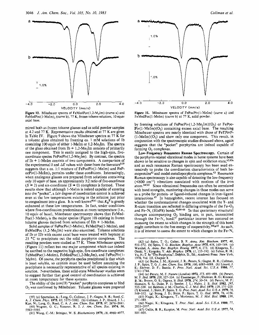
Both electronic absorption and MCD spectroscopy have been used to monitor the interactions of the “pocket” porphyrin com- plexes with O2 and CO. Previous work9 has shown that in general, CO and O2 adducts of other iron(I1) tetraarylporphyrin imidazole complexes display bands at ca. 425 nm in the Soret absorption spectra, with the molar extinction coefficients for the carbonyl adducts being approximately twice those of the oxygen complexes. Similar observations are seen in the Soret MCD spectra.46 In general, both the CO and O2 adducts give rise to S-shape bands similar in character to the A term band observed in oxy or car- bonmonoxymyoglobin, with the molar ellipticities of the carbonyl complexes being twice that of the oxygen adducts. Figure 7 shows the changes in the Soret MCD spectra occurring when 0, or CO is added to a solution of IIb containing 0.13 M 1-MeIm. Similar spectral changes are observed with the “small” and “tall” “pocket” complexes FePocPiv( I-MeIm) and FeTalPoc( 1,2-Me21m) so these MCD spectra are not reproduced here. Significantly, the CO and O2 MCD spectra for the well-characterized “tailed” “picket fence” systemgb shows behavior perfectly analogous to that of the FeM- edPoc(1-MeIm) complex (Figure 7). It has been previously shown that the “picket fence” complexes form stable O2 and CO adduct^.^ Therefore, the MCD spectra can be interpreted as being evidence supporting the conclusion that, just as with the iron(I1) “picket fence” porphyrins, stable O2 and CO complexes are formed in the iron(I1) “pocket” porphyrin series. It should be noted that the iron(I1) derivatives of both the “picket fence” and “pocket”

3044 J . Am. Chem. SOC., Vol. 105, No. 10. 1983 Collman et al.
Table 11. Stabilities of Selected Iron(I1) Porphyrin 0, Adducts
- 6 0 1
3 4 0 380 420 460 WAVELENGTH (nm)
Figure 7. MCD spectra of FeMedPoc in 1-MeIm (0.13 M ) (--); +02 (...); +co (-). porphyrins form not only stable, but also reversible, 0, complexes in toluene solution at room temperature. The five-coordinate iron(I1) porphyrin, FeP(B) can be exposed to 0, (forming FeP- (B)(O,)) then extensively bubbled with N, to regenerate the initial five-coordinate species FeP(B) (P = PocPiv, MedPoc, TPivP, etc.).
Although the general features of the Soret absorption and Soret MCD spectra of the six-coordinate carbonyl and oxygen adducts of the various iron(I1) “pocket” porphyrin imidazole complexes are similar to those for other analogous systems (e.g., FeTPivP- (1,2-Me2Im)), this is not the case in the visible region of the electronic absorption spectra. In the visible, both the O2 and CO adducts of the ”picket fence” systems show predominantly a single weak band in the 540-550-nm r e g i ~ n . ~ By contrast, the carbonyl and oxygen complexes derived from five-coordinate complexes of Ib, IIb, or IIIb show bands at about 520 and 560 nm, re- spectively.
It is important to stress that only at high excess concentrations of axial base (1-MeIm, 1,2-MezIm) do the iron(I1) “pocket” complexes Ib, IIb, IIIb form oxygen adducts which are sufficiently stable kinetically so as to allow determinations of O2 binding affinities under equilibrium conditions.
At low base concentration, oxidation to the pox0 iron(II1) porphyrin dimer occurs. A similar observation has been made with regard to the iron(I1) “capped” porphyrin complexes.’0 Apparently, large concentrations of axial bases serve to reduce the concentration of the rapidly oxidized,49 four-coordinate species.’O Since the nitrogenous bases bind with much greater affinity to the unencumbered side of the ”capped” and “pocket” porphyrins, high concentrations of axial base also serve to effect regioselective gaseous ligand binding within the protected cavity, as the axial base competitively inhibits 0, or CO from binding on the unencumbered side.50 Table I1 shows the half-lives of various iron(I1) “pocket” 0, complexes obtained in the usual way.” Although the rates of oxidation are extremely sensitive to im-
(49) (a) Alben, J. D.; Fuchsman, W. H.; Beaudreau, C. H.; Caughey, W. S. Biochemistry 1968, 7, 624-635. (b) Cohen, I . A.; Caughey, W. S. Ibid. 1968, 7,636-641. (c) Hammond, G. S.; Wu, C.-H. S. Adu. Chem. Ser. 1968, 77, 186-207. (d) Chin, D.-H.; LaMar, G. N.; Balch, A. L. J . Am. Chem. SOC. 1980, 102, 4344-4349.
(50) We have generally found (cf. ref 20b) that for complexes derived from Ib and IIb the CO and O2 affinities are independent of base concentration provided 0.1 5 [B] 5 1 M. In contrast to the behavior with imidazole-type ligands, we have found that toluene solutions of Ib and pyridine do not support the reversible binding of O2 at room temperature. This probably reflects the low K B for pyridine rather than some intrinsic property of the pyridine base. Pyridine supports reversible oxygenation in the ‘picket fence” series of com- p l e x e ~ . ~ ~
complex
FeTPivP(1 ,2-Me21M) FePiv, 5 CIm FePocPiv(1 ,2-Me21m) FePocPiv( 1-MeIm) FePocPiv(1-MeIm) FeMedPoc(1 -MeIm) Fe4Cu(l-MeIm) “chelated protoheme” FePocSCu(py) FePF3Cu(py) FeC,-cap(1-MeIm)
base concn, M
10-41 0
“tai1ed”Q, b
0.10 1 .OO O.1° 0.10
half-lives at 25 “C
1 month 3 days 1 day
36 h 7 h 2 days
2 1 2 h 210 s -10 min
1 day 5 h
ref
2a 2a this work this work this work this work 17 25a this work this work 10b
Toluene. Covalently attached imidazole ligand. Benzene. Aqueous cetyltrimethylammonium bromide suspension (2%).
e Covalently attached pyridine ligand.
I ” ” I ” ” l ” ” I ” ” l ” ” l 5 0 4 0 30 2 0 10 0 p p m
Figure 8. 100-MHz ‘H N M R spectra in toluene-d8 of FeMedPoc(1- MeIm-d,) (upper trace, a) and FePocPiv( 1-MeIm-d6) (lower trace, b).
purities, these data show that the sterically protected iron(I1) “pocket” porphyrins form long-lived oxygen complexes under conditions where the bimolecular oxidation49 to the pox0 dimer is suppressed. In fact, the “pocket” porphyrins are among the more stable of the known hemoprotein models. It is of interest to note that a t a given concentration of 1-MeIm, the oxygen complex derived from FePocPiv, Ib, is kinetically less stable than that derived from FeMedPoc, IIb. The reasons for this are as yet not entirely understood. A complete analysis of the rates of both base and OZzob binding is needed to clarify this point.
Proton Nuclear Magnetic Resonance. Proton nuclear magnetic resonance has been widely used to characterize both hemoproteins and simple iron(I1) porphyrin model c o m p l e x e ~ . ~ ~ In tetra- arylporphyrins, the degree of isotropic shift of the pyrrole @ protons serves as a convenient probe for both oxidation and spin ~ t a t e . ~ , - ~ ~ In five-coordinate high-spin iron(I1) porphyrins at or near room temperature, these resonances generally appear as sharp, well- resolved signals in the 45-60-ppm region (downfield from Me4Si according to the diamagnetic convention). Perdeuterated 1 -MeIm was synthesized and used for several IH NMR studies. Figure
(51) (a) LaMar, G. N.; Walker, F. A. In ’The Porphyrins”; Dolphin, D., Ed.; Academic Press: New York, 1978; Vol. IV, Chapter 2, and references therein. (b) LaMar, G. N.; Horrocks, W., Jr.; Holm, R. H., Eds. ‘NMR of Paramagnetic Molecules”; Academic Press: New York, 1973, and references therein.
Chem. SOC. 1973, 95, 63-75. (52) LaMar, G. N.; Eaton, G. R.; Holm, R. H.; Walker, F. A. J . Am.
(53) LaMar, G. N.; Walker, F. A. J . Am. Chem. SOC. 1973, 95, 1782-1 790.
(54) (a) Goff, H.; LaMar, G. N.; Reed, C. A. J . Am. Chem. SOC. 1977, 99, 3641-3646. (b) Goff, H.; LaMar, G. N. Ibid. 1977, 99, 6599-6606.
Synthesis and Characterization of “Pocket” Porphyrins
Table 111. Observed N M R Shifts of Pyrrole Protons in Selected Iron(II)/(III) Tetraarylporphyrin Complexes
spin chemical complex state shifta ref
FeTPP(C1) 2.5 78.95 5 2
[FeTPP(Im), ] +Cl- 0.5 - 16.65 5 3
FePiv34CIm 2 52-60 9
(FeTPP),O b 13.48 52
FeTPP 1 4.65 54a FeTPP( 2-MeIm) 2 52.2 54b
FePoc3CuIm 2 47 .5 -5 0.5 this work FePocPiv(1-MeIm) 2 45.5-49.0 this work FePocPiv(1m) 52.5-57.0 this work FeMedPoc(1 -MeIm) 2 53.0-56.0 this work
Fe3CuPoc(py) 2 34.5-40.0 this work FePocPiv(py) C 24.5-21.5 this work
FePF3Cu(py) 2 4 1 5 5
Vs. Me,%; downfield designated as positive shift. ’ Antiferro- magnetically coupled, S = 2.5. upon addition of pyridine t o Ib is not known.
8 shows the ‘H N M R spectra of FeMedPoc(1-MeIm-d6) and FePocPiv( l-MeIm-d6) at room temperature under conditions of excess axial base. The central position of the pyrrole p proton signals a t 54 and 47 ppm are similar to those seen for other high-spin five-coordinate iron(I1) porphyrins (Table 111). When the relative concentration of 1-MeIm-d6 is increased from 20 to 100 equiv, the pyrrole p signals of FeMedPoc, IIb, become broadened and less well resolved. This again suggests that under these conditions a quantity of the six-coordinate, bis-ligated FeMedPoc( 1-MeIm), complex exists in rapid equilibrium with the predominant five-coordinate species, FeMedPoc( 1 -MeIm). By contrast, no evidence of behavioral changes dependent on l-MeIm-d6 concentration are seen with FePocPiv( 1-MeIm). When CD2C12 solutions of FePocPiv(1m-d4) (containing -20 equiv of axial base) are cooled, the pyrrole p peaks are broadened and shifted to lower field by the Curie effect.55 In the temperature range investigated (-20 1 t I 30 “C), no evidence of the formation of six-coordinate species could be detected. Moreover, Curie plots of Av vs. l/temp proved linear where Av is the shift difference of a pyrrole /3 peak at a given temperature relative to that observed for the diamagnetic complex (pyrrole p protons at 8.7 * 0.2 ppm vs. Me,Si). In contrast to the imidazole complexes, well-defined ‘H N M R behavior is not exhibited when pyridine-d5 is used as axial base with Ib. At low ligand to porphyrin ratios (120 equiv), signals a t 24-27 ppm are observed which are ascribed to the pyrrole protons. At higher concentrations of pyridine-d,, these signals broaden beyond recognition and the spectrum resembles that of the diamagnetic complex. This behavior indicates that a mixture of five-coordinate (S = 2) and six-coordinate (S = 0) complexes probably pertains a t high pyridine-d5 concentration either due to binding of a second pyridine ligand or adventitious coordinating impurities. The formation of an S = 1 six-coordinate specieslga cannot be ruled out. The fact that at high relative concentrations of pyridine-d, a typical diamagnetic spectrum is observed suggests strongly that the latter explanation is not valid. It is worth noting that the pyrrole p proton signals occur at lower field for the rigorously five-coordinate pyridine “tailed” “picket fencens6 and pyridine “tailed” “pocket” porphyrin complexes (cf., Table 111) than they do for Ib with low concentrations of pyri- dine-d,.
Proton nuclear magnetic resonance was also used to determine magnetic susceptibilities in toluene solution. The Evans method as adapted for iron(I1) porphyrins by Brault and Rougee was employed,57 and specific results are given in the Experimental Section. In general, it was found that peff - 5pB for compounds assigned to be high-spin, S = 2, five-coordinate complexes on the
Exact nature of species formed
~ ~~
( 5 5 ) Jenson, J. P., in ref 5 1 b; Chapter 1. (56) Collman, J. P.; Brauman, J. I.; Doxsee, K. M.; Sessler, J. L.; Morris,
(57) (a) Evans, D. F. J . Chem. SOC. 1959, 2003-2005. (b) Brault, D.; R. M.; Gibson, Q. H. Inorg. Chem., in press.
Rougee, M. Biochemistry 1974, 13, 4598-4602.
J . Am. Chem. SOC., Vol. 105, No. 10, 1983 3045
\ I
1.0% I 1 I 1 I I 1 I I
-4 .0 -2.0 0.0 2.0 4 .0 VELOCITY ( m m / s )
Figure 9. Mossbauer spectra of FePocPiv(1-MeIm) and FePocPiv( 1- MeIm), (curve a) and FePocPiv(l,2-Me21m) (curve b), obtained at 77 K. Frozen toluene solutions containing 100 equiv of axial base were used.
Table IV. Mossbauer Spectral Parameters of Selected Iron(I1) Tetraarylporphyrinsa
6 , 6E, spin complex mm/s mm/s state ref
FeTPPd 0.50 1.50 1 59b FeTPP( 2-MeIm)d 0.92 2.26 2 59b FeTPP(piperidine), 0.50 1.44 0 59b FeTPivP(1 MeIm)(O,)d 0.27 2.04 0 59a FePocPiv(l-MeIm)’rC 0.81 2.36 2 this work FePocPiv(l-MeIm)d 0.81 2.19 2 this work FePocPiv(1 MeIm), b,c 0.46 1.65 0 this work FePocPiv(1-MeIm)(O ) b 0.23 2.23 0 this work FePocPiv(1 ,2-Me2Im)% 0.92 2.27 2 this work FePo~Piv(l,2-Me,Im)(O~)~ 0.24 2.32 0 this work FeMedPoc( l-MeIm)d 0.88 1 .88 2 this work FeMedPoc(1-MeIm), e 0.48 1.37 0 this work TeMedPoc(1 ,2-Me21m)= 1.01 2.15 2 this work FeMedPo~( l ,2 -Me , Im)~ 1.11 2.13 2 this work
a 77 K , frozen benzene or toluene solution unless noted other- wise. ’ 100 equivalents of axial base used. mixture of mono- and bis-ligated. equiv of axial base used.
Observed as 1 : 1 77 K, solid sample. e 10
basis of ‘H N M R and MCD experiments. This is as expected on the basis of work with other five-coordinate ~ y s t e m s . ~ , ~ ~ ~ When O2 or CO was admitted, no detectable paramagnetic shifts were seen. With the concentrations used and the estimated minimum noticeable shift (ca. 0.5 Hz on a 100-Hz instrument), the smallest detectable peff would be ca. 1.2 pe. Therefore, this value is reported in the Experimental Section when no paramagnetic shift was seen. The above results are similar to those obtained with the iron(I1) “picket fence” porphyrin^.^ They indicate that presumably dia- magnetic S = 0 O2 and CO complexes are formed from the iron(I1) “pocket” porphyrin complexes Ib and IIb.
Mossbauer Spectroscopy. Mossbauer spectroscopy has been used extensively to study the electronic nature of iron(I1) he- moproteins as well as the simple porphyrin complexes which serve as models for them.58 In order to probe the coordination geometry of the encumbered “pocket” porphyrins, we have prepared samples of FePocPiv, Ib, and FeMedPoc, IIb, 90% enriched with 57Fe. The 1-MeIm, 1,2-Me21m, and O2 complexes of these systems have been examined. Their zero-field Mossbauer spectra have been deter-
(58) (a) Hambright, P.; Bearden, A. J. In “Porphyrins and Metallo- porphyrins”; Smith, K. M., Ed.; Elsevier: New York, 1975; Chapter 12. (b) Miinck, E. In ‘The Porphyrins”; Dolphin, D., Ed.; Academic Press: New York, Vol. IV, Chapter 8. (c) Sams, J. R.; Tsin, T. B. Ibid.; Chapter 9.
3046 J . Am. Chem. Soc.. Vol. 105, No. 10, 1983 Collman et al.
/- " V A T
I 1 1 1 1 I I - 4 . 0 - 2 . 0 0.0 2.0 4 . 0
VELOCITY (mm/s)
Figure 10. Mossbauer spectra of FeMedPoc(l,2-M21m) (curve a) and F e M e d P ~ ( l - M e I m ) ~ (curve b), 77 K, frozen toluene solutions, 10 equiv axial base.
mined both as frozen toluene glasses and as solid powder samples at 4.2 and 77 K. Representative results obtained at 77 K are given in Table IV. Figure 9 shows the Mossbauer spectra a t 77 K for a toluene glass obtained by freezing ca. 1 mM solutions of Ib containing 100 equiv of either 1-MeIm or 1,2-Me21m. The spectra of the glass obtained from Ib + 1,2-Me21m consists of primarily one component. This is easily assigned to the high-spin, five- coordinate species FePocPiv( 1,2-Me21m). By contrast, the spectra of Ib + 1-MeIm consists of two components. A comparison of the experimental 6 and AE values with those from the l i t e r a t ~ r e ~ ~ ~ ~ suggests that a ca. 1:l mixture of FePocPiv(1-MeIm) and FeP- ocPiv( l-MeIm)2 pertains under these conditions. Interestingly, when analogous glasses are prepared from solutions containing only 10 equiv of base, an approximately 3:2 ratio of five-coordinate (S = 2) and six-coordinate (S = 0) complexes is formed. These results show that although 1-MeIm is indeed capable of entering into the "pocket", a fair degree of coordination control is achieved even at the low temperatures existing in the solution just prior to congealment into a glass. It is well-knownm6' that KBB is greatly enhanced at these low temperatures. In fact, under conditions where five-coordination predominates at room temperature (Le., 10 equiv of base), Mossbauer spectroscopy shows that FeMed- P ~ c ( l - M e I m ) ~ is the major species (Figure 10) existing in frozen toluene glasses derived from solutions of IIb + 1-MeIm.
Solid samples of FePocPiv( 1-MeIm), FeMedPoc( 1-MeIm), and FeMedPoc ( 1 ,2-Me21m) were also examined. Toluene solutions of Ib or IIb with excess axial base were treated with heptane at 25 "C to precipitate out the solid porphyrin complexes. The resulting powders were studied at 77 K. These Miissbauer spectra (Figure 11) reflect but one major component which can indeed be ascribed to the respective high-spin, five-coordinate complexes: FeMedPoc( 1 -MeIm), FeMedPoc( 1 ,2-Me21m), and FePocPiv( 1 - MeIm). Of course, the porphyrin species precipitated is that which is least soluble, so caution must be used before assuming the precipitate is necessarily representative of all species existing in solution. Nevertheless, these solid-state Mossbauer studies seem to suggest further that good control of coordination is achieved at room temperature for these systems.
The ability of the iron(I1) "pocket" porphyrin complexes to bind 0, was confirmed by Mossbauer. Toluene glasses were prepared
(59) (a) Spartalian, K.; Lang, G.; Collman, J. P.; Gagne, R. R.; Reed, C. A. J . Chem. Phys. 1975, 63, 5375-5382. (b) Collman, J. P.; Hoard, J . L.; Kim, N. ; Lang, G.; Reed, C. A. J . Am. Chem. SOC. 1975, 97, 2676-2681.
(60) Wagner, G. C. ; Kassner, R. J . 1. Am. Chem. SOC. 1974, 96, 5593-5595.
(61) Wang, C.-M.; Brinigar, W. S. Biochemistry 1979, 18, 4960-4977.
T ~ 1.0%
-4 .0 -2.0 0.0 2.0 4.0 VELOCITY ( m m / s )
Figure 11. Mossbauer spectra of FePocPiv(1-MeIm) (curve a) and FeMedPoc(1-MeIm) (curve b) at 77 K, solid powder.
by freezing solutions of FePocPiv( 1 ,2-Me21m)(02) or FePoc- Piv( l-MeIm)(02) containing excess axial base. The resulting Mossbauer spectra are nearly identical with those of FeTPivP- (1-MeIm)(02) and show only one component. This result, in conjunction with the spectroscopic studies discussed above, again suggests that the "pocket" porphyrins are indeed capable of forming O2 complexes.
Low-Frequency Resonance Raman Spectroscopy. Certain of the porphyrin-related vibrational modes in heme systems have been shown to be sensitive to changes in spin and oxidation and as such resonance Raman spectroscopy has been used ex- tensively to probe the coordination characteristics of both he- moproteins62 and model metalloporphyrin complexes.63 Resonance Raman spectroscopy is also capable of detecting the low-frequency (C600 cm-') vibrations associated with motions of the iron a t ~ m . ~ ~ ' . ' ~ Since vibrational frequencies can often be correlated with bond strengths, monitoring changes in these modes can serve as a probe for protein- or ligand-induced changes in iron-ligand interaction^.^^ In hemoglobin, recent interest has focused on whether the conformational changes associated with the T- and R-state transition are reflected in differing strengths of the F d 2 and Fe-N, (HisF8) bond^.^^,'^"^ In that quaternary structural changes accompanying O2 binding are, in part, transmitted through the Fe-Ne bond,67 particular interest has centered on assessing the extent to which changes in the strength of this bond might contribute to the free energy of cooperat i~i ty .~ 's*~~ As such, it is of interest to assess the extent to which changes in the Fe-N,
(62) (a) Spiro, T. G.; Gaber, B. P. Annu. Reu. Biochem. 1977, 46, 532-572. (b) Spiro, T. G . Biochim. Biophys. Acro 1975, 416, 169-189. (c) Warshel, A. Annu. Rev. Biophys. Bioeng. 1977, 6, 273. (d) Kitagawa, T.; Ozaki, Y.; Kyogoku, Y. Adu. Biophys. 1978, I / , 153-192. (e) Felton, R. H.; Yu, N.-T. In "The Porphyrins"; Dolphin, D., Ed.; Academic Press: New York, 1978; Vol. 111, Chapter 8 .
(63) (a) Burke, J. M.; Kincaid, J. R.; Peters, S.; Gagne, R. R.; Collman, J. P.; Spiro, T. G. J. Am. Chem. SOC. 1978, 100, 6083-6088. (b) Linard, J. E.; Shriver, D. F.; Basolo, F. Proc. Natl. Acad. Sri. U.S.A. 1980, 77, 174 1-1 744.
(64) (a) Perutz, M. F. Nature (London) 1972, 273, 495-499. (b) Perutz, M. F. Ibid. 1970,228, 327-334. (c) Eisenberger, P.; Shulman, R. G.; Kincaid, B. M.; Brown, G. S.; Ogawa, S. Ibid. 1978, 274, 30-34. (d) Perutz, M. F.; Hasnain, S. S.; Duke, P. J.; Sessler, J . L.; Hahn, J . E. Ibid. 1982, 295, 535-538. (e) Baldwin, J. M.; Chothia, C. J . Mol. Biol. 1979, 129, 175-220. (f) Kincaid, J.; Stein, P.; Spiro, T. G . Proc. Natl. Acad. Sci. U.S.A. 1979, 76, 549-552. (g) Stong, J. D.; Stein, P.; Spiro, T. G. Ibid. 1979, 76, 4156.
(65) Nagai, K.; Kitagawa, T.; Morimoto, M. J . Mol. Biol. 1980, 136,
(66) Nagai, K.; Kitagawa, T. Proc. Natl. Acad. Sci. U.S.A. 1980, 77,
(67) Gellin, B. R.; Karplus, M. Proc. Natl. Acad. Sci. U.S.A. 1977, 74,
271-289.
2033-2037.
801-805.
Synthesis and Characterization of “Pocket” Porphyrins
193
J . Am. Chem. Soc., Vol. 105, No. 10, 1983 3041
C
1 228
\ 288 I \ 323 I
M V
460 400 340 2 8 0 220 160
A V (cm-’)
Figure 12. Low-frequency resonance Raman spectra of FePocPiv(B) in CH2CI2 or benzene at 25 “C: (A) B = Im; (B) B = 1-MeIm; (C) B = 2-MeIm; (D) B = 1,2-Me21m. Spectra were obtained with 454.5-nm excitation, IO-cm-’ spectral band-pass, and 50.0-s time constant. The value in parenthesas shows shift in this peak observed when Im-d4 is used as axial ligand.
bond strength might be correlated with changes in the Fe-N, stretching frequency in model iron(I1) porphyrin complexes.68
The “pocket” porphyrins offer a unique system with which to probe changes in u(FeN,) as the result of changes in axial ligation. Substantial evidence has now been presented which shows that in derivatives of Ib the “pocket” sterically enforces five-coordi- nation about iron in the presence of an excess of various imidazole ligands. Therefore, with Ib the axial base can be systematically varied and the changes in u(Fe-N,) observed.
The results of the present resonance Raman study indicate that for five-coordinate iron( 11) porphyrins, the Fe-imidazole stretching frequencies lie in the 190-240-cm-l region (Figure 12). For each member of the series of compounds of the general formula Fe”PocPiv(B), where B = Im, I-MeIm, 2-MeIm, and 1,2-Me21m, we observe a set of bands 436, 369, 322, and 284 cm-’ common to each of the spectra. These are assigned to porphyrin modes which are insensitive to axial base characteristic^.^^ In addition to these, there is an envelope of bands centered about 200 cm-’ which varies in both frequency and intensity distribution as a function of axial base substitution.
We observe a band at 236 cm-I in the spectrum of FeIIPOcPivIm which shifts to 227 cm-’ in the spectrum of Fe”PocPiv( 1-MeIm), whereas a band at -200 cm-’ that occurs in both spectra gains intensity in the latter. The high-frequency bands are assigned to u(Fe-N,) on the basis of 3 f 1 cm-’ downshift in the 236-cm-I band in the spectrum of Fe”PocPiv(1m-d4). These assignments are congruent with those made earlier by Hori and Kitagawa in their study of “picket fence” porphyrins.68 The frequency shift on going from Im to 1-MeIm may be predicted on the basis of mass ratios alone, assuming a diatomic oscillator model.
(68) Hori, H.; Kitawaga, T. J . Am. Chem. SOC. 1980, 102, 3608-3613. (69) (a) Burke, J. M.; Kincaid, J. R.; Peters, S.; Gagne, R. R.; Collman,
J. P.; Spiro, T. G. J. Am. Chem. SOC. 1978, ZOO, 6083. (b) Wright, P. G.; Stein, P.; Burke, J . M.; Spiro, T. G. Ibid. 1979, 101, 3531.
1 , 1 1 1 1
480 400 340 280 220 160 A V (cm- ’ )
Figure 13. Low-frequency resonance Raman spectra of iron(I1) “pocket” porphyrin compounds in CH2C12 at 25 O C , same conditions as Figure 12: (A) Fe”PocPiv; (B) [ Fel”PocPiv] 20; (C) Fe”PocPiv(py).
A comparison of the Fe(I1) “pocket” complex formed with the unhindered ligand 1-MeIm with that formed with 2-MeIm, a hindered ligand of the same mass, reveals that u(Fe-N,) is 12 cm-I lower for the 2-MeIm adduct. Although this shift probably has its origin in the increased hindrance of the 2-methyl unit, it does not necessarily reflect a decreased Fe-N, bond strength. A de- crease in frequency on going from l-MeIm to 2-MeIm is expected neither on the basis of KB nor mass ratios. In the 200-cm-’ region no relative change of intensity is observed.
A detailed rationalization of the 12-cm-I difference in v(Fe-N,) for the 2-methyl and 1-methyl adducts is not currently possible. The most likely explanation is that in the 2-MeIm adduct, off- perpendicular “stretching” or “rocking” motions contribute sub- stantially to the Fe-N, normal mode. These effects should be less pronounced in the unhindered 1-MeIm adduct. Structural studiesZs of FeTPivP(Z-MeIm).EtOH indicate that the imidazole unit is in fact tilted off the normal to the porphyrin plane, so as to accommodate the sterically demanding 2-MeIm ligand. Currently, no structural information is available for the analogous “pocket” porphyrins.
In the spectrum of Fe”PocPiv( 1 ,2-Me21m) an intense band is observed at 193 cm-I. The band at 215 cm-’ in the spectrum of Fe”PocPiv(2-MeIm) is calculated to shift down to 209 cm-’ in the spectrum of the 1,2-MeJm adduct. A downshift of 22 cm-’ instead of 6 cm-’ might be explained as due to the extra steric hindrance arising from the mutual repulsion of the methyl groups of the disubstituted imidazole, the so-called “buttressing” e f f e ~ t . ~ ~ . ’ ~ However, the frequency-intensity patterns of the v(Fe-N,) band and the -200-cm-’ band of the preceding three compounds suggests the Occurrence of a Fermi resonance. In such a scheme, the 200-cm-I band would increase in intensity and shift to lower frequency as is observed, perhaps due to the proximity of a hidden u(FeN,) band at 209 cm-’ in the Fe11PocPiv(l,2-Me21m) complex.
Figure 13 shows the low-frequency resonance Raman spectra for FePOcPiv(py). As was observed in the ‘H NMR, the pyridine systems show anomalous behavior as compared to imidazole
(70) Similarly, in toluene solution, FeTPP( 1,2-Me21m) has a 2-fold lower CO affinity than FeTPP(2-MeIm) (cf. ref 21b).
3048 J. Am. Chem. Soc., Vol. 105, No. 10, 1983
Table V. CO Stretching Frequencies (IR) and PEP
Coilman et al.
compd
Mb(C0)’ Hb(C0)’
FeMedPoc(1 -.MeIm)C~f
Fe4Cu(l-MeIm) FeTalPoc(1-MeIm)‘ FePocPiv( 1-MeIm)‘ FeTPivP( 1-Me Im)‘ FeTPP(1 ,2-hle2 Im)‘ FePPIXDMe( 1-MeIm)‘ Fe(C,cap)(l-Melm)‘
”CC”, cm
1945 1951
1954
1960d 1963 1964f 1969 1970 1970 2002
(1909Ie
PEP, torr
0.004, 0.035b
6.5 X
0.014-0.025
0.1‘
1.5 x 10⁻³ “irreversible” 0.15
5.4 x 10⁻³
ref
33b, 77 33b, 77,
? 8 , 7 9 this work
17 this work this work 31 1 9 80 1 3
’ Hemoproteins in aqueous media. Values for the binding of the fourth CO and for the overall equilibrium, respectively. zene or perdeuterated benzene. d In neat 1-methylimidazole. e Obtained with ”CO. ylimidazole concentration when between 3 and 300 equiv of base were used.
systems. Since the coordination properties of pyridine are not yet defined, unambiguous assignment of u( Fe-N,) is not currently possible. The use of py-dj led to small changes ( 5 2 cm-l) in both the 182- and 228-cm-I bands.
In the stretching frequency assigned to u(Fe-N,), Nagai and Kitagawa6j have recently observed a 6-cm-I shift between deoxy T-state HbA (u(Fe-N,) = 215 cm-’) and deoxy R-state NES des-ARGI4’”-Hb (u(Fe-N,) = 221 cm-I). Valency hybrids were used66 to ascertain that the shift (ca. 15-20 cm-l) is greater for the LY subunit than for the 0 subunit (ca. 4-7 cm-I). These workers concluded that the Fe( II)-Nc bond is stretched in the process of an R to T transition, producing greater strain energy along the Fe-N, bond in the a subunit than in the /3 subunit. Results obtained with the “picket fence” model complexes were used to support the assignment of u(Fe-N,) in these hemoprotein sys- tems.68 Unlike the “pocket” porphyrins, the “picket fence” system constitutes only a limited arena in which axial base effects can be studied, since the iron(I1) “picket fence” porphyrins favor six-coordination in the presence of excess 1 -MeIm.9
In the new “pocket” model compounds, our ligand binding studies have suggested that factors other than lowered bond strengths can account for decreases in u(Fe-N,). Neither the “picket fence” nor the ”pocket” complexes are direct analogues of the “active site” for Hb. They are ortho-substituted tetra- phenyliron(I1) porphyrins, and as such, monomers free of site-site interactions and other protein related effects. Furthermore, the hindrance from the 2-methyl of an imidazole-type ligand has no exact biological counterpart. It is, however, of interest that similar differences in u(Fe-N,) were observed between the R-state model FePocPivP( 1-MeIm) and the T-state models FePocPiv(2-MeIm) (Au(Fe-N,) = 12 cm-l) and FePocPiv( 1,2-MezIm) (Au(Fe-Ne) = 27 cm-’, after correction for mass differences) as were seen in hemoglobin.65-66 To the extent that an analogy exists between H b and its model compounds, these findings indicate that, in the analysis of hemoprotein resonance Raman spectra, caution must be exercised in associating changes in Fe-N, frequencies with an increase in strain energy or the lowering of bond order in the Fe-N,(HisF8) linkage.
Interactions with CO. The CO affinities of complexes derived from the “pocket” porphyrins Ib and IIb are substantially lower than those of the analogous “picket fence” complexes.z0 This reduction in affinity is ascribed to the steric encumbrance afforded by the protecting “pocket”. As yet, we have been unsuccessful in our attempts to grow crystals suitable for X-ray diffraction e~per iments .~~ We have therefore considered spectroscopic probes which might be used to correlate CO affinities with putative steric distortions of the FeCO unit. Both CO stretching frequen-
Ben-
~ ( ~ 0 ) was found independent of l-meth-
! b -- I
2 1 0 200 190 180 PPM
Figure 14. Low-field region of 13C N M R spectra recorded in 1:l tolu- ene-d8:THF, at a field strength of 75 Hz; (spectrum a) S’FeMedPoc(l- MeIm)(”CO); (spectrum b) S6FeMedPoc( l-MeIm)(13CO).
cies3 1,33b,34,40 and 13C-S7Fe coupling constants4’ in iron(I1) por- phyrin carbonyl complexes have been suggested as being suitable probes for this task. The CO complexes of several iron(I1) “pocket” porphyrins have been investigated by these techniques and these results are now discussed.
The values of the CO stretching frequencies and CO affinities for FePocPiv( 1-MeIm)(CO), FeMedPoc( 1-MeIm)(CO), and FeTalPoc( 1-MeIm)(CO), as well as those for several compounds in the literature are collected in Table V. It is clear from these data that, at present, no simple correlations can be made between CO affinities, structure, and CO stretching frequencies in iron(I1) porphyrin carbonyl complexes.72 Of particular interest is the observation that the stretching frequency of FeMedPoc( 1 - MeIm)(CO) is lower than that of either the more encumbered FePocPiv( 1-MeIm)(CO) model or the totally unemcumbered FeTPivP( 1-MeIm)(CO) complex. The CO affinities of the “medium” “pocket” system are, however, intermediate between those of the “small” “pocket” and “picket fence” complexes.20b Surprisingly, the u(C0) values are reduced in the “pocket” com- plexes as compared to simple iron(I1) systems, whereas those of the various “capped” porphyrin carbonyl complexes are in- ~ r e a s e d . ’ ~ ~ ~ ~ It has been suggested that no distortion of the intrinsically linear FeCO unit occurs in the ”capped” porphyrins.” The high CO stretching frequencies were ascribed to electronic interactions with the aromatic Both aromatic electronic interactions and steric hindrance effects might contribute to the nature of the FeCO unit in carbonyl adducts of complexes derived from the “pocket” porphyrins Ib, IIb, and IIIb. Therefore, the behavior of u(C0) in the “pocket” series of porphyrins might reflect an admixture of these effects.
LaMar and c o - ~ o r k e r s ~ ~ have recently reported the values for the s7Fe-13C0 coupling constants obtained for 57PPIX( 1 - MeIm)(I3CO) and s7Mb(’3CO) obtained by I3C N M R spec- troscopy. These workers suggested that J(13C-57Fe) might be a convenient probe for assessing the extent to which an FeCO unit is distorted (Le., bent and/or tilted) from the normal linear binding geometry. It was further suggested that the similarity in J- (13C0-57Fe) values obtained in both the unhindered model, s7PPIX( l-MeIm)(13CO), and 57FeMb(13CO) indicates that no distortion of the FeCO unit pertains in Mb(C0) in solution. This proposal is in direct contrast to the substantial deviations from linearity seen in the solid state.37
The “pocket” and “picket fence” series of porphyrins offer an opportunity to test whether J(13CO-57Fe) correlates with CO affinities in iron(I1) porphyrin carbonyl complexes. The low-field 13C N M R spectra for FeTPP(1-MeIm)(13CO), FeTPivP(1- MeIm)(I3CO), FeMedPoc(l-MeIm)(13CO), and FePocPiv( 1- MeIm)(I3CO) were measured at 75 Hz, using both normal 56Fe
(71) A preliminary analysis of EXAFS data indicates that CO unit is bound in a nonlinear fashion in FePocPiv(Im)(CO). These results will be published later (by Linda Powers, Bell Labs).
(72) This finding supports a recent proposal to this effect by Chang.” (73) Jones, R. D.; Budge, J. R.; Ellis, P. E., Jr.; Linard, J. E.; Summerville,
D. A.; Basolo, F. J . Organomet. Chem. 1979, 181, 151-158.
Synthesis and Characterization of “Pocket” Porphyrins
and 90% 57Fe-enriched samples. Data were obtained at 23 OC in standard 5-mm tubes, using 1:l THF:toluene-d8 solutions of ca. 5 mM in metalloporphyrin. All spectra were proton decoupled and typically 5000-10000 transients were recorded. Figure 14 shows the 13C N M R spectra in the downfield region for both FeMedPoc( 1-MeIm)( l3CO) and the analogous complex prepared with 90% enriched 57Fe. Since 57Fe has a spin of I = 0.5 (56Fe has a spin of I = 0), a doublet is observed upon isotopic substi- tution. This allows the assignment of the peak at 204 ppm (downfield relative to tetramethylsilane) as being due to bound CO. The peak at 184 ppm is assigned to free CO in solution. The higher field region of the spectra contains the multitude of peaks arising from solvents, I-MeIm, etc. and is not shown.
The data in Table V allow for the following observations. Firstly, J(13C-57Fe) values do not, within error, correlate with CO affinities. Thus, to the extent that the lowered CO affinities observed in the “pocket” complexes are ascribable to differences in steric encumbrance, values for J(13C0-57Fe) do not correlate with the degree to which the CO unit is distorted from its normal linear binding configuration. This suggests that little information can be garnered by this technique with regard to the geometry of the Fe-CO unit in Mb(CO), in solution. It is interesting to note further that a clear trend in chemical shifts is observed in Table 111. The higher chemical shifts in the “pocket” porphyrins most probably reflect interactions between the ring current of the aromatic “pocket” and the bound carbonyl ligand, rather than any particular distortions of the C O moiety. This observed trend nonetheless indicates that the environment of the bound CO is different in the “pocket” complexes than in the unencumbered analogues, providing further evidence that gaseous ligands such as CO are bound in a regioselective manner inside the protecting “pocket”.
Experimental Section General Information. Proton magnetic resonance spectra were re-
corded on either a Varian Instruments XL-100 or Brucker HX-360 pulsed Fourier transform N M R spectrometer interfaced with a Nicolet 1180 data system. Carbon magnetic resonance spectra were recorded on a Nicolet 300 spectrometer interfaced with a Nicolet 1180E data system. Infrared spectra were obtained in benzene or benzene-d, solution in CaF, cells, using a Nicolet 7199 Fourier transform IR spectrometer interfaced with a Model 1180 data system.
Electronic spectra were recorded on a Cary 219 spectrophotometer thermostated to 25.0 f 0.3 O C with a Formascientific Model 2095 constant-temperature bath and circulator. MCD for toluene solutions were measured on a JASCO 5-40 circular dichrometer fitted with a 15-kG electromagnet. The spectra were recorded, manipulated, and smoothed on a Data General Nova Model 840 computer and stored on magnetic diskettes. [e ] , is in units of deg cm2 dmol-’ G-I.
Mossbauer spectra were recorded on $’Fe-enriched porphyrin com- plexes. Samples were prepared under Nz at Stanford either as solids diluted 1:l in BN or as frozen toluene glasses of ca. mM concentration in metalloporphyrin and shipped to University Park under liquid N,, where the usuals9 apparatus and techniques were used for data collection.
Resonance Raman spectra were recorded at 25 O C in benzene or CH,CI, solutions sealed in 9-in. N M R tubes, using irradiation at 454.5 nm in the back-scattering geometry. A IO-cm-’ spectral band-pass and 50.0-s time constant were employed. Low laser power levels and line focusing of the laser light were used to minimize possible artifacts due to laser heating. The frequencies were measured to f 1 cm-I by using the 703-cm-l line of CH2C12 or the 606-cm-l line of benzene as internal frequency standards.
Solution magnetic susceptibilities were measured on a Varian XL-100 N M R spectrometer, using a concentric tube arrangement consisting of a 1.7-mm inner diameter capillary tube nestled in a standard 5-mm N M R tube. The inner tube contained the metalloporphyrin in toluene, and the outer tube contained a 4:l mixture of toluene-d8 and toluene. The differences in the toluene methyl resonances Au could be measured to an accuracy of f 0 . 5 Hz.
Chromatographic separations were made by using either gravity columns or a medium-pressure liquid chromatograph consisting of a Cheminert metering pump, Altex glass column, and an Instrumentation Specialties Co. Model 328 fraction collector.
Melting points were obtained on a Mel-Temp apparatus and are un- corrected. Elemental analyses were performed by the Stanford Micro- analytical Laboratory or by the Analytische Laboratorien (Engelskirchen, W. Germany). Mass spectral analyses were performed at either the
J. Am. Chem. Soc., Vol. 105, No. 10, 1983 3049
Stanford Mass Spectrometry Laboratory, the Midwest Center for Mass Spectrometry (Lincoln, NE), or the Middle Atlantic Mass Spectrometry Laboratory (Baltimore, MD).
Iron insertions and subsequent manipulations requiring the exclusion of oxygen were carried out in a Vacuum Atmospheres drybox equipped with an MO-40 Dri-Train capable of maintaining an N, atmosphere of under 2 ppm 0, or H,O. The atmosphere was monitored with a Vacuum Atmospheres AO-3 16-C oxygen analyzer.
Materials. All solvents and reagents were purchased commercially and further purified as follows. DMF was distilled at reduced pressure from BaO under an N, atmosphere and stored over degassed 3-A mo- lecular sieves. CH2C12 was distilled from CaH, under N,. T H F was distilled from sodium benzophenone ketyl under N,. Benzene and toluene were distilled from sodium or potassium metal under N2. Heptane was stirred with 6 N H2S04 and then 0.5 M KMnO, in 6 N H,SO,, washed with dilute NaHCO, and H,O, dried over MgS04, and distilled from CaH, under N,. Thionyl chloride was distilled twice from triphenyl phosphite under Nz immediately prior to use. 2,6-Lutidine was passed through alumina and then distilled from BF,.Et20. 1-MeIm was heated at reflux with and distilled under reduced pressure from first phthalic anhydride (one time) and then KOH (two times), stored over activated 4-A molecular sieves for 1 month, and then trap-to-trap distilled under reduced pressure (ca. 5 X 10” torr). 1,2-Me2Im was distilled twice from Na metal at reduced pressure and subjected to 100 cycles of zone re- fining. Im and 2-MeIm were recrystallized from benzene. Pyridine was distilled from KOH under Nz. The S7Fe20j (90% enriched) was pur- chased from Oak Ridge National Laboratories and reduced to 57Fe powder, and the Fe and S7Fe powders were converted to FeBr, and 5 7 -
FeBr2 by literature p r~cedure . ’~ The I’CO gas (90% enriched) was purchased from Monsanto Stable Isotopes. Imidazole-d, and imidaz- ole-d4 were obtained by the exchange of imidazole with D20 ; imidaz- 0le-d4 was also purchased commercially (Stohler Isotopes). The 1- methyhmidazole-d6 was prepared from imidazole-d4 by treatment with CD,I according to the procedure of W a l l a ~ h . ’ ~ Silica gel type 62 was purchased from W. R. Grace, Inc. Silica gel type 60 was purchased from E. Merck & Co. Both were dried for 24 h at 55 “C prior to use. Woelm activity grade I neutral alumina was evacuated for 24 h and stored under N2 prior to use. The TLC was performed on commercially prepared silica gel plates purchased from Analtech, Inc.
Synthesis. 1,3,5-Tris(3’-(methyoxycarbonyl)propyl)benzene. Sodium hydride (0.85 g as a 60% dispersion in oil) was put in a dry 500-mL three-necked round-bottom flask equipped with a magnetic stirrer, reflux condenser, and nitrogen bubbler. After the NaH was washed with dry hexanes, 100 mL of dry DMF was added followed by the slow, dropwise addition of 4.5 cm3 of freshly distilled diethyl malonate; 1,3,5-tris(2‘- bromoethy1)benzene (0.2 g, 0.56 mmol) was added and the mixture heated at reflux for 14 h. After cooling, the resulting emulsion was extracted with ether (4 X 100 mL). The combined ether extracts were washed with brine (3 X 100 mL) and dried over Na2S04, and the solvent was removed to yield a pale yellow oil. TLC (silica, Et0Ac:hexanes 1.3 v/v) showed three spots. This crude oil was heated at reflux overnight in 80 cm3 of 2 N KOH,, containing 20 cm3 of EtOH. After cooling, the solution was acidified with dilute aqueous HC1 and taken to dryness in vacuo. The resulting salts were extensively extracted with acetone:eth- er:MeOH 1: l : l . After filtration and removal of the solvent, the resulting gum-like material was heated for 4 h at 180 ‘C. After the mixture cooled, 100 mL of MeOH containing 1 drop of H,SO, was added and the solution heated at reflux overnight. After cooling and removal of the solvent, the material was loaded onto a 0.5 X 8 in. column of grade 62 silica. After elution with Et0Ac:hexanes 1:3 (v/v). the leading band was taken to dryness yielding the pure triester (30 mg, 28% yield): N M R
3.68 (s, 9 H, OCH,), 6.83 (s, 3 H , aromatic); mass spectrum (m/e ) calcd
1,3,5-Tris(3’-carboxypropyl)benzene. The above triester (20 mg, 53 mmol) was dissolved in 20 mL of 2 N NaOH and heated at reflux overnight. After cooling, dilute HCI was added until precipitation was complete (pH ca. 4). The resulting slurry was taken to dryness in vacuo
(CDCI,) 6 1.90 (q, 6 H, CH,), 2.28 (t, 6 H, CH,), 2.55 (t, 6 H, CHZ),
for C~iH3006, 378, found, 378.
(74) Winter, G. Inorg. Synth. 1973, 14, 101-103. (75) Wallach, 0. Chem. Ber. 1882, 15, 644-646. (76) Albert, A. In “Physical Methods in Heterocyclic Chemistry”; Ka-
tritzky, A. R., Ed.; Academic Press: New York, 1963; Vol. 1. (77) (a) Antonini, E.; Brunori, M. “Hemoglobin and Myoglobin in Their
Reactions with Ligands”; Elsevier: New York, 1971. (b) LaGow, J.; Park- hurst, L. J. Biochemistry 1972, I Z , 4520-4525.
(78) Roughton, F. J. W. J . Physiol. (London) 1954, 126, 359-383. (79) Huestis, W. H.; Raftery, H. M. Biochemistry 1975, 14, 1886-1892. (80) Wang, J.-H.; Nakahara, A,; Fleischer, E. F. J . Am. Chem. SOC. 1958,
80, 1109-1 113.
3050 J . Am. Chem. SOC., Vol. 105, No. 10, 1983 Collman et al.
at room temperature. The solids were washed with acetone:EtOH 1:l (3 X 5 mL). The solution was filtered and taken to dryness, yielding the desired acid (17 mg, 95%): mp 93-95 OC; mass spectrum ( m / e ) calcd for C18H2406, 336, found, 336.
The acid was treated with S0C12 in the usual way to yield the triacid chloride which was used both to generate IIIa (vide infra) and to re- generate the trimethyl ester described above.
1,3,5-Tris(earboxymethyl-~,u-dZ)~nzene. 1,3,5-Benzenetriacetic acid (0.5 g, 1.6 mmol) was treated with 2 mL of 1 N NaOD in D 2 0 and heated at reflux for 1 week. The solution was taken to dryness in vacuo and redissolved in 2 mL of 1 N NaOD and the above exchange process repeated. The solution was then neutralized with DCI (generated by adding D20 to AIC1,) and extracted with ether (3 X 15 mL). The combined ether extracts were then taken to dryness to yield 0.35 g of the deuterated product; N M R (DzO) 6 4.5 (s, 3 H, aromatic).
1,3,5-Benzenetriacetyl Chloride. 1,3,5-Benzenetriacetic acid (1.35 g, 5.3 mmol) was stirred at room temperature for 24 h under N 2 with 50 cm3 of twice distilled (from triphenyl phosphite) thionyl chloride con- taining 1 drop of DMF. The excess S 0 C l 2 was pumped off and dry CHZC12 was added and removed by pumping. The resulting crude triacid chloride was pumped at 10" torr for 24 h and then used directly without further purification. This same procedure was used for the generation of all the benzenetriacid chlorides required in this study.
meso -Mono(a-o -pivalamidophenyl) tris( a,a,a-o -aminophenyl)- porphyrin (VII). meso-Tetrakis(a,cu,a,a-o-aminophenyl)porphyrin, VI (1.0 g, 1.5 mmol), was dissolved in 700 mL of CH2C12 with 0.80 mL of pyridine. Pivaloyl chloride (0.22 g, 1.8 mmol) was dissolved in 50 mL of C H Q 2 and 0.85 mL of pyridine and this solution was added dropwise. The resulting porphyrin solution was stirred under N 2 in an ice bath for 2 h. Then, 10% N H 4 0 H (150 mL) was added and the solution stirred for 0.5 h. The organic layer was separated and the aqueous layer ex- tracted with CH2CIz. The combined organic portion was stripped to dryness, washed with hexanes then dried in vacuo. Two such prepara- tions were combined and chromatographed by MPLC on a 2.5 X 50 cm column packed with silica gel 60 using 5:l CHZCl2:ether as eluent. The second band was collected and stripped to dryness at room temperature. Any remaining meso-mono(a,a,a,a-o-aminopheny1)porphyrin was sim- ilarly collected as the third band. Average yields were 735 mg (VII) and 670 mg of recovered meso-mono(a,a,a,a-o-aminopheny1)porphyrin for an overall yield of 60%, based on VI consumed. VI1 was identified by its N M R spectrum and its conversion to the known HzTPivPP: N M R (CDCI,) 6 2.6 (s, 2 H, pyrrole), 0.2 (s, 9 H , (CH,),C), 3.6 (s, br, 6 H , HN-), 7.9 (m, 25 H, pyrrole (3, phenyl, amide NH) . 5,10,15-(( 1,3,5-Benzenetriyltriacetyl)tris(a,a,a-o -aminophenyl))-
20-(a-o -pivalamidophenyl)porphyrin, H2PocPiv (Ia). meso-Mono(a-o- pivalamidophenyl) tris(a,a,cr-o-aminopheny1)porphyrin (VII) (400 mg, 0.53 mmol) was dissolved in 2.0 L of dry CHZCIz containing 5.0 mL of pyridine. Excess 1,3,5-benzenetriacetyl chloride (1.62 g, 5.3 mmol) was dissolved in 20 mL of CH2ClZ and loaded onto 1.5 g of activity 1 neutral alumina. The solution coming off the alumina was dripped directly into the porphyrin solution. The reaction was stirred under Nz in an ice bath until judged complete by TLC. (When the run was deemed complete, an additional 400-mg sample of VI1 could be added into the same flask and further quantities of the acid chloride added. This procedure allowed for large-scale preparations under high dilution conditions while con- serving solvent.) Ten-percent N H 4 0 H (200 mL) was added and the solution was stirred for 0.5 h. The organic layer was separated and the aqueous layer extracted with CHCI,. The combined organic portion was stripped to dryness, washed with hexanes, and then dried in vacuo. The crude product was dissolved in 50 mL of CHCI, and filtered through 1 g of type 62 silica to remove polymer. The filtered solution was evapo- rated to dryness, dissolved in 4:l CHC1,:acetone (40 mL), and chroma- tographed by MPLC on a 2.5 X 20 cm column packed with silica gel 60. The column was eluted with 4:1 CHCl,:acetone, yielding an average of 320 mg (63%) of pure Ia which could be recrystallized from CHC13/ EtOH to give purple needles: 'H N M R (CDCI,) 6 -2.60 (2 H, s, pyrrole H), 0.35 (9 H, s, C(CH,),), 0.75 (2 H, d, Jgcm = 30 Hz, methylene bridges), 1.50 (impurity), 2.22 (2 H, d, Jgcm = 30 Hz, methylene bridges), 3.20 (2 H, s, methylene bridges), 3.85 (1 H , s, triacetylphenyl moiety), 5.10 (2 H, s, amide NH), 6.10 (2 H , s, triacetylphenyl moiety), 7.10 (1 H, s, amide NH), and 7.75-8.90 (25 H, m, aromatic); UV A,, (CHCI,) 421 nm ( e 261 000), 516 (12900), 547 (2790), 588 (4180), and 644 (1230); mass spectrum ( m / e ) calcd, 956.37985; found, 956.38127. Anal. Calcd for C61H48N804-H20: C, 75.14; H, 5.17; N , 11.49. Found: C, 74.88; H, 5.15; N , 11.43. 5,10,15-( (1,3,5-Benzenetriyltriacetyl)tris(a,a,a-o -aminophenyl))-
20-(8-o-pivalamidophenyl)porphyrin. Pure Ia could be converted to a 1:l mixture of Ia and the "(3-picket pocket" compound by refluxing in toluene for 2 h. The mixture was chromatographed on type 62 silica with 3:1:0.25 CHCI,:hexane:CH,CN as the eluent and the second band was
collected. The "@-picket pocket" compound was identified by its thermal equilibration back to Ia and 'H N M R spectroscopy (CDCI3) 6 0.1 (s, 9 H, CH3C), other features same as for Ia. 5,10,15-( 1,3,5-Tris(carbamoylmetbylbenzene-a,a-d2) tris(a,a,a-o -
aminophenyl)-20-(a-o-pivalamidophenyl)porphyrin. "Deuteropocket" was made from VI1 (100 mg, 0.13 mmol) in a manner similar to Ia except using 1,3,5-tris(chloroformyl)benzene-a,a-d2 (0.43 g, I .4 mmol) in place of 1,3,5-benzenetriacetyl chloride. ' H N M R (CDC1,) 6 2.6 (s, 2 H , pyrrole, H), 0.35 (s, 9 H, (CH,),C), 3.85 (s, 1 H, triacetylphenyl moiety), 5.10 (s, 2 H, amide NH), 7.75-8.90 (m, 25 H, pyrrole (3, phenyl, amide NH).
meso -Mono(a-o -( (triphenylmethyl)amino)phenyl)tris(a,a,a-o - aminopbeny1)porphyrin (VIII). meso-Tetrakis(a,a,a,a-o-aminophenyl)- porphyrin (1.0 g, 1.5 mmol) was dissolved in 1.2 L of CH2C12 and 2.8 mL of triethylamine. Trityl bromide (480 mg, 1.5 mmol) was dissolved in 40 mL of CHZCIZ and added to the porphyrin solution dropwise. The resulting mixture was stirred under N 2 in an ice bath for 2 h. The solution was washed twice with 50 mL of H 2 0 , dried with MgS04, and then evaporated to dryness. Two such preparations were combined, dissolved in 40 mL of CHCI,, and chromatographed by MPLC on a 2.5 X 50 cm column packed with silica gel 60. The column was eluted with 3:7 ethyl acetate:hexane (v/v). The second to the last band was collected and stripped to dryness in vacuo at room temperature. Any remaining meso-tetrakis(a,a,a,a-o-aminophenyl)porphyrin was collected as the last band. Average yields were 1.12 g of VI11 and 0.75 g of VI for an overall yield of 65%, based on starting material consumed. Compound VI11 was identified by its conversion to IX and ' H N M R (CD3C13) 6 -2.65 (s, 2 H, pyrrole), 3.6 (s, br, 6 H, H2N-), 7-9.5 (m, 40 H , pyrrole (3, phenyl, amide NH). 5,10,15-( (1,3,5-Benzenetriyltriacetyl)tris(a,a,a-o -aminophenyl))-
20-(a-o-( (triphenylmethyl)amino)phenyl)porphyrin (IX). meso-Mono- (a+( (triphenylmethyl)amino)phenyl)tris(a,a,cu-o-aminophenyl)- porphyrin (350 mg, 0.38 mmol) was dissolved in 750 mL of CHZClZ and 1.0 mL of pyridine. Excess 1,3,5-benzenetriacetyl chloride (1.16 g, 3.8 mmol) was dissolved in 10 mL of CH2C12 and added dropwise to the porphyrin solution via a 1.5-g plug of activity 1 neutral alumina. The solution was stirred under Nz in an ice bath until the reaction was judged complete by TLC. The procedure was repeated twice more in the same flask in order to conserve solvent. Ten-percent N H 4 0 H (200 mL) was added and the solution stirred for 30 min. The organic layer was sepa- rated and the aqueous layer was extracted with CH2ClZ. The combined organic portion was stripped to dryness, washed with hexanes, and then dried in vacuo. Two such preparations were combined, dissolved in 30 mL of 1:4 acetone:CH2C12 and filtered through 5 g of type 62 silica to remove polymer. The filtered solution was chromatographed by MPLC on a 2.5 X 25 cm column packed with silica gel 60 and eluted with 1:9 acetone:CHzCl2. Average yields were 880 mg (33%) of pure IX. Com- pound IX was identified by its conversion to Xa and NMR spectrum (CDC13) 6 -2.65 (s, 2 H, pyrrole), 0.20 (d, 2 H , methylene bridges), 1.50 (impurity), 1.81 (d, 2 H , methylene bridges), 3.15 (s, 2 H , methylene bridges), 3.40 (s, 1 H, triacetylphenyl moiety), 4.90 (s, 2 H, amide NH), 5.27 (s, 7 H, amide NH), 5.95 (s, 2 H, triacetylphenyl moiety), 6.75-9.1 (m, 40 H, pyrrole (3, phenyl, amide NH). 5,10,15-(( 1,3,5-Benzenetriyltriacetyl)tris(a,a,a-o -aminophenyl))-
2O-(a-o-aminophenyl)porphyrin, H2AmPoc (Xa). 5,10,15-(( 1,3,5- BenzenetriyltriacetyI)tris(a,a,a-o-amidopheny~))-2O-(a-o-( (triphenyl- methy1)amino)phenyl)porphyrin (880 mg, 0.79 mmol) was dissolved in 90% aqueous acetic acid and stirred overnight. The solution was neu- tralized by careful addition of N H 4 0 H and extracted with CHZClz. The organic layer was dried with MgS04 and then stripped to dryness, yielding Xa quantitatively. Compound Xa was identified by conversion to Ia and 'H N M R spectroscopy. Compound Xa (1 mg, 1 X lo-' mmol) was dissolved in 4 mL of toluene and excess pivaloyl chloride (2 mg, 1.6 X mmol) added. The product was shown by TLC to be identical with Ia. Compound Xa was fairly insoluble in most organic solvents and as such the IH N M R spectrum was poorly resolved: (CDCI,) 6 2.65 (s, 2 H, pyrrole), 0-2.2 (impurity), 3.25 (s, 2 H , methylene bridges), 3.75 (s, 1 H , triacetylphenyl moiety), 4.0-4.5 (impurity), 5.2 (s, 1 H , amide NH), 6.25 (s, 2 H , triacetylphenyl moiety), 6.75-9.1 (m, 24 H, pyrrole @, phenyl). 5,10,15-( (1,3,5-Benzenetriyltriacetyl)tris(a,a,a-o -aminophenyl))-
20-(~-o-aminophenyl)porphyrin (Xb). The a-amino compound Xa (690 mg, 0.79 mmol) was dissolved in 100 mL of toluene and heated at reflux for 5 h. The porphyrin was stripped to dryness, dissolved in 25 mL of 1:9 MeOH:CH2CIz, and chromatographed by MPLC on a 2.5 X 25 cm column packed with silica gel 60. The column was eluted with 1:19 MeOH:CH2Clz and the first band collected, yielding 31 1 mg (45%) of pure Xb. Compound Xa was also collected and recycled. Compound Xb was identified by its thermal equilibration back to a 1:l mixture of Xa and Xb. Furthermore, when Xb was treated with excess pivaloyl chlo-
Synthesis and Characterization of "Pocket" Porphyrins
ride, the "@-picket pocket" compound was generated quantitatively. 5,10,15-( (1,3,5-Benzenetriyltriacetyl)tris(a,a,a-o-aminophenyl))-20-
j3-0-3-(N-imidazolyl)propylureidophenyl)porphyrin, H2Poc3CuIm (Va). The '@-amino pocket" porphyrin Xb (200 mg, 0.23 mmol) was dissolved in 75 mL of T H F in a three-necked round-bottom flask equipped with two stopcocks and a rubber septum. Phosgene (CAREFUL!) was blown over the solution for 20 min, and the excess was bubbled through aqueous base. Solvent was removed in vacuo and then 30 mL of T H F was added with a N 2 purge and removed in vacuo. The dark green residue was dissolved in 200 mL of T H F with 1 mL of pyridine. The following steps were carried out in a darkroom illuminated by a 40-W red light bulb. 3-(N-Imidazolyl)propylamine (500 mg, 3.98 mmol) was dissolved in 10 mL of T H F with a trace of pyridine and injected via syringe dropwise into the purple-brown porphyrin solution. The solution was stirred for 2 h and the solvent removed in vacuo. The residue was dissolved in CH2CI2, wahsed twice with H 2 0 , and chromatographed on a 3 X 22 cm column of type 62 silica gel. The column was eluted with 3:17 MeOH:CHCI, and the first major band was collected yielding 144 mg (63%) pure Va: N M R (2:l CDC13:CD30D) 6 2.65 (s, 2 H, pyrrole), 1.2 (m, 6 H, methylene, cyclophane, tail), 2.15 (d, 2 H, methylene bridge), 2.40 (t, 2 H, methylene, tail), 3.00 (m, 3 H), 3.80 (m, 3 H), 5.95 (s, 2 H), 6.20 (s, 1 H), 6.45 (s, 1 H), 6.75 (s, 1 H), 7.20 (s, 2 H), 7.3-9.0 (m, 24 H, pyrrole @, phenyl); mass spectrum ( m / e ) calcd for C63H49N1104, 1023.396; found, 1023.417 * 0.05 amu.
5 ~ 0 , 1 5 4 (1,3,5-Benzenetriyltriacetyl)tris(a,a,a-o -aminophenyl))- 20-(j3-0-3-(3-pyridyl)propyluidophenyl)porphyrin, H2PocXu(py) (IVa). The same procedure as for Va except 3-(3-pyridyl)propylamine (500 mg, 3.7 mmol) was used in place of 3-(N-imidazolyl)propylamine. The same chromatography conditions were used, but IVa proved to be the second major band: N M R (2:l CDCI,:CD,OD) 6 2.65 (s, 2 H, pyrrole), 0.80 (m, 2 H, CH, tail), 1.4 (m, 4 H), 1.80 (m, 2 H , CH, tail), 2.30 (d, 2 H, methylene bridges), 2.40 (t, 2 H, CH2 tail), 3.15 (s, 2 H, methylene bridge), 3.55 (s, 1 H), 4.20 (m, 2 H), 6.05 (s, 2 H , triacetylphenyl moiety), 6.80-9.00 (m, 29 H); mass spectrum ( m / e ) calcd for C65HSo- Nloo4 1034.5, found, 1034.5; for C65H50Nlo04~H+, 1035.5, found 1035.5.
5,10,15- ( 1,3,5-Tris( benzenetripropionyl) tris(a,a,a-o -aminophenyl))- 2O-(a-o-pivalamidophenyl)porphyrin, H2MedPoc (IIa). This was pre- pared from VI1 (400 mg, 0.53 mmol) by the same procedure as that used for Ia, except 1,3,5-tris(2'-chloroformylethyl)benzene (1.85 g, 5.3 mmol) was used in place of 1,3,S-benzenetriacetyl chloride. Average yields of 400 mg (75%) were obtained after chromatography and recrystallization from CHC12/heptane: 'H N M R (2:l CDCI,:C2D50D) 6 2.65 (s, 2 H , pyrrole H), 4 . 5 (m, br, 4 H, methylene bridge), 0.5 (s, 9 H, (CH,),C), 0.80 (m, 2 H, methylene bridge), 1.50 (m, 2 H, methylene bridge), 1.70 (impurity), 2.4 (m, 4 H , methylene bridge), 3.70 (s, 1 H, cyclophane), 5.20 (s, 2 H, amide NH), 5.90 (s, 2 H, cyclophane), 7.25-9.0 (m, 26 H, pyrrole @, phenyl, amide NH) ; mass spectrum ( m / e ) calcd for C64H5,N,04-H+, 999.4; found, 999.4.
Anal. Calcd for C6,HS4N8O4.H2O: C, 75.57; H, 5.55; N , 11.00. Found: C, 75.58; H, 5.61; N , 11.03.
5,10,15-( 1,3,5-Tris( benzenetributyryl)tris(a,a,a-o -aminophenyl))- ZO-(a-o-pivalamidophenyl)porphyrin, H2TalPoc (IIIa). This was pre- pared from VI1 (36 mg, 0.047 mmol) in the same manner as Ia except 1,3,5-tris(3'-chloroformylpropyl)benzene (1 8 mg, 0.048 mmol) was used in place of 1,3,5-benzenetriacetyl chloride. The yield of IIIa was 42 mg (85%) after chromatography: 'H N M R (CDCI,) 6 2.65 (s, 2 H, pyrrole H), 0.42 (s, 9 H, (CH,),C), 0.50-1.65 (m, 16 H , methylene bridge), 2.05 (m, 2 H, methylene bridge), 4.65 (s, 1 H , cyclophane), 5.70 (s, 2 H , cyclophane), 6.45 (s, 2 H, amide NH), 7.10-9.00 (m, 26 H , pyrrole @, phenyl, amide NH) ; mass spectrum ( m / e ) calcd for C67H60N804.H+, 1041.43; found, 1041. Anal. Calcd for C67H60N804-H20: C, 75.97; H, 5.90; N , 10.58. Found: C, 75.30; H, 5.91; N , 10.47.
5,10,15-((1,3,5-Benzenetriyltriacetyl)tris(a,a,a-o -aminophenyl))- 2 0 4 (a-o-pivalamidophenyl)porphyrinato)iron(II), FePocPiv (Ib). All iron insertions and subsequent manipulations were carried out in an N 2 drybox with less than 2.0 ppm of 02.
Ia (100 mg, 0.1 1 mmol) was dissolved in 10 mL of 1 : 1 THF:benzene with 0.25 mL of 2,6-lutidine. The solution was brought to reflux and excess FeBr, (100 mg, 0.5 mmol) was immediately added. The solution was heated at reflux for 1 min and then the solvent was removed in vacuo. The orange-brown residue was redissolved and chromatographed on 10 g of activity 1 neutral alumina. Elution with 1O:l:l to1uene:THFMeOH produced Ib in essentially quantitative yield: UV-vis Amr (toluene) 417 nm (e, 143.9), 443 (129.6), 538 (21.0). Anal. Calcd for C6H4,N804Fe.THF: C, 72.08; H, 5.03; N, 10.35; Fe, 5.16. Found: C, 72.22; H, 5.46; N , 9.28; Fe, 5.27.
FePocPiv(1m). Solutions were generated by the addition of 2-20 equiv of imidazole to the four-coordinate Fe'IPocPiv solution. Five-coordinate Fe"PocPiv(1m) was obtained in CH2C12 toluene, etc. FePocPiv(1m): UV-vis A,, (toluene) 438 nm (e 224.6), 541 (14.3), 561 (13.6); ' H
J . Am. Chem. Soc.. Vol. 105, No. 10, 1983 3051
NMR (CD2CI2 with 20 equiv of imidazole-d4) d 0.2-12.0 (m, -38 H), 52.5 (d, 4 H, pyrrole @), 54.5 (s, 2 H, pyrrole @), 57.0 (s, 2 H, pyrrole PI.
FePocPiv( 1-MeIm). FePocPiv with 2-104 equiv of 1-MeIm yielded five-coordinate FePocPiv(1-MeIm): UV-vis A,,, (toluene) 439 nm (c 320.1), 541 (21.4), 563 (19.6); 'H N M R (toluene-d8 with 100 equiv of I-MeIm-d,) 6 0-10 (m, -38 H), 45.5 (d, 4 H, pyrrole @), 47.0 (s, 2 H, pyrrole @), 49 (s, 2 H, pyrrole @); pen (toluene, 29 "C) = 5.03 pB. +CO:
(toluene, 29 "C) <1.2 pB. +02: A,,, (toluene) 424 nm (e 80.4), 559 (1 1.7); perf (toluene, 29 "C) < 1.2 pug.
FePocPiv(2-MeIm). FePocPiv with %lo4 equiv of 2-MeIm yielded
Amax 427 nm ( C 209), 519 (16.6); U(C0) 1965 f 2 Cm-' (C6D.5); /Jeff
five-coordinate FePocPiv(2-MeIm): A,, (toluene) 438 nm (e 331), 536 (14.9), 562 (18.0). . FePocPiv(1,t-Me,Im). FePocPiv with 2-104 equiv of 1,2-Me21m yielded five-coordinate FePocPiv(l,2-Me21m): A,,, (toluene) 438, 535 nm (c 16.0), 562 (19.3). peff = 5.02 pB. +CO: A,,, (toluene) 439, 519. +O,: A,,, (toluene) = 422, 561; fieff (toluene, 29 "C) < 1.2 pB.
FePocPiv(py). FePocPiv with 2-10, equiv of pyridine yielded FeP- ocPiv(py): A,,, (toluene) 436 nm (c 249), 534 (17.3), 561 (sh) (13.5); pcff (toluene with 20 equiv of pyridine, 29 'C) = 4.05 p ~ ; 'H N M R (toluene-d8 with 20 equiv of pyridine-ds) 6 0-10 (m, -38 H), 24.5 (s, 2 H, pyrrole @), 25.5 (s, 2 H, pyrrole @), 27.5 (s, 4 H, pyrrole 0). +CO: 'H N M R has features similar to H,PocPiv (Ia); 'H N M R (pyridine-ds) features similar to Ia.
5,10,15-( 1,3,5-Tris(benzenetripropionyl) tris(a,a,a-o -aminophenyl) )- 20- ((a- o -pivalamidophenyl) porphyrinato) iron(II), FeMedPoc (IIb). Fe(I1) was inserted into IIa in the usual way to produce FeMedPoc (IIb): A,,, (toluene) 418, 443, 539.
FeMedPoc(1-MeIm). FeMedPoc with 2-100 equiv of 1-MeIm pro- duced FeMedPoc(1-MeIm): A,,, (toluene) 439, 542, 561; peff (toluene with 20 equiv of 1-MeIm, 29 "C) = 4.88 pug, perf (toluene with 100 equiv of 1-MeIm) = 4.68 pB; 'H N M R (toluene-d, with 20 equiv of 1-MeIm- d6) 6 0-10.5 (m, -44 H), 53 (s, 4 H , pyrrole @), 56 (s, 4 H, pyrrole @). +CO: IH N M R had features similar to IIb; u(C0) 1954 f 2 cm-' (C6D6). + 0 2 : A,,, (toluene): 425, 554; Feff (toluene, 29 "C) < 1.2 pB. FeMedPoc with ?lo5 equiv of 1-MeIm produced FeMedPoc( 1-MeIm),: A,,, (toluene) 440, 545.
FeMedPoc(1,2-Me21m). FeMedPoc with 2-104 equiv of 1,2-MeIm produced FeMedPoc( 1,2-Me21m): A,,, (toluene) 440, 543, 533 (sh). +CO: A,,, (toluene) 423, 526. +02: 424, 557.
5,10,15-( 1,3,5-Tris( benzenetributyryl)tris(a,a,a-o-aminopheny1))- 20-((a-o -pivalamidophenyl)porphyrinato)iron(II), FeTalPoc (IIIb). Fe- (11) was inserted into H,TalPoc (IIIa) in the usual way to yield FeTalPoc (IIIb): A,,, (toluene) 515, 441, 530 (br), 564 (br).
FeTalPoc(l-MeIm)z. FeTalPoc with 2 3 equiv of 1-MeIm yielded FeTa lP~c( l -MeIm)~ : A,,, (toluene) 428, 533; 'H N M R had features similar to that of IIIa. +CO: u(C0) 1964 f 2 cm-I (C6D6).
FeTalPoc( 1,Z-Me21m). FeTalPoc with 2-1 20 equiv of 1 ,2-Me21m produced predominantly FeTalPoc( 1 ,2-Me21m): A,,, (toluene) 437, 53 1, 562. +CO: A,,, (toluene) 422, 512. +02: A,,, (toluene) 420, 551. FeTalPoc with 10 .1 M 1 ,2-Me21m produced appreciable FeTalPoc( 1,2- Me21m),: A,,, (toluene) 433, 533, 562, 580 (sh).
5,10,15-( (1,3,5-Benzenetriyltriacetyl)tris(a,a,a-o -aminophenyl))- 2O-(B-o - 3 - ( (3-pyridyl)propylureidophenyl)porphyrinato)iron(II), FePocdCu(py) (IVb). Fe(I1) was inserted into IVa in the usual manner to yield FePoc3Cu(py) (IVb): A, (THF) 433, 533; 'H NMR (THF-d,) d 0-12 (m, -37 H), 16.2 (s, 1 H), 17.5 (s, 1 H), 34.5 (s, 2 H, pyrrole @), 36.5 (s, 2 H , pyrrole @), 40.0 (d, 4 H, pyrrole p), 61.5 (s, 1 H, pyridine HI.
5,10,15-(( 1,3,5-Benzenetriyltriacetyl)tris(a,a,c~-o-aminophenyl))- 20-(@-0 -3-( (N-imidazolyl)propylureidophenyl)porphyrinato)iron(II), FePoc3CuIm (Vb). Fe(I1) was inserted into Va in the usual manner to yield FePoc3CuIm (Vb): A,,, (THF) 434, 534, 561 (sh); 'H N M R (THF-dg) 6 0-10 (m, -38 H), 47.5 (s , 2 H, pyrrole @), 48.5 (s, 2 H , pyrrole b), 49.5 (s, 2 H, pyrrole @), 50.5 (s, 2 H, pyrrole p), 58.5 (s, 7 H, imidazole H).
Acknowledgment. This work was supported by the National Insti tutes of Hea l th ( G r a n t G M 1 7 8 8 0 ) a n d by t h e Na t iona l Science Foundat ion ( G r a n t CHE78-09443) . M a s s spectral analyses were carr ied o u t a t t h e Midd le Atlant ic M a s s Spec- t rometry Laboratory (Johns Hopkins University), a Nat ional Science Foundation Shared Instrumentation Facility, and a t t he Midwest Center for Mass Spectrometry (University of Nebraska, Lincoln, NE). W e thank Prof. T. G. Spiro for use of t he facilities of t h e resonance R a m a n spectroscopic laboratory a t Princeton, supported by NIH Gran t HL12526. Other spectra were recorded o n instruments supported by the following grants: Mossbauer:
3052 J . Am. Chem. SOC. 1983, 105, 3052-3064
N I H HL16860 (Penn State); 360-MHz NMR: NSF GP23633 and NIH RR00711 (Stanford Magnetic Resonance Laboratory); XL-100 NMR: N S F GP28142 and CHE77-08810; NMC-300 WB NMR: N S F CHE81-09064 (Stanford University). We the 13C N M R studies.
thank Wes Sterman for synthetic assistance, Herbert Zimmerman for a gift of imidazole-d4, Prof. Jeffrey Schwartz (Princeton) for use of his nitrogen glovebox, and Tom Kodadek for assistance in
O2 and CO Binding to Iron(I1) Porphyrins: A Comparison of the “Picket Fence” and “Pocket” Porphyrins’”
James P. Collman,*lb John I. Brauman,*lb Brent L. Iverson,lb Jonathan L. Sessler,lb Roger M. Morris,le and Quentin H. Cibson*lc
Contribution from the Department of Chemistry, Stanford University, Stanford, California 94305, and the Section of Biochemistry, Molecular and Cell Biology, Division of Biological Sciences, Cornell University, Ithaca, New York 14853. Received July 23, 1982
Abstract: Kinetic and equilibrium O2 and CO binding behavior for various iron(I1) porphyrin systems are reported. Compared to the “picket fence” complexes, unprotected iron(I1) porphyrins have both lowered ligand affinities and lowered ligand association rates. This is explained in terms of a solvation effect which stabilizes the five-coordinate form in the unprotected iron(I1) porphyrins. Five-coordinate iron(I1) complexes of the sterically encumbered ”medium” and “small” “pocket” porphyrins show substantially reduced CO affinities as compared to the analogous open-cavity ”picket fence” complexes. Kinetic data indicate that the lowered CO affinities in the “pocket” complexes are primarily reflected in decreased association rates. By contrast, the “pocket” models show both decreased O2 dissociation and association rates as compared to the “picket fence” analogues. As such, the O2 affinities are virtually identical throughout the series of ”pocket” and “picket fence” complexes. These results are discussed in terms of distal-side steric interactions.
Understanding the role the protein plays in regulating the binding of small ligands to hemoproteins continues to be a topic of active interest.2” In particular, recent attention has focused on whether the heme cavity sterically discriminates between small ligands such as CO and 02, thereby affecting their relative binding affinitie~.~” Structural analyses in carbonylated hemoproteins reveal that the CO unit is bent and/or tilted from the perpen- dicular to the porphyrin plane, owing to interactions with the distal residues.’ In structures of oxygenated hemoproteins the O2 unit is found to be bent.* In Fe(I1) porphyrin systems, free from protein effects, the FeCO unit is linear and normal to the porphyrin plane,9 whereas O2 binds in a bent fashion.1° and oth-
( I ) (a) Abstracted from the Ph.D. thesis of J.L.S., Stanford University, 1982. (b) Department of Chemistry, Stanford University, Stanford, CA 94305. (c) Section of Biochemistry, Molecular & Cell Biology, Division of Biological Sciences, Cornell University, Ithaca, NY 14853.
(2) (a) Antonini, E.; Brunori, M. “Hemoglobin and Myoglobin in Their Reactions with Ligands”; Elsevier: New York, 1971. (b) Perutz, M. F. Annu. Rev. Biochem. 1979,48, 327-386. (c) Perutz, M. F. Proc. R . SOC. London, Ser. B 1980, 208, 135-162 and references therein. (d) Perutz, M. F. Br. Med. Bull. 1976, 32, 195-208.
(3) (a) Austin, R. H.; Beeson, K. W.; Eisenstein, L.; Frauenfelder, H.; Gunsalus, I. C. Biochemistry 1975,14, 5355-5373. (b) Alberding, N.; Chan, S. S.; Eisenstein, L.; Frauenfelder, H.; Good, D.; Gunsalus, I. C.; Nordlund, T. M.; Perutz, M. F.; Reynolds, A. H.; Sorensen, L. B. Biochemistry 1978, 17, 43-51.
(4) Baldwin, J. M.; Chothia, C. J. Mol. Biol. 1979, 129, 175-220. (5) Szabo, A. Proc. Natl. Acad. Sci. U.S.A. 1978, 75, 2108-2111. (6) Moffat, K.; Deatherage, J . F.; Seybert, D. W. Science (Washington,
(7) (a) Heidner, E. J.; Ladner, R. C.; Perutz, M. F. J . Mol. Biol. 1976, 104,707-722. (b) Norvell, J. C.; Nunes, A. C.; Schoenborn, B. P. Science (Washington, D.C.) 1975, 190, 568-570. (c) Huber, R.; Epp, 0.; Formanek, H. J. Mol. Biol. 1970, k2, 349-354. (d) Padlan, E. A,; Love, W. E. J. Biol. Chem. 1975, 249, 4067-4078.
(8) (a) Phillips, S. E. V. Nature (London) 1978, 273, 247-248. (b) Phillips, S. E. V.; Schoenborn, B. P. Ibid. 1981, 292, 81-82. (c) Shaanon, B. Ibid. 1982, 296, 683-684.
D.C.) 1979, 206, 1035-1042.
ersI3,l4 have proposed that in hemoproteins distortion of the FeCO unit reduces CO affinities without affecting the O2 affinity of the intrinsically bent FeOz group.2W,8,*0 This so-called “distal-side steric effect”15 could serve as an important detoxification mech- anism for CO.”
Studies with iron(I1) porphyrins offer the possibility of modeling various aspects of hemoprotein reactivityi6-@ as well as exploring specific structure-function relationships in well-characterized system^.'^*^^ In order to explore the role of distal steric effects
(9) (a) Peng, S.; Ibers, J. A. J . Am. Chem. SOC. 1976,98,8032-8036. (b) Hoard, J. L. Porphyrins Metalloporphyrins 1975, 351-358.
(10) (a) Jameson, G. B.; Rcdley, G. A,; Robinson, W. T.; Gagne, R. R.; Reed, C. A.; Collman, J. P. Inorg. Chem. 1978, 17, 850-857. (b) Jameson, G. B.; Molinaro, F. S.; Ibers, J . A,; Collman, J. P.; Brauman, J. I.; Rose, E.; Suslick, K. S. J . Am. Chem. SOC. 1980, 102, 3224-3237.
(11) Collman, J. P.; Brauman, J. I.; Doxsee, K. M. Proc. Natl. Acad. Sci.
(12) Collman, J. P.; Brauman, J. I.; Halbert, T. R.; Suslick, K. S. Proc. Natl. Acad. Sci. U.S.A. 1976, 73, 3333-3337.
(13) Tucker, P. W.; Phillips, S. E. V.; Perutz, M. F.; Houtchens, R.; Caughey, W. S. Proc. Natl. Acad. Sci. U.S.A. 1978, 75, 1076-1080.
(14) (a) Wallace, W. J.; Volpe, J . A.; Maxwell, J. C.; Caughey, W. S.; Charache, S. Biochem. Biophys. Res. Commun. 1976, 68, 1379-1385. (b) Caughey, W. S. Ann. N.Y. Acad. Sci. 1970, 174, 148-153.
(15) Traylor, T. G.; Berzinis, A. P. Proc. Natl. Acad. Sci. U.S.A. 1980,
(16) Collman, J. P.; Halbert, T. R.; Suslick, K. S. In “Metal Ion Activation of Dioxygen”; Spiro, T. G., Ed.; Wiley: New York, 1980; Chapter 1, and references therein.
(17) Jones, R. D.; Summerville, D. A,; Basolo, F. Chem. Rev. 1979, 79,
(18) Traylor, T. G. Acc. Chem. Res. 1981, 14, 102-109. (19) (a) Baldwin, J. E.; Almog, J.; Dyer, R. L.; Peters, J. J . Am. Chem.
SOC. 1975, 97, 226-227. (b) Almog, J.; Baldwin, J. E.; Huff, J . Ibid. 1975, 97, 227-228. (c) Ellis, P. E., Jr.; Linard, J. E.; Szymanski, T.; Jones, R. D.; Budge, T. R.; Basolo, F. Ibid. 1980, 102, 1889-1896. (d) Linard, J. E.; Ellis, P. E., Jr.; Budge, J. R.; Jones, R. D.; Basolo, F. Ibid. 1980, 102, 1896-1904.
(20) Collman, J. P.; Brauman, J. I.; Doxsee, K. M.; Sessler, J. L.; Morris, R. M.; Gibson, Q. H. Inorg. Chem., in press.
U.S.A. 1979, 76, 6035-6039.
77, 3171-3175.
139-179.
0002-7863/83/1505-3052$01.50/0 0 1983 American Chemical Society