sussman, meir (2009) coral disease pathogens of the indo
TRANSCRIPT
This file is part of the following reference:
Sussman, Meir (2009) Coral disease pathogens of the Indo-Pacific Ocean. PhD thesis, James Cook University.
Access to this file is available from:
http://eprints.jcu.edu.au/8197

This work is dedicated in memory of my father
Professor Zvi Sussman (1930-2006)
2
Coral disease pathogens of the Indo-Pacific Ocean
PhD Thesis Submitted by
Meir Sussman MSc Qld
in May 2009
for the degree of Doctor of Philosophy
in the School of Marine & Tropical Biology
James Cook University
3
Acknowledgments
This work was enabled by an Australian government International
Postgraduate Research Scholarship (IPRS; 2003-2006) through James Cook
University. I would like to thank Prof. Michael Kingsford, head of school of Marine
Biology and Aquaculture at James Cook University (2001-2006), head of school of
Marine and Tropical Biology (2007-2008) and the current head of school of Marine
and Tropical Biology for his welcoming invitation to study at JCU. I would also like
to thank Mrs. Barbara Pannach, research scholarships officer at JCU, for her kind
administrative assistance throughout my studies.
Laboratory work conducted during this study was performed at the
Australian Institute of Marine Science (AIMS) located at Cape Ferguson. I would like
to thank the following AIMS staff members for their enthusiastic support of my
project: Mick Adams, Beth Ballment, Steven Clark, Mick Donaldson, Jason Doyle,
Wendy Hiles-Stewart, Liz Howlett, Eric Matson, Tony Meckenna, Lesa Peplow, Tim
Simmond, David Stockham and Irena Zagorskis. Jason Doyle assisted in providing
knowledge and tools in the process of extracting active metaloproteases from bacterial
supernantants for nano LC/MS/MS and SDS-PAGE analyses and Irena Zagorskis
provided assistance in complying with AQIS regulations of import and storage.
Additional infection experiments were carried out at the JCU Marine and
Aquaculture Research Facilities Unit (MARFU) under the management of John
Morrison, to whom I am greatly indebted for adjusting the system to provide sterile
seawater in a running closed system. Field work during this study has been conducted
in many cites. I would like to thank the crew members of the AIMS RSV Lady Basten
for their assistance in field operations and diving. I would like to thank the managers
of the Lizard Island research station, Dr. Anne Hoggett and Dr. Lyle Vail, for their
continuous support and assistance during my project. Special thanks go to members of
the College of the Marshall Islands (CMI), in particular to Dean Jacobson, who
accompanied our research team on his atoll, and to the members of the Palau
International Coral Research Center (PICRC), Yim Golbuu and Steven Victor, for
their support in obtaining samples from Nikko Bay, instrument deployment and in
performing inoculation experiments to identify coral pathogens.
4
Protein analysis of samples obtained in this study was performed at the
Australian Proteome Analysis Facility at Macquarie University NSW. Special thanks
go to Chris Clarke for performing the assays and Louis Adler for analyzing the data.
Funding for this study has been provided by a JCU CRIG Grant and an ARC
Discovery Grant to Professor Bette Willis and through the kind support of the GEF
World Bank coral disease working group and its chairperson Professor Drew Harvell
from Cornell University, and its member Professor Laurie Raymundo from the
University of Guam (UGU).
This study would not have been possible without the generous assistance
from my colleagues at James Cook University and at the Australian Institute of
Marine Science: David Abrego, Shelley Anthony, Ray Berkelmans, Rose Cobb, Tim
Cooper, Nikolaus Császár, Jason Doyle, Walter Dunlap, Jessica Haapkylä, Emily
Howells, Alison Jones, Anke Klüter, Jos Mieog, Stephan Neal, Andrew Negri,
Matthew Payne, Helena Safavi, Sven Uthicke, Kenneth Wasmund, Miriam Weber,
Nicole Webster and Niel Young. I wish to thank Jos Mieog for supplying A. millepora
coral juveniles that were used in exposure experiments in this study. These corals
were inoculated with Symbiodinium cultures as planulae and reared on Magnetic
Island before taken back to the Australian Institute of Marine Science for
manipulative experimentation. Anke Klüter and her visiting student from Germany
Helena Safavi established the first Symbiodinium cultures at AIMS by designating a
culture room and by establishing the ultimate growth conditions and culture medium.
I would also like to pay a special salute to my PhD buddy Cathie Page. We started our
PhD's together in 2003 and are soon about to cross along side the final finishing line.
Cathie's care and companionship always made the study of coral diseases a bit more
cheerful and exciting. We have been through a lot of ups and downs throughout these
last years and shared many moments together, including long discussions on coral
health and disease, elaborate cooking feasts and trips to remote reefs with lizards…
I wish to express my special gratitude to both my supervisors: Professor
Bette Willis from JCU who is a world renown coral disease scientist from whom I've
learned so much, for her patience and generocity, and Dr. David Bourne from AIMS,
whose great knowledge on microorganisms, his creativity and professionalism
supported me all along this study, and his kind assistance enabled me to follow this
projects' path to its conclusion. I'm also grateful to Professor Yossi Loya from the Tel-

5
Aviv University who employed me after I left Australia while forcing me to sit down
and bring an endless journey to a happy end.
I wish to thank my brave children, Angel and Avshalom, who flew with me
to Australia and joined a Townsville high school with no friends and little knowledge
of English. They had to spend long periods of time on their own while I was away in
the field and graduated school with excellence gaining many memorable experiences
on the way. I would also like to thank Hamutal Prat, Naama and Omer for giving
their thumbs up along the projects' final stretch.
Finally, I would like to dedicate this work to my loving parents: Varda and
Zvi Sussman. Throughout my long journey from Israel to Australia, my parents
supported my PhD research by every possible mean. They have paid for my field trips
and associated research expenses and provided additional encouragement and support
for maintaining a home for myself and my children in Townsville. Without them, this
project would have not been completed. My father could not see this work come to its
actual conclusion before passing away in November 2006 from Leukemia at the age
of 76. His faith and commitment to me and to this study support and accompany me
to this very day and I am sure that he would have been very proud to see the fruits of
this study being harvested by so many people.
6
Abstract
Since the identification of coral diseases in the Caribbean in the early 1970's,
the number of reported coral disease syndromes, their prevalence and spread
worldwide have rapidly increased. Despite increasing reports of coral epizootics
resulting in mortalities, little is known about the direct causes of coral diseases.
Currently, the study of coral pathogens, their natural reservoirs and possible vectors
are still in their early infancy with only five causative agents identified and confirmed
by fulfilling Henle-Koch's postulates. Uncertainty regarding the causes of disease has
sparked a sharp debate about, whether coral diseases occurring in complex aquatic
environments are only caused by primary pathogens, or by secondary pathogens in
combinations with other factors, such as ocean warming or anthropogenic stress.
The aim of this study is to identify coral pathogens that are directly associated
with the following Indo-Pacific scleractinian coral diseases: black band disease
(BBD), red bands and white syndromes (WS's), and to clarify their role in disease
onset.
Filamentous cyanobacteria forming red and black bands on three scleractinian
corals from Palau were isolated, cultured and identified based on 16S rRNA gene
identity as belonging to a single ribotype. Following trials of a range of specialized
media and culture conditions, two media, Grund and ASN III, were identified as the
best for successful isolation and culturing. Cultured cyanobacteria were examined
under a light microscope to establish purity, color and morphological appearance.
DNA extraction and partial sequencing of the 16S rRNA gene of both red and black
cyanobacterial isolates demonstrated 100% sequence identity. These isolated strains
were also found to have 99% sequence identity with an uncultured cyanobacterial
strain previously identified by molecular techniques as belonging to a cyanobacterial
ribotype associated with BBD infected corals in the Caribbean. Based on these
findings, it is concluded that the classification of these two syndromes as separate
coral diseases be postponed until further evidence is collected.
Coral pathogens from white syndrome (WS) epizootics in the Indo-Pacific
were also investigated. Bacterial isolates were obtained from corals displaying disease
signs at three WS outbreak sites: Nikko Bay in the Republic of Palau, Nelly Bay in
the central Great Barrier Reef (GBR) and Majuro Atoll in the Republic of the
7
Marshall Islands, and used in laboratory-based infection trials involving exposure of
healthy corals to putative bacterial pathogens in order to satisfy three separate criteria
for establishing causality: Henle-Koch’s postulates, Evan’s rules and Hill’s criteria..
Phylogenetic 16S rRNA gene analysis demonstrated that all six pathogens identified
in this study were members of the γ-Proteobacteria family Vibrionacae, each with
greater than 98% sequence identity with the previously characterized coral bleaching
pathogen Vibrio coralliilyticus. Tests to determine the ability of putative coral
pathogens to adhere to corals demonstrated that only the pathogenic strains could
transit from aquaria seawater to coral mucus in less than 12 hours. Screening for
proteolytic activity of more than 150 coral derived bacterial isolates by a biochemical
assay and specific primers for a Vibrio family zinc-metalloprotease demonstrated a
significant association between the presence of isolates capable of proteolytic activity
and observed disease signs.
A Vibrio zinc-metalloprotease, derived from the supernatants of six identified
WS pathogens, demonstrated rapid photoinactivation of susceptible Symbiodinium
endosymbionts followed by lesions in coral tissue. Symbiodinium photosystem II
inactivation was diagnosed by an imaging pulse amplitude modulation fluorometer in
two bioassays, performed by exposing Symbiodinium cells and coral juveniles to non-
inhibited and EDTA-inhibited supernatants.
Sequencing of protein bands (using nano LC/MS/MS) retrieved from pathogen
supernatants (via protein electrophoresis) identified the Vibrio zinc-metalloprotease as
a member of the thermolysin family and a potential virulence factor in the infection
process. This virulence factor, which has been previously identified from numerous
Vibrio pathogens of fish, mollusks and humans, showed highest activity when
pathogen cultures were grown at 27ºC and inoculated into susceptible Symbiodinium
cultures that were acclimatized to the same temperature.
This is the first study to identify coral pathogens on the GBR by fulfilling
Henle-Koch's postulates, and the first study to investigate the phylogeny of
cyanobacterial strains isolated from corals displaying both red band and black band
disease signs. This study also presents novel findings on the aetiology of Indo-Pacific
coral diseases, in particular the role of a bacterial virulence factor in causing WS
disease signs and the potential effects of host and environmental conditions on its
performance. Findings from this study will enable better monitoring of Indo-Pacific

8
coral diseases and their spread in the future, including better understanding of coral
pathogen virulence mechanisms and coral disease aetiologies.

9
Table of Contents
Chapter 1
General Introduction – 15-37
1.1 Coral disease: a novel field of research - 15
1.2 Diseases in the marine environment: an historical perspective – 15-17
1.3 What constitutes a healthy coral? – 17-20
1.4 Climate change and anthropogenic disturbances – 20-22
1.5 Coral disease on the Great Barrier reef and in the Indo-Pacific Ocean – 22-23
1.6 Black band disease (BBD) - 23
1.7 Red band disease (RBD) - 24
1.8 White syndromes (WS’s) – 24-26
1.9 Atramentous necrosis, a new coral disease outbreak at Magnetic Island, GBR - 26
1.10 Other coral diseases currently affecting the Indo-Pacific region – 27
1.10.1 Skeletal eroding band (SEB) - 27
1.10.2 Brown band disease (BrB) – 27-28
1.10.3 Porites ulcerative white spot disease (PUWS) – 28
1.10.4 Porites pink spots / Porites trematodiasis (TRM) - 28
1.10.5 White plague “like” disease in the Gulf of Eilat – 28-29
1.11 Coral disease pathogens: what is currently known? – 29-30
1.12 Pathogens from the γ – Proteobacteria family Vibrionacae – 30-31
1.13 Coral pathogens from the γ – Proteobacteria family Vibrionacae – 31-33
1.14 What makes Vibrios good candidates for virulence? – 33-37
1.15 The aims of this study – 37
Chapter 2
A single cyanobacterial ribotype is associated with both red and black bands
on diseased corals from Palau – 38-51
2.1 Introduction – 38-39
2.2 Materials and methods – 39-42
2.2.1 Sample collection – 39-40
2.2.2 Media and conditions of growth – 40-41
2.2.3 DNA extraction, PCR amplification and sequencing – 41-42
10
2.2.4 Light Microscopy - 42
2.3 Results – 42-47
2.3.1 Media and conditions of growth- 42-44
2.3.2 Pigmentation and morphology of strains – 45-47
2.3.3 Taxonomic identity of strains - 47
2.4 Discussion – 48-51
Chapter 3
Coral pathogens identified for white syndrome (WS) epizootics in the Indo-
Pacific – 52-84
3.1 Introduction – 52-54
3.2 Materials and Methods – 54-61
3.2.1 Isolation and growth of bacteria from coral samples – 54-55
3.2.2 DNA extraction, PCR amplification and gene sequencing – 55-56
3.2.3 Phylogenetic analyses – 56
3.2.4 Infection experiments - 56
3.2.4.1 Inoculation experiment I: Testing for infectivity of bacterial isolates – 56-57
3.2.4.2 Inoculation experiment II: Replicated exposure trial to fulfil Henle-
Koch’s postulates and test for virulence – 57-58
3.2.5 Other rules and criteria for supporting causality used by this study - 59
3.2.6 Adhesion of bacterial isolates to corals - 59
3.2.7 The asocasein proteolytic assay – 59-60
3.2.8 Statistical analysis - 60
3.2.9 Coral pathogens – 60-61
3.3 Results – 61-78
3.3.1 Higher bacterial counts on WS corals – 61-63
3.3.2 Inoculation experiment I: Exposed colonies display disease signs – 63-65
3.3.3 Inoculation experiment II: Fulfilling Henle-Koch’s postulates – 66-69
3.3.4 Aetiology of WS - 70
3.3.4.1 Adhesion of pathogens to coral tissue – 70-72
3.3.4.2 Loss of Symbiodinium followed by tissue lesions – 72-75
3.3.4.3 Isolates associated with disease signs are proteolytically active – 76-78
11
3.3.5 Pathogens identified by this study form a taxonomic cluster – 78-79
3.4 Discussion – 80-84
Chapter 4
Vibrio Zinc-metalloprotease causes photoinactivation of coral endosymbionts
and coral tissue lesions – 85-129
4.1 Introduction – 85-89
4.2 Materials and Methods - 90-102
4.2.1 Coral pathogens - 90
4.2.2 Growth curve and proteolytic activities of bacterial supernatants - 90
4.2.3 Inhibition by 1,10 Phenanthroline monohydrate (1,10 Pt) and phenyl
methylsulfonyl fluoride (PMSF) - 91
4.2.4 Inhibition of proteolytic activity by EDTA and reactivation with ZnCl2 – 91-92
4.2.5 Isolation of Symbiodinium cultures from sampled corals – 92
4.2.6 Symbiodinium cultures – 92-93
4.2.7 PS II dark adapted quantum yields (Fv /Fm) and PS II inactivation (I) – 93-94
4.2.8 PS II effective light-adapted yields (F/ Fm’) and PS II inactivation (I) – 94-95
4.2.9 Taxonomic identities of Symbiodinium cultures - 95
4.2.10 Experimental coral juveniles – 96-97
4.2.11 Pathogen concentration experiment - 97
4.2.12 PS-II photoinactivation as a function of temperature – 98-99
4.2.13 Exposure of Symbiodinium cultures to 35ºC - 99
4.2.14 Temperature data logger Information – 99-100
4.2.15 Protein sequence retrieval – 100-102
4.2.16 Statistical Analysis - 102
4.3 Results – 103-122
4.3.1 Symbiodinium culture Z1 is most susceptible to bacterial PS II inactivation - 103
4.3.2 Pathogen supernatants have similar effects on Symbiodinium culture Z1 –
104-106
4.3.3 Pathogen proteolytic activity is inhibited by EDTA and reactivated by
ZnCl2 – 107-108
4.3.4 Symbiodinium PS II inactivation by pathogen supernatants is inhibited by
EDTA – 109
12
4.3.5 PS II inactivation is significantly greater when PS II centers are active - 109
4.3.6 Pathogen supernatants cause Symbiodinium PS II inactivation in hospite –
109-110
4.3.7 Tissue lesions and Symbiodinium loss caused by pathogen supernatant– 111-112
4.3.8 A biological dose response between P1 supernatant and Z1 PS II
inactivation - 113
4.3.9 Enzymatic kinetics supports PS II inactivation by P1 supernatant – 113-115
4.3.10 Temperature response of Symbiodinium cultures Z1 and Z4 to pathogen
supernatant P1 – 115-119
4.3.11 What will happen if seawater temperatures continue to rise? – 119-121
4.3.12 Virulent zinc-metalloprotease is a 86.141 kDa Vibrio thermolysin - 122
4.4 Discussion – 123-129
4.4.1 Bacterial caused PS II inactivation of Symbiodinium photosynthesis – 123-125
4.4.2 Bacterial caused tissue lesions and Symbiodinium loss – 125
4.4.3 White syndrome is a multifactorial coral disease - 126
4.4.4 The effect of seawater temperatures and global warming on Symbiodinium
susceptibility to PS-II inactivation and pathogen virulence – 126-127
4.4.5 Are Vibrio WS pathogens primary pathogens, opportunistic pathogens,
or secondary pathogens to other unknown causes? – 128-129
Chapter 5
General Discussion – 130-143
5.1 Coral pathogens demonstrate global distribution with questionable origins –
130-133
5.2 Closely affiliated pathogens cause various combinations of disease signs –
133-135
5.3 Traditional culturing techniques are still a necessary tool for medical
research – 136-137
5.4 Are all coral diseases multi-factorial diseases? – 137-140
5.5 The role of climate change in the aetiology of coral infectious diseases – 140-142
5.6 Suggestions for future research on coral disease - 142-143
13
References - 144-182
Appendices 183- 220
Appendix I: Hill's criteria -183
Appendix II: Evans rules – 183-184
Appendix III: Published papers from this study – 184-220
Sussman M, Bourne DG, Willis BL (2006) A single cyanobacterial ribotype is
associated with both red and black bands on diseased corals from Palau. Dis Aquat
Organ 69: 111-118.
Sussman M, Willis BL, Victor S, Bourne DG (2008) Coral pathogens identified for
white syndrome epizootics in the Indo-Pacific. Plos ONE 3(6): e2393 doi:
10.10371/journal.pone.0002393.
Sussman M, Mieog JC, Doyle J, Victor S, Willis BL, Bourne DG (2009) Vibrio zinc-
metalloprotease causes photoinactivation of coral endosymbionts and coral tissue
lesions. PLoS ONE 4(2): e4511. doi:10.1371/journal.pone.0004511.
List of Tables
Table 2.1 Media and conditions of growth employed in this study to culture
cyanobacteria associated with red band and BBD infected corals from
Palauan reefs - 44 Table 2.2 Properties of cyanobacterial strains associated with red band and BBD
infected corals from Palau - 47
Table 3.1 Inoculation experiment I - 63
Table 3.2 Inoculation experiment II - 66
Table 3.3 Adhesion experiment - 72
Table 3.4 Proteolytic activity of bacterial isolates (Nikko Bay Palau) - 76
Table 3.5 Proteolytic activity of bacterial isolates (Nelly Bay, GBR) - 77
Table 3.6 Proteolytic activity of bacterial isolates (Inoculation experiment) - 78
Table 4.1: Symbiodinium cultures Z1-Z4 - 87
Table 4.2 Vibrio white syndrome coral pathogens - 88
Table 4.3 Bioassay of Symbiodinium cultures; treatment allocation - 89
Table 4.4 PS II inactivation (I) of Symbiodinium cultures Z1-Z4 by pathogen
14
supernatants P1-P4 - 104
List of Figures
Figure 1.1 The disease Triad - 20
Figure 2.1 Cyanobacterial infections on corals collected from Palauan reefs - 45
Figure 2.2 Filamentous cyanobacteria isolated and cultured from Montipora sp. with
BBD and Pachyseris speciosa with a red band - 46
Figure 3.1 Bacterial density on corals sampled from the field - 62
Figure 3.2 Inoculation experiment I - 65
Figure 3.3 WS signs observed in the laboratory and in the field - 67
Figure 3.4 Inoculation experiment II - 69
Figure 3.5 Adhesion experiment - 71
Figure 3.6 Disease progression - 73
Figure 3.7 Disease transmissions - 75
Figure 3.8 Phylogenetic tree of proteolitically-active isolates - 79
Figure 4.1 PS II inactivation of Symbiodinium cultures by bacterial supernatants - 105
Figure 4.2 Neighbour joining phylogenetic tree of Symbiodinium cultures Z1-Z4 - 106
Figure 4.3 Inhibition of proteolytic activity of Vibrio pathogens - 108
Figure 4.4 PS II inactivation (I) of Symbiodinium in coral juveniles - 110
Figure 4.5 Effect of P1 supernatant on the juvenile coral host, A. millepora - 112
Figure 4.6 Dose responses between P1 supernatant and Z1 PS II inactivation - 114
Figure 4.7 Pathogen P1 growth conditions - 115
Figure 4.8 Effect of temperature on pathogen P1 and Symbiodinium
photoinactivation - 117
Figure 4.9 Effect of temperature on bacterial pathogens and Symbiodinium
Cultures - 118
Figure 4.10 Effect of temperature on pathogen P1 supernatant photoinactivation
of Symbiodinium - 119
Figure 4.11 Current and future temperatures on the Great Barrier Reef - 121
Figure 4.12 Zinc-metalloprotease - conserved domains - 122
15
Chapter 1
General Introduction
1.1 Coral disease: a novel field of research
The study of coral diseases is a novel field of research that has rapidly
developed ever since the first coral disease signs were observed in the Caribbean by
Anfred Antonius (1973, 1981a, 1981b) and others (Garrett and Ducklow 1975,
Mitchell and Chet 1975), and a direct link between coral disease abundance and reef
decline suggested (Dustan 1977, Dustan and Halas 1987, Porter and Meier 1992,
Aronson and Precht 1997, Aronson et al. 1998, Hayes and Goreau 1998, Shinn et al.
2000, Nugues 2002). Currently, the number of reported disease signs associated with
hard corals has increased to 29 in the Caribbean (Weil 2004) and to 7 in the Indo-
Pacific Ocean (Willis et al. 2004). Many of these disease syndromes can be identified
in the field by using traditional surveillance methods, such as viewing cards and
underwater photography (Gefcoral.org). However, the direct cause(s) of the increased
coral disease abundance on reefs is difficult to determine. This uncertainty has
resulted in an on going debate among scholars regarding the question of whether coral
diseases are caused by singular-specific agents, such as pathogens (Rosenberg and
Falkovitz 2004), or by general disease-contributing factors and stressors, such as
global warming and anthropogenic pollution (Lesser et al. 2007), or by various
combinations of both specific and non-specific factors (Harvell et al. 2004).
From over 35 currently known coral diseases, 5 have been identified as
infectious diseases and the respective causative agents characterized (Kushmaro et al.
1996, 1997a, Geiser et al. 1998, Patterson et al. 2002, Ben-Haim et al. 2002, 2003a,
2003b, Denner et al. 2003, Barash et al. 2005, Thompson et al. 2006). However,
beyond the identification of visual disease signs, little is known about how coral
diseases are caused or their aetiologies. Without this knowledge, little can be achieved
in determining the actual health of coral reefs and even less in preventing epizootics
from spreading across the oceans.
1.2 Diseases in the marine environment: an historical perspective
The study of diseases in the marine environment poses many challenges due to
the remoteness of sites, the complex structure of reef habitats and the multitude of
16
species involved (Richardson and Poloczanska 2008). Diseases of the aquatic
environment do not have a long historical record. In 1867 a typhus like disease; the
first identified in fish, was reported in Lake Geneva in Switzerland (Woo and Bruno
1999). Pestis salmonum and Pestis rubra angillarum; later described as vibriosis and
now known to be one of the most prevalent diseases of fish affecting more than 50
species (Woo and Bruno 1999), were the first diseases reported in salmon and eel.
Bacillus anguillarum (1817), later identified as Vibrio anguillarum (1909), was found
to be the causative agent of this disease. Furunculosis in cultured trout caused by
Aeromonas salamonicida was identified by Emmerich and Weibel in 1888-1889 as
Bacterium salamonicida (Lehmann and Neumann 1896), which led to the formation
of a Furunculosis committee in Great Britain in 1928 to help combat this major
disease. A systematic study of fish disease, which included disease agent
characterization, followed from the 1930’s (Woo and Bruno 1999). Viral diseases of
fish were first observed in the 1960’s as part of an improvement in diagnostic
capability (Wolf 1988).
Based on this historical development, fish diseases were separated into
categories based on the identification of causative agents: parasites, fungi, bacteria
and viruses (OIE 2006). Not surprisingly, coral disease studies have followed a
similar path with 5 bacterial coral disease agents and one fungal agent identified to
date (Sutherland et al. 2004, Thompson et al. 2006). In both fish and coral disease
studies, bacteria belonging to the γ–Proteobacteria family Vibrionaceae were among
the first pathogens (causing summer infections) to be identified, although more than
175 years apart.
The recognition that international trade in fish and aquaculture products has
contributed to the global spread of disease, has led to the signing of the Sanitary and
Phytosanitary (SPS) agreement under the World Trade Organization (WTO). The
need for standardization has produced the OIE (Office International Epizootics)
diagnostic manual for aquatic animal disease (OIE 2006), which is updated once
every 3 years and lists the current diseases that require notification. It is noteworthy
that in the same manner in which the number of coral diseases has sharply increased
in the past decades, so too have the number of fish, mollusc and crustacean diseases,
with listings by the OIE increasing from only 14 in 1997 to 31 in 2003 and 33 in
2006 (http://www.oie.int/eng/normes/fmanual/A00004.htm). However, 19 out of the
33 notifiable aquaculture diseases listed by the OIE in 2006 were caused by viruses,
17
accounting for 58% in total (69% in fish, 88% in crustaceans and 13% in molluscs),
compared with no viral diseases yet identified in corals (Davy et al. 2006).
Of concern is the fact that many aquaculture farms are in close proximity to
coral reefs, and both rely on good health in order to support large human populations
in developing countries. In 1995, global production of aquaculture was estimated at
over US$42 billion growing to US$ 70.3 billion in 2004
(http://www.fao.org/docrep/009/a087e/a0874e00.htm), 91.5% of which came from
developing countries in Asia. In order to protect the value of the world aquaculture
market and trade, most aquaculture diseases listed by the OIE manual have been
identified to a host-specific level and can be detected by using multiple diagnostic
tools (OIE 2006). Coral reefs have also an intrinsic economic value to nations. Within
Australia alone, much of the tourism industry, valued at AUS $85 billion in 2006/7
(Australian national accounts – 5249.0), promotes the diverse healthy reef systems on
the East and West coasts. However, in contrast to aquaculture systems, most coral
diseases can not be identified by any means other than the visual signs of disease, and
most are still believed to affect multiple coral host species (Weil 2004, Willis et al.
2004) and be caused by general stress (Lesser et al. 2007). A general lack of
knowledge on the fundamental factors causing coral diseases results in reefs being
vunerable to devastating epizootics.
1.3 What constitutes a healthy coral?
The most general definition of disease is any alteration from the normal state
of health (Lightner and Redman 1998). This definition might suit animals whose
normal state of health can be expected. However, for corals there is currently no
available information on what constitutes a normal state of health, or whether such a
normal fixed state of health actually exists. The Dorland medical dictionary’s
definition of disease, which describes disease as a definite morbid process, often with
a characteristic train of symptoms (signs in corals; Dorland 2007), might therefore
better apply to corals. In order to identify a characteristic train of signs in coral
disease, knowledge of disease aetiology is required. Currently, very little is known
about coral disease aetiologies with visible signs in the field only apparent at latter
phases of disease progression (Ainsworth et al. 2007a). With no knowledge of disease
aetiology or cause, the term syndrome is used to describe the often unfamiliar signs of
coral diseases (Willis et al 2004).
18
A syndrome is defined as a combination of symptoms (or signs) resulting
from a single cause or so commonly occurring together as to constitute a distinct
clinical entity (Lightner and Redman 1998). However, many diseases, especially
those known to occur in environmental settings, do not have a single cause or a
distinct clinical entity. Such diseases have been named multi-factorial diseases
(Thursfield 2005) and may have multiple causes set up in complex hierarchies that
may result in various clinical entities, including diseases whose impacts are chronic
or acute (Ainsworth et al., 2007a).
The period from 1884-1960, defined as the fourth period in the history of
medicine and veterinary medicine advancement (Thrusfield 2005) included the
acceptance of the microbial theory of disease, as epitomised by the Henle Koch’s
postulates (Koch 1891). The use of these postulates defined infectious diseases by
their single causative agent, and thus established medical control methods aimed at
identifying and combating causative agents. During this period rigorous laboratory
trials were developed to test putative agents that were isolated from infected hosts.
Such putative agents had to fulfil 4 postulates before being identified as causative
agents, namely:
1. That the putative agent can be isolated from infected hosts and is not found on
non-infected hosts.
2. That the putative agent can be cultured as a pure culture in the laboratory.
3. That the pure culture can cause disease signs or symptoms identical with those
observed in the field, when inoculated onto healthy hosts.
4. That the putative agent can then be re-isolated from laboratory infected hosts.
Most pathogens known to date have been identified by fulfilling Henle-Koch’s
postulates, including the identification of the HIV virus, which causes AIDS (Weiss
and Duesberg 1990, Cohen 1994, Novembre et al. 1997). However, many infectious
diseases, such as numerous enteric and respiratory diseases, have been found to be
caused by multiple agents (Kaiser et al. 1999) or to be caused by singular agents but
only under specific conditions (Israeli et al. 2001). Causes of some infectious diseases
remain unknown, such as feline dysautonomia (Nunn et al. 2004) or equine grass
sickness (Wlaschitz 2004). According to Thrusfield (2005), diseases which are
identified by lesions (like most coral diseases) and not by aetiology or cause, tend to
be caused by many agents or factors that may target multiple species. External disease
19
signs are often considered as “sub-optimal data” (Thrusfield 2005), which should only
be considered the starting point for elucidating disease cause and aetiology.
The fifth and current period (1960-Present) in the history of medicine
responded to the limitations of Henle-Koch’s postulates by introducing the
multifactorial theory of disease (Thrusfield 2005) and a new understanding towards
the risk factors involved in disease susceptibility, such as the association between
smoking and the development of lung cancer (Doll and Hill 1994, Doll et al. 2004).
Following these modern realizations, new disease control strategies have also been
developed (Wilesmith et al. 1988), which focus on increased surveillance and
monitoring of disease, including diseases in large eco-systems, such as forests
(Desheng et al. 2006) and coral reefs (Weil 2004, Willis et al. 2004). A shift has been
made towards the analysis of data and the management of health and disease in entire
populations. Examples of such health control methods, which target populations in
marine environments rather than individuals, are the establishment of marine
protected parks (MPA’s; Agardy 1994, Dee-Boersma and Parrish 1999, Graham et al.
2008).
Currently, many complex disease aetiologies are being investigated by
incorporating multiple paths, such as combining traditional methods based on
fulfilling Henle-Koch’s postulates with modern methods that scan for non-culturable
pathogens or virulence factors using molecular tools (Richardson et al. 2001, Wegley
et al. 2007), in conjunction with pathological tools (Work and Aeby 2006) and an
array of histopathological methods (Bythell et al. 2002). The advantage of obtaining
pathogen isolates for the study of disease aetiology is obvious. However, it may not
always be sufficient to understand why outbreaks are occurring at one environmental
setting and not in another.
In order to understand the complexity of disease causation, the concept of a
disease triad has been developed (Snieszko 1974), which identifies disease as a
isosceles-triangle, in which the three apexes represent the pathogen, the host
(susceptibility) and environmental factors contributing to disease (Fig. 1.1). In
practice however, most coral diseases have been identified in the past three decades
by looking at the coral host displaying signs of disease. More recent predator
outbreaks of Acanthaster planci (Endean 1973) and coral bleaching (Hoegh-Guldberg
1999) have directed attention towards environmental factors, such as temperature and
anthropogenic pollution. Only recently, the fear of emerging infectious diseases
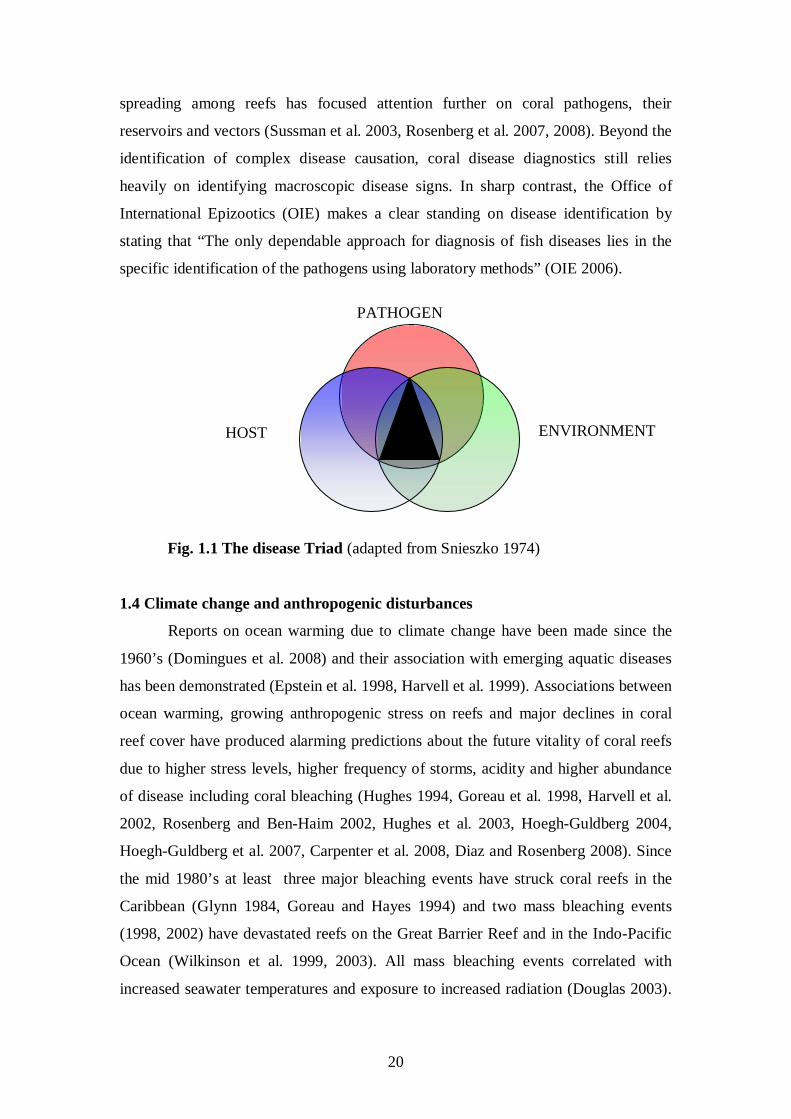
20
spreading among reefs has focused attention further on coral pathogens, their
reservoirs and vectors (Sussman et al. 2003, Rosenberg et al. 2007, 2008). Beyond the
identification of complex disease causation, coral disease diagnostics still relies
heavily on identifying macroscopic disease signs. In sharp contrast, the Office of
International Epizootics (OIE) makes a clear standing on disease identification by
stating that “The only dependable approach for diagnosis of fish diseases lies in the
specific identification of the pathogens using laboratory methods” (OIE 2006).
Fig. 1.1 The disease Triad (adapted from Snieszko 1974)
1.4 Climate change and anthropogenic disturbances
Reports on ocean warming due to climate change have been made since the
1960’s (Domingues et al. 2008) and their association with emerging aquatic diseases
has been demonstrated (Epstein et al. 1998, Harvell et al. 1999). Associations between
ocean warming, growing anthropogenic stress on reefs and major declines in coral
reef cover have produced alarming predictions about the future vitality of coral reefs
due to higher stress levels, higher frequency of storms, acidity and higher abundance
of disease including coral bleaching (Hughes 1994, Goreau et al. 1998, Harvell et al.
2002, Rosenberg and Ben-Haim 2002, Hughes et al. 2003, Hoegh-Guldberg 2004,
Hoegh-Guldberg et al. 2007, Carpenter et al. 2008, Diaz and Rosenberg 2008). Since
the mid 1980’s at least three major bleaching events have struck coral reefs in the
Caribbean (Glynn 1984, Goreau and Hayes 1994) and two mass bleaching events
(1998, 2002) have devastated reefs on the Great Barrier Reef and in the Indo-Pacific
Ocean (Wilkinson et al. 1999, 2003). All mass bleaching events correlated with
increased seawater temperatures and exposure to increased radiation (Douglas 2003).
PATHOGEN
ENVIRONMENT HOST
21
Thermal coral bleaching has also been replicated in experimental laboratory studies
by exposing healthy corals to heat and light stress (Hoegh-Guldberg and Smith 1989,
Glynn and D’Croz 1991, Iglesias-Prieto et al. 1992, Jones et al. 1998, Abrego et al.
2008). However, coral bleaching has also been shown to be caused by exposure of
healthy corals to bacterial pathogens at elevated temperatures (Kushmaro et al. 1998,
Banin 2000b, Israeli et al. 2001, Ben-Haim et al. 2003a) and to other stressors
(Hoegh-Guldberg and Smith 1989, Muscatine et al. 1991). Laboratory experimens
and modeling of seawater temperature data demonstrate that increased seawater
temperatures have a direct influence on coral disease abundance and progression
(Cervino 2004a, Boyett et al. 2007, Bruno et al. 2007). Bleached corals have also
been shown to develop coral disease signs, suggesting that susceptibility to disease
may be increased in compromised hosts (Muller et al. 2008). In addition, as seawater
temperatures continue to rise, new emerging coral diseases have been reported and
large scale epizootics have been documented in areas where no coral diseases have
been previously observed (Korrubel and Riegl 1998, Aeby 2005, Dalton and Smith
2006, Haapkylä et al. 2007, Sato et al. 2009), including in temperate regions (Hall-
Spencer et al. 2007). Nevertheless, lack of continuous data that document the impacts
of climate change on various targets in the marine environment, such as pathogen
abundance and spread, limits our understanding of how climate change may affect
coral reef health in the future if seawater temperatures continue to increase. Current
models forecast that by the end of the century, one third of reef building corals will
become extinct by direct and indirect effects of climate change (Carpenter et al. 2008)
including ocean acidification (Stone 2007).
In other more developed fields of disease research, such as human and
terrestrial wildlife medicine, the ability to predict the impact of climate change on
population health is supported by available data on pathogens and disease aetiology.
In a recent report released at the IUCN World Conservation Congress, named “The
Deadly Dozen: Wildlife Diseases in the Age of Climate Change” (The deadly dozen
2008), 12 infectious agents were listed by more than 60 scientists as contributing to
growing potential threats to both wildlife and human populations due to climate
change. According to the Wildlife Conservation Society (WCS), who published the
report, increased monitoring of health in wildlife ecosystems will provide a new lens
to document climate-related changes and will help governments, agencies, and
communities to detect and mitigate threats before they become disasters.
22
The second major source of stress impacting on marine life arises from the
discharge of anthropogenic effluents into nearshore environments. It has been
demonstrated for both fish and corals that eutrophication of these environments
represents a facilitating, predisposing and enabling factor for disease (Tomascik and
Sander 1987, Moeller 1990, Vethaak and Rheinallt 1992, Richmond 1993, Arkoosh et
al. 1998, Goreau et al. 1998, Richardson 1998a, Hughes and Connell 1999, Nyström
et al. 2000, Knowlton 2001, Harvell et al. 2002, Szmant 2002, Letters 2003, Loya et
al. 2004, Panek 2005, Kline et al. 2006, Smith et al. 2006, Voss and Richardson 2006,
Baker et al. 2007, Johnson et al. 2007, Knowlton and Jackson 2008). On the GBR and
throughout the Indo-Pacific, heavy rains results in large terrestrial flows into inshore
and fringing reefs, which carry nutrients from agricultural lands that promote
eutrophication and bacteria that directly impact on ecosystem health (Nairn 1993,
Lafferty et al. 2004, Fabricius 2005). Terrestrial flows may also contain pesticides and
sewage further deteriorating coral health (Grigg 1994, Wang et al. 2008). Fish
exposed to polluted water in the laboratory developed skin haemorrhages, opaqueness
of eyes and blindness (Grizzle et al. 1988), and Aeromonas, Pseudomonas and Vibrio
cells were isolated from these diseased fish. In another study, infected fish were found
as far away as 15 miles off-shore from the source of pollution (Mahoney et al. 1973).
Corals exposed to eutrophic stress in the field showed increased progression rates and
size of lesions caused by yellow band disease (Bruno et al. 2003), providing further
corroborative evidence that nutrient stress is an aggravating factor in disease
development.
1.5 Coral disease on the Great Barrier Reef and in the Indo-Pacific Ocean
Little research has been carried out on coral diseases on the GBR (Loya et al.
1984, Baird 2000). Some published notes on Indo-Pacific coral diseases relate to the
GBR in general (Antonius, 1985a, 1988a, 1988b, Coles, 1994, Littler and Littler 1995,
1996, Antonius 1999, Antonius and Lipscomb 2000, Raymundo et al. 2003). Willis
and collegues published an extensive review on coral diseases affecting GBR reefs in
2004 (Willis et al. 2004). Aeby (2006) and Kaczmarsky (2006) conducted similar
studies in the Northwestern Hawaiian Islands and in the Philippines, respectively. In
addition, reports on coral disease outbreaks from Hawaii (Aeby 2005, Friedlander et
al. 2005), Indonesia (Haapkylä et al. 2007) and the Gulf of Eilat (Barash et al. 2005)
have contributed to the acknowledgement that coral disease in the Indo-Pacific poses
23
a serious threat to coral populations. In contrast, studies of coral disease pathogens,
possible reservoirs or vectors on the GBR have not been attempted, mainly due to the
remoteness of outbreak sites, and the lack of expertise and funding needed for
conducting medically oriented microbial surveys. The Australian Institute of Marine
Science (AIMS) began to conduct coral disease surveillance in 1997 as part of its long
term monitoring program (LTMP). However, without any diagnostics or sampling,
little data have been collected beyond a visual inspection of disease abundance on
reefs (Sweatman et al. 2001), which has limited usefulness for determining the causes
and aetiologies of various coral diseases affecting corals of the GBR (Selig et al.
2006).
1.6 Black band disease (BBD)
Black band disease (BBD) was first reported from the Indo-Pacific in 1985
(Antonius 1985a) and from the GBR in 1994 (Dinsdale 1994, Littler and Littler 1996,
Miller 1996, Dinsdale 2002). It has a world wide distribution and has been studied for
the past 30 years demonstrating some of the early achievements in coral disease study,
but also many of its short comings. As suggested by early Caribbean studies
(Antonius 1981b, Rützler et al. 1983), BBD is a polymicrobial coral disease, which is
caused by a consortium (Richardson et al. 1997a, Richardson and Kuta 2003)
comprised of a dominant cyanobacterium (Phormidium corallyticum), which causes
the black band (Rützler and Santavy 1983, Taylor 1983, Kuta 2000) and other
bacteria, such as sulphur oxidizers and reducers (Carlton and Richardson 1995,
Richardson 1996, Schnell et al. 1996, Viehman and Richardson 2002, Viehman et al.
2006). Although accepted as causative agents for BBD, this group of microorganisms
has never been proven to actually cause the disease in laboratory experiments
(Antonius 1985b). P. corallyticum has also been found on healthy corals in the field in
contradiction to Henle-Koch's postulates (Richardson 1997b) and has only been
diagnosed using light microscopy to detect specific physiological traits. Molecular
studies undertaken in the Caribbean (Cooney et al. 2002, Frias-Lopez et al. 2002,
2004a) and in the Indo-Pacific (Frias-Lopez et al. 2003, Barneah et al. 2007) have all
failed to identify the presence of the specific P. corallyticum strain in samples from
BBD infected corals. In more recent studies conducted by the same authors, who
identified the former putative pathogen P. corallyticum, a new group of putative BBD
pathogens have been identified (Sekar et al. 2006, Myers et al. 2007, Voss et al. 2007),
24
suggesting that similar disease signs may be caused by different pathogens, or
alternatively, that the causative agents of BBD may change with time, as some corals
may develop resistance to infections, while certain cyanobacterial strains may become
out-competed by others. It may also suggest that disease identification may be biased
towards the tools and methods that are used for identification, suggesting that more
rigid disease identification and classifications are currently required (Ainsworth et al.
2007a, Work et al. 2008). The failure to induce black band disease signs by
conducting laboratory infections still prevents classifying this disease as an infectious-
polymicrobial disease (Aeby and Santavy 2006).
The abundance of BBD on the GBR has been reported by Willis et al. (2004)
and by Page and Willis (2006) and is typically low (under 1% prevalence) for the
northern and southern sectors of the GBR. The AIMS long term monitoring programe
(LTMP) reported less than 1 case of BBD infection per surveyed reef between the
years 1997 to 2001, during which surveys for coral disease were conducted at 48 sites
along the GBR (Sweatman et al. 2001).
The seemingly low prevalence of BBD from the GBR correlates well with
current data from the Caribbean (Weil et al. 2002) that estimate the wider abundance
of BBD on 19 different reefs to be under 0.5% (Weil 2004). Studies from the
Caribbean (Edmunds 1991, Bythell and Sheppard 1993, Bythell et al. 1993, Kuta and
Richardson 1997) demonstrate that coral mortalities caused by BBD outbreaks may
contribute to a major decline in reef community structure. Currently, no reports have
been made from the GBR regarding the phylogeny of cyanobacteria, or possibly other
putative agents, found on BBD infected corals.
1.7 Red band disease (RBD)
Red band disease (RBD) was first reported by Rützler et al. (1983) and later
by Richardson et al. in the Bahamas (1993, 1998a) and by Weil (2004) in the
Caribbean. It is assumed to be a novel coral disease, although little is known about its
cause or aetiology, but for the fact that bands progressing on diseased corals look red
and not black, the typical appearance of black band disease (BBD).
1.8 White syndromes (WS’s)
White syndrome (WS) is a general name given to patches or bands of bare
white coral skeleton resulting from yet unknown causes, reported from the GBR
25
(Willis et al. 2004) and from the Indo-Pacific Ocean (Aeby 2005). This name has
been adopted by the AIMS LTMP to differentiate between BBD infected corals,
bleached corals, skeleton exposed by predation and all other white syndromes. The
choice of this name follows on from experience in coral disease research gained in the
Caribbean, where 7 "white" coral diseases and plagues have been identified including
White Band I (Gladfelter 1982, Peters et al. 1983), White Band II (Ritchie and Smith
1998, Gil-Agudelo et al. 2006), White Plague I (Dustan 1977), White Plague II
(Richardson et al. 1998b, Richardson et al. 1998c), White Plague III (Richardson and
Aronson 2002) and additional white plague “like” diseases from the Caribbean
(Rodriguez-Martinez et al. 2001, Pantos et al. 2003) and from the Red Sea in Eilat
(Barash et al. 2005). The naming of all white plague (WP) and white band (WB)
diseases in the Caribbean has followed the chronological sequence of documenting
outbreaks, where different coral species have been shown to be susceptible to lesions
of varying characteristics. However, a superficial disease identification process based
on subjective observations has led to much confusion. Currently, causative agents
have been verified only for Caribbean white plague II (Denner et al. 2003), but for
none of the other syndromes. Lacking data on causative agents and disease aetiology
for Caribbean white plague and white band diseases makes it very hard to adopt the
Caribbean terminology for studies of similar disease signs found on corals in the
Indo-Pacific region, as suggested by Willis et al. (2004). However, regardless of
questions relating to either aetiology or cause, white diseases or syndromes both in the
Caribbean and in the Indo-Pacific Ocean pose substantial threats to coral reefs.
White band (WB) disease in the Caribbean has eliminated Acropora palmata
and Acropora cervicornis from reefs (Aronson and Precht 2001) leading to the listing
of WB-susceptible corals on the endangered species list (Precht et al. 2004). WP II
spread over 200 kilometer of reefs within 11 weeks during the 1995 Florida Key
outbreak, infecting 17 species and causing 38% morality (Richardson and Aronson
2002). White band disease was reported on the GBR by Baird (2000) and by
Antonious (1985a). A 20 fold increase in WS abundance was recorded between 1999
and 2003 by the AIMS LTMP on the GBR (almost 50 cases per reef) following the
2001/2 mass bleaching event (Willis et al. 2004). Willis et al. (2004) reported that WS
affected over 5% of all corals at Heron Island following the mass bleaching event of
2002/3. Outbreaks of WS’s have been reported since 2002/3 from many Indo-Pacific
locations, including the Northern Hawaiian Islands (Aeby 2005) and the Republic of
26
the Marshall Islands (Jacobson et al. 2006). Although WS's have been observed on
more than 15 coral species (Willis et al. 2004), local outbreaks typically affect tabular
coral species from the family Acroporidae, resulting in the naming of these disease
signs as Acropora White Syndrome (AWS) when they occur on Acropora hosts (Aeby
2005, Roff et al. 2006). However, causes for WS outbreaks still remain ambiguous.
Roff et al (2006) investigated diseased acroporid corals from Heron Island and
suggested that AWS occurs following a "shut down reaction” response programmed
by the coral. Other studies (Ainsworth et al. 2007b) detected DNA fragmentation of
coral cells affected by AWS, suggesting that lesions were caused by a coral
programmed cell death (PCD) initiated by the coral. Bruno et al. (2007) demonstrated
a positive correlation between coral cover, temperature and disease abundance on
GBR reefs, supporting the hypothesis that WS on the GBR may be an infectious
disease. Without precise knowledge of WS disease aetiology and it’s potential causes,
little can be said about whether WS is one disease or many, and whether similar
disease signs identified in the field are caused by one causative agent (or factor), or by
many. Without insight into the aetiology of WS, little can be said about the
association between WS disease abundance and environmental factors, such as ocean
warming or anthropogenic pollution, or any other host-related factor. Such knowledge
can aid in attempting to predict the future spread of WS’s in the Indo-Pacific region.
1.9 Atramentous necrosis, a new coral disease outbreak at Magnetic Island, GBR
Following the mass bleaching event in the Austral summer of 2002, a new
coral disease was observed on reefs fringing Magnetic Island (GBR). Jones et al.
(2004) identified large lesions on diseased Montipora aequituberculata colonies that
were later covered by sulphur deposits as the disease progressed. No causative agent
has yet been identified for this seemingly newly emerging disease, named
Atramentous necrosis (Jones et al. 2004). Recent work identified four distinct phases
of the disease, with the intial signs being white tissue that dies back progressively to
expose white skeleton and therefore fits the classification of a WS (Anthony et al.
2008). A study by Bourne (2005a) classified bacterial populations associated with
diseased corals on Magnetic Island, suggesting that WS on Magnetic Island
(Atramentous necrosis) may be bacterially caused. A coral disease affecting
Montipora spp. in Hawaii displaying similar disease signs has been named Montipora
White Syndrome – MWS (Jones 2007, Gochfeld and Aeby 2008).
27
1.10 Other coral diseases currently affecting the Indo-Pacific region
Greater surveillance efforts in the Indo-Pacific Ocean have produced
numerous reports of newly emerging coral diseases with names based on their
appearance, though characterization and establishment of either their cause or their
aetiologies has not been performed. Assignment of these names is required to simplify
any attempt to monitor outbreaks and follow their spread, though the current data
collected on unfamiliar disease signs may only lead to temporary classifications
(Work et al. 2008).
1.10.1 Skeletal eroding band (SEB)
The first report of this disease was made by Antonius and Lipscomb in 2001
and SEB is currently the most prevalent coral disease on the GBR affecting 2% of
corals from 82 scleractinian coral species surveyed between 2003-2006 (Page and
Willis 2008). Skeletal eroding band (SEB) manifests as dense aggregations of the
ciliate Halofolliculina corallasia (Riegel and Antonius 2003), which form a distinct
black band at the interface between recently exposed skeleton and apparently healthy
looking coral tissue. Similar disease signs have also been observed in the wider Indo-
Pacific (Winkler et al. 2004) and also in the Caribbean (Cróquer et al. 2006a, 2006b),
although Caribbean infections are potentially caused by another species from the
genus Halofolliculina. Currently, this disease is not classified as infectious to other
corals.
1.10.2 Brown band disease (BrB)
Brown band disease (BrB) was first described by Willis and collegues, who
observed macroscopic signs of the disease on three families of GBR corals (Willis et
al. 2004). Macroscopic signs of the disease include a brown zone adjacent to healthy
tissue on one side and to a zone of exposed white skeleton on the other. Occasionally,
a white zone is also observed between the brown band and the healthy tissue, which
may comprise bleached tissue and/or denuded skeleton. The brown zone contains a
protozoan ciliate which accumulates zooxanthellae intracellulary, resulting in the
characteristic brown colouring of the observed syndrome. The ciliates appear to ingest
coral tissue at the lesion interface, accumulating symbiotic zooxanthellae
(Symbiodinium) from coral endoderm (Bourne et al. 2008). Currently, no microbial
28
causative agents have been identified for this disease beyond coral infestation by
ciliates (Ulstrup et al. 2007) and fungi (Yarden et al. 2007).
1.10.3 Porites ulcerative white spot disease (PUWS)
This emerging disease targets specific hosts from the family Poritidae
(Raymundo et al. 2003). Novel PUWS disease signs are characterized by discrete
bleached, round foci, 3 to 5 mm in diameter, that may either regress or progress to
ulcerations the full thickness of the tissue layer that coalesce, occasionally resulting in
colony mortality. Raymundo et al. (2003) found a positive correlation between host
density and disease abundance. Currently, no causative agents have been identified
for PUWS, although grafting experiments (performed by tying together diseased and
healthy looking colonies in the field), demonstrated that this disease is communicable
(i.e., transmissible by direct contact with an affected coral, the coral's discharges or
by other indirect means) and possibly infectious (caused by the entrance into the body
of organisms, such as bacteria, protozoans, fungi, or viruses;
The causative agent of a disease affecting Favia spp. and Goniastrea spp.
corals in the Gulf of Eilat was identified by fulfilling Henle-Koch’s postulates (Barash
et al. 2005). Disease signs caused by the causative agent Thalassomonas loyana
Raymundo et al. 2003).
Until such evidence is provided, this disease may also be classified as WS.
1.10.4 Porites pink spots / Porites trematodiasis (TRM)
Porites trematodiasis is a coral disease caused by the infestation of coral tissue
by the digenetic trematode and parasite Podocotyloides stenometra (Friedlander et al.
2005, Aeby 1998, 2007). Disease signs include 3-5mm, pink to pale, swollen nodules
on the coral colony. This disease affects specific hosts from the family Poritidae.
However, little is known about TRM aetiology and the conditions required for large
scale outbreaks to occur. Similar disease signs have also been reported from the GBR
(Willis et al. 2004), although no evidence of a trematode was found, and from
Okinawa (Yamashiro 2000), where they were classified as growth anomalies
(Domart-Coulon et al. 2006), which were previously described in the Indo-Pacific
Ocean as neoplasia by Squires (1965) and as tumor formations on the GBR by Loya
et al. (1984).
1.10.5 White plague “like” disease in the Gulf of Eilat
29
(Thompson et al. 2006) include progressing tissue lesions which result in coral
mortalities. These disease signs are similar to reported signs of white plague II from
the Caribbean (Richardson et al. 1998c). However, the causative agent of this disease
has not been identified elsewhere.
1.11 Coral disease pathogens: what is currently known?
Studies attempting to isolate putative bacterial coral pathogens commenced in
the 1980’s (Peters et al. 1983), however the first bacterial coral pathogens were not
conclusively identified by fulfilling Henle-Koch’s postulates until the 1990’s
(Kushmaro et al. 1996). Identified coral pathogens include the fungal agent
Aspergillus sydowii, which causes the coenenchyme of sea fans to recede, exposing
the axial skeleton in laboratory infection trials involving the gorgonian Gorgonia
ventalina (Geiser et al. 1998) Identical disease signs were observed for the sea fan
disease in the field (Smith et al. 1996) and the fungal agent was also implicated.
The bacterial causative agent, Vibrio shiloi, has been identified by Kushmaro
et al. (1996) and shown to cause bleaching in laboratory exposure trials of the hard
encrusting coral Oculina patagonica. These disease signs were identical with
bleaching signs observed on infected colonies in the Mediterranean Sea. Although it
is currently well accepted that less than 5% of all marine microorganisms are
culturable on standard available media (Amann et al. 1995), the advantages of
maintaining pure or axenic pathogen cultures is self evident. It is a perquisite for the
fulfilment of Henle-Koch’s postulates (Koch 1891), but also a necessity when
attempting to study pathogen cell physiology, its virulence mechanisms and its
function under varying environmental conditions. It is also a necessity, when
attempting to design diagnostic tools of high validity that could be used for health
screening purposes. Isolating pathogens on growth media is also a relatively cost
efficient alternative to establish the identity of putative candidates. Such techniques
can be accomplished at field stations, thereby removing the possibility of transmitting
coral diseases by transporting infected corals or pathogens across reef regions.
Nevertheless, other methods for screening diseased individuals are currently available,
which are often used when direct isolation methods fail. These tools include cloning
and denaturing gradient gel electrophoresis – DGGE (Bourne 2005), fluorescent in
situ hybridization – FISH (Sussman et al. 2003), micro-arrays (Edge et al. 2005) and
metagenomics (DeLong 2005, Edwards and Rohwer 2005, Yokouchi et al. 2006).
30
For establishing causation of multi-factorial diseases involving either multiple
causative agents or enhanced host susceptibility as a consequence of environmental
conditions, more modern conventions and criteria have been established (Hill 1965,
Evans 1976) in order to overcome the many limitations arising when attempting to
fulfil Henle-Koch’s postulates in laboratory exposure trials (Casadevall and Pirofski
1999, Walker et al. 2006). New criteria rely on statistical significance for establishing
causation, rather than on direct cause and effect (Thrusfield 2005). For some diseases,
the maintenance of pathogen cultures may prove insufficient, for example when
susceptible hosts develop resistance towards one primary agent and become
susceptible towards another, such as in the case of V. shiloi, the causative agent for
the bleaching of O. patagonica in the Mediterranean Sea (Reshef et al. 2006).
1.12 Pathogens from the γ–Proteobacteria family Vibrionacae
Vibrios are prime candidates for bacterial infections in the marine
environment. Vibrio infections of fish (Egidius 1987), crustaceans (Brock 1992,
Goarant et al. 1998) and clams (Paillard 2004) have been studied for several decades.
From 21 diseases of shrimp reported from Asian aquaculture, all bacterial infections
(three of a total 21) were associated with vibrios (Lightner and Redman 1998). A
similar proportion (four Vibrio infections from a total of 24) was also reported from
the Americas (Lightner and Redman 1998). However, many Vibrio infections have
also been shown to be either opportunistic or secondary infections, which occur at a
latter part of disease progression (Saulnier et al. 2000). The shrimp industry has
provided significant evidence that many diseases are multifactorial, i.e., have mixed
etiologies (Lightner 1996). Viral infections in highly compacted shrimp aquaculture
facilities are often accompanied by bacterial and epicommensal infestations, which
end up being the cause of death in already severely compromised populations
(Beadling and Slifka 2004). In many cases, vibrios are among the dominant bacteria
isolated from such mortalities (Johansen and Sommer 2001).
Corals have been shown to die following exposure to elevated levels of
nutrients, which may cause a surge in bacterial abundance (including commensal
vibrios) in coral mucus (Smith et al. 2006). Vibriosis, also known as saltwater
furunculosis was first reported by Canestrini (1893). It took over 15 years until
Bergman (1909) characterized the disease and its etiological agent Vibrio anguillarum
(Snieszko 1975). The cost of Vibrio infections in closed aquaculture systems provided
31
the impetus to study of these pathogens, although they are also known to infect fish
populations in the wild (Haastein and Holt 1972). Currently, more than half a dozen
Vibrio species are listed as pathogens for invertebrates in the marine environment
(Amaro and Biosca 1996, Lambert et al. 1998, Venkateswaran et al. 1998, Liu 1999,
Villamil 2003, Wu 2004, Wang 2007), with pathogens, such as Vibrio vulnificus and
Vibrio parahaemoliticus, known to infect human beings as opportunistic pathogens
(Blake et al. 1980) causing high mortality rates (~60%) for compromised hosts
(Oliver 1989).
Of all known Vibrio pathogens Vibrio cholera is one of the best studied due to
its role as the causative agent of human Cholera epidemics for the past 1200 years,
including 7 pandemics since 857 AD. (Karaolis et al. 1994, Wachsmuth et al. 1994,
Colwell 1996). The study of Vibrio cholera contributed to current understanding of
the potential threat of pathogens commonly found in the environment to human
populations (Jiang et al. 2000, Codeco 2001). It also demonstrated the environmental
factors contributing to the spread of disease (Colwell 1996), and the role of gene
transfer in pathogen evolution (Waldor and Mekalanos 1996). Scientific discoveries
accomplished through the study of Vibrio cholera provided a clear path to follow for
the first studies of coral infectious diseases (Rosenberg 2005).
1.13 Coral pathogens from the γ–Proteobacteria family Vibrionacae
The most well characterized Vibrio infection of corals is the bleaching of the
Mediterranean encrusting coral, Oculina patagonica by the coral bleaching agent V.
shiloi (Kushmaro et al. 1996, 1997a, 2001). Although bacteria from the γ–
Proteobacteria family Vibrionacae were previously identified on bleached corals in
the Caribbean by Peters et al. (1983) and by Ritchie et al. (1994), the bleaching of O.
patagonica in the Mediterranean Sea is extensive and unique because it occurs
annually during the warm summer months (from May to September) and is followed
by full recovery of infected colonies during the winter. Recovery occurs when
seawater temperatures drop below 20°C (Kushmaro et al. 1997b, 1998), suggesting
that Vibrio virulence is temperature regulated (Toren et al. 1998, Israeli et al. 2001).
The pathogen, V. shiloi, has been shown to be capable of adhering to coral mucus
(Banin et al. 2001a), and to have an ability to penetrate into coral cells, where it
transforms into a viable but not culturable state - VBNC (Banin et al. 2000a, Oliver
2005). V. shiloi has also been shown to produce at least one specific toxin, a proline
32
rich peptide, which causes photoinhibition of Symbiodinium photosynthetic
endosymbionts that are located within the coral host tissue (Banin et al. 2001b, Ben-
Haim et al. 1999). The causative agent has been found to produce numerous other
virulence factors, including an anti-oxidant (superoxide dismutase – SOD), which is
critical for the pathogen’s survival in an oxygen rich environment, such as when in
close proximity to photosynthesizing cells (Banin et al. 2003, Reshef et al. 2008).
Following the identification of the first Vibrio coral bleaching agent, another coral
bleaching agent, Vibrio coralliilyticus, was characterized by Ben-Haim et al. (2002,
2003a) and shown to affect the Indo-Pacific coral Pocillopra damicornis by causing
bleaching at lower temperatures (≤28°C ) and cell lysis resulting in tissue lesions and
mortality at elevated temperatures (30°C; Ben-Haim et al. 2003a). This coral
pathogen, which was isolated in Zanzibar, Tanzania from infected corals, was found
to produce a proteolytic enzyme, which may cause photoinhibition of Symbiodinium
cells (Ben-Haim et al. 2003a). V. coralliilyticus has a world wide distribution,
including 3 strains causing coral bleaching isolated from infected corals in Eilat in the
Red Sea, and a strain isolated from a diseased oyster larvae (C. gigas) in the UK
(Ben-Haim et al. 2003a).
Recently, V. coralliitycus caused 100% mortality of Rainbow trout
(Oncorhychus mykiss) exposed to the pathogen in laboratory experiments, suggesting
a new hypothesis about its potential distribution in the wild (Austin 2005). Summer
zoonotics devastating populations of the gorgonian coral Paramuricea clavata along
western Mediterranean coasts in 1999 (Cerrano 2000) and in 2003 (Bally and
Garrabou 2007) were demonstrated to have been caused by a novel strain of V.
coralliilyticus isolated from infected gorgonian corals (Bally and Garrabou 2007).
In the Caribbean, isolates belonging to the γ–Proteobacteria family
Vibrionacae where found to be associated with corals displaying White Band II
disease signs (Ritchie and Smith 1995b). Recent experimental inoculation studies
demonstrated that the causative agent for WB II in Puerto Rico is a Vibrio harveyi
(Gil-Agudelo et al. 2006) and that infections are more likely to occur at elevated
seawater temperatures during the summer than in winter months when infected corals
were observed recovering (Gil-Agudelo et al. 2006). Vibrio isolates were also
identified as the causative agents for the Caribbean coral disease named Yellow
Blotch/Band – YB (Cervino et al. 2004a). Experimental exposure studies conducted
by Cervino et al. (2004a) demonstrated that this disease might be caused by a

33
consortium of Vibrio strains and not by a single pathogen, and that Yellow Blotch/
Band pathogens target the coral’s photosynthetic endosymbionts (Genus:
Symbiodinium) rather than coral tissue itself. Microbiological surveys conducted in
Zanzibar, Tanzania identified an elevated concentration of culturable Vibrio strains on
corals displaying white syndrome (WS) disease signs compared to healthy looking
corals (Piskorska et al. 2007). A significantly higher abundance of vibrios,
particularly Vibrio harveyi (strain LB4), was also shown to be associated with rapid
tissue necrosis (RTN) of Pocillopora damicornis (Luna et al. 2007). The presence of
Vibrio strains on compromised hosts has recently been demonstrated for corals
displaying growth anomalies (GA's; Breitbart et al. 2005), and in another study it has
been demonstrated that GA's are transmissible (Kaczmarsky and Richardson 2007).
Investigation of coral disease pathogens and their hosts has led to the
understanding that disease occurrence in eco-systems is a dynamic process, with
pathogens, hosts and environmental factors subjected to change. Recently, it has been
reported that the susceptible Mediterranean coral host, Oculina patagonica, can no
longer be artificially infected by its bleaching causing agent, V. shiloi (Reshef et al.
2006). Such reports have led to the conclusion that the coral host has become resistant
to its former pathogen (Reshef et al. 2006) and given rise to the establishment of the
coral probiotic hypothesis (Reshef et al. 2006) as part of a hologenome theory
(Rosenberg et al. 2007). The fact that currently coral bleaching in the Mediterranean
waters still occurs annually (as in the past) may suggest that coral hosts have become
susceptible to another ‘competing’ bacterium that causes similar bleaching signs, or
that bleaching was not bacterially-caused in the first place (Ainsworth et al. 2008).
1.14 What makes Vibrios good candidates for virulence?
The adaptability of vibrios to various environmental conditions and changes is
demonstrated by their ability to survive in multiple locations or hosts (Anderson and
May 1982, Thompson et al. 2004, Higgins et al. 2007). Vibrios can survive long
periods of dormancy in various forms including the viable but not culturable (VBNC)
state (Oliver 2005), in which they were shown to maintain their virulence (Oliver and
Bockian 1995). Some Vibrio strains have been shown to contain one chromosome and
another chromosome-like plasmid, both of which contain identical genes which allow
vibrios to switch “on” and “off” an entire set of expressional cascades directly
triggered by environmental signals (Heidelberg et al. 2000, Chen et al. 2003, Makino
34
et al. 2003, Ruby et al. 2005). Three “languages”, or signaling systems, assist vibrios
in direct communication among bacteria of the same species as well as in
communication with different bacterial species and even eukaryotic organisms
(Bassler 1999, Reading and Sperandio 2006). Vibrio “quorum sensing” (Dunlap 1999)
was first discovered through the ability of V. fisherei cells to switch on luminescence
at night when present at higher cell densities (Nealson et al. 1970) as mutalistic
symbionts in light-emitting organs of certain squids and fishes (Haygood 1993, Ruby
1996). The study of Vibrio “quorum sensing” and its specific signaling molecules,
which include acyl-homo-serine lactone (AHL) and autoinducers AI-I and AI-II
(Bassler et al. 1997) identified the involvement of cell density regulated gene
expression in the secretion of virulence factors. Such virulence factors within vibrios
include the upregulation of HapR (zinc-metalloprotease) and repression of ToxR
regulated Cholera toxin (CT) synthesis by Vibrio cholera (Zhu et al. 2002). The
ability of vibrios to acquire genetic material through horizontal transfer of plasmids
among bacteria (conjugation), via infection with bacteriophages (transduction) or by
natural transformation (Dröge et al. 1999) allows these organisms to easily penetrate
into new niches and quickly adapt to a rapidly changing environment (Thompson et al.
2004, Faruque et al. 2005). Some authors suggest that vibrios constantly migrate from
a solitary existence in the environment to one in hosts (Higgins et al. 2007) requiring
special genes for adhesion, penetration and for survival in hosts, compared to genes
adapted for solitary survival. The cholera paradigm (Colwell 1996) demonstrates that
Vibrio cholera cells can survive for long periods of time in the water column (Munro
and Colwell 1996) under varying conditions of nutrients and salinity (Singleton et al.
1982). They can also digest mucin (Nelson et al. 1976) and chitin (Nalin et al. 1979)
when adhering to copepods (Shukla et al. 1995) or plants (Shukla et al. 1995),
respectively. Thus cholera pandemics correlate well with elevated seawater
temperatures (Colwell 1996), which are associated with zooplankton and
phytoplankton
Few studies have investigated bacterial abundance on GBR reefs (Webster and
Hill 2001). One study of Vibrio prevalence on a GBR reefs has identified multiple
Vibrio species on both healthy and diseased corals (Bourne and Munn 2005),
blooms (Lobitz et al. 2000, Lipp et al. 2002). In aquaculture facilities
Vibrio virulence may be controlled by the use of antimicrobial peptides (Jia et al.
2000), yet in the marine environment, Vibrio prevalence and evolution is less studied
and understood.
35
suggesting that transition from a healthy state to a state of disease may not require an
infection by an external pathogen, but simply the transition of healthy hosts into
compromised-susceptible hosts. Another study by Bourne et al. (2008) demonstrated
an elevated abundance of vibrios on corals prior to their bleaching during a mass
bleaching event on Magnetic Island in 2001-2002, suggesting a possible elevated
abundance due to heat stress. Studies by Ritchie and colleagues have demonstrated
that healthy corals may harbour specific microbial populations (Ritchie and Smith
1997), which are capabale of utilizing specific coral-endosymbiont produced carbon
sources (Ritchie and Smith 1995a, Ritchie et al. 1995, 1996), while potentially
protecting corals against pathogen colonization (Ritchie 2006).
Vibrios have been shown to possess an array of potent virulence factors
(Debra and Linkous 1999, Mekalanos 1992, Nishibuchi and Kaper 1995). Of over 200
V. cholera serotypes present in the environment (Yamai et al. 1997, Reidl and Klose
2002), only two are known to contain all the necessary genes to cause the Cholera
pandemic; other less virulent V. cholera serotypes only cause minor infections (Singh
et al. 2001). One of the most well studied functions of Vibrio pathogens is their ability
to adhere to mucus, either mucus found on fish gills and fish skin (Bordas et al. 1998),
or mucus along the digestive duct (O'Toole 1999). Comprehensive studies on the
ability of pathogenic strains to adhere to their target host have demonstrated that only
pathogens can successfully colonize the mucus of fish by attachment, whereas non-
pathogens are only capable of displaying chemotaxis towards mucus, or mucus
digestion, but not attachment (Bordas et al. 1996). Bordas et al. (1996) have also
demonstrated that successful adhesion of pathogens to host mucus depends on
environmental factors, mainly on temperature and salinity (Bordas et al. 1996). It has
also been shown that specific compounds found in the host mucus (named: chemo-
attractants) are required for the successful adhesion of bacteria (O'Toole 1999). It is
therefore not surprising that Vibrio pathogens have also been shown to display
chemotaxis and adherence to coral mucus (Banin et al. 2001a) by attachment to a β-
D-galactopyranoside receptor present on the coral tissue. Banin et al. (2001a) also
demonstrated that pathogen attachment was only successful for corals in which
Symbiodinium endosymbionts were present. Sharon and Rosenberg (2008) concluded
that coral pathogens may require special conditions to successfully out-compete
bacterial populations that are the normal mucus-residents of healthy corals (Koren and
Rosenberg 2006, Ritchie 2006).
36
The secretion of proteolytic enzymes by Vibrio pathogens has also been
extensively studied, in particular the V. cholera HA/protease, a zinc-metalloprotease
(Booth et al. 1983), which is closely related to other Vibrio pathogen zinc-
metalloproteases (Norqvist et al. 1990, Milton et al. 1992, Hirono et al. 1996, Kim et
al. 2002, Binesse et al. 2008, Hasegawa et al. 2008). This enzyme is involved in a
number of functions, including digestion of connective tissue, attachment and
detachment and cell perturbation. The Vibrio zinc-metalloprotease belongs to the
thermolysin family of exocellular peptides and is defined as a pre-proenzyme
containing five subunits, which have all been suggested to contribute to virulence
(Marchler-Bauer et al. 2007, Varina et al. 2008). In addition, the fact that the
synthesis of zinc-metalloproteases by Vibrio pathogens is regulated by cell density
quorum sensing (Shao and Hor 2001, Kim et al. 2003, Milton 2006) supports placing
this group of proteolytic enzymes at the core of Vibrio species virulence. The
secretion of zinc-metalloprotease by Vibrio pathogens correlates with high cell
densities during the late exponential growth phase and early stationary phase of these
strains (Kreger and Lockwood 1981, Jeong et al. 2001, Denkin and Nelson 2004),
suggesting that the secretion of this proteolytic enzyme is required when pathogens
are invading, aggregating or swarming (Norqvist et al. 1990, Valiente et al. 2008). At
these times zinc-metaloprotease synthesis suppresses the synthesis of other virulence
genes, such as the cholera toxin – CT (Zhu et al. 2002), suggesting that all virulence
factors are not secreted by Vibrio pathogens at once and that each pathogen (or
serotype) may possess a unique set of virulence factors (Watanabe et al. 2004).
However, since the zinc-metalloprotease gene has been found in both pathogenic and
non-pathogenic Vibrio strains, it is not regarded as a major factor of virulence by all
authors (Booth and Finkelstein 1986), and the exact role of this key enzyme is yet to
be determined.
Vibrio coralliilyticus, the aetiological agent of bleaching of the Indo-Pacific
coral Pocillipora damicornis (Ben-Haim et al. 2002, 2003b) has been shown to
secrete a zinc-metalloprotease, whose partial protein sequence is closely related to the
V. cholera HA/protease.(Ben-Haim et al. 2003a). The coral pathogen zinc-
metalloprotease has been derived from a SDS gel sample containing the supernatant
from a culture of coral pathogens, which caused instant photoinhibition of
Symbiodinium cells in laboratory exposure trials (Ben-Haim et al. 2003a). Vibrio
shiloi the aetiological agent of bleaching of the Mediterranean coral Oculina
37
patagonica has been recently shown to possess numerous virulence genes, which
were also found to be absent from putative non-pathogenic strains (Reshef et al. 2008).
1.15 The aims of this study
To date, no attempts have been made to identify infectious diseases (i.e.,
diseases caused by micoorganisms) on the GBR, and only two coral pathogens have
been successfully identified from the entire Indo-Pacific region (Ben-Haim and
Rosenberg 2002, 2003b, Barash et al. 2005). This PhD study addresses this gap in
knowledge through investigation that addresses the following specific aims:
1. To identify the causative agents of corals displaying black band disease signs
on the Great Barrier Reef and red band disease signs in the Indo-Pacific Ocean
by culturing cyanobacteria isolated from infected corals.
2. To identify the causative agents of white syndromes (WS’s) currently
affecting coral reefs in the Republic of the Marshall Islands, the Republic of
Palau and on the Great Barrier Reef by culturing putative pathogens from
infected corals.
3. To identify virulence factors that are associated with coral disease signs
in order to investigate coral disease aetiology.
4. To determine the potential role of zinc-metalloproteases against coral and
Symbiodinium hosts by developing novel bio-assays utilizing cultured
Symbiodinium cells that have been isolated from disease-susceptible corals
and non-susceptible corals, and coral juveniles infected with known
Symbiodinium cells.
38
Chapter 2
A Single Cyanobacterial Ribotype is Associated with both
Red and Black Bands on Diseased Corals from Palau Sussman M, Bourne DG, Willis BL (2006) A single cyanobacterial ribotype is associated with
both red and black bands on diseased corals from Palau. Dis Aqua Org 69: 111-118
2.1 Introduction
Cyanobacteria are associated with at least two band diseases that are
differentiated macroscopically primarily based on color: black band disease (BBD),
the first coral disease to be identified (Antonious 1973); and red band disease (RBD),
first reported by Ruetzler et al. (1983) and later by Richardson in the Bahamas
(Richardson 1993, 1998a). Black band disease is a band of filamentous cyanobacteria
that progresses across live coral tissue, causing mortality of the coral along the
advancing front (Antonius 1981b). It has been recorded world-wide and is found on
21 species in the Caribbean (Weil 2004) and 40 species on the Great Barrier Reef.
(Page and Willis 2006). Red band disease (RBD) has been considered a separate
disease category (Santavy and Peters 1997, Garzon-Ferreira et al. 2001) and has been
recorded to affect 14 coral species in the Caribbean (Weil et al. 2002, Weil 2004).
However, researchers have had little success in culturing the cyanobacteria associated
with these putatively different coral diseases and thus it has not been possible to
evaluate the significance of their color differences.
Early studies suggested that the causative agent of BBD is a consortium of
microorganisms (reviewed in Richardson 2004). Phormidium corallyticum, a
filamentous cyanobacterium from the genus Oscillatoria, was initially identified as
the species forming the mat or band on infected corals (Rützler and Santavy 1983,
Taylor 1983). The band’s black appearance was suggested to be caused by a high
concentration of cells containing the light-harvesting red photosynthetic pigment
phycoerythrin (Richardson 1997a). Recent molecular analyses have failed to identify
P. corallyticum and other proposed consortium-members, such as the sulfide-
oxidizing bacteria Beggiatoa sp., in the black mat of BBD infected corals (Frias-
Lopez et al. 2004a). Another ribotype of cyanobacteria from the genus Oscillatoria
was shown to be associated with BBD infected corals from the Caribbean and Indo-
Pacific (Frias-Lopez et al. 2002, Cooney et al. 2002, Frias-Lopez et al. 2003). These
39
conflicting findings have led to an uncertainty regarding the identity of the
cyanobacteria associated with BBD and its role in causing infections (Frias-Lopez et
al. 2004a, 2004b).
Morphological observations of the cyanobacteria associated with red bands
identified two filamentous strains within the genus Oscillatoria (Richardson 1993,
1998a). These strains were reported as being visually distinct from P. corallyticum,
yet never identified to a species level. Rützler et al. (1983) visually identified
Schizothrix calcicola and S. mexicana on corals displaying red bands. Sutherland et
al. (2004) claimed that due to lower prevalence in the field, red band might be
grouped together with black band as a single classification. Harvell et al. (2004) was
the first to publish a red band photo of an infected Pachyseris speciosa colony from
Palau, emphasizing the need for further investigations. In recent surveys on the GBR,
Willis et al. (2004) classified red bands and other cyanobacterial syndromes as part of
a generalized cyanobacterial category due to uncertainty regarding their taxonomic
identity. Needless to say, not all cyanobacteria found on corals constitute a threat to
coral health (Lesser et al. 2004).
The aims of this study were to develop methods for culturing cyanobacteria
associated with band diseases in corals and to determine if cyanobacteria isolated
from red bands and BBD are genetically different. Isolation and culturing of
cyanobacteria from one black and two red bands on corals sampled from reefs in
Palau (Micronesia) tested whether pigmentation was: (1) independent of strain-host
interaction, and (2) stable under identical growth conditions. Results of this study
establish the first link between color, morphology and molecular-based taxonomic
identification of cyanobacterial strains isolated from both red band and BBD infected
corals.
2.2 Materials and methods
2.2.1 Sample collection
Samples from two corals infected with red bands (Porites sp. and Pachyseris
speciosa) and one infected with BBD (Montipora sp.) were collected by SCUBA on
Indo-Pacific reefs surrounding the island of Palau in January 2004. Samples were
sealed in plastic bags immediately after collection and transported to a laboratory at
the Palau International Coral Reef Center (PICRC). Samples were processed within
two hours of collection. Red and black cyanobacteria associated with the microbial
40
mat of each disease were sampled from the corals with sterile forceps and transferred
to agar plates.
2.2.2 Media and conditions of growth
Optimized conditions for growth of isolated cyanobacterial strains were
determined by comparison of growth responses in 13 different media (see Table 2.1)
under two light regimes. Media were chosen based on published recommendations for
optimizing cyanobacterial growth in cultures (reviewed in McLachlan, 1973).
Cultures were incubated at 26°C under either a 12:12 h fluorescent light regime or 24
hours of constant fluorescent illumination, both at 15μE m-2 s-1. Half the plates used in
this trial were scored (Vaara et al.1979). Scoring assists in purifying cultures from
contaminants while selecting for motility, which is a key characteristic of
cyanobacteria found on BBD and red band infected corals. Cultures were inspected
daily and graded for growth on a scale from low growth (+) to highest growth (+++++)
after a 7-day incubation period. The most successful media (Grund and ASN-III) and
conditions (constant lighting/scored plates) (see Table 2.1 for summary of results)
were chosen for studies of color expression under constant growth conditions. Grund
media (von Stosch 1963, 1969) consisted of enriched seawater (enriched with 10 mgl -
1 CaCl2 , 4 mgl -1 Na2HPO4, 1 gl -1 NaNO3, 2mgl -1 Disodium EDTA, 20 mgl -
1 Na2CO3) to which were added 1 ml micronutrient solution (A5 + CO) and 1 ml
vitamin solution (S-3) per liter of medium. The micronutrient solution A5 + CO
(Rippka et al. 1979) was made of an x 1000 stock solution and filter sterilized (2.86
gl -1 H3BO3 , 1.81 gl -1 MnCl2 . 4H2O, 0.22 gl -1 ZnSO4
. 7H2O, 0.39 gl -1
Na2MoO4. 2H2O, 0.079 gl -1 CuSO4
. 5H2O, 0.049 gl -1 Co(No3)2. 6H2O). The vitamin
solution S-3 (Provasoli 1963) was made as an x 1000 stock solution filter sterilized
and kept in the dark (100 mgl -1 nicotinic acid, 500 mgl -1 thiamine . HCl, 100 mgl -1
Ca. pantothenate, 10 mgl -1 PABA, 1 mgl -1 biotin, 5 gl -1 i-inositol, 2 mgl -1 folic
acid, 1 mgl -1 cyanocobalamin, 3 gl -1 thymine). In addition, 8 μM of Na2-EDTA /
FeCl3 were added to each 1 liter of medium (Rippka et al. 2000). ASNIII, a synthetic
media developed by Waterbury and Stanier (1981) was revised and made up of 1l
double distilled water (DDW), 427 mM NaCl, 6.7 mM KCl, 3.4 mM CaCl2, 14.2 mM
MgSO4, 9.8 mM MgCl2, 0.09 mM K2HPO4, 1 mM NaNO3, 0.19 mM Na2CO3. 8 μM
of Na2-EDTA / FeCl3, 1 ml micronutrient solution (A5 + CO) and 1 ml vitamin
solution (S-3) were added to each 1 liter of medium. Agar plates were prepared using
41
washed agar (Waterbury and Willey 1988). A x1.33 concentration (in 750 ml) of
liquid media (Grund or ASN-III) was filtered using Millipore 1000 ml Stericup®,
preheated to 50ºC and added to 250 ml of autoclaved DDW with 6 g washed agar.
Once dry, plates were scored with parallel lines using a flamed glass slide (Vaara et al.
1979) and incubated under unidirectional light. A liquid medium prepared from live
coral tissue in seawater was also tested and found to enhance growth. This medium
was autoclaved for 30 min and then sterile filtered (as above).
2.2.3 DNA extraction, PCR amplification and sequencing
DNA extraction was performed by a modified protocol from Wu et al. (2000).
Following an initial growth phase of 10-14 days, filaments from liquid media flasks
were inspected under light microscopy for strain purity, color and morphology. 200
mg of cyanobacteria culture RMS1 (P. speciosa), RMS2 (Porites sp.) and BMS1
(Montipora sp.) were collected from each flask and placed in 2 ml tubes with 1 ml
buffer A (100mM Tris, 50mM EDTA, 100mM NaCl at pH 8) for 10 min at room
temperature. N-lauroylsarcosine (sarkosyl) was added to a final concentration of 0.1%
and the samples incubated overnight at 4°C. Filaments were collected by
centrifugation at 10,000 rpm for 10 min, washed in 2 ml of TES (50 mM Tris, 5 mM
EDTA, 50mM NaCl at pH 8), centrifuged again for 5 min and resuspended in 1 ml
TES. Lysozyme was added to a final concentration of 0.5 mg ml-1 and the sample
incubated with RNase (20μg ml -1) at 37°C for 30-90 min with shaking. SDS was
added to a final concentration of 1%, stirred thoroughly, and kept at 37°C for 10 min.
50μg ml-1 Proteinase K was added and the sample was incubated overnight at 4°C (or
alternatively at 37°C for 1h). Extraction was performed by using the Promega Wizard
Genomic DNA purification kit® starting with protein precipitation (step 4 of Gram
negative protocol). After isopropanol and 70% ethanol precipitations, pellets were
dried in a DNA110 Speed Vac (Savant) for 15 min and rehydrated overnight at 4°C.
PCR amplification was performed following the protocol of Nübel et al. (1997).
Cyanobacterial specific primers used were the CY106F forward primer (CGG ACG
GGT GAG TAA CGC GTG A) and an equimolar mixture of CYA 781Ra (GAC TAC
TGG GGT ATC TAA TCC CAT T) and CYA 781Rb (GAC TAC AGG GGT ATC
TAA TCC CTT T) reverse primers. 50 μl reactions were made of 10 picomoles of
each primer, 5 μl of 10x PCR buffer (Taq PCR Core Kit, Qiagen with 15mM MgCl2),
42
50 nmol dNTP, 10 ng of template DNA (1:10 dilution of stock). 10 μl Q-Solution
(Qiagen) was added to each tube and DDW to the volume of 50 μl. 1U of Taq was
added at 80°C to minimize nonspecific annealing of primers following a 5 min hot
start at 94°C. An Eppendorf Mastercycler® was used for template amplification with
35 cycles of 1 min at 94°C, 1 min at 60°C and 1 min at 72°C. A final 5 min extension
step at 72 °C was performed. Reaction products were purified using DNAce Quick-
Clean® and sequenced using ET-Terminators (Amersham Biosciences) on
MegaBACE 1000 from Amersham Biosciences. Sequences were visualized using
CHROMAS® (Conor McCarthy http://trihul.sci.gu.edu au/P
~Pconor/chromas.html) and
aligned (Altschul et al. 1990) with existing database using BLAST
(http://www.ncbinih.nlm.gov/blast). Multiple alignments were performed using BCM
- multiple sequence alignments (http://dot.imgen.bcm.tmc.edu/multi-align/multi-
align.html). Partial 16S rRNA gene sequences were entered into the Genbank
database under the following accession numbers: AY839639, AY839640, AY839641
(for further detail see table 2.2).
2.2.4 Light Microscopy
Cyanobacterial filaments were examined using the Zeiss Axioshop® 2 Mot
Plus microscope equipped with an AxioCam® MRC5 digital camera using
AxioVision® 4 software. Cyanobacterial filaments were photographed prior to the
sampling for DNA extraction. Since this study aims to compare pigmentation among
cynaobacterial strains, no further software-manipulation of AxioVision® 4 calibrated
images has been undertaken and they appear as identical replications of originals.
2.3 Results
2.3.1 Media and conditions of growth
Thirteen different variations of solid and liquid media were evaluated in an
attempt to isolate and grow filamentous cyanobacteria associated with infected corals.
Cultures grew best in either Grund media (with seawater) or ASN III media (Table
2.1). Cyanobacteria did not grow on, or in, any of the media devoid of nitrogen (PCR-
S8, PCR-S11, PCR-Tu2). This demonstrates that cyanobacteria found on red band and
BBD infected corals do not fix nitrogen. Highest growth in liquid media (score:
+++++) was achieved in a zooxanthellae broth consisting of coral tissue autoclaved in
43
seawater. At 26°C and using fluorescent lights (15μE m-2 s-1), continuous (24 h)
lighting provided better results than a 12:12 h light:dark regime.
Washing agar with ethanol and acetone, as suggested by Waterbury and
Willey (1988), and scoring plates (Vaara et al. 1979) facilitated the motility of
cyanobacteria towards a unidirectional light source placed at an angle away from the
plates. Both red and black cyanobacterial filaments in scored plates progressed along
groves at a speed of up to 5cm/day. Non-scored plates placed directly under light
exhibited no growth. Supplementing the agar with 0.1% (wt/vol) Na2S and raising pH
to 9 (Vaara et al. 1979) did not result in enhanced motility. Nevertheless, strains
isolated from infected corals demonstrated an ability to survive in higher
concentrations of Na2S (750mg Na2S.9H2O per liter of medium). In liquid media,
filaments concentrated in clumps on the glass walls of flasks. They formed a straight
line at the interphase between media and air while orienting towards the light.
Placement of a sterile piece of coral skeleton in the flasks initiated settlement of the
filaments onto the skeleton.
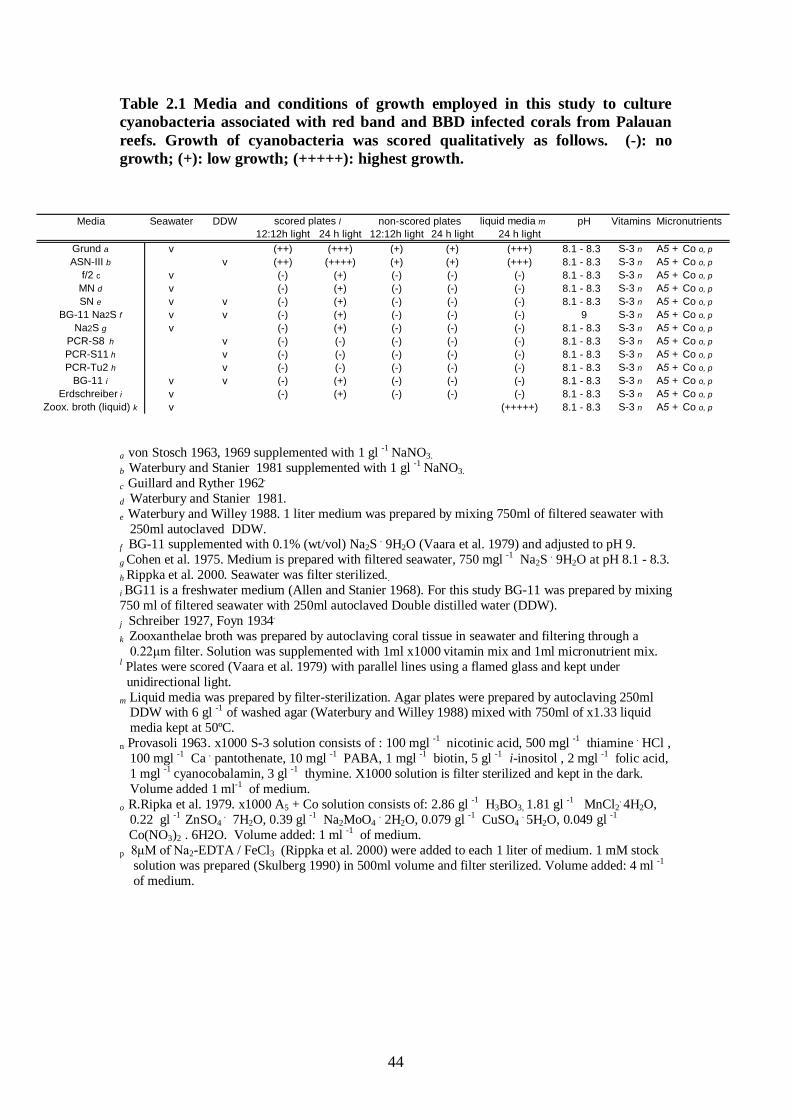
44
Table 2.1 Media and conditions of growth employed in this study to culture cyanobacteria associated with red band and BBD infected corals from Palauan reefs. Growth of cyanobacteria was scored qualitatively as follows. (-): no growth; (+): low growth; (+++++): highest growth.
a von Stosch 1963, 1969 supplemented with 1 gl -1 NaNO3.
b Waterbury and Stanier 1981 supplemented with 1 gl -1 NaNO3. c Guillard and Ryther 1962.
d Waterbury and Stanier 1981.
e Waterbury and Willey 1988. 1 liter medium was prepared by mixing 750ml of filtered seawater with
250ml autoclaved DDW.
f BG-11 supplemented with 0.1% (wt/vol) Na2S . 9H2O (Vaara et al. 1979) and adjusted to pH 9.
g Cohen et al. 1975. Medium is prepared with filtered seawater, 750 mgl -1 Na2S . 9H2O at pH 8.1 - 8.3. h Rippka et al. 2000. Seawater was filter sterilized.. i BG11 is a freshwater medium (Allen and Stanier 1968). For this study BG-11 was prepared by mixing 750 ml of filtered seawater with 250ml autoclaved Double distilled water (DDW). j
Schreiber 1927, Foyn 1934.
k Zooxanthelae broth was prepared by autoclaving coral tissue in seawater and filtering through a 0.22μm filter. Solution was supplemented with 1ml x1000 vitamin mix and 1ml micronutrient mix. l Plates were scored (Vaara et al. 1979) with parallel lines using a flamed glass and kept under unidirectional light. m Liquid media was prepared by filter-sterilization. Agar plates were prepared by autoclaving 250ml DDW with 6 gl -1 of washed agar (Waterbury and Willey 1988) mixed with 750ml of x1.33 liquid media kept at 50ºC. n Provasoli 1963. x1000 S-3 solution consists of : 100 mgl -1 nicotinic acid, 500 mgl -1 thiamine . HCl , 100 mgl -1 Ca . pantothenate, 10 mgl -1 PABA, 1 mgl -1 biotin, 5 gl -1 i-inositol , 2 mgl -1 folic acid, 1 mgl -1 cyanocobalamin, 3 gl -1 thymine. X1000 solution is filter sterilized and kept in the dark. Volume added 1 ml-1 of medium. o
R.Ripka et al. 1979. x1000 A5 + Co solution consists of: 2.86 gl -1 H3BO3, 1.81 gl -1 MnCl2. 4H2O,
0.22 gl -1 ZnSO4 . 7H2O, 0.39 gl -1 Na2MoO4 . 2H2O, 0.079 gl -1 CuSO4 . 5H2O, 0.049 gl -1
Co(NO3)2 . 6H2O. Volume added: 1 ml -1 of medium. p 8μM of Na2-EDTA / FeCl3 (Rippka et al. 2000) were added to each 1 liter of medium. 1 mM stock solution was prepared (Skulberg 1990) in 500ml volume and filter sterilized. Volume added: 4 ml -1
Media Seawater DDW scored plates l non-scored plates liquid media m pH Vitamins Micronutrients12:12h light 24 h light 12:12h light 24 h light 24 h light
Grund a v (++) (+++) (+) (+) (+++) 8.1 - 8.3 S-3 n A5 + Co o, pASN-III b v (++) (++++) (+) (+) (+++) 8.1 - 8.3 S-3 n A5 + Co o, p
f/2 c v (-) (+) (-) (-) (-) 8.1 - 8.3 S-3 n A5 + Co o, pMN d v (-) (+) (-) (-) (-) 8.1 - 8.3 S-3 n A5 + Co o, pSN e v v (-) (+) (-) (-) (-) 8.1 - 8.3 S-3 n A5 + Co o, p
BG-11 Na2S f v v (-) (+) (-) (-) (-) 9 S-3 n A5 + Co o, pNa2S g v (-) (+) (-) (-) (-) 8.1 - 8.3 S-3 n A5 + Co o, p
PCR-S8 h v (-) (-) (-) (-) (-) 8.1 - 8.3 S-3 n A5 + Co o, pPCR-S11 h v (-) (-) (-) (-) (-) 8.1 - 8.3 S-3 n A5 + Co o, pPCR-Tu2 h v (-) (-) (-) (-) (-) 8.1 - 8.3 S-3 n A5 + Co o, p
BG-11 i v v (-) (+) (-) (-) (-) 8.1 - 8.3 S-3 n A5 + Co o, pErdschreiber i v (-) (+) (-) (-) (-) 8.1 - 8.3 S-3 n A5 + Co o, p
Zoox. broth (liquid) k v (+++++) 8.1 - 8.3 S-3 n A5 + Co o, p
of medium.
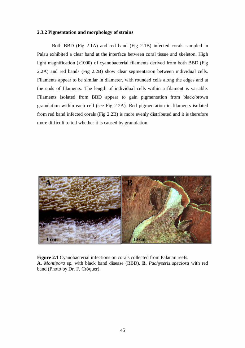
45
2.3.2 Pigmentation and morphology of strains
Both BBD (Fig 2.1A) and red band (Fig 2.1B) infected corals sampled in
Palau exhibited a clear band at the interface between coral tissue and skeleton. High
light magnification (x1000) of cyanobacterial filaments derived from both BBD (Fig
2.2A) and red bands (Fig 2.2B) show clear segmentation between individual cells.
Filaments appear to be similar in diameter, with rounded cells along the edges and at
the ends of filaments. The length of individual cells within a filament is variable.
Filaments isolated from BBD appear to gain pigmentation from black/brown
granulation within each cell (see Fig 2.2A). Red pigmentation in filaments isolated
from red band infected corals (Fig 2.2B) is more evenly distributed and it is therefore
more difficult to tell whether it is caused by granulation.
Figure 2.1 Cyanobacterial infections on corals collected from Palauan reefs. A. Montipora sp. with black band disease (BBD). B. Pachyseris speciosa with red band (Photo by Dr. F. Cróquer).
B
10 cm
AA
1 cm
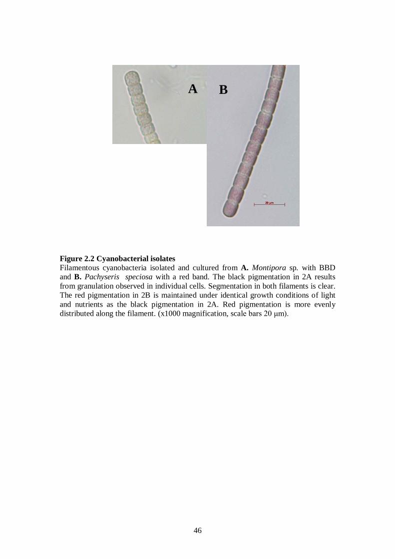
46
Figure 2.2 Cyanobacterial isolates Filamentous cyanobacteria isolated and cultured from A. Montipora sp. with BBD and B. Pachyseris speciosa with a red band. The black pigmentation in 2A results from granulation observed in individual cells. Segmentation in both filaments is clear. The red pigmentation in 2B is maintained under identical growth conditions of light and nutrients as the black pigmentation in 2A. Red pigmentation is more evenly distributed along the filament. (x1000 magnification, scale bars 20 μm).
A B
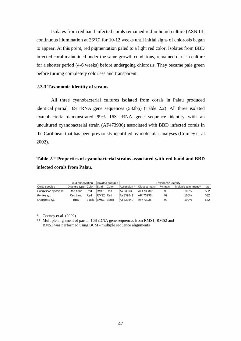
47
Isolates from red band infected corals remained red in liquid culture (ASN III,
continuous illumination at 26°C) for 10-12 weeks until initial signs of chlorosis began
to appear. At this point, red pigmentation paled to a light red color. Isolates from BBD
infected coral maintained under the same growth conditions, remained dark in culture
for a shorter period (4-6 weeks) before undergoing chlorosis. They became pale green
before turning completely colorless and transparent.
2.3.3 Taxonomic identity of strains
All three cyanobacterial cultures isolated from corals in Palau produced
identical partial 16S rRNA gene sequences (582bp) (Table 2.2). All three isolated
cyanobacteria demonstrated 99% 16S rRNA gene sequence identity with an
uncultured cyanobacterial strain (AF473936) associated with BBD infected corals in
the Caribbean that has been previously identified by molecular analyses (Cooney et al.
2002).
Table 2.2 Properties of cyanobacterial strains associated with red band and BBD
infected corals from Palau.
Field observation Isolated cultures Taxonomic identity
Coral species Disease type Color Strain Color Accession # Closest match % match Multiple alignment** bpPachyseris speciosa Red band Red RMS1 Red AY839639 AF473936* 99 100% 582Porites sp. Red band Red RMS2 Red AY839641 AF473936 99 100% 582Montipora sp. BBD Black BMS1 Black AY839640 AF473936 99 100% 582 * Cooney et al. (2002) ** Multiple alignment of partial 16S rDNA gene sequences from RMS1, RMS2 and BMS1 was performed using BCM - multiple sequence alignments
48
2.4 Discussion
Results reported here represent the first successful isolation and culturing of
filamentous cyanobacteria associated with both red band and BBD infected corals.
Our molecular studies show that cyanobacterial filaments isolated from corals
infected with red band and BBD share the same taxonomic identity. Observations of
cyanobacteria in culture suggest that a single cyanobacterial ribotype associated with
a coral disease can exhibit dual pigmentation under similar light and growth
conditions. Previous studies of cyanobacterial pigmentation have demonstrated that
complementary chromatic adaptation allows cyanobacteria to reversibly alter their
pigmentation from green-blue to red in response to light (see reviews by Grossman et
al. 1993a, 1993b). This has been demonstrated in the filamentous cyanobacterium,
Fremyella diplosiphon (Kehoe and Grossman 1994). The color of cells can also be in
response to other environmental factors, such as elevated or low nutrient levels
(nitrogen, phosphorus and sulfur; Collier and Grossman 1992).
Consistency of color, either red or brown/black, of cyanobacteria cultured over
the 12 weeks of our study suggests that complementary chromatic adaptation doesn’t
adequately explain our observations. Had light and growth conditions determined
color expression, a complementary chromatic adaptation should have occurred and
both red and brown/black cyanobacterial cultures would have converged to a similar
pigmentation. The only shift in pigmentation suggestive of a complementary
chromatic adaptation that we observed was from red to light green in some cultures
growing on agar plates. This change was usually rapid and irreversible. Red
cyanobacteria from red band infected corals never exhibited the dark brown/black
pigmentation seen in the cultures originating from BBD infected corals.
Synechococcus sp. strain SH-94-5 reacts to nitrogen depletion by upregulating the
accumulation of beta-carotene, thus resulting in a pigmentation change to red. During
exponential growth, this strain down regulates production of phycobiliproteins and
becomes green (Miller et al. 2002). Whether such pigmentation changes occur in
cyanobacteria infesting coral tissue in the field remains unknown. Currently, there are
no reports of BBD infected corals transforming into red bands, or red bands
transforming into black bands.
Two lines of evidence suggest that cyanobacterial growth in culture is
optimized by the presence of substrate for settlement. First, cyanobacterial strains
49
grown in liquid cultures settled on the glass flasks along a line at the interface
between the liquid media and air and initiated this growth characteristic on the side
closest to the light. Secondly, addition of a sterile piece of coral skeleton to the media
flasks initiated immediate settlement of the filaments. This preference for substrate
might explain why shaking of cultures in liquid media has proven unsuccessful for
growth. However, settlement of cyanobacterial filaments on coral skeletons in the
laboratory was different from settlement observed in the field, where filaments
concentrate only at the interface between coral tissue and skeleton leaving white
exposed skeleton behind the moving front (as in Figs 2.1A-B). The settlement pattern
observed in the laboratory is possibly explained by the distribution of nutrients inside
media flasks. Cyanobacteria progressing along the border of degrading coral tissue are
exposed to high nutrient levels (Taylor 1983). The high density of filaments forming
the bands on infected corals also results in shading (Richardson et al. 2001), although
there are conflicting reports of bands progressing towards the light or away from it
(Antonius 1981b). Filament aggregation has been suggested by Castenholz (1982) as
a mechanism adopted by cyanobacteria to increase shading in order to attain lower
light levels needed for optimal photosynthetic activity. In our laboratory trials, in
contrast, filaments tended to dissociate when placed in grooves on scored agar plates,
with individual filaments separating and traveling up to 5 cm/day towards the light,
eventually spreading over the entire agar surface rather than aggregating.
The speed with which cyanobacterial filaments isolated from both red band
and BBD infected corals travel across agar indicates that band-progression in the field
(only 0.3-1 cm/day) might be limited by other factors. The degree of motility of
filaments in this study might explain how members of this taxonomic ribotype
outcompete other cyanobacteria when settling on corals. Further study should be
directed at determining the relationship between motility towards light and the
propensity for aggregation and how these might be affected by variation in light and
nutrient levels.
As in previous studies that have attempted to determine the identity of
filamentous cyanobacteria associated with BBD (Cooney et al. 2002, Frias-Lopez et
al. 2002, 2003), this study used partial sequencing of the 16S rDNA gene. Given that
we sampled only two red bands from the field, our results do not imply that all red
band infections carry the same cyanobacteria; further samples from red bands are
required to test the generality of our results. Although we have shown that dual
50
pigmentation can be exhibited by a single ribotype, other less dominant
cyanobacterial strains that we were unable to isolate and culture on the media used,
may have been present in our original field samples. Our aim in this study was to
isolate and culture the dominant color forming strains from infected corals in the field
and to establish a reference point in the relation between cyanobacterial color,
taxonomic identity and disease classification, rather than to identify the entire
microbial complexity of cyanobacterial mats on infected corals. Cloning of extracted
DNA from field samples using universal primers targeted at a larger region of the 16S
rRNA gene would provide a more comprehensive analysis of cyanobacterial
populations associated with both red band and BBD infected corals. If pathogenicity
and virulence are to be studied, an attempt should be made to explore the genes
linking identity and function, as has been recently demonstrated for BBD by Frias-
Lopez et al. (2004b). In the case of Vibrio cholera it has already been shown that the
16S rRNA gene based identity of strains does not necessarily relate to virulence
(Colwell and Huq 1994, Singh et al. 2001).
This study has identified dual pigmentation expressed by a single ribotype
found on both red band and BBD infected corals but did not determine the mechanism
involved. The fact that red bands are far less prevalent in the field than BBD might
point towards a genetic variation that result in both a common and a rare phenotype.
The strains isolated and cultured by this study belong to a ribotype that has
been previously identified by molecular tools on BBD infected corals in the
Caribbean (Cooney et al. 2002, Frias-Lopez et al. 2002). The strains isolated from
both red bands and BBD in this study were found by BLAST to share less than 90%
match-identity with P. corallyticum (AF474001), previously suggested as the
causative agent of BBD (Richardson and Kuta 2003), though later re-named as a
Geitlerinema sp., which has been shown to produce microcystin toxins (Richardson et
al. 2007, Voss et al. 2007). The results of this study call for examination of the role of
P. corallyticum/Geitlerinema sp. in BBD infections by comparing its ability to infect
healthy corals with the newly isolated strains presented here (RMS1, RMS2 and
BMS1).
The maintenance of new cultures associated with coral disease opens many
doors for further research including examining the potential virulence and toxicity of
these new strains isolated from red bands and BBD, their geographical distribution
and the ways in which strains are transmitted. Based on the results of this study, we

51
strongly urge postponing the classification of red cyanobacterial bands as a separate
coral disease until further evidence is collected.
52
Chapter 3
Coral pathogens identified for white syndrome (WS)
epizootics in the Indo-Pacific Sussman M, Willis BL, Victor S, Bourne DG (2008) Coral pathogens identified forWhite Syndrome (WS) epizootics in the Indo-Pacific. PLoSONE 3(6): e2393.doi:10.1371/journal.pone.0002393
3.1 Introduction
Reports on coral disease continue to rise (Harvell et al. 2002), however the
causes for these diseases and the methods by which to investigate them are still
heavily debated (Cervino et al. 2004a, Rosenberg and Falkovitz 2004, Lesser et al.
2007). Most efforts are directed towards traditional surveillance (Morens et al. 2004),
with comparatively less research directed towards developing strategies for active
engagement in coral reef health management, disease prevention and cure (Palumbi
2005, Pandolfi et al. 2005, Efrony et al. 2007). Unfortunately, a lack of knowledge of
coral disease causative agents propels this debate to a stand still. To date, only five
bacterial species and one fungal agent have been determined as causative agents for
coral infectious diseases (Kushmaro et al. 1996, Geiser et al. 1998, Patterson et al.
2002, Ben-Haim et al. 2002, 2003a, Denner et al. 2003, Barash et al. 2005, Thompson
et al. 2006), and currently no diagnostic tools or management efforts are able to
validate these findings at a level required for active intervention (Hiney 1997, Hiney
and Smith 1998).
The study of disease in complex environmental settings is often difficult.
Modern studies have cast a shadow on traditional culturing methods that are required
to satisfy Henle-Koch’s postulates (Koch 1891), namely that a putative pathogen is
first isolated on growth medium and then used in pure culture to duplicate disease
signs in laboratory controlled infections. In many cases, more than 200 years after
Henle-Koch’s own revolution, these experiments often fail, requiring the introduction
of modern rules and criteria in order to establish disease causation (Hill 1965, Evans
1976). These are often based on statistical associations rather than on “cause and
effect”.
Most microorganisms cannot be easily cultured (Amman et al. 1995) and other
disease components, namely host susceptibility and environmental factors may jointly
contribute to successful infections in what is known as the “disease triad” (Snieszko
53
1974). To this end, modern diagnostic tools have been developed that can be applied
to enhance our knowledge of coral disease without targeting either a single or a
cultivable agent. These tools include cloning and denaturing gradient gel
electrophoresis (Bourne 2005a), fluorescent in situ hybridization (Sussman et al.
2003), microarrays (Edge et al. 2005) and metagenomics (DeLong 2005, Edwards and
Rohwer 2005, Yokuchi et al. 2006), just to name a few, and are used to either detect
new pathogens or validate their presence once detected. Nevertheless, the benefits
from isolating and culturing pathogens are still many, especially when precise disease
identification for health control purposes is needed (OIE 2006).
The study of epidemiology has revolutionized many concepts associated with
disease studies (Thrusfield 2005) including some of the terminology used in
infectious disease classifications. Traditional distinctions between primary vs.
secondary, exogenous vs. endogenous and opportunistic agents (Dubos 1965, Stainer
et al. 1971) are being replaced by schemes classifying the genes involved in
infectivity (the ability to physically infect a host; Thomas and Elkinton 2004) and
virulence (the severity of disease outcome inflicted by infection; Day 2002). Modern
studies have demonstrated that host, pathogen and environment form a constantly
evolving disease equilibrium (Osawa et al. 2000) contributing to a growing list of
newly emerging infectious diseases (Cohen 2000). The hierarchy of causation has
been translated into causal models and complex outbreaks are now considered as
multi-factorial, comprised of an often-unknown range of component causes
(Thrusfield 2005), which need to be explored both independently and in conjunction
with other causes. Nevertheless it remains a paradox, that despite the growing
complexity in our understanding of disease causation, it is often expected that
emerging infectious outbreaks be successfully curtailed before causation is fully
established (Wilesmith 1993), shifting the focus from cure of individuals to disease-
prevention in entire populations.
The aims of this study were therefore twofold: firstly, to identify possible
causative agents for white syndromes widespread throughout the Indo-Pacific by
combining both traditional microbial tools such as culturing with biochemical and
molecular methods, and secondly, to investigate the aetiology of WS in order to
recommend the development of novel diagnostic tools that could be implemented and
validated in an active coral reef health management plan targeted “to protect against
disease in the framework of the concept of ecosystem management” (FAO 1995).
54
Since 2003, a variety of white syndromes have been reported from numerous
locations throughout the Indo-Pacific and under various names (Jones et al. 2004,
Willis et al. 2004, Aeby 2006, Roff et al. 2006, Haapkylä et al. 2007). Willis et al.
(2004) suggested the use of a common term: white syndrome (WS), for Indo-Pacific
scleractinian coral diseases displaying acute tissue loss exposing white skeleton in the
absence of other disease signs or established causation. Three independent WS
outbreaks were chosen for this three year study (2003-2006) in order to determine
whether WS is one disease or possibly many, and whether a standard disease
investigation protocol could be developed that could be used in future monitoring and
management efforts.
3.2 Materials and Methods
3.2.1 Isolation and growth of bacteria from coral samples
For inoculation experiment I, ten fragments (2-10 g wet weight) from corals
displaying WS disease signs and ten fragments (2-10 g wet weight) from corals
lacking WS disease signs were collected from depths between 3-15 m at each of the
following locations: 1) Nelly Bay fringing reef (S19 10’ E 146 52’) at Magnetic
Island in the central section of the Great Barrier Reef (GBR) in September 2003; 2)
Majuro Atoll the Republic of the Marshall Islands (N 9 00’ E 168 00’) in August 2004;
and 3) Nikko Bay, an enclosed bay among rock islands in the Republic of Palau (N 7
30’ E 134 30’) in February 2005. WS mainly affected plate colonies of Pachyseris
speciosa in Palau, tabular species of Acropora (A. cytherea, A. hyacinthus and A.
clathrata) in the Marshall Islands and plate colonies of Montipora aequituberculata at
Nelly Bay GBR. At each site, samples were transported from the reef to the laboratory
in sterile containers.
For calculating the abundance of bacteria associated with diseased and non-
diseased fragments, the following sub-samples were obtained at each site: healthy
tissue from coral fragments with no disease (CON, n=3); tissue adjacent to lesions on
coral fragments with WS disease signs (INF, n=3); healthy tissue on coral fragments
displaying disease signs (H, n=3); lesion interface on coral fragments displaying
disease signs (I, n=3); and exposed coral skeleton on coral fragments displaying
disease signs (S, n=3).
Samples were crushed and diluted with 10 ml of 0.22 µm filtered seawater
(Millipore, USA), and then vortexed for 3 min at maximum speed before being left to
55
settle for 3 min [26]. Supernatant (100µL) was streaked on agar plates containing a
general heterotrophic bacterial medium (Marine Agar: 1.8% Marine Broth, Difco-
2216, USA 0.9% NaCl, 1.8% Agar Bacto, Difco-214010, USA) and thiosulfate citrate
bile salts sucrose (TCBS) agar, a Vibrionacea selective growth medium (Difco, USA).
Plating was conducted in triplicates of serial dilutions (1x10⁻¹ – 1x10⁻⁶) followed by
incubation overnight at 30ºC. Cultivable strains were quantified by counting colony
forming units (CFU’s) and the density of bacteria associated with corals was
determined as mean CFU’s per 1 ml of crush derived from 1 g (wet weight) of coral
tissue (CFU’s ml-1 g-1
Genomic DNA extraction from pure cultures of bacterial isolates retrieved by
this study was performed using the Wizard genomic DNA purification kit
). Single CFU’s were picked from both Marine Agar (MA) and
TCBS plates and transferred to fresh MA plates for further analyses.
Single isolates were grown in 250ml sterile flasks containing sterile marine
broth (MB) media incubated at 30 ºC for 18h (i.e. to end of the logarithmic phase)
with constant shaking (150rpm). Cell density in pure cultures was determined by
plating triplicates of serial dilutions on MA and by measuring absorbance (595nm) in
sterile microtitre well plates (n=6).
Additional bacterial isolates from fragments displaying signs of ongoing tissue
loss in association with WS and from healthy fragments (controls) were retrieved for
screening purposes from corals at Heron Island, GBR (March 2004) and at Dip Reef
GBR (November 2004).
3.2.2 DNA extraction, PCR amplification and gene sequencing
® (Promega,
USA) as per the manufacturer's instructions. The 16S rRNA gene was amplified by
using universal primers 27F and 1492R. (Lane 1991). In addition, primers HA-F (5’ –
CATGAGGTCAGCCACGGTTTTACTGAGCAG) and HA-R (5’ –
CGCGCGGTTAAACACGCCACTCGAATGGTGAAC (Invitrogen, NZ) targeting a
~ 225 bp region including the zinc binding site of Vibrio-family zinc-
metalloproteasses (Häse and Finkelstein 1991) were used to screen all bacterial
genomic DNA. PCR reactions (50 µL) were run on an Eppendorf Mastercycler with
the reaction mix consisting of 10 pmol of each primer, 5 µL of 10xPCR buffer with
15 mM MgCl2, 50 nmol dNTP, 10 ng template DNA and 1U Taq (iTaq, Intron
Biotechnology, Korea). DDW (Milli-Q, millipore) was added to the volume of 50 µL.
Cycling conditions consisted of: 1) 27F/1492R - a 5 min denaturation step at 94ºC
56
followed by 30 cycles of 1 min at 94ºC, 1 min at 52ºC and 1 min at 72ºC and
concluded by a 7 min extension step at 72ºC; and 2) HA-F/HA-R - a 5 min
denaturation step at 94ºC followed by 30 cycles of 20 sec at 94ºC, 20 sec at 55ºC and
1 min at 72ºC, concluded by a 5 min extension step at 72ºC. Amplified bands of the
correct size were confirmed on a 1% ethidium bromide stained TAE agarose gel and
amplified gene products were sequenced at MACROGEN Inc. (Seoul, Korea) on an
ABI PRISM 3730XL analyzer (96 capillary-Applied Biosystems, CA, USA) using the
ABI PRISM BigDyeTM Terminator Cycle Sequencing Kit. Retrieved gene sequences
were aligned for closest matches using BLAST (Altschul et al. 1997). In total, 152
partial sequences were retrieved from coral fragments displaying WS signs and from
controls sampled at the three-infection sites.
3.2.3 Phylogenetic analyses
Sequences were checked for chimera formation with the CHECK_CHIMERA
software of the Ribosomal Database Project (Maidak et al. 1996). Sequence data were
aligned to the most similar sequence using the BLAST database algorithm (Altschul
et al. 1997), and then further analysed with the ARB software package (Ludwig et al.
2004). Tree topologies were evaluated by reconstructing phylogenies using maximum
likelihood evolutionary distance analysis (Phylip Distance Method with Jukes and
Cantor model) of aligned near full-length sequences (>1200 bp). Regions of
ambiguous sequence (N) were removed from the analysis. Bootstrap values were
obtained for branching patterns using the Phylip software package (version 3.65;
Retief 2000) and values ≥50% were included for main nodes of the tree.
3.2.4 Infection experiments
Infection experiments were run as incurred matrices in 2 consecutive stages,
described below as inoculation experiments I and II.
3.2.4.1 Inoculation experiment I: Testing for infectivity of bacterial isolates
To screen bacteria for infectivity, i.e., the ability to initiate visual disease signs
(lesions) regardless of their severity (Thomas and Elkinton 2004) , 20 isolates
retrieved from coral samples at each of the three sites (10 most abundant isolates on
both MA and TCBS plates from both healthy and diseased colonies at each site) were
grown to end logarithmic phase in MB (as described above) and inoculated
57
individually into 7L sterile aerated tanks (final inoculum concentration = 1 x 10⁶ cells
ml-1
To fulfil Henle-Koch’s postulates, a large multi-replicated exposure trial using
successful putative pathogens that initiated disease signs in inoculation experiment I
were grown as pure cultures and inoculated (final inoculum concentration = 1 x
10
) containing 4-6 healthy fragments of corals collected from sites without disease
signs (i.e. healthy fragments of Pachyseris speciosa from a healthy Palau site, healthy
Acropora cytherea fragments from a healthy Marshall Islands site, and healthy
Montipora aequituberculata fragments from a healthy Nelly Bay site). Prior to
bacterial inoculation, coral fragments were acclimatized for 5 days to allow recovery
from handling and fragmentation following a protocol by Kushmaro et al. (1997a).
Each of the 20 culture inoculations was tested in two tanks [n = 168-252 fragments
per site, N = 21 inoculation treatments including 1 negative control treatment]. The
negative control tanks contained coral fragments with no bacteria added. Seawater in
the tanks was replaced every 48 h and tanks were observed and photographed for 140
h in order to detect developing disease signs. At the end of each experiment,
infectivity was calculated as the proportion of exposed fragments per tank that
became infected. Both infected and non-infected fragments were crushed and
individual CFU’s were picked and transferred to fresh MA plates for further analyses
and DNA extraction, as previously described. Bacterial strains causing disease signs
in this experiment were given the simplified names: P1-P7 (P1 from M.
aequituberculata in Nelly Bay GBR; P2 and P7 from A. cytherea in Majuro Atoll the
Republic of the Marshall Islands; and P3-P6 from P. speciosa in Nikko Bay Palau)
and were inoculated as pure cultures in the following experiments under these names.
Bacterial strain P6 was isolated from seawater above diseased P. speciosa colonies at
Nikko Bay Palau. It caused infections in a separate experiment and was therefore
added to the list of putative pathogens. Strain P7 from the Marshall Island caused
partial disease signs on only one A. cytherea fragment (out of four fragments) when
inoculated into two tanks (n = 4 fragments per tank). Inoculation experiment I was
repeated using this isolate (n =12 fragments in each of 3 tanks) and it was removed
from the putative pathogen list after failing to cause infections.
3.2.4.2 Inoculation experiment II: Replicated exposure trial to fulfil Henle-
Koch’s postulates and test for virulence
6 cells ml-1) into multiple tanks with colony fragments (of P. speciosa, M.
58
aequituberculata A. hyacinthus) collected from non-disease sites and acclimatized for
5 days. The number of fragments allocated to inoculation tanks at each site (n) was
between 80-360, distributed as 4-6 fragments per tank, and the number of tanks per
inoculation/control treatments (N) was between 4 and 12. At each site, four negative
controls were run including: one treatment comprising tanks to which no bacteria
were added, two treatments comprising tanks to which control bacterial strains were
added at identical concentrations, and one treatment comprising tanks to which sterile
bacterial media was added (1 ml MB per 1 L seawater) as potential “growth
enhancer” for putative pathogens that might be already present on experimental coral
fragments. Tanks were maintained at temperatures identical to those measured at
infection sites. Fragments in each tank were observed and photographed for the entire
length of the experiment and developing disease signs were recorded. The
experiments were terminated following mortality in infection tanks. Case mortality
rate, or virulence (the proportion of infected fragments in each tank that died; Day
2002) and the mean proportion of infected fragments per tank were calculated.
Pathogenicity (the proportion of exposed fragments that died) was calculated
following the formula of Thomas and Elkinton (2004): Pathogenicity = infectivity x
virulence (where pathogenicity = # dying / # exposed, infectivity = # infected / #
exposed and virulence = # dying / # infected). Finally, LT50 (the estimated time it
takes to kill 50% of the infected fragments) was calculated as a temporal measure of
virulence. Both infected and non-infected fragments were crushed and streaked on
agar plates to determine mean CFU’s, as previously described. Total DNA was
extracted from retrieved isolates for elucidation of taxonomic identity (16S rRNA
gene sequence). Complete alignment (100%) of the 16SrRNA gene sequences
retrieved from bacteria re-isolated from infected fragments and the 16S rRNA gene
sequences of inoculated bacteria (P1-P6) allowed the fulfilment of Henle-Koch’s
postulates (Koch 1891), namely, that:
1. An organism found only on infected corals could be isolated, taxonomically
identified, and grown in pure culture.
2. The isolated organism reproduced disease signs when inoculated onto healthy
corals.
3. An isolate retrieved from coral fragments that developed disease signs in
inoculation experiments is demonstrated to be identical (by analysis of 16SrRNA
partial gene sequences) with the organism used for inoculations.
59
3.2.5 Other rules and criteria for supporting causality used by this study
Results of experiments and screenings conducted in this study were used to
evaluate compliance with Evans’ rules (1976) and Hill’s criteria (1965), defined as
alternative requirements for establishing disease causation. Both Evans’ rules and
Hill’s criteria are listed in the Index Section.
3.2.6 Adhesion of bacterial isolates to corals
To further test the physical ability of putative pathogens to migrate towards
coral hosts, to adhere and to survive the initial contact with the coral host, before
initial signs of infection are observed, mean bacterial CFU’s were quantified from
random sub samples of tank seawater (N=4 seawater sub-samples per treatment, each
taken from a different tank) following inoculation with the six coral pathogens (P1-P6)
identified in Inoculation Experiment I (final inoculum concentration = 1 x 10⁶ cells
ml-1). One ml of tank seawater was collected at inoculation time (t=0 h), 1 h post
inoculation (t=1 h) and then at 12 h intervals (t=12 h, t=24 h, t=36 h) from four
infection tanks. 100 µL aliquots from each sample were spread in triplicates on agar
plates containing Marine Agar and TCBS, as described previously. Mean CFU’s
ml-1 g-1 wet weight were determined from three crushed coral samples per treatment at
corresponding times (Banin et al. 2001a). In addition, control bacterial strains were
also tested to determine if they adhered to coral fragments following inoculation into
tanks. Finally, both putative pathogens and control bacteria were inoculated into four
seawater tanks (per bacterial treatment) lacking coral fragments to test their ability to
survive and remain suspended in the water column for the experiment’s duration.
Seawater samples were collected from these tanks and plated in triplicate as described
previously to determine mean bacterial density in seawater (CFU’s ml-1
The proteolytic activities of supernatants derived from 152 isolated bacterial
strains retrieved by this study were tested by the asocasein assay based on a protocol
by Windle and Kelleher (1997). Briefly, 1 ml of bacterial cultures grown to end
logarithmic phase were centrifuged at 10,000g (Eppendorf 5415D centrifuge) for 5
min. Supernatant was removed and filtered through a 0.22µm filter (Millipore, USA).
100µL supernatant was incubated for 30min at 30ºC with 5g ml
).
3.2.7 The asocasein proteolytic Assay
-1 of asocasein as
substrate (Sigma, USA) dissolved in Tris-Hcl (50 mM pH 8) containing 0.04% NaN3
60
(wt vol-1). The reaction was terminated by adding 10% (wt vol-1) of trichloroacetic
acid (TCA) to a final concentration of 6.7% (wt vol-1) and incubating samples for 1
min. Samples were then centrifuged for 3 min at 10,000g and transferred to 700 µL of
525 mM NaOH. Absorbance of six replicates from triplicate culture samples was
measured in 96 microtitre well plates at 450 nm using a Wallac spectrophotometer
(Perkin Elmer, USA). Blank controls were prepared from supernatant derived from
E.coli cultures boiled at 100ºC for 10 min, treated with 5mg ml-1 asocasein and
directly thereafter by TCA. Protease activity was calculated as proteolytic units, when
1U = 1000 x (OD450 CFU-1) x 10 9
Six coral pathogen strains that were identified by this study were submitted to
the public collection of BCCM/LMG at the Gehnt University, Belgium and are
available for acquisition under the following accession numbers: LMG23691 - isolate
P2 from a WS infected Acropora cytherea in Majuro Atoll the Republic of the
Martshall Islands, LMG23692 - isolate P5 from a WS infected Pachyseris speciosa in
Nikko Bay Palau, LMG 23693 - isolate P4 from a WS infected P. speciosa in Nikko
Bay Palau, LMG 23694 - isolate P6 from seawater above a WS infected P. speciosa
in Nikko Bay Palau, LMG 23695 - isolate P3 from a WS infected P. speciosa in
Nikko Bay Palau and LMG 23696 - isolate P1 from a WS infected Montipora.
aequituberculata in Nelly Bay the GBR. 16S rRNA gene sequences of all coral
(Denkin and Nelson 1999). Isolates were divided
into 3 groups based on their proteolytic activity: High activity (>3U), medium activity
(1-3U) and no activity (<1U).
3.2.8 Statistical analysis
Means and Standard Errors (SE) for bacterial colony forming unit (CFU)
counts and for the proportion of infected colonies were compared among treatments
using One-Way ANOVA (Statistica, StatSoft, Inc. USA). Colony forming unit (CFU)
counts are presented in this study using logarithmic scales.
The association between categorical values related to bacterial isolates
retrieved independently from diseased and non-diseased corals and demonstrating
positive or negative proteolytic activity was estimated using 2x2 contingency tables
(Pearson Chi-square). Significant results were determined when α ≤ 0.05.
3.2.9 Coral pathogens
61
pathogens identified by this study were submitted to Genbank
(www.ncbi.nih.nlm.gov/Genbank/) under the following accession numbers: P1
(LMG23696) - EU372917, P2 (LMG23691) - EU372935, P3 (LMG23695) -
EU372934, P4 (LMG23693) - EU372931, P5 (LMG23692) - EU372933, P6
(LMG23692) - EU372932. 16S rRNA gene sequences retrieved from isolates
demonstrating positive results for proteolytic activity in both screening tests
conducted by this study (asocasein assay and PCR amplification) were submitted to
Genbank under the following accession numbers: EU372918-EU372930, EU372936-
EU372939, and are presented in Fig. 3.8.
3.3 Results
3.3.1 Higher bacterial counts on WS corals
Densities of cultivable bacteria (measured as CFU’s ml-1 g-1 wet weight)
associated with corals sampled from each of the three Indo-Pacific outbreak sites
examined in this study were significantly higher on corals displaying disease signs
than on those lacking disease signs (Fig.3.1A-C). Mean CFU’s from Pachyseris
speciosa samples collected from Nikko Bay Palau (Fig. 3.1A) plated on a general
heterotrophic Marine Agar (MA) were ~20 times higher for diseased corals (mean
1.50 ± 0.42 x 10⁶ CFU’s ml-1 g-1) than for corresponding samples lacking disease
signs (mean 8.0 ± 0.5 x 10⁴ CFU’s ml-1 g-1). A ~200 fold difference was observed
when the same samples were plated on TCBS agar selective for members of the
family Vibrionacae (mean 4.42 ± 1.84 x 10⁵ and mean 2.0 ± 0.1 x 10³ CFU’s ml-1 g-1,
respectively), suggesting higher Vibrio densities on diseased corals.
Cultivable bacterial densities were also found to be significantly higher on
Montipora aequituberculata fragments (Nelly Bay GBR) displaying visual WS
disease lesions, compared to coral fragments lacking lesions. Diseased fragments
sampled from the interface (I) between lesions and healthy tissue (Fig 3.1 B), gave
rise to ~7 times more Vibrio CFU’s counts (mean 4.92 ± 1.53 x 10² CFU’s ml-1 g-1)
than the corresponding healthy fragments (H) from the same corals (mean 6.8 ± 1.3 x
101 CFU’s ml-1 g-1). Fragments sampled from exposed coral skeleton (S) gave rise to
~50 times more CFU’s (mean 3.42 ± 0.77 x 10³ CFU’s ml-1 g-1) than healthy
fragments (H) from the corresponding corals.
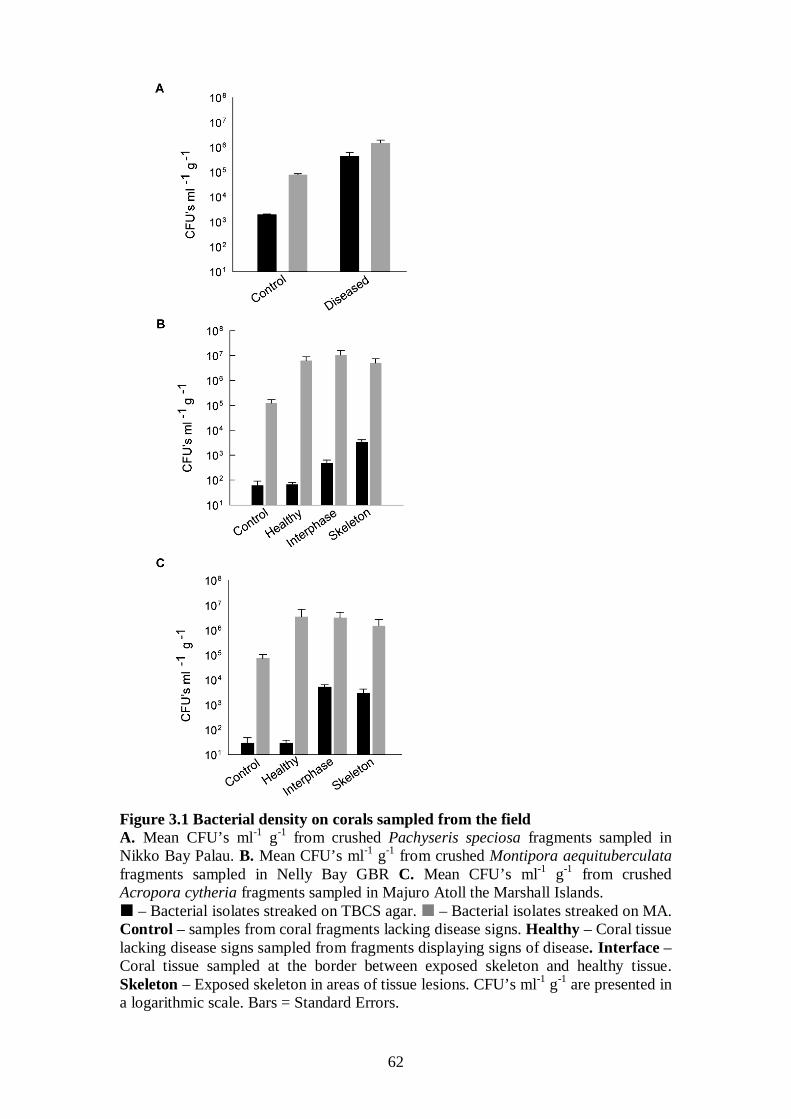
62
Figure 3.1 Bacterial density on corals sampled from the field A. Mean CFU’s ml-1 g-1 from crushed Pachyseris speciosa fragments sampled in Nikko Bay Palau. B. Mean CFU’s ml-1 g-1 from crushed Montipora aequituberculata fragments sampled in Nelly Bay GBR C. Mean CFU’s ml-1 g-1 from crushed Acropora cytheria fragments sampled in Majuro Atoll the Marshall Islands. – Bacterial isolates streaked on TBCS agar. – Bacterial isolates streaked on MA. Control – samples from coral fragments lacking disease signs. Healthy – Coral tissue lacking disease signs sampled from fragments displaying signs of disease. Interface – Coral tissue sampled at the border between exposed skeleton and healthy tissue. Skeleton – Exposed skeleton in areas of tissue lesions. CFU’s ml-1 g-1 are presented in a logarithmic scale. Bars = Standard Errors.
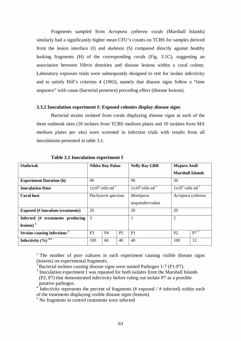
63
Fragments sampled from Acropora cytherea corals (Marshall Islands)
similarly had a significantly higher mean CFU’s counts on TCBS for samples derived
from the lesion interface (I) and skeleton (S) compared directly against healthy
looking fragments (H) of the corresponding corals (Fig. 3.1C), suggesting an
association between Vibrio densities and disease lesions within a coral colony.
Laboratory exposure trials were subsequently designed to test for isolate infectivity
and to satisfy Hill’s criterion 4 (1965), namely that disease signs follow a “time
sequence” with cause (bacterial presence) preceding effect (disease lesions).
3.3.2 Inoculation experiment I: Exposed colonies display disease signs
Bacterial strains isolated from corals displaying disease signs at each of the
three outbreak sites (10 isolates from TCBS medium plates and 10 isolates from MA
medium plates per site) were screened in infection trials with results from all
inoculations presented in table 3.1.
Table 3.1 Inoculation experiment I Outbreak Nikko Bay Palau Nelly Bay GBR Majuro Atoll
Marshall Islands
Experiment Duration (h) 96 96 36
Inoculation Dose 1x10⁶ cells ml 1x10⁻¹ 6 cells ml 1x10⁻¹ 6 cells ml-1
Coral host Pachyseris speciosa Montipora
aequituberculata
Acropora cytherea
Exposed (# inoculum treatments) 20 20 20
Infected (# treatments producing
lesions)
3 1
1 2
Strains causing infections P3 2 P4 P5 P1 P2 P7 3
Infectivity (%) 100 4.5 60 40 40 100 12
1 The number of pure cultures in each experiment causing visible disease signs (lesions) on experimental fragments. 2 Bacterial isolates causing disease signs were named Pathogen 1-7 (P1-P7). 3 Inoculation experiment I was repeated for both isolates from the Marshall Islands (P2, P7) that demonstrated infectivity before ruling out isolate P7 as a possible putative pathogen. 4 Infectivity represents the percent of fragments (# exposed / # infected) within each of the treatments displaying visible disease signs (lesions) 5 No fragments in control treatments were infected
64
All five P. speciosa fragments (Nikko Bay Palau) inoculated with isolate P3
(1x10⁶ bacteria ml-1
) developed disease signs following exposure for 96 h, while
treatments with isolates P4 and P5 demonstrated lower infectivity (Fig. 3.2). Coral
fragments in control treatments (n=17) including treatments with 7 other TCBS
derived isolates and 10 isolates from MA plates remained unaffected for the duration
of the experiment. Healthy fragments of M. aequituberculata (Nelly Bay GBR) were
only infected by one strain (P1) of the 20 strains tested, with 40% of fragments
displaying disease signs after a 96 h exposure to P1. 100% and 12% of healthy A.
cytherea fragments (Majuro Atoll Marshall Islands) exposed to strains P2 and P7,
respectively, displayed disease signs after 36 h. A repeat of the experiment with
strain P7 resulted in no further positive results and therefore the strain was eliminated
as a possible putative pathogen. Results from inoculation experiment I satisfied Hill’s
criterion 4 (1965) of “time sequence” (cause precedes effect) by demonstrating
successful infectivity following putative pathogen inoculations.
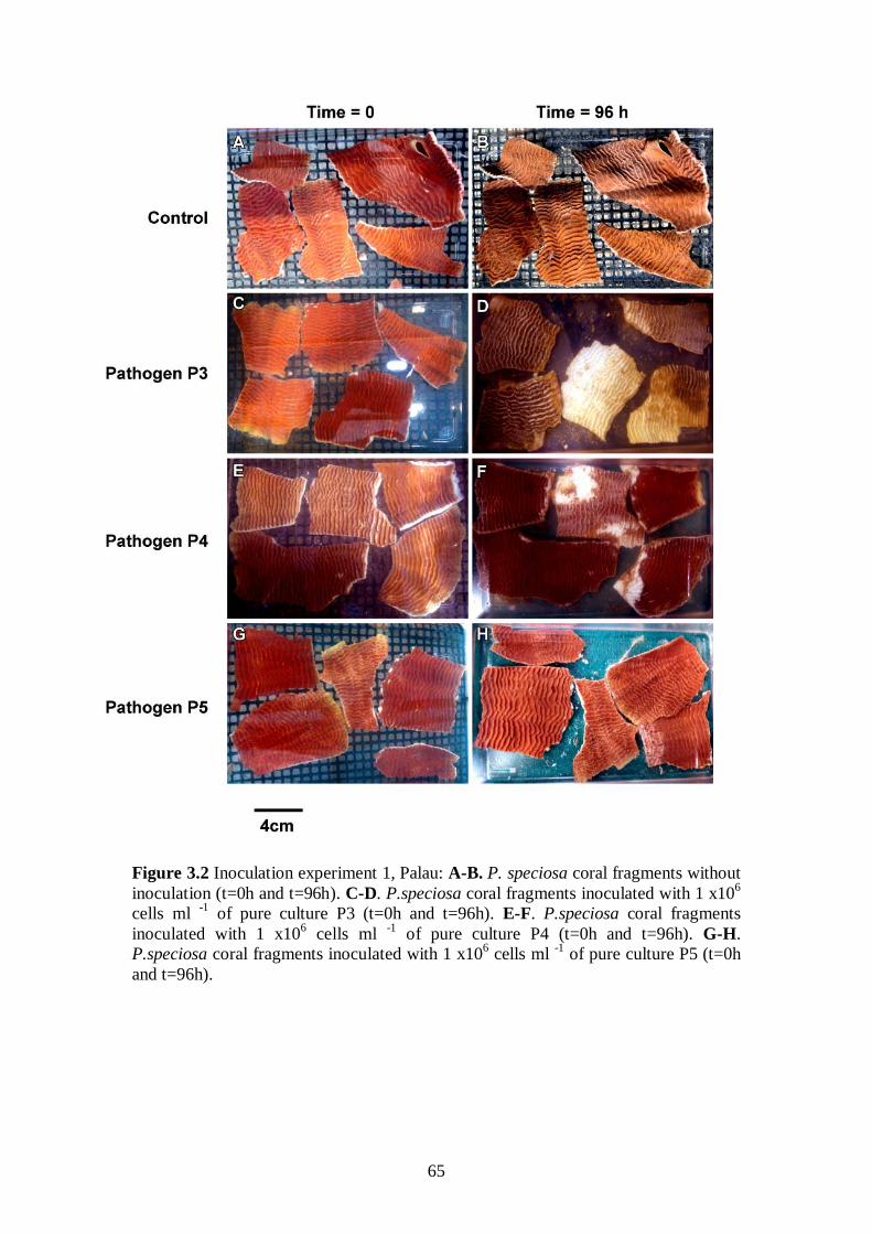
65
Figure 3.2 Inoculation experiment 1, Palau: A-B. P. speciosa coral fragments without inoculation (t=0h and t=96h). C-D. P.speciosa coral fragments inoculated with 1 x10⁶ cells ml -1 of pure culture P3 (t=0h and t=96h). E-F. P.speciosa coral fragments inoculated with 1 x10⁶ cells ml -1 of pure culture P4 (t=0h and t=96h). G-H. P.speciosa coral fragments inoculated with 1 x10⁶ cells ml -1
of pure culture P5 (t=0h and t=96h).
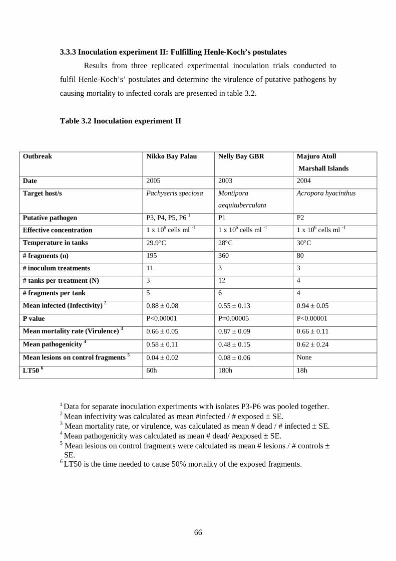
66
3.3.3 Inoculation experiment II: Fulfilling Henle-Koch’s postulates
Results from three replicated experimental inoculation trials conducted to
fulfil Henle-Koch’s’ postulates and determine the virulence of putative pathogens by
causing mortality to infected corals are presented in table 3.2.
Table 3.2 Inoculation experiment II
1 Data for separate inoculation experiments with isolates P3-P6 was pooled together. 2 Mean infectivity was calculated as mean #infected / # exposed ± SE. 3 Mean mortality rate, or virulence, was calculated as mean # dead / # infected ± SE. 4 Mean pathogenicity was calculated as mean # dead/ #exposed ± SE. 5 Mean lesions on control fragments were calculated as mean # lesions / # controls ± SE. 6
Outbreak
LT50 is the time needed to cause 50% mortality of the exposed fragments.
Nikko Bay Palau Nelly Bay GBR Majuro Atoll
Marshall Islands
Date 2005 2003 2004
Target host/s Pachyseris speciosa Montipora
aequituberculata
Acropora hyacinthus
Putative pathogen P3, P4, P5, P6 P1 1 P2
Effective concentration 1 x 10⁶ cells ml 1 x 10⁻¹ 6 cells ml 1 x 10⁻¹ 6 cells ml -1
Temperature in tanks 29.9°C 28°C 30°C
# fragments (n) 195 360 80
# inoculum treatments 11 3 3
# tanks per treatment (N) 3 12 4
# fragments per tank 5 6 4
Mean infected (Infectivity) 0.88 ± 0.08 2 0.55 ± 0.13 0.94 ± 0.05
P value P<0.00001 P=0.00005 P<0.00001
Mean mortality rate (Virulence) 3 0.66 ± 0.05 0.87 ± 0.09 0.66 ± 0.11
Mean pathogenicity 0.58 ± 0.11 4 0.48 ± 0.15 0.62 ± 0.24
Mean lesions on control fragments 0.04 ± 0.02 5 0.08 ± 0.06 None
LT50 60h 6 180h 18h
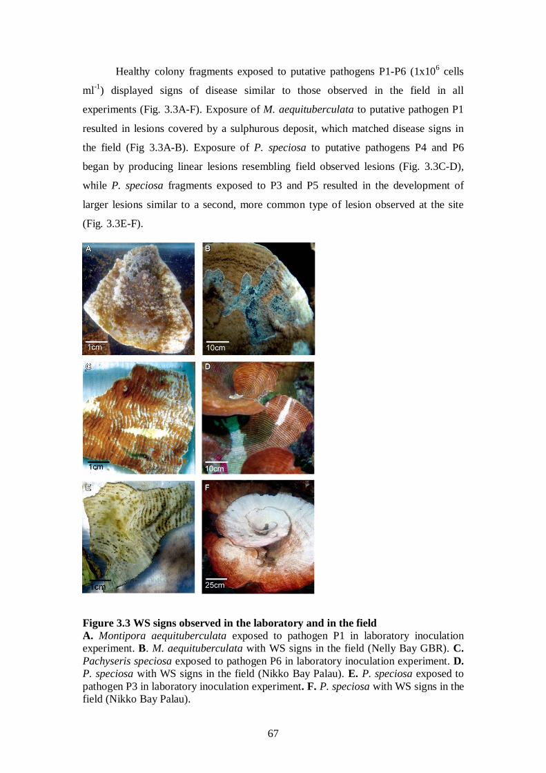
67
Healthy colony fragments exposed to putative pathogens P1-P6 (1x10⁶ cells
ml-1
Figure 3.3 WS signs observed in the laboratory and in the field A. Montipora aequituberculata exposed to pathogen P1 in laboratory inoculation experiment. B. M. aequituberculata with WS signs in the field (Nelly Bay GBR). C. Pachyseris speciosa exposed to pathogen P6 in laboratory inoculation experiment. D. P. speciosa with WS signs in the field (Nikko Bay Palau). E. P. speciosa exposed to pathogen P3 in laboratory inoculation experiment. F. P. speciosa with WS signs in the field (Nikko Bay Palau).
) displayed signs of disease similar to those observed in the field in all
experiments (Fig. 3.3A-F). Exposure of M. aequituberculata to putative pathogen P1
resulted in lesions covered by a sulphurous deposit, which matched disease signs in
the field (Fig 3.3A-B). Exposure of P. speciosa to putative pathogens P4 and P6
began by producing linear lesions resembling field observed lesions (Fig. 3.3C-D),
while P. speciosa fragments exposed to P3 and P5 resulted in the development of
larger lesions similar to a second, more common type of lesion observed at the site
(Fig. 3.3E-F).
68
Coral fragments inoculated with control strains (non-pathogenic) and un-
inoculated control fragments did not develop signs of WS lesions (Fig. 3.4 IA-B, 3.4
IIA-B) in contrast to lesion signs and mortality observed in all treatments with
putative pathogens (Fig 3.4 IC-D, 3.4 IIC-J). Bacterial isolates from infected
fragments retrieved at the conclusion of the experimental exposure, demonstrated
100% 16S rRNA gene sequence identity to inoculated strains. Recovery of inoculated
strains from infected fragments fulfilled Henle-Koch’s postulates for all six proposed
agents examined in this study.
The proportion of exposed fragments per tank that became infected (infectivity)
varied among the experiments, with 88% of fragments exposed to P3-P6, 55% of
fragments exposed to P1 and 94% of fragments exposed to strain P2 becoming
infected (Table 3.2). Pathogenicity (proportion of exposed fragments that died)
measured 58%, 48% and 62%, and mortality rate, or virulence (proportion of infected
fragments that died) equalled 66%, 87% and 66% for putative pathogens from Palau,
Magnetic Island and the Marshall Islands, respectively (Table 3.2). Similarly, the
times needed for 50% of the fragments to experience mortality (LT50) were 60h,
180h and 18h, respectively (Table 3.2).
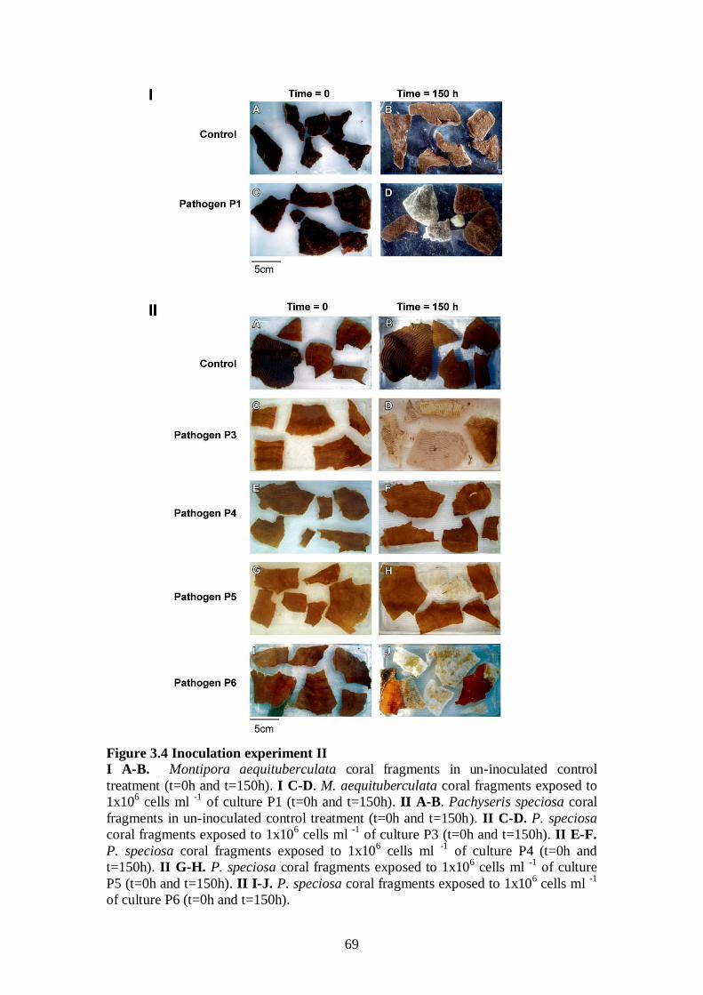
69
Figure 3.4 Inoculation experiment II I A-B. Montipora aequituberculata coral fragments in un-inoculated control treatment (t=0h and t=150h). I C-D. M. aequituberculata coral fragments exposed to 1x10⁶ cells ml -1 of culture P1 (t=0h and t=150h). II A-B. Pachyseris speciosa coral fragments in un-inoculated control treatment (t=0h and t=150h). II C-D. P. speciosa coral fragments exposed to 1x10⁶ cells ml -1 of culture P3 (t=0h and t=150h). II E-F. P. speciosa coral fragments exposed to 1x10⁶ cells ml -1 of culture P4 (t=0h and t=150h). II G-H. P. speciosa coral fragments exposed to 1x10⁶ cells ml -1 of culture P5 (t=0h and t=150h). II I-J. P. speciosa coral fragments exposed to 1x10⁶ cells ml -1 of culture P6 (t=0h and t=150h).
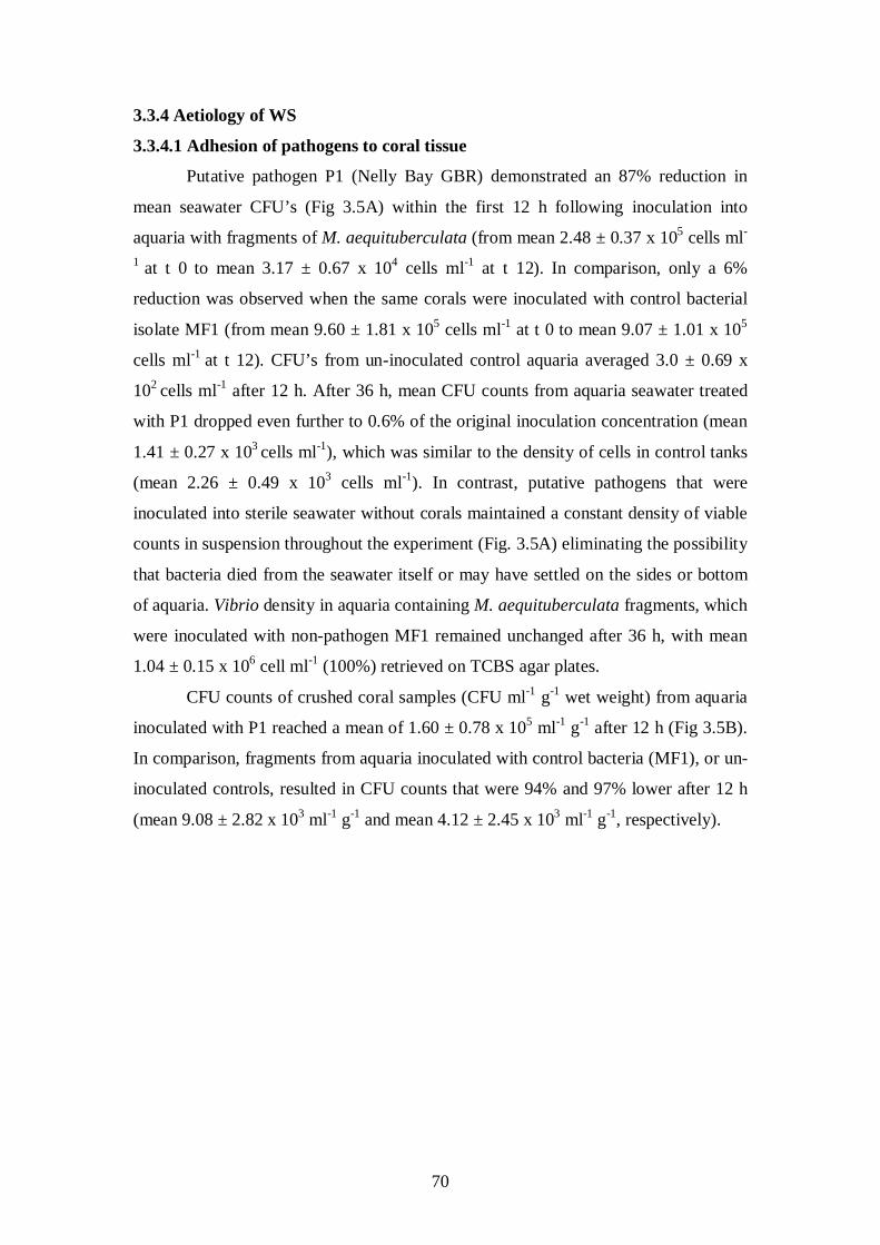
70
3.3.4 Aetiology of WS
3.3.4.1 Adhesion of pathogens to coral tissue
Putative pathogen P1 (Nelly Bay GBR) demonstrated an 87% reduction in
mean seawater CFU’s (Fig 3.5A) within the first 12 h following inoculation into
aquaria with fragments of M. aequituberculata (from mean 2.48 ± 0.37 x 10⁵ cells ml-
1 at t 0 to mean 3.17 ± 0.67 x 10⁴ cells ml-1 at t 12). In comparison, only a 6%
reduction was observed when the same corals were inoculated with control bacterial
isolate MF1 (from mean 9.60 ± 1.81 x 10⁵ cells ml-1 at t 0 to mean 9.07 ± 1.01 x 10⁵
cells ml-1 at t 12). CFU’s from un-inoculated control aquaria averaged 3.0 ± 0.69 x
102 cells ml-1 after 12 h. After 36 h, mean CFU counts from aquaria seawater treated
with P1 dropped even further to 0.6% of the original inoculation concentration (mean
1.41 ± 0.27 x 10³ cells ml-1), which was similar to the density of cells in control tanks
(mean 2.26 ± 0.49 x 10³ cells ml-1). In contrast, putative pathogens that were
inoculated into sterile seawater without corals maintained a constant density of viable
counts in suspension throughout the experiment (Fig. 3.5A) eliminating the possibility
that bacteria died from the seawater itself or may have settled on the sides or bottom
of aquaria. Vibrio density in aquaria containing M. aequituberculata fragments, which
were inoculated with non-pathogen MF1 remained unchanged after 36 h, with mean
1.04 ± 0.15 x 10⁶ cell ml-1 (100%) retrieved on TCBS agar plates.
CFU counts of crushed coral samples (CFU ml-1 g-1 wet weight) from aquaria
inoculated with P1 reached a mean of 1.60 ± 0.78 x 10⁵ ml-1 g-1 after 12 h (Fig 3.5B).
In comparison, fragments from aquaria inoculated with control bacteria (MF1), or un-
inoculated controls, resulted in CFU counts that were 94% and 97% lower after 12 h
(mean 9.08 ± 2.82 x 10³ ml-1 g-1 and mean 4.12 ± 2.45 x 10³ ml-1 g-1
, respectively).
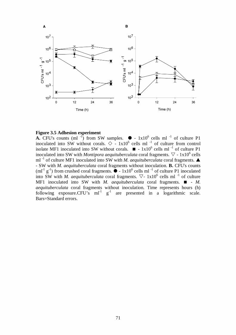
71
Figure 3.5 Adhesion experiment A. CFU's counts (ml P
-1P) from SW samples. - 1x10 P
6P cells ml P
-1P of culture P1
inoculated into SW without corals. - 1x10P
6P cells ml P
-1P of culture from control
isolate MF1 inoculated into SW without corals. - 1x10P
6P cells ml P
-1P of culture P1
inoculated into SW with Montipora aequituberculata coral fragments. - 1x10 P
6P cells
ml P
-1P of culture MF1 inoculated into SW with M. aequituberculata coral fragments.
- SW with M. aequituberculata coral fragments without inoculation. B. CFU's counts (mlP
-1P gP
-1P) from crushed coral fragments. - 1x10 P
6P cells ml P
-1P of culture P1 inoculated
into SW with M. aequituberculata coral fragments. - 1x10 P
6P cells ml P
-1P of culture
MF1 inoculated into SW with M. aequituberculata coral fragments. - M. aequituberculata coral fragments without inoculation. Time represents hours (h) following exposure.CFU’s mlP
-1P gP
-1P are presented in a logarithmic scale.
Bars=Standard errors.
72
Table 3.3 summarizes the data from adhesion experiments conducted with
putative pathogens and controls isolated from the three infection sites examined in
this study.
Table 3.3 Adhesion experiment
P
1 PThe precent (%) of cultivable putative pathogens in seawater at t=12h was
calculated by the formula: CFU’s mlP
-1P [t=12h] / CFU’s mlP
-1P [t=0h].
P
2 PThe precent (%) of cultivable putative pathogen on corals was calculated by the
formula: Total CFU’s on coral fragments [t=12h] / Total original inoculation [t=0h]. P
3 P The precent (%) of missing cultivable cells was calculated by the formula:
100 – (% CFU’s in sweater [t=12h] + % CFU’s on coral fragments [t 12h]).
3.3.4.2 Loss of Symbiodinium followed by tissue lesions
Detailed photographs taken of A. hyacinthus fragments infected
experimentally with P2 (Fig 3.6A-C) revealed 2 distinct disease-phases. An initial
loss of Symbiodinium, visible as tissue paling was observed after 9-12 h of exposure
(Fig 3.6 B-C) followed by developing tissue lesions. Similar patterns of paling were
also observed when P. speciosa fragments were exposed to P3 (Fig 3.6D). Paling and
loss of Symbiodinium commenced in coenosarc tissue (tissue between polyps) in
distinct linear patterns starting 12 h post inoculation and corresponding with the peak
in viable CFU counts retrieved from coral tissue. These early signs of disease then
Outbreak Nikko Bay,
Palau
Nelly Bay, GBR Majuro Atoll,
Marshall Islands
Cultivable putative pathogens in SW at
t=12h (% from original inoculation at t= 0h) P
1
13.5 12.0 13.0
Cultivable putative pathogens on corals at
t=12h (increase factor from CFU’s at t=0h)
1049 x
Increase
876 x
Increase
484 x
Increase
Cultivable putative pathogens on corals at
t=12h (% from original inoculation at t=0h) P
2
0.9 0.7 0.8
% of cultivable putative pathogens missing from
total original inoculation P
3P
85.6 87.3 86.2
73
developed into lesions that resembled those observed in the field (Fig 3.6E-F),
suggesting that disease progression was consistent (Hill’s criterion 2; Hill 1965) and
followed measurable steps (Evans’ Rule F; Evans 1976).
Figure 3.6 Disease progression A. Acropora hyacinthus fragmennt inoculated with 1 x 10 P
6P cells ml P
-1P of culture P2
(t=0h). B. Loss of Symbiodinium from A. hyacinthus inoculated with 1 x 10 P
6P cells ml P
-
1P of culture P2 (t=12h). C. Polyp and surrounding tissue - loss of Symbiodinium from
A. hyacinthus inoculated with 1 x 10 P
6P cells ml P
-1P of culture P2 (t=12h). D. Loss of
Symbiodinium cells from Pachyseris speciosa inoculated with 1 x 10P
6P cells ml P
-1P of
culture P3 (t=12h). E. Tissue lesions on P. speciosa inoculated with 1 x 10 P
6P cells ml P
-1P
of culture P3 (t=24h). F. Exposed skeleton on P. speciosa inoculated with 1 x 10 P
6P
cells ml P
-1P of culture P3 (t=60h).
In all experimental treatments inoculated with putative pathogens P1-P6, the
proportion of fragments displaying acute disease signs (lesions) increased with time to
between 55% and 94% of fragments per tank (Fig 3.7A-C) conforming with Evans’
rules D and E (Evans 1976), namely that disease occurs, temporally, following
specific incubation times and that the number of new cases and the severity of
outcome should correlate positively with time. The proportion of P. speciosa
fragments from Palau, M. aequituberculata fragments from Nelly Bay and A.
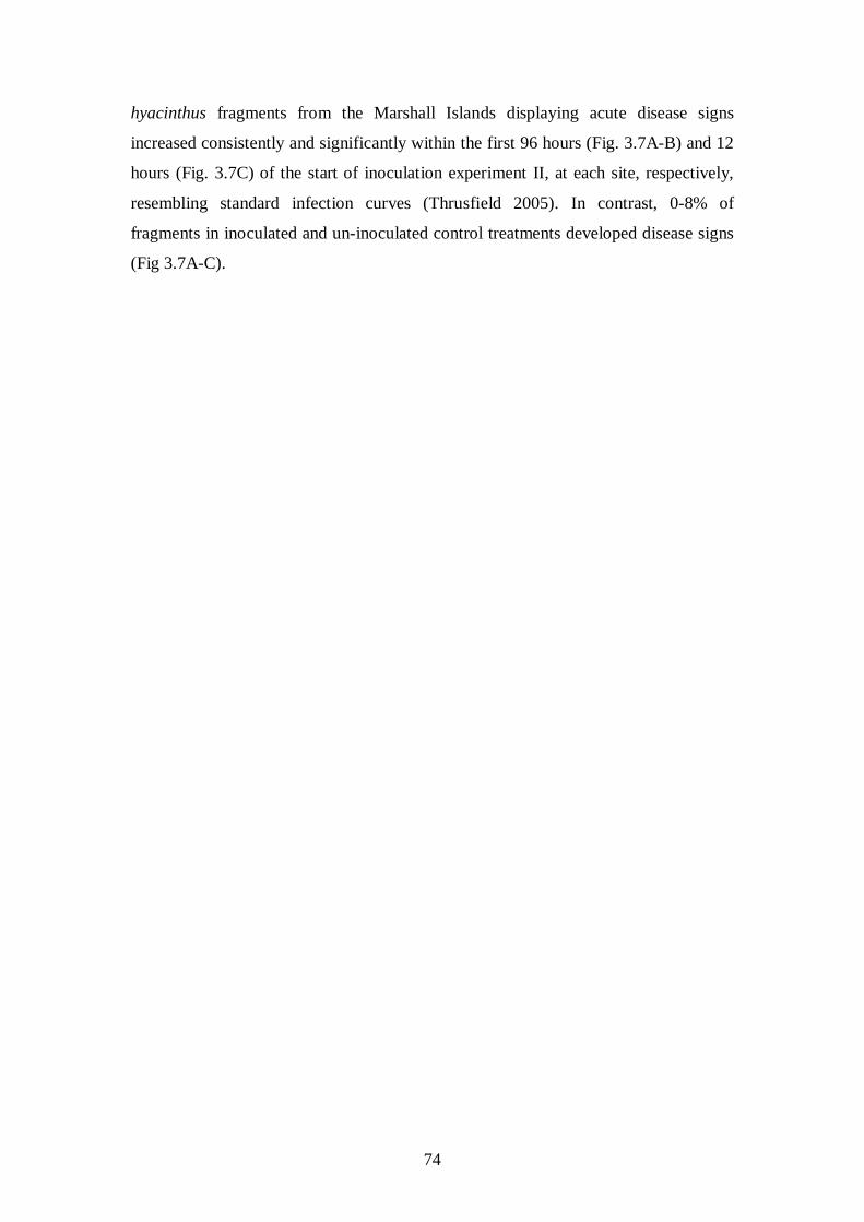
74
hyacinthus fragments from the Marshall Islands displaying acute disease signs
increased consistently and significantly within the first 96 hours (Fig. 3.7A-B) and 12
hours (Fig. 3.7C) of the start of inoculation experiment II, at each site, respectively,
resembling standard infection curves (Thrusfield 2005). In contrast, 0-8% of
fragments in inoculated and un-inoculated control treatments developed disease signs
(Fig 3.7A-C).
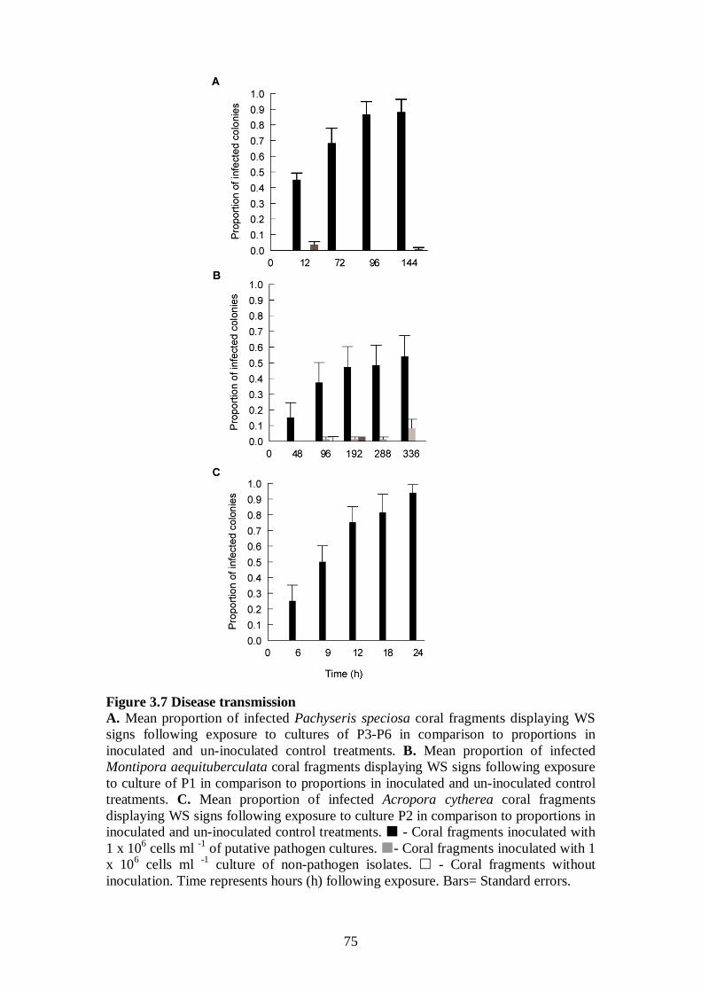
75
Figure 3.7 Disease transmission A. Mean proportion of infected Pachyseris speciosa coral fragments displaying WS signs following exposure to cultures of P3-P6 in comparison to proportions in inoculated and un-inoculated control treatments. B. Mean proportion of infected Montipora aequituberculata coral fragments displaying WS signs following exposure to culture of P1 in comparison to proportions in inoculated and un-inoculated control treatments. C. Mean proportion of infected Acropora cytherea coral fragments displaying WS signs following exposure to culture P2 in comparison to proportions in inoculated and un-inoculated control treatments. - Coral fragments inoculated with 1 x 10P
6P cells ml P
-1P of putative pathogen cultures. - Coral fragments inoculated with 1
x 10 P
6P cells ml P
-1P culture of non-pathogen isolates. - Coral fragments without
inoculation. Time represents hours (h) following exposure. Bars= Standard errors.
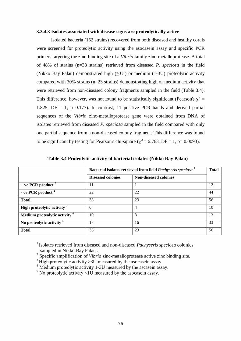
76
3.3.4.3 Isolates associated with disease signs are proteolytically active
Isolated bacteria (152 strains) recovered from both diseased and healthy corals
were screened for proteolytic activity using the asocasein assay and specific PCR
primers targeting the zinc-binding site of a Vibrio family zinc-metalloprotease. A total
of 48% of strains (n=33 strains) retrieved from diseased P. speciosa in the field
(Nikko Bay Palau) demonstrated high (≥3U) or medium (1-3U) proteolytic activity
compared with 30% strains (n=23 strains) demonstrating high or medium activity that
were retrieved from non-diseased colony fragments sampled in the field (Table 3.4).
This difference, however, was not found to be statistically significant (Pearson's χ2 =
1.825, DF = 1, p=0.177). In contrast, 11 positive PCR bands and derived partial
sequences of the Vibrio zinc-metalloprotease gene were obtained from DNA of
isolates retrieved from diseased P. speciosa sampled in the field compared with only
one partial sequence from a non-diseased colony fragment. This difference was found
to be significant by testing for Pearson's chi-square (χ2
= 6.763, DF = 1, p= 0.0093).
Table 3.4 Proteolytic activity of bacterial isolates (Nikko Bay Palau)
1 Isolates retrieved from diseased and non-diseased Pachyseris speciosa colonies sampled in Nikko Bay Palau . 2 Specific amplification of Vibrio zinc-metalloprotease active zinc binding site. 3 High proteolytic activity >3U measured by the asocasein assay. 4 Medium proteolytic activity 1-3U measured by the ascasein assay. 5
No proteolytic activity <1U measured by the asocasein assay.
Bacterial isolates retrieved from field Pachyseris speciosa Total 1
Diseased colonies Non-diseased colonies
+ ve PCR product 11 2 1 12
- ve PCR product 22 2 22 44
Total 33 23 56
High proteolytic activity 6 3 4 10
Medium proteolytic activity 10 4 3 13
No proteolytic activity 17 5 16 33
Total 33 23 56
77
Similar results were obtained by screening field isolates from Nelly Bay GBR
(Table 3.5). Bacteria demonstrating high and medium proteolytic activity by the
asocasein assay made up 70% of all isolates retrieved from coral skeletons (S)
exposed by WS disease at Nelly Bay GBR and 57% of all isolates from the lesion
interfaces (I), compared with only 24% of all isolates obtained from healthy (H) tissue
fragments on diseased colonies, demonstrating a significant difference in proteolytic
activity between isolates associated with disease signs (I+S) and healthy (H) tissue
(Pearson's χP
2 P= 6.446, DF = 1, p= 0.011). A significant difference was also obtained
for the same 38 isolates when screened by the molecular method using PCR primers
(Pearson's χP
2P = 12.518, DF = 1, p<0.0001).
Table 3.5 Proteolytic activity of bacterial isolates
P
1 PIsolates retrieved from diseased field samples (H=healthy segment on diseased
fragment, I=interphase between lesion and healthy tissue, S=exposed skeleton). P
2P Specific amplification of Vibrio zinc-metalloprotease active zinc binding site.
P
3 PHigh proteolytic activity >3U measured by the asocasein assay.
P
4P Medium proteolytic activity 1-3U measured by the ascasein assay.
P
5P No proteolytic activity <1U measured by the asocasein assay.
Finally, screenings by the molecular method performed on DNA extracted
from 56 isolates retrieved from both infected and non-infected fragments at the
conclusion of inoculation experiment II in Palau (Table 3.6), demonstrated that results
obtained by screening field isolates were consistent with screening laboratory derived
isolates (Pearson's χP
2P = 6.725, DF = 1, p=0.010). Thus, in both field and laboratory
Isolates retrieved from diseased M. aquetuberculata
coral fragments collected in the field (Nelly Bay, GBR)
- ve Disease SignsP
1 + ve Disease SignsP
1
Healthy on diseased Interphase Skeleton Total
+ ve PCR product P
2 0 4 4 8
- ve PCR product P
2 21 3 6 30
Total 21 7 10 38
High proteolytic activityP
3 2 4 5 11
Medium Proteolytic activityP
4 3 0 2 5
No proteolytic activityP
5 16 3 3 22
Total 21 7 10 38
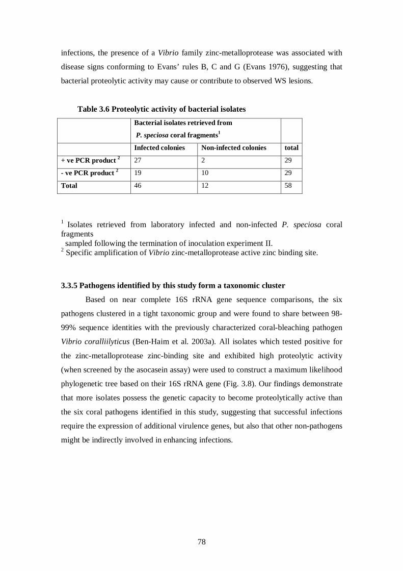
78
infections, the presence of a Vibrio family zinc-metalloprotease was associated with
disease signs conforming to Evans’ rules B, C and G (Evans 1976), suggesting that
bacterial proteolytic activity may cause or contribute to observed WS lesions.
Table 3.6 Proteolytic activity of bacterial isolates Bacterial isolates retrieved from
P. speciosa coral fragmentsP
1
Infected colonies Non-infected colonies total
+ ve PCR product P
2 27 2 29
- ve PCR product P
2 19 10 29
Total 46 12 58
P
1 PIsolates retrieved from laboratory infected and non-infected P. speciosa coral
fragments sampled following the termination of inoculation experiment II. P
2P Specific amplification of Vibrio zinc-metalloprotease active zinc binding site.
3.3.5 Pathogens identified by this study form a taxonomic cluster
Based on near complete 16S rRNA gene sequence comparisons, the six
pathogens clustered in a tight taxonomic group and were found to share between 98-
99% sequence identities with the previously characterized coral-bleaching pathogen
Vibrio coralliilyticus (Ben-Haim et al. 2003a). All isolates which tested positive for
the zinc-metalloprotease zinc-binding site and exhibited high proteolytic activity
(when screened by the asocasein assay) were used to construct a maximum likelihood
phylogenetic tree based on their 16S rRNA gene (Fig. 3.8). Our findings demonstrate
that more isolates possess the genetic capacity to become proteolytically active than
the six coral pathogens identified in this study, suggesting that successful infections
require the expression of additional virulence genes, but also that other non-pathogens
might be indirectly involved in enhancing infections.
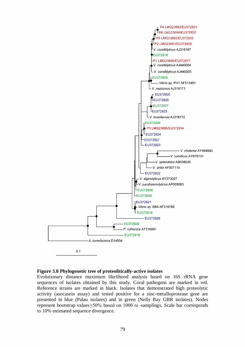
79
Figure 3.8 Phylogenetic tree of proteolitically-active isolates Evolutionary distance maximum likelihood analysis based on 16S rRNA gene sequences of isolates obtained by this study. Coral pathogens are marked in red. Reference strains are marked in black. Isolates that demonstrated high proteolytic activity (asocasein assay) and tested positive for a zinc-metalloprotease gene are presented in blue (Palau isolates) and in green (Nelly Bay GBR isolates). Nodes represent bootstrap values ≥50% based on 1000 re -samplings. Scale bar corresponds to 10% estimated sequence divergence.
80
3.4 Discussion
This study reports the successful isolation and identification of bacterial
infectious agents implicated in a group of widespread Indo-Pacific coral diseases that
affect numerous species at various geographical locations. Six coral pathogens were
identified with close 16S rRNA gene phylogenetic affiliation with the previously
identified coral pathogen V. coralliilyticus (Ben-Haim et al. 2003a). Vibrio pathogens
have been previously demonstrated to cause fish, eel, shrimp and human mortalities
(Milton et al. 1992, Amaro and Biosca 1996, Faruque et al. 1998, Lightner 1988,
Linkous and Oliver 1999). Seasonal bleaching of the coral Oculina patagonica in the
Mediterranean Sea has been shown to be caused by V. shiloi (Rosenberg and
Falkovitz 2004, Kushmaro et al. 1996, 1997a, 2001) and V. coralliilyticus has been
identified as the aetiological agent of Pocillopora damicornis bleaching in the Indian
Ocean (Ben-Haim et al. 2002, 2003a, 2003b). Other coral diseases in the Caribbean,
such as White Band Disease type II, Yellow Blotch/Band and Dark Spots Disease,
were found to be associated with elevated Vibrio spp. prevalence (Ritchie and Smith
1998, Gil-Agudelo et al. 2006, 2007), suggesting the involvement of Vibrio strains in
numerous coral diseases including coral bleaching (Ritchie et al. 1994, Rosenberg and
Falkovitz 2004, Barneah et al. 2007, Bourne et al. 2008).
This study has found high prevalence of Vibrio spp. to be associated with WS
signs in all diseased colonies sampled from three WS outbreaks. An association
between bacterial presence and disease signs does not provide proof that bacteria
actually cause the disease. However, such an association already fulfils requirements
put forth by Evans’ rule A (Evans 1976), namely that the "prevalence of the disease
should be significantly higher in those exposed to the putative cause than in cases
controls not so exposed". It also complies with the Read (1994) definition of virulence,
which highlights an agent’s contribution to reduction in host fitness caused by
exploitation.
When bacterial strains were inoculated into tanks with healthy coral fragments,
only putative pathogens adhered to coral tissue and a consistent peak in cultivable
Vibrio abundance, 500-1000 fold greater than was found for control strains, was
observed on fragments exposed to putative pathogens 12 h post inoculation. This
experiment confirmed Hill's criterion of "time sequence" (Hill 1965), suggesting that
following exposure and prior to the development of visual disease signs (lesions),
putative pathogens were able to migrate towards the coral fragments, adhere to coral
81
tissue and survive initial contact in a viable state. In contrast, control strains were
unable to perform this transmission, suggesting that motility towards corals and
adhesion may be regarded as traits involved in pathogenicity. Initial visual signs of
tissue paling and lesions were observed following a peak in cultivable bacterial
abundance for all six putative pathogens, demonstrating a common aetiology of
adhesion followed by disease progression. Despite this peak in cultivable Vibrio
abundance 12 h post exposure, less than 1% of the original inoculation was retrieved
by plating coral fragments, potentially indicating that Vibrio cells entered a viable but
non-culturable (VBNC) state (Israeli et al. 2001), or alternatively, died.
The colonization of target hosts by Vibrio pathogens has been studied in detail,
particularly the ability of Vibrios to adhere to mucus found either inside the gastro-
internal track or externally on fish or corals (Bordas et al. 1996, Banin et al. 2001a).
Denkin and Nelson (1999) have demonstrated that the transcription of zinc-
metalloprotease by the fish pathogen V. anguillarum is regulated by mucus and can
only occur after adhesion is completed. This duality in Vibrio function is often
referred to as the “transmission-virulence trade-off” (Anderson and May 1982) and
highlights the fact that the ultimate goal of Vibrio pathogenicity is not to kill a host, or
to complete a necessary biological life-cycle within it, but to re-enter the environment
in larger numbers and initiate a new cycle of infections (Higgins et al. 2007). It
explains why Vibrio pathogens are commonly found in environmental reservoirs
(Sussman et al. 2003), or transmitting through the water column, like pathogen P6
isolated in this study from seawater above infected corals at Nikko Bay Palau.
This study has demonstrated that 55%-94% of coral fragments exposed to
pathogens cultured from diseased corals at their respective field sites become infected
and that 66%-87% of those infected die, compared with significantly lower infection
and mortality for fragments exposed to control bacterial strains (0-8%). These results
conform to Evans' rule G (Evans 1976), requiring that experimental reproduction of
the disease should occur in higher incidence in those exposed to the putative cause
than in those not so exposed. However, a proportion of exposed fragments did not
develop disease signs, demonstrating that the probability of becoming infected may
not be equal among healthy colony fragments collected from the field, and that other
host related factors potentially contribute to successful infections. Such unknown
factors can be explored in future inoculation trials.
82
The presence of Vibrio spp. on both healthy and diseased corals has led to the
conclusion by some authors (Bourne and Munn 2005, Ainsworth et al. 2007a) that
Vibrio infections of corals may be opportunistic in nature. This assumption fits well
into models of disease occurring in environmental settings, where multiple factors,
such as host density (Bruno et al. 2007) and temperature (Colwell 1996) have been
shown to influence the probability of successful infections. Combinations of virulent
and a-virulent Vibrio strains are found readily in environmental samples (Stelma et al.
1992) with non-clinical V. cholera strains found to be capable of causing infections
despite lacking the cholera toxin gene (Honda et al. 1989). Many Vibrios specialize in
multiple host attachment and detachment (Nelson et al. 1976, Tamplin et al. 1990,
Kirn et al. 2005), suggesting a broad scope for potential coral infections by Vibrios
including possible host shifts due to fish depletion from coral reefs (Connolly et al.
2005). Amaro and Biosca (1996) have demonstrated that Vibrio vulnificus biotype 2 is
both a primary pathogen for eels and an opportunistic pathogen for humans,
indicating that the identification of opportunistic pathogens requires rigorous testing.
Nevertheless, none of the claims to define Vibrio coral infections as opportunistic
have so far provided conclusive evidence to show that suspects (identified by
molecular screening methods) found on healthy corals are in fact pathogenic (whether
opportunistic or not), or that only compromised hosts become infected. In addition,
not all coral mortalities are caused by infectious agents, but rather by exposure to
extreme conditions, such as pesticides or high nutrient levels (Negri et al. 2005, Kline
et al. 2006), which may result in indirect shifts in microbial abundance. Infectious
outbreaks can be distinguished from non-infectious ones by plotting infection curves
(Thrusfield 2005) to demonstrate a bell-shape increase and decrease in incidence rate
with time.
This study did not find evidence for the presence of coral pathogens on
healthy corals in the field, nor evidence that exposed fragments might be successfully
infected due to stress other than the direct exposure to the pathogens themselves.
Control treatments in all inoculations remained healthy, including a proportion of
those exposed to pathogens. Further studies are recommended to determine the
prevalence of pathogens in field samples by developing diagnostic tools to target
specific virulence genes in large scale screening efforts. These studies could then
determine the proportion of exposed corals in the field that develop acute disease
83
signs and should become an integral part of establishing acute vs. chronic disease
prevalence in environmental studies.
This is the first study to diagnose proteolytic activity as a possible component
of the aetiology of WS through the screening of more than 150 isolates from both
diseased and non-diseased corals. Zinc-metalloproteases have been characterized as
virulence factors in many Vibrio family pathogens, such as V. cholera (Finkelstein
and Hanne 1982), V. vulnificus (Miyoshi and Shinoda 1988), V. harveyi (Teo et al.
2003) and V. anguillarum (Denkin and Nelson 2004). Vibrio zinc-metalloproteases
are involved in cleavage of connective tissue (Finkelstein et al. 1983), para-cellular
perturbation (Wu et al. 1996), swarming and adhesion to mucus (Kim et al. 2007) and
detachment (Finkelstein et al. 1992). The coral bleaching pathogens V. shiloi and V.
coralliilyticus have been previously shown to harbour a zinc-metalloprotease (Ben-
Haim et al. 1999, 2003a) along with other toxins that cause photosynthetic inhibition
of coral Symbiodinium (Banin et al. 2001b). Serratia marcescens, the aetiological
agent of acroporid serratiosis (coral White Pox disease; Patterson et al. 2002),
resulting in acute tissue lesions, also possesses a virulent zinc-metalloprotease capable
of connective tissue degradation (Kamata et al. 1985). However, it has been shown
that both clinical and non-clinical strains possess zinc-metalloprotease genes (Booth
and Finkelstein 1986), suggesting that it may not be the only virulence factor to cause
successful infections. This study provided similar results, underlining the need to
search for additional virulence factors in future studies.
Recent studies by Ainsworth et al. (2007b) did not detect bacteria associated
with WS lesions of diseased corals sampled at Heron Island on the GBR, using direct
microscopic techniques. In contrast, samples of WS corals obtained from Heron
Island in this study for screening purposes demonstrated an abundance of Vibrio spp.
isolates on WS lesions, including proposed putative pathogens that are proteolytically
active and possess a zinc-metalloprotease gene. These contradicting findings
underline the importance of ‘comparative validation’ (Dane 1990) in disease research
and the need for standardized protocols for disease detection using better diagnostic
tools.
Further histopathological studies by Ainsworth et al. (Ainsworth et al. 2007a,
2007b) utilizing commercial labelling kits have found that coral fragments displaying
WS signs test positive for DNA fragmentation. These observations led to the
hypothesis that WS is potentially the result of coral programmed cell death. However,
84
further proof is needed in order establish whether DNA fragmentation (or apoptosis)
in corals is cause or effect. The induction of apoptosis by bacterial pathogens
(Salmonella Sp., E. coli, Shigella sp., C. difficile, L. monocytogenes, C. parvum and
others) has been previously demonstrated by many studies (Keenan et al. 1986,
Fiorentini et al. 1998, Kim et al. 1998, Valenti et al. 1999, McCole et al. 2000),
suggesting a possible link between bacterial infections and apoptosis. This link can be
tested in future pathogen-exposure trials and used to design novel diagnostic protocols
for WS, which would target bacterial enzymes causing DNA fragmentation.
In summary, this study demonstrated consistent results in applying cost
effective culturing techniques combined with biochemical and molecular tools
towards successful pathogen isolation, coral disease investigation and sample
screening. Future research should be conducted to explore the virulence components
of all six pathogens identified in this study and to test the contribution of multiple
factors (pathogen, environment and host related) to the aetiology of WS. Enhanced
monitoring and management of WS outbreaks will not only benefit coral health, but
would also further validate results obtained in this study.
85
Chapter 4
Vibrio Zinc-metalloprotease causes photoinactivation of
coral endosymbionts and coral tissue lesions Sussman M, Mieog JC, Doyle J, Victor S, Willis BL, Bourne DG (2009) Vibrio zinc-
metalloprotease causes photoinactivation of coral endosymbionts and coral tissue lesions.
PLoSONE4(2):e4511.doi:10.1371/journal.pone.0004511
4.1 Introduction
Of nine coral infectious diseases, whose pathogens have been characterized by
fulfilling Henle-Koch’s postulates (Koch 1891), six (including pathogens identified
for Indo-Pacific WS's; previous chaper 3) are caused by agents from the family
Vibrionacae (Kushmaro et al. 2001, Ben-Haim et al. 2003, Bally and Garrabou 2007,
Sussman et al. 2008), adding to the many previously characterized Vibrio infections
of shrimps (Goarant et al. 1998), clams (Paillard 2004) and fish (Egidius 1987), which
date back to 1817 (Woo and Bruno 1999). Other coral disease signs in the Caribbean
(Cervino et al. 2004a, Gil-Agudelo 2006) have also been associated with the presence
of Vibrio agents. The study of coral disease signs in Zanzibar (Piskorska et al. 2007),
bleached corals on the Great Barrier Reef (GBR; Bourne et al. 2008), black band
disease signs on corals in the Gulf of Aquaba (the Red Sea; (Barneah et al. 2007) and
even growth anomalies on Hawaiian corals (Breitbart et al. 2005) have all
demonstrated significant correlation between disease signs and an elevated abundance
of Vibrio strains. These newly emerging coral diseases, either caused or associated
with members of the Vibrionacae family have sparked a debate on the origin of Vibrio
pathogens and their role in the aetiology of coral diseases: Are Vibrio pathogens the
primary causative agents of all these diseases, or are they opportunistic pathogens?
Alternatively, are they secondary infections to other unknown causes? (Harvell et al.
2004, Bourne and Munn 2005, Smith et al. 2006, Lesser et al. 2007, Ainsworth et al.
2007, Rosenberg et al. 2007, 2008, Muller et al. 2008, Work et al. 2008)
In the previous chapter (Chapter 3; Sussman et al. 2008) we identified two
novel V. coralliilyticus strains and four additional Vibrio pathogens as causative
agents of three Indo-Pacific coral white syndromes (WS’s). A link was demonstrated
between WS disease signs on corals and the presence of Vibrio strains possessing a
zinc-metalloprotease gene (Chapter 3; Sussman et al. 2008). Protein homologues of
86
this gene have been identified as key virulence factors of Vibrio pathogens of fish
(Norqvist et al. 1990), shrimp (Aguirre-Guzmán et al. 2004), mollusks (Binesse et al.
2008) and humans (Crowther et al. 1987) acting to digest mucin and other connective
tissue components, such as collagen IV (Miyoshi et al. 1998) and fibronectin
(Finkelstein et al. 1983). These enzymes have also been shown to perturb paracellular
barrier functions (Wu et al. 1996) and cause tissue necrosis (Milton et al. 1992)
including pathogen detachment from epithelial mucus (Finkelstein et al. 1992). Ben-
Haim et al. (2003a) suggested that V. coralliilyticus, the bleaching agent of the coral
Pocillopora damicornis, expresses a V. cholera–like zinc-metalloprotease, which
causes rapid photosystem II (PS II) inactivation of Symbiodinium endosymbionts.
However, little is known about either the kinetics or the specificity of this reaction,
and under which conditions it is likely to occur. Numerous studies have demonstrated
that the zinc-metalloprotease gene is present in Vibrio pathogenic strains, but also in
non-pathogenic strains (Booth and Finkelstein 1986, Sussman et al. 2008), suggesting
that this gene may not be considered an essential virulence factor (Milton et al. 1992,
Shao and Hor 2000).
In this chapter we test this hypothesis and the role of zinc-metalloprotease in
the pathogenicity of coral WS's by developing two novel bioassays. Symbiodinium
cells from four coral hosts at two locations on the GBR were isolated and grown in
cultures (Z1-Z4; table 4.1) before being exposed in 96 well microtitre plates to
bacterial supernatants derived from four coral pathogens (P1-P4; Table 4.2) that have
been characterized in chapter 3 (Sussman et al. 2008) as the causative agents of coral
WS’s on Pacific reefs, i.e., on the GBR (P1), in the Republic of the Marshall Islands
(P2) and in Palau (P3-P4). In order to test the effects of pathogen supernatants on
Symbiodinium cells living in hospite, a second bioassay is developed by rearing
juveniles of Acropora millepora and infecting them with specific Symbiodinium
isolates from clades C and D (Little et al. 2004).
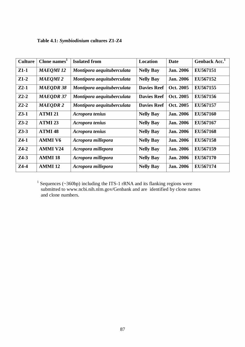
87
Table 4.1: Symbiodinium cultures Z1-Z4
1
submitted to Sequences (~360bp) including the ITS-1 rRNA and its flanking regions were
www.ncbi.nih.nlm.gov/Genbank and are identified by clone names and clone numbers.
Culture Clone names Isolated from 1 Location Date Genback Acc.1
Z1-1 MAEQMI 12 Montipora aequituberculata Nelly Bay Jan. 2006 EU567151
Z1-2 MAEQMI 2 Montipora aequituberculata Nelly Bay Jan. 2006 EU567152
Z2-1 MAEQDR 38 Montipora aequituberculata Davies Reef Oct. 2005 EU567155
Z2-2 MAEQDR 37 Montipora aequituberculata Davies Reef Oct. 2005 EU567156
Z2-2 MAEQDR 2 Montipora aequituberculata Davies Reef Oct. 2005 EU567157
Z3-1 ATMI 21 Acropora tenius Nelly Bay Jan. 2006 EU567160
Z3-2 ATMI 23 Acropora tenius Nelly Bay Jan. 2006 EU567167
Z3-3 ATMI 48 Acropora tenius Nelly Bay Jan. 2006 EU567168
Z4-1 AMMI V6 Acropora millepora Nelly Bay Jan. 2006 EU567158
Z4-2 AMMI V24 Acropora millepora Nelly Bay Jan. 2006 EU567159
Z4-3 AMMI 18 Acropora millepora Nelly Bay Jan. 2006 EU567170
Z4-4 AMMI 12 Acropora millepora Nelly Bay Jan. 2006 EU567174
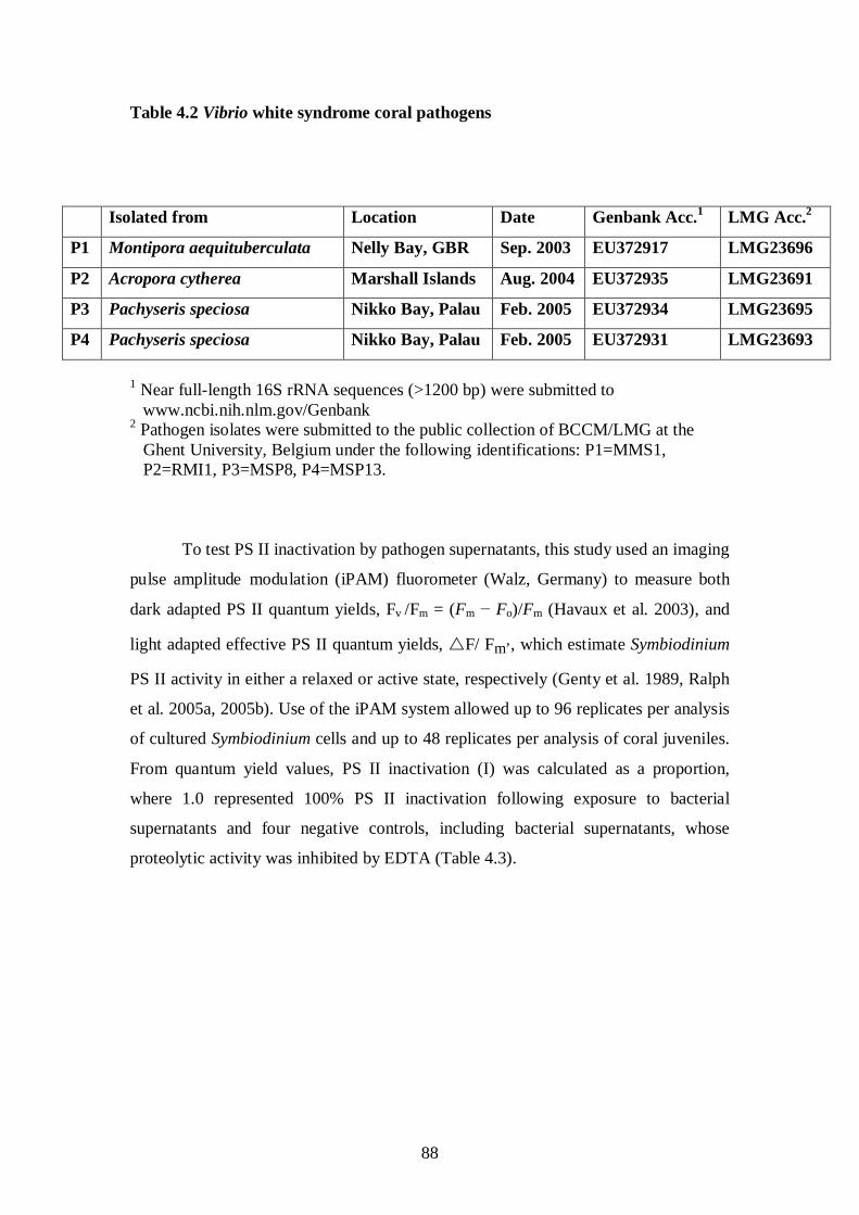
88
Table 4.2 Vibrio white syndrome coral pathogens
P
1P Near full-length 16S rRNA sequences (>1200 bp) were submitted to
11Twww.ncbi.nih.nlm.gov11T/Genbank P
2P Pathogen isolates were submitted to the public collection of BCCM/LMG at the
Ghent University, Belgium under the following identifications: P1=MMS1, P2=RMI1, P3=MSP8, P4=MSP13.
To test PS II inactivation by pathogen supernatants, this study used an imaging
pulse amplitude modulation (iPAM) fluorometer (Walz, Germany) to measure both
dark adapted PS II quantum yields, Fv /Fm = ( 10TF10Tm − 10TF10To)/ 10TF10Tm (Havaux et al. 2003), and
light adapted effective PS II quantum yields, F/ Fm’, which estimate Symbiodinium
PS II activity in either a relaxed or active state, respectively (Genty et al. 1989, Ralph
et al. 2005a, 2005b). Use of the iPAM system allowed up to 96 replicates per analysis
of cultured Symbiodinium cells and up to 48 replicates per analysis of coral juveniles.
From quantum yield values, PS II inactivation (I) was calculated as a proportion,
where 1.0 represented 100% PS II inactivation following exposure to bacterial
supernatants and four negative controls, including bacterial supernatants, whose
proteolytic activity was inhibited by EDTA (Table 4.3).
Isolated from Location Date Genbank Acc. P
1 LMG Acc.P
2
P1 Montipora aequituberculata Nelly Bay, GBR Sep. 2003 EU372917 LMG23696
P2 Acropora cytherea Marshall Islands Aug. 2004 EU372935 LMG23691
P3 Pachyseris speciosa Nikko Bay, Palau Feb. 2005 EU372934 LMG23695
P4 Pachyseris speciosa Nikko Bay, Palau Feb. 2005 EU372931 LMG23693
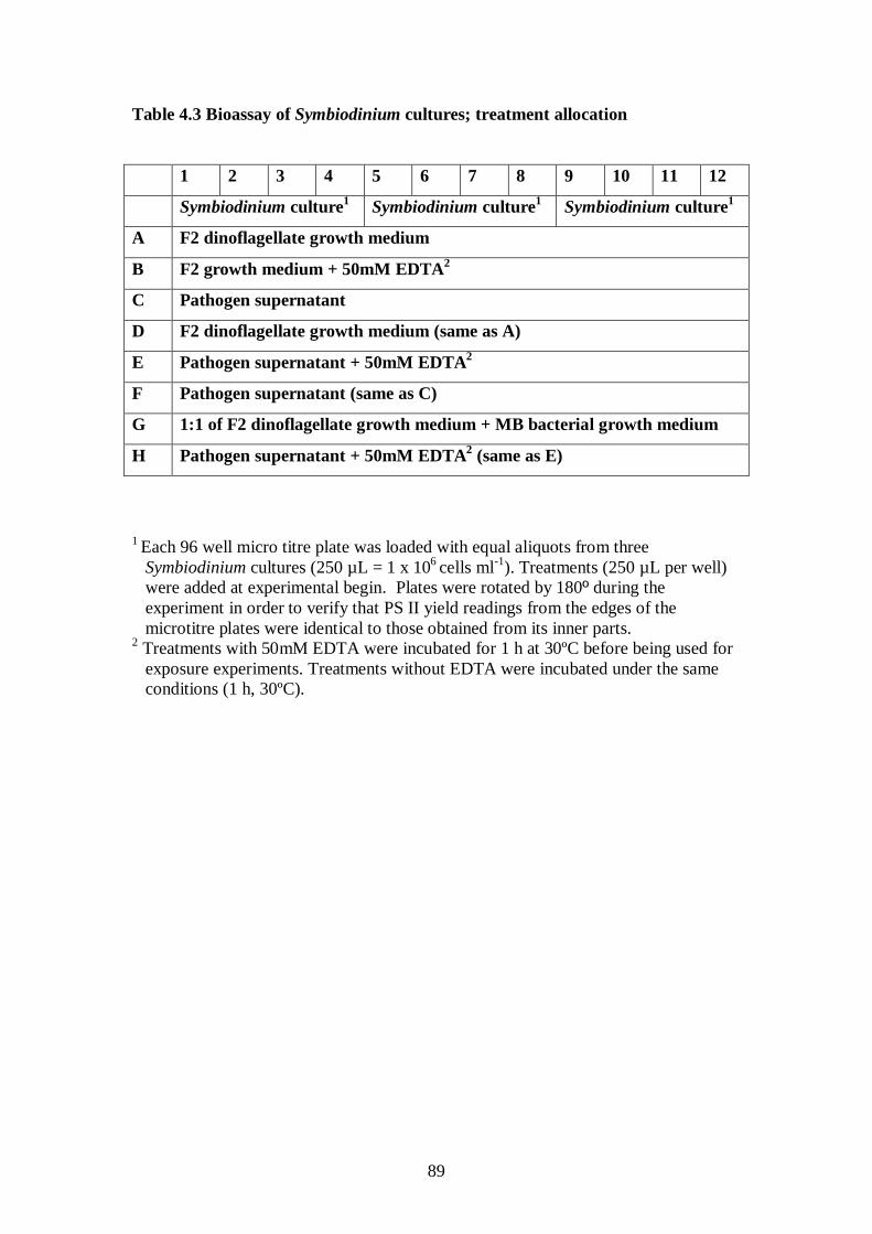
89
Table 4.3 Bioassay of Symbiodinium cultures; treatment allocation
1 2 3 4 5 6 7 8 9 10 11 12
Symbiodinium culture Symbiodinium culture1 Symbiodinium culture1 1
A F2 dinoflagellate growth medium
B F2 growth medium + 50mM EDTA2
C Pathogen supernatant
D F2 dinoflagellate growth medium (same as A)
E Pathogen supernatant + 50mM EDTA2
F Pathogen supernatant (same as C)
G 1:1 of F2 dinoflagellate growth medium + MB bacterial growth medium
H Pathogen supernatant + 50mM EDTA2 (same as E)
1 Each 96 well micro titre plate was loaded with equal aliquots from three Symbiodinium cultures (250 µL = 1 x 10⁶ cells ml-1). Treatments (250 µL per well) were added at experimental begin. Plates were rotated by 180º during the experiment in order to verify that PS II yield readings from the edges of the microtitre plates were identical to those obtained from its inner parts.
2 Treatments with 50mM EDTA were incubated for 1 h at 30ºC before being used for exposure experiments. Treatments without EDTA were incubated under the same conditions (1 h, 30ºC).
90
4.2 Materials and Methods
4.2.1 Coral pathogens
Four coral pathogen strains (P1-P4; Table 4.2), identified in the previous
chapter (Sussman et al. 2008) as causative agents for white syndrome diseases (WS’s)
affecting Indo-Pacific scleractinian corals by fulfilling Henle-Koch’s postulates, were
examined in this study. As highlighted previously, the 16SrRNA gene sequences of
all four coral pathogen strains can be found in GenBank under accession numbers:
EU372917,EU372931,EU372934,EU372935
(http://www.ncbi.nih.nlm.gov/Genebank). These isolates correspond to accession
numbers LMG23691, LMG23693, LMG23695 and LMG23696, in the public culture
collection of BCCM/LMG at Ghent University, Belgium (Table 4.2).
4.2.2 Growth curve and proteolytic activities of bacterial supernatants
Each of the four bacterial pathogens (P1-P4) was inoculated into a general
heterotrophic bacterial medium, Marine Broth-2216 (Difco, USA) and grown to end
logarithmic phase at 27ºC with shaking (150rpm). Tests performed to determine the
optimal growth conditions for pathogens P1-P4, demonstrated that culture
supernatants expressed the strongest proteolytic activity when incubated for 18 h to
end logarithmic phase (Fig. 4.7A). Bacterial cell density was determined by colony
forming unit counts (CFU; described in M&M chapter 3; Sussman et al. 2008) and by
constructing a cell density calibration curve of absorbance (595nn) vs. CFU (Fig.
4.7B). Absorbance (595nm) of serial culture dilutions was measured in sterile
microtitre 96 well plates (n=6) using a Wallac Victor 2 1420 multi label counter
spectrophotometer (Perkin Elmer, USA). Bacterial supernatants used in exposure
experiments were obtained by centrifugation (12,000 x g, 20 min, 4ºC) and serial
filtration through 0.45µm and 0.22µm filters (Millipore, USA). These solutions were
defined as bacterial supernatants P1-P4, and their protease activity was measured by
the asocasein assay (Windle and Kelleher 1997) as proteolytic units (Denkin and
Nelson 1999), where 1U = 1000 x (OD450 x CFUP
-1P) x 10 P
9P. Protein concentrations in
all bacterial supernatants (P1-P4) were determined by the Biorad protein assay
(Biorad laboratories, USA). Bacterial supernatant aliquots were stored at -20ºC until
used.
91
4.2.3 Inhibition by 1,10 Phenanthroline monohydrate (1,10 Pt) and phenyl
methylsulfonyl fluoride (PMSF)
Inhibition of proteolytic activity by bacterial supernatants was tested by using
three inhibitors: 1,10 Phenanthroline monohydrate (1,10 Pt),phenyl methylsulfonyl
fluoride (PMSF) and EDTA. 1,10 Phenanthroline monohydrate (1, 10 Pt; SIGMA)
was dissolved in DDW (Millipore). Pathogen supernatants were incubated for 1 h at
30ºC with 1,10 Pt in a final concentration of 5mM (Farrell and Crosa 1991).
Proteolytic activity was measured by the asocasein assay (Windle and Kelleher 1997,
Denkin and Nelson 1999). PMSF (SIGMA), an alkaline serine protease inhibitor, was
dissolved in ethanol and incubated for 1 h at 30ºC with pathogen supernatants in a
final concentration of 5mM (Kuo-Kau et al. 1997). Following incubation, reactions
were assayed for proteolytic activity by the asocasein assay (Windle and Kelleher
1997, Denkin and Nelson 1999).
4.2.4 Inhibition of proteolytic activity by EDTA and reactivation with ZnCl2
Bacterial supernatants (P1-P4) were exposed to treatments with four
concentrations of EDTA (5mM, 10mm, 25mM and 50mM). Triplicate samples of
each treatment were incubated for 1 h at 30ºC and then tested for proteolytic activity
by the asocasein assay (Windle and Kelleher 1997, Denkin and Nelson 1999). Control
treatments included bacterial supernatant with no EDTA. Treatments of pathogen
supernatants inhibited by adding 50mM EDTA and incubation (1h, 30ºC) were used
as negative control treatments in all exposure experiments conducted in this study.
The ability to reactivate the proteolytic activity of the P1 pathogen by adding divalent
cations was tested by incubating P1 supernatant with 50mM EDTA (1 h at 30ºC ) and
adding five concentrations of ZnCl2 (5mM, 10mM, 25mm, 50mM and 100mM).
Samples were incubated for 1h at 30ºC and then tested for proteolytic activity by the
asocasein assay (Windle and Kelleher 1997, Denkin and Nelson 1999). Inhibition of
pathogen P1 supernatant by EDTA reached 98% following exposure to 50mM EDTA.
At lower concentrations, inhibition dropped to 87%, when P1 supernatant was
exposed to a 5mM concentration and 91% and 96% when exposed to 10mM and
25mM, respectively. When 10mM ZnCl2 were added to EDTA-inhibited samples, P1
supernatant recovered 77% of its proteolytic activity prior to EDTA inhibition. When
treated with other ZnCl2 concentrations, recovery was significantly lower, with 61%
recovery following the addition of 5mM ZnCl2 and 35% recovery following the
92
addition of 25mM ZnCl2. Following the addition of higher ZnCl2 concentrations
(50mM and 100mM), no recovery was detected by the asocasein assay (Windle and
Kelleher 1997, Denkin and Nelson 1999), suggesting that the P1 harbors a zinc-
metalloprotease that needs an optimal concentration of ZnCl2 for activity. To test this,
P1 supernatant was incubated with five concentration of ZnCl2 (5mM, 10mM, 25mm,
50mM and 100mM) without prior inhibitory treatment with EDTA. Results
demonstrated that the proteolytic activity of P1 supernatant was significantly affected
by excess ZnCl2 in all treatments, with a 95% loss of activity measured in the 10mM-
100mM concentrations and 81% activity-loss when 5mM ZnCl2 were added to the
supernatant (data not shown). Similar inhibition was demonstrated when exposing the
H. pylori zinc-metalloprotease to zinc concentrations above 1mM (Windle and
Kelleher 1997). Other zinc peptides are also inhibited by excess zinc (competitive
inhibition), which is proposed to result from the formation of zinc monohydroxide
multidentate complexes which bridge the catalytic zinc to a side chain on the active
site (Larsen and Auld 1991).
4.2.5 Isolation of Symbiodinium cultures from sampled corals
Colonies of Montipora aequituberculata (Z1), Acropora tenius (Z3) and
Acropora millepora (Z4) were collected in sterile containers at Nelly Bay, Magnetic
Island, GBR (S19 10’ E 146 52’), an inshore fringing reef. Additional colonies of
Montipora aequituberculata (Z2) were collected at Davies Reef, GBR (S18°81’,
E147°67’), a midshelf reef located less than 100 km away (Table 4.1). Coral tissue
was removed by airbrush, centrifuged three times (3000 x g, 5 min) and resuspended
in 0.22µm filtered SW (25ºC). Coral nematocysts were removed by two consecutive
filtrations (20µm; Millipore, USA) using a vacuum pump.
4.2.6 Symbiodinium cultures
F2 dinoflagellate growth medium for Symbiodinium was prepared by
modification of F2 and Erdschreiber media (Guilard and Ryther 1962). Briefly,
seawater supplemented with 4mg l-1 Na2HPO4, 1g l-1 NaNO3, 1ml l-1 from a x1000
concentrated A5 + CO micronutrient solution (described in chapter 2; Sussman et al.
2006), 2.5 mg l-1 GeO2, 80mg l-1 G-Penicillin, 80mg l-1 Streptomycin, 40mg l-
1 Amphotericin, 0.4mg l-1 Thiamine-HCl, 2µg l-1 Biotin and 2µg l-1 Vitamin B12
93
(cyanocobalamin). The growth medium was 0.22µm filtered and stored at 4ºC in the
dark. Before use, 0.22µm filtration was repeated.
Symbiodinium cultures Z1-Z4 in F2 medium were inoculated into sterile 24
well plates (3ml per well), covered and sealed. Plates were incubated at 27ºC under
12h:12h light:dark irradiance (90 pmol photons m-2 s-1). Cells were inspected daily
and contaminated plates were discarded. Culture purity was enhanced by limiting
dilution steps. Briefly, Symbiodinium cells were pipetted from wells into sterile
0.22µm filtered F2 medium in new 24 well plates and cultured for 7 days. This step
was repeated for 4 months, during which samples were periodically obtained for
molecular analysis (see 4.2.9) to ensure the taxonomic consistency of clades in each
culture. Prior to experimental exposures, Symbiodinium cells were quantified (n=10)
using a Neubauer haemocytometer and adjusted to one concentration (1x10⁶ cells ml-1
96 well microtitre plates containing Symbiodinium cells (1x10
)
by adding F2 medium before transferring cultures into sterile 96 well microtitre plates
(250 µL per well). An attempt was made to maintain the original Symbiodinium
populations that were associated with the host coral at the time of isolation rather than
to purify and maintain single axenic cultures (Santos et al. 2001), which would have
less ecological relevance when tested for their susceptibility to pathogen supernatants.
In order to confirm the taxonomic identity of Symbiodinium types in each culture,
cloning of Symbiodinium DNA was performed at the time of isolation from corals and
prior to using the incubated cultures for experimental procedures. Experiments
exposing Symbiodinium cells to bacterial supernatants and controls were repeated
twice to confirm the consistency of results. A full description of treatments is
presented in table 4.3.
4.2.7 PS II dark adapted quantum yields (Fv /Fm) and PS II inactivation (I) 6 cells ml-1) were
incubated in the dark (1 h) and centrifuged (5 min at 3000 x g). F2 medium was
discarded and wells were loaded with treatment solutions. Plates were exposed in a
Maxi imaging-pulse-amplitude-modulation (iPAM) fluorometer (Walz, Germany) to a
saturation light pulse (Gain=1-2, Intensity=1-2, Saturation Pulse=7) at 5 min intervals
and dark adapted PS II quantum yields were calculated by using the formula: Fv/Fm =
(Fm – F0) / Fm (Havaux et al. 2003), where Fm = maximal fluorescent yield, and F0 =
Dark fluorescent yield. From Fv /Fm values, PS II inactivation values (I) were
calculated as proportions by using the formula: I (Fv /Fm ) = (Fv /Fm at time 0 – Fv /Fm
94
at time n) / Fv /Fm at time 0, where 1.0 represented 100% PS II inactivation, following
exposure to proteolytically-active and EDTA-inhibited bacterial supernatants and
three additional controls (Table 4.3).
4.2.8 PS II effective light-adapted yields (F/ Fm’) and PS II inactivation (I)
The identical procedure for sample preparation before measurement of dark
adapted yields was repeated before measuring effective light adapted yields. This step
confirmed the results obtained from reading photosynthetic inactivation as a
proportion of dark adapted yields. Some authors also consider it as a better estimate
for photosynthetic function (Iglesias-Prieto et al. 2004), because quantum yields are
measured when the cells are photosynthetically active. 96 well microtitre plates were
prepared as described above. Each plate was dark adapted first and Fm, F1 and dark
adapted quantum yields (Fv/Fm) were recorded at 5 min intervals for a period of 30
min, until consistent levels were obtained. Plates were then centrifuged as described
above and returned to the imaging PAM chamber for initial light adapted
measurements. An actinic light source of 90 pmol m -2 s -1
A similar protocol was used by Schreiber et al. (2006) to measure PS II
photoinhibition caused by the toxic effects of diuron, suggesting that since quantum
yields are calculated from the ratio of fluorescent values before (Ft) and after (Fm’)
was switched on in the
measuring chamber and cultures were exposed to a saturation light pulse at 5 min
intervals for a period of 30 min until it was confirmed that readings of effective light
adapted quantum yields were stable (Gain=1-2, Intensity=1-2, Saturation Pulse=7).
Plates were then removed from the chamber and centrifuged. F2 medium was
discarded from the plates and without further delay, plates were returned to the
imaging PAM to be loaded with treatment solutions. Plates remained in the imaging
PAM chamber under illumination for the entire duration of the experiment. The
continuous measurement at 5 min intervals was preferred to the alternative of dark
adapting the samples before each single light adapted reading, due to the nature of the
experiment. Although photochemical quenching was not fully relaxed, this procedure
allowed closer surveillance of the continuous effects of bacterial supernatant on PS II
photosynthesis, as it might occur under environmentally relevant conditions, where
corals are constantly exposed to light during the day and for longer periods during the
summer compared to winter.
95
firing a constant saturation pulse, results are independent of signal amplitudes.
According to Schreiber et al. (2006), 100 sec intervals between consequent saturation
pulses (SP) were sufficient to allow complete reoxidation of QA and re-establishment
of the original Ft levels. Light adapted effective quantum yields (F/ Fm’) were
calculated by the formula: (Fm’-Ft) / Fm’ (Genty 1989), where Fm’ = maximal
fluorescent yield under light conditions and Ft = fluorescence before a saturating
pulse. PS II inactivation (I) was calculated (as a proportion) from light adapted
effective quantum yields (F/ Fm’) as described above. An alternative method for
calculating PS II inactivation by comparing PS II quantum yields of treatments with
PS II quantum yields of negative controls at corresponding times (Schreiber et al.
2006) was tested and provided similar results.
4.2.9 Taxonomic identities of Symbiodinium cultures
DNA was extracted from Symbiodinium cultures incubated at 27ºC or directly
from corals (Wilson et al. 2002) and amplified using primers targeting the ribosomal
RNA (rRNA) internal transcribed spacer 1 region (ITS-1; (van Oppen et al. 2001).
PCR products were cloned (pCR 2.1 TOPO kit, Invitrogen, CA) and inserts
containing plasmid DNA were amplified with a 5’- tet fluorescently labelled ITS-1
forward primer and then screened on a single strand conformation polymorphism
(SSCP) gel before sequencing (Ulstrup and van-Oppen 2003). Retrieved nucleotide
sequences (~360 bp) including the ITS-1 rRNA and its flanking regions were edited
using Chromas Lite software version 2.01 (Technelysium) and aligned using ClustalX
version 1.83 (Thompson et al. 1994). Distance matrices were calculated using the
DNADIST program in PHYLIP (Felsenstein 1989) and phylogenetic trees were
generated from distance matrices using the neighbour-joining method (Saitou and Nei
1987) and Kimura substitution algorithm (Kimura 1983). Bootstrapping with 1000
replicates was performed using SeqBoot as integrated in PHYLIP (Retief 2000) and
values ≥50% were included for main nodes of the tree. Ribosomal RNA sequences of
Symbiodinium microadriaticum amplified with the ITS-1 primers and cloned were
submitted to Genbank (http://ncbi.nih.nlm.gov/Genbank) under the accession numbers
EU567151-567152, EU567155-567160, EU567167-567168, EU567170, EU567174
(Table 1). Reference Symbiodinium types for phylogenetic analyses were obtained
96
from the following authors: AJ311944 (Pochon et al. 2001), AF380532, AF380537,
AF380543, AF380546 (van-Oppen et al. 2001), DQ238587 (Santiago-Vazquez et al.
2007), AY457958 (Fabricius et al. 2004), AF334660 (LaJeunesse 2001), AF396629
(Santos et al. 2003), EF455526, EF455528 (Magalon et al. 2007), out group
Heterocapsa sp. FK6-D47 AB084097 (Yoshida et al. 2003).
4.2.10 Experimental coral juveniles
Rearing coral juveniles (Acropora millepora) and infecting them with
Symbiodinium clades D and C1 was performed following the protocol of Little et al.
(2004). Gametes were collected following spawning of eight (hermaphroditic)
colonies of Acropora millepora, during the spawning event in October, and mixed
together for fertilization. Fertilized larvae were raised in filtered seawater in two
separate 500 L tanks in a temperature controlled (27° C) aquaria room. When the
larvae were elongated and searching for settlement sites (four days after spawning),
preconditioned and autoclaved terracotta tiles (preconditioned for six weeks at
Magnetic Island, GBR) were placed on the bottom of the two 500 L tanks to provide
settlement surfaces. Symbiodinium clade strains D and C1 were obtained from adult A.
millepora and A. tenuis colonies from Magnetic Island, respectively, by airbrushing
the coral tissue and isolating Symbiodinium cells out of the coral-Symbiodinium slurry
by centrifugation (5min. 3500 x g). These Symbiodinium cells were offered at 10P
8P
cells tankP
-1 Pthree and five days after spawning and were acquired by newly settled
juveniles. The terra-cotta tiles were attached to a fringing reef (Nelly Bay, Magnetic
Island, GBR) in a zone where Acropora spp. were present and were then collected
nine months after settlement (in July) for experimental exposure. Tile racks were
removed from the reef and carried in large containers to the outdoor aquaria at the
Australian Institute of Marine Science (AIMS). Individual tiles were separated and
placed to recover in outdoor tanks with a flow-through seawater system. Following
five days, individual juveniles were scraped from the tiles and placed in sterile 48
well plates. These plates were further suspended in flow-through tanks for recovery
until juveniles attached to the bottom surface of the wells. In this setup, juveniles
experimentally infected with either Symbiodinium D or C1 cultures originated from
crosses involving the same parent corals, thereby minimizing potential host genetic
differences that may influence the physiology of the holobiont (host-symbiont
partnership). The rearing and infection of coral juveniles used in this chapter was
97
performed by PhD student Jos Mieog at the Australian Institute if Marine Sience
(AIMS), as part of a collaborative effort.
Individual A. millepora juveniles infected with Symbiodinium clades D and C1
were placed in 48 well plates and exposed to the following treatments (n=4): 1. F2
dinoflagellate medium; 2. P1 supernatant diluted 1:1 with sterile seawater; 3. P3
supernatant diluted 1:1 with sterile seawater; 4. P1 supernatant diluted 1:1 with sterile
F2 medium, treated with 50mM EDTA and incubated for 1 h at 30ºC; 5. P3
supernatant diluted 1:1 with sterile F2 medium, treated with 50mM EDTA and
incubated for 1 h at 30ºC; 6. Bacterial medium (LB) mixed 1:1 with dinoflagellate
medium (F2). All EDTA and non-EDTA treatments were incubated for 1 h at 30ºC
prior to use. Plates were acclimatized for five days prior to exposure. Measurements
and calculation of PS II dark and light adapted quantum yields and PS II inactivation
were performed as described above. For measurements of PS II effective light adapted
quantum yields (F/ Fm’), an actinic light source of 5 pmol m -2 s -1, identical to light
intensity in the field, was switched on in the measuring chamber of the imaging PAM.
Well plates containing A. millepora juveniles identical to those exposed to pathogen
supernatants and controls under the imaging PAM were exposed and photographed
under identical conditions (5-pmol m-2 s-1
Symbiodinium cultures (Z1 and Z4) were prepared as described above.
Pathogen supernatant concentrations were prepared by diluting 0.22µm filtered P1
supernatant with modified F2 medium to end concentrations of 50%, 25%, 10%, 5%,
1% and 0.1% from original stock. Effective light adapted quantum yield (F/ Fm’) was measured under illumination as described above and PS II inactivation (I) was
calculated. I of Symbiodinium culture Z1 and concentrations of P1, as proportions of
1.0, were plotted resulting in a parabolic curve. Reciprocating data for Z1 PS II
inactivation (I) and P1 concentrations resulted in a Lineweaver-Burk (1934) - ‘like’
linear plot, commonly used to describe the relation between substrate concentration (S)
and reaction velocity (V). The term ‘like’ is used in this study, since neither the
substrate for bacterial supernatants nor the products of their catalytic activity were
determined. It was thus assumed that both supernatant dose and PS II inactivation (I)
, 27ºC) at 30 min intervals using a dissecting
microscope (x 1.6) and a digital camera.
4.2.11 Pathogen concentration experiment
98
values are good estimates of S and V. The linear equation (y=ax+b) was used to
determine 1/km’, when y = 0 and, with km’ defined as the concentration of P1 needed
to cause a 50% PS II inactivation (I) of the susceptible Symbiodinium culture Z1
within 10 min following exposure.
4.2.12 PS-II photoinactivation as a function of temperature
For this experiment to be of environmental relevance, both bacterial pathogens
and Symbiodinium cultures were required to be incubated and then mixed at the same
temperatures. P1 was inoculated into MB media and grown to end logarithmic phase
at 5 different temperatures (20ºC, 25ºC, 27ºC, 30ºC and 35ºC), which cover the range
of seawater temperatures typical of sites where WS outbreaks occur. Pathogen
supernatant P1 was derived from culture as previously described. Symbiodinium
cultures Z1 and Z4 were acclimatized to the corresponding temperatures over a period
of 5 days. Exposure of Symbiodinium cultures to pathogen supernatant P1 was
conducted in a temperature controlled room and effective light adapted quantum
yields were measured using the Maxi Imaging PAM. PS-II photoinactivation (I) was
calculated as described previously. The following parameters were quantified prior to
the commencement of the experiment in order to determine the direct effect of
temperature on its components (bacteria and Symbiodinium):
1. Bacterial cell density, reflecting pathogen P1 growth at 5 different temperatures.
Cell density was quantified by plating serial dilutions of triplicate samples and by
spectrophotometry (as described previously).
2. The relative level of proteolytic activities in bacterial supernatants filtered from
cultures that were incubated at the various temperatures, as determined by the
asocasein assay.
3. The physical state of Symbiodinium cells in cultures acclimatized to the five
different temperatures prior to their exposure to bacterial supernatant, determined
by measuring dark adapted maximum quantum yields (Fv /Fm), light adapted
effective quantum yields (F/ Fm’) and calculating excitation pressure (Q) over
photosystem II by the formula: Q = 1- (∆F/Fm’)/(Fv/Fm), when ∆F/Fm’ is the light
dependent reduction of the effective quantum yield of photosystem II (Iglesias-
Prieto 2004).
99
Cultures of Symbiodinium Z1 (1x10⁶ cells ml-1) were exposed in 96 well
micro-titre plates to 7 treatments (n=12 wells per treatment) consisting of four
concentrations of P1 bacterial supernatant and three controls: P1 supernatant
incubated with 50mM EDTA (1h 30ºC), Symbiodinium cultures in dinoflagellate
growth medium (F2) and in a 1:1 mix of bacterial growth medium (LB) and
dinoflagellate growth medium (F2). Pathogen P1 supernatant concentrations were
prepared by diluting 0.22µm filtered P1 supernatant with modified F2 medium to
concentrations of 100%, 50%, 10% and 1%. Each experiment was performed at each
of the five selected temperatures (20ºC, 25ºC, 27ºC, 30ºC and 35ºC; 1 plate per
temperature, n=2 repetitions), deriving five Lineweaver-Burk “like” plots, which
demonstrated a gradient in enzymatic kinetics as a function of temperature. Km', the
apparent concentration needed to achieve a 50% photoinactivation of Z1 by P1
supernatant following a 10 min exposure was calculated from the linear regression
equations obtained for the five temperatures.
4.2.13 Exposure of Symbiodinium cultures to 35ºC
96 well micro titre plates containing Symbiodinium cultures Z1 and Z4
(1x10⁶ cells ml-1) were incubated at 35ºC under a 12h:12h light:dark irradiance (90
pmol photons m-2 s-1
Seawater temperature data were obtained from In situ data loggers deployed
on reefs. The Australian Institute of Marine Science (AIMS) and the Great Barrier
Reef Marine Park Authority (GBRMPA) maintain the SeaTemps monitoring program,
which is coordinated by Dr Ray Berkelmans (
) for a period of 25 days to test Symbiodinium ability to withstand
elevated temperatures. F2 media in wells was replaced every five days with fresh
media (35ºC) and PS-II performance was estimated by measuring both dark adapted
maximum quantum yields (Fv /Fm) and effective light adapted quantum yields (F/
Fm’) at five day intervals (as described above). Following this experiment, sub-
aliquots from each Symbiodinium culture were cloned (as described above) in order to
determine whether a shift in the Symbiodinium populations in each culture had
occurred following exposure to elevated temperatures (35ºC).
4.2.14 Temperature data logger Information
[email protected]). Data
loggers instantaneously record sea temperatures every 30 minutes and are exchanged
100
and downloaded approximately every 12 months. Temperature loggers on the reef-flat
are placed just below Lowest Astronomical Tide (LTD) level. They are also deployed
on the upper reef slope (in depths of 5–9 m) and in deep reef-slopes (in depths of ~20
m). Loggers are calibrated against a certified reference thermometer after each
deployment and are generally accurate to 0.1-0.2ºC. Mean Daily Seawater
Temperature (MDST) was calculated by averaging data for all loggers at each site per
one day. 38 data logger sites from which seawater temperatures have been recorded
for at least five years (including the two major bleaching events on the GBR in the
austral summers of 1997/8 and 2001/2), were selected and the number of days in
which the mean daily seawater temperature (MDST) equalled or exceeded 27ºC and
remained below or equalled 30ºC was calculated for each station per year, beginning
in the austral winter (1 June-31 May). This temperature span (27ºC ≥X≤ 30ºC)
corresponded best with the optimal temperature range suggested by this study for
bacterial zinc-metalloprotease activity causing Symbiodinium photoinactivation.
Annual seawater temperature data (number of days per year in which 27ºC ≥ MDST ≤
30ºC) was analysed from 9 data-logger reef sites: Low Isles (S16º20', E145º30'),
Myrmidon Reef (S18°26, E147°38’), Kelso reef (S18°45’, E146°99’), Cattle Bay
(S18°57’, E146°48’), Davies Reef (S18°81’, E147°67’), Geoffrey Bay, Magnetic
Island (S19°16’, 146°86’), Nelly Bay, Magnetic Island (S19°17’, E146°85’), Turner
Cay I (S21°7’, E152°56’) and Halfway Reef (S23°2’, 150°97’).
The daily span of sunlight (in min per day) was obtained for the city of
Townsville, which corresponds to the latitude of Davies Reef.
4.2.15 Protein sequence retrieval
Bacterial cultures P1-P4 were grown (1.8 L) and crude extracts were derived
by ammonium sulphate precipitation (Wu et al. 1996). Proteins in a total volume of
100 ml of supernatant derived from each of four pathogen cultures were precipitated
by slowly adding ammonium sulphate to achieve the final % saturations of 20%, 40%,
60%, 80% and 100%. This procedure was undertaken to determine the optimal
ammonium sulphate concentration needed to precipitate bacterial proteolytic enzymes
from their corresponding supernatants without compromising proteolytic activity.
Samples were stirred for 2 h and then centrifuged at 5000 x g for 1 h at 4ºC.
Supernatants were discarded and the remaining precipitate was diluted by adding
101
200µl of 0.22µm-filtered phosphate buffered saline (PBS: 10mM Potassium
Phosphate 150mM NaCl pH7.4). Samples were tested for proteolytic activity by the
asocasein assay (Windle and Kelleher 1997, Denkin and Nelson 1999) and the
optimum % saturation of ammonium sulphate was chosen for the production of higher
concentrations of active crude extracts from all pathogen supernatants. Pathogens P1-
P4 measured the highest proteolytic activity by the asocasein assay when precipitated
in a 20% saturation of ammonium sulphate.
For the production of proteolytic active crude extracts, 1.8 L volumes of
bacterial cultures were incubated and crude extracts, diluted in 45ml 0.22µm-filtered
PBS (7.4 pH), were derived (as described above). Each sample was then ultra-filtered
using three Amicon 5,000M MWCO ultra centrifugal filter devices (Millipore, USA).
Samples were centrifuged at 4000 x g in a swinging bucket Alegra X-15R centrifuge
(Beckman Coulter, CA) at 4ºC for 45 min to derive a total final crude extract volume
of 600µL from each pathogen supernatant, corresponding to a x3000 concentration of
the original pathogen cultures. 1µL of each sample was then diluted in 99µL of
0.22µm-filtered DDW (Millipore, USA) and tested to confirm proteolytic activity by
the asocasein assay (Windle and Kelleher 1997, Denkin and Nelson 1999). Protein
concentrations were measured with a protein assay kit from Bio-Rad and samples
were frozen at -80ºC and sent to the Australian Proteome Analysis Facility
(Macquarie University NSW, Australia) for fast protein liquid chromatography (FPLC)
analysis. The samples were run on a Superdex 75 10/300 GL Column and the buffer
used was PBS (pH7.4). The column was tested with low molecular weight standards
(GE Biosciences) before the sample was injected (Albumin 67kDa, Ovalbumin 43kDa,
Chymotrypsinogen A 25kDa and Ribonuclease A 13.7 kDa). The samples were
prepared by filtering through a 0.45µm cellulose acetate microcentrifuge filter system
unit at 10,000 x g for 1 min. The method flow rate was 0.5ml for 70 min and fraction
size was 400uL (48 seconds). The sample was run on a Waters Alliance HT 2795
system with a PDA detector and Gilson FC204 fraction collector. 10µL from all 72
fractions from each sample were assayed for proteolytic activity by the asocasein
assay (Windle and Kelleher 1997, Denkin and Nelson 1999) and results were
superimposed on the 280nm chromatograms generated for each sample for fraction
selection. Selected samples were run on zymogen gels containing 0.1% Na-casein co-
polymerized in gels as substrate revealing zones of hydrolysis. 15 active fractions
from all four pathogens were re-run on a 12% SDS-PAGE following the method by
102
Laemmli (Laemmli 1970). 15 Bands were excised using a sterile blade and sent to the
Australian Proteome Analysis Facility for nano-liquid chromatography peptide
separation and mass spectrometry. Gel plugs were de-stained followed by tryptic
digestion for 16h at 37ºC. Digested peptides were separated by nano-LC using a
CapLC system (Agilent 1100 Series, Agilent Technologies, Germany). Sample (39μL)
were injected onto a peptide trap (Michrome peptide Captrap) for pre-concentration
and desalted with 0.1% formic acid at 10μL/min. The peptide trap was then switched
into line with the analytical column containing C18 RP silica (SGE ProteCol C18,
300A, 3μm, 150μm x 10 cm). Peptides were eluted from the column using a linear
solvent gradient, with steps, from H2O:CH3CN (95:5; + 0.1% formic acid) to
H2O:CH3CN (20:80, + 0.1% formic acid) at 500 nLmin-1
Means and standard errors (SE) for bacterial colony forming unit (CFU)
counts, for absorption readings (bacterial cell density and proteolytic activity), for PS
II dark adapted quantum yields and light adapted effective quantum yields were
compared among treatments using One-Way ANOVA (Statistica, StatSoft, Inc. USA).
CFU counts are presented in this study using logarithmic scales. Means and standard
errors (SE) for PS II inactivation (as a proportion of 1.0) in all exposure experiments
(treatments and controls) were compared using multivariate repeated measures
MANOVA (Statistica, StatSoft, Inc. USA), which does not rest on the assumption of
sphericity and compound symetry (Huynh and Feldt 1970). Four multivariate tests of
significance were applied (Wilks' Lambda, Pillai-Bartlett Trace, Hotelling-Lawley
Trace, and Roy's Largest Root) with non-significant results used to overrule any
previous assumptions of statistical significance. Significant results were determined
when α ≤ 0.05.
over a 45 min period. The
LC eluent was subject to positive ion nano flow electrospray analysis on an Applied
Biosystems QSTAR XL mass spectrometer (ABI, CA, USA). The QSTAR was
operated in an information dependant acquisition mode (IDA). In IDA mode a
TOFMS survey scan was acquired (m/z 400-2000, 1.0s), with the four largest multiple
charged ions (counts >25) in the survey scan sequentially subjected to MS/MS
analysis. MS/MS spectra were accumulated for 1 s (m/z 50-2000).
LC/MS/MS data were searched using Mascot (Matrix Science, London, UK)
and bacterial entries in the NCBI non-redundant protein database (Altschul et al.
1997).
4.2.16 Statistical Analysis
103
4.3 Results
4.3.1 Symbiodinium culture Z1 is most susceptible to bacterial PS II inactivation
Symbiodinium culture Z1 isolated from the WS susceptible coral host
Montipora aequituberculata at Nelly Bay, an inshore reef off Magnetic Island in the
central GBR, was the most severely affected of the four Symbiodinium cultures tested
when exposed to P1 supernatant under illumination (p<0.01; Fig. 4.1A). For
Symbiodinium culture Z1, inactivation (I) of PS II (measured as light adapted
quantum yields) was greater than 95% (mean I (Z1 F/ Fm’) = 0.968 ± 0.016)
following exposure to P1 supernatant for 10 min in two independent experiments, and
total PS II inactivation resulted after 20 min (Fig 4.1A). A significant (~40%;
p<0.0001) difference in mean I was measured between this culture from Nelly Bay
and culture Z2 isolated from the same coral species found at Davies Reef, a GBR
midshelf reef located less than a 100 km away, where no signs of WS on M.
aequituberculata have been observed [mean I (Z2 F/ Fm’) = 0.587 ± 0.021
following exposure to P1 supernatant for 10 min in two independent experiments].
The impact of P1 on culture Z3, which was isolated from the coral Acropora tenuis at
Nelly Bay, where it has not been observed with signs of WS, was similar to its impact
on Symbiodinium culture Z2 throughout the experiment (p=0.426). Symbiodinium
culture Z4 isolated from the coral Acropora millepora at Nelly Bay, where it has not
been observed with WS signs at this site, was the least affected (~3%; p<0.01) of all
Symbiodinium cultures tested in this study [mean I (Z4 F/ Fm’) = 0.034 ± 0.019
following exposure to P1 supernatant for 10 min in two independent experiments].
Control treatments with dinoflagellate growth medium F2 (Fig. 4.1A) and bacterial
growth medium (MB) resulted in limited or no PS II inactivation of the respective
cultures [mean I (F2 F/ Fm’) = 0.001 ± 0.001; p<0.01, and mean I (MB F/ Fm’) =
0 following exposure for 10 min in two independent experiments].
Cloning and sequencing the ribosomal RNA (rRNA) internal transcribed
spacer 1 (ITS-1) region of Symbiodinium from cultures Z1-Z4 identified Z1 and Z2 as
two distinct types phylogenetically affiliated with Symbiodinium clade A (Fig. 4.2).
Culture Z3 was phylogentically affiliated with Symbiodinium clade C, and Z4
contained two Symbiodinium types affiliated with Symbiodinium clades A and D (Fig
4.3).
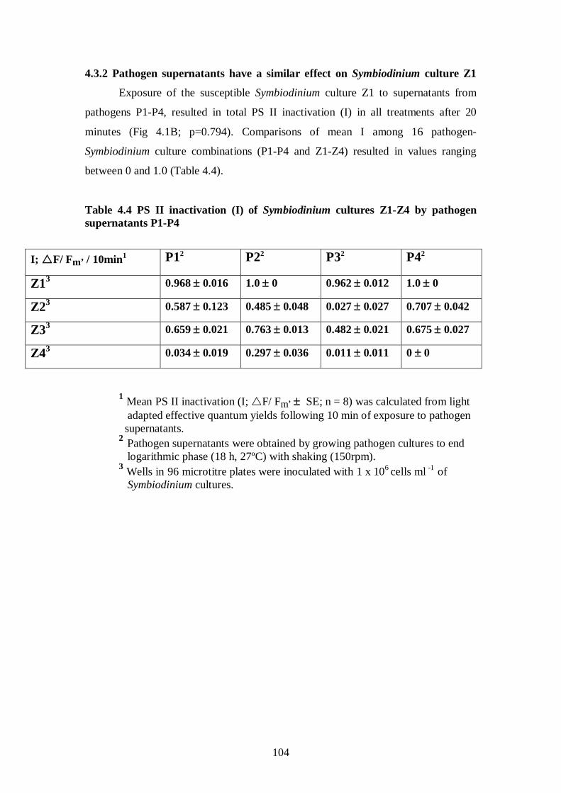
104
4.3.2 Pathogen supernatants have a similar effect on Symbiodinium culture Z1
Exposure of the susceptible Symbiodinium culture Z1 to supernatants from
pathogens P1-P4, resulted in total PS II inactivation (I) in all treatments after 20
minutes (Fig 4.1B; p=0.794). Comparisons of mean I among 16 pathogen-
Symbiodinium culture combinations (P1-P4 and Z1-Z4) resulted in values ranging
between 0 and 1.0 (Table 4.4).
Table 4.4 PS II inactivation (I) of Symbiodinium cultures Z1-Z4 by pathogen supernatants P1-P4
I; F/ Fm’ / 10min P11 P22 P32 P42 2
Z1 0.968 ± 0.016 3 1.0 ± 0 0.962 ± 0.012 1.0 ± 0
Z2 0.587 ± 0.123 3 0.485 ± 0.048 0.027 ± 0.027 0.707 ± 0.042
Z3 0.659 ± 0.021 3 0.763 ± 0.013 0.482 ± 0.021 0.675 ± 0.027
Z4 0.034 ± 0.019 3 0.297 ± 0.036 0.011 ± 0.011 0 ± 0
1 Mean PS II inactivation (I; F/ Fm’ ± SE; n = 8) was calculated from light adapted effective quantum yields following 10 min of exposure to pathogen supernatants. 2 Pathogen supernatants were obtained by growing pathogen cultures to end logarithmic phase (18 h, 27ºC) with shaking (150rpm). 3 Wells in 96 microtitre plates were inoculated with 1 x 10⁶ cells ml -1
of Symbiodinium cultures.
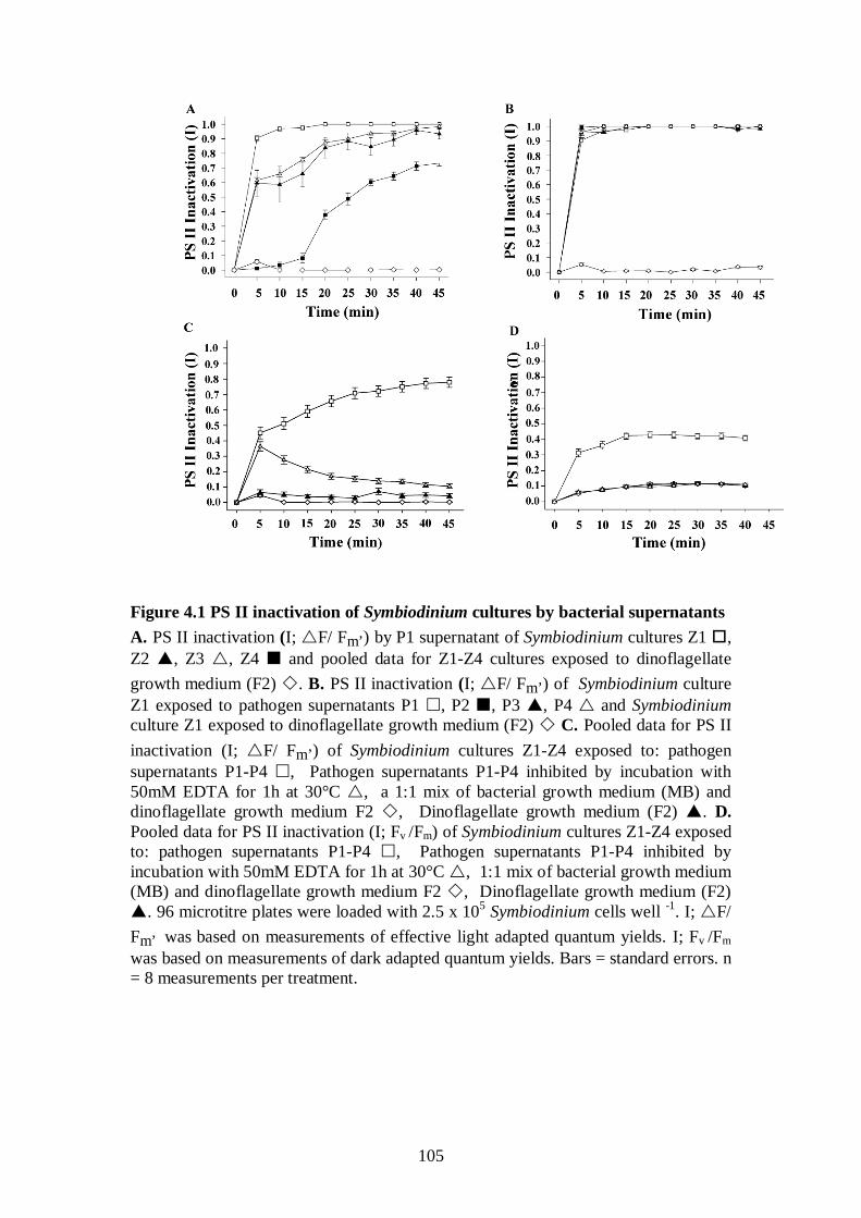
105
Figure 4.1 PS II inactivation of Symbiodinium cultures by bacterial supernatants A. PS II inactivation (I; F/ Fm’) by P1 supernatant of Symbiodinium cultures Z1 , Z2 , Z3 , Z4 and pooled data for Z1-Z4 cultures exposed to dinoflagellate growth medium (F2) . B. PS II inactivation (I; F/ Fm’) of Symbiodinium culture Z1 exposed to pathogen supernatants P1 , P2 , P3 , P4 and Symbiodinium culture Z1 exposed to dinoflagellate growth medium (F2) C. Pooled data for PS II inactivation (I; F/ Fm’) of Symbiodinium cultures Z1-Z4 exposed to: pathogen supernatants P1-P4 , Pathogen supernatants P1-P4 inhibited by incubation with 50mM EDTA for 1h at 30°C , a 1:1 mix of bacterial growth medium (MB) and dinoflagellate growth medium F2 , Dinoflagellate growth medium (F2) . D. Pooled data for PS II inactivation (I; Fv /Fm) of Symbiodinium cultures Z1-Z4 exposed to: pathogen supernatants P1-P4 , Pathogen supernatants P1-P4 inhibited by incubation with 50mM EDTA for 1h at 30°C , 1:1 mix of bacterial growth medium (MB) and dinoflagellate growth medium F2 , Dinoflagellate growth medium (F2) . 96 microtitre plates were loaded with 2.5 x 10⁵ Symbiodinium cells well -1. I; F/ Fm’ was based on measurements of effective light adapted quantum yields. I; Fv /Fm was based on measurements of dark adapted quantum yields. Bars = standard errors. n = 8 measurements per treatment.
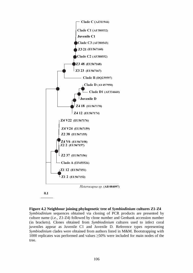
106
Figure 4.2 Neighbour joining phylogenetic tree of Symbiodinium cultures Z1-Z4 Symbiodinium sequences obtained via cloning of PCR products are presented by culture name (i.e., Z1-Z4) followed by clone number and Genbank accession number (in brackets). Clones obtained from Symbiodinium cultures used to infect coral juveniles appear as Juvenile C1 and Juvenile D. Reference types representing Symbiodinium clades were obtained from authors listed in M&M. Bootstrapping with 1000 replicates was performed and values ≥50% were included for main nodes of the tree.
107
4.3.3 Pathogen proteolytic activity is inhibited by EDTA and reactivated by
ZnCl2
EDTA was the most potent inhibitor of proteolytic activity of bacterial
supernatants P1-P4 when tested by the asocasein assay (Windle and Kelleher 1997,
Denkin and Nelson 1999) incorporating three standard protease inhibitors (EDTA,
1,10 Pt and PMSF; Fig. 4.3A). Proteolytic activity was reduced by 98% with the
addition of 50mM EDTA to the supernatant of pathogen P1 (1h incubation at 30ºC;
Fig. 4.3B) and by ~80%, on average, for pathogens P1-P4 (Fig. 4.3A). Addition of
10mM ZnCl2 reversed the chelating effect of EDTA and reactivated the proteolytic
activity of P1 supernatant to 77% of its original capacity (Fig 4.3C). This result
combined with results from the screenings of the previous chapter (Chapter 3 section
3.3.4.3; Sussman et al. 2008), which detected the active zinc binding site of a
metalloprotease using specific primers targeting the DNA in all pathogens (P1-P4),
confirmed the presence of a zinc-metalloprotease. Following the addition of higher
ZnCl2 concentrations (50mM and 100mM), no recovery was detected by the asocasein
assay, suggesting that the P1 zinc-metalloprotease requires an optimal concentration
of ZnCl2 for activity [for more information see M&M 4.2.4]. Other divalent cations
(NiCl2, MnCl2, MgCl2, CaCl2, CuCl2, HgCl2 and FeCl2) failed to reactivate the P1
zinc-metalloprotease following inhibition by EDTA (data not shown).
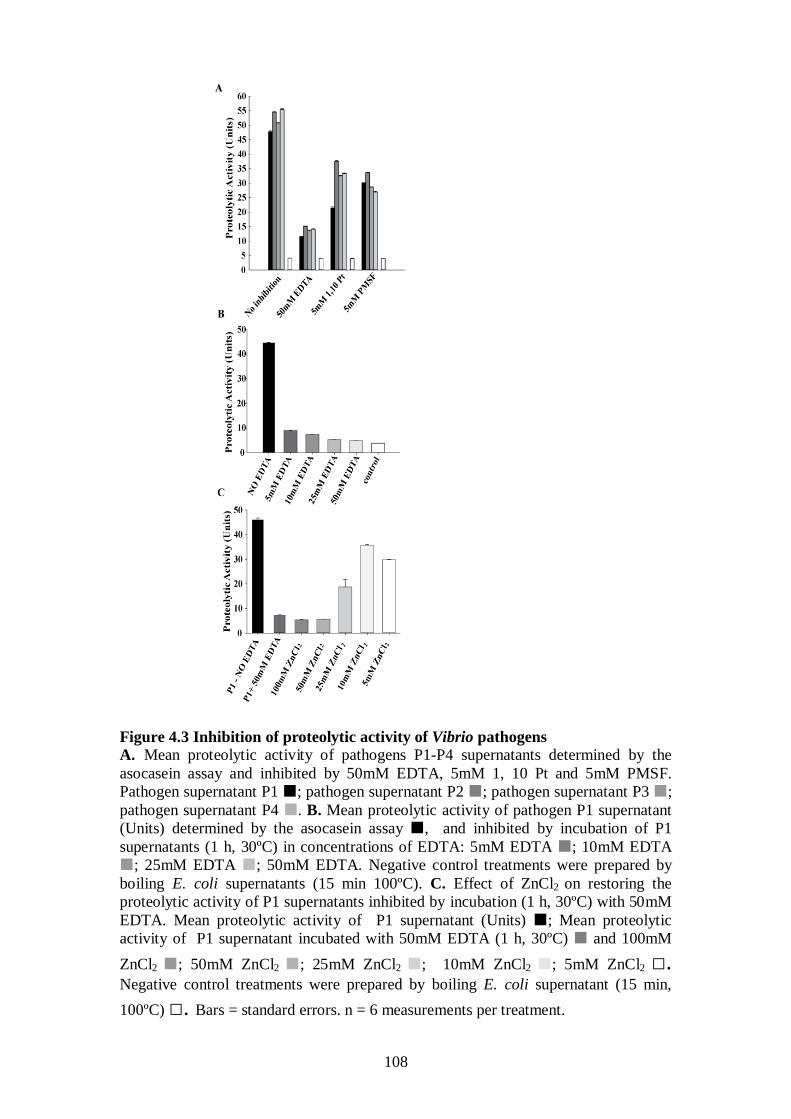
108
Figure 4.3 Inhibition of proteolytic activity of Vibrio pathogens A. Mean proteolytic activity of pathogens P1-P4 supernatants determined by the asocasein assay and inhibited by 50mM EDTA, 5mM 1, 10 Pt and 5mM PMSF. Pathogen supernatant P1 ; pathogen supernatant P2 ; pathogen supernatant P3 ; pathogen supernatant P4 . B. Mean proteolytic activity of pathogen P1 supernatant (Units) determined by the asocasein assay , and inhibited by incubation of P1 supernatants (1 h, 30ºC) in concentrations of EDTA: 5mM EDTA ; 10mM EDTA ; 25mM EDTA ; 50mM EDTA. Negative control treatments were prepared by boiling E. coli supernatants (15 min 100ºC). C. Effect of ZnCl2 on restoring the proteolytic activity of P1 supernatants inhibited by incubation (1 h, 30ºC) with 50mM EDTA. Mean proteolytic activity of P1 supernatant (Units) ; Mean proteolytic activity of P1 supernatant incubated with 50mM EDTA (1 h, 30ºC) and 100mM ZnCl2 ; 50mM ZnCl2 ; 25mM ZnCl2 ; 10mM ZnCl2 ; 5mM ZnCl2 □. Negative control treatments were prepared by boiling E. coli supernatant (15 min, 100ºC) □. Bars = standard errors. n = 6 measurements per treatment.
109
4.3.4 Symbiodinium PS II inactivation by pathogen supernatants is inhibited by
EDTA
Limited PS II inactivation was observed in all Symbiodinium cultures after 45
min when bacterial supernatant P1 was incubated with 50mM EDTA (Fig. 4.1C). This
was in contrast to strong PS II inactivation when all cultures were exposed to non-
chelated supernatants (p<0.01) [mean I (Z1, P1 EDTA, F/ Fm’) = 0.119 ± 0.017,
mean I (Z2, P1 EDTA, F/ Fm’) = 0, mean I (Z3, P1 EDTA, F/ Fm’) = 0.267 ±
0.015 and mean I (Z4, P1 EDTA, F/ Fm’) = 0]. The EDTA inhibition of proteolytic
activity was not significantly different among the four pathogen supernatants tested
(P1-P4; p=0.566), supporting the hypothesis that they share a common virulence
mechanism. Pooling all I (F/ Fm’) data for Symbiodinium cultures (Z1-Z4) exposed
to four pathogen supernatants (P1-P4) clearly demonstrated that PS II inactivation (I)
is caused by bacterial supernatants but was absent in controls in 16 experiments (Fig
4.1C; p<0.001). Addition of 50mM EDTA to pathogen supernatants resulted in
significantly lower PS II inactivation (p<0.01). PS II inactivation was not eliminated
completely, as shown by levels of I approaching non-EDTA treated supernatants in
the first 5 min following exposure (Fig 4.1C). However, I in EDTA treatments
diminished as time progressed, suggesting that the initial I values were due to EDTA
not chelating all zinc cations available in supernatants and therefore preventing
complete inhibition of the supernatant activity.
4.3.5 PS II inactivation is significantly greater when PS II centers are active
Pathogen supernatant-exposure experiments under illumination, equal to the
light intensity in the culturing incubator (90 µmol photons m-2 s-1
As Symbiodinium cells may function differently when free-living compared to
when in hospite, a second bioassay system was developed, comprised of coral
juveniles (Acropora millepora) harbouring Symbiodinium endosymbionts from clades
), resulted in
significantly higher I of all Symbiodinium cultures (Fig. 4.1C; p<0.001) in comparison
to I calculated from identical control and supernatant exposure experiments that were
conducted by measuring quantum yields (Fv /Fm) in the dark (Fig. 4.1D).
4.3.6 Pathogen supernatants cause Symbiodinium PS II inactivation in hospite
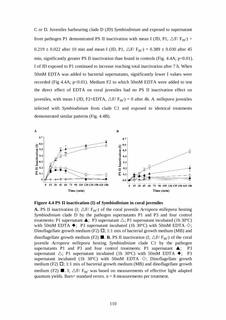
110
C or D. Juveniles harbouring clade D (JD) Symbiodinium and exposed to supernatant
from pathogen P1 demonstrated PS II inactivation with mean I (JD, P1, F/ Fm’) =
0.219 ± 0.022 after 10 min and mean I (JD, P1, F/ Fm’) = 0.389 ± 0.030 after 45
min, significantly greater PS II inactivation than found in controls (Fig. 4.4A; p<0.01).
I of JD exposed to P1 continued to increase reaching total inactivation after 7 h. When
50mM EDTA was added to bacterial supernatants, significantly lower I values were
recorded (Fig 4.4A; p<0.01). Medium F2 to which 50mM EDTA were added to test
the direct effect of EDTA on coral juveniles had no PS II inactivation effect on
juveniles, with mean I (JD, F2+EDTA, F/ Fm’) = 0 after 4h. A. millepora juveniles
infected with Symbiodinium from clade C1 and exposed to identical treatments
demonstrated similar patterns (Fig. 4.4B).
Figure 4.4 PS II inactivation (I) of Symbiodinium in coral juveniles A. PS II inactivation (I; F/ Fm’) of the coral juvenile Acropora millepora hosting Symbiodinium clade D by the pathogen supernatants P1 and P3 and four control treatments: P1 supernatant ; P3 supernatant ; P1 supernatant incubated (1h 30ºC) with 50mM EDTA ; P3 supernatant incubated (1h 30ºC) with 50mM EDTA ; Dinoflagellate growth medium (F2) ; 1:1 mix of bacterial growth medium (MB) and dinoflagellate growth medium (F2) . B. PS II inactivation (I; F/ Fm’) of the coral juvenile Acropora millepora hosting Symbiodinium clade C1 by the pathogen supernatants P1 and P3 and four control treatments: P1 supernatant ; P3 supernatant ; P1 supernatant incubated (1h 30ºC) with 50mM EDTA ; P3 supernatant incubated (1h 30ºC) with 50mM EDTA ; Dinoflagellate growth medium (F2) ; 1:1 mix of bacterial growth medium (MB) and dinoflagellate growth medium (F2) . I; F/ Fm’ was based on measurements of effective light adapted quantum yields. Bars= standard errors. n = 8 measurements per treatment.
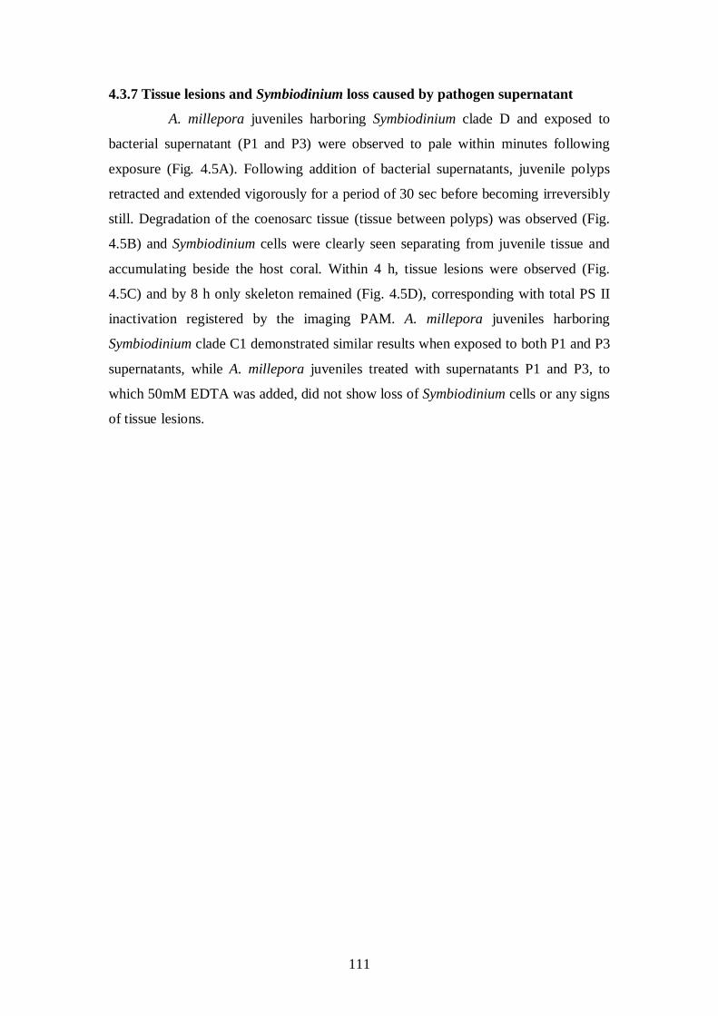
111
4.3.7 Tissue lesions and Symbiodinium loss caused by pathogen supernatant
A. millepora juveniles harboring Symbiodinium clade D and exposed to
bacterial supernatant (P1 and P3) were observed to pale within minutes following
exposure (Fig. 4.5A). Following addition of bacterial supernatants, juvenile polyps
retracted and extended vigorously for a period of 30 sec before becoming irreversibly
still. Degradation of the coenosarc tissue (tissue between polyps) was observed (Fig.
4.5B) and Symbiodinium cells were clearly seen separating from juvenile tissue and
accumulating beside the host coral. Within 4 h, tissue lesions were observed (Fig.
4.5C) and by 8 h only skeleton remained (Fig. 4.5D), corresponding with total PS II
inactivation registered by the imaging PAM. A. millepora juveniles harboring
Symbiodinium clade C1 demonstrated similar results when exposed to both P1 and P3
supernatants, while A. millepora juveniles treated with supernatants P1 and P3, to
which 50mM EDTA was added, did not show loss of Symbiodinium cells or any signs
of tissue lesions.
112
Figure 4.5 Effect of P1 supernatant on the juvenile coral host, Acropora millepora A. millepora juvenile infected with Symbiodinium clade D exposed under a dissecting microscope to P1 supernatant. A. Before exposure. B. 2h following exposure. C. 4 h following exposure. D. 8 h following exposure. Bar = 2mm (x1.6 enlargement).
113
4.3.8 A biological dose response between P1 Supernatant and Z1 PS II
inactivation
Significant PS II inactivation of Z1 was measured by exposure to P1
supernatant concentrations as low as 1% of the original supernatant stock (Fig. 4.6A;
p<0.001), with mean I (Z1, P1, 1%, F/ Fm’) = 0.226 ± 0.028 following 10 min
exposure. Total PS II inactivation of Z1 was measured in all P1 concentrations equal
and above 25% following a 10 min exposure. In contrast, recovery of photosynthetic
activity was detected in Z1 exposed to P1 concentrations of 5% and lower. Full
recovery of photosynthetic activity was measured in Z1 cells exposed to 1% and 5%
concentrations of P1 following 5 h and 24 h, respectively (p>0.1). In sharp contrast to
the susceptible Symbiodinium culture Z1, Symbiodinium culture Z4 was only affected
by higher P1 concentrations, with total PS II inactivation measured for P1
concentrations of 50% and 100% following 2.5 h and 45 min, respectively (Fig 4.6B).
Proteolytic activity of bacterial supernatants, measured by the asocasein assay
(Windle and Kelleher 1997, Denkin and Nelson 1999), was found in this study to
correlate to culture cell density, with maximum activity measured when cultures
reached their end logarithmic growth phase (18 h) and when cell density reached
1x10⁹ cells ml-1 (Fig. 4.7A-B).
4.3.9 Enzymatic kinetics supports PS II inactivation by P1 supernatant
To explain the high efficiency of P1 in causing PS II inactivation against in
its susceptible Z1 target, we plotted Z1 I (Z1, F/ Fm’) as a factor of P1 dose and
obtained a parabolic curve (Fig. 4.6C) often common in catalytic reactions. Taking
the reciprocals of P1 dose and Z1 I resulted in a linear Lineweaver-Burk (1934) -
‘like’ plot (Fig. 4.6D; R2=0.9991), where apparent km’ represents the effective P1
supernatant concentration causing a 50% PS II inactivation of Z1 following 10 min of
exposure.
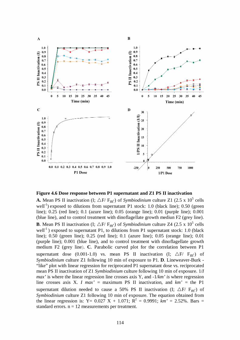
114
Figure 4.6 Dose response between P1 supernatant and Z1 PS II inactivation A. Mean PS II inactivation (I; F/ Fm’) of Symbiodinium culture Z1 (2.5 x 10⁵ cells well-1) exposed to dilutions from supernatant P1 stock: 1.0 (black line); 0.50 (green line); 0.25 (red line); 0.1 (azure line); 0.05 (orange line); 0.01 (purple line); 0.001 (blue line), and to control treatment with dinoflagellate growth mediun F2 (grey line). B. Mean PS II inactivation (I; F/ Fm’) of Symbiodinium culture Z4 (2.5 x 10⁵ cells well-1 ) exposed to supernatant P1, to dilutions from P1 supernatant stock: 1.0 (black line); 0.50 (green line); 0.25 (red line); 0.1 (azure line); 0.05 (orange line); 0.01 (purple line); 0.001 (blue line), and to control treatment with dinoflagellate growth medium F2 (grey line). C. Parabolic curved plot for the correlation between P1 supernatant dose (0.001-1.0) vs. mean PS II inactivation (I; F/ Fm’) of Symbiodinium culture Z1 following 10 min of exposure to P1. D. Lineweaver-Burk -“like” plot with linear regression for reciprocated P1 supernatant dose vs. reciprocated mean PS II inactivation of Z1 Symbiodinium culture following 10 min of exposure. 1/I max’ is where the linear regression line crosses axis Y, and -1/km’ is where regression line crosses axis X. I max’ = maximum PS II inactivation, and km’ = the P1 supernatant dilution needed to cause a 50% PS II inactivation (I; F/ Fm’) of Symbiodinium culture Z1 following 10 min of exposure. The equation obtained from the linear regression is: Y= 0.027 X + 1.071; R2 = 0.9991; km’ = 2.52%. Bars = standard errors. n = 12 measurements per treatment.
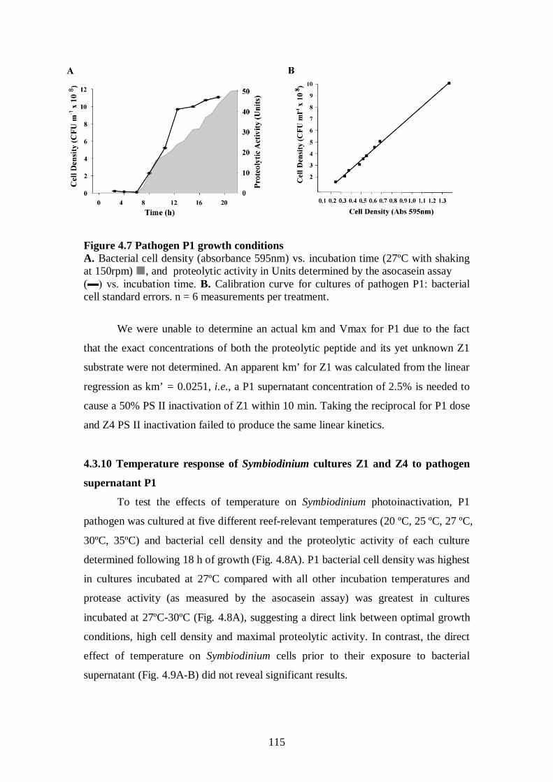
115
Figure 4.7 Pathogen P1 growth conditions A. Bacterial cell density (absorbance 595nm) vs. incubation time (27ºC with shaking at 150rpm) , and proteolytic activity in Units determined by the asocasein assay (▬) vs. incubation time. B. Calibration curve for cultures of pathogen P1: bacterial cell standard errors. n = 6 measurements per treatment.
We were unable to determine an actual km and Vmax for P1 due to the fact
that the exact concentrations of both the proteolytic peptide and its yet unknown Z1
substrate were not determined. An apparent km’ for Z1 was calculated from the linear
regression as km’ = 0.0251, i.e., a P1 supernatant concentration of 2.5% is needed to
cause a 50% PS II inactivation of Z1 within 10 min. Taking the reciprocal for P1 dose
and Z4 PS II inactivation failed to produce the same linear kinetics.
4.3.10 Temperature response of Symbiodinium cultures Z1 and Z4 to pathogen
supernatant P1
To test the effects of temperature on Symbiodinium photoinactivation, P1
pathogen was cultured at five different reef-relevant temperatures (20 ºC, 25 ºC, 27 ºC,
30ºC, 35ºC) and bacterial cell density and the proteolytic activity of each culture
determined following 18 h of growth (Fig. 4.8A). P1 bacterial cell density was highest
in cultures incubated at 27ºC compared with all other incubation temperatures and
protease activity (as measured by the asocasein assay) was greatest in cultures
incubated at 27ºC-30ºC (Fig. 4.8A), suggesting a direct link between optimal growth
conditions, high cell density and maximal proteolytic activity. In contrast, the direct
effect of temperature on Symbiodinium cells prior to their exposure to bacterial
supernatant (Fig. 4.9A-B) did not reveal significant results.
116
Symbiodinium cultures Z1 and Z4 were concurrently acclimatized to the same
temperatures for 5 days and exposed to four dilutions of P1 supernatant (1%, 10%,
50% and 100%) with incubations at all five corresponding temperatures. The Z1
Symbiodinium culture exposed to non-diluted supernatant P1 for 10 min under
illumination demonstrated the highest and most rapid photoinactivation at all five
temperature treatments (Fig. 8B; p<0.0001) compared with culture Z4 exposed to P1
for 10 min. For Z1 incubations, the strongest photoinactivation was recorded for 27ºC
acclimatized cells exposed at 27 ºC to P1 supernatant derived from a culture incubated
at 27 ºC (mean I (Z1, P1, 27 ºC, F/ Fm’) = 0.889 ± 0.005 after 10 min).
Photoinactivation of Z1 was significantly weaker after 10 min in the 30 ºC and 35 ºC
treatments, with mean I (Z1, P1, 30 ºC, F/ Fm’) = 0.847 ± 0.007; p=0.0019 and
mean I (Z1, P1, 35 ºC, F/ Fm’) = 0.706 ± 0.006; p<0.0001, respectively. It was also
significantly weaker for cultures acclimatized to lower temperatures. The PS-II
activity of Symbiodinium culture Z4 was not affected when exposed at 35 ºC to P1
cultures grown at 35 ºC (Fig. 4.8B).
In separate control treatments, Symbiodinium cultures Z1 and Z4 remained
photosynthetically active following a 25 day exposure to 35ºC. Our findings suggest
that maximum heat stress may not be an important facilitating factor for WS
infections, which are more likely to initiate within the optimal 25ºC-30ºC temperature
range. Apparent Km’ values calculated for the various temperature treatments (Fig.
4.10) indicate that P1 supernatant concentrations of 2.44-3.32% (for temperatures
ranging between 25ºC–30ºC) are needed to cause a 50% photoinactivation of Z1 after
10 min. Much higher concentrations of P1 (7.58% and over100%) were calculated to
cause a 50% photoinactivation at 20ºC and 35ºC, respectively, suggesting that above
30ºC, poor performance by Vibrio pathogens would limit the probability of successful
infections by an estimated factor of x 1/50, when all other conditions remain constant.
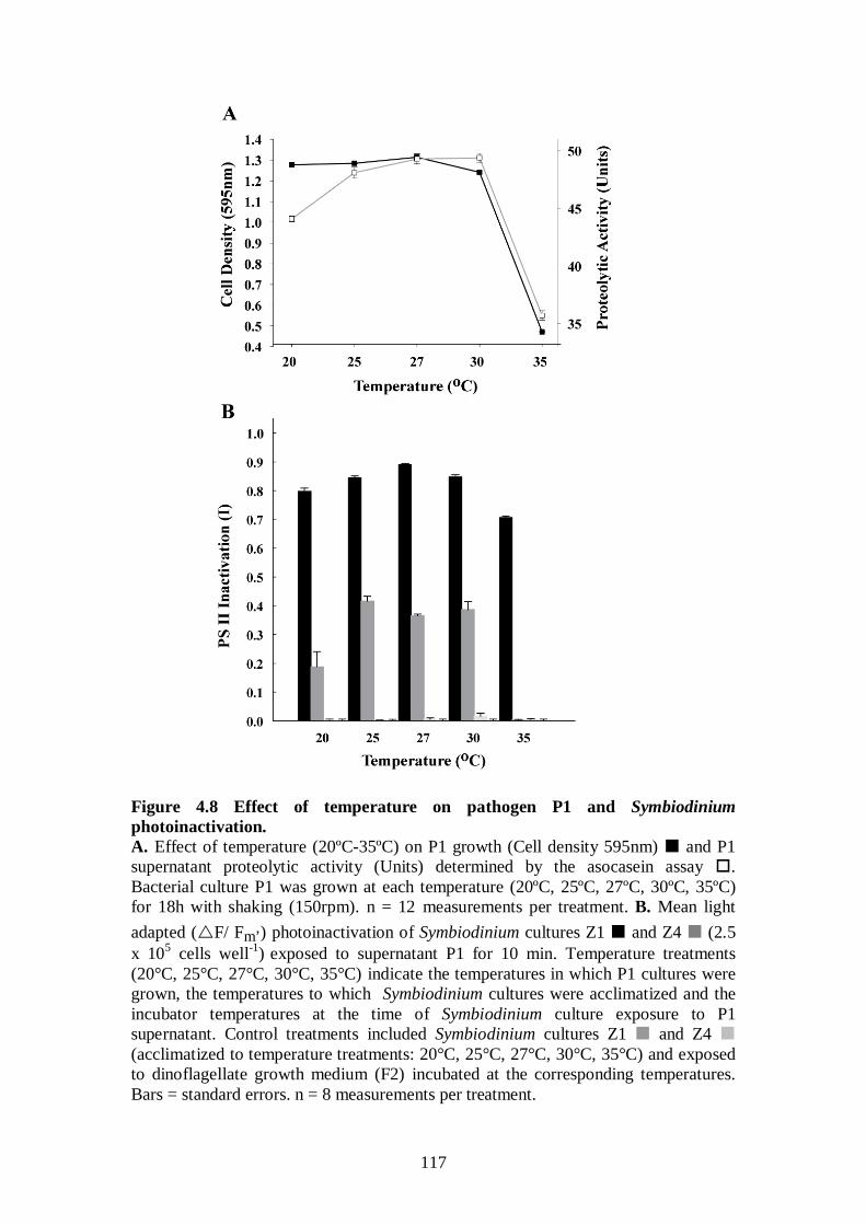
117
Figure 4.8 Effect of temperature on pathogen P1 and Symbiodinium photoinactivation. A. Effect of temperature (20ºC-35ºC) on P1 growth (Cell density 595nm) and P1 supernatant proteolytic activity (Units) determined by the asocasein assay . Bacterial culture P1 was grown at each temperature (20ºC, 25ºC, 27ºC, 30ºC, 35ºC) for 18h with shaking (150rpm). n = 12 measurements per treatment. B. Mean light adapted (F/ Fm’) photoinactivation of Symbiodinium cultures Z1 and Z4 (2.5 x 10⁵ cells well-1) exposed to supernatant P1 for 10 min. Temperature treatments (20°C, 25°C, 27°C, 30°C, 35°C) indicate the temperatures in which P1 cultures were grown, the temperatures to which Symbiodinium cultures were acclimatized and the incubator temperatures at the time of Symbiodinium culture exposure to P1 supernatant. Control treatments included Symbiodinium cultures Z1 and Z4 (acclimatized to temperature treatments: 20°C, 25°C, 27°C, 30°C, 35°C) and exposed to dinoflagellate growth medium (F2) incubated at the corresponding temperatures. Bars = standard errors. n = 8 measurements per treatment.
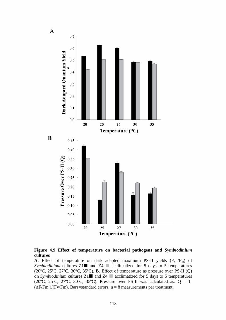
118
Figure 4.9 Effect of temperature on bacterial pathogens and Symbiodinium cultures A. Effect of temperature on dark adapted maximum PS-II yields (Fv /Fm) of Symbiodinium cultures Z1 and Z4 acclimatized for 5 days to 5 temperatures (20ºC, 25ºC, 27ºC, 30ºC, 35ºC). B. Effect of temperature as pressure over PS-II (Q) on Symbiodinium cultures Z1 and Z4 acclimatized for 5 days to 5 temperatures (20ºC, 25ºC, 27ºC, 30ºC, 35ºC). Pressure over PS-II was calculated as: Q = 1- (∆F/Fm’)/(Fv/Fm). Bars=standard errors. n = 8 measurements per treatment.
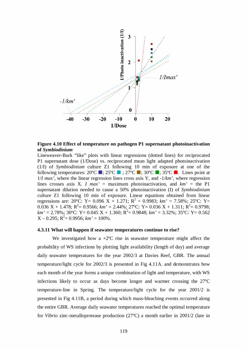
119
Figure 4.10 Effect of temperature on pathogen P1 supernatant photoinactivation of Symbiodinium Lineweaver-Burk “like” plots with linear regressions (dotted lines) for reciprocated P1 supernatant dose (1/Dose) vs. reciprocated mean light adapted photoinactivation (1/I) of Symbiodinium culture Z1 following 10 min of exposure at one of the following temperatures: 20ºC ; 25ºC ; 27ºC ; 30ºC ; 35ºC . Lines point at 1/I max’, where the linear regression lines cross axis Y, and -1/km’, where regression lines crosses axis X. I max’ = maximum photoinactivation, and km’ = the P1 supernatant dilution needed to cause a 50% photoinactivation (I) of Symbiodinium culture Z1 following 10 min of exposure. Linear equations obtained from linear regressions are: 20ºC: Y= 0.096 X + 1.271; R2 = 0.9983; km’ = 7.58%; 25ºC: Y= 0.036 X + 1.478; R2= 0.9566; km’ = 2.44%; 27ºC: Y= 0.036 X + 1.311; R2= 0.9798; km’ = 2.78%; 30ºC: Y= 0.045 X + 1.360; R2= 0.9848; km’ = 3.32%; 35ºC: Y= 0.562 X – 0.295; R2
We investigated how a +2ºC rise in seawater temperature might affect the
probability of WS infections by plotting light availability (length of day) and average
daily seawater temperatures for the year 2002/3 at Davies Reef, GBR. The annual
temperature/light cycle for 2002/3 is presented in Fig 4.11A. and demonstrates how
each month of the year forms a unique combination of light and temperature, with WS
infections likely to occur as days become longer and warmer crossing the 27ºC
temperature-line in Spring. The temperature/light cycle for the year 2001/2 is
presented in Fig 4.11B, a period during which mass-bleaching events occurred along
the entire GBR. Average daily seawater temperatures reached the optimal temperature
for Vibrio zinc-metalloprotease production (27ºC) a month earlier in 2001/2 (late in
= 0.9956; km’ > 100%. 4.3.11 What will happen if seawater temperatures continue to rise?
120
October), compared to 2002/3 (late in November), and continued to rise beyond 30ºC
in February (when mass-bleaching occurred).
However, if seawater temperatures continue to rise, +2ºC above the currently
prevailing temperatures (Fig. 4.11C), the optimal temperature range for Vibrio
pathogens is expected to shift towards the winter, with seawater temperatures
remaining above 25ºC throughout the entire year. By calculating the seasonal length
of optimal conditions for coral Vibrio pathogens (number of days per year with
average seawater temperatures between 27ºC-30ºC; Fig. 4.11D), we found that
currently longer and shorter seasons of optimal conditions for WS pathogens oscillate
along the GBR, possibly contributing to increased WS transmission and abundance
during longer seasons of optimal conditions, and coral host recovery during shorter
seasons. Not surprisingly, the two mass bleaching events that have occurred along the
GBR in recent decades (1997/8, 2001/2) correlate with peaks of longer warm seasons
(Fig. 4.11D). For coral reefs in the southern GBR sectors (Halfway Island S23°2’ and
Turner Cay I S21°7’, E152°56’), changes between warm and cold years have been
dramatic. For example, less than 10 days with optimal mean temperatures for Vibrio
pathogens were measured in 1999/2000 increasing to over 100 days in the mass
bleaching year of 2001/2002 (Fig. 4.11D). Since 2003/2004, the length of optimal
seasons for Vibrio pathogens has increased on those reefs with no colder years in
between.
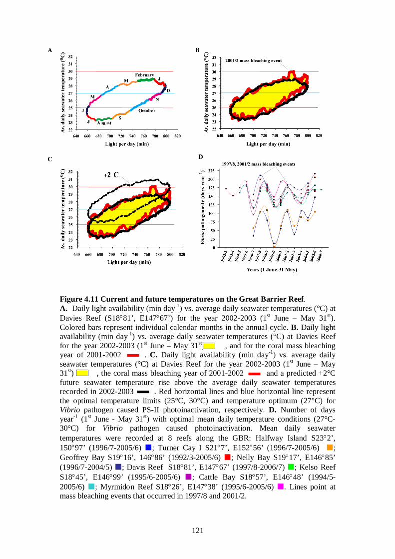
121
Figure 4.11 Current and future temperatures on the Great Barrier Reef. A. Daily light availability (min day-1) vs. average daily seawater temperatures (°C) at Davies Reef (S18°81’, E147°67’) for the year 2002-2003 (1st June – May 31st). Colored bars represent individual calendar months in the annual cycle. B. Daily light availability (min day-1) vs. average daily seawater temperatures (°C) at Davies Reef for the year 2002-2003 (1st June – May 31st) , and for the coral mass bleaching year of 2001-2002 . C. Daily light availability (min day-1) vs. average daily seawater temperatures (°C) at Davies Reef for the year 2002-2003 (1st June – May 31st) , the coral mass bleaching year of 2001-2002 and a predicted +2°C future seawater temperature rise above the average daily seawater temperatures recorded in 2002-2003 . Red horizontal lines and blue horizontal line represent the optimal temperature limits (25°C, 30°C) and temperature optimum (27°C) for Vibrio pathogen caused PS-II photoinactivation, respectively. D. Number of days year-1 (1st June - May 31st
) with optimal mean daily temperature conditions (27°C-30°C) for Vibrio pathogen caused photoinactivation. Mean daily seawater temperatures were recorded at 8 reefs along the GBR: Halfway Island S23°2’, 150°97’ (1996/7-2005/6) ; Turner Cay I S21°7’, E152°56’ (1996/7-2005/6) ; Geoffrey Bay S19°16’, 146°86’ (1992/3-2005/6) ; Nelly Bay S19°17’, E146°85’ (1996/7-2004/5) ; Davis Reef S18°81’, E147°67’ (1997/8-2006/7) ; Kelso Reef S18°45’, E146°99’ (1995/6-2005/6) ; Cattle Bay S18°57’, E146°48’ (1994/5-2005/6) ; Myrmidon Reef S18°26’, E147°38’ (1995/6-2005/6) . Lines point at mass bleaching events that occurred in 1997/8 and 2001/2.
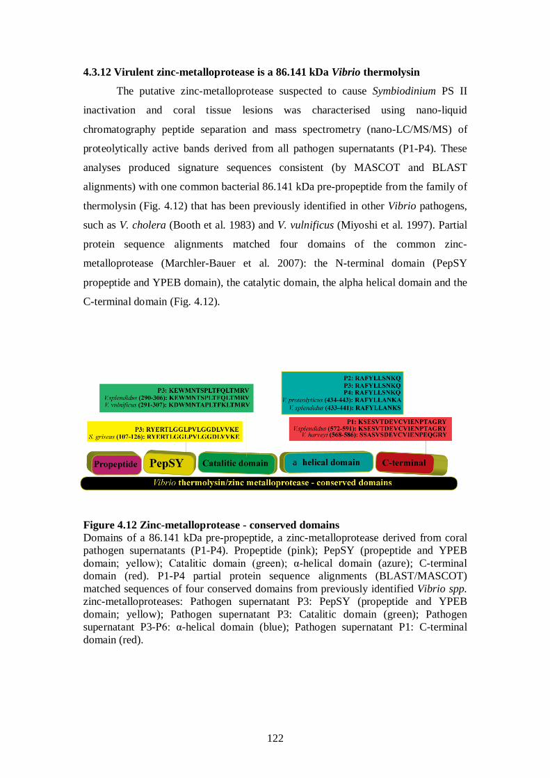
122
4.3.12 Virulent zinc-metalloprotease is a 86.141 kDa Vibrio thermolysin
The putative zinc-metalloprotease suspected to cause Symbiodinium PS II
inactivation and coral tissue lesions was characterised using nano-liquid
chromatography peptide separation and mass spectrometry (nano-LC/MS/MS) of
proteolytically active bands derived from all pathogen supernatants (P1-P4). These
analyses produced signature sequences consistent (by MASCOT and BLAST
alignments) with one common bacterial 86.141 kDa pre-propeptide from the family of
thermolysin (Fig. 4.12) that has been previously identified in other Vibrio pathogens,
such as V. cholera (Booth et al. 1983) and V. vulnificus (Miyoshi et al. 1997). Partial
protein sequence alignments matched four domains of the common zinc-
metalloprotease (Marchler-Bauer et al. 2007): the N-terminal domain (PepSY
propeptide and YPEB domain), the catalytic domain, the alpha helical domain and the
C-terminal domain (Fig. 4.12).
Figure 4.12 Zinc-metalloprotease - conserved domains Domains of a 86.141 kDa pre-propeptide, a zinc-metalloprotease derived from coral pathogen supernatants (P1-P4). Propeptide (pink); PepSY (propeptide and YPEB domain; yellow); Catalitic domain (green); α-helical domain (azure); C-terminal domain (red). P1-P4 partial protein sequence alignments (BLAST/MASCOT) matched sequences of four conserved domains from previously identified Vibrio spp. zinc-metalloproteases: Pathogen supernatant P3: PepSY (propeptide and YPEB domain; yellow); Pathogen supernatant P3: Catalitic domain (green); Pathogen supernatant P3-P6: α-helical domain (blue); Pathogen supernatant P1: C-terminal domain (red).
123
4.4 Discussion
4.4.1 Bacterial caused PS II inactivation of Symbiodinium photosynthesis
Bacterial causative agents for coral diseases have been identified in previous
studies (Kushmaro et al. 1996, Geiser et al. 1988, Ben-Haim and Rosenberg 2002,
Patterson et al. 2002, Denner et al. 2003, Barash et al. 2005, Bally and Garrabou 2007,
Sussman et al. 2008), including specific virulence mechanisms that enable coral
colonization and disease progression (Banin et al. 2001a, 2001b, 2003), however, this
study is the first to investigate the clinical effect of a virulence factor derived from
multiple causative agents and applied to multiple targets. Our principal findings
demonstrate that PS II inactivation of susceptible Symbiodinium cells (Z1) by
pathogen supernatants is significantly higher than PS II inactivation of non-
susceptible targets (Z2-Z4). Susceptibility of Symbiodinium cells to bacterial PS II
inactivation was supported by demonstrating a biological dose response (Hill 1965).
Partial peptide sequencing of proteolytically active fractions derived from WS
pathogen supernatants identified a common 86.141 kDa zinc-metalloprotease from the
family thermolysin, which is suspected of causing Symbiodinium PS II inactivation
and coral tissue lesions. Nevertheless, our preliminary exposure trials could not
determine the exact process by which bacterial zinc-metalloproteases affect
Symbiodinium photosynthesis. A similar thermolysin derived from Bacillus
thermoproteolyticus rokko has been reported to selectively cleave chloroplast outer
envelope membrane (OEM) proteins causing PS II photoinhibition (Cline et al. 1984).
Tests performed with B. thermoproteolyticus rokko thermolysin revealed that it can
not penetrate through the chloroplast OEM (Cline et al. 1984), but can affect about 20
OEM polypeptides (Joyard et al. 1983), including components of a protein import
apparatus (Tranel et al. 1995) that may indirectly influence PS II performance.
This study identified specific Lineweaver-Burk - ‘like’ kinetics between P1
supernatant and its susceptible Z1 target, suggesting that PS II inactivation of
Symbiodinium Z1 is potentially the result of a specific bond between an enzyme and
its target substrate. Failure to produce similar kinetics between P1 supernatant and a
non-susceptible (Z4) target, supports the specificity of this bond and may possibly
explain why Acropora millepore coral hosts harboring culture Z4 Symbiodinium at
Nelly Bay (GBR), or Montipora aequituberculata colonies harboring Z2
Symbiodinium at Davies Reef (GBR) have not been observed with WS disease signs,
whereas M. aequituberculata colonies from Nelly Bay, which harbor susceptible Z1
124
Symbiodinium, often display WS disease signs. Nevertheless, further studies are
needed in order to determine the specific substrate of the zinc-metalloprotease
identified in WS coral pathogens in this study. Symbiodinium PS II inactivation by
low pathogen supernatant concentration (≤5%) was found to be reversible in this
study. Recovery of full photosynthetic capacity of Symbiodinium cells within 24 h,
after short-term PS II inactivation following low concentration exposures, suggests
that zinc-metalloprotease damage to PS II might be repairable, as demonstrated for PS
II repair by heat shock proteins (Schroda et al. 1999). Alternatively, PS II inactivation
may be caused by enzymatic cleavage of Symbiodinium cell membranes, resulting in
an irreparable cellular collapse and Symbiodinium mortality, as observed by Cervino
et al. (2004b) for yellow blotch/band infections of Montastraea spp. corals in the
Caribbean. Our study, however, could not find adequate support for this hypothesis,
since total PS II inactivation occurred less than 20 sec following exposure to bacterial
supernatants. Further studies are needed to examine the pathology of Symbiodinium
exposed to coral pathogen zinc-metalloproteases. Damage to Symbiodinium PS II has
been shown to be caused by a variety of factors including light and heat stress (Barber
1992, Lesser 1996, asada 1996, Warner 1999, Hill et al, 2004, Takahashi 2008)
associated with mass coral bleaching (Hoegh-Guldberg and Smith 1989, Gates et al.
1992, Brown 1997, Hoegh-Guldberg 1999, Fitt et al. 2001), and by numerous
bacterial toxins (Banin et al. 2001b, Ben-Haim et al. 2003a) suggesting that PS II
damage may result from independent disease aetiologies. In order to test the
hypothesis that these factors act in synergism, specific diagnostics must be designed,
such as monoclonal antibodies that will register zinc-metalloprotease signals in the
field.
In this study, Symbiodinium isolates affiliated with clade A were found to be
both susceptible and non-susceptible to bacterial PS II inactivation, in contrast to
findings by Stat et al. (2008), speculating that clade A Symbiodinium associations
with diseased corals are closer to parasitism than to mutualism than similar
associations of corals with clade C Symbiodinium. Better knowledge of Symbiodinium
physiology and disease aetiology will assist in identifying why specific types are more
susceptible to bacterial PS II inactivation. Further studies including the cloning and
sequencing of the of zinc-metalloprotease genes from WS pathogens will enable
validation of our current findings, potentially by utilizing mutant coral pathogen
strains that lack the zinc-metalloprotease gene, or by utilizing differential expression
125
and coral pathogen zinc-metalloproteases expressed by a vector system in additional
exposure trials (Foulongne et al. 2004) aimed at fulfilling Koch’s molecular postulates
(Falkow 1988).
4.4.2 Bacterial caused tissue lesions and Symbiodinium loss
In this study, visual observations and iPAM measurements of exposed coral
juveniles revealed three distinct phases of disease: 1. Symbiodinium PS II inactivation;
2. paling of coral tissue through loss of Symbiodinium cells; and 3. spread of coral
tissue lesions culminating in mortality. These signs, which were expressed by coral
juveniles in response to bacterial supernatants, were identical to WS disease signs
observed on adult corals in the field (Willis et al. 2004, Anthony et al. 2008) and
during pathogen inoculation experiments (chapter 3; Sussman et al. 2008), and further
support our findings of the previous chapter, which identified Vibrio pathogens as the
primary causative agents of WS's (Sussman et al. 2008). This is the first study to
successfully replicate WS disease signs by using cell free supernatants.
Vibrio zinc-metalloproteases are known to perform dual functions similar to
the duality of function demonstrated by coral zinc-metalloproteases in this study, i.e.,
in causing both Symbiodinium PS II inactivation and coral tissue lesions. For example,
in the human pathogen Vibrio cholera, a virulent zinc-metalloprotease has been
named hemagglutinin/protease because of its dual capacity to cause both
hemagglutination and proteolytic cleavage (Finkelstein and Hanne 1982, Miyoshi et
al. 1997). The V. vulnificus elastase/protease has also been shown to possess dual
functions, enhancing vascular permeability and causing hemorrhagic damage
(Kothary and Kreger 1985, Oliver et al. 1986). Numerous studies demonstrate that
Vibrio zinc-metalloproteases are synthesized as inactive precursors that mature
outside the bacterial cell following several processing stages which may alter their
function (Miyoshi and Shinoda 2000), such as the cleavage of a C-terminal 10 kDa
peptide from the V. vulnificus zinc-metalloprotease (VVP; Miyoshi et al. 2001) by a
specific processing protease (Varina et al. 2008), which mediates effective binding of
V. vulnificus VVP to its substrate. Future studies will determine whether coral
pathogen zinc-metalloproteases undergo a similar maturing process.
126
4.4.3 White syndrome is a multifactorial coral disease
Findings from this current study support the classification of coral WS as a
multifactorial disease with multiple component causes (Thrusfeld 2005). We found
the expression of zinc-metalloprotease by WS coral pathogens to be cell density
dependant, with greatest proteolytic activities measured at the end logarithmic phase,
when bacterial cell density in cultures reached 1x10⁹ cells ml-1. Based on dose
response experiments, we calculated that the steady state concentration of coral
pathogen derived zinc-metalloprotease required to cause a rapid and irreversible 50%
PS II inactivation of susceptible Symbiodinium cells following 10 min of exposure is
equal to the dose produced by a bacterial concentration of ~5x10⁷ cells ml-1
The findings by Bruno et al. (2007), that both temperature and host density
influence WS disease prevalence, support the definition of WS as a multifactorial
disease. These two factors, plus the requirements for primary causative agents at
elevated concentrations and for susceptible target Symbiodinium types, may all
contribute to facilitating WS epizootics. Elevated seawater temperatures have been
shown to be a major contributing factor to Vibrio cholera pandemics (Colwell 1996)
. This
calculation, although preliminary, implies that WS disease signs are unlikely to occur
in the field unless susceptible populations of Symbiodinium cells are exposed to a high
concentration of pathogenic bacteria. Thus, the progressing band of exposed coral
skeleton typical of WS signs in the field (Willis et al. 2004, Anthony et al. 2008), can
be explained by the presence of high densities of pathogens at the interface between
exposed skeleton and healthy looking tissue in progressing lesions. In the previous
chapter, we found that Vibrio cell densities associated with a lesion interface were
more than a 100 times higher than the cell densities found on healthy-looking tissue
(Chapter 3 section 3.3.1; Sussman et al. 2008). In a field study, Roff et al. (2008)
provided evidence that coral tissues remain photosynthetically active when they are
less than 10 cm away from the interface of a progressing WS tissue lesion, suggesting
that pathogen zinc-metalloprotease concentrations may be diluted to a non-effective
dose away from a progressing tissue lesion interface. Reductions in zinc-
metalloprotease concentrations when conditions for optimal pathogen growth are
impared may also explain how corals can recover from WS infections.
4.4.4 The effect of seawater temperatures and global warming on Symbiodinium
susceptibility to PS-II inactivation and pathogen virulence
127
and a necessary factor for triggering the virulence of the coral pathogen Vibrio shiloi
(Israeli et al. 2001).
Based on our findings, we postulate that optimal temperatures for Vibrio WS
pathogen growth contribute directly to cell density dependant synthesis of zinc-
metalloproteases that are required for infections. Our study suggests that optimal
temperatures for Vibrio pathogens should be summed to the length of season in which
optimal temperature conditions prevail, suggesting that both pathogen growth and
pathogen transmission may be temperature and light dependant. Many Vibrio caused
coral infections reported to date initiate and develop during spring and summer
(Kushmaro et al. 1996, 1998, Rosenberg and Ben-Haim 2002, Ben-Haim et al. 2003a,
Sussman et al. 2003), when days become longer and warmer. Many other Vibrio
infections in the marine environment correlate with seawater temperatures within the
optimal range of 25ºC–30ºC established in this study, including V. cholera outbreaks
which have been shown to peak at 30ºC (Colwell 1996).
In sharp contrast, no Vibrio caused infectious diseases have been reported at
elevated temperatures above 30ºC. This study tested the performance of WS
pathogens at 20ºC, 25ºC, 27ºC, 30ºC and 35ºC and determined that the probability of
WS infections occurring at elevated temperatures (above 30ºC) is limited. This study
could not detect any temperature related stress affecting Symbiodinium cultures
exposed to elevated temperatures (35ºC), however, it did detect slower growth rates of
Vibrio pathogens and limited extra-cellular zinc-metalloprotease secretion at 35ºC. It
is currently unknown how Vibrio pathogens might evolve as seawater temperatures
increase, therefore making it very hard to predict the impact of global warming on the
spread and prevalence of WS on Indo-Pacific reefs. Nevertheless, current seawater
temperatures are probably optimal for the growth, virulence and spread of Vibrio WS
pathogens, potentially explaining the large impact of this disease at present. Only
precise monitoring of reefs using diagnostic tools that are able to detect Vibrio
pathogens and their virulence factors could enable to determine whether global
warming will remain a driving force enhancing the impact of WS on Indo-Pacific
reefs in the future.
128
4.4.5 Are Vibrio WS pathogens primary pathogens, opportunistic pathogens, or
secondary pathogens to other unknown causes?
Detection of Vibrio strains on both healthy and diseased populations of fish
(Thune et al. 1993), shrimps (Karunasagar et al. 1994) and corals (Bourne and Munn
2005) has led to the conclusion that Vibrio infections are opportunistic in nature
(Lightner 1993). The term ‘opportunistic infection’ was first defined by Utz in 1962
relating to fungal infections (Utz 1962), but has since been modified to include
additional pathogen-host interactions, particulary those that can be represented by a
‘damage-response framework’ (Casadevall and Pirofski 2003), which defines
pathogenicity and host-susceptibility as coupled variant traits. This study identified
the variant traits of Symbiodinium hosts exposed to a common zinc-metalloprotease.
However, it has also been found that the presence of a zinc-metalloprotease gene can
be detected in DNA retrieved from non-pathogenic strains, which were unable to
cause disease signs in controlled exposure trials (see chapter 3 section 3.3.4.3;
Sussman et al. 2008). The presence of a zinc-metalloprotease gene in non-pathogenic
Vibrio strains, suggests that the expression of other virulence genes is necessary for
successful infections to occur. Thus we conclude that, although all WS pathogens
identified possess a zinc-metalloprotease gene sufficient to cause rapid
photoinactivation and coral tissue lesions, not all Vibrio strains possessing this gene
can be classified as primary causative agents of WS. Genetic studies support the
variant traits of Vibrio pathogens. From over 200 V. cholera serotypes, only a few
have been shown to cause cholera pandemics, while others, possessing partial
combinations of virulent genes, were shown to cause a gradient of attenuated disease
symptoms (Singh et al. 2001, Reidl and Klose 2002). Work by Austin et al. (2005)
demonstrated that the coral pathogen V. coralliilyticus (Ben-Haim et al. 2003a, Bally
and Garrabou 2007), which was also identified in this study (Chapter 3 section 3.3.5;
Sussman et al. 2008) also affects rainbow trout (Oncorhynchus mykiss) and Artemia
nauplii by causing mortalities in animal models, suggesting it may target multiple
species that are not necessarily compromised hosts by possessing broad pathogenicity.
In contrast, the virulence of the coral bleaching agent V. shiloii (Kushmaro et al. 2001)
was not adequate to infect Oncorhynchus mykiss and Artemia nauplii (Austin et al.
2005). In addition, V. shiloi has recently been reported to have stopped infecting its
known coral host Oculina patagonica in the Mediterranean Sea (Reshef et al. 2006),
suggesting a shift in the ‘damage-response framework’ (Casadevall and Pirofski
129
2003), defined by Rosenberg et al. (2007) as the ‘hologenome theory of evolution‘,
i.e., the failure of variant pathogen-host traits to continue producing expected disease
signs. Pathogen-coral interactions may be further complicated considering the fact
that pathogens in the marine environment perform under different conditions than
those confronted by terrestrial agents (McCallum et al. 2004, Murray 2008), and in
particular, Vibrio coral agents, whose survival strategy might be aimed at specializing
in ‘adaptability’ (Higgins et al. 2007), rather than in an obligatory ‘selectivity’
towards specific hosts.
In conclusion, our findings support classifying coral WS’s as multifactorial
diseases, which are caused by primary Vibrio pathogens. Based on findings from this
study, Vibrio pathogens may be involved in numerous coral disease aetiologies as
pathogens of variant traits, and may operate as primary, opportunistic, or as secondary
agents. Their ubiquity and modes of action underline the need for further
collaborative studies to explore the complexity of roles performed by Vibrio zinc-
metalloproteases in both coral health and disease.
130
Chapter 5
General Discussion
5.1 Coral pathogens: global distributions with potentially multiple origins?
This study is the first to identify coral pathogens from the Great Barrier Reef
and from remote locations in the Indo-Pacific Ocean, including the Republic of the
Marshall Islands and the Republic of Palau. Principal findings from this study
demonstrate that coral pathogens on the GBR and across the Indo-Pacific Ocean are
closely related to bacterial pathogens identified from other reef locations, such as the
Caribbean and the Mediterranean Sea. Based on 16S rRNA gene sequencing,
cyanobacteria associated with corals displaying signs of black band disease (BBD) on
the GBR and in Palau were closely affiliated with cyanobacterial strains associated
with BBD infected corals in the Caribbean (Frias-Lopez et al. 2003) and in the Gulf of
Aqauba (Barneah et al. 2007). Similarly, bacterial pathogens causing WS disease
signs on the GBR (Magnetic Island) and in the Republic of the Marshall Islands were
found to be closely affiliated with the coral bleaching pathogen V. coralliilyticus
identified in Zanzibar and in the Gulf of Aquaba (Ben-Haim et al. 2002, 2003a). V.
coralliilyticus has also been identified as the causative agent of gorgonian mortalities
in the Mediterranean Sea (Bally and Garrabou, 2007), of fish and shrimp mortalities
in laboratory infection trials (Austin et al. 2005) and of oyster disease in Kent, UK
(Thompson et al. 2001). It is not clear, however, if such global distributions are the
result of coral pathogen transmission across geographical regions, or the result of
genetic similarity among multiple coral pathogens, each with a separate origin and a
potentially highly varied arsenal of virulence genes resulting in niche specificity.
Many studies of infectious disease support the hypothesis that diseases
emerge in one location and are then carried across regions by vectors. For example,
the devastating effects of West Nile viral infections on avian wildlife populations in
North America (Rappole and Hubálek, 2003) have been attributed to transport of the
virus to New York by a human vector in 1999 (Lanciotti et al. 1999). Avian Flu virus
infections have also been shown to spread across geographical regions by migrating
birds (Olsen et al. 2006). In the marine environment, coral disease pathogens have
been identified in ballast water brought into the Gulf of Mexico (Aguirre-Macedo et
al. 2008). Modeling of disease spread and transmission based on the 'novel pathogen
131
hypothesis of infectious diseases' (Alford 2001), which assumes that a pathogen has
recently spread into a new geographic area, has been a key factor in developing
strategies to combat the spread of infectious diseases across geographical regions, for
example in the development of strict isolation and quarantine measures (Fraser et al.
2004), as in the case of SARS (Seto et al. 2003), or quarantine and vaccination
measures, as in the case of rabies (Perrin et al. 1999). The lack of rabies on inhabited
islands, i.e., Great Britain, Hawaii and Australia (Johnson et al. 2002), provides
corroborative evidence that the current distribution of the rabies pathogen is the result
of spread across geographical regions. There are also records of numerous other
spreading infectious diseases that have accumulated throughout the last 30 years
(Cohen 2000). However, other infectious diseases may evolve from existing
autochthonous pathogens found in environmental reservoirs that become virulent
under numerous compounding conditions (Colwell and Huq 2001, Rachowicz et al.
2005). Although not fully established, Vibrio infections in general (Wittman et al.
1995) and Vibrio cholera infections in particular (Politzer 1959, Huq et al. 1990,
Kaper et al. 1995, Rivera et al. 2001) may result from autochthonous pathogens,
which were found in endemic reservoirs (Lipp et al. 2002).
Black band disease infections of corals were first identified in the Caribbean
(Antonius 1973) and subsequently reported from the Indo-Pacific Ocean within the
following decade (Antonius 1985a). The appearance of BBD in widely separated reef
regions within such a time frame is consistent with the possibility that cyanobacterial
strains are present normally within the environment. Detection of bacterial rRNA
sequences in both healthy and diseased corals on the Great Barrier Reef (GBR), which
were closely affiliated with rRNA sequences derived from corals from other
geographical regions (Jones et al. 2004, Bourne and Munn 2005, Bourne 2005),
supports the hypothesis that at least some coral pathogens may be autochthonous. In
this study, the bacterial causative agent of white syndrome (WS) infections on reefs in
the Republic of the Marshall Islands, a secluded Atoll in the Indo-Pacific where a
major WS epizootic has been occurring since 2003 (Jacobson et al. 2006), was found
to be phylogenetically related to similar Vibrio strains that were recovered from
various locations, including diseased oysters in the English Channel (Thomspson et al.
2001). The Marshall Islands coral epizootic occurred on a site that is densely
populated with large plating acroporid corals and was in proximity to the atoll's
sewage outlet and waste disposal ground, two factors that have been shown
132
previously to be associated with the prevalence of coral diseases (Peters 1984, Bruno
et al. 2003, Kaczmarsky et al. 2005, Klaus et al. 2005, 2007 Voss and Richardson
2006). In addition, the Marshall Islands epizootic was found to be more active during
the warm months of the year, suggesting a direct correlation with elevated seawater
temperatures (Jacobson et al. 2006), as has also been found on the GBR for WS
abundance (Bruno et al. 2007) and for BBD abundance and rates of progression
(Boyett et al. 2007; Sato et al. 2009).
In contrast, a recent expedition to the Northern Line Islands surveyed
microbial communities along a gradient of increasing human population, which was
inversely correlated with the number of sharks and other top predators, and found that
the greatest abundance of coral diseases occurred on reefs surrounding populated and
polluted atolls, where over-fishing and sewage disposal often occurs (Sandin et al
2008). They also demonstrated that Vibrio isolates could only be cultured from
seawater samples obtained on populated reefs, where the abundances of virus-like
particles and heterotroph-dominated bacteria in seawater samples were 10 times
higher than on unpopulated atolls (Dinsdale et al. 2008), suggesting that Vibrio
pathogens may not typically be members of healthy bacterial communities on corals.
Nevertheless, the abundance of bacteria in seawater samples and the prevalence of
disease cannot give clear indications as to the origin of coral pathogens. Currently the
use of 16S rRNA gene sequencing for phylogenic determination is effective (due to
the gene's variable structure and the availability of a large sequence-database).
However, for the identification of pathogen-virulence among identical species, 16S
rRNA resolution might not be sufficient. Thompson and colleagues (2005)
demonstrated that V. coralliilyticus strains possess high variability for the rpoA, recA
and pyrH genes, suggesting that 16S rRNA taxonomy can not indicate the origin and
composition of virulence genes; whether these are the result of endemic speciation or
a worldwide distribution. Additional tools must be developed in order to test the
hypothesis that some coral pathogens are globally distributed and can be detected in
endemic reservoirs, versus the alternative hypothesis that spread of coral pathogens
across geographical regions is a major contributing factor facilitating coral disease
outbreaks. Other tools should also be developed in order to estimate the evolutionary
rate of coral pathogens, which can be significantly higher than previously estimated.
Although based on preliminary findings, results from this study are consistent
with both BBD and WS outbreaks originating from endemic pathogens, which
133
become successful at causing coral infections under a complex series of compounding
factors. However, further studies are needed in order to verify this hypothesis.
Clarification of the origin of coral pathogens would determine future management
strategies aimed at arresting the spread of coral disease on Indo-Pacific reefs and their
relationship to other management tools, such as the construction of protected marine
parks (Coelho and Manfrino 2007). Regardless of the origin of coral pathogens, it
would be sensible to assume that the accumulation of stressors affecting reefs, such as
elevated seawater temperatures caused by global warming and anthropogenic impacts,
would positively enhance the susceptibility of corals to infectious diseases.
5.2 Closely affiliated pathogens cause various combinations of disease signs
Findings from this study described phylogenically consistent cyanobacterial
isolates from two macroscopically different disease lesions: 1. a typical progressing
BBD lesion; and 2. a progressing red band disease lesion. The cyanobacterial strains
isolated from the classic BBD lesion and from the red band lesion both possessed
almost identical (>99%) 16S rRNA gene sequences. Despite this close affiliation of
isolates derived from diseased corals, this study could not verify whether both the red
and black appearing cyanobacteria were part of identical disease aetiologies, causing
similar tissue lesions on corals. However, results from this study clearly demonstrate
that assigning disease names should be considered with great caution and should not
be based on disease sign appearance alone, especially when signs are only observed in
the field (Richardson 1993, Littler and Littler 1995, Cervino et al. 1998, Jones et al.
2004), to avoid misidentifications of disease.
Early studies performed in the Caribbean by Rützler and Santavy (1983) and
by Rützler et al. (1983), as well as later studies performed by others (Richardson et al.
1991a, Richardson and Kuta 2003, Sekar et al. 2006, Myers et al. 2007), identified
cyanobacterial strains associated with BBD that differ from those identified in more
recent studies (Cooney et al. 2002, Frias-Lopez et al. 2002, 2003). In combination
with the presence of BBD cyanobacteria on healthy corals (Richardson 1997b), these
conflicting findings raise questions about whether BBD is, in fact, primarily caused
by cyanobacterial strains. Recent work, however, established that tissue loss may be
the result of the combined effects of cyanobacterial toxins (Sekar et al. 2006,
Richardson et al. 2007) and anaerobic conditions resulting from sulfate production of
microbial consortia (Viehman and Richardson 2002, Viehman et al. 2006). These
134
findings lead to the distinct possibility that BBD is a poly-microbial disease, i.e. the
result of two or more microorganisms acting in consortia to cause the lesions. Further
studies are needed in order to determine the exact role of cynobacteria in intiating
BBD, and it may yet be concluded that cyanobacterial propagation on diseased corals
follows other primary infections, infestations or predation (Aeby and Santavy 2006),
such as the occurence of Caribbean rapid wasting disease (RWD) following parrotfish
white spot biting (PWSB; Cervino et al. 1998).
Findings from this study also demonstrate that the coral disease WS, which is
characterized by progressing tissue lesions culminating in coral mortality, is caused
by Vibrio agents that secrete an extracellular thermolysin that causes rapid
photoinactivation of Symbiodinium endosymbionts. This virulent effect cannot be
observed in the field preceding other observed disease signs, including paling of coral
tissue and the formation of lesions. In addition, not all WS disease signs caused by
thermolysin-secreting Vibrio pathogens have the exact identical appearance in the
field. Some lesions produced during the pathogen re-infection experiments appeared
as patchy tissue loss on the host, while others formed the more typical line of tissue
loss progressing across coral colonies. In some cases, tissue paling, which precedes
the progressing band, is more apparent in lab experiments than in infections observed
in the field, although Anthony et al. (2008) observed paling in the field for the coral
disease atramentous necrosis, which has the same disease aetiology.
Currently, most coral diseases are identified in the field by visual observations
and classified by variation in characteristics of progressing tissue lesions. Results
from this study confirm that present coral disease identification is strongly biased
towards macroscopic disease signs with little diagnostic capacity for distinguishing
other aetiologies resulting in compromised coral health. For example, similar tissue
lesions on corals in the Caribbean have been identified in the past as two distinct
diseases, white band disease 1 (WBD1; Peters et al. 1983) and white band disease 2
(WBD2; Ritchie and Smith 1998), based on the fact that field observations could only
distinguish paling tissues preceding the progressing line of tissue loss in the case of
corals infected by WBD2 and not by WBD1. Based on findings from this study,
which demonstrate that macroscopic signs of at least two coral diseases (BBD and
WS) can vary in the field, the naming of the two Caribbean white band diseases as
separate aetiological entities should be re-examined. Conversely, in a study of coral
tissue lesions in the Gulf of Aquaba (Red Sea), Ainsworth et al. (2007a) detected
135
signs of DNA fragmentation in some cases of coral tissue lesions by using molecular
markers, but not in others, suggesting separate aetiologies of disease signs that might
mistakenly look identical.
Accumulating data on the pathogenicity of Vibrio isolates has demonstrated
that presumably the same bacterial species, i.e., Vibrio coralliilyticus, is involved in
causing multiple diseases of multiple species. V. coralliilyticus was first identified as
the causative agent of coral bleaching in Zanzibar (Ben-Haim et al. 2002, 2003b). It
was later identified as the causative agent of gorgonian coral mortalities in temperate
waters in the Mediterranean Sea (Bally and Garrabou 2007) and the causative agent of
WS in the Pacific (chapter 3; Sussman et al. 2008). In addition, V. coralliilyticus
strains tested positive as the causative agent of fish and shrimp mortalities in exposure
experiments (Austin et al. 2005), although the virulent strains tested were not isolated
from field infected animals. V. coralliilyticus is known to be associated with oyster
disease (Thompson et al. 2001), and Vibrio coralliilyticus affiliated sequences have
also been retrieved from healthy looking corals (Bourne and Munn 2005). These
findings complicate the understanding of coral disease, and have resulted in a number
of hypotheses to explain the onset of disease, including those that characterize Vibrio
strains as primary causative agents of disease, and those that question the role of
Vibrio pathogens in cases where bacteria cannot be found on diseased corals
(Ainsworth et al. 2008). Findings from this study clearly demonstrate that infectious
diseases caused by Vibrio pathogens can be classified as multi-factorial diseases
(Thrusfeld 2005), with pathogen virulence influenced by temperature, light
availability and the presence of susceptible target hosts, in addition to other
environmental factors, such as host density, which have been demonstrated to play a
role in disease occurrence by other studies (Bruno et al. 2007). It is therefore difficult
to conclude if coral pathogens all perform the same roles in various disease
aetiologies, highlighting the need for further studies to fill gaps in these areas. Among
the novel tools that will help to determine the origin and function of multiple virulent
strains are sequencing protocols of higher resolution than those currently used in
taxonomic identification, which are based on ribosomal (rRNA) gene sequencing.
136
5.3 Traditional culturing techniques are still a necessary tool for biomedical
research on corals
In this study, a protocol involving travel to the site of a coral disease outbreak
to collect samples from both diseased and healthy colonies has been demonstrated to
be a highly successful strategy for disease investigation. Since diseased corals are not
brought to hospitals, as in the case of other wildlife species, the isolation of putative
pathogens from corals displaying disease signs in the field is a cost effective approach
that may potentially lead to the identification of causative agents. Growing these
isolates and transporting them to appropriate laboratory facilities is of great value for
further examinations of their role in potential disease outbreaks.
The advantages of this methodology are numerous, including:
1. It is much more likely to find pathogens on freshly infected samples.
2. General and selective growth media for bacteria are commercially available.
3. Identifying the real causative agent is much more useful than obtaining proof
of its statistical existence by employing molecular diagnostic means.
However, the disadvantages of this methodology are also numerous:
1. Not all pathogens are culturable.
2. Specific growth media and/or methodology have to be developed in order to
successfully isolate and grow potential microbial agents, including many
bacteria, fungi and even viruses, and this process is time consuming.
3. Isolated putative agents may gain or lose virulence in association with
artificial growth conditions, or preservation.
4. Diseases may be multifactorial (with environmental and host components),
preventing the detection of causative agents in laboratory exposure trials.
5. Non infectious diseases have causes that need to be established by other means.
This study used protocols aimed at identifying causative agents of coral
disease by fulfilling Henle-Koch's postulates (Koch 1891), and by satisfying other
rules and criteria (Hill 1965, Evans 1976). The successful isolation of cyanobacteria
associated with BBD and red bands from Palau and the GBR required testing
numerous media and selecting the ones most suitable for optimal growth. Similar
methodologies resulted in obtaining cultures of Symbiodinium endosymbionts from
coral samples that were later used in exposure experiments. These experiments would
not have been possible without the prior isolation of both pathogens and target
Symbiodinium populations, including the ability to grow them in culture. Furthermore,
137
these cultures are available for continuing experimentation in the future. Since this
study identified coral diseases which are multi-factorial in nature, and since protocols
aiming to identify pathogens by fulfilling Henle-Koch's postulates are often criticized
for failure to duplicate environmental conditions that might be the primary source of
disease, it is recommended to include a validation step in procedures to identify coral
pathogens. Validation will require the development of specific diagnostic protocols
that would confirm (in large scale studies) the presence or absence of pathogens on
infected and non-infected corals in the field. The justification for such a validation
step is that it only takes one sample to be able to retrieve a pathogen, which may
duplicate disease signs, but statistical validation is required to confirm that the
identified pathogen is in fact the real cause of an epizootic affecting an entire
population by demonstrating its abundance on a sub-sample of all infected corals and
its absence from a sub-sample of healthy corals. This step can also be tuned to
identify the presence and absence of specific virulence genes (and their expression)
and can rely on modern molecular tools, such as metagenomics and transcriptomics
(in particular those novel methods that can run multiplex analyses of numerous
samples performed in single platform runs at lower costs). We recommend
performing this validation step only once sufficient information is acquired on the
pathogen in question, its virulence and the aetiology of disease. This study did not
undertake this extensive validation step, which is planned for future studies. The
improved Henle-Koch'e postulates would be defined as follows:
1. The agent is present in all cases of disease and absent (or at low background levels)
in healthy cases.
2. The agent can be isolated in pure culture from infected cases.
3. The agent can replicate the same disease signs in a healthy candidate.
4. The same agent can then be re-isolated from a newly infected host.
5. The presence of pathogens on all infected corals and their absence (or low
abundance) on healthy corals should be validated by developing specific
diagnostic tools and by conducting sampling efforts which take into account the
estimated prevalence of disease.
5.4 Are all coral diseases multi-factorial diseases?
Results from this study suggest that a number of coral diseases are multi-
factorial in origin and require the synchronization of multiple disease-enabling or
138
disease-causing factors for successful epizootics to occur in the field. This study
failed to infect healthy corals with cyanobacteria isolated from BBD infected corals
and kept in culture. Although BBD was first reported almost four decades ago, many
attempts to test the pathogenicity of isolated cyanobacteria in controlled infection
experiments during the intervening years have not produced conclusive results
(Taylor 1983).
Some authors have claimed that an entire bacterial consortium is required to
cause successful BBD infections (Richardson et al. 1997a, Richardson 2004),
however, although the identities of consortium members have been suggested
(Richardson 1996, 2004), no successful attempts to infect healthy corals with the full
consortium have been reported. In addition, most proposed consortium members, such
as sulfur oxidizers and reducers, are commonly found on both healthy (Frias-Lopez et
al. 2002) and diseased corals (Viehman et al. 2006), including diseased corals from
Bermuda displaying disease signs other than BBD (Garrett and Ducklow 1975). In
many locations where BBD has been observed in the field during this study (2003-
2006), BBD has been observed to be associated with the occurrence of other diseases,
such as WS infections in Palau and on the GBR (Lizard Island), and following coral
tissue infestation by ciliates associated with brown band disease (Willis et al. 2004,
Bourne et al. 2008). These observations, although preliminary, might suggest that
cyanobacteria comprising the progressing BBD band may not be primary causative
agents of disease. Aeby and Santavy (2006) have reported that successful laboratory
infections with cyanobacteria isolated from BBD infected corals only occurred
following predation of healthy corals by fish, similar to observed fungal infections on
Caribbean corals following parrot fish bites (Cervino et al. 1998). Another study by
Ainsworth et al. (2007a), suggested that many healthy looking colonies in the Gulf of
Aquaba (Red Sea) are chronically infected with various strains of cyanobacteria. It
might, therefore, be conceivable that BBD infections do not require primary
cyanobacterial agents or vectors to initiate disease signs, but only a combination of
disease facilitating factors related to both host and environment. Records of BBD
epizootics, mainly in the Caribbean, suggest that infected coral colonies are not
clustered (Edmunds 1991, Porter et al. 2001), in contrast to other infectious diseases
(Kulldorff and Nagarwalla 1995, Jolles et al. 2002). A similar report has been made
for WP infections in St. Lucia by Nugues (2002). However, other studies report
opposite observations on clustering (Rützler and Santavy 1983, Antonius 1985, Kuta
139
and Richardson 1996, Bruckner and Bruckner 1997a, Bruckner et al. 1997),
suggesting that further studies are needed in order to determine the prevalence of
BBD associated cyanobacteria in field reservoirs and the aetiological steps which are
required for infections to develop (Plowright and Sokolow 2008) and spread
(Whittemore et al. 1987).
Recent studies have also detected toxins in cyanobacterial strains that have
been isolated from corals displaying signs of black band disease (Richardson et al.
2007). These studies emphasise the critical role of cyanobacteria in causing coral
tissue mortality regardless of their exact role within the black band disease web of
causation (McMahon et al. 1960). Such findings can contribute to the development of
accurate diagnostic tools that could identify specific virulence factors in field samples,
which would aid in gaining a better understanding of the aetiology of multi-factorial
coral diseases, such as BBD, and enable future attempts at disease prevention
(Richardson 1998a).
This study concentrated on identifying bacterial virulence factors that are
involved in disease progression following successful infections. A zinc-
metalloprotease from the family of themolysins was derived from pathogen
supernatants, identified, and its capacity to cause Symbiodinium photosynthetic
inactivation and coral tissue lesions was demonstrated in replicated exposure trials.
The ability to inhibit zinc-metalloprotease activity by EDTA enabled the development
of two novel bioassays; a bioassay based on cultures of Symbiodinium cells and one
based on cultures of coral juveniles. Possessing a bacterial virulence factor involved
in WS aetiology allowed testing the hypothesis that WS is a multi-factorial coral
disease by varying the exposure conditions of corals and Symbiodinium cultures in the
presence of EDTA-inhibited and non-inhibited pathogen supernatants. Following
these experiments, this study identified the following disease contributing factors for
WS:
1. Host factors
Susceptible Symbiodinium strains are more vulnerable to photosystem
inactivation than non-susceptible strains. Some corals may be resistant to
pathogen infections, or to pathogen virulence. Host density may become a
necessary factor to enable disease transmission through the water column.
2. Pathogen factors
The pathogen virulence factor, i.e., zinc-metalloprotease, is excreted at high
140
bacterial cell densities, requiring close to optimal growth conditions for
disease transmission and progression.
3. Environmental conditions
Light availability increases the effect of pathogen virulence on Symbiodinium
PS II centers. Optimal temperatures for pathogen growth increase the
virulence of pathogens. Optimal temperatures for pathogen growth (27ºC) on
GBR reefs extend over more days in warmer years than in colder years, adding
to the potential of disease transmission. An alternating cyclic occurrence of
warmer years, with optimal conditions for pathogen growth and disease
transmission, followed by colder years, allowing for coral recovery, may
potentially support the persistent occurrence and spread of WS on GBR reefs.
Other studies have also identified WS as a multi-factorial disease with
temperature and host densities contributing to disease prevalence observed in the field
(Bruno et al. 2007). This study has identified potential disease contributing factors by
conducting laboratory exposure trials. Nevertheless, the laboratory-based findings
from this study require additional validation steps by identifying the contribution of
pathogen strains, light, temperature and susceptible hosts in targeted field studies that
would determine:
1. Pathogen abundance in the field.
2. Abundance of susceptible hosts in the field.
3. The direct influence of environmental factors on infections and on coral
mortality vs. survivorship in the field.
5.5 The role of climate change in the aetiology of coral infectious diseases
The steep increase in reports on coral diseases worldwide (Harvell et al. 1999,
2002) and the devastating impact of diseases, such as white band and white plague, on
Caribbean reefs (Gladfelter 1982, Aronson and Precht 2001, Gardner et al. 2003,
Laferty et al. 2004, Weil 2004) have concided with a growing public awareness of
climate change (McMichael et al. 2006) and the threats to coral reefs that may unfold
in the future as seawater temperatures continue to rise and ocean acidification
increases (Wilkinson 2004, Stone 2007, Hoegh-Guldberg et al. 2007, Diaz and
Rosenberg 2008, Carpenter et al. 2008).
141
Growing awareness of the value and health of marine environments has
contributed immensely to the volume of surveillance efforts on reefs (Ward and
Laferty 2004) and to the scientific quality of reports on coral health and disease.
Nevertheless, despite successful attempts to model the future of reefs in conjunction
with ocean warming (Hoegh-Guldberdg 1999, Knowlton 2001, Kleypas et al. 2001)
and ocean acidification (Hoegh-Guldberdg et al. 2007), little is known about the
possible effects of environmental co-factors (such as global warming) on the spread of
coral infectious diseases and the health of marine environments. However, this
knowledge gap has, at least, been recognized (Harvell et al. 2004, Laferty et al. 2004,
Coelho and Manfrino 2007, Richardson and Poloczanska 2008). Unlike non-
infectious coral diseases such as coral bleaching, which has been demonstrated to be
caused directly by sea warming (Brown 1997, Fitt et al. 2001), far less is known about
the causes and aetiology of coral infectious diseases, other than that they are assumed
to be complex in nature (Bruno et al. 2007, Plowright et al. 2008, Sussman et al. 2009)
and mostly unpredictable.
Observations conducted during this four year study of numerous sites
throughout the Indo-Pacific where coral disease epizootics have been recorded,
suggest that the emergence of coral diseases cannot be attributed to climate change
alone. On many reefs, coral epizootics occur in one bay or at one site, but not in
adjacent bays or sites, which are presumably under the same influence of ocean
warming. These observations also engender a measure of hope, suggesting that coral
infectious diseases might potentially be prevented by controlling some of their more
accessible causes, such as host density (Bruno et al. 2007), vectors of transmission
(Sussman et al. 2003), or anthropogenic stressors (Pandolfi et al. 2005).
This study demonstrated that the optimal temperatures for WS pathogen
growth, elevated virulence and zinc-metalloprotease production remain within the
range of 25ºC-30ºC. Not surprisingly, this temperature range currently prevails on
many Indo-Pacific reefs, possibly explaining the current rise in WS abundance and
spread on the GBR (Willis et al. 2004), including its direct link to ocean warming
(Bruno et al. 2007). Current seawater temperatures, as presented for the GBR in this
study, demonstrate that longer seasons of optimal growth conditions for coral
pathogens may result in increased disease transmission and spread, as reported for
other infectious diseases (Lindgren and Gustafson 2001). However, unless Vibrio WS
pathogens adapt to even higher seawater temperatures (above 30ºC), they may
142
become vulnerable (Webster and Hill 2007), making it unlikely that the prevalence of
WS will continue to increase in the future.
In this study, it was suggested that both coral WS and BBD may possess
complex aetiologies. It was also demonstrated that light and temperature conditions
on reefs change daily throughout the year, potentially suggesting that the future
effects of ocean warming on coral reef health may be more complex than initially
anticipated (Rosenberg et al. 2007), or as suggested by other studies (Baker et al.
2004, Berkelmans and van Oppen 2006). The study of causal hierarchies among coral
disease-facilitating factors may potentially indicate that ocean warming may not be
the dominant driver of all coral diseases, such as demonstrated for autumn outbreaks
of WP disease in Puerto Rico, presumably driven by hurricane occurrence (Bruckner
and Bruckner 1997b). As the dominance of WS on Indo-Pacific reefs may subside,
new coral infectious diseases may emerge, suggesting that continued collection of
data on disease and disease aetiology will remaine a priority.
5.6 Suggestions for future research on coral disease
This study is the first to identify coral pathogens on the GBR and on reefs in
Palau and in the Republic of the Marshall Islands. It is also the first study to identify
the taxonomic identity of cyanobacteria associated with BBD infections on the GBR
and in Palau, as well as the identity of cyanobacteria associated with red bands in
Palau. For such vast territories of coral reefs, this preliminary study is only the
beginning.
The main recommendation resulting from this study is that a monitoring
system to document the health of reefs, which is based on microbiological
characterization, is greatly needed, and that this monitoring system should be
combined with existing and future efforts to monitor coral health. In addition, access
to sites where epizootics are occurring should be prioritized in order to allow for data
and sample collection. Based on findings from this study, which has identified the
taxonomic identity of six WS pathogens plus one of their virulence factors, as well as
strains associated with BBD and red bands on corals, novel diagnostic tools should be
developed for specific detection of pathogen reservoirs and infected corals. If
pathogens are transmitted onto reefs that have previously been free of these agents,
attempts should be made to limit the spread of disease. If endemic pathogens are
natural inhabitants of these coral reefs, attempts should be made to understand the
143
conditions which spark local epizootics, their virulence and the ability of diseased
reefs to recover.
In conclusion, the documented decline in coral reef cover worldwide and the
future threats to coral reef health from ocean warming, acidification and
anthropogenic pollution, pose an imminent need to study the role of coral infectious
diseases in coral reef deterioration, resilience and evolution. Due to the many factors
involved, it is not an easy task, and beyond surveillance efforts of disease outbreaks,
novel protocols for coral disease research must be developed and tested. To protect
coral reefs from future threats and aid in reef restoration, parallel investigations are
required in disease-related fields, which do not include the study of causative agents,
but the study of coral host immunity, coral genomics, coral proteonomics and
environmental factors that may facilitate disease. Results obtained in this study
demonstrate that better data collection using novel diagnostic tools, combined with
elaborate laboratory screening trials performed with novel bioassays, used in
conjunction with both old and new microbiological protocols have the potential to fill
knowledge gaps and advance the study of coral infectious diseases.
144
Bibliography A Abrego D, Ulstrup KE, Willis BL, van Open MJH (2008) Species specific interactions between algal endosymbionts and coral hosts define their bleaching response to heat and light stress. Proc R Soc B: Biol Sci 275: 2273-2282. Aeby GS (1998) A digenean metacercaria from the reef coral, Porites compressa, experimentally identified as Podocotyloides stenometra. Journal of Parasitology 84: 1259-1261. Aeby GS (2005) Outbreak of coral disease in the Northwestern Hawaiian Islands. Coral Reefs 24: 481 Aeby GS (2006) Baseline levels of coral disease in the Northwestern Hawaiian Islands. Atol Res Bull 543: 471-488. Aeby GS, Santavy DL (2006) Factors affecting susceptibility of the coral Montastraea faveolata to black-band disease. MEPS 318: 103-110. Aeby GS (2007) Spatial and temporal patterns of Porites trematodiasis on the reefs of Kaneohe Bay, Oahu, Hawaii. Bull Mar Sci 80: 209-218.
Agardy MT (1994) Advances in marine conservation: The role of marine protected areas. Tends Ecol Evol 9: 267-270.
Aguirre-Guzmán G, Mejia Ruíz H, Ascencio F (2004) A review of extracellular virulence product of Vibrio species important in diseases of cultivated shrimp. Aquaculture Res 35: 1395-1404.
Aguirre-Macedo ML, Vidal-Martinez VM, Herrera-Silveira JA, Valdés-Lozano DS, Herrera-Rodríguez M, Olvera-Novoa MA (2008) Ballast water as a vector of coral pathogens in the Gulf of Mexico: The case of the Cayo Arcas coral reef. Mar Pollt Bull 56: 1570-1577.
Ainsworth TD, Kramasky-Winter E, Loya Y, Hoegh-Guldberg O, Fine M (2007a) Coral disease diagnostics: What’s between a plague and a band? App Environ Microbiol 73: 981-992. Ainsworth TD, Kvennefors EC, Blackall LL, Fine M, Hoegh-Guldberg O (2007b) Disease and cell death in white syndrome of Acroporid corals on the Great Barrier Reef. Mar Biol 151: 19-29.
145
Ainswoth TD, Fine M, Roff G, Hoegh-Guldberg O (2008) Bacteria are not the primary cause of bleaching in the Mediterranean coral Oculina patagonica. The ISME Journal 2: 67–73 doi:10.1038/ismej Alford RA (2001) Testing the novel pathogen hypothesis. In R. Speare (ed). Developing management strategies to control amphibian diseases: decreasing the risks due to communicable diseases. P.20. School of Public Health and Tropical Medicine, James Cook University, Townsville, Australia. Allen MM, Stanier RY (1968) Selective isolation of blue-green algae from water and soil. J Gen Microbiol.51: 203-209. Altschul SF, Gish W , Miller W, Myers EW, Lipman DJ (1990) Basic local alignment search tool. J Mol Biol 215: 403-410. Altschul SF, Madden TL, Schaeffer AA, Zhang J, Zhang Z, Miller W, Lipman DJ (1997) Gapped BLAST and PSI-BLAST: a new generation of protein database search programs. Nucleic Acids Res 25: 3389-3402. Amann RI, Ludwig W, Schleifer KH (1995) Phylogenetic identification and in situ detection of individual microbial cells without cultivation. Microbiol Rev 59: 143-169. Amaro C, Biosca EG (1996) Vibrio vulnificus biotype 2, pathogenic for eels, is also an opportunistic pathogen for humans. Appl and Environ Microbiol 62: 1454-1457. Anderson RM, May RM (1982) Coevolution of hosts and parasites. Parasitology 85: 411-426. Anthony SL, Page CA, Bourne DG, Willis BL (2008) Newly characterized distinct phases of the coral disease ‘atramentous necrosis’ on the Great Barrier Reef. Dis Aquat Org 81: 255-259. Antonius A (1973) New observations on coral destruction in reefs. In: 10th Meeting of the Association of Island Marine Laboratories of the Caribbean (Abs Ass Is Mar Lab Carib), University of Puerto Rico 10:3. Antonius A (1981a) Coral reef pathology: a review. Proc 4th Int Coral Reef Sym 2: 1-6. Antonius A (1981b) The "band" diseases in coral reefs. Proc 4th Int Coral Reef Sym 2: 7-14. Antonius A (1985a) Coral disease in the Indo-Pacific: a first recording. PSZNI Mar Ecol 6: 197-218. Antonius (1985b) Black band disease infection experiments on hexacorals and octocorals. Proc 5th Int Coral Reef Sym 6: 155-160. Antonius A (1988a) Distribution and dynamics of coral diseases in the eastern Red Sea. Proc 6th Int Coral Reef Sym 2: 293-298.
146
Antonius A (1988b) Black band disease behavior on Red Sea reef corals. Proc 6th
Aronson RB, Precht WF (2001) White-band disease and the changing face of Caribbean coral reefs. Hydrobiologia 460: 25-38.
Int Coral Reef Sym 3: 145-150. Antonius A (1999) Halofolliculina corallasia, a new coral-killing ciliate on Indo-Pacific reefs. Coral Reefs 18: 300.
Antonius A, Lipscomb D (2001) First protozoan coral-killer identified in the Indo-Pacific. Atoll Res Bull 481: 1-21.
Arkoosh MR, Casillas E, Clemons E, Kagley AN, Olson R, Reno P, Stein JE (1998) Effect of Pollution on Fish Diseases: Potential Impacts on Salmonid Populations. J Aqua Anim Heal 10: 182-190.
Aronson RB, Precht WF (1997) Stasis, biological disturbance, community structure of a Holocene coral reef. Paleobiology 23: 326-346. Aronson RB, Precht WF, Macintyre I (1998) Extrinsic control of species replacement on a Holocene reef in Belize: the role of coral disease. Coral Reefs 17: 223-230.
Asada K (1996) Radical production and scavenging in the chloroplasts. In: Baker,N.R. (ed.) Photosynthesis and the Environment. Kluwer Academic Publishers, Dordrecht, The Netherlands, pp. 123–150. Austin B, Austin D, Sutherland R, Thompson F, Swings J (2005) Pathogenicity of vibrios to rainbow trout (Oncorhynchus mykiss, Walbaum) and Artemia nauplii. Environ Microbiol 7: 1488-1495. Australian National Accounts - 5249.0: Tourism Satellite Account, 2006-07 http://www.abs.gov.au/AusStats/[email protected]/MF/5249.0
B Baird A (2000) Microbial menace. CRC Reef Research paper. 2pp
Baker AC, Starger CJ, McClanahan TR, Glynn PW (2004) Coral's adaptive response to climate change. Nature 430: 741.
Baker DM, MacAvoy SE Kim K (2007) Relationship between water quality, δ 15 N, and aspergillosis of Caribbean sea fan corals. MEPS 343: 123-130. Bally M, Garrabou J (2007) Thermodependent bacterial pathogens and mass mortalities in temperate benthic communities: a new case of emerging disease linked to climate change. Global Change Biology 13: 2078-2088.
147
Banin E, Israely T, Kushmaro A, Loya Y, Orr E, Rosenberg E (2000a) Penetration of the coral-bleaching bacterium Vibrio shiloi into Oculina patagonica. Appl Env Microbiol 66: 3031-3036. Banin E, Ben-Haim Y, Israeli T, Loya Y, Rosenberg E (2000b) Effect of the environment on the bacterial bleaching of corals. Water Air and Soil Pollution 123: 337-352. Banin E, Israely T, Fine M, Loya Y, Rosenberg E (2001a) Role of endosymbiotic zooxanthellae and coral mucus in the adhesion of the coral-bleaching pathogen Vibrio shiloi to its host. FEMS Microbiol Lett 199: 33-37. Banin E, Khare SK, Naider F, Rosenberg E (2001b) Proline-rich peptide from the coral pathogen Vibrio shiloi that inhibits photosynthesis of zooxanthellae. Appl Environ Microbiol 67: 1536-1541. Banin E, Vassilakos D, Orr E, Martinez RJ, Rosenberg E (2003) Superoxide dismutase is a virulence factor produced by the coral bleaching pathogen Vibrio shiloi. Curr Microbiol 46: 418-422. Barash Y, Sulam R, Loya Y, Rosenberg E (2005) Bacterial Strain BA-3 and a filterable factor cause a white plague-like disease in corals from the Eilat coral reef. Aquat Microb Ecol 40: 183-189. Barber J, Andersson B (1992) Too much of a good thing: light can be bad for photosynthesis. 19TTrends Biochem Sci19T 20T17: 20T 61–66. Barneah O, Ben-Dov E, Kramarsky-Winter E, Kushmaro A (2007) Characterization of black band disease in Red Sea stony corals. Environ Microbiol 9:1995-2006. Bassler BL, Greenberg EP, Stevens AM (1997) Cross-species induction of luminescence in the quorum-sensing bacterium Vibrio harveyi. J Bacteriol 179: 4043- 4045. Bassler BL (1999) How bacteria talk to each other: regulation of gene expression by quorum sensing. Current Opinion in Microbiology 2: 582-587. Beadling C, Slifka MK (2004) How do viral infections predispose patients to bacterial infections? Current Opinion in Infectious Diseases 17: 185-192. Ben-Haim Y, Banim E, Kushmaro A, Loya Y, Rosenberg E (1999) Inhibition of photosynthesis and bleaching of zooxanthellae by the coral pathogen Vibrio shiloi. Environ Microbiol 1: 223-229. Ben-Haim Y, Rosenberg E (2002) A novel Vibrio sp. pathogen of the coral Pocillopora damicornis. Mar Biol 141: 47-55. Ben-Haim Y, Zicherman-Keren M, Rosenberg E (2003a) Temperature-regulated bleaching and lysis of the coral Pocillopora damicornis by the novel pathogen Vibrio coralliilyticus. Appl Environ Microbiol 69: 4236-4242.
148
Ben-Haim, Thompson FL, Thompson CC, Cnockaert MC, Hoste B, Swings J, Rosenberg E. (2003b) Vibrio coralliilyticus sp. nov., a temperature-dependent pathogen of the coral Pocillopora damicornis. Int J Syst Evol Microbiol 53: 309-315. Bergman AM (1909) Die rote Beulenkrankheit des Aals. Ber K Bayer Biol VersStn 2: 10-54.
Berkelmans R, van Open MJH (2006) The role of zooxanthellae in the thermal tolerance of corals: a `nugget of hope' for coral reefs in an era of climate change. PNAS B 273:2305-2312.
Binesse J, Delsert C, Saulnier D, Champomier-Vergès MC, Zagorec M, Munier- Lehmann H, Mazel D, Le Roux F (2008) The metalloprotease Vsm is the main Toxic factor for Vibrio splendidus extracellular products. Appl Environ Microbiol 74: 7108- 7117. Blake PA, Weaver RE, Hollis DG (1980) Diseases of Humans (Other than Cholera) Caused by Vibrios. Ann Rev Microbiol 34: 341-367. Booth BA, Boesman-Finkelstein M, Finkelstein RA (1983) Vibrio cholerae soluble hemagglutinin/protease is a metalloenzyme. Inf Immu 42: 639-644. Booth BA, Finkelstein RA (1986) Presence of hemag-glutinin/protease and other potential virulence factors in 01 and non-O1 Vibrio cholerae. J Infect Dis 154: 183-186. Bordas MA, Balebona MC, Zorrilla I, Borrego JJ, Moriñigo MA (1996) Kinetics of adhesion of selected fish-pathogenic Vibrio strains of skin mucus of gilt-head sea bream (Sparus aurata L.). Appl Environ Microbiol 62: 3650-3654. Bordas MA, Balebona MC, Rodriguez-Maroto JM, Borrego JJ, Moriñigo MA (1998). Chemotaxis of pathogenic Vibrio strains towards mucus surfaces of gilt-head sea bream (Sparus aurata L.). Appl Environ Microbiol 64: 1573-1575. Bourne DG (2005) Microbial assessment of a disease outbreak on coral from Magnetic Island (Great Barrier Reef, Australia). Coral Reefs 24: 304-312. Bourne DG, Munn CB (2005) Diversity of bacteria associated with the coral Pocillopora damicornis from the Great Barrier Reef. Environ Microbiol 7: 1162-1174. Bourne DG, Lida Y, Uthicke S, Smith-Keune C (2008) Changes in coral associated microbial communities during a bleaching event. ISME J 23T2: 350- 23T363.
22TBourne DG, Boyett HV, Henderson ME, Muirhead A, Willis 22TBL (2008) Identification
of a ciliate (Oligohymenophorea: Scuticociliatia) associated with brown band disease on corals of the Great Barrier Reef. Appl Environ Microbiol 74: 883-888.
149
Boyett HV, Bourne DG, Willis BL (2007) Elevated temperature and light enhance progression and spread of black band disease on staghorn corals of the Great Barrier Reef. Mar Biol 151: 1711-1720. Breitbart M, Bhagooli R, Griffin S, Johnston I, Rohwer F (2005) Microbial communities associated with skeletal tumors on Porites compressa. FEMS Microbiol Lett 243: 431-436. Brock JA (1992) Current diagnostic methods for agents and diseases of farmed marine shrimp. In: Fulks W, Main K_(Eds), Proceedings of the Asian Interchange Program Workshop on the Diseases of Cultured Penaeid Shrimp. Asian Interchange Program, The Oceanic Institute, Oahu, HI, pp. 209–231. Brown BE (1997) Coral bleaching: causes and consequences. Coral Reefs 16: S129-S138. Bruckner AW, Bruckner RJ (1997a) The persistence of black band disease in Jamaica: impact on community structure. Proc 8th Int Coral Reef Sym 1: 601-606. Bruckner AW, Bruckner RJ (1997b) Outbreak of coral disease in Puerto Rico. Coral Reefs. 16: 260. Bruckner AW, Bruckner RJ, Williams EH jr. (1997) Spread of black band disease epizootic through the coral reef system in St. Ann's Bay, Jamaica. Bull Mar Sci 61: 919-928. Bruno JF, Petes LE, Harvell CD, Hettinger A (2003) Nutrient enrichment can increase the severity of coral diseases. Ecol Lett 6: 1056-1061. Bruno JF, Selig ER, Casey KS, Page CA, Willis BL, Harvell CD, Sweatman H, Melendy AM (2007) Thermal stress and coral cover as drivers of coral disease outbreaks. PloS Biol 5:e124. Bythell JC, Barer MR, Cooney RP, Guest JR, Donnell AG, Pantos O, Le Tissier MDA (2002) Histopathological methods for the investigation of microbial communities associated with disease lesions in reef corals. Lett in Appl Microbiol 34: 359-364. Bythell JC, Sheppard C (1993) Mass mortality of Caribbean shallow corals. Mar Pollut Bull 26: 296-297. Bythell JC, Gladfelter EH, Bythell M (1993) Chronic and catastrophic natural mortality of three common Caribbean reef corals. Coral Reefs 12: 143-152.
C Canestrini G (1893) La malattia dominante della anguill. Atti Inst Veneto Sci 51: 809-814.
150
Carlton RG, Richardson LL (1995) Oxygen and sulfide dynamics in a horizontally migrating cyanobacterial matt-black band disease of corals. FEMS Microbiol Ecol 18: 155-162. Carpenter KE, Abrar M, Aeby G, Aronson RB, Banks S, Bruckner A, Chiriboga A, Cortes J, Delbeek JC, DeVantier L, Edgar GJ, Edwards AJ, Fenner D, Guzman HM, Hoeksema BW, Hodgson G Johan O, Licuanan WY, Livingstone SR, Lovell ER, Moore JA, Obura DO, Ochaavillo D, Polidoro BA, Precht WF, Quibilan MC, Reboton C, Richards ZT, Rogers AD, Sanciangco J, Sheppard A, Sheppard C, Smith J, Stuart S, Turak E, Veron JEN, Wallace C, Weil E, Wood E (2008) One-third of reef building corals face elevated extinction risk from climate change and local impacts. Science 321: 560-563. Casadevall A, Pirofski L (1999). Host-pathogen interactions: Redefining the basic concepts of virulence and pathogenicity. Infect Immun 67: 3703-3713. Casadevall A, Pirofski LA (2003) The damage-response framework of microbial pathogenesis. Nature Rev Microbiol 1: 17-24. Castenholz RW (1982) Motility and taxes. In: Carr NG, Whitton BA (eds) The biology of cyanobacteria. University of California Press, Berkely pp 413-440. Cerrano C, Bavestrello G, Bianchi CN, Cattaneo-vietti R, bava S, Morganti C, Morri C, Picco P, Sara G, Schiaparelli S (2000) A catastrophic mass-mortality episode of gorgonians and other organisms in the Ligurian Sea (North-western Mediterranean), summer 1999. Ecol Lett 3: 284-293.
Cervino JM, Goreau TJ, Hayes RL, Kaufman L, Nagelkerken I, Patterson K, Porter JW, Smith GW, Quirolo C (1998) Coral disease. Science 280: 499.
Cervino JM, Hayes RL, Polson SW, Polson SC, Goreau TJ, Martinez RJ, Smith GW (2004a) Relationship of Vibrio species infection and elevated temperatures to yellow blotch/band disease in Caribbean corals. Appl Environ Microbiol 70: 6855-6864. Cervino JM, Hayes R, Goreau TJ, Smith GW (2004b) Zooxanthellae regulation in yellow blotch/band and other coral diseases contrasted with temperature related bleaching: In Situ destruction vs. expulsion. Symbiosis 37: 63-85. Chen CY, Wu KM, Chang YC, Chang CH, Tsai HC, Liao TL, Liu YM, Chen HJ, Shen ABT, Li JC (2003) Comparative genome analysis of Vibrio vulnificus, a marine pathogen, Genome Research. Cold Spring Harbor Lab, pp. 2577-2587. Cline K, Werner-Washburne M, Andrews J, Keegstra K (1984) Thermolysin is a suitable protease for probing the surface of intact pea chloroplasts. Plant Physiol 75: 675-678. Codeco CT (2001) Endemic and epidemic dynamics of cholera: the role of the aquatic reservoir. BMC Infec Dis 1.
151
Coelho VR, Manfrino C (2007) Coral community decline at a remote Caribbean island: Marine no-take reserves are not enough. Aquatic Conserv: Mar Freshw Ecosyst 17: 666-685.
Cohen J (1994) Fulfilling Koch's postulates. Science 266: 1647. Cohen ML (2000) Changing patterns of infectious diseases. Nature 406: 762-767. Cohen Y, Padan E, Shilo M (1975) Facultative anoxygenic photosynthesis in the cyanobacetrium Oscillatoria limnetica. J Bacteriol 123: 855-861. Coles SL (1994) Extensive coral disease outbreak at Fahl Island, Gulf of Oman, Indian Ocean. Coral Reefs, 13: 242-242. Collier JL, Grossman AR (1992) Chlorosis induced by nutrient depravation in Synechococcus sp. Strain PCC 7942: not all bleaching is the same. J Bacteriol 174: 4718-4726. Colwell RR, Huq A (1994) Environmental reservoir of Vibrio cholera: the causative agent of cholera. Ann. N.Y. Acad Sci. 740: 44-54. Colwell RR (1996) Global climate and infectious disease: the Cholera paradigm. Science 274: 2025-2031. Colwell R, Huq A (2001) Marine ecosystems and Cholera. Hydrobiologia 460: 141-145. Connolly SR, Hughes TP, Bellwood DR, Karlson RH (2005) Community structure of corals and reef fishes at multiple scales. Science 309: 1363-1365. Cooney RP, Pantos O, Tissier MDAL, Barer MR, O'Donnell AG, Bythell JC (2002) Characterization of the bacterial consortium associated with black band disease in coral using molecular microbiological techniques. Environ Microbiol 4: 401-413.
Cox-Foster DL and others (2007) A metagenomic survey of microbes in honey bee colony collapse disorder. Science 318: 283-287.
Cróquer A, Bastidas C,Lipscomb D, Rodriguez-Martinez RE, Jordan-Dahlgren E, Guzman HM (2006a) First report of folliculinid ciliates affecting Caribbean scleractinian corals. Coral Reefs 25: 187-191. Cróquer A, Bastidas C,Lipscomb D (2006b) Folliculinid ciliates: a new threat to Caribbean corals? Dis Aqat Org 69: 75-78. Crowther RS, Roomi NW, Fahim REF, Forstner JF (1987) Vibrio cholera metalloprotease degrades intestinal mucin and facilitates enterotoxin-induced secretion from rat intestine. Biochim. Biophys. Acte 924: 393-402.
152
D Dalton SJ, Smith SDA (2006) Coral disease dynamics at a subtropical location, Solitary Islands Marine Park, eastern Australia. Coral Reefs, 25: 37-45. 22TDane FC (1990) Measurement. In: Research methods. Brooks Cole Publ, Pacific Grove, CA, pp. 258-259. Davy SK, Burchett SG, Dale AL, Davies P, Davy JE, Muncke C, Hoegh-Guldberg O, Wilson WH (2006) Viruses: agents of coral disease? Dis Aqua Org 69: 101-110. Day T (2002) On the evolution of virulence and the relationship between various measures of mortality. Proc R Soc Lond B 269: 1317-1323.
Dee-Boersma P, Parrish JK (1999) Limiting abuse: marine protected areas, a limited solution. Ecol Econ 31: 287-304.
DeLong EF (2005) Microbial community geonomics in the ocean. Nature Rev Microbiol 3: 459-46. Denner EBM, Smith GW, Busse HJ, Schaumann P, Narzt T, Polson SW, Lubitz W, Richardson LL (2003) Aurantimonas coralicida gen. nov., sp. Nov., the causative agent of white plague type II on Caribbean scleractinian corals. Int J Syst Evol Microbiol 53: 1115-1122. Denkin SM, Nelson DR (1999) Induction of protease activity in Vibrio anguillarum by gastrointestinal mucus. 23TAppl Environ Microbiol 65: 3555-3560. Denkin SM, Nelson DR (2004) Regulation of Vibrio anguillarum empA metalloprotease expression and its role in virulence. Appl Environ Microbiol 70: 4193-4204. Desheng L, Kelly M, Peng, G (2006) A spatial-temporal approach to monitoring disease spread using multi-temporal high spatial resolution imagery. Remote Sensing of Environment 101: 167-180. Diaz RJ, Rosenberg R (2008) Spreading dead zones and consequences for marine ecosystems. Science 321: 926-929. Dinsdale EA (1994) Coral disease on the Great Barrier Reef. Joint Scientific Conference on Science, Management and Sustainability of Marine Habitats in the 21st Century. Dinsdale EA (2002) Abundance of black band disease on corals from one location on the Great Barrier Reef: a comparison with abundance in the Caribbean region. Proc 9th Int Coral Reef Sym 2: 1239-1243. Dinsdale EA, Pantos O, Smriga S, Edwards RA, Angly F, Wegley L, Hatay M, Hall D, Brown E, Haynes M (2008) Microbial ecology of four coral atolls in the northern
153
Line Islands. PLoS ONE 3(2): e1584 doi:10.1371/journal.pone.0001584. Doll R, Hill AB (1964) Mortality in relation to smoking. Ten years’ observations of British doctors. Br Med J 1: 1399-1410, 1460-1467. Doll R, Peto R, Boreham J, Sutherland I (2004) Mortality in relation to smoking: 50 years’ observations on male British doctors. Br Med J 328: 1519-1528. Domart-Coulon IJ. Traylor-Knowles N, Peters E, Elbert D, Downs CA, Price K, Stubbs J, McLaughlin S, Cox E, Aeby G, Brown PR, Ostrander GK (2006) Comprehensive characterization of skeletal tissue growth anomalies of the finger coral Porites compressa. Coral Reefs 25: 531-543. Domingues CM, Church JA, White NJ, Gleckler PJ, Wijffels SE, Barker PM, Dunn JR (2008) Improved estimates of upper-ocean warming and multi-decadal sea-level rise. Nature 453: 1090-1093. Dorland Newman WA (2007) Dorland’s illustrated medical dictionary. Elsevier Health Sciences, Philadelphia, USA. Douglas AE (2003) Coral bleaching – how and why? Mar Poll Bull 46:385-392. Dröge M, Pühler A, Selbitschka W (1999). Horizontal gene transfer among bacteria in terrestrial and aquatic habitats as assessed by microcosm and field studies. Biology and Fertility of Soils 29: 221-245. Dubos R (1965) Man Adapting.Yale University Press, New Haven. Dunlap PV (1999) Quorum regulation of luminescence in Vibrio fischeri. J Mol Microbiol Biotech 1: 5-12. Dustan P (1977) Vitality of coral reef populations of Key Largo, Florida: Recruitment and mortality. Environ Geol 2: 51-58. Dustan P, Halas JC (1987) Changes in the reef-coral community of Carrysfort reef, Key Largo, Florida 1974-1982. Coral Reefs 6: 91-106.
E Edge SE, Morgan MB, Gleason DF, Snell TW (2005) Development of a coral cDNA array to examine gene expression profiles in Monastraea faveolata exposed to environmental stress. Mar Pollut Bull 51: 507-523. Edmunds PJ (1991) Extent and effect of black-band disease on a Caribbean reef. Coral Reefs 10: 161-165. Edmunds PJ (2000) Recruitment of scleractinians onto the skeletons of of corals killed by black band disease. Coral Reefs 19: 69-74.
154
Edwards RA, Rohwer F (2005) Viral metagenomics Nature Rev Microbiol 3: 504-510. Efrony R, Loya Y, Bacharach E, Rosenberg E (2007) Phage therapy of coral disease. Coral Reefs 26: 7-13. Egidius E (1987) Vibriosis: Pathogenicity and pathology. A review. Aquaculture 67:15-28. Endean R (1973) Population explosions of Acanthaster planci and associated destruction of hermatypic corals in the Indo-West Pacific region. In: OA Jones and R Endean (eds). Biology and Geology of coral reefs, Academic Press 2: 389-438. Epstein PR, Sherman K, Spanger-Siegfried E, Langston A, Prasad S, McKay B (1998) Marine ecosystems: emerging diseases as indicators of change. Health, Ecological and Economic Dimensions (HEED), NOAA Global Change Program pp.85. Evans AS (1976) Causation and disease: the Henle-Koch postulates revisited. Yale J Biol Med 49: 175-95.
F Fabricius KE, Mieog JC, Colin, Idip D, van Oppen MJ (2004) Identity and diversity of coral endosymbionts (zooxanthellae) from three Palauan reefs with contrasting bleaching, temperature and shading histories. Mol Ecol 13: 2445-2458. Fabricius KE (2005) Effects of terrestrial runoff on the ecology of corals and coral reefs: review and synthesis. Mar Pollut Bull 50: 125-146. Falkow S (1988) Molecular Koch’s postulates applied to microbial pathogenicity. Rev Infect Dis 2: S274-276. FAO (1995) The challenge of sustainable forest management: what future for world’s forests. FAO, Rome, 128pp. Farrell DH, Crosa JH (1991) Purification and characterization of a secreted protease from the pathogenic marine bacterium Vibrio anguillarum. Biochem 30: 3432-3436. Faruque SM, Albert MJ, Mekalanos JJ (1998) Epidemiology, genetics and ecology of Vibrio cholera. Microbiol Mol Biol Rev 62: 1301-1314. Faruque SM, Bin Naser I, Fujihara K, Diraphat P, Chowdhury N, Kamruzzaman M, Qadri F, Yamasaki S, Ghosh AN, Mekalanos JJ (2005) Genomic sequence and receptor for the Vibrio cholerae phage KSF-1F: Evolutionary divergence among filamentous vibriophages mediating lateral gene transfer. J Bacteriol 187: 4095-4103. Felsenstein J (1989) PHYLIP-phylogeny inference package. Cladistics 5: 164-166.
155
Finkelstein RA, Hanne LF. (1982) Purification and characterization of the soluble hemagglutinin (Cholera lectin) produced by Vibrio cholera. Infec Immu 36: 1199- 1208. Finkelstein RA, Boesman-Finkelstein M, Holt P. (1983) Vibrio cholera hemagglutinin/protease hydrolyzes fibronectin and ovomucin: FM Burnet revisited. PNAS USA 80: 1092-1095. Finkelstein RA, Boesman-Finkelstein M, Chang Y, Hässe CC (1992) Vibrio cholera hemagglutinin/protease, colonial variation, virulence, and detachment. Infect Immu 60: 472-478. Fiorentini C, Fabbri A, Falzano L, Fattorossi A, Matarrese P, Rivabene R, Donelli G (1998) Clostridium difficile toxin B induces apoptosis in intestinal cultured cells. Infect Immun 66: 2600-2665. Fitt WK, Brown BE, Warner ME, Dunne RP (2001) Coral bleaching: interpretation of thermal tolerance limits and thermal thresholds in tropical corals. Coral Reefs 20: 51-65. Foulongne V, Michaux-Charachon S, Jumas-Bilak E, O’Callaghan D, Ramuz M (2004) Stratégies pour la recherche de gènes de virulence chez les bactéries pathogènes. Pathologie Biologie 52: 104-114. Foyn B (1934) Lebenszyklus, Cytologie und Sexualitat der Chlorophycee Cladophora suhriana Kutzing. Arch Protistenkd 83: 1-56.
Fraser C, Riley S, Anderson RM, Ferguson NM (2004) Factors that make an infectious disease outbreak controllable. PNAS 101: 6146-6151.
Frias-Lopez J, Zerkle AL, Bonheyo GT, Fouke BW (2002) Partitioning of bacterial communities between seawater and healthy, black band diseased, and dead coral surfaces. Appl Environ Microbiol 68: 2214-2228. Frias-Lopez J, Bonheyo GT, Jin Q, Fouke BW (2003) Cyanobacteria associated with coral black band disease in Caribbean and Indo-Pacific Reefs. Appl Environ Microbiol 69: 2409-2413. Frias-Lopez J, Klaus JS, Bonheyo GT, Fouke BW (2004a). Bacterial community associated with black band disease in corals. Appl Environ Microbiol 70: 5955-5962. Frias-Lopez J, Bonheyo GT, Jin Q, Fouke BW (2004b) Identification of differential gene expression in bacteria associated with coral black band disease by using RNA- arbitrarily primed PCR. Appl Environ Microbiol 70: 3687-3694. Friedlander A, Aeby G, Brainard R, Clark A, DeMartini E, Godwin S, Kenyon J, Kosaki R, Maragos J, Vroom P (2005) The state of coral reef ecosystems of the Northwestern Hawai’ian Islands. The state of coral reef ecosystems of the United States and Pacific freely associated states: 270–311.
156
G Gardner TA, Cote IM, Gill JA, Grant A, Watkinson AR (2003) Long-term region-wide declines in Caribbean corals. Science 301: 958-960. Garett P, Ducklow H (1975) Coral disease in Bermuda. Nature 253: 349-350. Garzon-Ferreira J, Gil-Agudelo DL, Berrios ML, Zea S (2001) Stony coral diseases observed in southwestern Caribbean reefs. In Porter: JW (ed) The ecology and etiology of newly emerging marine diseases. Developments in Hydrobiology. Kluwer, Dordrecht, pp 65-69. Gates RD, Baghdasarian G, Muscartine L (1992) Temperature stress causes host cell detachment in symbiotic cnidarians: implications for coral bleaching. Biol Bull 182: 324-332. Geiser DM, Taylor JW, Ritchie KB, Smith GW. (1998) Cause of sea fan death in the West Indies. 13TNature 13T14T39414T: 137-138. Genty B, Briantais JM, Baker NR (1989) 22TThe relationship between the quantum yield of photosynthetic electron transport and quenching of chlorophyll fluorescence. 22T 10TBiochim Biophys Acta10T 22T990: 22T87-92.
Gil-Agudelo DL, Smith GW, Weil E (2006) The white band disease type II pathogen in Puerto Rico. Rev Biol Trop 54 supl 3: 59-67.
Gil-Agudelo DL, Fonseca DP, Weil E, Garzón-Ferreira J, Smith GW (2007) Bacterial communities associated with the mucopolysaccharide layers of three coral species affected and unaffected with dark spots disease. Can J Microbiol 53: 465-71. Gladfelter W (1982) White-band disease in Acropora palmate, implications for structure and growth of shallow reefs. Bull Mar Sci 32: 639-643.
Glynn PW (1984) Wide spread coral mortality and the 1982-83 El Nino warming event. Environ Conser 11: 133-146.
Glynn PW, D’Croz L (1991) Experimental evidence for high temperature stress as the cause of El-Nino coincident coral mortality. Coral Reefs 8: 181-191.
Goarant C, Régnier F, Brizard R, Marteau AL (1998) Acquisition of susceptibility to Vibrio penaeicida in Penaeus stylirostris postlarvae and juveniles. Aquaculture 169: 291-296. Gochfeld D.J, Aeby GS (2008) Antibacterial chemical defenses in Hawaiian corals provide possible protection from disease. MEPS 362: 119-128.
Goreau TJ, Hayes RL (1994) Coral bleaching and ocean "Hot Spots". Ambio 23: 176-180.
157
Goreau TJ, Cervino J, Goreau M, Hayes R, Hayes M, Richardson LL, Smith GW, DeMeyer K, Nagelkerken I, Garzon FJ, Gil D, Garrison G, Williams EH, Bunkley WL, Quirolo C, Patterson K, Porter JW, Porter K (1998) Rapid spread of diseases in Caribbean coral reefs. Rev Biol Trop 46 Sup 5: 157-171. Graham NAJ, McClanahan TR, MacNeil MA, Wilson SK, Polunin NVC, Jennings S, Chabanet P, clark S, Spalding MD, Letourner Y, Bigot L, Galzin R, Öhman MC, Garpe KC, Edwards AJ, Sheppard CRC (2008) Climate Warming, Marine Protected Areas and the Ocean-Scale Integrity of Coral Reef Ecosystems. PLoS ONE 3(8): e3039. doi:10.1371/journal.pone.0003039. Green E, Bruckner A (2000) The significance of coral disease epizootiology for coral reef conservation. Biol Conser 96: 347-361. Grigg RW (1994) Effects of sewage discharge, fishing pressure and habitat complexity on coral ecosystems and reef fishes in Hawaii. MEPS 103: 25-34. Grizzle JM, Horowitz SA, Strength DR (1988) Caged Fish as Monitors of Pollution; Effects of chlorinated effluent from a wastewater treatment plant. JAWRA 24: 951- 959. Grossman AR, Schaefer MR, Chiang GG and Collier JL (1993a) Environmental effects on the light-harvesting complex of cyanobacteria. J Bacteriol 175: 575-582 Grossman AR, Schaefer MR, Chiang GG and Collier JL (1993b) The phycobilisome, a light-harvesting complex responsive to environmental conditions. Microbiol Rev 57: 725-749. Guillard RRL, Ryther JH (1962) Studies of marine planktonic diatoms. I. Cyclotella nana Hustedt and Detonula confervacea (Cleve) Gran Can J Microbiol 8: 229-239.
H Haapkylä J, Seymour AS, Trebilco J, Smith D (2007) Coral disease prevalence and coral health in the Wakatobi Marine Park, South-East Sulawesi, Indonesia. J Mar Bio Assoc.UK. 87: 403-414. Haastein T, Holt G (1972) The occurrence of Vibrio disease in wild Norwegian fish. J Fish Biol 4: 33-37. Hall-Spencer JM, Pike J, Munn CB (2007) Diseases affect cold-water corals too: Eunicella verrucosa (Cnidaria: Gorgonacea) necrosis in SW England. Dis Aqua Org 76: 87-97.
Harvell CD, Kim K, Burkholder J, Colwell RR, Epstein PR, Grimes J, Hofmann EE, Lipp EK, Osterhaus ADME, Overstreet R, Porter JW, Smith GW, Vasta GR (1999)
158
Emerging marine diseases - climate links and anthropogenic factors. Science 285: 1505-1510. Harvell CD, Mitchell CE, Ward JR, Altizer S, Dobson AP, Ostfeld RS, Samuel MD (2002) Climate warming and disease risk for terrestrial and marine biota. Science 296: 2158-2162. Harvell1 D, Aronson R, Baron N, Connell J, Dobson A, Ellner S, Gerber L, Kim K, Kuris A, McCallum H, Lafferty K, McKay B, Porter J, Pascual M, Smith G, Sutherland K, Ward J (2004) The rising tide of ocean diseases: unsolved problems and research priorities. Front Ecol Environ 2: 375–382. Hasegawa H, Lind EJ, Boin MA, Häse CC (2008) The Extracellular metalloprotease of Vibrio tubiashii is a major virulence factor for Pacific oyster (Crassostrea gigas) Larvae? Appl and Environ Microbiol 74: 4101-4110. Hässe CC, Finkelstein RA (1991) Cloning and nucleotide sequence of the Vibrio cholerae hemagglutinin/protease (HA/protease) gene and construction of an HA/protease-negative strain. 23TJ Bacteriol 173: 3311-3317 23T. Havaux M, Lütz C, Grimm B (2003) Chloroplast membrane photostability in 10TchlP 10T transgenic tobacco plants deficient in tocopherols. 9T 4T9TPlant Physiol4T 8T1325T8T: 300–310. 5T Hayes R, Goreau T (1998) The significance of emerging diseases in the tropical reef ecosystem. Rev Biol Trop 46 Sup 5: 173-185. Haygood MG (1993) Light a organ symbioses in fishes. Crit Rev Microbiol 19: 191- 216. Heidelberg JF, Eisen JA, Nelson WC, Clayton RA, Gwinn ML, Dodson RJ, Haft DH, Hickey EK, Peterson JD, Umayam L (2000) DNA sequence of both chromosomes of the cholera pathogen Vibrio cholerae. Nature 406: 477-483. Higgins DA, Pomianek ME, Krami CM, Taylor RK, Semmelhack MF, Bassler BL (2007) The major Vibrio cholerae autoinducer and its role in virulence factor production. Nature 450: 883-886. Hill AB (1965) The environment and disease: Association or causation? Proc R SocMed 58: 295-300. Hill R, Larkum AWD, Frankart C, Kühl M, Ralph PJ (2004) Loss of functional Photosystem II reaction centres in zooxanthellae of corals exposed to bleaching conditions: using fluorescence rise kinetics. Photosynth Res 82: 59-72. Hiney MP (1997) How to test a test: Methods of field validation for non-culture- based detection techniques. Bull Eur Assoc Fish Pathol 17: 245-250. Hiney MP, Smith PR (1998) Validation of Polymerase Chain Reaction – based techniques for proxy detection of bacterial fish pathogens: Framework, problems and possible solutions for environmental applications. Aquaculture
159
162: 41-68.
Hirono I, Masuda T, Aoki T (1996) Cloning and detection of the hemolysin gene of Vibrio anguillarum. Microb Pathog 21: 173-182.
Hoegh-Guldberg O, Smith GJ (1989) The effect of sudden changes in temperature, irradiance and salinity on the population density and export of zooxanthellae from the reef corals Stylophora pistillata (Esper 1797) and Seriatopora hystrix (Dana 1846). J Exper Mar Biol Ecol 129: 279-303. Hoegh-Guldberg O (1999) Climate change, coral bleaching and the future of the world’s coral reefs. Mar and Freshwater Res 50: 839-866.
Hoegh-Guldberg O (2004) Coral reefs in a century of rapid environmental change. Symbiosis 37: 1–31.
Hoegh-Guldberg O, Mumby PJ, Hooten AJ, Steneck RS, Greenfield P, Gomez E, Harvell CD, Sale PF, Edwards AJ, Caldeira K, Knowlton N, Eakin CM, Iglesias-Prieto R, Muthiga N, Bradbury RH, Dubi A, Hatziolos ME (2007)
Honda T, Lertpocasombat K, Hata A, Miwatani T, Finkelstein RA (1989) Purification and characterization of a protease produced by Vibrio cholera non-01 and comparison with a protease of V. cholera 01. Infec Immun 57: 2799-2803. Hughes TP (1994) Catastrophes, phase shifts and large scale degradation of a Caribbean coral reef. Science 265:1547-1551. Hughes TP, Connell JH (1999) Multiple stressors on coral reefs: A long-term perspective. Limno Ocean 44: 932-940. Hughes TP, Baird AH, Bellwood DR, Card M, Connolly SR, Folke C, Grosberg R, Hoegh-Guldberg O, Jackson JB, Kleypas J, Lough JM, Marshall P, Nystrom M, Palumbi SR, Pandolfi JM, Rosen B, Roughgarden J (2003) Climate change, human impacts, and the resilience of coral reefs. Science 301: 929-933. Huq A, Colwell RR, Rahman R, Ali A, Chowdhury MAR, Parveen S, Sack DA, Russek-Cohen E (1990) Detection of Vibrio cholerae O1 in the aquatic environment by fluorescent monoclonal antibody and culture method. Appl Environ Microbiol 56: 2370–2373. Huynh H, Feldt LS (1970) Conditions under which mean square ratios in repeated measurements designs have exact F-distribution. J Am Stat Assoc 65: 1582-1589.
I Iglesias-Prieto R, Matta JL, Robins WA, Trench RK (1992) Photosynthetic response to elevated temperature in the symbiotic dinoflagellate Symbiodinium microadriaticum in culture.
Coral reefs under rapid climate change and ocean acidification. Science 318: 1737- 1742.
PNAS USA 89: 10302–10305.
160
Iglesias-Prieto R, Beltrán VH, LaJeunesse TC, Reyes-Bonilla H, Thomé PE (2004) Different algal symbionts explain the vertical distribution of dominant of reef corals in the eastern Pacific. Proc R Soc Lond B 271: 1757-1763. Israeli T, Banin E, Rosenberg E (2001) Growth, differentiation and death of Vibrio shiloi in coral tissue as a function of seawater temperature. Aqua Microb Ecol 24: 1-8.
J Jacobson DM, Smith LW, Dailer ML, Smith JE, Herzfeld I, Smith CM, Sansone F, Whelan KR, Miller J, Warra R (2006). Fine scale temporal and spatial dynamics of a Marshall Islands coral disease outbreak: Evidence for temperature forcing. EOS, Transactions, Am Geophysical Union 87: 36. Jeong HS, Jeong KC, Choi HK, Park KJ, Lee KH, Rhee JH, Choi SH (2001) Differential expression of Vibrio vulnificus elastase gene in a growth phase-dependent manner by two different types of promoters. J Biol Chem 276: 13875-13880. Jia X, Patrzkat A, delvin RH, Ackerman PA, Iwama GK, Hancock REW (2000) Antimicrobial peptides protect coho salmon from Vibrio anguillarum infections. Appl Environ Microbiol 66: 1928-1932. Jiang SC, Matte M, Matte G, Huq A, Colwell RR (2000) Genetic diversity of clinical and environmental isolates of Vibrio cholerae determined by amplified fragment length polymorphism fingerprinting. Appl Environ Microbiol 66: 148-153. Johansen LH, Sommer AI (2001) Infectious pancreatic necrosis virus infection in Atlantic salmon Salmo salar post-smolts affects the outcome of secondary infections with infectious salmon anaemia virus or Vibrio salmonicida. Dis Aqua Org 47: 109- 117.
Johnson N, Lipscombe DW, Stott R, Gopal Rao G, Mansfield K, Smith J, McElhinney L, Fooks AR (2002) Investigation of a human case of rabies in the United Kingdom. J Cli Vir 25: 351-356. Johnson PTJ, Chase JM, Dosch KL, Hartson RB, Gross JA, Larson DJ, Sutherland DR, Carpenter SR (2007) Aquatic eutrophication promotes pathogenic infection in amphibians. PNAS 104: 15781-15786.
Jolles AE, Sullivan P, Alker AP, Harvell CD (2002) Disease transmission of aspeergillosis in sea fans: inferring process from spatial pattern. Ecology 83: 2373-2378.
Jones C (2007) Reefs in trouble: Whiter shades of pale. Science, 318: 1716. Jones RJ, Hoegh-Guldberg O, Larkum AWD, Schreiber U (1998) Temperature- induced bleaching of corals begins with impairment of the CO2 fixation mechanism in
161
zooxanthellae. Plant Cell Environ 21: 1219–1230. Jones RJ, Bowyer J, Hoegh-Guldberg O, Blackall LL (2004) Dynamics of a temperature-related coral disease outbreak. MEPS 281: 63-77.
Joyard J, Billecocq A, Bartlett SG, Block MA, Chua NH, et al. (1983) Localization of polypeptides to the cytosolic side of the outer envelope membrane of spinach chloroplasts. J Biol Chem 258: 10000-10006.
K Kaczmarsky LT, Draud M, Williams EH (2005) Is there a relationship between proximity to sewage effluent and the prevalence of coral disease? Carib J Sci 41: 124-137. Kaczmarsky L, Richardson LL (2007). Transmission of growth anomalies between Indo-Pacific Porites corals. J Invert Pathol 94: 218-221. Kaiser L, Couch RB, Galasso GJ, Glezen WP, Webster RG, Wright PF, Hayden FG (1999) First International Symposium on Influenza and Other Respiratory Viruses: summary and overview: Kapalua, Maui, Hawaii, December 4-6, 1998. Antiviral Res 42: 149-175. Kamata R, Yamamoto T, Matsumoto K, Maeda HA (1985) Serratial protease causes vascular permeability reaction by activation of the Hageman factor–dependent pathway in guinea pigs. Infect Immun 48: 747-753
Kim JM, Eckmann L, Savidge TC, Lowe DC, Witthoft T, Kagnoff MF (1998)
. Kaper JB, Morris JG Jr., Levine MM (1995). Cholera. Clin Microbiol Rev 8: 48–86. Karaolis DK, Lan R, Reeves PR (1994) Molecular evolution of the seventh-pandemic clone of Vibrio cholerae and its relationship to other pandemic and epidemic V. cholerae isolates. J Bacteriol 176: 6199-6206. Karunasagar I, Malathi PR, Karunasagar I (1994) Mass mortality of Penaeus mondon larvae due to antibiotic-resistant Vibrio harveyi infection. Aquaculture 128: 203-209. Keenan KP, Sharpnack DD, Collins H, Formal SB, O'Brien AD (1986) Morphologic evaluation of the effects of Shiga toxin and E. Coli Shiga like-toxin on the rabbit intestine. Am J Pathol 125: 69-80. Kehoe DM, Grossman AR (1994) Complementary chromatic adaptation: photoperception to gene regulation. Semin Cell Biol 5: 303-313. Kim CM, Park RY, Chun HJ, Kim SY, Rhee JH et al. (2007) Vibrio vulnificus metalloprotease VvpE is essentially required for swarming. FEMS Microbiol Lett 269: 170-179.
162
Apoptosis of human intestinal epithelial cells after bacterial invasion. J Clin Investig 102: 1815-1823. Kim SK, Yang JY, Cha J (2002) Cloning and sequence analysis of a novel metalloprotease gene from Vibrio parahaemolyticus 04. Gene 283: 277-286. Kim SY, Lee SE, Kim YR, Kim CM, Ryu PY, Choy HE, Chung SS, Rhee JH (2003) Regulation of Vibrio vulnificus virulence by the LuxS quorum-sensing system. Mol Microbiol 48: 1647-1664. Kimura M (1983) The neutral theory of molecular evolution. Cambridge University Press, Cambridge, United Kingdom. Kirn TJ, Jude BA, Taylor RK (2005) A colonization factor links Vibrio cholerae environmental survival and human infection. Nature 23T438: 863-866.
Klaus JS, Frias-Lopez J, Bonheyo GT, Heikoop JM, Fouke BW (2005) Bacterial communities inhabiting the healthy tissues of two Caribbean reef corals: interspecific and spatial variation. Coral Reefs 24: 129-137.
17TKlaus JS, Janse I, Heikoop JM, Sanford RA, Fouke BW (2007) 17TCoral microbial communities, zooxanthellae and mucus along gradients of seawater depth and coastal pollution. Environ Microbiol 9: 1291-1305.
Kleypas JA, Buddenmeier RW, Gattuso JP (2001) The future of coral reefs in an age of global change. Int J Erath Sci 90: 426-437. Kline DI, Kuntz NM, Breitbart M, Knowlton N, Rohwer F (2006) Role of elevated organic carbon levels and microbial activity in coral mortality. MEPS 314: 119-125. Knowlton N (2001) The future of coral reefs. PNAS 98: 5419-5425 Knowlton N, Jackson JBC (2008) Shifting baselines, local impacts, and global change on coral reefs. PLoS Biol, 6(2): e54. Koch R (1891) Über bakteriologische Forschung. Verhandlungen des X internationalen medicinischen Congresses, 1: 35, August Hirschwald, Berlin. Koren O, Rosenberg E (2006) Bacteria associated with mucus and tissues of the coral Oculina patagonica in summer and winter. Appl Environ Microbiol 72: 5254-5259. Korrubel JL, Riegl B (1998) A new coral disease from the southern Arabian Gulf. Coral Reefs 17: 22-22. Kothary MH, Kreger AS (1985) Production and partial characterization of an elastolytic protease of Vibrio vulnificus. Infec Immun 50: 534-540. Kreger A, Lockwood D (1981) Detection of extracellular toxin (s) produced by Vibrio vulnificus. Infect Immun 33: 583-590. Kulldorff M, Nagarwalla N (1995) Spatial disease clusters: detection and inference. Statistics in Medicine 14: 799-810.
163
Kuo-Kau L, Shu-Ru Y, Ping-Chung L (1997) Alkaline serine protease is an exotoxin of Vibrio alginolyticus in Kuruma Prawn, Penaeus japonicus. Curr Microbiol 34: 110-117. Kushmaro A, Loya Y, Fine M, Rosenberg E (1996) Bacterial infection and coral bleaching. Nature 380: 396. Kushmaro A, Rosenberg E, Fine M, Loya Y (1997a) Bleaching of the coral Oculina patagonica by Vibrio AK-1. MEPS 147: 159-165. 0T. Kushmaro A, Fine M, Toren A, Landau L, Haim YB, Rosenberg E, Loya Y (1997b) The role of temperature in bacterial bleaching of corals. Am Zoo Annual Meeting January 3-7, 37: 13A. 0TKushmaro A, Rosenberg E Fine M, Ben-Haim Y, Loya Y (1998) 0TEffect of temperature on bleaching of the coral Oculina patagonica by Vibrio AK-1. 0T MEPS 171: 131-137 Kushmaru A, Banin E, Loya Y, Stackebrandt E, Rosenberg E (2001) Vibrio shiloi sp. nov., the causative agent of bleaching of the coral Oculina patagonica. Int J Sys Evol Microbiol 51: 1383-1388. Kuta KG, Richardson LL (1996) Abundance of distribution of black band disease on coral reefs in the northern Florida Keys. Coral Reefs 15: 219-223. Kuta KG, Richardson LL (1997) Black band disease and the fate of diseased coral colonies in the Florida Keys. Proc 8th Int Coral Reef Sym pp. 601-606. Kuta K (2000) Black band disease of corals: Ecology and physiology of Phormidium corallyticum PhD dissertation, Florida International University.
L Laemmli UK (1970) Cleavage of structural proteins during the assembly of the head of bacteriophage T4. Nature 227: 680-685. Lafferty KD, Porter JW, Ford SE (2004) Are diseases increasing in the ocean? Ann Rev Ecol Evol Syst 35: 31-54. LaJeunesse TC (2001) Investigating the biodiversity, ecology, and phylogeny of endosymbiotic dinoflagellates of the genus Symbiodinium using the ITS region: In search of a 'species' level marker. J Phycol 37: 866-880. Lambert C, Nicolas JL, Cilia V, Corre S (1998) Vibrio pectenicida sp. nov., a pathogen of scallop (Pecten maximus) larvae. Int J Syst Evo Microbiol 48: 481-487. 26TLanciotti RS, Roehrig JT, Deubel V, Smith J, Parker M, Steele K, Crise B, Volpe KE, Crabtree MB, Scherret JH, Hall RA, MacKenzie JS, Cropp CB, Panigrahy B,
164
Ostlund E, Schmitt B, Malkinson M, Banet C, Weissman J, Komar N, Savage HM, Stone W, McNamara T, Gubler DJ
Lesser MP, Mazel CH, Gorbunov MY, Falkowski PG (2004) Discovery of symbiotic nitrogen-fixing cyanobacteria in corals
(1999) Origin of the West Nile virus responsible for an outbreak of encephalitis in the Northeastern United States. Science 286: 2333-2337. Lane DJ (1991) 16S/23S rRNA sequencing. In: Stackebrandt E, Goodfellow M (eds.) Nucleic acid techniques in bacterial systematics. Wiley, New York, pp 115–175. Larsen KS, Auld DS (1991) Characterization of an inhibitory metal binding site in carboxypeptidase A Biochem 30: 2610-2613. Lehmann KB, Neumann R (1896) Atlas und Grundriss der Bakteriologie und Lehrbuch der speciellen bakteriologischen Diagnostik. Verlag von JF Lehmann, München, Deutschland. Lesser MP (1996) Elevated temperatures and ultraviolet radiation cause oxidative stress and inhibit photosynthesis in symbiotic dinoflagellates. Limnol Oceanogr 41: 271-283.
Science 305: 997-1000. Lesser MP, Bythell JC, Gates RD, Johnstone RW, Hoegh-Guldberg O (2007) Are infectious diseases really killing corals? Alternative interpretation of the experimental and ecological data. J Exp Mar Biol Ecol 346: 36-44. Letters E (2003) Nutrient enrichment can increase the severity of coral diseases. Ecology Letters 6: 1056-1061. Lightner DV (1996) A handbook of shrimp pathology and diagnostic procedures for diseases of cultured penaeid shrimp. World Aquaculture Society, Baton Rouge, LA, USA, 305 pp. Lightner DV (1988) Vibrio disease of penaeid shrimp. In: Sindermann CJ, Lightner DV (eds.) Disease diagnostics and control in North American marine aquaculture. Elsevier, Amsterdam, The Netherlands, pp 42-47. Lightner DV, Redman RM (1998) Shrimp diseases and current diagnostic methods. Aquaculture 164: 201-220. Lightner DV (1993) Diseases of cultured shrimp. In: McVey PV (Ed.) CRC Handbook of Mariculture CRC Press, Boca Raton, FL, USA pp. 393–486.
Lindgren E, Gustafson R (2001) Tick-borne encephalitis in Sweden and climate change. The Lancet 358: 16-18.
Lineweaver H, Burk D (1934) The determination of enzyme dissociation constants. J Am Chem Soc 56: 658-666. Linkous DA. Oliver JD (1999) Pathogenesis of Vibrio vulnificus. FEMS Microbiol
165
Letters 174: 207-214. Lipp EK, Huq A, Colwell RR (2002) Effects of global climate on infectious disease: the cholera model. Clin Microbiol Rev 15: 757-770. Little AF, van Oppen MJ, Willis BL (2004) Flexibility in algal endosymbioses shapes growth in reef corals. Science 304: 1492-1494. Littler MM, Littler DS (1995) Impact of CLOD pathogen on Pacific coral reefs. Science 267: 1356-1360. Littler MM, Littler DS (1996) Black band disease in the South Pacific. Coral reefs 15: 20. Liu PC, Lee KK (1999) Cysteine protease is a major exotoxin of pathogenic luminous Vibrio harveyi in the tiger prawn Penaeus monodon. Lett Appl Microbiol 28: 428-430. Lobitz B, Beck L, Huq A, Wood B, Fuchs G, Faruque ASG, Colwell R (2000) Climate and infectious disease: Use of remote sensing for detection of Vibrio cholerae by indirect measurement. PNAS 97: 1438-1443. Loya Y, Bull G, Pichon M (1984) Tumor formations in scleractinian corals. Helgolander Meeresuntersuchungen 37: 99-112. Loya Y, Lubinevsky H, Rosenfeld M, Kramarsky-Winter E (2004) Nutrient enrichment caused by in situ fish farms at Eilat, Red Sea is detrimental to coral reproduction. Mar Pollut Bull 49: 344-353. 22TLudwig W, Strunk O, Westram R, Richter L, Meier H, Yadhukumar, Buchner A, Lai T, Steppi S, Jobb G, Förster W, Brettske I, Gerber S, Ginhart AW, Gross O, Grumann S, Hermann S, Jost R, König A, Liss T, Lüßmann R, May M, Nonhoff B, Reichel B, Strehlow R, Stamatakis A, Stuckmann N, Vilbig A, Lenke M, Ludwig T, Bode A, Schleife KH22T (2004) ARB: A software package environment for sequence data. Nucleic Acids Research 32: 1363-1371.
17TLuna GM, Biavasco F, Danovaro R (2007)17T Bacteria associated with the rapid tissue necrosis of stony corals. Environ Microbiol 9: 1851-1857.
M Magalon H, Flot JF, Baudry E (2007) Molecular identification of symbiotic dinoflagellates in Pacific corals in the genus Pocillopora. Coral Reefs 26: 551-558. Mahoney JB, Midlige FH, Deuel DG (1973) A fin rot disease of marine and euryhaline fishes in the New York Bight. Trans Am Fish Soc 102: 596-605. Maidak BL, Larsen N, McCaughey M.J, Overbeek R, Olsen GJ, Fogel K, Blandy J, Woese CR (1994) The Ribosomal Database Project (RDP). Nuc Acid Res 22: 3485-3487.
166
Maidak BL, Olsen GJ, Larsen N, Overbeek R, McCaughey M.J, Woese R (1996) The Ribosomal Database Project (RDP). Nucleic Acid Res 24: 82-85. Makino K, Oshima K, Kurokawa K, Yokoyama K, Uda T, Tagomori K, Lijima Y, Najima M, Nakano M, Yamashita A (2003) Genome sequence of Vibrio parahaemolyticus: a pathogenic mechanism distinct from that of V. cholerae. The Lancet, 361: 743-749. Marchler-Bauer A, Anderson JB, Derbyshire MK, DeWeese-Scott C, Gonzales NR, Gwadz M, Hao L, He S, Hurwitz DI, Jackson JD (2007) CDD: a conserved domain database for interactive domain family analysis. Nuc Acid Res 35: D237. McCallum H, Harvell CD, Dobson A (2003) Rates of spread of marine pathogens. Ecology Letters 6: 1062-1067. McCallum HI, Kuris A, Harvell CD, Lafferty KD, Smith GW, Porter J (2004) Does terrestrial epidemiology apply to marine systems? Trend Ecol Evol 19: 585-591. McCole DF, Eckmann L, Laurent F. Kangnoff MF (2000) Intestinal epithelial cell apoptosis following Cryptosporidium parvum infection. Infec Immun 68: 1710- 1713. McLachlan J (1973) Growth media - marine, In: Stein JR (ed.) Handbook of phycological methods, culture methods and growth measurements, Cambridge University Press, Cambridge Eng, pp 25-51. McMahon B, Pugh TF, Ipsen J (1960) Epidemiologic methods, Little Brown and Co, Boston.
McMichael AJ, Woodruff RE, Hales S (2006) Climate change and human health: present and future risks. The Lancet 367: 859-869.
Mekalanos JJ (1992) Environmental signals controlling expression of virulence determinants in bacteria. J Bacteriol 174: 1-7. Miller I (1996) Black band disease on the Great Barrier Reef. Coral Reefs 15: 58. Miller SR, Martin M, Touchton J, Castenholz RW (2002) Effects of nitrogen availability on pigmentation and carbon assimilation in the cyanobacterium Synechococcus sp. Strain SH-94-5. Arch Microbiol 177: 392-400. Milton DL, Norqvist A, Wolf-Watz H (1992) Cloning of a metalloprotease gene involved in the virulence mechanism of Vibrio anguillarum. J Bacteriol 174: 7235- 7244. Milton DL (2006) Quorum sensing in vibrios: Complexity for diversification. Int J Med Microbiol 296: 61-71. Mitchell R, Chet I (1975) Bacterial attack of corals in polluted sea-water. Microb Ecol 2: 227-233.
167
Miyoshi S, Shinoda S (1988) Role of the protease in the permeability enhancement by Vibrio vulnificus. Microbiol Immunol 32: 1025-1032. Miyoshi S, Wakae H, Tomochika K, Shinoda S (1997) Functional domains of a zinc metalloprotease from Vibrio vulnificus. J Bacteriol 179: 7606-7609. Miyoshi S, Nakazawa H, Tomochika K, Shinoda S (1998) Characterization of the hemorrhagic reaction caused by Vibrio vulnificus metalloprotease, a member of the thermolysin family, Infect Immun 66: 4851–4855. Miyoshi SI, Shinoda S (2000) Microbial metalloprotease and pathogenesis. Microbes and Infection 2: 91-98. Miyoshi S, Kawata K, Tomochika S, Shinoda S, Yamamoto S (2001) The C-terminal domain promotes hemorrhagic damage caused by Vibrio vulnificus metalloprotease. Toxicon 39:1883-1886. Möller H (1990) Association between diseases of flounder (Platichthys flesus) and environmental conditions in the Elbe estuary, FRG. ICES J Mar Sci 46: 187-199. Morens DM, Gregory K, Folkers K, Fauci AS (2004) The challenge of emerging and re-emerging infectious diseases. Nature 430: 242- 249. Muller E, Rogers C, Spitzack A, van Woesik R (2008) Bleaching increases likelihood of disease on Acropora palmata (Lamarck) in Hawksnest Bay, St John, US Virgin Islands. Coral Reefs 27: 191-195. Munro PM, Colwell RR (1996) Fate of Vibrio cholerae O1 in seawater microcosms. Water Research 30: 47-50. Murray AG (2008) Using simple models to review the application and implications of different approaches used to simulate transmission of pathogens among aquatic animals. Prevent Vet Med doi:10.1016/j.prevetmed.2008.09.006 Muscatine L, Grossman D, Doino J (1991). Release of symbiotic algae by tropical sea anemones and corals after cold shock. MEPS 77: 233–243. Myers JL, Sekar R, Richardson LL (2007) Molecular detection and ecological significance of the cyanobacterial genera geitlerinema and leptolyngbya in black band disease of corals. Appl Environ Microbiol 73: 5173-5182.
N Nalin DR, Daya V, Reid A, Levine MM, Cisneros L (1979) Adsorption and growth of Vibrio cholerae on chitin. Infect Immun 25: 768-770. Nairn O (1993) Seasonal responses of a fringing reef community to eutrophication (Reunion Island, Western Indian Ocean). MEPS 99: 137-151.
168
Nealson KH, Platt T, Hastings JW (1970) Cellular Control of the synthesis and activity of the bacterial luminescent system. J Bacteriol 104: 313-322. Negri A, Vollhardt C, Humphrey C, Heyward A, Jones R, Eaglesham G, Fabricius K (2005) Effects of the herbicide diuron on the early life history stages of coral. 23TMar Pollut Bull 51: 370-383. Nelson ET, Clements JD, Finkelstein RA (1976) Vibrio cholerae adherence and colonization in experimental cholera: electron microscopic studies. Infect Immun 14: 527-547. Nishibuchi M, Kaper JB (1995) Thermostable direct hemolysin gene of Vibrio parahaemolyticus: a virulence gene acquired by a marine bacterium. Infect Immun 63: 2093-2099. Norqvist A, Norrman B, Wolf-Watz H (1990) Identification and characterization of a zinc metalloprotease associated with invasion by the fish pathogen Vibrio anguillarum. Infect Immun 58: 3731-3736. Novembre FJ, Saucier M, Anderson DC, Klumpp SA, O'Neil SP, Brown II CR, Hart CE, Guenthner PC, Swenson RB, McClure HM (1997) Development of AIDS in a chimpanzee infected with human immunodeficiency virus type 1. J Virol 71: 4086-4102. Nübel U, Garcia-Pichel F and Muyzer G (1997) PCR primers to amplify 16S rDNA genes from cyanobacteria. Appl Environ Microbiol 63: 3327-3332. Nugues MM (2002) Impact of coral disease outbreak on coral communities in St. Lucia: what and how much has been lost? MEPS 229: 61-71. Nunn F, Cave TA, Knottenbelt C, Poxton IR (2004) Association between Key-Gaskell syndrome and infection by Clostridium botulinum type C/D. Vet Rec 155: 111-115. Nyström M, Folke C, Moberg F (2000) Coral reef disturbance and resilience in a human-dominated environment. Trend Ecol Evol 15: 413-417.
O OIE (2006) World Organization for Animal Health Manual of diagnostic tests for aquatic animals. http://www.oie.int/eng/normes/fmanual/A_00004.htm Oliver JD, Wear JE, Thomas MB, Warner M, Linder K (1986) Production of exocellular enzymes and cytotoxicity by Vibrio vulnificus. Diag Microbiol Infec Dis 5: 99-111. Oliver JD (1989) Vibrio vulnificus In: Doyle MP (ed) Foodborne Bacterial Pathogens Marcel Dekker Inc, NY pp. 569–599.
169
Oliver JD, Bockian R (1995) In vivo resuscitation and virulence towards mice of viable but nonculturable cells of Vibrio vulnificus. Appl Environ Microbiol 61: 2620- 2623. Oliver JD (2005) The viable but nonculturable state in bacteria. J Microbiol 43: 93- 100. Olsen B, Munster VJ, Wallensten A, Waldenström J, Osterhaus ADME, Fouchier RAM (2006) Global patterns of influenza A virus in wild birds. Science 312: 384-388. van Oppen MJ, Palstra FP, Piquet AM, Miller DJ (2001) Patterns of coral-dinoflagellate associations in Acropora: significance of local availability and physiology of Symbiodinium strains and host-symbiont selectivity. Proc Biol Sci 1478: 1759-67. Erratum in: Proc R Soc Lond B Biol Sci 268: 2617 (2001). Osawa RO, Iyoda S, Nakayama SI, Wada A, Yamai S, Watanabe (2000) Genotypic variations of Shiga toxin-converting phages from enterohaemorrhagic Escherichia coli O157: H7 isolates. Mol Epidemiol 49: 565-574. O'Toole R (1999). The Chemotactic response of Vibrio anguillarum to fish intestinal mucus is mediated by a combination of multiple mucus components. J Bacteriol 181: 4308-4317.
P Page C, Willis B (2006) Distribution, host range and large-scale spatial variability in black band disease prevalence on the Great Barrier Reef, Australia. Dis Aqu Organ 69: 41-51. Page CA, Willis BL (2008) Epidemiology of skeletal eroding band on the Great Barrier Reef and the role of injury in the initiation of this widespread coral disease. Coral Reefs 27: 257-272. Paillard C (2004) A short-review of brown ring disease, a vibriosis affecting clams, Ruditapes philippinarum and Ruditapes decussatus. Aqua Liv Res 17: 467-475. Palumbi SR (2005) Germ theory for ailing corals. Nature 434:713-714. Palumbi SR (2005) Environmental science: Germ theory for ailing corals. Nature 434: 713-715. Pandolfi JM, Jackson JBC, Baron N, Bradbury RH, Guzman HM, Hughes TP, Kappel CV, Micheli F, Ogden JC, Possingham HP (2005) Are U.S. Coral Reefs on the Slippery Slope to Slime? Science 307: 1725-1726. Panek FM (2005) Epizootics and disease of coral reef fish in the tropical Western Atlantic and Gulf of Mexico. Rev Fish Sci 13: 1-21.
170
Pantos O, Cooney RP, Le Tissier MDA, Barer MR, O'Donnell AG, Bythell JC (2003) The bacterial ecology of a plague-like disease affecting the Caribbean coral Monatstrea annularis. Environ Microbiol 5: 370-382. Patterson K, Porter JW, Ritchie KB, Polson SW, Mueller E, Peters E, Santavy D, Smith GW (2002) Etiology of white pox, a lethal disease of the Caribbean elkhorn coral Acropora palmate. PNAS 99: 8725-8730. Peters EC, Oprandy JJ, Yevich PP (1983) Possible causal agent of white band disease in Caribbean acroporid corals. J Inverterb Pathol 41: 394-396. Peters EC (1984) A survey of cellular reactions to environmental stress and disease in Caribbean scleractinian corals. Helgoland Marine Research 37: 113-137. Piskorska M, Smith G, Weil E (2007) Bacteria associated with the coral Echinopora lamellosa (Esper 1795) in the Indian Ocean-Zanzibar region. Afric J Environ Sci Tech 1: 093-098. Plowright RK, Sokolow SH, Gorman ME, Daszak P, Foley JE (2008) Causal inference in disease ecology: investigating ecological drivers of disease emergence. Front Ecol Environ 6: 420-429. Pochon XC, Pawlowski J, Zaninetti L, Rowan RG (2001) High genetic diversity and relative specificity among Symbiodinium-like endosymbiotic dinoflagellates in soritid foraminiferans Mar Biol 139: 069-1078. Politzer R (1959) Cholera. World Health Organization, Geneva, Switzerland. Porter JW, Meier OW (1992) Quantification of loss and change in Floridian reef coral populations. Am Zool 32: 625-640. Porter JW, Dustan P, Jaap WC, Patterson KL, Kosmynin V, Meier OW, Patterson ME, Parsons M (2001) Patterns of spread of coral disease in the Florida Keys. Hydrobiologia 460: 1-24. Precht WF, Robbart ML, Aronson RB (2004) The potential listing of Acropora species under the US Endangered Species Act. Mar Pollut Bull 49: 534-536. Provasoli L (1963) Growing marine seaweeds. In: DeVirville D, Feldman J (eds) Proc Int Seaweed Symp , Pergamon Press, Oxford, 4: 9-17.
R Rachowicz LJ, Hero JM, Alford RA, Taylor JW, Morgan JAT, Vredenburg VT, Collins JP, Briggs CJ (2005) The novel and endemic pathogen hypotheses: competing explanations for the origin of emerging infectious diseases of wildlife. Conservation Biology 19: 1441-1448.
171
Ralph PJ, Larkum AWD, Kuhl M (2005a) Temporal patterns in effective quantum yield of individual zooxanthellae expelled during bleaching. JEMBE 316: 17-28. Ralph PJ, Schreiber U, Gademann R, Kühl M, Larkum AWD (2005b) Coral photobiology studied with a new imaging pulse amplitude modulated fluorometer .J. Phycol 41: 335-342. 17TRappole H, Hubálek Z (2003) 17TMigratory birds and West Nile virus. J Appl Microbiol 94: 47-58. Raymundo LJ, Harvell CD, Reynolds TL (2003) Porites ulcerative white spot disease: description, prevalence, and host range of a new coral disease affecting Indo-Pacific reefs. Dis Aquat Organ 56: 95-104. Read AF (1994) The evolution of virulence. Trends Microbiol 2: 73-76.
Reading NC, Sperandio V (2006) Quorum sensing: the many languages of bacteria. FEMS Microbiol Lett 254: 1-11. Reidl J, Klose KE (2002) Vibrio cholerae and cholera: out of the water and into the host. FEMS Microbiol Rev 26: 125-139. Reshef L, Koren O, Loya Y, Zilber-Rosenberg I, Rosenberg E (2006) The coral probiotic hypothesis. Environ Microbiol 8: 2068-2073. Reshef L, Ron E, Rosenberg E (2008) Genome analysis of the coral bleaching pathogen Vibrio shiloi. Arch Microbiol 190: 185-194. Retief JD (2000) Phylogenetic analysis using PHYLIP. Methods in Molecular Biology 132: 243-258. Richardson AJ, Poloczanska ES (2008) Under-resourced, under threat. Science 320: 1294-1295. Richardson LL (1993) Red band disease: A new cyanobacterial infestation of corals. Am Acad Und Sci 12th Ann Sci Div Symp pp.153-160. Richardson LL (1996) Horizontal and vertical migration patterns of Phorimidium corallyticum and Beggiatoa spp. Associated with black-band disease. Microbial Ecol 32: 323-335. Richardson LL, Kuta KG, S Schnell S, Carlton RG (1997a) Ecology of the black band disease microbial consortium. Proc 8th Int Coral Reef Sym 1: 597-600. Richardson LL (1997b) Occurrence of the black band disease cyanobacterium on healthy corals of the Florida Keys. B Mar Sci 61: 485-490. Richardson LL (1998a) Coral disease: what is really known? TREE 13: 438-443.
172
Richardson LL, Goldberg WM, Kuta KG (1998b) Florida's mystery coral-killer explained. Nature 392: 557-558. Richardson LL, Goldberg WM, Carlton RG, Halas JC (1998c) Coral disease outbreak in the Florida Keys: plague type II. Rev Biol Trop 46 Sup 5: 187-198. Richardson LL, Smith GW, Ritchie KB, Carlton RG (2001) Integrating microbiological, microsensor, molecular and physiologic techniques in the study of coral disease pathogenesis. Hydrobilogia 460: 71-89. Richardson LL, Aronson RB (2002) Infectious diseases of reef corals. Proc 9th Int Coral Reef Sym 2: 1225-1230. Richardson LL, Kuta KG (2003) Ecological physiology of the black band disease cyanobacterium Phormidium corallyticum. FEMS Microbiol Ecoly 43: 287-298. Richardson LL (2004) Black Band Disease In: Rosenberg E, Loya Y (eds) Coral health and disease. Springer-Verlag, Heildelberg pp. 325-336. Richardson LL, Sekar R, Myers JL, Gantar M, Voss JD, Kaczmarsky L, Remily ER, Boyer GL, Zimba PV (2007) the presence of the cyanobacterial toxin microcystin in black band disease of corals. FEMS Microbiol Lett 272: 182-187. Richmond RH (1993) Coral reefs- present problems and future concerns resulting from anthropogenic disturbance. Am Zool 33: 524-536. Riegel B, Antonius A (2003) Halofolliculina skeleton eroding band (SEB): a coral disease with fossilization potential? Coral Reefs 22: 48. Rippka R, Deruelles J, Waterbury JB, Herdman M, Stanier RY (1979) generic assignments, strain histories and properties of pure cultures of cyanobacteria. J Gen Microbiol 111: 1-61. Rippka R, Coursin T, Hess W, Lichtle C, Scanlan D, Palinska KA, Iteman I, Partensky F, Houmard J and Herdman M (2000) Prochlorococcus marinus Chishol et al. 1992 subsp. pastoris subsp. nov. strain PCC 9511, the first axenic chlorophyll a2/b2 - containing cyanobacterium (Oxyphotobacteria). Int J Sys Evo Microbiol 50: 1833-1847. Ritchie KB, Dennis JH, McGrath T, Smith GW (1994) Bacteria associated with bleached and nonbleached areas of Montastrea annularis. Proc Symp Nat Hist Bahamas 5: 75–80. Ritchie KB, Smith GW (1995a) Carbon-source utilization of coral-associated marine heterotrophs. J Mar Biotechnol 3: 107–109. Ritchie KB, Smith GW (1995b) Preferential carbon utilization by surface bacterial communities from water mass, normal and white-band diseased Acropora cervicornis. Mol Mar Biol Biotechnol 4: 345–352.
173
Ritchie KB, Gerace DT, Smith GW (1995) Grouping of bacterial heterotrophs from scleractinian corals using metabolic potentials. Proc Assoc Mar Labs Caribb 26: 224–236. Ritchie KB, Holley RA, McGrath T, Smith GW (1996) INT-linked dehydrogenase activity in the surface mucopolysaccharide layers of Bahamian scleractinian corals. Proc Symp Nat Hist Bahamas 6: 18–27. Ritchie KB, Smith GW (1997) Physiological comparisons of bacterial communities from various species of scleractinian corals. Proc 8th Int Coral Reef Symp 1: 521–526. Ritchie KB, Smith GW (1998) Description of type II white band disease in acroporid corals. Rev Bio Trop 46: 199-203. Ritchie KB (2006) Regulation of microbial populations by coral surface mucus and mucus-associated bacteria. MEPS 322: 1-14. Rivera ING, Chun J, Huq A, Sack RB, Colwell RR (2001) Genotypes associated with virulence in environmental isolates of Vibrio cholera. Appl Environ Microbiol 67: 2421-2429. Rodriguez-Martinez RE, Banaszak A, Jordan-Dahlgren E (2001) Necrotic patches affect Acropora palmata (Scleractinia: Acroporidae) in the Mexican Caribbean. Dis Aquat Organ 47: 229-234. Roff G, Hoegh-Guldberg O, Fine M (2006) Intra-colonial response to Acroporid “white syndrome” lesions in tabular Acropora spp.(Scleractinia). Coral Reefs 25: 255- 264. Roff G, Ulstrup KE, Fine M, Ralph PJ, Hoegh-Guldberg O (2008) Spatial heterogeneity of photosynthetic activity within diseased corals from the Great Barrier Reef. J Phycol 44: 526-538. Rosenberg E, Ben-Haim Y (2002) Microbial diseases of corals and global warming. Environ Microbiol 4: 318-326. Rosenberg E, Falkovitz, L (2004) The Vibrio shiloi/Oculina patagonica model system of coral bleaching. Annu Rev Microbiol 58: 143-59 Review. Rosenberg E (2005) Coral symbiosis: The best and worst of three kingdoms. In: McFall-Ngai MJ, Henderson B, Ruby EG (eds) The Influence of Cooperative Bacteria on Animal Host Biology, Cambridge University Press p.116. Rosenberg E, Koren O, Reshef L, Efrony R, Zilber-Rosenberg I (2007) The role of microorganisms in coral health, disease and evolution. Nat Rev Microbiol 5: 355-362. Rosenberg E, Kushmaro A, Kramarsky-Winter E, Banin E, Loya Y (2008) The role of microorganisms in coral bleaching ISME J 3: 139-146.
174
Ruby EG (1996) Lessons from a cooperative bacterial bacterial animal association: The Vibrio fischeri-Euprymna scolopes light organ symbiosis. Ann Rev Microbiol 50: 591-624. Ruby EG, Urbanowski M, Campbell J, Dunn A, Faini M, Gunsalus R, Lostroh P, Lupp C, McCann J, Millikan D (2005) Complete genome sequence of Vibrio fischeri: A symbiotic bacterium with pathogenic congeners. PNAS 102: 3004-3009. Rützler K, Santavy DL (1983) The black band disease of Atlantic reef corals. I. Description of the cyanophyte pathogen. PSZNI Mar Ecol 4: 301-319. Rützler K, Santavy DL, Antonius A (1983) The black-band disease of Atlantic reef corals. III. Distribution, ecology and development. PSZNI Mar Ecol 4: 329-358.
S Saitou N, Nei M (1987) The neighbour-joining method: a new method for reconstructing phylogenetic trees. Mol Biol Evol 4: 406-425. Sandin SA, Smith JE, DeMartini EE, Dinsdale EA, Donner SD, Friedlander AM, Konotchick T, Malay M, Maragos JE, Obura D, Pantos O, Paulay G, Richie M, Rohwer F, Schroeder RE, Walsh S, Jackson JBC (2008) Baselines and Degradation of Coral Reefs in the Northern Line Islands. PLoS ONE 3(2): e1548. doi:10.1371/journal.pone.0001548. Santavy D, Peters E (1997) Microbial pests: coral disease in the Western Atlantic. Proc 8th Int Coral Reef Sym 1: 607-612. Santiago-Vazquez LZ, Newberger NC, Kerr RG (2007) Cryopreservation of the dinoflagellate symbiont of the octocoral Pseudopterogorgia elisabethae. Mar Biol 152: 549-556. Santos SR, Taylor DJ, Coffroth MA (2001) Genetic comparisons of freshly isolated versus cultured symbiotic dinoflagellates: implications for extrapolating to the intact symbiosis. J Phycol 37: 900-912. Santos SR, Kinzie RA III, Sakai K, Coffroth MA (2003) Molecular characterization of nuclear small subunit (18S)-rDNA pseudogenes in a symbiotic dinoflagellate (Symbiodinium, Dinophyta). J Eukaryot Microbiol 50: 417-421. Saulnier D, Haffner P, Goarant C, Levy P, Ansquer D (2000) Experimental infection models for shrimp vibriosis studies: a review. Aquaculture 191: 133-144. Sato Y, Bourne DG, Willis BL (2009) Dynamics of seasonal outbreaks of black band disease in the assemblage of Montipora species at Pelorus Island (Great Barrier Reef, Australia). Proc R Soc B (in press).
175
Schnell S, Assmus B, Richardson LL (1996) Role of sulfate reducing bacteria in the black band disease of corals. In: Biospecktrum. Annual meeting of the VAAM (Vereinigung fuer allgemeinde ind angewandte Mickrobiologie) and GBCH (Gesselschaft fuer biologische Chemie) p.116 Schreiber E (1927) Die reinkultur von marinem Phytoplankton und deren Bedeutung fur die Erforschung der Produktionfahigkeit des Meerwassers. Wiss Meeresuntersuch N.F. 16: 1-34. Schreiber U, Quayle P, Schmidt S, Escher BI, Mueller JF (2006) Methodology and evaluation of a highly sensitive algae toxicity test based on multiwell chlorophyll fluorescence imaging. Biosen Bioelect 22: 2554-2563. Schroda M, Vallon O, Wollman FA, Beck CF (1999) A chloroplast-targeted heat shock protein 70 (HSP70) contributes to photoprotection and repair of photosystem II during and after photoinhibition. Plant Cell 11: 1165-1178. Sekar R, Mills DEK, Remily ER, Voss JD, Richardson LL (2006) Microbial communities in the surface mucopolysaccharide layer and the black band microbial mat of black band-diseased Siderastrea siderea. Appl Environ Microbiol 72: 5963- 5973. Selig ER, Harvell CD, Bruno JF, Willis BL, Page CA, Casey KS, Sweatman H (2006) Analyzing the relationship between ocean temperature anomalies and coral disease outbreaks at broad spatial scales. In: Phinney JT, Hoegh-GuldbergO. Kleypas JA, Skirving W, Strong (eds.) Coral reefs and climate change; science and management AE American Geophysical Union, Washington DC. pp. 111-128 Seto W, Tsang D, Yung R, Ching T, Ng T, Ho M, Ho L, Peiris J (2003) Effectiveness of precautions against droplets and contact in prevention of nosocomial transmission of severe acute respiratory syndrome (SARS). The Lancet 361: 1519-1520. Shao CP, Hor LI (2000) Metalloprotease is not essential for Vibrio vulnificus virulence in mice. Infect Immu 68: 3569-3573. Shao CP, Hor LI (2001) Regulation of metalloprotease gene expression in Vibrio vulnificus by a Vibrio harveyi LuxR Homologue. J Bacteriol 183: 1369-1375. Sharon G, Rosenberg E (2008) Bacterial growth on coral mucus. Curr Microbiol 56: 481-488. Shinn EA, Smith GW, Prospero JM, Betzer P, Hayes ML, Garisson V, Barber RT (2000) African dust and the demise of Caribbean coral reefs. Geophys Res Lett 27: 3029-3032. Shukla BN, Singh DV, Sanyal SC (1995) Attachment of non-culturable toxigenic Vibrio cholerae O1 and non-O1 and Aeromonas spp. to the aquatic arthropod Gerris spinolae and plants in the River Ganga, Varanasi. FEMS Immun Med Microbiol 12: 113-120.
176
Singh DV, Matte MH, Matte GR, Jiang S, Sabeena F, Shukla BN, Sanyal SC, Huq A, Colwell RR (2001) Molecular analysis of Vibrio cholerae O1, O139, non-O1, and non-O139 strains: Clonal relationships between clinical and environmental isolates. Appl Environ Microbiol 67: 910-921. Singleton FL, Attwell RW, Jangi MS, Colwell RR (1982) Influence of salinity and organic nutrient concentration on survival and growth of Vibrio cholerae in aquatic microcosms. Appl Environ Microbiol 43: 1080-1085. Skulberg OM (1990) Culture collections of Algae, Norwegian instittue of water research, Catalogue of strains 1990. Oslo: Norwegian institute of water research (NIVA). Smith GW, Ives L, Nagelkerken I, Ritchie K (1996) Aspergillosis associated with Caribbean sea fan mortalities. Nature 382: 487. Smith JE, Shaw M, Edwards RA, Obura D, Pantos O, Sala E, Sandin SA, Smira S, Hatay M, Rohwer FL (2006) Indirect effects of algae on coral: algae-mediated, microbe-induced coral mortality. Ecol Lett 9: 835-845. Snieszko SF (1974) The effects of environmental stress on outbreaks of infectious diseases of fishes. J Fish Biol 6: 197-208. Snieszko SF (1975) History and present status of fish diseases. J Wildlife Dis 11: 446- 459. Squires DF (1965) Neoplasia in a coral? Science 148: 503-505. Stainer RY, Doudoroff M, Adelberg EA (1971) General microbiology. Macmillan, London, UK. Stat M, Morris E, Gates RD (2008) Functional diversity in coral-dinoflagellate symbiosis. PNAS 105: 9256-9261. Stelma GN Jr, Reyes AL, Peeler JT, Johnson CH, Spaulding PL (1992) Virulence characteristics of clinical and environmental isolates of Vibrio vulnificus. Appl Environ Microbiol 58: 2776-2782. Stone R (2007) Ecology: A world without corals? Science, 316: 678. von Stosch HA (1963) Wirkung von Jod und Arsenit auf Meeresalgen in Kultur. In: DeVirville D & Feldman J (eds), Proc Int Seaweed Symp, Pergamon Press, Oxford, 4: 142-150. von Stosch HA (1969) Observations on Corallina, Jania and other red algae in culture. In: Margalef R (ed) Proc Int Seaweed Symp, Direccion General de Pesca Maritima, Madrid, 6: 389-399.
177
Sussman M, Loya Y, Fine M, Rosenberg E (2003) The marine fireworm Hermodice carunculata is a winter reservoir and spring-summer vector for the coral-bleaching pathogen Vibrio shiloi. Environ Microbiol 5: 250-255. Sussman M, Bourne DG, Willis BL (2006) A single cyanobacterial ribotype is associated with both red and black bands on diseased corals from Palau. Dis Aqua Org 69: 111-118 Sussman M, Willis BL, Victor S, Bourne DG (2008) Coral pathogens identified forWhite Syndrome (WS) epizootics in the Indo-Pacific. PLoSONE 3(6): e2393.doi:10.1371/journal.pone.0002393 Sussman M, Mieog JC, Doyle J, Victor S, Willis BL, Bourne DG (2009) Vibrio zinc-metalloprotease causes photoinactivation of coral endosymbionts and coral tissue lesions. PLoSONE4(2):e4511.doi:10.1371/journal.pone.0004511 Sutherland KP, Porter JW, Torres C (2004 ) Disease and immunity in Caribbean and Indo-Pacific zooxanthellate corals. MEPS 266: 273-302 Review. Sweatman H, Cheal A, Coleman G, Delean S, Fitzpatrick B, Miller I, Ninio R, Osborne K, Page C, Thompson A (2001) Long-Term monitoring og the Great Barrier Reef . Status Report Number 5, 2001. Szmant AM (2002) Nutrient enrichment on coral reefs: Is it a major cause of coral reef decline? Estuaries and Coasts 25: 743-766.
T Takahashi S, Whitney S, Itoh S, Maruyama T, Badger M (2008) Heat stress causes inhibition of the de novo synthesis of antenna proteins and photobleaching in cultured Symbiodinium. Proc Natl Acad Sci USA 105: 4203-4208. Tamplin ML, Gauzens AL, Huq A, Sack DA, Colwell RR (1990) Attachment of Vibrio cholerae serogroup O1 to zooplankton and phytoplankton of Bangladesh waters. 4TAppl Environ Microbiol. 4T8T565T8T: 1977–19805T. Taylor DL (1983) The black band disease of Atlantic reef corals II. Isolation, cultivation and growth of Phormidium corallyticum. PSZNI Mar Ecol 4: 321-328.
Teo JW, Zhang LH, Poh CL. (2003) Cloning and characterization of a metalloprotease from Vibrio harveyi strain AP6. Gene 303: 147-56.
The Deadly Dozen (2008) The Wildlife Conservation Society sounds the alarm on wildlife-human disease threats in the age of climate change. 11Thttp://www.wcs.org/media/file/DEADLYdozen_screen.pdf11T
Thomas SR, Elkinton JS (2004) Pathogenicity and virulence. J Invert Pathol 85: 146-151.
178
Thompson FL, Hoste B, Vandemeulebroecke K, Swings J (2001) Genomic diversity amongst Vibrio isolates from different sources determined by fluorescent amplified fragment length polymorphism. Syst Appl Microbiol 24: 520–538. Thompson FL, Iida T, Swings J (2004) Biodiversity of vibrios. Microbiol Mol Biol Rev 68: 403–431.
Thompson FL, Barash Y, Swabe T, Sharon G, Swings J, Rosenberg E (2006) Thalassomonas loyana sp. Nov., a causative agent of the white plague-like disease of corals on the Eilat coral reef. Int J Sys Evol Microbiol 56: 365-368.
Thompson FL, Gevers D, Thompson CC, Dawyndt P, Naser S, Hoste B, Munn CB, Swings J (2005) Phylogeny and molecular identification of vibrios on the basis of multilocus sequence analysis. Appl Environ Microbiol 71: 5107-5115.
Thompson JD, Gibson TJ, Plewniak F, Jeanmougin F, Higgins DG (1994) The ClustalX windows interface: flexible strategies for multiple sequence alignment aided by quality analysis tools. Nucl Acids Res 22: 4673-4680. Thrusfield M (2005) Veterinary Epidemiology 3rd Edition. Blackwell Science Ltd., Oxford, UK. Thune RL, Stanley LA, Cooper RK (1993) Pathogenesis of gram-negative bacterial infections in warm water fish. Annu Rev Fish Dis 3: 37-68. Tomascik T, Sander F (1987) Effects of eutrophication on reef-building corals. Mar Biol 94: 53-75. Toren A, Landau L, Kushmaro A, Loya Y, Rosenberg E (1998) Effect of temperature on adhesion of Vibrio Strain AK-1 to Oculina patagonica and on coral bleaching. Appl Environ Microbiol 64: 1379-1384. Tranel PJ, Froehlich J, Goyal A, Keegstra K (1995) A component of the chloroplastic protein import apparatus is targeted to the outer membrane via a novel pathway. EMBO 14: 2436-2446.
U Ulstrup KE, van Oppen MJH (2003) Geographic and habitat partitioning of genetically distinct zooxanthellae (Symbiodinium) in Acropora corals on the Great Barrier Reef. Mol Ecol 12: 3477-3484. Ulstrup KE, Kühl M, Bourne DG (2007) Zooxanthellae harvested by ciliates asociated with brown band syndrome of corals remain photosynthetically competent? Appl Environ Microbiol 73: 1968-1975. Utz J (1962) Symposium on opportunistic fungal infections. Lab Invest 11: 1017-
179
1242.
V Vaara T, Vaara M and Niemela S (1979) Two improved methods for obtaining axenic cultures of cyanobacteria. Appl Environ Microbiol 38: 1011-1014. Valenti P, Greco R, Pitari G, Rossi P, Ajello M, Melino G, Antonini G (1999) Apoptosis of Caco-2 intestinal cells invaded by Listeria monocytogenes: protective effect of lactoferrin. Exp Cell Res 250: 197-202. Valiente E, Lee CT, Hor Li, Fouz B, Amaro C (2008) Role of the metalloprotease Vvp and the virulence plasmid pR99 of Vibrio vulnificus serovar E in surface colonization and fish virulence. Environ Microbiol 10: 328-338. Varina M, Denkin SM, Staroscik AM, Nelson DR (2008) Identification and characterization of Epp, the Secreted processing protease for the Vibrio anguillarum EmpA Metalloprotease? J Bacteriol 190: 6589-6597. Venkateswaran K, Dohmoto N, Harayama S (1998) Cloning and nucleotide sequence of the gyrB gene of Vibrio parahaemolyticus and its application in detection of this pathogen in shrimp. Appl Environ Microbiol 64: 681-687. Vethaak AD, ap Rheinallt T (1992) Fish disease as a monitor for marine pollution: the case of the North Sea. Rev Fish Biol Fish 2: 1-32. Viehman TS, Richardson LL (2002) Motility patterns of Beggiatoa and Phormidium corallyticum in black band disease. Proc 9th Int Coral Reef Sym 2: 1251-1255.
Viehman S, Mills DK, Meichel GW, Richardson LL (2006) Culture and identification of Desulfovibrio sp. From corals infected by black band disease on Dominican and Florida Keys reefs. Dis Aqua Org 69: 119-127.
Villamil L, Figueras A, Toranzo AE, Planas M, Novoa B (2003) Isolation of a highly pathogenic Vibrio pelagius strain associated with mass mortalities of turbot, Scophthalmus maximus (L.), larvae. J Fish Dis 26: 293-303. Voss JD, Richardson LL (2006) Nutrient enrichment enhances black band disease progression in corals. Coral Reefs 25: 569-576. Voss JD, Mills DEK, Myers JL, Remily ER, Richardson LL (2007) Black band disease microbial community variation on corals in three regions of the wider Caribbean. Microb Ecol 54: 730-739.
W Wachsmuth IK, Blake PA, Olsvik O (eds) Vibrio Cholerae and Cholera: Molecular to Global Perspectives. 1994, ASM Press.
180
Waldor MK, Mekalanos JJ (1996) Lysogenic conversion by a filamentous phage encoding cholera toxin. Science, 272: 1910. Walker L, LeVine H, Jucker M (2006) Koch’s postulates and infectious proteins. Acta Neuropathologica, 112: 1-4. Wang D, Miao X, Li QX (2008) Analysis of organochlorine pesticides in coral (Porites evermanni) samples using accelerated solvent extraction and gas chromatography/ion trap mass spectrometry. Arch Environ Contam Tox 54: 211-218. Wang Q, Liu Q, Ma Y, Rui H, Zhang Y (2007) LuxO controls extracellular protease, haemolytic activities and siderophore production in fish pathogen Vibrio alginolyticus. J Appl Microbiol 103: 1525-1534. Ward J, Lafferty KD (2004) The elusive baseline of marine disease: Are diseases in ocean ecosystems increasing? PLoS Biol 2: e120. doi:10.1371/journal.pbio.0020120 Warner M, Fitt WK, Schmidt GW (1999) Damage to photosystem II in symbiotic dinoflagellates: A determinant of coral bleaching. Proc Natl Acad Sci USA 96: 8007-8012. Watanabe H, Miyoshi S, Kawase T, Tomochika K, Shinoda S (2004) High growing ability of Vibrio vulnificus biotype 1 is essential for production of a toxic metalloprotease causing systemic diseases in humans. Microbial Pathoge 36: 117-123. Waterbury JB, Stanier RY (1981) In: Starr MP, Stolp H, Truper HG, Balows A and Schlegel HG (eds) The Prokaryotes. Springer-Verlag, Berlin, Vol 1 p. 221. Waterbury J, Willey J (1988) Isolation and growth of marine planktonic cyanobacteria. Methods Enzymol 167: 100-105. Webster NS, Hill RT (2001) The culturable microbial community of the Great Barrier Reef sponge Rhopaloeides odorabile is dominated by an a-Proteobacterium. Mar Biol 138: 843-851. Webster N, Hill R (2007) Vulnerability of marine microbes on the Great Barrier Reef to climate change. Climate Change and the Great Barrier Reef. Great Barrier Reef Marine Park Authority and the Australian Greenhouse Office: Townsville: 97–120. Wegley L, Edwards R, Rodriguez-Brito B, Liu H, Rohwer F (2007) Metagenomic analysis of the microbial community associated with the coral Porites astreoides. Environ Microbiol 9: 2707-2719. Weil E, Urreiztieta I, Garzon-Ferreira (2002) Geographic variability in the incidence of coral and octocoral disease in the wider Caribbean. Proc 9th Int Coral Reef Sym 2: 1231-1238. Weil E (2004) Coral reef diseases in the wider Caribbean: status and prognosis. In: Rosenberg E, Loya Y (eds) Coral health and disease. Springer-Verlag, Heildelberg.
181
Weiss RA, Duesberg JHW (1990) HIV and AIDS. Nature 345: 659.
Wilesmith JW, Wells GAH, Cranwell MP, Ryan JBM (1988) Bovine spongiform encephalopathy: epidemiological studies. Vet Rec 123: 638-644.
Wilesmith JW (1993) BSE: Epidemiological approaches, trials and tribulations. Prevent Vet Med 18: 33-42.
Wilkinson C, Linden O, Cesar H, Hodgson G, Rubens J, Strong AE (1999) Ecological and socioeconomic impacts of 1998 coral mortality in the Indian Ocean: An ENSO impact and a warning of future change? Ambio 28: 188-196.
Wilkinson C (2004) Status of coral reefs of the world. Australian Institute of Marine Science, Townsville, Australia.
Willis BL, Page CA, Dinsdale EA (2004) Corals disease on the Great Barrier Reef . In: Rosenberg E, Loya Y (eds) Coral health and disease. Springer-Verlag Heildelberg.
Wilson KJ, Li Y, Whan V, Lehnert SA, Byrne K. et al. (2002) Genetic mapping of the black tiger shrimp Penaeus monodon with Amplified Fragment Length Polymorphisms. Aquaculture 204: 297-309. Windle HJP, Kelleher D (1997) Identification and characterization of a metalloprotease activity from Helicobacter pylori. Infect Immun 65: 3132-3137. Winkler R, Antonius A, Abigail Renegar D (2004) The skeleton eroding band disease on coral reefs of Aqaba, Red Sea. Mar Ecol 25: 129-144.
Whittemore AS, Friend N, BW Brown Jr., Holly EA (1987) A test to detect clusters of disease. Biometrika 74: 631-635.
Wittman RJ, Flick GJ (1995) Microbial Contamination of Shellfish: Prevalence, Risk to Human Health, and Control Strategies. Ann Rev Pub Health 16: 123-140.
Wlaschitz S (2004) grass sickness: a literature review. Wiener Tierartzliche Monatsschrift 91: 14-22.
Wolf K (1988). Fish Viruses and Fish Viral Diseases, Cornell University Press, Ithaca, New York, USA.
Woo PTK, Bruno DW (1999) Fish diseases and disorders, Vol. 3: Viral bacterial and fungal infections. Cabi publishing, Oxford, UK.
Work TM, Aeby GS (2006) Systematically describing gross lesions in corals. Dis Aqua Organ 70: 155-160. Work TM, Richardson LL, Reynolds TL, Willis BL (2008) Biomedical and veterinary science can increase our understanding of coral disease. J Exper Mar Biol Ecol 362:
182
63-70. Wu H, Ma Y, Zhang Y, Zhang H (2004) Complete sequence of virulence plasmid pEIB1 from the marine fish pathogen Vibrio anguillarum strain MVM425 and location of its replication region. J Appl Microbiol 97: 1021-1028. Wu X, Zarka A, Boussiba S (2000) A simplified protocol for preparing DNA from filamentous cyanobacteria. Plant Mol Biol Rep 18: 385-392. Wu Z, Milton D, Nybom P, Sjo A, Magnusson KE (1996) Vibrio cholera hemagglutinin/protease (HA/protease) causes morphological changes in cultured epithelial cells and perturbs their paracellular barrier function. Microb Pathogen 21: 111-123.
Y Yamai ST, Okitsu T, Shimada T, Katsube Y (1997) Distribution of serogroups of Vibrio cholera non-O1 non O139 with specific reference to their ability to produce cholera toxin and addion of novel serogroups. Kansenshogaku Zasshi 10: 1037-1045. Yamashiro H, Yamamoto M, van Woesik R (2000) Tumor formation on the coral Montipora informis. Dis Aqua Org 41: 211-217. Yarden O, Ainsworth TD, Roff G, Leggat W, Fine M, Hoegh-Guldberg O (2007) Increased prevalence of ubiquitous ascomycetes in an acropoid coral (Acropora formosa) exhibiting symptoms of brown band syndrome and skeletal eroding band disease? Appl Environ Microbiol 73: 2755-2757. Yokuchi H, Fukuoka Y, Mukoyama D, Calugay R, Takeyama H, Matsunaga T (2006) Whole-metagenome amplification of a microbial community associated with scleractinian coral by multiple displacement amplification using phi 29 polymerase. Environ Microbiol 8: 1155-1163. Yoshida T, Nakai R, Seto H, Wang MK, Iwataki M et al. (2003) Sequence analysis of 5.8S rDNA and the internal transcribed spacer region in dinoflagellate Heterocapsa sp. (Dinophyceae) and development of selective PCR primers for the bivalve killer Heterocapsa circularisquama. Microbes Environ18: 216-222.
Z Zhu J, Miller MB, Vance RE, Dziejman M, Bassler BL, Mekalanos JJ (2002) Quorum-sensing regulators control virulence gene expression in Vibrio cholerae. PNAS 99: 3129-3134.
183
Appendices
Appendix I: Hill’s criteria (Hill 1965)
1. Time sequence: Cause must precede effect.
2. Strength of association: a strong positive statistical association between a factor and
disease.
3. Biological gradient: a dose response relationship between a factor and disease.
4. Consistency: If an association exists in a number of different circumstances, then a
causal relationship is probable
5. Compatibility with existing knowledge: It is more reasonable to infer that a factor
causes a disease if a plausible biological mechanism has been identified.
Appendix II: Evans' Rules (Evans 1976)
A. The proportion of individuals with the disease should be significantly higher in
those exposed to the supposed cause than in those that are not.
B. Exposure to the supposed cause should be present more commonly in those with
than those without the disease, when all other risk factors are held constant.
C. The number of new cases of disease should be significantly higher in those
exposed to the supposed cause than in those not so exposed, as shown in
prospective studies.
D. Temporally, the disease should follow exposure to the supposed cause with a
distribution of incubation periods on a bell shaped curve.
E. A spectrum of host responses, from mild to severe, should follow exposure to the
supposed cause along a logical biological gradient.
F. A measurable host response (e.g., antibody, cancer cells) should appear regularly
following exposure to the supposed cause in those lacking this response before
exposure, or should increase in magnitude if present before exposure; this pattern
should not occur in individuals not so exposed.
G. Experimental reproduction of the disease should occur with greater frequency in
animals or man appropriately exposed to the supposed cause than in those not so
exposed; this exposure may be deliberate in volunteers, experimentally induced in
184
the laboratory, or demonstrated in a controlled regulation of natural exposure.
H. Elimination (e.g., removal of a specific infectious agent) or modification (e.g.,
alteration of a deficient diet) of the supposed cause should decrease the frequency
of occurrence of the disease.
I. Prevention or modification of the host’s response (e.g., by immunization or use of
specific lymphocyte transfer factor in cancer) should decrease or eliminate the
disease that normally occurs on exposure to the supposed cause.
J. All relationships and associations should be biologically and epidemiologically
credible.
Appendix III: Published Papers from this Study
Sussman M, Bourne DG, Willis BL (2006) A single cyanobacterial ribotype is
associated with both red and black bands on diseased corals from Palau. Dis Aquat
Organ 69: 111-118.
Sussman M, Willis BL, Victor S, Bourne DG (2008) Coral pathogens identified for
white syndrome epizootics in the Indo-Pacific. Plos ONE 3(6): e2393 doi:
10.10371/journal.pone.0002393.
Sussman M, Mieog JC, Doyle J, Victor S, Willis BL, Bourne DG (2009) Vibrio zinc-
metalloprotease causes photoinactivation of coral endosymbionts and coral tissue
lesions. PLoS ONE 4(2): e4511. doi:10.1371/journal.pone.0004511.