surprise? early visual novelty processing is not modulated by attention
TRANSCRIPT

Surprise? Early visual novelty processing is not modulated byattention
(Article begins on next page)
The Harvard community has made this article openly available.Please share how this access benefits you. Your story matters.
Citation Tarbi, Elise C., Xue Sun, Phillip J. Holcomb, and Kirk R. Daffner.2010. “Surprise? Early Visual Novelty Processing Is NotModulated by Attention.” Psychophysiology 48 (5) (September29): 624–632. doi:10.1111/j.1469-8986.2010.01129.x.http://dx.doi.org/10.1111/j.1469-8986.2010.01129.x.
Published Version doi:10.1111/j.1469-8986.2010.01129.x
Accessed April 11, 2016 4:02:54 PM EDT
Citable Link http://nrs.harvard.edu/urn-3:HUL.InstRepos:12608066
Terms of Use This article was downloaded from Harvard University's DASHrepository, and is made available under the terms and conditionsapplicable to Other Posted Material, as set forth athttp://nrs.harvard.edu/urn-3:HUL.InstRepos:dash.current.terms-of-use#LAA
Surprise? Early visual novelty processing is not modulated byattention
Elise C. Tarbi1, Xue Sun1, Phillip J. Holcomb2, and Kirk R. Daffner1,*1Brigham Behavioral Neurology Group, Division of Cognitive and Behavioral Neurology,Department of Neurology, Brigham and Women's Hospital, Harvard Medical School, 221Longwood Avenue, Boston, MA 021152Department of Psychology, Tufts University, 490 Boston Avenue, Medford, MA 02155
AbstractThis study investigated the influence of direction of attention on the early detection of visualnovelty, as indexed by the anterior N2. The anterior N2 was measured in young subjects (n=32)under an Attend and Ignore condition. Subjects were presented standard, target/rare, andperceptually novel visual stimuli under both conditions, but under the Ignore condition, attentionwas directed towards an auditory n-back task. The size of the anterior N2 to novel stimuli did notdiffer between conditions and was significantly larger than the anterior N2 to all other stimulustypes. Furthermore, under the Ignore condition, the anterior N2 to visual novel stimuli was notaffected by the level of difficulty of the auditory n-back task (3-back vs. 2-back). Our findingssuggest that the early processing of visual novelty, as measured by the size of the anterior N2, isnot strongly modulated by direction of attention.
We have proposed a model of visual novelty processing that involves several stages thatcould be identified because of the excellent temporal resolution of event-related potentials(ERPs). The stages include: relatively early detection of a mismatch between perceptuallynovel stimuli and stored representations (indexed by the anterior N2); voluntary allocationof resources dependent on the context in which the novel event occurs (indexed by the P3);and, when appropriate, more sustained processing of novelty (indexed by late slow waveactivity) (Chong et al., 2008). There is growing consensus that the anterior N2 serves as anearly marker of stimulus unfamiliarity/unusualness, or more broadly, a discrepancy betweenthe current stimulus and stored representations (Daffner et al., 2000; Ferrari, Bradley,Codispoti, & Lang, 2010; Nittono, Shibuya, & Hori, 2007). Folstein and Van Petten (2008)summarize this concept in their review article in which they describe the anterior N2response as reflecting a mismatch between a stimulus and available mental templates formedby either long-term or short-term experience.
There continues to be disagreement in the literature regarding the extent to which the earlydetection of visual novelty, as indexed by the anterior N2, is modulated by direction ofattention. In their review article, Folstein and Van Petten (2008) argue that there has been noevidence that the anterior N2 response to visual stimuli is generated “in the absence of focalattention to the eliciting stimulus” (p.159). They point out that this observation distinguishesthe mismatch operations of the anterior N2 component in the visual modality from those ofthe mismatch negativity (MMN) component in the auditory modality, which is elicited in
*Corresponding author: Kirk R. Daffner, M.D., Division of Cognitive and Behavioral Neurology, Department of Neurology,Brigham and Women's Hospital, Harvard Medical School, 221 Longwood Avenue, Boston, MA 02115, U.S.A. (617) 732-8060(phone); (617) 738-9122 (fax); [email protected].
NIH Public AccessAuthor ManuscriptPsychophysiology. Author manuscript; available in PMC 2012 May 1.
Published in final edited form as:Psychophysiology. 2011 May ; 48(5): 624–632. doi:10.1111/j.1469-8986.2010.01129.x.
NIH
-PA Author Manuscript
NIH
-PA Author Manuscript
NIH
-PA Author Manuscript

response to ignored acoustic deviants. They review the results of several sequentialmatching tasks which found that change or mismatch between pairs of stimuli along task-relevant dimensions enhanced the anterior N2 more than along task-irrelevant dimensions,which at times did not elicit any discernable N2 (Fu, Fan, & Chen, 2003; Wang, Cui, Wang,Tian, & Zhang, 2004). For example, Wang and colleagues (2004) presented pairs of stimulito subjects who were asked to determine matches by attending to designated features andfound that although the anterior N2 was elicited under all mismatch conditions, its amplitudewas larger when the mismatched features were task-relevant. In accordance with this theory,Folstein, Van Petten, and Rose (2008) conducted two experiments in which participantswere instructed to categorize animal-like figures according to one or two stimulusdimensions (in Experiment 1 and 2 respectively). They found that when novel features wereembedded in a dimension not essential for stimuli classification (Experiment 1), the N2 wasnot enhanced. Conversely, when novel features were relevant to the assigned categorizationrule (Experiment 2) this was associated with large N2s. These results led the researchers tosuggest that it is necessary for novel features to occur in a task-relevant dimension in orderto see enhancements in the N2 and thus, early novelty detection in the visual modality isdependent on the focus of attention.
In contrast, Chong et al. (2008) found that the size of the anterior N2 response was notinfluenced by experimental manipulation of the degree to which novel visual stimuli weretask relevant. Subjects participated in one of two conditions of a subject-controlled noveltyoddball paradigm. Both conditions had the same formal task requirements (button press toadvance to the next stimulus and foot-pedal to respond to a target), but the instructionsemphasized different goals under each condition that determined the potential meaning ofthe novel stimuli. Under the ‘Target-Focused’ condition, subjects were told that the purposeof the experiment was to see how quickly and accurately individuals react to targets whenthey are exposed to a variety of distracter images. In this context, novel stimuli served astask-irrelevant distracters. Under the ‘Curiosity-Focused’ condition, subjects were informedthat the purpose of the experiment was to learn how curious people are about the things intheir environment. Here the novel stimuli could be understood as potential invitations toexplore. They found that, in contrast to the subsequent P3 component and posterior slowwave activity, the size of the anterior N2 response to novel stimuli did not differ betweenconditions. Their results suggested that the anterior N2 component was not modulated byexperimental context, and that early identification of stimulus novelty reflected a relativelyautomatic process.
However, inferences from this study were limited by the fact that under both conditionsattention was directed to stimuli in the visual modality. This left open the possibility thatenhancement of the anterior N2 component simply requires that novel stimuli are presentedunder attend conditions, regardless of their degree of task relevance. The current studyprovided an opportunity to further investigate the extent to which the early detection ofnovelty, as indexed by the N2, is dependent on direction of attention. The anterior N2 wasmeasured in young adult subjects under two conditions that manipulated attention. Underboth conditions, subjects were presented standard, target/rare, and perceptually novel visualstimuli. Under the Attend condition, subjects responded to designated visual targets. Underthe Ignore condition, subjects performed a difficult n-back task in the auditory modalitywhile passively being exposed to the visual stimuli. We compared the anterior N2 to novelstimuli under both conditions to help distinguish between competing theories concerningwhether it is influenced by direction of attention.
If early novelty processing, as indexed by the anterior N2, is modulated by the direction ofattention, one would hypothesize that its amplitude in response to novel visual stimuli wouldbe larger under the Attend condition than under the Ignore condition. Moreover, if the
Tarbi et al. Page 2
Psychophysiology. Author manuscript; available in PMC 2012 May 1.
NIH
-PA Author Manuscript
NIH
-PA Author Manuscript
NIH
-PA Author Manuscript

attentional demands of the primary auditory task were increased, one would expect that thesize of the N2 to novel stimuli in the ignored channel would be diminished. The most robustversion of this hypothesis would suggest that early novelty processing, as indexed by theN2, is entirely dependent on the focus of attention, and would lead to the expectation of anextremely attenuated or absent anterior N2 response under the Ignore condition. In addition,one would predict that under this condition, the N2 response to novel stimuli would notdiffer from that in response to repetitive standard stimuli. By contrast, if early noveltyprocessing is not modulated by direction of attention, one would expect that the anterior N2to novel visual stimuli would not differ between the Attend and the Ignore condition.
The P3 response to novel and target visual stimuli also was investigated to determine if thepattern was different from that seen in the anterior N2 across conditions. The P3a, ananteriorly oriented P3 component, has been viewed as reflecting the process of reorientingattention to deviant events (Escera, Alho, Winkler, & Naatanen, 1998; Naatanen, 1990), oras indexing the call of an executive control system to shift mental set (Barcelo, Escera,Corral, & Perianez, 2006; Barcelo, Perianez, & Knight, 2002). The P3b, a posteriorlyoriented P3 component, has been interpreted as reflecting the process of updatingrepresentations in working memory (Donchin, 1981; Donchin & Coles, 1988), or asindexing the process of categorizing an event (Chao, Nielsen-Bohlman, & Knight, 1995;Ford, 1978; Kok, 2001; Squires, Hillyard, & Lindsay, 1973). Since it has been shown thatdirection of attention strongly modulates both the P3a and P3b components (Duncan-Johnson & Donchin, 1977; Hillyard & Kutas, 1983; Holdstock & Rugg, 1995; Johnson,1986; Woods, Knight, & Scabini, 1993), we hypothesized that the P3 to novel visual stimuliwould be larger under the Attend condition than under the Ignore condition.
MethodsParticipants
Thirty-two English-speaking subjects participated in this study (mean age = 21.6 ± 2.0years; 16 males). All were free of CNS diseases or major psychiatric disorders based onDSM-IV criteria (American Psychiatric Association, 1994). They had a mean of 15.1 ±1.3years of education. Written informed consent was obtained from all subjects before thestudy. Subjects were paid for their time.1
Experimental ProceduresThe experimental procedures used were analogous to the ones described in Riis et al. (2009).Each subject participated in two experimental conditions, the Attend condition and theIgnore condition, whose order was counterbalanced. Under both conditions, 250 linedrawings, white on black background, were presented in 5 blocks of 50, each at the center ofa high-resolution computer monitor. All stimuli subtended a visual angle of approximately2.75° along their longest dimension. Each task included three categories of visual stimuli: 1)a repetitive standard stimulus (a square under Attend, a rectangle with the long sidevertically oriented under Ignore)—70% frequency, 2) an infrequent target/rare stimulus (adiamond under Attend, a rectangle with the long side horizontally oriented under Ignore)—
1Participants completed a neuropsychological test battery that included assessments of estimated IQ (AMNART (Ryan & Paolo,1992)), global cognitive status (MMSE (Folstein, Folstein, & McHugh, 1975)), frontal-executive functioning (Digit Span, WAIS-III(Wechsler, 1997a)), Controlled Oral Fluency (COWAT (Ivnik, Malec, Smith, Tangalos, & Petersen, 1996)), semantic access(Category Fluency (animals) (Spreen & Strauss, 1998)), verbal and visual memory (Logical Memory II, WMS-III (Wechsler, 1997b)),Visual Retention Test (Youngjohn, Larrabee, & Crook, 1993), and language (Boston Naming Test (Tombaugh & Hubley, 1997)).Neuropsychological test scores were standardized using age-matched norms. On average, the sample had an AMNART estimated IQof 119.2 (5.5). On tests of neuropsychological performance, the group had an overall composite percentile score of 59.3 (15.1) (basedon the mean percentile score relative to age-matched norms for the Digit Span WAIS-III, Controlled Oral Fluency (COWAT),Category Fluency (animals), Logical Memory II WMS-III, Visual Retention Test, and Boston Naming Test).
Tarbi et al. Page 3
Psychophysiology. Author manuscript; available in PMC 2012 May 1.
NIH
-PA Author Manuscript
NIH
-PA Author Manuscript
NIH
-PA Author Manuscript

approximately 15% frequency, and 3) novel stimuli, randomly drawn from a set of unusual/unfamiliar line drawings (e.g., impossible or fragmented objects) shown only one time each—approximately 15% frequency, many of which came from the collection of drawings thathave been used by Kroll and Potter (1984) and Kosslyn et al. (1994). All visual stimuliappeared within a fixation box subtending a visual angle of approximately 3.5° x 3.5° thatremained on the screen at all times. Three different sets of novel stimuli were used,counterbalanced across subjects and conditions. The standard and target/rare stimuli werevaried across conditions to help prevent subjects from responding to rare stimuli as they hadto targets under the Attend condition. Under both conditions, each visual stimulus waspresented for 600 ms and was followed by a period in which only the fixation box remainedon the screen (1000–1900 ms; mean (SD): 1310 (192) ms).
Under the Attend condition, subjects focused their attention on the visual stimuli andresponded to rare stimuli, designated as targets, with a foot pedal press. Left/right foot pedalpress was counterbalanced across subjects. Under the Ignore condition, subjects performed adifficult n-back task in the auditory modality in which a series of letters were binaurallypresented (digitized voice) for 200 ms by computer via headphones during the period whenonly the fixation box appeared on the screen. To reduce the influence of the ERPs elicited byauditory stimuli on the ERPs elicited by visual stimuli (Woldorff, 1993), the time betweenthe onset of the auditory stimuli and the onset of the visual stimuli was varied between 500and 900 ms. Subjects performed a 3-back task during blocks 1 and 2, and a 2-back taskduring blocks 3–5, and responded to targets by foot pedal press (left/right foot pedal presswas counterbalanced across subjects). N-back auditory target letters were presented atapproximately 12% frequency and occurred randomly with respect to the presentation ofrare, novel, or standard visual stimuli.
While performing the auditory n-back task, subjects were instructed that, in order tominimize artifacts from eye activity, they should keep their gaze at the center of the fixationbox on the computer monitor in which the visual stimuli were presented. Thus, under theIgnore condition, subjects passively viewed the same types of visual stimuli as thoseresponded to in the Attend condition. However, in the Ignore condition, these visual stimuliwere irrelevant to the assigned task. Under both conditions, subjects were asked to respondquickly while trying to be as accurate as possible.
Although the types of visual stimuli presented were physically the same under bothconditions, their roles differed as a function of the task assigned to subjects. Under theAttend condition, the infrequent target/rare stimuli required a response (foot pedal press) andare therefore referred to as target stimuli. Under the Ignore condition, the same kinds ofstimuli used as targets under the Attend condition were task-irrelevant, as the paradigmrequired attending to the auditory modality. They are referred to as rare, and not target,stimuli because subjects were not instructed to generate a behavioral response to the visualevents.
ERP RecordingsAn electrode cap (Electro-Cap International, Eaton, OH, USA) was used to hold 35 tinelectrodes to the scalp, whose locations were based on the International 10–20 system.Electrodes were arranged in 5 columns (midline, 2 inner lateral, 2 outer lateral), each with 7antero-posterior sites. All sites were referenced to the left mastoid, and the impedancebetween each recording site and reference was reduced to less than 5K ohms. An electrodewas placed beneath the left eye (referenced to an electrode placed above the left eye) tomonitor for blinks. Another electrode was placed to the right of the subjects’ right eye(referenced to an electrode placed to the left of the left eye) to monitor for lateral eyemovement. A final electrode was placed over the right mastoid (referenced to the left one) to
Tarbi et al. Page 4
Psychophysiology. Author manuscript; available in PMC 2012 May 1.
NIH
-PA Author Manuscript
NIH
-PA Author Manuscript
NIH
-PA Author Manuscript
monitor for asymmetrical mastoid activity. (None was identified.) The EEG was amplifiedby an SA Instrumentation (San Diego, CA, USA) system, using a band filter with negative3dB cutoffs of 0.01 and 40 Hz, and continuously digitized (200 Hz) by a computer yielding1280 ms of data from each electrode site, beginning 100 ms before stimulus onset.
Data AnalysisBehavioral data were collected under both conditions. In the Attend condition, target hitrate, false alarm rate, accuracy rate (target hit rate – false alarm rate), and median reactiontime were calculated in the 200–1800 ms interval post-stimulus presentation. In the Ignorecondition, auditory n-back target hit rate, false alarm rate, accuracy rate, and medianreaction time were calculated in the 200–1800 ms interval post-stimulus presentation for the2-back and 3-back tasks separately. Although the same measures were obtained under theAttend and Ignore conditions, behavioral findings were not compared across conditions asthe task in the visual modality under the Attend condition was much easier (0-back) than thetask in the auditory modality under the Ignore condition (2 or 3-back).
A continuous record of the raw EEG was stored on hard disk. Off-line, EEG epochs for thethree stimulus type (visual novel, target/rare, and standard events) were averaged separately.This report will focus on the response to visual stimuli under both conditions, and only datacollected from the midline electrode sites will be reviewed. Trials with eye movements oramplifier blocking were excluded from data analysis. (Of note, there were no differencesacross conditions for the number of events averaged for each stimulus type after trials withartifacts were rejected.) A blink correction program using principal component analysis wasemployed that computed the impact of the blink on the waveform in each channel (Dale,1994).
The temporal intervals used to define the anterior N2 in each task were determined afterreviewing individual and grand mean ERPs. The group’s mean local negative peak latencywas determined across midline electrode sites between 210–350 ms after stimulus onset inresponse to each visual stimulus type for both conditions. The anterior N2 component foreach subject was then computed as the mean amplitude of the 80 ms interval centered on thelocal negative peak latency for each visual stimulus type as determined above. Thecomponent was measured over fronto-central midline electrode sites with respect to theaverage of the 100 ms pre-stimulus baseline. Thus, the anterior N2 in response to standardand novel stimuli was defined as the mean amplitude between 245–325 ms after stimulusonset, and the anterior N2 in response to target/rare stimuli was defined as the meanamplitude between 255–335 ms after stimulus onset.2 To assess the effects of direction ofattention on novelty processing, the anterior N2 elicited by each type of visual stimuli wasmeasured under the Attend and Ignore conditions.
The temporal intervals used to define the P3 in each task were established in a similarfashion. After reviewing individual and grand mean ERPs, it was determined that the P3peaked at different times under the Attend and Ignore conditions. As a result, the group’smean local positive peak latency was determined across midline electrode sites at separateintervals for each condition: between 250–400 ms under the Ignore and between 300–500ms under the Attend. The P3 for each subject was then computed as the mean amplitude ofthe 80 ms interval centered around the local positive peak latency for each visual stimulustype. The component was measured over all midline electrode sites with respect to theaverage of the 100 ms pre-stimulus baseline.
2When the anterior N2 elicited by standard and target/rare stimuli is discussed, it refers to the amplitude of the waveforms in the N2latency range observed in response to these stimuli.
Tarbi et al. Page 5
Psychophysiology. Author manuscript; available in PMC 2012 May 1.
NIH
-PA Author Manuscript
NIH
-PA Author Manuscript
NIH
-PA Author Manuscript
Data were analyzed using repeated-measures analyses of variance (ANOVAs) withcondition (Attend condition and Ignore condition), stimulus type (novel, target/rare, andstandard visual stimuli), and electrode site (for N2 analyses: Fz, FCz, Cz; for P3 analyses:FPz, Fz, FCz, Cz, CPz, Pz, Oz) as within-subject variables. The 2-back and 3-back taskswere collapsed in all analyses, except when investigating the impact of task difficulty in theIgnore condition. Analyses that yielded significant interactions between condition, stimulustype, or electrode site resulted in planned contrasts between the levels of the variable. TheGeisser-Greenhouse correction was applied for all repeated measures with greater than 1degree of freedom.
ResultsBehavior
A summary of the behavioral findings can be found in Table 1 (including mean hit rate,mean false alarm rate, mean accuracy rate, and median reaction time under both the Attendand Ignore conditions). Under the Ignore condition, when subjects focused on the auditoryn-back task, performance differed as a function of task difficulty, with subjects performingmore accurately and responding more quickly on the 2-back task compared to the 3-backtask (mean accuracy rate: F(1,31) = 17.33, p< 0.0005, ηp
2 = 0.36; median reaction time:F(1,31) = 15.87, p< 0.0005, ηp
2 = 0.34).
ERP DataMidline ERPs in response to novel, target/rare, and standard stimuli under each conditionare illustrated in Figure 1. Figure 2 compares the midline ERPs in response to novel stimuliunder both conditions.
Effect of stimulus type on the anterior N2Attend condition—There was an effect of stimulus type under the Attend condition, withnovel stimuli eliciting a larger anterior N2 than either standard or target stimuli (F(2,62) =51.48, p< 0.0001, ε = 0.90, ηp
2 = 0.62; novel vs. target: F(1,31)= 73.71, p< 0.0001, ηp2 =
0.70; novel vs. standard: F(1,31) = 54.67, p< 0.0001, ηp2 = 0.64). The anterior N2 elicited
by standard and target stimuli also differed, with the anterior N2 to standard stimuli beinglarger (target vs. standard: F(1,31) = 10.59, p< 0.005, ηp
2 = 0.26). Additionally, there was aneffect of electrode site, with the N2 having maximal amplitude at Fz/FCz (F(2,62) = 3.49,p< 0.05, ε = 0.79, ηp
2 = 0.10; Fz ≈ FCz > Cz). The scalp distribution of the anterior N2 didnot differ between stimulus types (Stimulus type x Electrode site interaction: F(4,124) =2.14, p> 0.12, ε = 0.52, ηp
2 = 0.07).
Ignore condition—Under the Ignore condition, there was an effect of stimulus type, withnovel stimuli eliciting a larger anterior N2 than either standard or rare stimuli (F(2,62) =29.55, p< 0.0001, ε = 0.92, ηp
2 = 0.49; novel vs. rare: F(1,31) = 31.24, p< 0.0001, ηp2 =
0.50; novel vs. standard: F(1,31) = 45.56, p< 0.0001, ηp2 = 0.60). The anterior N2 elicited
by standard and rare stimuli did not differ (rare vs. standard: F(1,31) = 1.37, p> 0.24, ηp2 =
0.04). Additionally, there was an effect of electrode site, with the largest amplitude for theN2 at Cz (F(2,62) = 9.99, p< 0.001, ε = 0.77, ηp
2 = 0.24; Cz > FCz ≈ Fz). There was nodifference in the scalp distribution across stimulus types (Stimulus type x Electrode siteinteraction: F(4,124) = 1.03, p> 0.37, ε = 0.61, ηp
2 = 0.03).
As illustrated in Figure 3, the amplitude of the anterior N2 to novel stimuli was not modifiedby n-back task difficulty (effect of 2-back vs. 3-back: F(1,31) = 0.19, p> 0.66, ηp
2 < 0.01).Furthermore, there was no difference in the anterior N2 scalp distribution between the 2-
Tarbi et al. Page 6
Psychophysiology. Author manuscript; available in PMC 2012 May 1.
NIH
-PA Author Manuscript
NIH
-PA Author Manuscript
NIH
-PA Author Manuscript

back and 3-back tasks (N-back x Electrode site interaction: F(2,62) = 0.80, p> 0.44, ε = 0.96,ηp
2 = 0.03).
Effects of direction of attention (Attend vs. Ignore condition) on the anterior N2In comparing the Attend and Ignore conditions, the most pertinent results involvedinteractions between stimulus type and condition (F(2,62) = 11.47, p< 0.0001, ε = 0.95, ηp
2
= 0.27), and between condition and electrode site (F(2,62) = 15.03, p< 0.0001, ε = 0.73, ηp2
= 0.33). To better understand the nature of the interaction between stimulus type andcondition, each stimulus type was examined separately across conditions. The interactionbetween condition and electrode site was present because the anterior N2 was more centrallydistributed in the Ignore condition (Cz max) and more anteriorly distributed under theAttend condition (Fz/FCz max). The magnitude of this difference was similar for allstimulus types (Stimulus Type x Condition x Electrode site interaction: F(4,124) = 0.52, p>0.59, ε = 0.50, ηp
2 = 0.02).
Novel stimuli—The size of the anterior N2 to novel stimuli did not vary with the directionof attention (main effect of Condition: F(1,31) = 0.28, p> 0.60, ηp
2 < 0.01). A condition byelectrode site interaction (F(2,62) = 8.19, p< 0.005, ε = 0.67, ηp
2 = 0.21) was presentbecause there was no difference in the size of the anterior N2 to novel stimuli betweenconditions at Fz and FCz (effect of Condition, Fz: F(1,31) = 0.13, p> 0.71, ηp
2 < 0.01; FCz:F(1,31) = 0.02, p>0.89, ηp
2 < 0.01), but a trend towards a difference at Cz, with the N2 tonovel stimuli under the Ignore condition being slightly larger than the N2 to novel stimuliunder the Attend condition (F(1,31) = 3.40, p< 0.08, ηp
2 = 0.10).
Target/Rare stimuli—The amplitude in the N2 latency range to target/rare stimuli waslarger under the Ignore condition than under the Attend condition (effect of Condition:F(1,31) = 23.62, p< 0.0001, ηp
2 = 0.43). A condition by electrode site interaction (F(2,62) =3.74, p= 0.05, ε = 0.63, ηp
2 = 0.11) was present because the magnitude of the effect ofconditions was largest at Cz (effect of Condition, Fz: F(1,31) = 13.65, p< 0.001, ηp
2 = 0.31;FCz: F(1,31) = 19.20, p< 0.001, ηp
2 = 0.38; Cz: F(1,31) = 25.39, p< 0.0001, ηp2 = 0.41).
Standard stimuli—The amplitude in the N2 latency range to standard stimuli also waslarger under the Ignore condition than under the Attend condition (F(1,31) = 6.83, p< 0.05,ηp
2 = 0.18). A condition by electrode site interaction (F(2,62) = 9.01, p< 0.005, ε = 0.74, ηp2
= 0.23) was due to the fact that the anterior N2 to standard stimuli did not differ betweenconditions at Fz (F(1,31) = 1.35, p> 0.25, ηp
2 = 0.04), but was larger under the Ignorecondition than under the Attend condition at FCz and Cz (effect of Condition, FCz: F(1,31)= 5.33, p< 0.05, ηp
2 = 0.15; Cz: F(1,31) = 15.09, p< 0.001, ηp2 = 0.33).
Effects of direction of attention (Attend vs. Ignore condition) on the P3Novel stimuli—The size of the P3 to novel stimuli was larger under the Attend conditionthan under the Ignore condition (effect of Condition: F(1,31) = 23.92, p< 0.0001, ηp
2 =0.44). A condition by electrode site interaction (F(6,186) = 12.65, p< 0.0001, ε = 0.40, ηp
2 =0.29) was present because the magnitude of the effect of condition was largest posteriorly(CPz-Oz).
Target/Rare stimuli—The P3 response to target stimuli under the Attend condition waslarger than that to rare stimuli under the Ignore condition (F(1,31) = 37.74, p< 0.0001, ηp
2 =0.55). A condition by electrode site interaction (F(6,186) = 15.23, p< 0.0001, ε = 0.53, ηp
2 =0.33) was present because the magnitude of the effect of condition was largest at posteriorelectrode sites (CPz/Pz).
Tarbi et al. Page 7
Psychophysiology. Author manuscript; available in PMC 2012 May 1.
NIH
-PA Author Manuscript
NIH
-PA Author Manuscript
NIH
-PA Author Manuscript

DiscussionAlthough there is growing consensus that the anterior N2 component indexes the mismatchbetween a presented stimulus and representations held in memory, several issues continue tobe debated. Our study sought to examine the extent to which early novelty processing, asmeasured by the anterior N2, is modulated by the direction of attention. To help assess therelative merits of competing theories, young adult subjects completed two experimentaltasks that manipulated attention. Subjects were exposed to the same kinds of visual stimuliunder both conditions (standard, target/rare, and perceptually novel), but under the Ignorecondition, subjects directed attention to a difficult n-back task in the auditory modality,whereas under the Attend condition subjects focused attention on the visual stimuli in orderto respond to designated targets.
As reviewed in the Introduction, we considered the outcomes that would be predicted byseveral competing hypotheses regarding the extent to which the anterior N2 is influenced bydirection of attention. The results of our study are most supportive of the hypothesis thatearly novelty processing, as indexed by the anterior N2, is not strongly modulated by thedirection of attention. First, there was no difference in the amplitude of the anterior N2 tonovel visual stimuli between the Attend and Ignore conditions. Second, under the Ignorecondition, the size of the anterior N2 component varied considerably across stimulus types,with only perceptually novel stimuli eliciting an enhanced response. Lastly, the anterior N2to novel visual stimuli did not diminish as a function of task difficulty in the attendedauditory channel.3 These results suggest that changing the attentional load did not impactthe processing of visual novelty as indexed by the anterior N2 component.
In contrast to the anterior N2, the P3 response to novel visual stimuli did vary acrossconditions, with novel visual stimuli under the Attend condition eliciting a much larger P3response than novel visual stimuli under the Ignore condition. This was consistent with ourhypothesis and research in the field that the direction of attention strongly modulates the sizeof the P3 response. It strongly suggests that the N2 and P3 are indexing different aspects ofnovelty processing.
The results of this study are in keeping with previous findings in our lab (Chong et al., 2008;Daffner et al., 2000). For example, we have reported that unlike the P3 component, theanterior N2 response does not appear to be influenced by task relevance, but rather reflects arelatively automatic process. In the study by Chong and colleagues (2008), subjectscompleted two experimental tasks in the visual modality that had the same formal taskrequirements, but divergent instructions that manipulated the significance of novel stimuli.The anterior N2 did not vary across conditions. Similarly, in the current study, the amplitudeof the N2 did not differ between two experimental tasks that contained the same kinds ofvisual stimuli, but in which attention was directed either to visual or auditory events.
Another difference between the current study and the one reported by Chong and colleagues(2008) was the way in which the task was paced. Whereas in the study done by Chong andcolleagues subjects controlled how long they viewed the visual stimuli, in the current study,viewing time was predetermined. Nevertheless, in both cases, in contrast to the P3component, the anterior N2 to novel visual stimuli was not modulated by stimulussignificance or direction of attention. Our findings indicate that whether the task is self-paced or experimenter-paced, task-relevance and direction of attention affect the P3component, but not the anterior N2, in response to novel visual stimuli.
3Although unlikely, it is possible that subjects performed better on the 2-back task than on the 3-back task as a result of a practiceeffect, and not task difficulty, as the 3-back task always preceded the 2-back task.
Tarbi et al. Page 8
Psychophysiology. Author manuscript; available in PMC 2012 May 1.
NIH
-PA Author Manuscript
NIH
-PA Author Manuscript
NIH
-PA Author Manuscript

The findings of the current study are not consistent with the argument made in the reviewarticle by Folstein and Van Petten (2008) that anterior N2 responses to visual stimuli aredependent upon focal attention to the eliciting stimuli. Our results can be partially reconciledwith the observations made by Folstein and Van Petten (2008) by considering differences inthe way in which the terms “novel stimuli” and “focal attention” are used. For instance,Folstein and Van Petten (2008) support their claim by citing the sequential matching tasksconducted by Fu et al. (2003) and Wang et al. (2004), who found that mismatches alongtask-relevant features of a stimulus enhanced the anterior N2 much more than thosemismatches along task-irrelevant dimensions. The stimuli used by Fu et al. (2003) werecircular square-wave gratings and the stimuli used by Wang et al. (2004) were familiarcolored shapes. Thus, in both of these studies, the stimuli used were easily recognizable, andonly “novel” in terms of deviation from the immediate context of the experiment.
In contrast to the studies described above, novel stimuli in our experiment were highlyunusual/unfamiliar line drawings (e.g., impossible or fragmented objects). Rare stimuli weresimple geometric figures (i.e., rectangle with the long side horizontal) that deviated fromimmediate context (i.e., frequent, repetitive ‘standard’ rectangles with the long side vertical).Rare stimuli were presented as infrequently as novel stimuli. However, under the Ignorecondition, the anterior N2 in response to rare stimuli did not differ from that in response tostandard stimuli, both of which were much smaller than the anterior N2 in response to novelstimuli. This suggests that infrequency of presentation was not critical to the elicitation ofthe anterior N2. Rather, the unusual/unfamiliar nature of the novel stimuli appears to be thesource of the enhanced anterior N2, even under the Ignore condition.
A similar observation was made in an earlier study from our lab (Daffner et al., 2000),which examined factors that modulate the size of the anterior N2 component under Attendconditions. Infrequency of stimulus presentation and deviance from immediate context bysimple geometric figures had limited influence, but deviance from long-term experience byhighly unusual stimuli had the most marked impact on N2 amplitude. At the time, wesuggested that the amplitude of the visual N2 was strongly linked to unfamiliarity ordeviation from representations held in long term storage. Perhaps it is this aspect of noveltythat elicits an anterior N2 response even under Ignore conditions. Some evidence suggeststhat unlike the processing of other stimulus types, there are only limited early selectiveattention effects for novel stimuli, which Woods (1992) speculated might reflect the fact thatneurons responsive to novelty may be located outside of the sensory association corticesmediating selective attention. The existence of a neural mechanism to single out highlynovel events, even when they occur outside of the focus of attention, would be consistentwith the crucial role that detection of novelty is hypothesized to play in survival and thepromotion of cognitive competence (Berlyne, 1960; Daffner et al., 2006; Daffner, Scinto,Weintraub, Guinessey, & Mesulam, 1994; Holdstock et al., 1995; Sokolov, 1963; Woods etal., 1993).
Another important difference between our study and the studies conducted by Fu et al.(2003) and Wang et al. (2004) involves the way in which focal attention was manipulated. Inthe studies cited, all stimuli were presented within the visual modality and focal attentioninvolved which dimension (e.g., orientation vs. spatial frequency) was to be preferentiallyprocessed. In our investigation, focal attention involved which modality (auditory vs. visual)was to be preferentially processed. One could argue that because subjects had their gazeaimed at the middle of the monitor where visual stimuli were presented, it was impossible toignore the occurrence of visual events. Although this is likely the case, it does not negate thefact that direction of attention varied substantially across the two conditions, but the size ofthe anterior N2 to novel visual stimuli did not. Moreover, gaze also was aimed at the rarevisual stimuli under the Ignore condition. However, this stimulus type did not elicit a robust
Tarbi et al. Page 9
Psychophysiology. Author manuscript; available in PMC 2012 May 1.
NIH
-PA Author Manuscript
NIH
-PA Author Manuscript
NIH
-PA Author Manuscript

anterior N2 response. Taken together, these results suggest that the anterior N2 component isparticularly sensitive to perceptually novel visual stimuli and is not enhanced by focal visualattention.
Although the amplitude of the anterior N2 did not differ between the Attend and Ignoreconditions, its scalp distribution did. It was more centrally distributed under the Ignorecondition (Cz max) and more anteriorly distributed under the Attend condition (Fz/FCzmax). As reviewed below, there are at least two non-mutually exclusive explanations for thisphenomenon. One possibility is that the anterior N2 may be an index of two differentaspects of information processing, which are associated with divergent sources of neuralactivation that lead to different scalp distributions under the Attend and Ignore conditions.We favor an alternative explanation that the anterior N2 indexes only one basic cognitiveoperation whose scalp distribution varies as a function of overlapping ERP components thatare differentially elicited under various conditions.
Consistent with the first explanation, Nittono et al. (2007) raise the possibility that theanterior N2 may represent two components that index somewhat different processes, onewith a frontal, and the other a central scalp distribution. They suggest that the frontal N2component may be particularly sensitive to stimulus unfamiliarity, whereas the central N2component may be especially sensitive to stimulus complexity. In the context of ourexperiment, this would mean that under the Ignore condition, the N2 elicited by novelstimuli would be a marker of their complexity, whereas under the Attend condition, the N2elicited by novel stimuli would be a marker of their unfamiliarity. If this were true, it wouldsuggest that under the Ignore condition, the brain automatically registers the complexity ofthe novel stimuli, which elicits a central N2, but does not mark its degree of unfamiliarity(i.e., lack of a ready match to stored representations), an operation that may be moredependent upon the allocation of focal attention.
The alternative explanation, which we consider more likely, is that the differential scalpdistribution of the anterior N2 between conditions reflects the impact of an overlapping P3component under the Attend condition. Greater central-posterior positivity (from the P3component) would reduce the size of the N2 response over those regions of the scalp, givingit a more frontally distributed appearance. Consistent with this view is the observation thatthe P3 response to novel visual stimuli was more posteriorly distributed under the Attendcondition than under the Ignore condition. Additionally, the difference in N2 scalpdistribution between conditions did not vary as a function of stimulus type (no interactionbetween condition, site, and stimulus type). Thus, the difference in scalp distribution underthe Attend relative to the Ignore condition was not limited to the processing of novelty, butmore likely reflected a non-specific effect of directing attention to the visual task. It hasbeen well established that the P3 component is much larger under Attend than Ignoreconditions (Chong et al., 2008; Knight & Scabini, 1998), which was observed in the currentstudy.
Also of note, under the Ignore condition, the N2 to novel stimuli is followed by a smallpositive deflection (P3a) and subsequent negativity, the latter of which is not seen in theAttend condition. We suspect that the subsequent negativity may be absent under the Attendcondition due to the overlapping P3b in the same temporal window. The functionalsignificance of this subsequent negativity remains to be determined. We have suggestedpreviously (Daffner et al., 1998) that it may be a remnant in young adults of the anterior Ncwave observed in younger children, which Courchesne (1978) suggested is elicited bypotentially ‘attention-getting or interesting events,’ a description which aptly characterizesthe novel stimuli in our paradigm. Alternatively, it may reflect what has been labeled thepicture N300, which has been understood to indicate the degree of effort necessary to
Tarbi et al. Page 10
Psychophysiology. Author manuscript; available in PMC 2012 May 1.
NIH
-PA Author Manuscript
NIH
-PA Author Manuscript
NIH
-PA Author Manuscript
integrate nonverbal figural material into higher level conceptual representations (Daffner etal., 2000; Holcomb & McPherson, 1994; McPherson & Holcomb, 1999). The picture N300is especially robust in response to visual stimuli (e.g. non-objects) that deviate from long-term prior experience, much like the unfamiliar novel stimuli used in the current study.
One concern about the current study’s findings of no difference in the amplitude of theanterior N2 component under the Attend and Ignore conditions is that they lead to support ofa null hypothesis. There are several reasons why we do not believe that our results simplyreflect a Type-II statistical error, and that if the study were better powered it would bringabout the rejection of the null hypothesis in favor of its alternative that attention enhancesthe anterior N2. Of note, there was not even a suggestion of a weak trend toward asignificant difference between conditions in the size of the anterior N2 response to novelstimuli, with the effect of condition having an F value of less than 1. There was aninteraction between condition and electrode site that was due to a trend at Cz toward adifference between conditions. However, the effect was actually contrary to the hypothesisthat the N2 is enhanced by attention, as the size of the N2 was smaller under the Attend thanthe Ignore condition.
Future investigations should address several other limitations in the current study. Althoughsubjects were presented the same types of visual stimuli under both conditions, only theIgnore condition included exposure to auditory stimuli. We reduced the likelihood that theelectrophysiological response to auditory events would have an impact on pertinent ERPs inresponse to the visual stimuli by varying the jitter between the presentation of auditory andvisual stimuli (Woldorff, 1993). Although the influence of overlapping electrophysiologicactivity remains a possibility, there is no reason to believe that it would affect one stimulustype more than the others. Also noteworthy is the fact that under the Attend condition allnon-novel, rare stimuli were targets, which confounded ‘rareness’ with ‘targetness’. Futurestudies should include rare visual stimuli that are neither targets nor novels. The inclusion ofthis stimulus type would allow one to address whether attended rare stimuli, like those in thesequential matching task conducted by Wang and colleagues (2004), would elicit a largeranterior N2 than ignored rare stimuli. We would predict that perceptually novel stimuliwould still generate a much larger anterior N2 response than attended simple rare stimuli.Moreover, if the scalp distribution of the anterior N2 did not differ between perceptuallynovel and simple rare stimuli, it would argue against the notion (Nittono et al., 2007) that anN2 component with a more central distribution is a marker of stimulus complexity.Although outstanding issues remain, the current study confirms the brain’s exquisitesensitivity to novelty, with robust early processing that occurs even in the absence of focalattention.
AcknowledgmentsThis research was funded in part by NIA grant 1R01 AGO17935 and by generous support from D. Wimberly and S.Muss. The authors thank Katie Gartner for her excellent administrative assistance.
ReferencesAmerican Psychiatric Association. Diagnostic and Statistical Manual of Mental Disorders. 4th Edition.
4th ed.. Washington, D.C: American Psychiatric Association; 1994.Barcelo F, Escera C, Corral MJ, Perianez JA. Task switching and novelty processing activate a
common neural network for cognitive control. Journal of Cognitive Neuroscience. 2006; 18:1734–1748. [PubMed: 17014377]
Barcelo F, Perianez JA, Knight RT. Think differently: a brain orienting response to task novelty.NeuroReport. 2002; 13:1887–1892. [PubMed: 12395085]
Berlyne, D. Conflict, Arousal and Curiosity. New York, NY: McGraw-Hill; 1960.
Tarbi et al. Page 11
Psychophysiology. Author manuscript; available in PMC 2012 May 1.
NIH
-PA Author Manuscript
NIH
-PA Author Manuscript
NIH
-PA Author Manuscript

Chao LL, Nielsen-Bohlman L, Knight RT. Auditory event-related potentials dissociate early and latememory processes. Electroencephalography and Clinical Neurophysiology. 1995; 96:157–168.[PubMed: 7535221]
Chong H, Riis JL, McGinnis SM, Williams DM, Holcomb PJ, Daffner KR. To ignore or explore: top-down modulation of novelty processing. J.Cogn Neurosci. 2008; 20:120–134. [PubMed: 17919081]
Courchesne E. Neurophysiological correlates of cognitive development: changes in long-latencyevent-related potentials from childhood to adulthood. Electroencephalography and ClinicalNeurophysiology. 1978; 45:468–482. [PubMed: 81749]
Daffner KR, Mesulam MM, Calvo V, Faust R, Scinto LFM, Holcomb PJ. An electrophysiologicalindex of stimulus unfamiliarity. Psychophysiology. 2000; 37:737–747. [PubMed: 11117454]
Daffner KR, Mesulam MM, Scinto LFM, Cohen LG, Kennedy BP, West WC, et al. Regulation ofattention to novel stimuli by frontal lobes: an event-related potential study. NeuroReport. 1998;9:787–791. [PubMed: 9579666]
Daffner KR, Ryan KK, Williams DM, Budson AE, Rentz DM, Wolk DA, et al. Increasedresponsiveness to novelty is associated with successful cognitive aging. Journal of CognitiveNeuroscience. 2006; 18:1759–1773. [PubMed: 17014379]
Daffner KR, Scinto LFM, Weintraub S, Guinessey J, Mesulam MM. The impact of aging on curiosityas measured by exploratory eye movements. Archives of Neurology. 1994; 51:368–376. [PubMed:8155014]
Dale, AM. Dissertation Abstracts International 55-07B. 1994. Source localization and spatialdiscriminant analysis of event-related potentials: linear approaches (brain cortical surface); p. 2559
Donchin E. Surprise!…surprise? Psychophysiology. 1981; 18:493–513. [PubMed: 7280146]Donchin E, Coles MGH. Is the P300 component a manifestation of context updating? Behavioral and
Brain Sciences. 1988; 11:357–374.Duncan-Johnson CC, Donchin E. On quantifying surprise: the variation in event-related potentials with
subjective probability. Psychophysiology. 1977; 14:456–467. [PubMed: 905483]Escera C, Alho K, Winkler I, Naatanen R. Neural mechanisms of involuntary attention to acoustic
novelty and change. Journal of Cognitive Neuroscience. 1998; 10:590–604. [PubMed: 9802992]Ferrari V, Bradley MM, Codispoti M, Lang PJ. Detecting novelty and significance. Journal of
Cognitive Neuroscience. 2010; 22:404–411. [PubMed: 19400680]Folstein JR, Van Petten C. Influence of cognitive control and mismatch on the N2 component of the
ERP: a review. Psychophysiology. 2008; 45:152–170. [PubMed: 17850238]Folstein JR, Van Petten C, Rose SA. Novelty and conflict in the categorization of complex stimuli.
Psychophysiology. 2008; 45:467–479. [PubMed: 18047482]Folstein MF, Folstein SE, McHugh PR. "Mini-Mental State". A practical method for grading the
cognitive state of patients for the clinician. Journal of Psychiatric Research. 1975; 12:189–198.[PubMed: 1202204]
Ford, JM. Does P300 reflect template match/mismatch?. In: Otto, DA., editor. MultidisciplinaryPerspectives in Event-Related Brain Potential Research. Washington, DC: U.S. GovernmentPrinting Office; 1978. p. 181-183.
Fu S, Fan S, Chen L. Event-related potentials reveal involuntary processing of orientation changes inthe visual modality. Psychophysiology. 2003; 40:770–775. [PubMed: 14696730]
Hillyard SA, Kutas M. Electrophysiology of cognitive processing. Annual Review of Psychology.1983; 34:33–61.
Holcomb PJ, McPherson WB. Event-related brain potentials reflect semantic priming in an objectdecision task. Brain Cogn. 1994; 24:259–276. [PubMed: 8185897]
Holdstock JS, Rugg MD. The effect of attention on the P300 deflection elicited by novel sounds.Journal of Psychophysiology. 1995; 9:18–31.
Ivnik RJ, Malec JF, Smith GE, Tangalos EG, Petersen RC. Neuropsychological tests' norms above age55: COWAT, BNT, MAE Token, WRAT-R Reading, AMNART, STROOP, TMT, and JLO. TheClinical Neuropsychologist. 1996; 10:262–278.
Johnson R. A triarchic model of P300 amplitude. Psychophysiology. 1986; 23:367–384. [PubMed:3774922]
Tarbi et al. Page 12
Psychophysiology. Author manuscript; available in PMC 2012 May 1.
NIH
-PA Author Manuscript
NIH
-PA Author Manuscript
NIH
-PA Author Manuscript

Knight RT, Scabini D. Anatomic bases of event-related potentials and their relationship to noveltydetection in humans. Journal of Clinical Neurophysiology. 1998; 15:3–13. [PubMed: 9502509]
Kok A. On the utility of P3 amplitude as a measure of processing capacity. Psychophysiology. 2001;38:557–577. [PubMed: 11352145]
Kosslyn SM, Alpert NM, Thompson WL, Chabris CF, Rauch SL, Anderson AK. Identifying objectsseen from different viewpoints: a PET investigation. Brain. 1994; 117:1055–1071. [PubMed:7953588]
Kroll JF, Potter MC. Recognizing words, pictures, and concepts: a comparison of lexical, object andreality decisions. Journal of Verbal Learning and Verbal Behavior. 1984; 23:39–66.
McPherson WB, Holcomb PJ. An electrophysiological investigation of semantic priming with picturesof real objects. Psychophysiology. 1999; 36:53–65. [PubMed: 10098380]
Naatanen R. The role of attention in auditory information processing as revealed by event-relatedpotentials and other brain measures of cognitive function. Behavioral and Brain Sciences. 1990;13:201–288.
Nittono H, Shibuya Y, Hori T. Anterior N2 predicts subsequent viewing time and interest rating fornovel drawings. Psychophysiology. 2007; 44:687–696. [PubMed: 17532803]
Riis JL, Chong H, McGinnnis S, Tarbi E, Sun X, Holcomb PJ, et al. Age-related changes in earlynovelty processing as measured by ERPs. Biological Psychology. 2009; 82:33–44. [PubMed:19463888]
Ryan J, Paolo A. A screening procedure for estimating premorbid intelligence in the elderly. TheClinical Neuropsychologist. 1992; 6:53–62.
Sokolov EN. Higher nervous functions: the orienting reflex. Annual Review of Physiology. 1963;25:545–580.
Spreen, O.; Strauss, E. A Compendium of Neuropsychological Tests: Administration, Norms, andCommentary. 2nd ed.. Vol. vols. 599. New York: Oxford University Press; 1998.
Squires KC, Hillyard SA, Lindsay PH. Vertex potentials evoked during auditory signal detection:relation to decision criteria. Perceptual Psychophysiology. 1973; 14:25–31.
Tombaugh TN, Hubley AM. The 60-item Boston Naming test: norms for cognitively intact adults aged25 to 88 years. Journal of Clinical and Experimental Neuropsychology. 1997; 19:922–932.[PubMed: 9524887]
Wang Y, Cui L, Wang H, Tian S, Zhang X. The sequential processing of visual feature conjunctionmismatches in the human brain. Psychophysiology. 2004; 41:21–29. [PubMed: 14692997]
Wechsler, D. Wechsler Adult Intelligence Scale. WAIS-III. Administration and Scoring Manual. 3rded.. San Antonio, TX: The Psychological Corporation; 1997a.
Wechsler, D. Wechsler Memory Scale. WMS-III. Administration and Scoring Manual. 3rd ed.. SanAntonio, TX: The Psychological Corporation; 1997b.
Woldorff MG. Distortion of ERP averages due to overlap from temporally adjacent ERPs: analysis andcorrection. Psychophysiology. 1993; 30:98–119. [PubMed: 8416067]
Woods DL, Alho K, Algazi A. Intermodal selective attention. I. Effects on event-related potentials tolateralized auditory and visual stimuli. Electroencephalography and Clinical Neurophysiology.1992; 82:341–355. [PubMed: 1374703]
Woods DL, Knight RT, Scabini D. Anatomical substrates of auditory selective attention: behavioraland electrophysiological effects of posterior association cortex lesions. Cognitive Brain Research.1993; 1:227–240. [PubMed: 8003922]
Youngjohn JR, Larrabee GJ, Crook TH. New adult- and education-correction norms for the BentonVisual Retention Test. The Clinical Neuropsychologist. 1993; 7:155–160.
Tarbi et al. Page 13
Psychophysiology. Author manuscript; available in PMC 2012 May 1.
NIH
-PA Author Manuscript
NIH
-PA Author Manuscript
NIH
-PA Author Manuscript
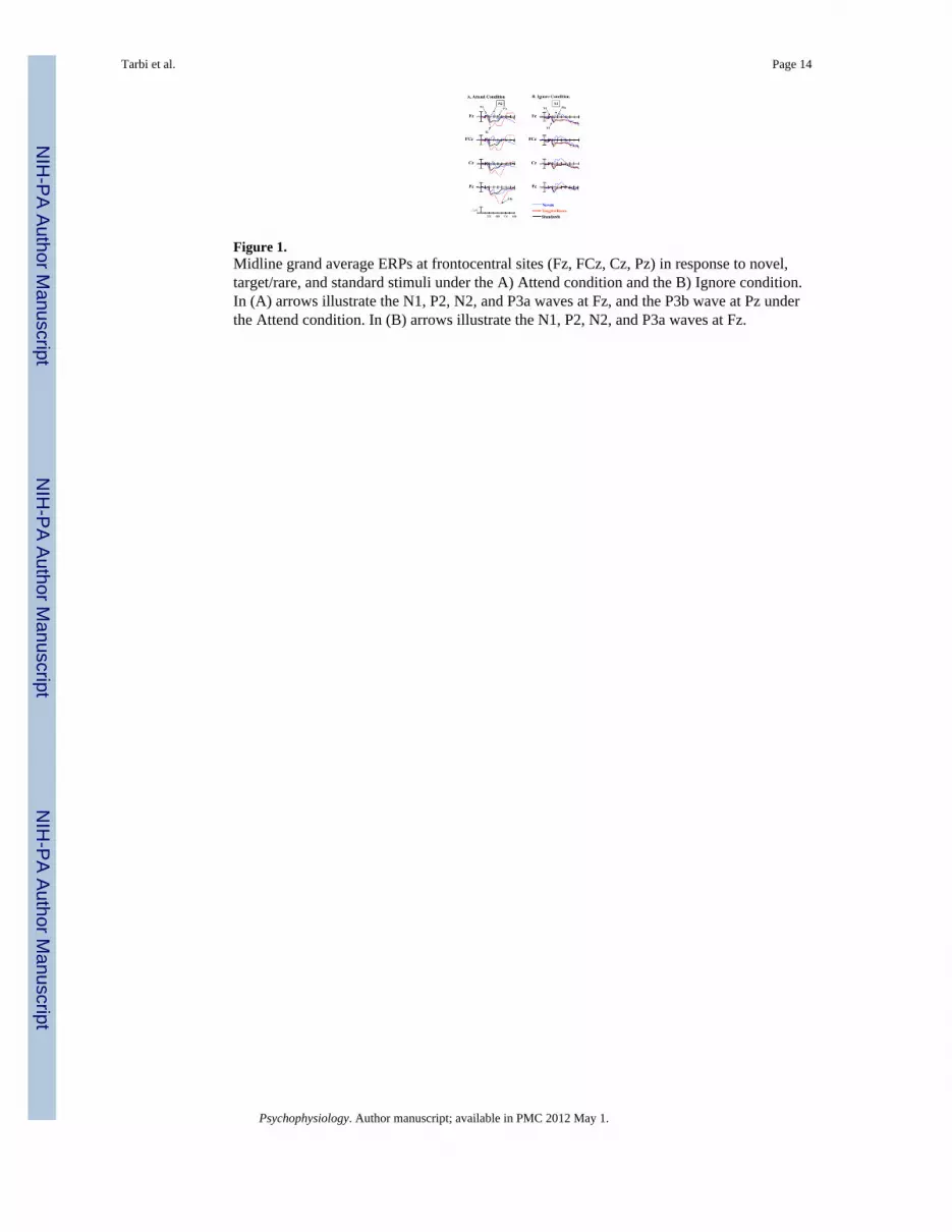
Figure 1.Midline grand average ERPs at frontocentral sites (Fz, FCz, Cz, Pz) in response to novel,target/rare, and standard stimuli under the A) Attend condition and the B) Ignore condition.In (A) arrows illustrate the N1, P2, N2, and P3a waves at Fz, and the P3b wave at Pz underthe Attend condition. In (B) arrows illustrate the N1, P2, N2, and P3a waves at Fz.
Tarbi et al. Page 14
Psychophysiology. Author manuscript; available in PMC 2012 May 1.
NIH
-PA Author Manuscript
NIH
-PA Author Manuscript
NIH
-PA Author Manuscript

Figure 2.Midline grand average ERPs at frontocentral sites (Fz, FCz, Cz) in response to novel stimuliunder the Attend vs. Ignore conditions.
Tarbi et al. Page 15
Psychophysiology. Author manuscript; available in PMC 2012 May 1.
NIH
-PA Author Manuscript
NIH
-PA Author Manuscript
NIH
-PA Author Manuscript
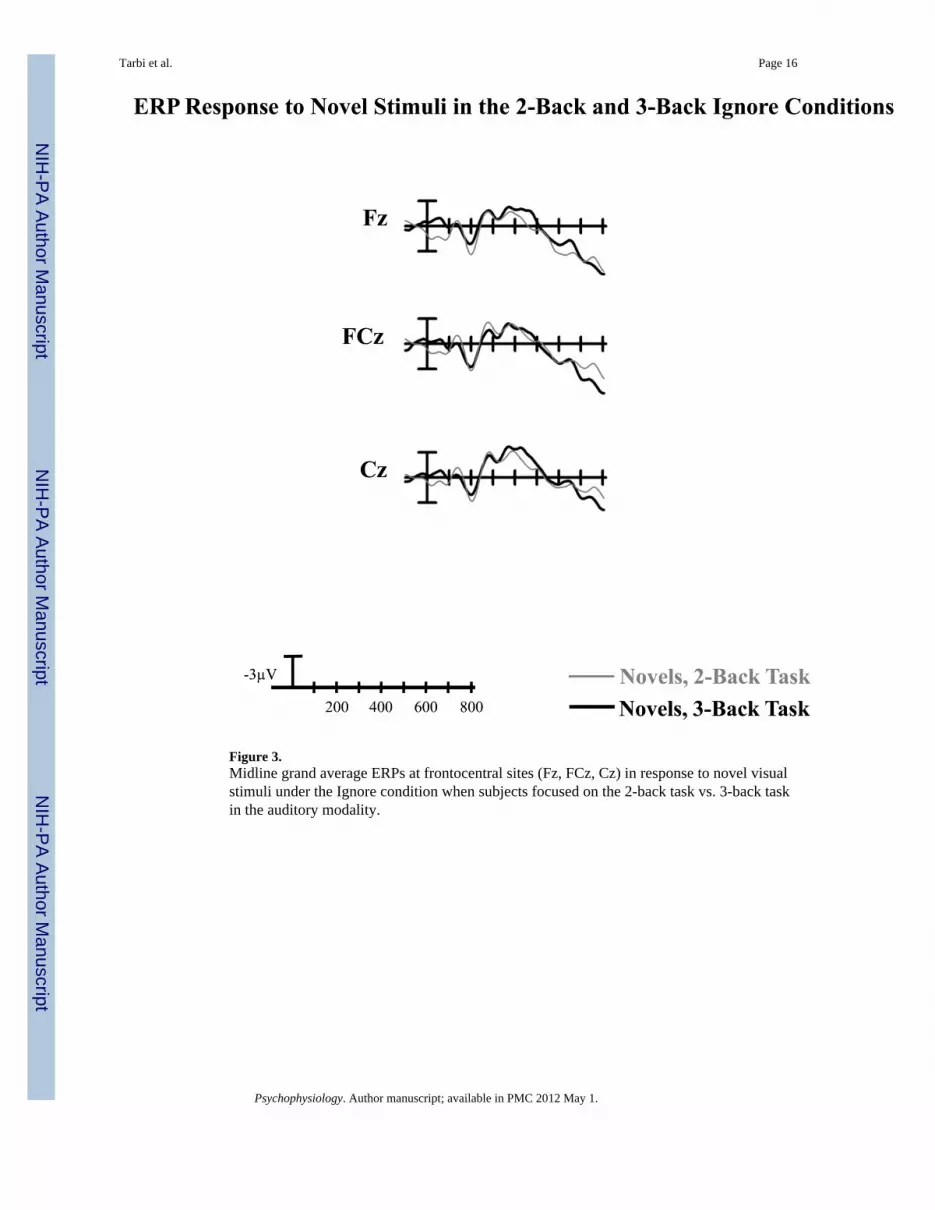
Figure 3.Midline grand average ERPs at frontocentral sites (Fz, FCz, Cz) in response to novel visualstimuli under the Ignore condition when subjects focused on the 2-back task vs. 3-back taskin the auditory modality.
Tarbi et al. Page 16
Psychophysiology. Author manuscript; available in PMC 2012 May 1.
NIH
-PA Author Manuscript
NIH
-PA Author Manuscript
NIH
-PA Author Manuscript

NIH
-PA Author Manuscript
NIH
-PA Author Manuscript
NIH
-PA Author Manuscript
Tarbi et al. Page 17
Table 1
Behavioral Data
Mean (SD)
Attend:
Mean Hit Rate 0.99 (0.02)
Mean False Alarm Rate < 0.001 (< 0.010)
Mean Accuracy Rate 0.98 (0.02)
Median Reaction Time 719 ms (119)
Ignore:
2-Back Task
Mean Hit Rate 0.81 (0.18)
Mean False Alarm Rate 0.04 (0.02)
Mean Accuracy Rate 0.78 (0.19)
Median Reaction Time 867 ms (116)
3-Back Task
Mean Hit Rate 0.66 (0.20)*
Mean False Alarm Rate 0.06 (0.04)*
Mean Accuracy Rate 0.59 (0.20)*
Median Reaction Time 1028 ms (223)*
*Denotes significant differences between the 2-back and 3-back tasks (p < 0.001)
Psychophysiology. Author manuscript; available in PMC 2012 May 1.