surgical treatment for medically refractory myasthenic blepharoptosis
TRANSCRIPT

© 2014 Shimizu et al. This work is published by Dove Medical Press Limited, and licensed under Creative Commons Attribution – Non Commercial (unported, v3.0) License. The full terms of the License are available at http://creativecommons.org/licenses/by-nc/3.0/. Non-commercial uses of the work are permitted without any further
permission from Dove Medical Press Limited, provided the work is properly attributed. Permissions beyond the scope of the License are administered by Dove Medical Press Limited. Information on how to request permission may be found at: http://www.dovepress.com/permissions.php
Clinical Ophthalmology 2014:8 1859–1867
Clinical Ophthalmology Dovepress
submit your manuscript | www.dovepress.com
Dovepress 1859
O r i g i n a l r e s e a r C h
open access to scientific and medical research
Open access Full Text article
http://dx.doi.org/10.2147/OPTH.S69883
surgical treatment for medically refractory myasthenic blepharoptosis
Yusuke shimizu1
shigeaki suzuki2
Tomohisa nagasao1
hisao Ogata1
Masaki Yazawa1
norihiro suzuki2
Kazuo Kishi1
1Department of Plastic and reconstructive surgery, 2Department of neurology, Keio University school of Medicine, Tokyo, Japan
Purpose: Currently, only a few reports have recommended surgery as a suitable treatment
for blepharoptosis associated with myasthenia gravis. The present study aims to introduce our
surgical criteria, surgical options, outcomes, and precautions for medically refractory myas-
thenic blepharoptosis.
Patients and methods: Eight patients who failed to respond to at least 2 years of medical
treatment and who underwent blepharoptosis surgery, from January 2008 to December 2011,
were enrolled in this study. Medical records, photographs, and questionnaire results regarding
postoperative status were evaluated. Of the eleven procedures performed, four involved frontal
suspension, four involved external levator advancement, one involved nonincisional transcon-
junctival levator advancement, and two involved subbrow blepharoplasty with orbicularis oculi
muscle tucking. The margin reflex distance improved postoperatively in seven patients.
Results: Seven patients had very minimal scarring, and one had minimal scarring. Five patients
showed no eyelid asymmetry, one had subtle asymmetry, and two had obvious asymmetry. Seven
patients were very satisfied, and one patient was satisfied with the overall result. Postoperative
complications included mild lid lag with incomplete eyelid closure, prolonged scar redness, and
worsened heterophoria. No patient experienced postoperative exposure keratitis or recurrent
blepharoptosis during the study period.
Conclusion: Our results indicate that blepharoptosis surgery is effective for patients with
myasthenia gravis, especially those with residual blepharoptosis despite multiple sessions of
medical treatments. We recommend that neurologists and surgeons collaborate more system-
atically and discuss comprehensive treatment plans to increase the quality of life for patients
with myasthenia gravis.
Keywords: ptosis, myasthenia gravis, ocular myasthenia, blepharoplasty
IntroductionAt present, many studies of blepharoptosis surgery have been published. However, few
reports have described specific procedures or precautions for blepharoptosis surgery
in patients with myasthenia gravis (MG).1–5 MG is an autoimmune disorder in which
the body’s own antibodies block the transmission of nerve impulses to muscles, caus-
ing fluctuating weakness and muscle fatigue.6 Based on retrospective case reviews,
blepharoptosis is the first sign of MG in approximately 50% of patients,7–9 and more
than 90% of patients experience ocular symptoms during the course of their disease.10
To improve the quality of vision, medical treatment is administered first, typically with
acetylcholine esterase inhibitors (AchE-I). However, some patients require additional
therapy because of the limited efficacy of AcheE-I in some patients. Moreover, while
corticosteroids are very effective in the short term, long-term adverse effects occur in
up to 38% of cases.11,12 Thus, neurologists often struggle to treat patients with medi-
cally refractory longstanding blepharoptosis.13–15
Correspondence: Yusuke shimizuDepartment of Plastic and reconstructive surgery, Keio University school of Medicine, 35 shinanomachi, shinjuku, Tokyo 160-8582, JapanTel +81 3 5363 3814Fax +81 3 3352 1054email [email protected]
Journal name: Clinical OphthalmologyArticle Designation: Original ResearchYear: 2014Volume: 8Running head verso: Shimizu et alRunning head recto: Surgery for myasthenic blepharoptosisDOI: http://dx.doi.org/10.2147/OPTH.S69883

Clinical Ophthalmology 2014:8submit your manuscript | www.dovepress.com
Dovepress
Dovepress
1860
shimizu et al
In such cases, an oculoplastic surgeon may collaborate
with neurologists to perform blepharoptosis surgery. How-
ever, a recent Japanese multicenter survey noted a very low
rate of blepharoptosis surgery in patients with MG (19 out of
676 [2.8%]).16 Through surveys and interviews11 at Japanese
neurological centers, we found that neurologists often do
not closely collaborate with plastic or oculoplastic surgeons
in cases of myasthenic blepharoptosis. In general, neurolo-
gists are understandably unfamiliar with the efficacy and
drawbacks of blepharoptosis surgery and may hesitate to
recommend this as a surgical option.
The present report aims to acquaint neurologists with
the efficacy and drawbacks of blepharoptosis surgery, in an
attempt to encourage a closer relationship with oculoplastic
surgeons. In addition, to inform surgeons about the surgical
approaches for medically refractory longstanding myasthenic
blepharoptosis, this report presents our criteria, procedure
options, outcomes, and precautions of the surgery, from our
experiences.
Materials and methodsPatientsWe examined 220 Japanese patients who were definitively
diagnosed with MG at the MG clinic of Keio University
Hospital, from January 2008 to December 2011. Among
them, eight patients (3.6%) who failed to respond to
medical treatment and underwent blepharoptosis surgery
were enrolled in the study. Three patients who underwent
blepharoptosis surgery but whose MG was not followed at
the clinic were excluded from the study. The eight patients
were also among 19 surgical patients described in a previous
Japanese multicenter study.16 All patients were followed for
at least 2 years. All clinical information was collected after
the patients had provided written informed consent, and the
study was approved by the institutional review board of Keio
University, School of Medicine.
MethodsMedical records and photographs pertaining to patients’
blepharoptosis surgery were reviewed. The pre- and 1-year
postoperative margin reflex distance (MRD) at the time of
most severe MG were measured. Questionnaires were also
given to the patients concerning the ease of eyelid opening
(over both the short term [1 year] and long term [1 year]
postoperatively), esthetic outcome (scarring, asymmetry),
overall satisfaction with the surgery, and any noted compli-
cations. Three-point Likert scales were used to assess the
ease of eyelid opening (much easier, easier, worse), scarring
(very minimal, minimal, visible), eyelid asymmetry (none,
subtle, obvious), and overall satisfaction with the surgery
(very satisfied, satisfied, dissatisfied). To evaluate scarring,
the patients answered the questionnaires with their attending
neurologists at the outpatient clinics. To encourage unbiased
opinions from the patients, the surgeons were not present at
questionnaire completion.
surgical criteriaIf a patient with blepharoptosis with an etiology suggestive
of MG was first referred to the plastic surgery department
rather than the neurology department, then the attend-
ing plastic surgeon consistently consulted a neurologist.
Therefore, none of the patients underwent blepharoptosis
surgery before receiving a definitive diagnosis of MG.
Patient selection for surgical treatment was carefully judged
by the attending neurologist according to the following four
criteria: (1) stable general condition, (2) at least 2 years of
prior medical treatment by neurologists before surgery, (3)
continuous blepharoptosis with minimal circadian changes,
and (4) quality of life severely disrupted by blepharoptosis
(Table 1).
surgical optionsThe surgical procedure was selected through discussion among
several board-certified plastic surgeons, in light of the patient’s
condition. The surgical options used at Keio University
Hospital are illustrated in Figure 1. These surgical procedure
options are basically the same as those used for blepharoptosis
surgery without MG. Although the final decision was always
made after careful consultation with the patient, we placed a
significant emphasis on levator function when MG was most
severe, as the primary factor for determining the choice of
surgical procedure. In addition, MRD, skin thickness and
redundancy, and patient preference were taken into account.
Eyebrow height, lash–brow distance, bilateral difference in
orbital shape, coexisting disease other than MG, and medical
therapies, such as antiplatelet agents, were also considered.
Table 1 surgical criteria for myasthenic blepharoptosis of Keio University hospital
Element Content
general condition stableMedical treatment at least 2 years of prior medical treatment
before surgeryCircadian change Continuous blepharoptosis, with minimal
circadian changesQuality of life severely disrupted

Clinical Ophthalmology 2014:8 submit your manuscript | www.dovepress.com
Dovepress
Dovepress
1861
surgery for myasthenic blepharoptosis
surgical procedureWe used several surgical procedures to treat blepharoptosis
in patients with MG: frontal suspension, external levator
advancement, nonincisional levator advancement, and sub-
brow blepharoplasty. In frontal suspension, we preferably
used autogenous fascia lata. The harvested fascia lata was
tailored to an inverted Y shape, and the separated caudal
legs were then fixed to the tarsus, while the cephalic end
was grafted inside the eyebrow through a suborbital septum
tunnel. In cases where the fascia lata was avoided, several
nylon or GORE-TEX® sutures were used to suspend the
eyelid from the eyebrow.
In external levator advancement, we used the classic
transcutaneous levator advancement technique described by
Carraway and Vincent,17 which has broad utility. In nonin-
cisional levator advancement, we used a transconjunctival
levator advancement technique,18 which has the potential to
reduce postoperative downtime since no major incision of the
skin nor conjunctiva is required. In subbrow blepharoplasty,
we removed a certain amount of skin and orbicularis oculi
muscle to reduce lateral hooding and the weight of the upper
eyelid skin mounted on to the eyelash. The orbicularis oculi
muscle was tucked to elevate the eyelid margin.
ResultsEleven blepharoptosis surgeries were performed on eleven
eyes of eight patients. Three patients were male, and five were
female. The age at operation ranged from 38 to 78 years (mean
67.4 years, median 76 years). The age at MG diagnosis ranged
from 22 to 75 years (mean 56.6 years, median 63.5 years).
The time to surgery after MG diagnosis ranged from 2 to 30
years (mean 10.1 years, median 5 years). The clinical features
of the patients with MG are outlined in Table 2.
Of the eleven procedures performed, four involved
frontal suspension, four involved external levator advance-
ment, one involved nonincisional transconjunctival levator
advancement, and two involved subbrow blepharoplasty with
orbicularis oculi muscle tucking. MRD improved postopera-
tively in all patients except patient 7, who underwent subbrow
blepharoplasty. The surgical outcomes of the patients with
MG are presented in Table 3.
Postoperative complications were noted in several
patients, including mild lid lag with incomplete eyelid clo-
sure, prolonged scar redness, and worsened heterophoria. No
patients presented postoperative corneal exposure keratitis,
and none underwent surgery for overcorrection. Although
distinct recurrent ptosis did not develop in any patients during
the study period, one patient (patient 2) underwent additional
skin resection surgery, to achieve the maximum effect of
eyelid elevation, 25 months after the initial surgery.
Case reportsCase 2A 71-year-old man was diagnosed with MG after developing
right blepharoptosis and diplopia. The Myasthenia Gravis
Foundation of America (MGFA) grade was 1 (quantitative
myasthenia gravis [QMG] score of 7, at its most severe).19
Although AchE-I slightly improved the ptosis, severe bleph-
aroptosis remained, especially in the evening. At 73 years
of age, the patient also developed right Bell’s palsy, which
caused persistent right brow ptosis and paretic ocular syn-
kinesis. This combination of blepharoptosis and brow ptosis
severely disrupted the patient’s quality of vision. At 75
years of age, the patient was referred to the plastic surgery
department. Levator function was 12 mm, and MRD was
0 mm (Figure 2A). Since the patient was not particularly
Levator function <4 mm
Frontal suspension Levator advancement
Externallevator advancement
Nonincisionallevator advancement
Subbrow blepharoplasty
Levator function ≥4 mm
If a decrease infunction is expected
If a patient prioritizeda maximum effect
If a patient prioritizedshort downtime
MRD <2.0 mm• At least MRD ≥2.0 mm
• Skin thickness/redundancy
Figure 1 The surgical options used for blepharoptosis surgery at Keio University hospital.

Clinical Ophthalmology 2014:8submit your manuscript | www.dovepress.com
Dovepress
Dovepress
1862
shimizu et al
Tab
le 2
Clin
ical
feat
ures
of e
ight
Mg
pat
ient
s w
ith b
leph
arop
tosi
s su
rger
y
Cas
e
Num
ber
Age
at
surg
ery
(yea
rs)
Sex
MG
ons
et
age
(yea
rs)
Tim
e to
sur
gery
aft
er
MG
dia
gnos
is (
year
s)M
GFA
gr
ade
QM
G
scor
eA
chR
PSL
max
(m
g/da
y)P
SL
(mg/
day)
Ach
E-1
(m
g/da
y)O
ther
138
F22
161
9+
200
02
75M
714
17
+0
012
0r
ight
inco
mpl
ete
faci
al
pals
y, a
sthm
a3
77M
6017
18
+10
060
474
F67
51
5-
00
605
77M
742
15
+0
018
0D
iabe
tes
mel
litus
, hy
pert
ensi
on6
43F
375
2a11
-15
660
hyp
erth
yroi
dism
778
F75
23a
17+
105
180
Dai
ly u
se o
f cyc
losp
orin
e8
77F
4730
3a21
+30
518
0Bl
ind
(rig
ht e
ye)
Abb
revi
atio
ns:
ach
e-i,
acet
ylch
olin
e es
tera
se i
nhib
itor;
ach
r,
acet
ylch
olin
e re
cept
or;
F, f
emal
e; M
, m
ale;
Mg
, m
yast
heni
a gr
avis
; M
gFa
, M
yast
heni
a g
ravi
s Fo
unda
tion
of a
mer
ica;
QM
g,
quan
titat
ive
mya
sthe
nia
grav
is;
Psl,
pr
edni
solo
ne.
Tab
le 3
sur
gica
l out
com
e of
eig
ht M
g p
atie
nts
Cas
e
Num
ber
Affe
cted
ey
eSu
rgic
al
proc
edur
eLe
vato
r fu
ncti
on
(mm
)
MR
DP
osto
pera
tive
sta
tus
(que
stio
nnai
re)
Com
plic
atio
ns
Pre
-op
(mm
)P
ost-
op
(mm
)E
yelid
ope
ning
Eye
lid
scar
ring
Eye
lid
asym
met
ryO
vera
ll sa
tisf
acti
onSh
ort
term
(
1 ye
ar)
Long
ter
m
(1
year
)
1r
/lFs
(r
/l)
0/0
(r/l
)-1
/-1
(r/l
)2.
5/2.
5 (r
/l)
Muc
h ea
sier
Muc
h ea
sier
Ver
y m
inim
aln
one
Ver
y sa
tisfie
d2
rel
a12
02.
5M
uch
easi
erM
uch
easi
erV
ery
min
imal
non
eV
ery
satis
fied
3l
Fs0
-23.
5M
uch
easi
erM
uch
easi
erV
ery
min
imal
non
eV
ery
satis
fied
Mild
lid
lag
4r
ela
101.
53.
0M
uch
easi
erM
uch
easi
erV
ery
min
imal
subt
leV
ery
satis
fied
5r
nil
a8
0.5
2.5
Muc
h ea
sier
Muc
h ea
sier
Ver
y m
inim
aln
one
Ver
y sa
tisfie
d6
lel
a8
1.5
2.0
easi
erea
sier
Ver
y m
inim
alO
bvio
usSa
tisfie
d7
r/l
sBB
(r/l
)9/
9 (r
/l)
2.0/
2.0
(r/l
)2.
0/2.
0 (r
/l)
easi
erea
sier
Min
imal
non
eV
ery
satis
fied
Prol
onge
d sc
ar
redn
ess
8r
/lFs
/ela
(r
/l)
0/10
(r
/l)
0/3.
0 (r
/l)
4.0/
3.0
(r/l
)M
uch
easi
erM
uch
easi
erV
ery
min
imal
Obv
ious
Ver
y sa
tisfie
dh
eter
opho
ria
appe
aran
ce
Abb
revi
atio
ns: E
LA, e
xter
nal l
evat
or a
dvan
cem
ent;
FS, f
ront
al s
uspe
nsio
n; L
, lef
t; M
G, m
yast
heni
a gr
avis
; MR
D, m
argi
n re
flex
dist
ance
; NIL
A, n
onin
cisi
onal
leva
tor
adva
ncem
ent;
R, r
ight
; SBB
, sub
brow
ble
phar
opla
sty.
Clinical Ophthalmology 2014:8 submit your manuscript | www.dovepress.com
Dovepress
Dovepress
1863
surgery for myasthenic blepharoptosis
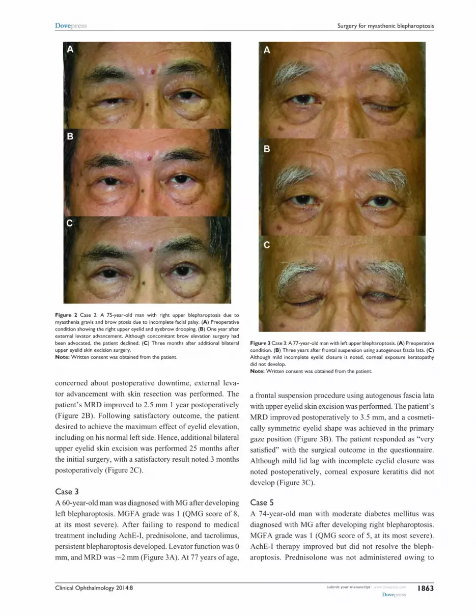
concerned about postoperative downtime, external leva-
tor advancement with skin resection was performed. The
patient’s MRD improved to 2.5 mm 1 year postoperatively
(Figure 2B). Following satisfactory outcome, the patient
desired to achieve the maximum effect of eyelid elevation,
including on his normal left side. Hence, additional bilateral
upper eyelid skin excision was performed 25 months after
the initial surgery, with a satisfactory result noted 3 months
postoperatively (Figure 2C).
Case 3 A 60-year-old man was diagnosed with MG after developing
left blepharoptosis. MGFA grade was 1 (QMG score of 8,
at its most severe). After failing to respond to medical
treatment including AchE-I, prednisolone, and tacrolimus,
persistent blepharoptosis developed. Levator function was 0
mm, and MRD was -2 mm (Figure 3A). At 77 years of age,
a frontal suspension procedure using autogenous fascia lata
with upper eyelid skin excision was performed. The patient’s
MRD improved postoperatively to 3.5 mm, and a cosmeti-
cally symmetric eyelid shape was achieved in the primary
gaze position (Figure 3B). The patient responded as “very
satisfied” with the surgical outcome in the questionnaire.
Although mild lid lag with incomplete eyelid closure was
noted postoperatively, corneal exposure keratitis did not
develop (Figure 3C).
Case 5A 74-year-old man with moderate diabetes mellitus was
diagnosed with MG after developing right blepharoptosis.
MGFA grade was 1 (QMG score of 5, at its most severe).
AchE-I therapy improved but did not resolve the bleph-
aroptosis. Prednisolone was not administered owing to
Figure 2 Case 2: a 75-year-old man with right upper blepharoptosis due to myasthenia gravis and brow ptosis due to incomplete facial palsy. (A) Preoperative condition showing the right upper eyelid and eyebrow drooping. (B) One year after external levator advancement. although concomitant brow elevation surgery had been advocated, the patient declined. (C) Three months after additional bilateral upper eyelid skin excision surgery.Note: Written consent was obtained from the patient.
Figure 3 Case 3: a 77-year-old man with left upper blepharoptosis. (A) Preoperative condition. (B) Three years after frontal suspension using autogenous fascia lata. (C) although mild incomplete eyelid closure is noted, corneal exposure keratopathy did not develop. Note: Written consent was obtained from the patient.

Clinical Ophthalmology 2014:8submit your manuscript | www.dovepress.com
Dovepress
Dovepress
1864
shimizu et al
diabetes mellitus. Levator function was 8 mm, and MRD
was 0.5 mm in the most severe condition (Figure 4A). At
77 years of age, nonincisional transconjunctival levator
advancement was performed since the patient preferred a less
invasive procedure without skin incision. Care was taken not
to elevate the eyelid too high since the patient demonstrated
a weak Bell’s phenomenon (Figure 4B). Although slight
recurrent blepharoptosis was noted 1 year postoperatively,
MRD had improved to 2.0 mm (Figure 4C). The patient
did not demand additional surgery and reported satisfaction
with the result.
Case 7A 75-year-old woman was diagnosed with MG after develop-
ing bilateral upper eyelid ptosis. MGFA grade was 3a (QMG
score of 17, at its most severe). Although the patient had taken
AchE-I, prednisolone, and cyclosporine for more than 2 years,
her bilateral blepharoptosis did not improve. Levator function
was 9 mm, and MRD was 2.0 mm bilaterally (Figure 5A).
The patient did not demonstrate Bell’s phenomenon,
although external ocular muscle movement was not impaired.
At 78 years of age, a subbrow skin excision (13×52 mm)
with orbicularis oculi muscle tucking was performed.
Although the MRD did not change, and prolonged scar red-
ness persisted nearly 1 year after the operation (Figure 5B),
the patient felt “easier” eyelid opening at both short-
(1 year) and long-term (1 year) follow up.
Case 8A 47-year-old woman was diagnosed with MG after develop-
ing bilateral upper eyelid ptosis. MGFA grade was 3a (QMG
score of 21, at its most severe). Although several medical
treatments were administered, right external ophthalmoplegia
with severe blepharoptosis, and mild left blepharoptosis did
not improve. At 77 years of age, the patient demonstrated
fixed heterophoria (Figure 6A). Furthermore, the right
eye was blinded due to optic neuritis (etiology unknown),
and Bell’s phenomenon was absent. Levator function was
0 mm in the right eye and 10 mm in the left, while MRD was
0 and 3.0 mm, respectively. Although the right eye had lost
visual function, the patient desired blepharoptosis surgery
for esthetic purposes. Hence, frontal suspension, using nylon
sutures, with upper eyelid skin resection was performed on
the right eye, while external levator advancement was per-
formed on the left eye. In the right eye, care was taken not to
excessively elevate the eyelid, to avoid postoperative corneal
Figure 4 Case 5: a 77-year-old man with right upper blepharoptosis and weak Bell’s phenomenon. (A) Preoperative condition. (B) immediately after the nonincisional levator advancement operation. eye swelling was minimal. (C) Mild recurrent blepharoptosis was noted 1 year postoperatively.Note: Written consent was obtained from the patient.
Figure 5 Case 7: a 78-year-old woman with bilateral blepharoptosis and absence of Bell’s phenomenon. (A) Preoperative condition. (B) although prolonged redness of the scar was noted even 1 year postoperatively, the patient felt easier eyelid opening.Note: Written consent was obtained from the patient.

Clinical Ophthalmology 2014:8 submit your manuscript | www.dovepress.com
Dovepress
Dovepress
1865
surgery for myasthenic blepharoptosis
exposure. Although the heterophoria became more apparent
(Figure 6B), the patient felt “much easier” eyelid opening in
the left eye and was “very satisfied” with both the esthetic and
functional outcome. Since the right eye was nonfunctional,
diplopia did not develop postoperatively.
DiscussionBlepharoptosis substantially reduces the quality of life of
patients with MG.20–22 Despite multiple medical treatments,
patients often experience longstanding residual blepharopto-
sis. In such cases, surgical treatment may play an important
role. However, lack of information about the efficacy and
adverse effects of blepharoptosis surgery may cause many
neurologists to hesitate to inform their longstanding patients
about this surgical option.
Generally, blepharoptosis surgery is not very difficult for
trained surgeons, and the rate of major functional complica-
tions, such as lagophthalmos and exposure keratopathy, is
usually not high. Moreover, possible complications can be
effectively managed by conservative therapy or additional
surgeries if necessary. Bradley et al reviewed the surgical
outcome of 16 blepharoptosis procedures in ten patients with
MG and concluded that blepharoptosis surgery can be an
appropriate treatment option for MG refractory to medical
treatment.2 However, the indications and specific criteria for
Figure 6 Case 8: a 77-year-old woman with right severe blepharoptosis and heterophoria. (A) Preoperative condition. (B) One year after right frontal suspension using nylon sutures, and left external levator advancement. although the heterophoria became more apparent, diplopia did not develop since the right eye had been blinded due to persistent optic neuritis.Note: Written consent was obtained from the patient.
blepharoptosis surgery in patients with MG remain unclear
and should be established.
The present study presents four surgical criteria accord-
ing to the specific characteristics of MG. First, the patient’s
general condition must be considered since clinical mani-
festations of MG are variable, ranging from ocular myas-
thenia to myasthenic crisis, in which patients experience
life-threatening respiratory insufficiencies.23 Second, medical
treatment with strict follow up must be undertaken by an
attending neurologist for at least 2 years before surgery. It
is reported that 50% to 60% of patients who initially present
with ocular symptoms progress to develop generalized mus-
cle weakness and that the vast majority will do so within the
first 1 to 2 years of diagnosis.6–8 Third, blepharoptosis should
be consistent with minimal circadian change; otherwise, the
surgical effect could collapse. Finally, the patient’s quality
of life should certainly be weighed, with neurologists and
surgeons carefully considering a patient’s quality of vision.
While the criteria may vary by institute, the development of
uniform criteria should be fostered.
Levator function at the most severe condition of MG
should be the primary factor in surgical procedure decision
making. In addition, MRD, skin condition, eyebrow height,
lash–brow distance, orbital shape, coexisting disease, and
medication use should also be considered. A patient with
poor levator function (4 mm) should be treated by frontal
suspension. However, the procedure may also be used in
cases wherein the levator function is currently 4 mm,
but a decrease in function is expected. Thus, patients with
moderate levator function (5–8 mm) can be treated by either
levator advancement or frontal suspension. Alternatively, a
patient with at least a 2.0 mm MRD and thick eyelid skin
can be treated by subbrow blepharoplasty with orbicularis
oculi muscle tucking. Although subbrow blepharoplasty
generally cannot directly improve MRD, it is advanta-
geous in that it is typically associated with short downtime
compared with levator advancement and imposes less risk
of developing corneal exposure keratopathy. In the case
of levator advancement, “external” and “nonincisional”
procedures may be an option. Both procedures share the
same principle that advancement of the levator aponeurosis
and muscle eventually elevates the upper eyelid. However,
we generally apply an external procedure for candidates
who desire to reduce the upper eyelid skin weight by
excision, to achieve the maximum effect of eyelid eleva-
tion. In contrast, we employ a nonincisional procedure for
candidates who desire a minimally invasive operation with
less downtime.

Clinical Ophthalmology 2014:8submit your manuscript | www.dovepress.com
Dovepress
Dovepress
1866
shimizu et al
This study demonstrates the efficacy of blepharoptosis
surgery for patients with MG. In terms of functional outcome,
MRD was improved 1 year postoperatively in all cases except
for case 7. In case 7, although MRD did not improve, the patient
felt easier eyelid opening. No patients developed postoperative
exposure keratitis or underwent surgery for overcorrection.
Although mild lid lag with incomplete eyelid closure was
noted in one patient, it did not become problematic. In terms of
esthetic outcome, eyelid scarring was “very minimal”, except
in patient 7 who had “minimal” scarring after subbrow skin
excision. Additionally, the scar in this case demonstrated red-
ness for a relatively longer period, taking more than 1 year to
subside. This patient’s poor wound healing ability was likely
due to daily use of prednisolone and cyclosporine. Five patients
had “no” eyelid asymmetry and one had “subtle” asymmetry.
Although two patients (MGFA 2a and 3a) demonstrated “obvi-
ous” asymmetries, these were evident before surgery and were
not caused by the surgery. In terms of overall satisfaction, seven
patients were “very satisfied” and one patient was “satisfied”
with the result. No patients were “unsatisfied.”
It should be noted that particular precautions must be
taken during these operations, in order to avoid postoperative
lagophthalmos or corneal exposure keratitis. This is important
since such patients often show the absence of Bell’s phenom-
enon, owing to external ophthalmoplegia. In patients with
heterophoria, the risk of postoperative worsening of diplopia
must also be considered. In such cases, eyelid elevation should
be corrected to an appropriate extent and not overcorrected.
The major limitation of the present study is the relatively
small number of patients who actually underwent the opera-
tion. We are considering future studies with a greater number
of patients and with longer follow-up periods.
In conclusion, blepharoptosis surgery for patients with
MG may be a good choice of treatment if performed with
appropriate surgical criteria and procedure decision making.
The surgery is especially effective for patients who have
residual blepharoptosis despite multiple medical treatments.
Therefore, in order to increase the quality of life of patients
with MG, we recommend that neurologists and surgeons
collaborate more systematically and discuss comprehensive
treatment plans.
AcknowledgmentsThis work was supported by a grant from the Japanese
Ministry of Education, Science, Sports, and Culture (grant
number 23591255) and a Neuroimmunological Disease
Research Committee grant from the Japanese Ministry of
Health, Labour, and Welfare.
DisclosureThe authors report no conflicts of interest in this work.
References 1. Kapetansky DI. Surgical correction of blepharoptosis in myasthenia
gravis. Am J Ophthalmol. 1972;74(5):818–820. 2. Bradley EA, Bartley GB, Chapman KL, Waller RR. Surgical correction
of blepharoptosis in patients with myasthenia gravis. Ophthal Plast Reconstr Surg. 2001;17(2):103–110.
3. Carter SR, Meecham WJ, Seiff SR. Silicone frontalis slings for the correction of blepharoptosis: indications and efficacy. Ophthalmology. 1996;103(4):623–630.
4. Yu CC, Chen SG, Chen TM. Frontalis slings with palmaris tendon as an adjuvant treatment for myasthenic blepharoptosis: a case report. Ann Plast Surg. 2007;58(5):577–579.
5. Asamura S, Kakizaki H, Enjyo M, Hashimoto T, Isogai N. Frontalis sling procedure for ocular myasthenia gravis. Clin Ophthalmol. 2012; 6:575–577.
6. Benatar M, Kaminski H. Medical and surgical treatment for ocular myasthenia. Cochrane Database Syst Rev. 2012;12:CD005081.
7. Bever CT Jr, Aquino AV, Penn AS, Lovelace RE, Rowland LP. Prog-nosis of ocular myasthenia. Ann Neurol. 1983;14(5):516–519.
8. Oosterhuis HJGH. Myasthenia Gravis. Groningen: Groningen Neuro-logic Press; 1997.
9. Benatar M, Kaminski HJ; Quality Standards Subcommittee of the American Academy of Neurology. Evidence report: the medical treat-ment of ocular myasthenia (an evidence-based review): report of the Quality Standards Subcommittee of the American Academy of Neurol-ogy. Neurology. 2007;68(24):2144–2149.
10. Osserman KE. Ocular myasthenia gravis. Invest Ophthalmol. 1967; 6(3):277–287.
11. Sghirlanzoni A, Peluchetti D, Mantegazza R, Fiacchino F, Cornelio F. Myasthenia gravis: prolonged treatment with steroids. Neurology. 1984;34(2):170–174.
12. Evoli A, Batocchi AP, Minisci C, Di Schino C, Tonali P. Thera-peutic options in ocular myasthenia gravis. Neuromuscul Disord. 2011;11(2):208–216.
13. Kaminski HJ, Daroff RB. Treatment of ocular myasthenia: steroids only when compelled. Arch Neurol. 2000;57(5):752–753.
14. Roach ES. Treating ocular Myasthenia Gravis with inadequate evidence. Arch Neurol. 2007;64(12):1794–1795.
15. Nagane Y, Utsugisawa K, Suzuki S, et al. Topical naphazoline in the treat-ment of myasthenic blepharoptosis. Muscle Nerve. 2011;44(1):41–44.
16. Shimizu Y, Suzuki S, Utsugisawa K, et al. Is surgical intervention safe and effective in the treatment of myasthenic blepharoptosis? A multi-center survey in Japan. Eur Neurol. 2014;71(5–6):259–261.
17. Carraway JH, Vincent MP. Levator advancement technique for eyelid ptosis. Plast Reconstr Surg. 1986;77(3):394–403.
18. Shimizu Y, Nagasao T, Asou T. A new non-incisional correction method for blepharoptosis. J Plast Reconstr Aesthet Surg. 2010;63(12): 2004–2012.
19. Jaretzki A, Barohn RJ, Ernstoff RM, et al. Myasthenia gravis: recom-mendations for clinical research standards. Task Force of the Medical Scientific Advisory Board of the Myasthenia Gravis Foundation of America. Neurology. 2000;55(1):16–23.
20. Paul RH, Nash JM, Cohen RA, Gilchrist JM, Goldstein JM. Quality of life and well-being of patients with myasthenia gravis. Muscle Nerve. 2011;24(4):512–516.
21. Mullins LL, Carpentier MY, Paul RH, Sanders DB; Muscle Study Group. Disease-specific measure of quality of life for myasthenia gravis. Muscle Nerve. 2008;38(2):947–956.
22. Masuda M, Utsugisawa K, Suzuki S, et al. The MG-QOL15 Japanese version: validation and associations with clinical factors. Muscle Nerve. 2012;46(2):166–173.
23. Lacomis D, Silvestri NJ, Wolfe GI. What’s in the Literature? J Clin Neuromuscul Dis. 2014;16(1):32–41.

Clinical Ophthalmology
Publish your work in this journal
Submit your manuscript here: http://www.dovepress.com/clinical-ophthalmology-journal
Clinical Ophthalmology is an international, peer-reviewed journal covering all subspecialties within ophthalmology. Key topics include: Optometry; Visual science; Pharmacology and drug therapy in eye diseases; Basic Sciences; Primary and Secondary eye care; Patient Safety and Quality of Care Improvements. This journal is indexed on
PubMed Central and CAS, and is the official journal of The Society of Clinical Ophthalmology (SCO). The manuscript management system is completely online and includes a very quick and fair peer-review system, which is all easy to use. Visit http://www.dovepress.com/testimonials.php to read real quotes from published authors.
Clinical Ophthalmology 2014:8 submit your manuscript | www.dovepress.com
Dovepress
Dovepress
Dovepress
1867
surgery for myasthenic blepharoptosis