supportive periodontal therapy
TRANSCRIPT
SUPPORTIVE
PERIODONTAL
THERAPY
Surupa Dutta, et al.
Medical and Research Publications

Supportive Periodontal Therapy
Written by
Surupa Dutta Ass. Prof.
Abhijit Chakraborty Head Of The Department, Prof.
Himadri Chakrabarty Prof.
Ravi Prakash B S Prof.
Manoj Kumar Singh Prof.
Mekhala Mukherjee Ass.Prof.
Rekha Puttannavar Assoc. Prof.
Kumari Rupam Assoc. Prof
Kaushik Dutta Assoc. Prof
Jawaid Badr Assoc. Prof
Supportive Periodontal Therapy
Medical and Research Publications
Copyright © 2021 Surupa Dutta, et. al.
All rights reserved. No part of this publication may be reproduced, distributed, or transmitted in
any form or by any means, including photocopying, recording, or other electronic or mechanical
methods, without the express written permission of the publisher except for the use of brief
quotations in a book review..
First printing, 2021.
Published by
Medical and Research Publications,
124SpencerRd,Stoke-on-TrentST42BE,
UnitedKingdom.
www.medicalandresearch.com
Email: [email protected]
CONTENTS
SL.NO. TOPIC PAGE NO.
1. INTRODUCTION 1
2. HISTORY 3
3. AIMS AND OBJECTIVES 4
4. GOALS AND RATIONALE 6
5. FREQUENCY AND EFFICACY 8
6. RISK ASSESSMENT 10
7. CLASSIFICATION 18
8. COMPLIANCE 20
9. MAINTENANCE PHASE 26
10. RETREATMENT 37
11. SUPPORTIVE CARE OF DENTAL IMPLANTS 43
12. SUMMARY 49
13. CONCLUSION 51
14. REFERENCES 52
1
INTRODUCTION
The healthy periodontium which consists of four principal components i.e. the gingiva,
periodontal ligament, alveolar bone, and cementum, provides the support which is necessary to
maintain teeth in function. Periodontitis is defined as “an inflammatory disease of the supporting
tissues of the teeth caused by specific microorganisms or groups of specific microorganisms,
resulting in progressive destruction of the periodontal ligament and alveolar bone with increased
probing depth, recession, or both. Since virtually all cases of periodontal disease are infectious
disorders, they can be prevented or effectively treated by controlling pathogenic microbes
residing in supra-gingival and sub-gingival plaque. The treatment of periodontal and peri-implant
diseases involves cause-related therapy, together with professional management, which may be
undertaken nonsurgical or surgically to achieve optimal health, function, and esthetics.
After complete active periodontal treatment patients may or may not be effective in
removing plaque, which further requires adherence to a maintenance program that reduces the
risk of future disease progression. The maintenance and recall phase of periodontal therapy was
renamed as “SUPPORTIVE PERIODONTAL THERAPY” which expresses the essential
need for therapeutic measures to facilitate the patient’s own efforts to control periodontal
infection.
With the evidence from a number of studies, it can be concluded that “Tooth loss in
periodontitis patients is inversely proportional to the frequency of maintenance visits”.1 Once the
tooth loss occurs dental implants will become the treatment of choice in totally or partially
edentulous patients. The survival of implants is high, but not free of complications. They are also
susceptible to inflammatory diseases caused by biofilm accumulation.
The periodontal and peri-implant diseases progress either due to an absence of treatment
or inadequate long-term management, resulting in attachment loss and bone loss which further
compromise the patient-related outcomes like tooth retention, esthetics, and function.
Maintenance care is a critical phase of periodontal therapy. The long-term preservation of the
dentition is closely associated with the frequency and quality of Supportive Periodontal
Treatment. Due to the chronic nature of periodontitis and the inability to predict disease
progression, it is necessary to continuously adjunct monitoring and providing appropriate
treatment to prevent recurrence of the disease. Thus, the established principles of periodontal
maintenance are considered as a gold standard of care. Supportive periodontal care is therefore
2
largely founded on the chronic nature of the disease, patient maintenance and clinician ability to
deliver the appropriate management.
Similarly, periodontal maintenance allows for monitoring of dental implants, as well as
evaluation of mechanical and biological aspects of implant support and restoration.2,3 Numerous
studies have demonstrated the efficacy of periodontal maintenance, and have shown that
recurrent periodontitis can be prevented or limited by optimal personal oral hygiene or through
periodic PM. Maintenance care requires patients' understanding of the purpose of the
maintenance program, time and effort on the part of the dentist and staff side. The more often
patients present for recommended supportive periodontal treatment (SPT), the less likely they are
to lose teeth.
The present library dissertation attempts to review the importance of “Supportive
Periodontal Therapy” in the periodontal and peri-implant treatment procedure.

3
HISTORY
The periodontal maintenance program is also known as the recall and maintenance phase,
but the name was changed to supportive periodontal treatment at the 1989 World Workshop in
Clinical Periodontics.1
According to the Glossary of Periodontal terms 1986, Supportive periodontal therapy was
known as Preventive Treatment and was defined as “Procedures performed at selected intervals
to assist the periodontal patients in maintaining oral health”. In 1992, supportive periodontal
therapy was defined as “an extension of periodontal therapy”.
According to Jan Lindhe, Supportive periodontal therapy is defined as “Therapeutic
measures to support the patient's own efforts to control and to avoid re-infection”.4 The
prevention of periodontal disease requires as positive a program as that required for the
elimination of the periodontal disease.
It was noted that a maintenance program should provide adequate therapy for previously
existing periodontal conditions. Initially, the patient should be provided with thorough
prophylaxis and complete reinforcement instructions in oral hygiene procedures every 3months.
The 3 months interval should be increased, maintained, or decreased depending on an evaluation
of the stability of the supporting structures. Close monitoring will indicate the appropriate time
interval for each patient, and if necessary retreatment determined for those areas that may be
deteriorating.
4
AIM AND OBJECTIVE
AIMS:
1.Prevent the occurrence of new diseases.
2.Prevent the recurrence of the previous disease.
THERAPEUTIC OBJECTIVES
1.To prevent the Progression and recurrence of periodontal disease in patients who have
previously been treated for gingivitis and periodontitis.
2.To reduce the incidence of tooth loss by monitoring the patient‟s dentition and any prosthetic
replacements of the natural teeth.
3.To diagnose and manage, promptly, other diseases or conditions found within or related to the
oral cavity.
4.To prevent the loss of dental implants after clinical stability has been achieved.
OBJECTIVES
1.Preservation of alveolar bone support: As evaluated with a radiograph, bone height may not
only be maintained but also improved when proper maintenance is provided after periodontal
therapy.5
2.Maintenance of stable clinical attachment levels: Despite all the variability associated with
clinical measurement maintenance of stable clinical attachment levels represent a reasonable
clinical indicator to evaluate the stability of results.
3.Control inflammation: Without proper maintenance, dental plaque will re-accumulate and
inflammation would be re-established in periodontal tissues.6 On the contrary well-maintained
patient will have low levels of inflammation after therapy.5
5
4.Re-evaluation and reinforcement of proper home care: Although 3-4 month recall seems to
compensate for improper plaque control as far as its effects on clinical attachment levels are
concerned.7 However, the better the oral hygiene the patient maintains, the better the possibility
of maintaining a stable result. With training and positive reinforcement, the level of plaque
control can be improved in most patients, however, it may take several sessions with some
patients.
5.Maintenance of a healthy and functional oral environment: In addition to the evaluation of
periodontal parameters, the mouth and dentition should be thoroughly inspected and assessed for
changes over time. This may require consultation with other specialties &/or treatment. Any
patient concerns or complaints should be addressed by the dental team during these visits.
6
GOALS AND RATIONALE
RATIONALE FOR SUPPORTIVE PERIODONTAL THERAPY
1.Incomplete Subgingival Plaque Removal: This leads to the continuous loss of attachment,
without the appearance of clinical gingival inflammation. Bacteria remaining even after the
scaling, root planing and flap surgery recolonize the pocket and cause recurrent disease.
2.Bacteria associated with periodontitis can be transmitted between spouses and other family
members.
3.The microscopic nature of the dentogingival unit after periodontal treatment. After periodontal
surgical procedures, the tissue heals by the formation of long junctional epithelium which is
weaker in inflammatory conditions and gets separated easily which leads to recurrence of the
pocket formation.
4.Subgingival scaling alters the microflora of periodontal pockets.
Few studies stated that:
A.There is a decrease in the proportion of motile rods and spirochetes and elevation in the
proportion of coccoid rods.
B.Pocket debridement suppresses components of the subgingival microflora associated with
periodontitis.
All these alterations are said to be present for a short duration of time, and the periodontal
pathogens may return to baseline levels within days or months.
7
GOALS OF SUPPORTIVE PERIODONTAL THERAPY
The goals of periodontal or peri-implant therapy are to ensure that the periodontal or peri-
implant tissues are maintained in a state of health, with the achievement of an acceptable degree
of disease stability, patient comfort and function.
Due to multi-factorial etiology and the complex nature and progression of the periodontal
and peri-implant disease, there are no universally agreed clinical parameters of success.
The goals of successful periodontal recall regimens for teeth may be achieved with:
1.Stabilization of plaque score at 20-40%
2.Stabilization of bleeding scores at 10-25%
3.Probing depth reductions and maintenance of probing depth between 1-2mm (at 30%)
4.A residual probing depth of <5mm.
5.Gains in clinical attachment levels (Wilson 1996).
The goals of successful periodontal recall regimens for implants may be achieved with:
• Probing depth no greater than 5mm
• The absence of bleeding on probing
• The absence of implant mobility
• Absence of pain
• The absence of continuous radiolucency surrounding the implant
• Annual vertical loss of bone height no greater than 0.2m.
8
FREQUENCY AND EFFICACY
A counterpoint to the efficacy of frequent supportive periodontal treatment visits has
been offered in a limited number of studies that found no difference in the progression of disease
in patients seen less frequently compared with those who received more frequent supportive
periodontal treatment. Most of the patients in this last group of studies maintained relatively high
levels of oral hygiene.
For patients with a previous history of periodontitis, the results from a number of clinical
trials suggest that the frequency of supportive periodontal treatment should be less than 6
months. Intervals of 2 weeks, 2-3 months, 3 months, 3-4 months, 3-6months and 4-6 months
have been proposed and studied. These data indicate that most patients with a previous history of
chronic periodontitis should be seen at least four times a year because that interval will result in a
decreased likelihood of progressive disease as compared with patients seen less frequently.8
Specific microorganisms are associated with periodontitis. The subgingival population of
these bacteria is suppressed following the root planing used during supportive periodontal
treatment but may return to baseline levels within days to months later.9
The average time for the return to baseline is between 9 and 11 weeks, but this return can
vary dramatically in different patients. If the clinician wishes to prevent the re-establishment of
suspected pathogens, supportive periodontal treatment intervals of 3 months or less appear to be
required.
Patients treated for periodontitis who comply with suggested supportive periodontal
treatment intervals will experience less attachment loss and tooth loss than patients who do not
demonstrate compliance with the supportive periodontal treatment schedule. This body of data
supports the concept that it is advantageous if supportive periodontal treatment visits are
performed every 3months. Supportive periodontal treatment intervals can be tailored to the
individual needs of each patient and further modified based on ongoing clinical studies.10
The average time required for a supportive phase session is 1 hour and it may be divided as
follows:
• 15minutes – examination and evaluation of oral hygiene.

9
• 30minutes – selective scaling and root planing polishing and fluoride application and
• 15minutes – evaluation, and determination of future needs.
10
RISK ASSESSMENT
A Risk is defined as the probability of an individual's developing a given disease or
experiencing a health status change over a specified period.11
Risk assessment is presented as a way of examining risks so that they may be better
avoided, reduced, or otherwise managed.
Risk implies uncertainty, so that risk assessment is largely concerned with uncertainty
and hence with a concept of probability that is hard to grasp. The results of even the simplest risk
assessments need to be compared with similar assessments of commonplace situations to give
them some meaning. The results of risk assessments will necessarily be in the form of an
estimate of probabilities for various events, usually injurious. The goal in performing a risk
assessment is to obtain such estimates.
Risk can be identified in terms of risk factors, risk indicators, or risk predictors .11
Terminology: Three types of causation (of a disease) are generally identified:12
l. A sufficient cause,
2.A necessary cause, and
3.A risk factor.
A Sufficient Cause refers to any condition, characteristic, or exposure in the presence of
which, the disease will always occur. This is the strongest type of causal relationship and is
relatively rare.
Examples: Genetic anomalies or conditions.
A Necessary Cause is any condition, characteristic, or exposure that must be present for
a disease to manifest itself.
An example of this is the organism Mycobacterium tuberculosis, which is a prerequisite for a
person to develop tuberculosis. However, many people can carry this organism in their bodies
without any symptoms of the disease.
11
A Risk Factor is any characteristic, behavior, or exposure with an association to a
particular disease (are confirmed through experiments or randomized controlled trials.
Risk Indicator is a term used to describe a potential risk factor identified to be
associated with disease from case-control or cross-sectional studies.
A risk factor that can be used to predict the future course of the disease, such as an
increased probability of disease, is known as a Risk Marker.
Some risk factors can be modified to reduce one’s risk of initiation or progression of the
disease, such as smoking cessation or improved oral hygiene to reduce the risk of periodontal
destruction, while other factors cannot be modified, such as genetic factors. A risk factor that
cannot be modified is often referred to as a Determinant.
A Risk Predictor is a factor that has no current biological plausibility as a causative
agent but has been associated with disease on a cross-sectional or longitudinal basis. Risk
predictors may be either marker of disease or other historical measures of the disease.
Examples are the number of missing teeth or past evidence of periodontal disease. The number
of missing teeth is a risk predictor for the disease but has little or no biological plausibility as a
causative agent for periodontitis.
The AAP Guidelines describe risk assessment as „increasingly important in periodontal
treatment planning and should be part of every comprehensive dental and periodontal
evaluation‟.
It was concluded regarding the status of periodontal risk assessment, which remains true
to this day, i.e a „Universally accepted objective method of calculating a risk of developing
or worsening periodontal disease at a future date does not exist.‟ And, all such models for
calculating risk and determining prognosis are probabilistic and thus are inherently limited in
their practical application to the individual patient.
12
THE RISK ASSESSMENT PROCESS
A 4-step process for identification of high-risk individuals given by the University of North
Carolina.
• The first step, Identification of risk factors, Clues to these factors usually come from
clinical impressions, animal and in vitro laboratory experiments, and from prevalence
surveys of populations. When a disease is found to have multiple risk factors (as is the
case for Periodontitis), testing the ability of one risk factor at a time to identify
individuals at risk gives an incomplete picture and the development of a risk assessment
model becomes necessary.
• The second step, Development of a risk assessment model or models, i.e putting
together the relevant risk factors into a multivariate model that identifies the combination
of factors that will most efficiently distinguish between those who are at high or low risk
of developing the disease.
• The third step involves Screening population groups for the factors included in the risk
assessment model and using the model to predict each individual's risk of developing
a disease. This step is called assessment.
• The fourth step, Targeting, involves the application of some health promotion/disease
prevention regimen or treatment procedure to the individuals at increased risk with an
evaluation of the effectiveness of the intervention.
Periodontal Risk Calculator:
Types of risk factors: modifiable or non-modifiable.
Modifiable Risk Factors: Smoking, DM, microorganism and periodontal diseases,
socioeconomic status, psychological factors, stress, nutrition.
Non-Modifiable Risk Factors: Genetic factors, osteoporosis, aging, other systemic conditions.
Risk Models: To meet the objective of incorporating risk assessment into the diagnostic process,
numerous risk assessment models have been introduced during the past decade

13
1.Periodontal Risk calculator: Example: Previser
Author(s)/
Year
Risk model Risk variables Notes
Page et al.
(2002).13
Periodontal
Risk
Calculator
(PRC)
11 factors: Age, smoking
history, DM, history of
periodontal surgery, BOP,
furcation involvements,
subgingival restorations, vertical
intrabony defects, root calculus,
PD, radiographic bone loss.
Only the deepest
PD and greatest
bone loss per
sextant are entered
for PD and
radiographic bone
levels.
Based on 11 parameters, "numeric risk and disease severity scores" are calculated that
establish both an assessment of risk as well as a quantification of disease severity. These, in turn,
are coupled with suggested treatment options for the clinician.
As an example of this, the PreViser Periodontal Risk Calculator has been in use for well
over a decade, has been widely published and its algorithm has also been previously adapted for
use by the American Academy of Periodontology for a Web-based “self-assessment tool” – the
“Gum Disease Risk Assessment Test”.
2.Periodontal Risk Assessment
Author(s)/
Year
Risk model Risk variables Notes
Lang &
Tonetti
(2003).14
Periodontal
Risk
Assessment
(PRA)
6 factors: full-mouth
BOP %, PD ≥ 5mm,
tooth loss,
radiographic bone
loss-to-age ratio,
systemic and/or
genetic conditions,
smoking
All sites of BOP and PD ≥
5mm must be entered.
Alveolar bone loss is limited
to the most severe posterior
site. Binary designation for
“systemic and/orgenetic
conditions” category. Six-
point scale for each factor
14
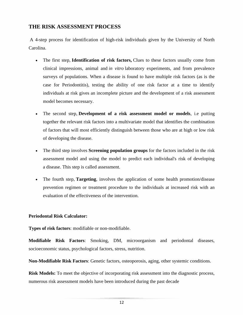
Figure: 3 Functional diagrams of a medium risk maintenance patient - A moderate PRA patient
has at least two parameters in the moderate category, but at most one parameter in the higher-risk
category (Adapted from Lang &Tonetti 2003).
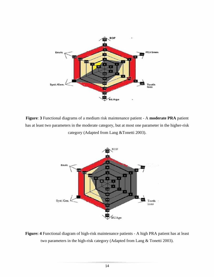
Figure: 4 Functional diagram of high-risk maintenance patients - A high PRA patient has at least
two parameters in the high-risk category (Adapted from Lang & Tonetti 2003).
15
3.Modified PRA
Author(s)/
Year
Risk model Risk variables Notes
Chandra
(2007).15
Modified
PRA
8 factors: full-mouth
BOP %, PD 5mm,
tooth loss, CAI to age
ratio, smoking, DM,
dental status –
systemic factors
interplay,
psychosocial factors
DM is separated from
systemic conditions. Alveolar
bone loss is not evaluated.
Five-point scale for each
factor.
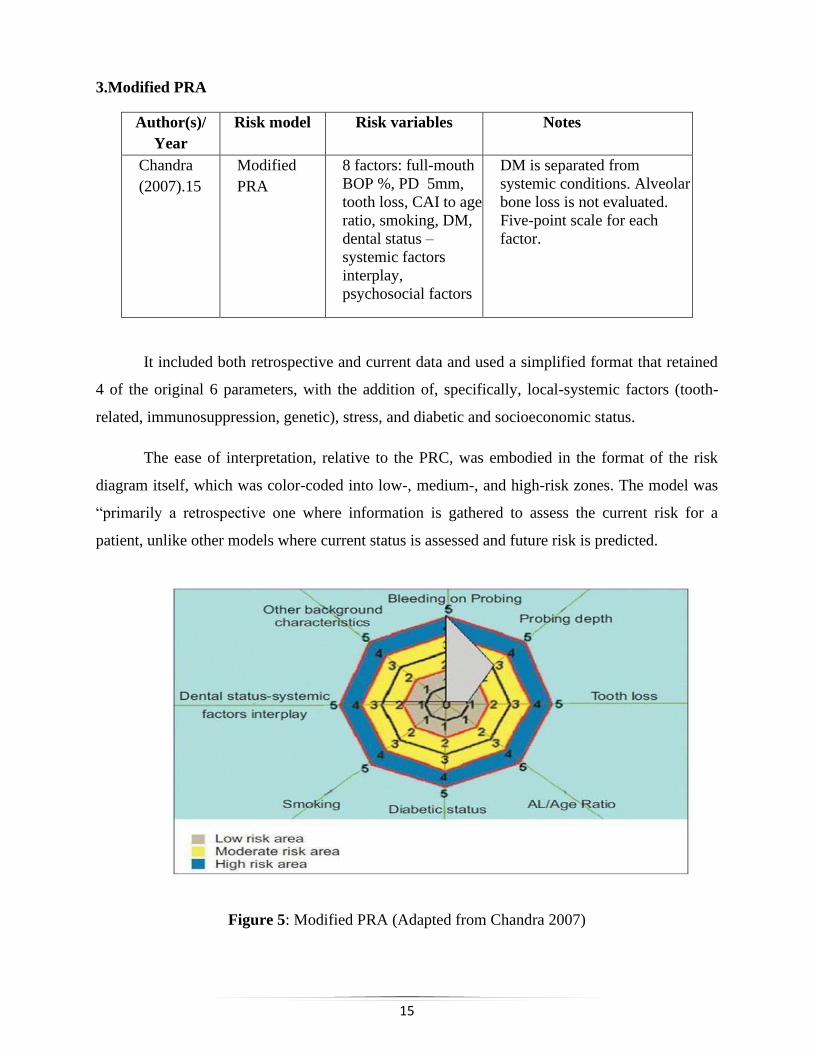
It included both retrospective and current data and used a simplified format that retained
4 of the original 6 parameters, with the addition of, specifically, local-systemic factors (tooth-
related, immunosuppression, genetic), stress, and diabetic and socioeconomic status.
The ease of interpretation, relative to the PRC, was embodied in the format of the risk
diagram itself, which was color-coded into low-, medium-, and high-risk zones. The model was
“primarily a retrospective one where information is gathered to assess the current risk for a
patient, unlike other models where current status is assessed and future risk is predicted.
Figure 5: Modified PRA (Adapted from Chandra 2007)
16
4.Unife
Author(s)/
Year
Risk model Risk variables Notes
Trombelli
(2009).16
Unife 5 factors: BOP %,
PD ≥ 5mm,
radiographic bone
loss to age ratio,
smoking, DM
All sites of BOP and PD ≥
5mm must be entered.
Alveolar bone loss included
for one interproximal site of
each tooth.
A numeric value for each parameter was calculated, based on its extent or severity, and
patients were assigned to 1 of 5 risk categories derived from the sum of those values, i.e., 1(low),
2 (low-medium), 3 (medium), 4 (medium-high), or 5 (high).
5.Dento Risk
Author(s)
/Year
Risk
model
Risk variables
Lindskg
et al.
(2010)17
DentoRisk 20 factors: Systemic Predictors: Age in relation to history of
chronic periodontitis, family history of chronic periodontitis,
systemic disease and related diagnoses, result of skin
provocation test, patient cooperation and disease awareness,
socioeconomic status, smoking, clinician experience
local Predictors: bacterial plaque (oral hygiene), endodontic
pathology, furcation involvements, vertical intrabony
defects, radiographic marginal bone levels, PD, BOP,
marginal dental restorations, increaded tooth mobility,
missing teeth, abutment teeth, presence of purulence
This model differs from others in that an assessment is first calculated for the patient‟s
overall dentition (level I). If an elevated risk is detected, a prognosis for annualized attachment
loss for each individual tooth (level II) is then computed. This information can then be used
during the treatment planning appointment, and provide the patient and clinician with a current
and the future prognostication (based on completion of successful therapy).
17
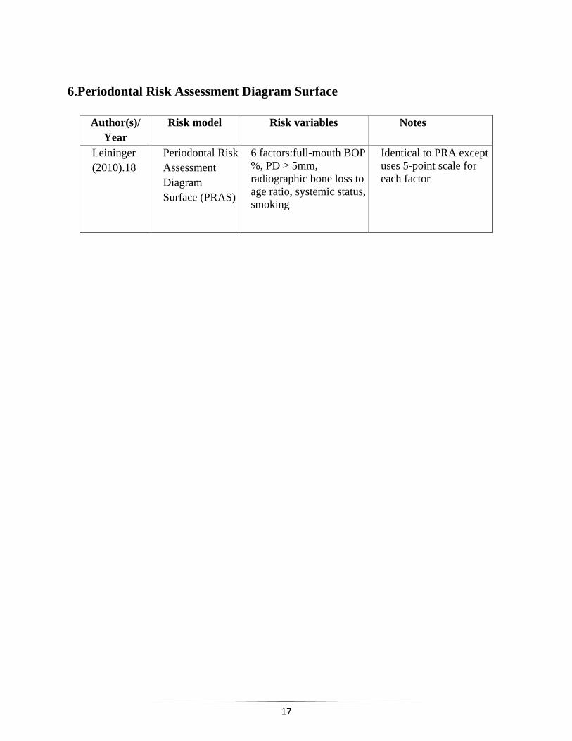
6.Periodontal Risk Assessment Diagram Surface
Author(s)/
Year
Risk model Risk variables Notes
Leininger
(2010).18
Periodontal Risk
Assessment
Diagram
Surface (PRAS)
6 factors:full-mouth BOP
%, PD ≥ 5mm,
radiographic bone loss to
age ratio, systemic status,
smoking
Identical to PRA except
uses 5-point scale for
each factor
18
CLASSIFICATION
Classification of post-treatment patients:
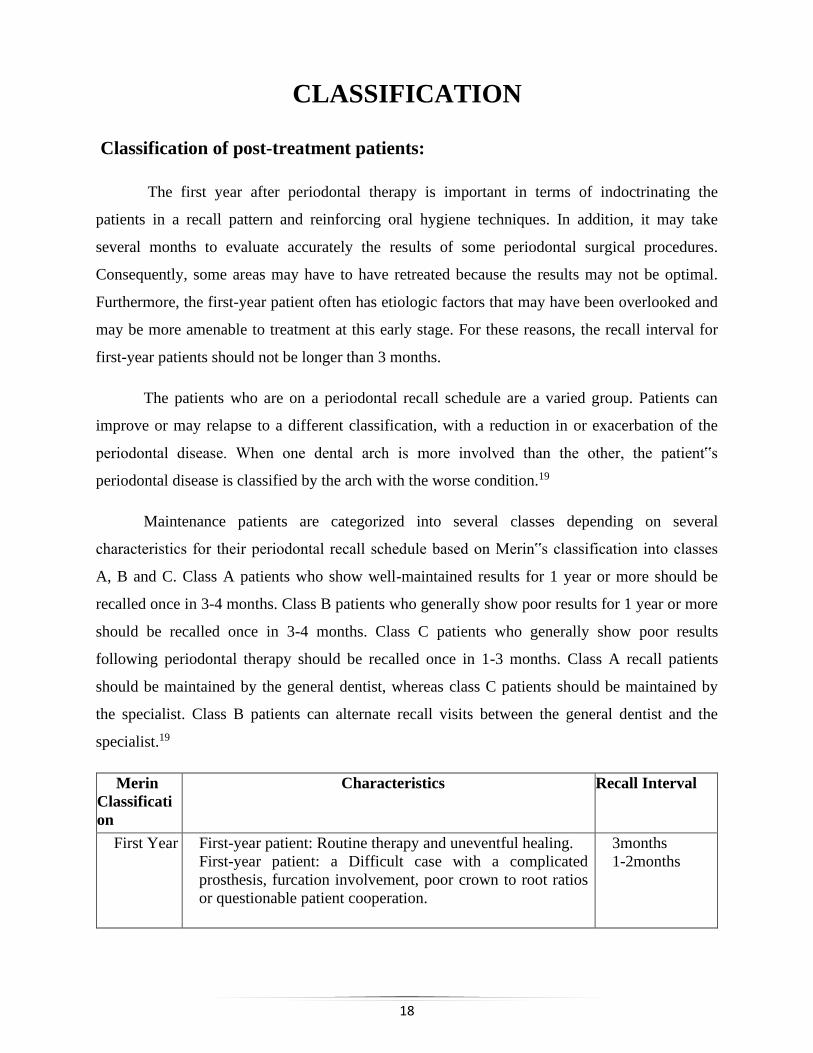
The first year after periodontal therapy is important in terms of indoctrinating the
patients in a recall pattern and reinforcing oral hygiene techniques. In addition, it may take
several months to evaluate accurately the results of some periodontal surgical procedures.
Consequently, some areas may have to have retreated because the results may not be optimal.
Furthermore, the first-year patient often has etiologic factors that may have been overlooked and
may be more amenable to treatment at this early stage. For these reasons, the recall interval for
first-year patients should not be longer than 3 months.
The patients who are on a periodontal recall schedule are a varied group. Patients can
improve or may relapse to a different classification, with a reduction in or exacerbation of the
periodontal disease. When one dental arch is more involved than the other, the patient‟s
periodontal disease is classified by the arch with the worse condition.19
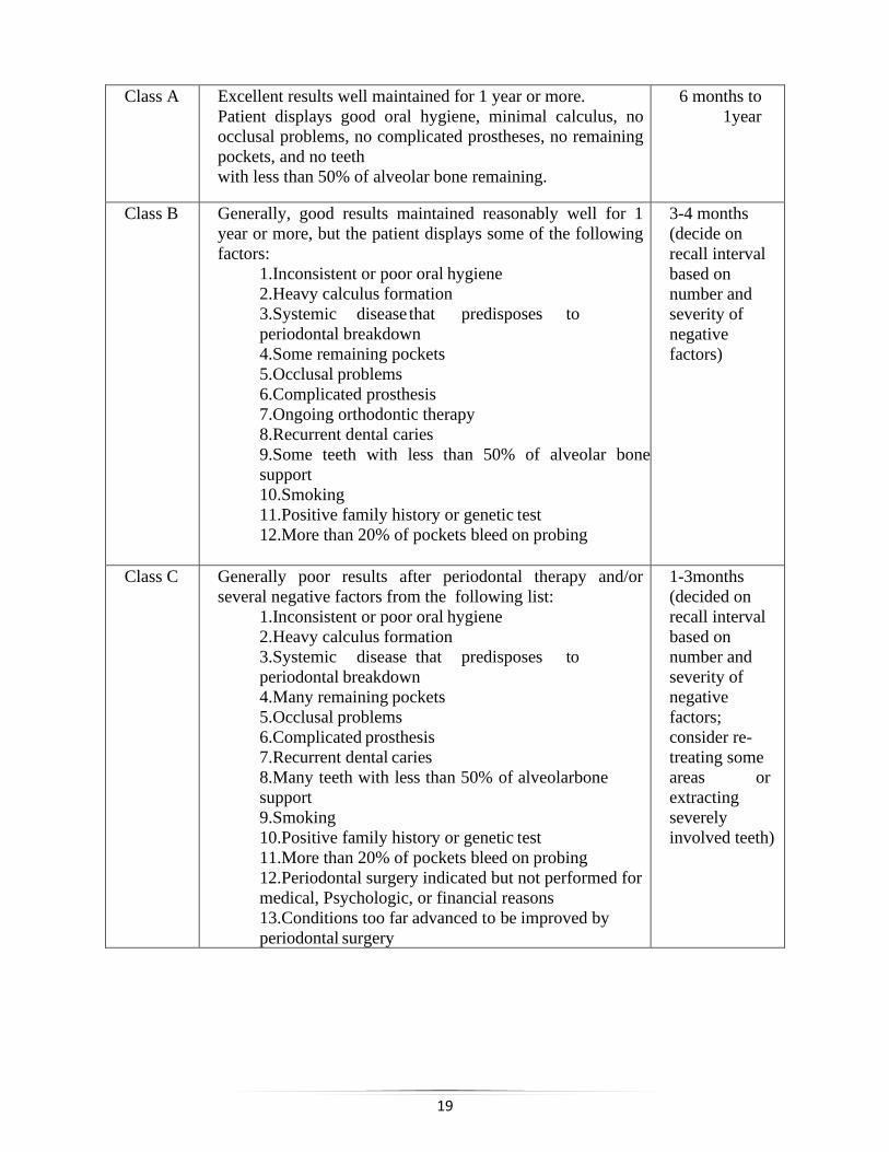
Maintenance patients are categorized into several classes depending on several
characteristics for their periodontal recall schedule based on Merin‟s classification into classes
A, B and C. Class A patients who show well-maintained results for 1 year or more should be
recalled once in 3-4 months. Class B patients who generally show poor results for 1 year or more
should be recalled once in 3-4 months. Class C patients who generally show poor results
following periodontal therapy should be recalled once in 1-3 months. Class A recall patients
should be maintained by the general dentist, whereas class C patients should be maintained by
the specialist. Class B patients can alternate recall visits between the general dentist and the
specialist.19
Merin
Classificati
on
Characteristics Recall Interval
First Year First-year patient: Routine therapy and uneventful healing.
First-year patient: a Difficult case with a complicated
prosthesis, furcation involvement, poor crown to root ratios
or questionable patient cooperation.
3months
1-2months
19
Class A Excellent results well maintained for 1 year or more.
Patient displays good oral hygiene, minimal calculus, no
occlusal problems, no complicated prostheses, no remaining
pockets, and no teeth
with less than 50% of alveolar bone remaining.
6 months to
1year
Class B Generally, good results maintained reasonably well for 1
year or more, but the patient displays some of the following
factors:
1.Inconsistent or poor oral hygiene
2.Heavy calculus formation
3.Systemic disease that predisposes to
periodontal breakdown
4.Some remaining pockets
5.Occlusal problems
6.Complicated prosthesis
7.Ongoing orthodontic therapy
8.Recurrent dental caries
9.Some teeth with less than 50% of alveolar bone
support
10.Smoking
11.Positive family history or genetic test
12.More than 20% of pockets bleed on probing
3-4 months
(decide on
recall interval
based on
number and
severity of
negative
factors)
Class C Generally poor results after periodontal therapy and/or
several negative factors from the following list:
1.Inconsistent or poor oral hygiene
2.Heavy calculus formation
3.Systemic disease that predisposes to
periodontal breakdown
4.Many remaining pockets
5.Occlusal problems
6.Complicated prosthesis
7.Recurrent dental caries
8.Many teeth with less than 50% of alveolarbone
support
9.Smoking
10.Positive family history or genetic test
11.More than 20% of pockets bleed on probing
12.Periodontal surgery indicated but not performed for
medical, Psychologic, or financial reasons
13.Conditions too far advanced to be improved by
periodontal surgery
1-3months
(decided on
recall interval
based on
number and
severity of
negative
factors;
consider re-
treating some
areas or
extracting
severely
involved teeth)
20
COMPLIANCE
The ultimate aim of any medical/dental therapy is to achieve certain desired outcomes in
the patients concerned. These desired outcomes are part of the objectives in the management of
the diseases or conditions. However, despite all the best intentions and efforts on the part of the
healthcare professionals, those outcomes might not be achievable if the patients are non-
compliant which may have serious and detrimental effects from the perspective of disease
management. Hence, therapeutic compliance has been a topic of clinical concern since the 1970s
due to the widespread nature of non-compliance with therapy.
The success of non-surgical, surgical, and supportive periodontal therapy is associated
with patient compliance. The prognosis of patients is critically dependent on the patient's‟
attitude desire to retain natural teeth, willingness, and ability to maintain good oral hygiene.19
Definitions:
Compliance has been defined as “The extent to which a person’s behavior coincides with
medical or health advice”.
Adherence is defined as “The ability and willingness to abide by a prescribed therapeutic
regimen”.
“Concordance” -- Compared with “compliance”, the term concordance makes the patient the
decision-maker in the process and denotes patients-prescribers agreement and harmony.20
Therapeutic non-compliance occurs when an individual‟s health-seeking or maintenance
behavior lacks congruence with the recommendations as prescribed by a healthcare provider.
Types of compliance:
1.Non-compliance:- the patient does not comply at all.
2.Erratic compliance: - patient complies occasionally.
3.Complete compliance: - patient complies 75% of time .21
21
Type of non-compliance
1. Receiving a prescription but not filling it.
2. Taking an incorrect dose.
3. Taking medication at the wrong times.
4. Increasing or decreasing the frequency of doses.
5. Stopping the treatment too soon.
6. Delaying in seeking healthcare..
7. Non-participation in clinic visits
8. Failure to follow the doctor's instructions.
9. “Drug holidays”, which means the patient stops the therapy for a while and then restarts the
therapy.
10. “White-coat compliance”, which means patients are compliant to the medication regimen
around the time of clinic appointments.
The first study on the degree of compliance with supportive periodontal treatment schedules
was published by Wilson et al. in 1984.21 Of the approximately 1000 patients followed for up to
8 years, only 16% complied with suggested supportive periodontal treatment intervals, 34%
never came back for maintenance, and the rest complied erratically.
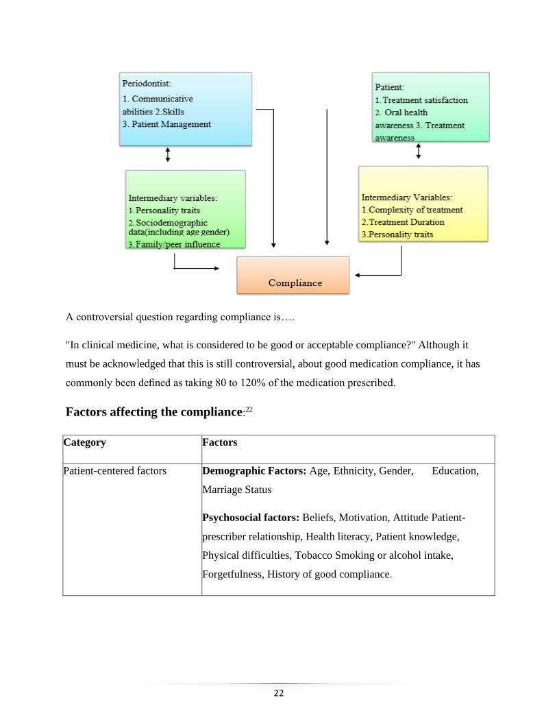
Various fixed and intermediate variables that affect the patient compliance: (Hoogstraten et al.
2005).
22
A controversial question regarding compliance is….
"In clinical medicine, what is considered to be good or acceptable compliance?" Although it
must be acknowledged that this is still controversial, about good medication compliance, it has
commonly been defined as taking 80 to 120% of the medication prescribed.
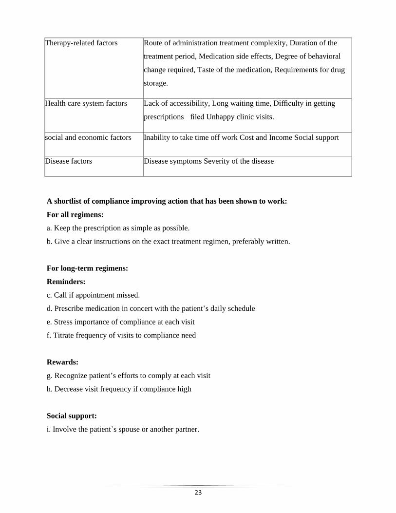
Factors affecting the compliance:22
Category Factors
Patient-centered factors Demographic Factors: Age, Ethnicity, Gender, Education,
Marriage Status
Psychosocial factors: Beliefs, Motivation, Attitude Patient-
prescriber relationship, Health literacy, Patient knowledge,
Physical difficulties, Tobacco Smoking or alcohol intake,
Forgetfulness, History of good compliance.
23
Therapy-related factors Route of administration treatment complexity, Duration of the
treatment period, Medication side effects, Degree of behavioral
change required, Taste of the medication, Requirements for drug
storage.
Health care system factors Lack of accessibility, Long waiting time, Difficulty in getting
prescriptions filed Unhappy clinic visits.
social and economic factors Inability to take time off work Cost and Income Social support
Disease factors Disease symptoms Severity of the disease
A shortlist of compliance improving action that has been shown to work:
For all regimens:
a. Keep the prescription as simple as possible.
b. Give a clear instructions on the exact treatment regimen, preferably written.
For long-term regimens:
Reminders:
c. Call if appointment missed.
d. Prescribe medication in concert with the patient’s daily schedule
e. Stress importance of compliance at each visit
f. Titrate frequency of visits to compliance need
Rewards:
g. Recognize patient’s efforts to comply at each visit
h. Decrease visit frequency if compliance high
Social support:
i. Involve the patient’s spouse or another partner.
24
Possible methods of improving compliance - The following methods have been developed to
increase compliance which has proved successful in private practice. They are generalities and
must be individualized to each patient and therapist.
Simplify:
The simpler the required behavior, the more likely it is to be carried out.
Accommodate:
The more dentists practice and their suggestions fit the patients‟ needs, the more likely patients
are to comply. Satisfied patients tend to comply with more of the recommended therapy than
dissatisfied patients.
Remind patients of appointments:
The reason for a failure for a given appointment may be from a patient side or from the
dentist side which creates problems for both the patient and the dentist.
Patients break appointments for various reasons. Other factors that may contribute are
age, race, psychosocial problems and the percentage of previous noncancelled appointments.
Communication is a key element along which avoids this general problem. Appropriate vehicles
for appointment reminders include postcards and telephone contact.
Keep records of compliance:
Patients can “get lost in the system” and efforts should be made to keep up with them.
This often requires advanced systems, and a computer for appointment control and tracking
missed visits. Communication with the patient should be initiated as quickly as possible when
non-compliant behavior is noted. The sooner the patient is contacted after missing the
appointment, the more likely they are to keep their new appointment.
Inform:
Providing written informed consent is useful in reducing non-compliance. Telling the
patient the causes of the disease process and their role in its treatment improves compliance. In
25
addition, find out what the patient's goals are for their teeth and then show them how they may
achieve their goals only if they participate in the management of the disease.
Provide positive reinforcement:
Positive feedback and constructive guidance can help the patient to do better when
compared to a negative approach to their compliance problem.
Identify potential non-compliers:
If any patient is suspicious regarding compliance (not following), then discuss the
problems which he/she may encounter due to it. Then track these patients closely. Ensure the
dentist’s involvement. There is evidence that, in some cases, dentists are more likely to
encourage compliance than dental hygienists. Noncompliance decreased by 50% when these
general approaches were applied to a private periodontal practice over 5 years.
26
MAINTENANCE PHASE
Once the periodontal therapy is completed, the principal concern is to maintain achieved
periodontal health by preventing recurrence, which is referred as the "Maintenance Phase of
Periodontal Therapy."
“Once a state of oral health has been established, periodic evaluation is necessary for the
continued health of the supporting structures of the teeth”.24
In 1916 Widman stated that "If one succeeds in having the patient carry out effective
mouth hygiene after the operation, there is no return of pyorrhea"
Bunting in 1928 emphasized that „procedures must be performed thoroughly to keep the
teeth clean from secondary deposits, so that diseases recurrence is limited.
During the past 20 years, the main thrust of maintenance is oral cleanliness because
plaque and calculus are believed to be intimately associated with the development of periodontal
disease. However, with increasing knowledge of periodontal disease, methods for maintaining
periodontal health become more sophisticated.
A MAINTENANCE PROGRAM:
The main assumption of the maintenance program is that “Adequate therapy has been
provided for whatever periodontal disease existed”.
Initially, the patient should be provided with thorough prophylaxis and complete
reinforcement instructions in oral hygiene procedures every 3 months. The 3-month interval
should be increased, maintained, or decreased depending on the evaluation of the stability of the
supporting structures.
In determining the optimum interval, the three most critical factors are:
(1) The degree of inflammation in the gingival tissues,
(2) Amount of plaque and calculus accumulation, and
(3) Changes in gingival crevice depth and level of the attachment apparatus.2
27
According to clinical studies, close monitoring is important to develop a time interval that is
appropriate for each individual to maintain the health of the periodontium.
1. Patients with good oral hygiene and healthy and stable periodontium the maintenance
appointments can be prolonged.
2. The patients with suboptimal plaque control and a concomitant high prevalence of
bleeding sites recalled more frequently,
3. Patients with healthy gingival conditions but with a severely reduced height of
periodontal support are recalled at short intervals (not exceeding 3-4 months) to exclude
or at least reduce the risk of tooth loss.
Factors to be recorded and evaluated at maintenance visit:
1.Changes in general health status,
2.Scoring of plaque and calculus accumulation; and
3.Notation of problem area(s) for edema, bleeding, pocket development, attachment loss, tooth
mobility, and oral tissue changes.
Management of time during maintenance visit (generally 1 hour) –
The first 10-15 minutes – For clinical evaluation of the periodontal and caries conditions.
The second 30-40 minutes - used to clean and polish all supragingival tooth surfaces, following
the instrumentation of the subgingival sites that have been diagnosed as being inflamed.
Last 5-15minutes - used to provide adjunctive preventive measures such as topical application of
fluoride or chemical plaque control agents. In addition to the evaluation of the periodontal and
caries conditions, the vitality of abutment teeth for fixed bridgework should be checked.25 A
small percentage of integrated dental implants ultimately fail either due to trauma (from the
occlusion or an ill-fitting prosthesis) or from an infection similar to periodontitis, or from a
combination of these factors.
A typical maintenance visit for implants consist of -
1.Updating the patient‟s medical and dental history.
2.Review of oral hygiene and modification if needed.
3.Examination of implant and peri-implant tissues.
4.Evaluation of patient complaints in the area of implants.
28
5.Evaluation implant stability: manually or by using computerized devices.
Setting Maintenance Intervals:
A. Patients with both teeth and implants should see the periodontist as often as necessary to keep
the periodontium or peri-implant tissues healthy.
B. Totally edentulous patients with implants should be seen at least once per year.10
Re-education:
During the recall visits the plaque score record in previous visits are used as an
educational tool, highlighting to the patient-specific areas where plaque is accumulating. Many
patients just require ‘fine-tuning’ of their oral hygiene along with specific advice needed for the
more difficult areas i.e. interproximal areas, furcation areas, and root surfaces. The importance of
daily plaque removal from these areas must be emphasized.26
Re-motivation:
Compliance is the most difficult aspect of periodontal care. Patients usually respond well
to periodontal therapy during the active phase of treatment, but patients who are well educated in
oral hygiene techniques and who demonstrate an ability to achieve low plaque levels, show
decreased compliance over time.
Patients slip back easily into their old ways. Re-motivation and positive reinforcement of
patients are necessary to maintain the high standard of oral hygiene required for periodontal
health. Re-motivation involves reminding the patient about the nature of the periodontal disease
and the potential consequences of the untreated disease, the relationship between plaque and
periodontal disease and the patient's power to prevent disease progression with good oral hygiene
practice. This information and advice should be given in a language that is non-threatening, and
in layman's terms. Positive reinforcement and acknowledgment of the successes achieved by the
patient are important for ensuring on-going compliance.
It may be difficult to change a non-complying patient into a very compliant one, but with
re- motivation, at least the small improvement in oral hygiene behavior achieved is maintained.
Therefore, regular supragingival prophylaxis and calculus removal should be performed at
appropriate intervals based on individual needs during maintenance care.
29
Sub-gingival debridement should be confined to:
Sites that show an increased probing depth, Sites that demonstrate re-infection and suppuration,
Sites that are difficult for the patient to access and demonstrate persistent bleeding on probing,
for example, furcation areas and pockets >4 mm.22
Adjunctive use of antimicrobial agents:
The evidence-based rationale supporting the use of adjunctive antimicrobials within maintenance
care is increasing day by day.
Use of antimicrobials during SPT by the professional:
Subgingival irrigation:
Slots and Jorgensen (2000) advised using mechanical debridement followed by subgingival
irrigation with povidine-iodine during Supportive periodontal therapy appointments of
periodontitis patients due to bactericidal potential in areas with difficult access.
Disadvantage:
Due to the short-term effect of subgingival irrigations, additional anti-microbial means should be
indicated when a more prolonged antimicrobial effect is desired.
Sustained release delivery systems:
Local antimicrobial therapy is an alternative approach aimed at providing antimicrobial
concentration adequate to penetrate the biofilm in the periodontal pocket for prolonged time
periods.
Kasaj et al. 2007 evaluated the effectiveness of a controlled-release chlorhexidine chip as
adjunctive therapy to scaling and root planing with a newly developed ultrasonic device in
supportive periodontal therapy. The target sites were randomly treated with either a newly
developed piezo-driven ultrasonic device Vector or ultrasonic system (VUS) + Chlorhexidine
chip or Vector or ultrasonic system alone without adjunctive antimicrobial treatment. The
average reduction of Probing depth and improvement in Clinical attachment level was greater in
the Vector or ultrasonic system + Chlorhexidine chip sites than in sites treated with the Vector or
ultrasonic alone at 1, 3 and 6 months. These data suggest that the Chlorhexidine chip application
30
following Supportive periodontal therapy with the tested ultrasonic device is beneficial in
improving periodontal parameters in patients on Supportive periodontal therapy.
Minocycline is a bacteriostatic, antimicrobial agent, which is available in a gel and microsphere
formulation for local application within periodontal pockets. A case-control study comparing the
efficacy of a 2% minocycline gel versus scaling and root planing (SRP) alone, in treating sites
with pocket depths greater than or equal to 5 mm, with bleeding on probing during a 12- month
period of maintenance care, resulted in similar clinical outcome.
Similar results have been found with the adjunctive use of a 25% metronidazole gel in
combination with SRP.
Systemic antibiotic:
The use of antibiotics during Supportive periodontal therapy should be reserved for
patients experiencing periodontal breakdown and recurrence of the disease.
Nakajima et al 2016 examined the short-term and long-term microbiological and clinical
effects of systemic sitofloxacin and azithromycin (AZM) on active periodontal pockets during
supportive periodontal therapy. These results indicate that monotherapy of systemic Sitofloxacin
and Azithromycin could be an alternative treatment during supportive periodontal therapy.
Use of antimicrobials for personal Supportive periodontal therapy:
Mouth rinses
Sangeetha et al. 2015 Patients who, as a result of therapy, have only shallow periodontal
pockets should concentrate on supragingival plaque control and elimination of pathogenic
bacteria from the oral reservoir.
Chlorhexidine rinses for 8 days may be recommended after each Supportive periodontal
therapy appointment, to ensure prevention of re-infection during the 3–4-month interval between
Supportive periodontal therapy appointments.
Irrigation:
Braun and Ciancio (1992) the daily use of supragingival irrigation with antimicrobial agents may
partially benefit patients with deep periodontal pockets during Supportive periodontal therapy,
and the use of subgingival tips for selected deep pockets may augment the effect of irrigation due
to deeper penetration properties.
31
Disadvantages:
The manual complexity of personal subgingival irrigation
Low compliance
Cost and possible side-effects (risk for abscess formation and bacteremia)
Toothpaste:
Using a toothbrush as a delivery device, it was found that toothpastes can penetrate only up to
0.9 mm into the periodontal pockets. No recommendation can be concluded for use of specific
toothpaste during Supportive periodontal therapy for periodontitis patients.
Polishing, fluorides, determination of recall interval:
The recall hour is concluded with polishing the entire dentition to remove all remaining
soft deposits and stains. Following polishing, fluorides should be applied in high concentration in
order to replace the fluorides which might have been removed by instrumentation from
superficial layers of the teeth. Fluoride or chlorhexidine varnishes may be applied to prevent root
surface caries in areas of gingival recession. The determination of future Supportive periodontal
therapy visits must be based on the patient’s risk assessment.
Therefore, current evidence supports the adjunctive use of antimicrobial agents during
maintenance care. In addition to the superior clinical outcomes reported with their use,
adjunctive agents can offer further benefits like shorter clinical time and post-operative patient
sensitivity as they reduce the requirement for repeated sub-gingival debridement.
Disadvantages: Expensive, Risk of bacterial resistance (on repeated use).
Therefore used only for the maintenance care of patients with good plaque control in
residual pockets of 5 mm or greater. Non-responding sites, or those with persistent bleeding,
should be specially targeted.
Application of topical fluorides:
Gingival recession and exposure of the root surface is a common side-effect of
periodontal disease or periodontal disease therapy. Gingival recession increases the risk for root
caries and predisposes to cervical sensitivity. In one study, 82% of patients on periodontal
supportive care had evidence of root caries (either treated or untreated) and the number of root
lesions in an individual was related to his/her plaque score.27 In these cases regular application of
32
topical fluoride will help to prevent root caries and may relieve some of the symptoms of
sensitivity.
General guidelines for dental treatment planning have been published based on five
treatment phases.
I. Systemic II. Acute III. Cause-related IV. Surgical V. Corrective and VI. Maintenance.
Systemic phase:
A. Consultation with patients‟ physician
B. Pre-medication
C. Stress/fear management
D. Any necessary treatment considerations for systemic disease.
Acute phase:
A. Emergency treatment for pain and infection
B. Addressing the urgent chief complaint.
Cause-related phase:
A. Oral hygiene education, patient motivation, and risk assessment
B. Mutual goal-setting for acceptable outcomes/end-points of therapy
• Implementation of strategies for risk reduction
C. Excavation of deep carious lesions
• determine restorability
D. Extraction of hopeless teeth along with non-surgical periodontal debridement
E. Removal of plaque retentive factors
F. Necessary endodontic and occlusal therapy
G. Post-treatment re-evaluation
• Objective assessment of endpoints of therapy.
Surgical corrective phase
A. Resective/regenerative and implant surgical procedures
B. Post-surgical re-Evaluation
33
• Objective assessment of endpoints of therapy
C. Definitive prosthodontic restoration.
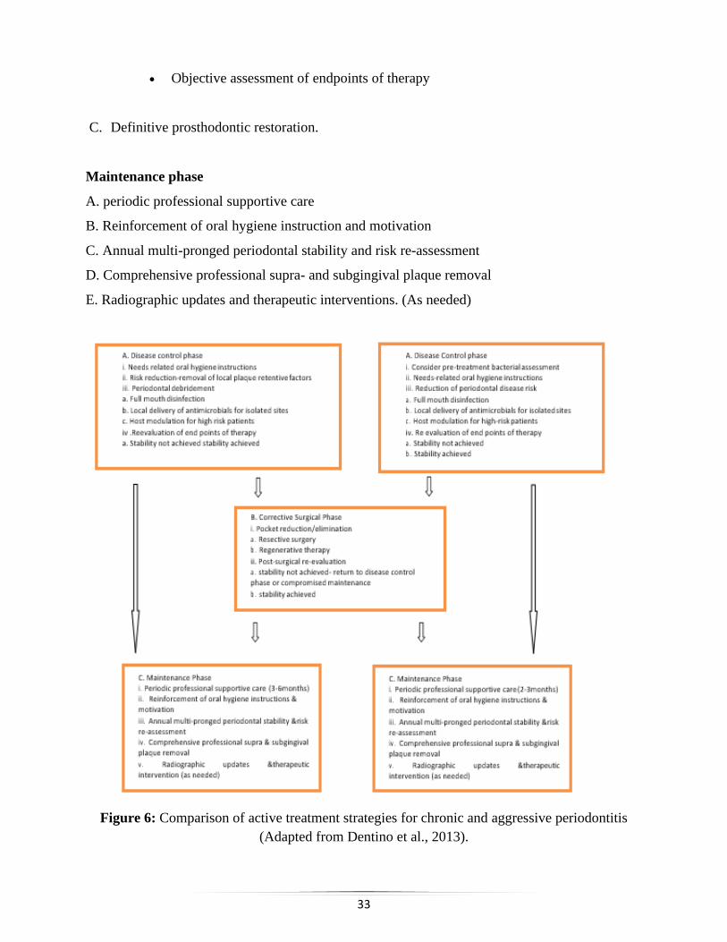
Maintenance phase
A. periodic professional supportive care
B. Reinforcement of oral hygiene instruction and motivation
C. Annual multi-pronged periodontal stability and risk re-assessment
D. Comprehensive professional supra- and subgingival plaque removal
E. Radiographic updates and therapeutic interventions. (As needed)
Figure 6: Comparison of active treatment strategies for chronic and aggressive periodontitis
(Adapted from Dentino et al., 2013).
34
Frequency and the time allotted for periodontal maintenance:
Many patients presenting with recurrent gingivitis without additional attachment loss
after definitive periodontal therapy may be adequately maintained with Periodontal Maintenance
performed semi-annually and for patients with a history of periodontitis, numerous clinical
studies suggest that Periodontal Maintenance should be performed at intervals of less than 6
months.
Intervals of 2 weeks, 2-3 months, 3 months, 3-4 months, 3-6 months, 4-6 months, and up
to 18 months have been evaluated in general data suggest that most patients with a previous
history of periodontitis should obtain Periodontal Maintenance at least 4 times per year, since
that interval will result in a decreased likelihood of progressive disease, compared to patients
receiving Periodontal Maintenance on a less frequent basis. Finally, it can be concluded that the
periodontal maintenance schedules should be individualized, with the Periodontal Maintenance
intervals tailored to the needs of each patient.
Although pocket debridement suppresses components of the subgingival microflora
associated with periodontitis, periodontal pathogens may return to baseline levels within days or
months. The return of pathogens to pretreatment levels generally occurs in approximately 9- 11
weeks but can vary dramatically among patients. The time required for periodontal maintenance
appointments should be dictated by factors such as the number of teeth or implants, patient
cooperation, oral hygiene efficacy and compliance, systemic health, the previous frequency of
periodontal maintenance, instrumentation access, history of disease or complications, and the
distribution and depth of the Sulcus.
The following items may be included in a periodontal maintenance visit, subject to previous
examination, history and the judgment of the clinician:
A. Review and update of medical and dental history
B. Clinical examination (to be compared with previous baseline measurements)
1.Extraoral and intraoral examination and recording of results
2.Dental examination and recording of results
a) Tooth mobility, fremitus, and occlusal factors
b) Coronal and root caries assessment
c) Restorative and prosthetic factors, such as defective restorations
d) Other tooth-related problems, such as open contacts or malpositioned teeth
35
3.Periodontal examination and recording of results: Probing depth, bleeding on probing, general
levels of plaque and calculus, evaluation of furcations. exudate, other signs of disease
progression, microbial testing if indicated gingival recession, and attachment level if indicated.
4.Examination of dental implants and peri-implant tissues and recording of results: Probing
depths, bleeding on probing, examination of prosthesis/abutment components, evaluation of
implant stability, occlusal examination, other signs and symptoms of disease activity Example:
Pain and suppuration.
C. Radiographic examination
1.Radiographs should be current, based on the diagnostic needs of the patient, and should permit
appropriate evaluation and interpretation of the status of the oral structures, including teeth,
periodontium, and dental implants.
2.The frequency and the number of radiographs needed is decided based on individual patient
needs.
3.Radiographic abnormality should be noted.
D. Assessment of disease status or changes by reviewing the clinical and radiographic
examination findings, compared to baseline
E. Assessment of personal oral hygiene
To assess the individual oral Hygiene patients should perform their hygiene regimen
immediately before the recall appointment. Plaque control must be reviewed and corrected until
the patient demonstrates the necessary proficiency.
F. Treatment
1.Subgingival and supragingival plaque and calculus removal.
2.Behavioral modification:
a) Oral hygiene re-instruction
b) Adherence to suggested Periodontal Maintenance intervals
c) Counselling on control of risk factors e.g. Smoking, nutrition, stress
3.Selective scaling or root planing if indicated
36
4.Occlusal adjustment, if indicated
5.Use of systemic antibiotics, local antimicrobial agents, or irrigation procedures as necessary
6.Root desensitization, if indicated
7.Surgical therapy (or discontinuation of periodontal maintenance and treatment of recurrent
disease), if indicated.
G. Communication
1.Informing the patient regarding the current status of oral condition and need for additional
treatment if indicated
2.Consultation with other health care practitioners who may be providing additional therapy or
participating in the Periodontal Maintenance program.
H. Planning
1.For most patients with a history of periodontitis, recall intervals should be planed according to
individual needs.
2.Based on the evaluation of clinical findings and assessment of disease status, PM frequency
may remain the same, be modified, or the patient may return to mechanical, chemical, surgical
and/or nonsurgical treatment.

37
RETREATMENT
Treatment implies “some procedure used to relieve or cure a disease” and Retreatment
simply means treating “again, anew or once more”. As periodontal disease tends to be chronic
and “episodic”, the retreatment aspect of patient care becomes a significant part of the
maintenance program.28
Even after long periods of maintenance, there is a tendency for periodontal disease to
recur. Good oral hygiene and adequate supportive care reduce the rate of relapse but do not
eliminate it. Unfortunately, no available clinical tests can predict if or where the recurrent disease
will occur.
Can the recurrent periodontal disease be totally prevented?
Not with present knowledge, but the occurrence and severity can be lessened with proper
supportive periodontal treatment. All complex periodontal cases should have a thorough
evaluation at regular intervals, the frequency being determined by the individual situation. This
examination may take place once a year for a poor response patient and 2- 3 years for a stable
case. The appointment for thorough evaluation should be scheduled at 2-3 weeks after a regular
supportive periodontal treatment visit.
The regular, short “check‟ by the periodontist in typical supportive periodontal treatment is all
too often too superficial to be of much value.
Reasons for Regression
• The most common cause of failure is the inability of the patient to keep the bacterial
population of the crevicular areas at a permissible level. The first step to take in
retreatment is to review the patient's oral hygiene regimen.
• Failure to smooth the involved roots during the original treatment is often a cause of
pocket recurrence. Even a good brushing and flossing technique will not keep
incompletely treated roots free of significant amounts of bacterial irritants.
• The choice of an improper surgical technique will usually result in a relative failure.
38
• Certain types of periodontal disease appear to have an occlusal factor that must be
controlled. Equilibration, the control of bruxism, and possibly splinting must be
accomplished in such cases before considering additional surgery.
• Further surgery performed before the occlusal factor is controlled and before the
supporting tissue has had time to respond to occlusal therapy will accelerate the disease
process.21
Criteria for Failure
A treated case that is failing, either generally or in certain areas, is characterized by one or all of
the following symptoms:
1. Sulcus that bleed when probed. This probing should be done 1 to 2 weeks after a
preventive treatment.
2. Sulcus get progressively deeper. This can be determined by comparing sulcus depth with
the previous probing.
3. Bone loss. This can be determined by comparing old and new radiographs that have been
properly taken with the paralleling technique.
4. Increased tooth mobility. Gradual increases in tooth mobility values should alert the
therapist that the periodontium is undergoing destructive changes.
The recurrence of disease occurs due to the inadequacy of the original treatment or
sometimes the clinician will encounter a new disease in a previously healthy area. Most
problems occur, however, in previously treated sites. The clinical signs of breakdown are the
same as those encountered before the original treatment. They are:
1. Periodontal pockets that bleed or show exudate when gently probed
2. Periodontal pockets that get progressively deeper
3. Alveolar bone loss (diagnosed by comparing radiographs obtained at different times)
4. Increased tooth mobility, and
5. Presence of plaque, gingivitis and subgingival calculus.27
According to Chace 1996 criteria for SPT are:
1.If clinical signs of recurring disease are slight, it is prudent not to make an immediate decision
as to whether or not the patient needs retreatment.
39
The patient may be having a lapse in oral hygiene or perhaps be behind in supportive periodontal
treatment visits. In such a case, the patient should be given an appointment in 2-3 weeks to see
whether the clinical signs of breakdown are still present. Another session of oral hygiene efforts
is all that is needed.
2.If the patient’s oral hygiene is inadequate and, the original therapy was less than ideal.
The recurrent periodontitis is generally associated with mild gingival inflammation and edema,
as well as a moderate increase of probing depth. Then only the oral hygiene maintenance by the
patient is may not sufficient, i.e. after the second session the examination of the patient if the
situation has not markedly improved, the patient may be referred for debridement and root
planing in conjunction with access periodontal surgery.
3.If the patient's oral hygiene and general oral health are good but who present one or two
isolated areas in which probing depths have increased 2 mm or more and gentle probing
produces bleeding or exudate. The first step in the treatment of this type of breakdown is to
debride the pocket with a fine, sharp current, irrigate the pocket and, in some instances, pack the
area with a tetracycline hydrochloride fiber. If healing is incomplete and a surgical procedure is
indicated, but the redebridement procedure is a valuable first step.
4.Recurrent disease in a segment of the dentition rather is another type of breakdown
encountered in supportive periodontal treatment. If improved oral hygiene and additional scaling
and root planing do not return the probing depths to an acceptable level, surgical flaps should be
raised for access to more definitive debridement.
The optimal way to decide when to retreat would be to use a method that accurately
predicts future attachment loss. Unfortunately, this sort of test does not exist.Two measurements
are used: changes in clinical attachment loss (probing depth plus gingival recession) or probing
depth alone and tissue signs, especially bleeding upon probing and suppuration.
Based on research work done by Lang and Tonetti 2003 guidelines for specific therapy
decisions can be used for patients on supportive periodontal treatment. This approach has
improved the communication between the dental hygienist and periodontist in one office for
many years and is used as a basis for retreatment and instituting supportive periodontal treatment
intervals.

40
Therapy Decisions Based on Probing Depths or Clinical attachment loss Changes For
Patients With Plaque Associated Gingivitis or Chronic Periodontitis: 10
Wilson & kornman.,1996
1. Baseline probing means:
• The probing depths found at initial examination if no periodontal therapy is done; or
• The probing depths found at least 1 year after periodontal therapy.
2. Surgery assumes that :
• Probing depths are 6mm or greater and there are signs
• Root surface has been root planned as thoroughly as possible using closed methods;
Base Line
No Possible Aggresive 2 mm or greater
Complete
Same SPT intervals
No change for 2
Consecutive visits
Lengthen the
interval by one
month
Root
Errattic
Compiler
s
Root planing 1 mm
Same probing
depths Reduced probing
depths
Improved
or stable
Same
depth
with
Surger
SPT
Radiographic
bone loss
SPT
SPT
Osseous
surgery or
regenerative
procedures
No
radiographic
Gingivectomy,
Gingivoplasty
SPT
41
• The patient is a good surgical candidate; and
• The patient is maintaining reasonable plaque control.
3. Gingivectomy/gingivoplasty assumes that:
• Through root planing will be done at surgery; and
• Soft tissues only are removed.
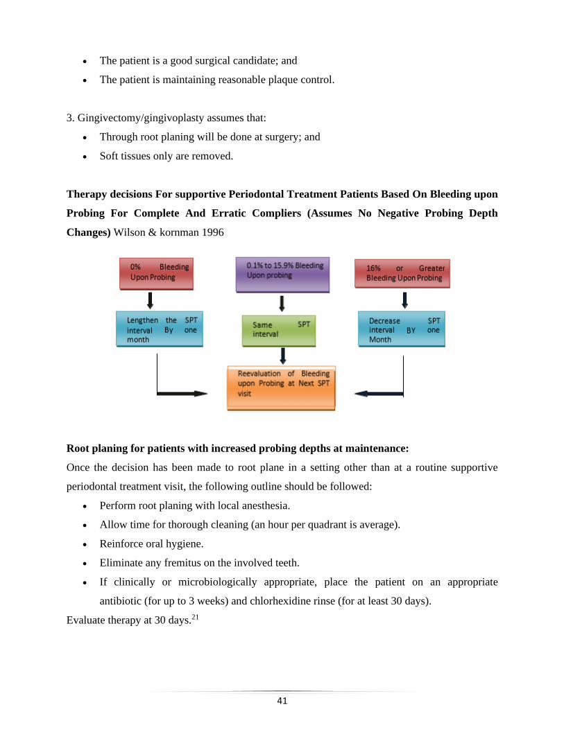
Therapy decisions For supportive Periodontal Treatment Patients Based On Bleeding upon
Probing For Complete And Erratic Compliers (Assumes No Negative Probing Depth
Changes) Wilson & kornman 1996
Root planing for patients with increased probing depths at maintenance:
Once the decision has been made to root plane in a setting other than at a routine supportive
periodontal treatment visit, the following outline should be followed:
• Perform root planing with local anesthesia.
• Allow time for thorough cleaning (an hour per quadrant is average).
• Reinforce oral hygiene.
• Eliminate any fremitus on the involved teeth.
• If clinically or microbiologically appropriate, place the patient on an appropriate
antibiotic (for up to 3 weeks) and chlorhexidine rinse (for at least 30 days).
Evaluate therapy at 30 days.21
42
Important clinical parameters for monitoring periodontal health during supportive
periodontal treatment are:
• Loss of attachment of 2 mm or more and the associated deepening of the periodontal
pocket or gingival recession
• Bleeding on probing
• Suppuration or exudate and
• Including the gingival recession, furcation involvement, caries, open contacts and status
of occlusion and arch relationship, including any anomalies.
Less objective parameters to consider in determining whether retreatment is appropriate. The
subjective parameters often incorporate the therapist’s experience, emotions, and clinical savvy
or intuition.
Included in this group of parameters are:
clinical history; loss of alveolar bone; crown-root ratio; increase in mobility; changes in the
patient’s immune system and response; effectiveness in daily removal of bacterial plaque;
smoking; patient’s age; root surface smoothness; evidence of calculus or root surface accretions;
patient systemic disease or complications; patient medicines; patient compliance with treatment
recommendations, including scheduling of supportive periodontal treatment visits; new clinical
procedures; and ability to pay for professional services (either with insurance coverage, co-
payment or fee for service).10
43
SUPPORTIVE CARE OF DENTAL IMPLANTS
All dental implants are at risk of developing peri-implant diseases as they are placed in
the microbe-laden oral environment. Patients who have undergone successful implant therapy
should receive individualized, systematic and continuous supportive care of the peri-implant
tissues. Patients at higher risk for peri-implantitis, such as those with partially edentulous and
pre-existing chronic periodontitis, should be identified and monitored closely.29
Following the restoration of an implant, the patient should be re-evaluated regularly (i.e.,
every 3 to 4 months) during the first year. After the first year, the response of the peri-implant
tissues should be assessed, at which time the appropriate frequency of periodontal maintenance
should be determined.
Assessment:
Updating of Medical and Dental Histories:
The assessment begins with updating the patient’s medical and dental histories, to ensure that all
concomitant conditions and therapies are known and to identify patients in high-risk categories.
1.Soft-Tissue Assessment:
Signs of gingival inflammation, such as redness, swelling, alterations of contour and
consistency, aberrant gingival form or the presence of a fistula.
2.Plaque Index:
Plaque monitoring is performed and documented at every maintenance visit, to allow
longitudinal assessment of oral hygiene.
Plaque indices commonly used for evaluating plaque on implants: 30
O’ Leary colleagues
% score = no. tooth surfaces with the plaque/ no. of tooth surface present b 100
Lindquist and colleagues
0=no visible plaque
1= local plaque accumulation
2= general plaque accumulation>25%
44
Mombelli and colleagues
0 = no visible plaque
1= plaque recognized by running probe over the smooth margin of the implant
2= visible plaque
3= abundance of soft matter
Rough-surfaced implants accumulate greater amounts of plaque than smooth-surfaced implants,
which may increase the risk for peri-implantitis. Bacterial adhesion has also been shown to be
influenced by surface roughness in vitro, with higher subgingival bacterial load occurring on
rough surfaces.
3.Clinical Probing Depth:
Probing is an important and reliable diagnostic parameter in the longitudinal monitoring of peri-
implant soft tissues
4.Bleeding on Probing:
A prospective study of implants confirmed that, similar to the situation for natural teeth, absence
of bleeding on probing had high negative predictive value and thus can be interpreted to
represent stability of the peri-implant soft tissues.
Gerber and colleagues demonstrated that 0.15 N of pressure might represent the threshold (i.e.,
minimum pressure) to avoid false-positive readings for bleeding on probing around oral
implants.
5.Stability of the Soft-Tissue Margins:
Any apical migration of the gingival margin should be noted and monitored, even though there is
no evidence that gingival stability is important for implant survival over the long term.
6.Mobility:
Mobility should be assessed either manually or by automated means such as the Periotest dental
measuring instrument (Siemens, Bensheim, Germany) or the Ostell instrument (Ostell,
Gothenburg, Sweden). If only one implant in a multiunit splinted prosthesis has mobility, the
mobility may be masked. Therefore, it has been suggested that fixed, multiunit, retrievable
45
implant-retained prostheses be removed periodically to assess mobility, gingival health and
hygiene status.
The cause of any mobility should always be ascertained, specifically whether it is due to failure
of the prosthesis or failure of osseointegration. If the implant as a whole becomes mobile, it is
deemed to have failed and should be removed.
7.Occlusion:
Eccentric guidance should be used to ensure optimal distribution of the potentially destructive
effects of excursive occlusal parafunction. If technical complications occur, they should be
treated accordingly. Parafunctional habits should be documented and treated since the
application of excessive concentrated force can cause rapid and substantial peri-implant bone
loss.
8.Bone Level:
If clinical signs suggest the presence of peri-implantitis, radiography of the site should be
performed to confirm the diagnosis.
To facilitate the accurate reading of radiographs (should they be needed in the future), it is
important to establish baseline bone levels after implant placement and again after insertion of
the prosthesis.30
The precise interventions provided during supportive implant therapy visits will be determined
by the findings obtained following a thorough examination of the peri-implant tissues and a
careful assessment of the risk factors for peri-implantitis. A complete examination of the status
of dental implants includes the same general steps performed during a routine examination of the
natural dentition.
It is clear that biofilm forms rapidly on both smooth and rough implant surfaces.
It is critically important that a patient-specific program of professional care be established to
prevent the development of microbe-associated peri-implant diseases.
46
The program should include:
Individual oral hygiene instructions; Control of relevant risk factors; and
Provision of professional preventive interventions, including maintenance care.
The primary goal of a program of supportive implant therapy is to prevent the
development of peri-implantitis. This is especially important because once peri-implantitis
occurs it is extremely difficult to treat. Indeed, there is no reliable evidence on the best way to
treat this condition. It has generally been assumed that the best way to keep peri-implant tissues
healthy is to place affected patients on a well-designed supportive implant therapy program that
stresses excellent oral hygiene and periodic recall visits for professional removal of biofilm
deposits from implant surfaces.
This approach has also been advocated for reversing the course of peri-implant mucositis.
Several protocols have been proposed for supportive implant therapy programs but there is no
consensus on what specific interventions are required for the best results.29 The indication for the
appropriate treatment strategy has been demonstrated in patient studies leading to the
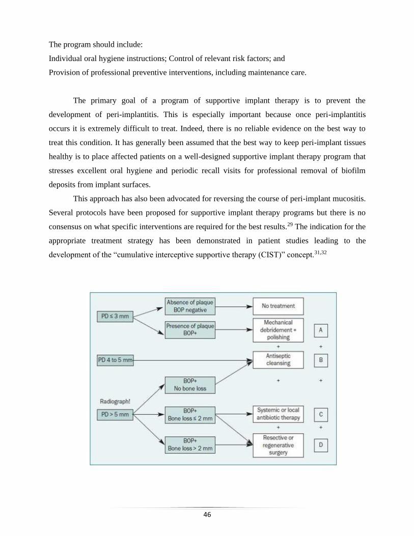
development of the “cumulative interceptive supportive therapy (CIST)” concept.31,32
47
• In part A of the CIST protocol, typically initiated when plaque and BOP are present but
PDs are 3mm or less, patients are re-instructed in oral hygiene and motivated to initiate
and continue maintenance;
Mechanical debridement is performed using non-metallic curettes; and polishing is done
by using a rubber cup and nonabrasive polishing paste.
• Part B, when PDs of 4 to 5 mm are found, consists of antiseptic treatment. Here, chemical
plaque control is performed using chlorhexidine digluconate, typically as mouthrinses
with 0.1% to 0.2% chlorhexidine for 30sec using approximately 10ml, application of
local chlorhexidine gel (0.2%), and/or local irrigation with chlorhexidine(0.2%), 2 times
a day for 3 to 4 weeks.
• Protocol C, systemic or local antibiotic treatment, is initiated when PDs are greater than
5mm. in addition, radiography should be used to supplement clinical findings. Typical
systemic treatment is with Ornidazole (1000mg, OD) or metronidazole (250mg, TID) for
10 days, or a combination of amoxicillin (375mg TID) and metronidazole (250mg TID)
for 10 days.
Local treatment might include local application of antibiotics using a controlled-release
device for 10 days, e.g: tetracycline fibers and minocycline microspheres.
Once treatment modalities A, B, and C have been completed, a surgical approach (D)
may be considered. Surgical therapy for peri-implantitis should be performed in conjunction with
systemic antibiotics and implant surface decontamination. If regenerative treatment is chosen, a
barrier membrane technique alone or in combination with autogenous grafts and/or bone
substitutes may be considered. Respective surgery may be considered when the peri-implant
defect is not suitable for regenerative techniques.33
In 2004 it was modified and called AKUT-concept by Lang et al.33 The basis of this
concept is a regular recall of the implanted patient and repeated assessment of plaque, bleeding,
suppuration, pockets and radiological evidence of bone loss.34
48
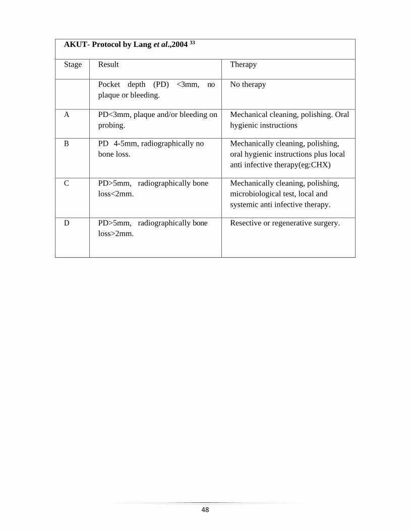
AKUT- Protocol by Lang et al.,2004 33
Stage Result Therapy
Pocket depth (PD) <3mm, no
plaque or bleeding.
No therapy
A PD<3mm, plaque and/or bleeding on
probing.
Mechanical cleaning, polishing. Oral
hygienic instructions
B PD 4-5mm, radiographically no
bone loss.
Mechanically cleaning, polishing,
oral hygienic instructions plus local
anti infective therapy(eg:CHX)
C PD>5mm, radiographically bone
loss<2mm.
Mechanically cleaning, polishing,
microbiological test, local and
systemic anti infective therapy.
D PD>5mm, radiographically bone
loss>2mm.
Resective or regenerative surgery.
49
SUMMARY
Supportive Periodontal Treatment is the group of procedures performed at selected
intervals to assist the periodontal patient in maintaining oral health. Formerly referred to as recall
maintenance, preventive maintenance or supportive periodontal therapy, this periodic assessment
is established following the initial active periodontal therapy and it includes an update of the
medical and dental histories, extra-oral and intra-oral soft tissue examination, dental
examination, periodontal evaluation, radiographic review, removal of the bacterial flora deposits
from crevicular and pocket areas, scaling and root planing where indicated, polishing of the teeth
and a review of the patient’s plaque control efficacy. These procedures aim to prevent the
recurrence and progression of periodontal disease and to prevent or reduce the incidence of tooth
loss.1
Several investigations have indicated that only a minority of periodontal patients comply
with the prescribed supportive periodontal care. Wilson & crouch 1987 indicated that a complete
compiler group retained more teeth than did erratic compliers. Miyamoto et al 2006 suggested
that the progress and recurrence of periodontal disease can be prevented in complete compliers
by using SPT whereas Ramfjord et al 1982 suggested that periodic SPT can prevent the
recurrence of periodontal disease even in patients with poor oral hygiene.
Paramount to the success of the supportive periodontal treatment program is
communication with the patient about the state of his or her periodontal health. Assessment of
disease status by reviewing the current clinical and radiographic examination findings compared
with baseline is important. The word “care” must always be an integral part of the health care
profession. It has been stated that the “C” in “care” stands for concern, the “A” for availability,
the “R” for 62 being responsive to the needs of the patient and the “E” for equitable. A service or
treatment (such as the supportive periodontal treatment visit) must be performed in such a way
that the cost is equitable to the patient and the health care provider.
To improve compliance, the demands on patients can be simplified by pre appointing the
next supportive periodontal treatment visit before the patient leaves the office, seeking out the
patient‟s concerns and acting upon them, maintaining good records and communication with the
patient and others involved health care professionals, informing the patient of the consequences
of noncompliance, attempting to identify noncompliers before corrective treatment is necessary
50
and continuing to offer positive reinforcement and support to the patient on their periodontal
status. In short, excellence is the goal of a supportive periodontal treatment program: taking
exceptional care of patients and constantly innovating treatment care with new ideas and
services.
A successful long-term maintenance program is based on semantics and good
communication. This involves: 1) informing the patients of their current periodontal status and
any alterations in treatment, if indicated; 2) consultation with other health care providers who
will be providing additional dental care or participating in the supportive periodontal treatment
program, and 3) future planning. For patients with a history of active periodontitis, visits at 3-
month intervals are recommended. However, the scheduling of future patient supportive
periodontal treatment visits should be based on the evaluation of clinical findings and assessment
of disease status. Supportive periodontal treatment visit frequency may be modified, or the
patient may be returned to active treatment. The success of any supportive periodontal treatment
program is based on periodic evaluation and appropriate retreatment if indicated. The ultimate
goal of treatment is the preservation of the dentition in health and function.
Dental implants require constant maintenance and monitoring, which further involves
assessment of the patient's general and oral health, professional implant maintenance, and
diligent patient home care as critical factors that will ensure the long-term success of implants
and a predictable replacement for natural teeth.
51
CONCLUSION
Periodontal disease has a greater tendency to recur for which careful maintenance is as
important as skilful original treatment. Active and supportive periodontal treatments are the two
faces of the same coin. All complex periodontal cases should have a thorough evaluation at
regular intervals, the frequency being determined by the individual situation. Adequate steps
should be taken to ensure that the patients understand their disease, the treatment options
available, and the consequences if appropriate treatment is not done.
A serious limitation in the clinical application of adjunctive therapy or different time
intervals in Supportive periodontal therapy is the lack of clear guidelines and protocols, as
pointed out by many authors. Further knowledge regarding susceptibility and progression of
periodontal disease in a specific site, based on individual patient risk factors will ensure optimal
outcomes and cost-effective institution of a Supportive periodontal therapy regime.
Greater attention should be given to the methodology used to assess Supportive
periodontal therapy. Duration of follow-up is of paramount importance when adding adjunctive
treatments to Supportive periodontal therapy, as many adjuncts demonstrate short-term
effectiveness but fail to demonstrate long-term improvement in clinical outcomes. Studies should
focus on the clinical significance of results, in order to place the effectiveness of adjunctive
therapy in perspective.
So it can be concluded from clinical studies in dental institutions and private practices,
that periodontal therapeutic success is underpinned by an ongoing program of Supportive
periodontal therapy. This encompasses systematic and regular monitoring of periodontal
parameters in order to detect and intercept any new or recurrent disease. Individual variations to
disease susceptibility will determine the frequency and level of professional input required.
52
References
1.Wilson TG, Glover ME, Malik AK, Schoen JA & Dorsett D. Tooth loss in maintenance
patients in a private periodontal practice. J Periodontol 1987;58: 231–35.
2.Axelsson P, Lindhe J. Effect of controlled oral hygiene procedures on caries and periodontal
disease in adults. Results after 6 years. J Clin Periodontol 1981a; 8: 239–48.
3.Axelsson P, Lindhe J. The significance of maintenance care in the treatment of periodontal
disease. J Clin Periodontol 1981b;8:281–94.
4.Lindhe J, Nyman, S. Long‐term maintenance of patients treated for advanced periodontal
disease. J Clin Periodontol 1984;11:504–14.
5.Rosling B, Nyman S, Lindhe J, Jern B. The healing potential of the periodontal tissues
following different techniques of periodontal surgery in plaque‐free dentitions. J Clin
Periodontol 1976;3:233–50.
6.Nyman S, Lindhe J, Rosling, B. Periodontal surgery in plaque‐infected dentitions. J Clin
Periodontol 1977;4: 240–49.
7.Ramfjord SP, Morrison EC, Burgett FG et al. Oral hygiene and maintenance of periodontal
support. J Periodontol 1982;53:26–30.
8.Hill RW, Ramfjord SP, Morrison EC et al. Four types of periodontal treatment compared over
two years. J Periodontol 1981;52:655–77.
9.Slots J, Mashimo P, Levine MJ, Genco RJ. Periodontal therapy in humans. I. Microbiological
and clinical effects of a single course of periodontal scaling and root planing, and of adjunctive
tetracycline therapy. J Periodontol 1979;50:495–509.
10.Wilson TG, Kornman KS. Retreatment. Periodontol 2000.1996;12:119.
11.Becker W, Becker BE, Berg LE. Periodontal treatment without maintenance. A retrospective
study in 44 patients. J Periodontol 1984;55:505–9.
12.Pihlstrom BL, McHugh RB, Oliphant TH, Ortiz‐Campos C. Comparison of surgical and
non‐surgical treatment of periodontal disease. A review of current studies and additional results
after 6. years. J Clin Periodontol 1983;10:524–41.
13.Page RC, Krall EA, Martin J, Mancl L, Garcia RI. Validity and accuracy of a risk calculatorin
predicting periodontal disease. J Am Dent Assoc 2002;133:569-76.
14.Lang NP, Tonetti MS. Periodontal risk assessment (PRA) for patients in supportive
periodontal therapy. Oral Health Prev Dent 2003;1:7-16.
53
15.Chandra RV. Evaluation of a novel periodontal risk assessment model in patients presenting
for dental care. Oral Health Prev Dent 2007;5:39-48.
16.Trombelli L, Farina R, Ferrari S, Pasetti P, Calura G. Comparison between two methods for
periodontal risk assessment. Minerva Stomatol 2009;58:277-87.
17.Lindskog S, Blomlof J, Persson I, Niklason A,Hedin A, Ericsson L, et al. Validation of an
algorithm for chronic periodontitis risk assessment and risk predictors. J Periodontol
2010;81:837-47.
18.Leininger M, Tenenbaum H, Davideau IL.Modified periodontal risk assessment score: long
term predictive value of treatment outcomes. A retrospective study. J Clin Periodontol
2010;37:427-35.
19.Merin RL. Supportive periodontal treatment. In: Newman MG, Takei HH, Carranza FA (Eds)
Carranza‟s Clinical Periodontology, 9th edition. Philadelphia, PA, USA: WB Saunders;2003.pp
966-77.
20.Miyamoto T, Kumagai T, Lang MS, et al. Compliance as a prognostic indicator. II. Impact of
patient's compliance to the individual tooth survival. J Periodontol. 2010;81:1280–88.
21.Chace R. Retreatment in periodontal practice. J Periodontol.1977;48:410.
22.Wilson TG Jr, Glover ME, Schoen J, et al. Compliance with maintenance therapy in a private
periodontal practice. J Periodontol. 1984;55:468.
23.Jin J, Sklar GE, Min Sen Oh V, Chuen Li S. Factors affecting therapeutic compliance: A
review from the patient's perspective. Ther Clin Risk Manag. 2008;4(1):269‐86.
24.Shick R. Maintenance phase of periodontal therapy. J Periodontol 1981;52(9):576-83.
25.Lang NP, Suvan JE, Tonetti MS. Risk factor assessment tools for the prevention of
periodontitis progression. A systematic review. J Clin Periodontol 2015;42(in press).
26.Christensen GJ. To refer or not to refer, that is the question.Clinicians Report. 2016;9(7):1–5.
27.Rosling B, Nyman S, Lindhe J, Jern B. The healing potential of the periodontal tissues
following different techniques of periodontal surgery in plaque‐free dentitions.J Clin
Periodontol 1976;3:233–50.
28.Kerr NW. Treatment of chronic periodontitis. 45% failure rate. British Dental Journal
1981;150:222–4.
29.Armitage GC, Xenoudi P. Post-treatment supportive care for the natural dentition and dental
implants. Periodontology 2000. 2016;71:164–184.
54
30.Todescan S, Lavigne S, Kelekis-Cholakis A. Guidance for the maintenance care of dental
implants: clinical review. J Can Dent Assoc. 2012;78:c107.
31.Mombelli A, Mühle T, Brägger U, et al. Comparison of periodontal and peri-implant probing
by depth-force pattern analysis. Clin Oral Implants Res. 1997;8:448–54.
32.Mombelli A. Microbiology and antimicrobial therapy of peri-implantitis. Periodontol 2000.
2002;28:177–189.
33.Lang NP, Berglundh T, Heitz-Mayfield LJ, Pjetursson BE, Salvi GE, Sanz M. Consensus
statements and recommended clinical procedures regarding implant survival and complications.
Int J Oral Maxillofac Implants. 2004;19 Suppl:150‐154.
34.Smeets R, Henningsen A, Jung O, Heiland M, Hammächer C, Stein JM. Definition, etiology,
prevention and treatment of peri-implantitis--a review. Head Face Med. 2014;10:34.