studies on paramphistomiasis in ruminants - kafrelsheikh
TRANSCRIPT
Studies On Paramphistomiasis In Ruminants. Magdy H. Al-Gaabary et al.,
116
Kafrelsheikh, Vet. Med. J., 3rd Sci. Congress. 10-12 May 2009, pp. (116-136)
STUDIES ON PARAMPHISTOMIASIS IN RUMINANTS
Magdy H. Al-Gaabary, Salama A. Osman and Amera G.M. El-Tonoby
Department Of Animal Medicine, Faculty Of Veterinary Medicine,
Kafrelsheikh University, Kafr El-Sheikh 33516, Egypt
ABSTRACT
This study was carried out through one year from January 2008 to
December 2008 at Kafr El-Sheikh Governorate to determine some
epidemiological and clinical features concerning paramphistomiasis
in ruminants. Moreover, different treatment trails were conducted to
evaluate their efficacy. Out of 944 examined animals (316 cattle 218
buffalos and 410 sheep), eggs of paramphistomes were detected in
260 (27.43%), the prevalence rate was 38.92%, 41.74% and 10.98%
among cattle, buffaloes and sheep respectively. Geographically, the
disease was distributed variably among different areas of Kafr El-
Sheikh. Concerning the sex predisposition, the prevalence of the
disease was significantly higher (p<0.05) in females (41.61%) than
males (27.45%). The prevalence of paramphistomiasis was differed
significantly among different age groups and different seasons.
Clinically, mild infected animals were apparently normal; the moderate
and severely infected animals developed diarrhea, emaciation,
submandibular edema, rough coat and decreased milk yield in
variable degrees correlated to the faecal egg count. Paramphistomes
species which detected were Paramphistomum cervi, Carmyerius
gregarious, and Cotylophoron cotylophorum. Histopathological changes
in infected animals were in the form of mononuclear cell infiltration in
the sub mucosa of the ruminal papillae, necrosis and degeneration in
the gland of the duodenum. It was observed that oxyclozamide was
97.9% effective against mature paramphistomes whereas niclosamide
failed to cure completely any of the infected animals.
Studies On Paramphistomiasis In Ruminants. Magdy H. Al-Gaabary et al.,
117
INTRODUCTION
Helminthiasis is one of the most important groups of parasitic diseases
in several countries. Among these infections, paramphistomes are the
most common and pathogenic (Manna et al., 1994).
Paramphistomiasis is caused by digenetic flukes belong to the family
Paramphistomidae. Adult paramphistomes are the main parasites in the
rumen and reticulum of sheep, goats, cattle and water buffaloes. Light
infection dose not cause serious damage to the animals, but massive
number of immature paramphistomes can migrate through the intestinal
tract causing acute parasitic gastroenteritis with high morbidity and
mortality rates, particularly in young animals (Hanna et al., 1988). Mature
Paramphistomes are also responsible for ruminitis, irregular rumination,
unthriftiness, loss of body condition, decrease in milk production and
reduction of fertility (Zinsstag et al., 1997).
Paramphistomiasis is distributed all over the world, but its highest
prevalence has been reported in tropical and subtropical regions, particu-
larly in Africa, Asia, Australia, Eastern Europe and Russia (Sey et al.,
1997).
Diagnosis of paramphistomiasis is mainly based on faecal examina-
tion (Hanna et al., 1988). While early diagnosis of such trematode is so
difficult where, the egg output is not present in faeces until the fluke
reach the maturity (Hafeez et al., 2006).
To control paramphistomes infection in livestock a variety of
anthelmintics as resorantel and rafoxanide (Soulsby, 1982), thiophonate
and albendazole (Mahapatra et al., 1990) triclobendazole (Galdhar et
al., 2002), niclozamide (Reddy and Hafeez, 1986) have been used with
varying results. But the literature on the efficacy of oxyclozanide against
paramphistomiasis is meager except a few (Prasad and Bharti 2001).
Studies On Paramphistomiasis In Ruminants. Magdy H. Al-Gaabary et al.,
118
Economic losses caused by Paramphistomes infection has not been
estimated, but may be greater than those caused by many other parasites
(Hanna et al., 1988).
So, the aim of the present work was directed to study some epidem-
iological features and clinico-pathological aspects associated with Para-
mphistomiasis in cattle, buffaloes and sheep in addition to evaluate some
trials for treatment of naturally infected animals.
MATERIALS AND METHODS
Animals:
A total of 944 animals (316 cattle, 218 buffaloes and 410 sheep) of
different ages and sex belong to Kafr El-Sheikh Governorate were used
in this study. These animals were subjected to clinical, epidemiological
and parasitological investigation against paramphistomiasis during the
period from January, 2008 to December 2008.
Faecal samples:
Individual faecal sample was collected directly from the rectum of
each animal. Each sample was labeled and transported as soon as possible
to the Laboratory of Infectious Diseases, Faculty Veterinary Medicine,
Kafrelsheikh University for macroscopic and microscopic examination.
Samples for histopathological examinations:
At Kafr El-Sheikh abattoir, Rumens, reticulums and duodenums
showed gross lesions from paramphistomes infected animals were collected
and fixed in 10% neutral buffered formalin solution.
Studies On Paramphistomiasis In Ruminants. Magdy H. Al-Gaabary et al.,
119
Collection of the flukes:
Rumens and reticulums of 220 animals (172 buffaloes, 43 cattle, 5
sheep) freshly slaughtered in Kafrelsheikh abattoir were examined for
the presence of the ruminal flukes. The collected flukes were transported
to the laboratory in plastic container provided with physiological saline.
The collected flukes were washed several times in tap water to remove
the debris and ruminal content according to Asanji (1990) then prepared
for identification. The flukes were examined under microscope for
identification according the key of Yamaguti (1958).
Epidemiological investigation:
Prevalence rate, age and sex susceptibility relationships as well as
the seasonality of paramphistomiasis were estimated according to Martin
(1987).
Clinical examination:
All animals under study were subjected to clinical examination
according to Kelly (1984).
Parasitological examination:
Faecal examination, total egg counts and identification of the reco-
vered flukes were carried out according to Yamaguti (1958) and Soulsby
(1982).
Histopathological examination:
The specimens for histopathological examination were embedded
in paraffin wax. Five microns thick paraffin section were prepared and
stained with haematoxylene and Eosin (H & E) then examined microsco-
pically according to Drurag and Wallington (1980).
Studies On Paramphistomiasis In Ruminants. Magdy H. Al-Gaabary et al.,
120
Treatment trails:
Seventy three animals (22 cattle, 23 buffaloes and 28 sheep) of 2-6
years naturally infected with paramphistomes were used for treatment
trials. Mixed infection with other parasites was excluded. These animals
were divided into two groups. The first group consisted of 48 animals
(15 sheep, 17 cows and 16 buffaloes) which treated using oxyclozanide
(zanil) (Shering plough veterinaire) at a dose rate of 10 mg/kg body
weight. The second group consisted of 25 animals (13 sheep, 4 buffaloes
and 8 cattle) which treated using niclosamide (Adwia) at dose 100 mg/kg
body weight. Faecal egg counts were estimated on day zero treatment
and then at 7, 14, 21 and 28 days post treatment. The efficacy of the drug
was evaluated on the basis of faecal egg count.
Statistical analysis:
The obtained data had been analyzed statistically using chi-square
and student t-test according to Snedecor and Cochran (1980).
RESULTS AND DISCUSSION
Paramphistomiasis has been a neglected trematode infectious disease;
recently, it emerged as an important cause of productivity loss (Anuracp-
reeda et al., 2008).
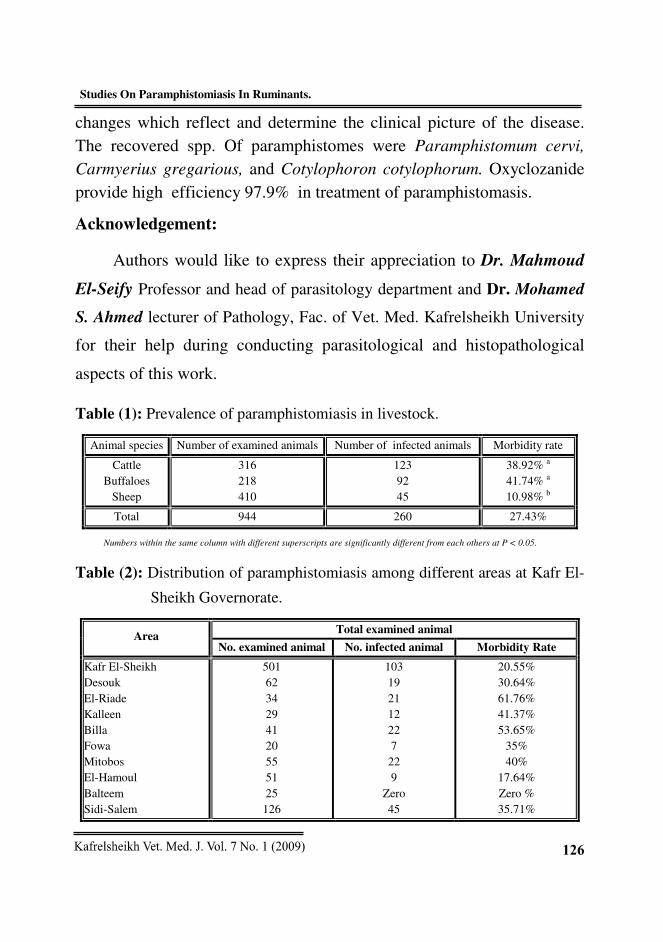
Total prevalence which recorded in this study was 27.43%. Table
(1) revealed that the prevalence was significantly higher (P < 0.001) in
cattle and buffaloes than that of sheep, whereas the variation was not
significant between cattle and buffaloes. This variation of the disease
prevalence among different species may be attributed to the host
specificity in addition to the rate of exposure where, cattle and buffaloes
were exposed similarly compared to a little exposure of sheep. Lower
Studies On Paramphistomiasis In Ruminants. Magdy H. Al-Gaabary et al.,
121
rates were recorded by Agosti et al. (1980) who recorded 16.9%
cumulative incidence and Kozakiewicz (1980) who recorded 3.06%
prevalence from 1971-1973 and 17.29% from 1976-1978.
Regarding to the prevalence rate in cattle, it was 38.92%. Similar rates
were recorded by Bouvry and Rau (1984) who recorded 34% prevalence
rate. Lower prevalence was recorded by Vartic et al. (1982) who recorded
3% prevalence rate; Juyal et al. (2003) who recorded 4.46% prevalence
rate; Dube et al. (2004) who recorded 25.41% prevalence rate and Haridy
et al. (2006) who recorded 7.3% prevalence rate of paramphistomiasis.
Higher rates were recorded by Manna et al. (1994); Dube et al. (2004)
and Stripalwit et al. (2007) who recorded 56.5%, 80% and 78.38% prev-
alence rates respectively.
Regarding to the prevalence of the disease among examined buffaloes,
the prevalence was 41.74%. Higher rates were recorded by Luc and
Thang (1999) and Ameni et al. (2001) who recorded 72.7% and 75%
prevalence rates respectively. Lower rates were recorded by El-Refaii
(1993); Manna et al. (1994); Juyal et al. (2003); Haridy et al. (2006)
and Khan et al. (2006) who recorded 9%, 27.4%, 6.59%, 10% and
28.33% prevalence rate respectively.
The prevalence of the disease among sheep was 10.98%. Similar
rate was recorded by Vartic et al. (1982) who recorded 9-11% prevalence
rate. Higher prevalence were recorded by Manna et al. (1994) and
Wang et al. (2006) who recorded 55.9% and 48.8% prevalence rates
respectively. Lower rates were recorded by Moghoddar and Khanitapeh
(2003) and Haridy et al. (2006) who recorded 1.09% and 4% prevalence
rates respectively.
Studies On Paramphistomiasis In Ruminants. Magdy H. Al-Gaabary et al.,
122
As shown in Table (2), the prevalence of the disease was differed
among different areas of Kafr El-Sheikh Governorate where higher
prevalence was recorded in El-Riade (61.76%) and Biala (53.65%)
compared to zero% in Baltim, this variation among different area might
be related to the environmental conditions which facilate the presence
and propagation of the intermediate host (Al-Gaabary and Nasr, 1997).
Climatic changes (Rangel-Ruiz et al., 2003) and husbandry practices
(Wang et al., 2006).
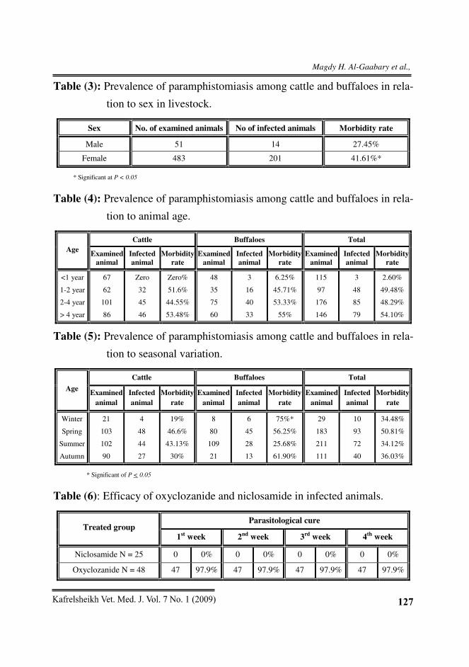
Concerning the sex predispoition of paramphistomiasis, significant
(P < 0.05) increase was recorded in female (41.61%) than males
(27.45%) in livestock (Table 3). Similar observations were reported
previously by Asanji et al. (1989) and Galdhar and Roy (2005) who
recorded that the prevalence of paramphistomiasis was generally higher
in females than males. On the contrary, Sevimi et al. (2005); kumari and
Hafeez (2005) and Khan et al. (2006) recorded that the prevalence of
paramphistomiasis in males was higher than that in females. The higher
prevalence in females may be attributed to stress factors (parturition and
lactation) to which the females were exposed.
Concerning the disease prevalence among examined cattle and buffaloes
in relation to their ages, the prevalence of paramphistomiasis was zero % in
cattle less than one year, 51.6% in cattle from 1-2 year, 44.55% in cattle from
2-4 year and 53.48% in cattle more than 4 year. Whereas the prevalence rates
in buffaloes were 6.25% in age group less than 1 year, 45.71% in age group 1-
2 year, 53.33% in age group 2-4 years and 55% in buffaloes more than 4 years
(Table 4). Similar result were previously reported by Agosti et al. (1980)
who recorded that all cases of paramphistomiasis were observed in adult
cattle whereas no cases were reported in calves; Ferre et al. (1997) who
Studies On Paramphistomiasis In Ruminants. Magdy H. Al-Gaabary et al.,
123
recorded that the risk of paramphistomiasis infestation was increased
with increasing the animal age; Amer et al. (2002) who recorded that the
incidence of paramphistomiasis in cattle over 2½ years was 46.77%
while it was 28.9% in cattle under 2½ year and Galdhar and Roy (2005)
who recorded 1.25% prevalence in animal above 6 years followed by
calves of one year (4.34%). On the other hand, Sobih and Hassan (1992)
recorded 2.9% and zero % prevalence rates in cattle and buffaloes in
yearling animals and 1.7% and zero % in animal over 3 year and Khan et
al. (2006) who recorded that the disease was prevalent in younger
buffaloes below two year compared to older buffaloes more than two
year. The lower rate of infection in young animals may be attributed to
the little chance of exposure as well as the long prepatent period of the
paramphistomes species with subsequent absence of the diagnostic eggs.
Concerning the disease prevalence in relation to different seasons,
the prevalence was statistically (P < 0.05) differ among different seasons.
The disease was higher in spring (50.81%), followed by autumn
(36.03%) then winter (34.48%) and lastly summer (34.12%) (Table 5).
Similar findings were previously recorded by Pal and Qayyum (1993)
who recorded highest paramphistomes infection rate during winter
(84.18%) followed by autumn (41.76%) then spring (37.25%) and finally
in summer (32.86%) and Wang et al. (2006) who recorded that summer
showed the peak season for paramphistomes infection in sheep. This
variation among different studies might be related to environmental
conditions which facilitate the presence of the intermediate host (Al-
Gaabary and Nasr, 1997) climatic and geographical parameters which
affect the hatchability of paramphistomes eggs (Dutta et al., 1995 and
Hirani et al., 1999).
Studies On Paramphistomiasis In Ruminants. Magdy H. Al-Gaabary et al.,
124
The clinical findings which recorded in this study were greatly
related to the degree of infestation; mildly infected ones showed no
clinical signs while moderately and severely infected animals showed
emaciation, diarrhoea, pale mucous membrane, submandibular oedema
and decreased milk yield. These signs may be attributed to the damage
and necrosis of gastro intestinal mucosa which results from direct effect
of the parasite which lead to impairment of digestion and absorption
resulting in production loss. Submanidublar oedema which observed may
be attributed to hypoproteinemia which resulted due to leakage of protein
through the damaged mucous membrane of the duodenum. Decreased
milk yield is due to decrease of volatile fatty acids (Amer et al., 2002).
Similar signs were observed previously by Hanna et al. (1988) who
recorded that light paramphistomiasis infection did not cause serious
damage to the animal and Amer et al. (2002) who recorded that pale
mucous membrane, weakness, weight loss, decreased milk yield in
paramphistomes infected animals.
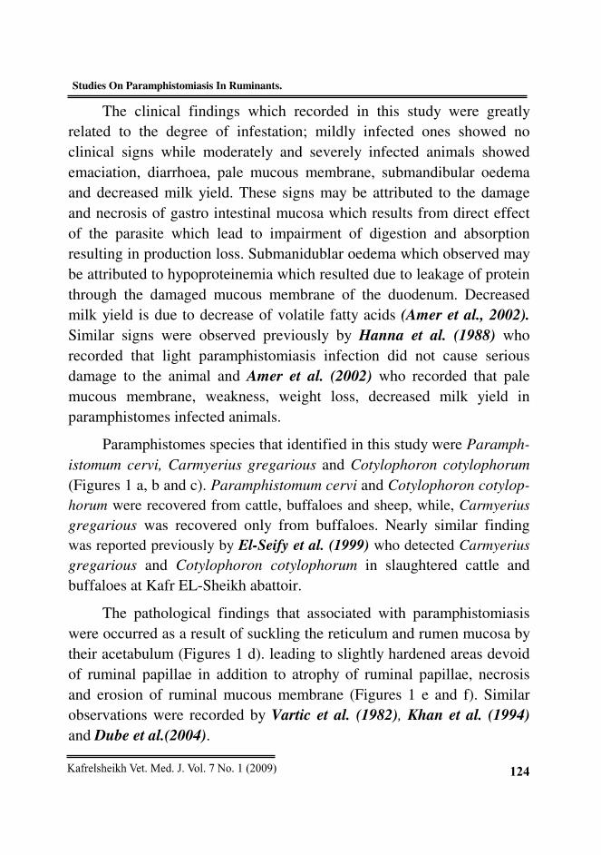
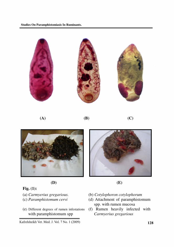
Paramphistomes species that identified in this study were Paramph-
istomum cervi, Carmyerius gregarious and Cotylophoron cotylophorum
(Figures 1 a, b and c). Paramphistomum cervi and Cotylophoron cotylop-
horum were recovered from cattle, buffaloes and sheep, while, Carmyerius
gregarious was recovered only from buffaloes. Nearly similar finding
was reported previously by El-Seify et al. (1999) who detected Carmyerius
gregarious and Cotylophoron cotylophorum in slaughtered cattle and
buffaloes at Kafr EL-Sheikh abattoir.
The pathological findings that associated with paramphistomiasis
were occurred as a result of suckling the reticulum and rumen mucosa by
their acetabulum (Figures 1 d). leading to slightly hardened areas devoid
of ruminal papillae in addition to atrophy of ruminal papillae, necrosis
and erosion of ruminal mucous membrane (Figures 1 e and f). Similar
observations were recorded by Vartic et al. (1982), Khan et al. (1994)
and Dube et al.(2004).
Studies On Paramphistomiasis In Ruminants. Magdy H. Al-Gaabary et al.,
125
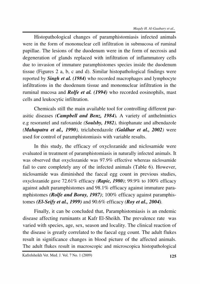
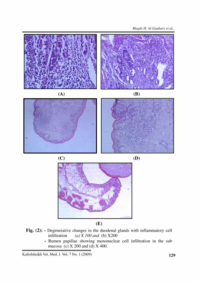
Histopathological changes of paramphistomiasis infected animals
were in the form of mononuclear cell infiltration in submucosa of ruminal
papillae. The lesions of the duodenum were in the form of necrosis and
degeneration of glands replaced with infiltration of inflammatory cells
due to invasion of immature paramphistomes species inside the duodenum
tissue (Figures 2 a, b, c and d). Similar histopathological findings were
reported by Singh et al. (1984) who recorded macrophages and lymphocyte
infiltrations in the duodenum tissue and mononuclear infiltration in the
ruminal mucosa and Rolfe et al. (1994) who recorded eosinophils, mast
cells and leukocytic infiltration.
Chemicals still the main available tool for controlling different par-
asitic diseases (Campbell and Benz, 1984). A variety of anthelmintics
e.g resorantel and rafoxanide (Soulsby, 1982), thiophanate and albendazole
(Mahapatra et al., 1990), triclabendazole (Galdhar et al., 2002) were
used for control of paramphistomiasis with variable results.
In this study, the efficacy of oxyclozanide and niclosamide were
evaluated in treatment of paramphistomiasis in naturally infected animals. It
was observed that oxyclozanide was 97.9% effective whereas niclosamide
fail to cure completely any of the infected animals (Table 6). However,
niclosamide was diminished the faecal egg count in previous studies,
oxyclozanide gave 72.61% efficacy (Rapic, 1980); 99.9% to 100% efficacy
against adult paramphistomes and 98.1% efficacy against immature para-
mphistomes (Rolfe and Boray, 1987); 100% efficacy against paramphis-
tomes (El-Seify et al., 1999) and 90.6% efficacy (Roy et al., 2004).
Finally, it can be concluded that, Paramphistomiasis is an endemic
disease affecting ruminants at Kafr El-Sheikh. The prevalence rate was
varied with species, age, sex, season and locality. The clinical reaction of
the disease is greatly correlated to the faecal egg count. The adult flukes
result in significance changes in blood picture of the affected animals.
The adult flukes result in macroscopic and microscopica histopathological
Studies On Paramphistomiasis In Ruminants. Magdy H. Al-Gaabary et al.,
126
changes which reflect and determine the clinical picture of the disease.
The recovered spp. Of paramphistomes were Paramphistomum cervi,
Carmyerius gregarious, and Cotylophoron cotylophorum. Oxyclozanide
provide high efficiency 97.9% in treatment of paramphistomasis.
Acknowledgement:
Authors would like to express their appreciation to Dr. Mahmoud
El-Seify Professor and head of parasitology department and Dr. Mohamed
S. Ahmed lecturer of Pathology, Fac. of Vet. Med. Kafrelsheikh University
for their help during conducting parasitological and histopathological
aspects of this work.
Table (1): Prevalence of paramphistomiasis in livestock.
Animal species Number of examined animals Number of infected animals Morbidity rate
Cattle
Buffaloes
Sheep
316
218
410
123
92
45
38.92% a
41.74% a
10.98% b
Total 944 260 27.43%
Numbers within the same column with different superscripts are significantly different from each others at P < 0.05.
Table (2): Distribution of paramphistomiasis among different areas at Kafr El-
Sheikh Governorate.
Total examined animal Area
No. examined animal No. infected animal Morbidity Rate
Kafr El-Sheikh
Desouk
El-Riade
Kalleen
Billa
Fowa
Mitobos
El-Hamoul
Balteem
Sidi-Salem
501
62
34
29
41
20
55
51
25
126
103
19
21
12
22
7
22
9
Zero
45
20.55%
30.64%
61.76%
41.37%
53.65%
35%
40%
17.64%
Zero %
35.71%
Studies On Paramphistomiasis In Ruminants. Magdy H. Al-Gaabary et al.,
127
Table (3): Prevalence of paramphistomiasis among cattle and buffaloes in rela-
tion to sex in livestock.
Sex No. of examined animals No of infected animals Morbidity rate
Male 51 14 27.45%
Female 483 201 41.61%*
* Significant at P < 0.05
Table (4): Prevalence of paramphistomiasis among cattle and buffaloes in rela-
tion to animal age.
Cattle Buffaloes Total
Age Examined
animal
Infected
animal
Morbidity
rate
Examined
animal
Infected
animal
Morbidity
rate
Examined
animal
Infected
animal
Morbidity
rate
<1 year
1-2 year
2-4 year
> 4 year
67
62
101
86
Zero
32
45
46
Zero%
51.6%
44.55%
53.48%
48
35
75
60
3
16
40
33
6.25%
45.71%
53.33%
55%
115
97
176
146
3
48
85
79
2.60%
49.48%
48.29%
54.10%
Table (5): Prevalence of paramphistomiasis among cattle and buffaloes in rela-
tion to seasonal variation.
Cattle Buffaloes Total
Age Examined
animal
Infected
animal
Morbidity
rate
Examined
animal
Infected
animal
Morbidity
rate
Examined
animal
Infected
animal
Morbidity
rate
Winter
Spring
Summer
Autumn
21
103
102
90
4
48
44
27
19%
46.6%
43.13%
30%
8
80
109
21
6
45
28
13
75%*
56.25%
25.68%
61.90%
29
183
211
111
10
93
72
40
34.48%
50.81%
34.12%
36.03%
* Significant of P < 0.05
Table (6): Efficacy of oxyclozanide and niclosamide in infected animals.
Parasitological cure Treated group
1st week 2nd week 3rd week 4th week
Niclosamide N = 25 0 0% 0 0% 0 0% 0 0%
Oxyclozanide N = 48 47 97.9% 47 97.9% 47 97.9% 47 97.9%
Studies On Paramphistomiasis In Ruminants. Magdy H. Al-Gaabary et al.,
128
(A) (B) (C)
(D) (E)
Fig. (1):
(a) Carmyerius gregarious. (b) Cotylophoron cotylophorum
(c) Paramphistomum cervi (d) Attachment of paramphistomum
spp. with rumen mucosa
(e) Different degrees of rumen infestations
with paramphistomum spp
(f) Rumen heavily infected with
Carmyerius gregarious
Studies On Paramphistomiasis In Ruminants. Magdy H. Al-Gaabary et al.,
129
(A) (B)
(C) (D)
(E)
Fig. (2): - Degenerative changes in the duodenal glands with inflammatory cell
infiltration (a) X 100 and (b) X200
- Rumen papillae showing mononuclear cell infiltration in the sub
mucosa (c) X 200 and (d) X 400.
Studies On Paramphistomiasis In Ruminants. Magdy H. Al-Gaabary et al.,
130
REFERENCES
- Agosti, M.; Cavalletii, E. and Pozza, O. (1980). Bovine paramphisto-
miasis in the province of Milan. Clinical and epizootiological investig-
ations. Clinica-Veterinaria, 103(5): 284-296.
- Al-Gaabary, M.H. and Nasr, M.Y. (1997). Epidemiological, clinco-
biochemical and electrophoretic studies on paramphistomiasis in
buffaloes in Kafr El-Sheikh province. Zagazig Veterinary Journal,
25(3): 34-40.
- Ameni, G.; Erko, B. and Bogale, T. (2001). Preliminary study on the
major bovine trematode infection around Kemissie, North eastern
Ethiopia and treatment trial with Praziquantel. Bulletin of Animal
Health and Production in Africa, 49(2): 62-67.
- Amer, A.; Osman, S.A. and Harfoush, M.A. (2002). Epizootiological
and serological studies on Fascioliasis and Paramphistomiasis in
cattle. Zagazig Veterinary Journal, 30(1): 107-116.
- Anuracpreeda, P.; Wanichanon, C. and Sobhon, P. (2008). Paramp-
histomum cervi: Antigenic profile of adults as recognized by infected
cattle sera. Experimental Parasitology, 118: 203-207.
- Asanji, M.F. (1989). Paramphistomiasis of cattle in sierra leone. Cameroon
and seasonal fluctuation in its prevalence. Bulletin of Animal Health
and Production in Africa, 37(4): 327-331.
- Asanji, M.F. (1990). Experimental determination of Snail intermediate
host of live stock paramphistomes and their distribution in snails in
nature. Bull. Anim. Health. Prod. Afr. 38: 151-157.
- Bouvry, M. and Rau, M.E. (1984). Paramphistomum spp. in dairy
cattle in Quebec. Can. Vet. J., 25: 353-356.
Studies On Paramphistomiasis In Ruminants. Magdy H. Al-Gaabary et al.,
131
- Campbell, W.C. and Benz, G.W. (1984). Ivermectin. A review of
efficacy and safety. J. Vet. Pharmacol. Therap. 7: 611-612.
- Dreyfuss, G.; Novobisky, A.; Vignoles, P.; Bellet, V.; Koudela, B.
and Rondelaud, D. (2007). Prevalence and intensity of infection in
the lymnaeid snail Omphiscola glabra experimentally infected with
Fasciola hepatica, Fascioloides magna and Paramphistomum
daupneyi of Helminthology. 81: 7-12.
- Drurag, R. and Wallington, E. (1980). Carlton’s histological techniques.
4th
ed. Oxford University Press, New York.
- Dube, S.; Dlamini, N.R.; Masanganisek, K.E. and Dube, C. (2004).
Abattoir studies on paramphistomes recovered from cattle in
Masuingo and Monicaland province of Zimbabwe. Folia Veterinaria,
48(3): 123-129.
- Dutta, S.; Majumdar, P. and Basak, D.K. (1995). Studies on the
incidence of paramphistomiasis in cattle (Bos indicus) in West Bengal.
Indian Journal of Veterinary Medicine, 15(2): 84-86.
- El-Refaii, A.H. (1993). Entamoeba bovis liebetanz 1905 recorded from
large ruminants in Egypt. J. Egypt. Soc. Parasitol, 23(1): 239-245.
- El-Seify, M.; El-Bahy, N.M.; Abdo-Rabo, T.M. and El-Shahawy,
I.S. (1999). Some morphological studies on Paramphistomidea in Kafr
El-Sheikh governorate. Alex. J. Vet. Sci., 15(5): 981-993.
- Ferre, I.; Shen, S.L.; Gu, Y.F.; Chen, L.; Xi, J.; Wang, B.Y.;
Gonzalez Gallego, J. and Meo, X.Z. (1997). Prevalence of Fasciola
hepatica and Paramphistomum spp. infection in water buffalo in
Anhui and Jiangsu provinces (China). Proceedings 5th
World Buffalo
Congress. Royal Palace, Caserta, Italy, 574-577.
Studies On Paramphistomiasis In Ruminants. Magdy H. Al-Gaabary et al.,
132
- Galdhar, C.N. and Roy, S. (2005). Studies on prevalence of bovine
paramphistomiasis in Chattisgarh state India. Indian Veterinary
Journal, 82(9): 938-940.
- Galdhar, C.N.; Roy, S.; Ali, S.L.; Maiti, S.K. and Awasthi, B.K.
(2002). National symposium and annual convention of ISVM on
recent trends in diagnostic and therapeutics of animal disease. Feb. 14-
16, Bikaner.
- Hafeez, M.; Ramesh, D.; Reddy, C.V.S.; Devi, D.A.; Chandra, M.S.
and Sameena, S.K. (2006). Detection of Paramphistomum epiclitum
infection in sheep by dot. Elisa, Indian Journal of Animal Sciences,
76(9): 962-963.
- Hanna, R.E.B.; Williamson, D.S.; Mattison, R.G. and Nizami, W.A.
(1988). Seasonal reproduction in Paramphistomum epiclitum and
Gastrothylax crumenifer, rumen paramphistomes of Indian water
buffalo and comparison with billiary Paramphistome gigantocotyle
explanatum. International Journal for Parasitology, 18: 513-521.
- Haridy, F.M.; El-Sherbiny, J.T. and Morsy, T.A. (2006). Some parasitic
flukes infecting farm animals in Al-Santa center, Gharbia Governorate,
Egypt. Journal of the Egyptian Society of Parasitology, 36 (1): 259-
264.
- Hirani, N.D.; Katariya, M.A.; Abdullah Patel, Hasnani, J.J.;
Kathiria, L.G.; Patel, P.V. and Patel, A. (1999). Prevalence of
gastrointestinal parasitic infections in cattle and buffaloes of Kheda
district of Gujar. Journal of Veterinary Parasitology, 13: 147-149.
- Juyal, P.D.; Gupta, M.P.; Kaurk, K.B. and Hassan, S.S. (2003).
Epidemiological status of paramphistomiasis in domestic ruminants.
Punjab Veterinary Journal 2: 100-102.
Studies On Paramphistomiasis In Ruminants. Magdy H. Al-Gaabary et al.,
133
- Kelly, W.R. (1984). Veterinary clinical diagnosis. 3rd
ed. Baillieve
Tindal.
- Khan, A. and Anjum, A.D. (1994). Liver paramphistomiasis in a
buffalo in Pakistan. Buffalo Journal, 10(2): 185-188.
- Khan, U.J.; Akhtar, T.; Maqbool, A. and Anees, A. (2006). Epidemi-
ology of Paramphistomiasis in buffaloes under different managemental
condition at four district of Punjab province, Pakistan Iranian Journal
of Veterinary Research, 7(3): 68-72.
- Kozakiewicz, B. (1980). The dynamic of occurrence of paramphistom-
iasis in cattle and sheep in Poznan area. Poland Medycyna Weterynar-
yjna, 36(3): 144-146.
- Kumari, P.S. and Hafeez, M.D. (2005). Prevalence of Paramphistom-
iasis in cattle in Chittor district of Andhra Pradesh. Indian Journal of
Parasitic Disease, 29(1): 1-8.
- Luc, P. and Thang, T. (1999). Paramphistomatidae infestations of
buffaloes in North Vietnam. Knoa Hocky Thuat Thuy, 6(1): 57-62.
- Mahapatra, P.K.; Misra, S.C.; Panda, M.R. and Rao, A.T. (1990).
Indian Vet. J. 67: 756. Cited after Roy et al. (2004).
- Manna, A.K.; Pramanik, S. and Mukherjee, G.S. (1994). Incidence
of Paramphistomiasis in west Bengal. Indian Journal of Animal
Health, 33(2): 87-89.
- Martin, W.S. (1987). Veterinary epidemiology. 3rd ed. Iowa State
University, Congress Library.
- Moghoddar, N. and Khanitapeh, N. (2003). Prevalence of Paramphi-
stomes in sheep and goat in the Fars province of Iran. Iranian Journal
of Veterinary Research, 4(2): 166-170.
Studies On Paramphistomiasis In Ruminants. Magdy H. Al-Gaabary et al.,
134
- Pal, A.R. and Qayyum, M. (1993). Distribution of gastrointestinal
amphistomes and cestodes in small ruminants grazed on irrigated and
non irrigated pasture-zones. Proceeding of Pakistan Congress of
Zoology 13: 307-313.
- Prasad, K.D. and Bharti, P. (2001). Assessment of oxyclozanide efficacy
against chronic natural fascioliasis or paramphistomiasis in cattle and
buffaloes. Journal of Research Birsa Agricultural University, 13(2):
245-248.
- Rangel Rulz, L.J.; Albores Brahms, S.T. and Gamboa Aguilar, J. (2003).
Seasonal trends of Paramphistomum cervi in Tabasco, Mexico. Veter-
inary Parasitology, 116(3): 217-222.
- Rapic, D. (1980). Action of Nilsan on paramphistomiasis in cattle.
Praxis Veterinaria, 28(1/2): 15-18.
- Reddy, M.R. and Hafeez, M. (1986). Niclosamide an effective drug
against amphistomasis of sheep. Cheiron, 15(1): 29-30.
- Rolfe, P.; Boray, J. and Collins, G. (1994). Pathology of infection with
Paramphistomum ichikaway in sheep. International Journal Parasitology,
24(7): 995-1004.
- Rolfe, P.F. and Boray, J.C. (1987). Chemotherapy of Paramphistomiasis
in cattle. Australian Veterinary Journal, 64(11): 328-332.
- Sey, O.; Prasitirat, P.; Romratanapun, S. and Mohkaew, K. (1997).
Morphological studies and identification of rumen flukes of cattle in
Thailand. Rivista Di Parasitologia, XIV(2): 247-256.
- Singh, R.P.; Saha, B.N.; and Jha, G.J. (1984). Histopathology of the
duodenum and rumen of goats during experimental infections with
paramphistomum cervi. Veterinary Parasitology, 15(1): 39-46.
Studies On Paramphistomiasis In Ruminants. Magdy H. Al-Gaabary et al.,
135
- Snedecor, G.W. and Cochran, W.G. (1980). Statistical methods. 8th
ed. The Iowa State University Press, USA.
- Sobih, M.A. and Hassan, M.G. (1992). Prevalence of gastrointestinal
parasites among cattle and buffaloes in Ismailia governorate. Zag. Vet.
J., 20(2): 299-310.
- Soulsby, E.J.L. (1982). Helminths, Arthropoda and Protozoa of
domesticated animals. 7th
ed. Baillier, Tindal and Cassel, London.
- Stripalwit, P.; Wongsawad, C.; Wongsawad, P. and Anuntalabnochai,
P. (2007). High annealing temperature random amplified polymorphic
DNA analysis of three paramphistome flukes from Thiland. Experim-
ental Parasitology, 115(1): 98-102.
- Vartic, N.; Zagreanu, C.; Trica, Z. and Suteu, E. (1982). Epizootiol-
ogical, pathological, clinical, therapeutic studies of paramphistomiasis
in cattle and sheep. Seminarul Amerliorarea Technologia Si
Pathologia Rumenga-toarelor, Volumul, VII: 29-30.
- Wang, C.R.; Qiu, J.H.; Zhu, X.Q.; Han, X.H.; Ni, H.B.; Zhoo, J.P.;
Zhou, Q.M.; Zhang, H.W. and Lun, Z.R. (2006). Survey of
Helminths in adult sheep in Heilongjiang Province, People’s Republic
of China. Veterinary Parasitology, 140: 378-382.
- Yamaguti, S. (1958). Systema helminthium, vol. 2, the digenetic trematodes
of vertebrates past II. Interscience Publishers Inc., New York.
- Zinsstag, J.; Ankers, P.H.; Dempfie, L.; Njie, M.; Kaufmann, J.; Itty, P.;
Pfister, K. and Pandey, V.S. (1997). Effect of strategic gastrointestinal
nematode control on growth of N’Dama cattle in Gambia. Veterinary
Parasitology, 68: 143-153.
Studies On Paramphistomiasis In Ruminants. Magdy H. Al-Gaabary et al.,
136
pa��a@À@áßìn�îÐߊbjÛa@æa‡í‡i@òib–⁄a@óÜÇ@pb�aŠ†
������� �� � ����– ���� ���� ��� –������� ����� ���� ����
����� �� � �� ����� ��� !"# /%&�� ��# !����
������� ����� � ������ ����� ��� ��� ������ ����� ��� ��� !� ���"#�
��� $� %���� &'� (����� )* +��� $���2008 ����� �� 2008 ����� �� �*�, ��
-'�� )* �������� ���. ��� %���� ��� . ��� 01��944 $�1 )314 3��4�5 218
36����410 !��75 ( ���" $� 9�:��� 01���� ��;�� !�260 (<�� ���" ����� $�1 27.43
% ���" ���� (���38.92 % 341.74 3 10.98 )���� ��� !��7.� 6������ ��4�. )*
>� ��� �?*�1� @��� $� ���" ���� (����A )* ���" ���� ���5 (��� B1 �������
!C�� @��� )* ���" 9 &��� !�� '�� ���� @��� . ���" $� ��;�� !� 9�:��� 01����
41.73 % &��4� B�� $�27.45 ����� $� . (��� B1 ��5 �� ��� $� ���" ���� (����
���" ����2.59 % � !�� $� &D (���1� )*49.48 % � $���� !�� $� �� (���1� )*
48.86 % $� �� (���1� )*2E4 (��� ��5� $�� 53.79 % F��5 $� ��� (���1� )*
!��5 .��� F��� &"* )* ���" ���� ��� (��� B1 �� �� &"* $� ���" ���� (�)43
(% ���� &"* ��'�)32.25 (% ���>� &"* !G)31 (% &"* )* ���" ���� &D (����
�"�)3.32 .(% +��� $�� (���C $� H��5 B'G ��� ����� $��5 9�:��� 01����
Paramphistomum cervi, Cotylophoron cotylophorum Carmyerius gregarious . I,�5
����� ���" (� (���1� �* (���� � ����� ���. (���� )���� 01��
&@:� $� %���� ������ ���. (��� � %��>� ���" (� (���1� �* �:��> )* (��@�
�� $��� -��� ��D� &�:� � $��� $� ��@� . @����� ���, ���� �����G�����:� 01�� �:?5
)���� +��� ��>7 )* . ��><� �'� $� ���:��J �'�� $� (����� ���� �,5 $�� �
�>� ��GJ ��7 )* @����� �C���� . ��� ��,4� )* ���� ����* ��@��� ���� -'� �:?5
�> ���� �C� B1 &�C� &���� ��97.9 % !��� ��,4� )* &>* K�� ��@���� ��4�� ����4�
&�C� ���.