structure of glyceraldehyde-3-phosphate dehydrogenase from the archaeal hyperthermophile...
TRANSCRIPT
RIKEN–UK structural genomics
Acta Cryst. (2009). F65, 1227–1233 doi:10.1107/S1744309109047046 1227
Acta Crystallographica Section F
Structural Biologyand CrystallizationCommunications
ISSN 1744-3091
Structure of glyceraldehyde-3-phosphatedehydrogenase from the archaeal hyperthermophileMethanocaldococcus jannaschii
Ali D. Malay,a‡ Yoshitaka
Bessho,a,b Mark J. Ellis,c
Svetlana V. Antonyuk,d
Richard W. Strange,d S. Samar
Hasnain,d Akeo Shinkai,b
Balasundaram Padmanabhana*§
and Shigeyuki Yokoyamaa,b,e*
aSystems and Structural Biology Center,
Yokohama Institute, RIKEN, 1-7-22 Suehiro,
Tsurumi, Yokohama 230-0045, Japan, bRIKEN
SPring-8 Center, Harima Institute, 1-1-1 Kouto,
Sayo, Hyogo 679-5148, Japan, cSTFC Daresbury
Laboratory, Warrington, Cheshire WA4 4AD,
England, dMolecular Biophysics Group, School
of Biological Sciences, University of Liverpool,
Liverpool L69 7ZB, England, and eDepartment
of Biophysics and Biochemistry, Graduate
School of Science, The University of Tokyo,
Bunkyo-ku, Tokyo 113-0033, Japan
‡ Present address: Global Edge Institute,
Tokyo Institute of Technology, 4259, S2-17,
Nagatsuda, Midori-ku, Yokohama,
Kanagawa 226-8503, Japan.
§ Present address: Aptuit Laurus Private Ltd,
ICICI Knowledge Park, Turkapally, Shameerpet,
Hyderabad 500 078, India.
Correspondence e-mail:
Received 25 June 2009
Accepted 7 November 2009
PDB Reference: glyceraldehyde-3-phosphate
dehydrogenase, 2yyy, r2yyysf.
The X-ray crystal structure of glyceraldehyde-3-phosphate dehydrogenase
(GAPDH) from the hyperthermophilic archaeon Methanocaldococcus jannaschii
(Mj-GAPDH) was determined to 1.81 A resolution. The crystal belonged to
space group C2221, with unit-cell parameters a = 83.4, b = 152.0, c = 118.6 A. The
structure was solved by molecular replacement and was refined to a final R
factor of 17.1% (Rfree = 19.8%). The final structure included the cofactor
NADP+ at the nucleotide-binding site and featured unoccupied inorganic and
substrate phosphate-binding sites. A comparison with GAPDH structures from
mesophilic sources suggested that Mj-GAPDH is stabilized by extensive
electrostatic interactions between the C-terminal �-helices and various distal
loop regions, which are likely to contribute to thermal stability. The key
phosphate-binding residues in the active site of Mj-GAPDH are conserved in
other archaeal GAPDH proteins. These residues undergo a conformational shift
in response to occupancy of the inorganic phosphate site.
1. Introduction
Glyceraldehyde-3-phosphate dehydrogenase (GAPDH; EC 1.2.1.12)
catalyzes the reversible oxidative phosphorylation of glyceraldehyde
3-phosphate to 1,3-bisphosphoglycerate in the presence of inorganic
phosphate and the cofactor NADH or NADPH as part of the classic
pathways of glycolysis and gluconeogenesis (Harris & Waters, 1976).
In addition, GAPDH acts as a ‘moonlighting enzyme’ that has been
implicated in diverse cellular pathways ranging from transcriptional
activation and membrane trafficking to apoptosis (Sirover, 1999,
2005; Tisdale, 2002; Zheng et al., 2003).
Numerous structures of the highly conserved GAPDH proteins
from bacteria and eukaryotes have been characterized (Murthy et al.,
1980; Skarzynski et al., 1987; Duee et al., 1996; Tanner et al., 1996;
Antonyuk et al., 2003). In addition, several GAPDH structures from
archaea have been reported (Isupov et al., 1999; Charron et al., 2000).
The archaeal GAPDHs belong to a different class to their bacterial/
eukaryotic counterparts. Although the two classes share a similar
basic architecture, the archaeal GAPDHs exhibit some distinct
characteristics such as the translocation of important active-site
residues to different structural elements and the presence of extra
secondary-structure elements. The evolutionary relationship between
the archaeal and bacterial/eukaryotic GAPDHs is still an open
question (Arcari et al., 1993; Littlechild et al., 2004).
In this study, we report the crystal structure of GAPDH from
the hyperthermophilic archaeon Methanocaldococcus jannaschii
(Mj-GAPDH) determined at 1.81 A resolution.
2. Materials and methods
2.1. Cloning, expression and purification
The gene encoding the Mj-GAPDH protein (gi:15669333) was
amplified via PCR using M. jannaschii DSM 2661 genomic DNA and
was cloned into the pET-21a expression vector (Merck Novagen,
Darmstadt, Germany). The expression vector was introduced into
Escherichia coli BL21-CodonPlus(DE3)-RIL strain (Stratagene, La
Jolla, California, USA) and the recombinant strain was cultured# 2009 International Union of Crystallography
All rights reserved
in 4.5 l LB medium containing 30 mg ml�1 chloramphenicol and
50 mg ml�1 ampicillin. The harvested cells (16.3 g) were lysed by
sonication in 30 ml 20 mM Tris–HCl buffer pH 8.0 containing
500 mM NaCl, 5 mM �-mercaptoethanol and 1 mM phenylmethyl-
sulfonyl fluoride on ice. The cell lysate was heat-treated at 343 K for
13 min and centrifuged at 15 000g for 30 min at 277 K. The super-
natant was desalted by fractionation on a HiPrep 26/10 column (GE
Healthcare Biosciences). The sample was applied onto a Toyopearl
SuperQ-650M column (Tosoh, Tokyo) equilibrated with 20 mM Tris–
HCl buffer pH 8.0 and was eluted with a linear (0–0.3 M) gradient of
NaCl. The target sample, which eluted in the 0.26 M NaCl fraction,
was then applied onto a Resource Q column (GE Healthcare Bio-
sciences) equilibrated with 20 mM Tris–HCl buffer pH 8.0 and was
eluted with a linear gradient of 0–0.5 M NaCl. The fractions that
eluted in 0.33 M NaCl were further purified using a hydroxyapatite
CHT20-I column (Bio-Rad Laboratories) with a linear gradient of
0.01–0.5 M potassium phosphate buffer pH 7.0. The target sample,
which eluted in the 0.09 M potassium phosphate fraction, was con-
centrated and applied onto a HiLoad 16/60 Superdex 200 pg column
(GE Healthcare Biosciences) equilibrated with 20 mM Tris–HCl
buffer pH 8.0 containing 200 mM NaCl. The protein sample was
analyzed by SDS–PAGE and was confirmed by N-terminal amino-
acid sequencing. After concentration to approximately 20 mg ml�1 by
ultrafiltration, the protein yield was 26.2 mg from 16.3 g of cells.
2.2. Protein crystallization
Crystallization was performed using the microbatch-under-oil
method at 291 K. 0.5 ml crystallization reagent was mixed with 0.5 ml
23.2 mg ml�1 protein solution and was covered with 15 ml silicone and
paraffin oil. In the preliminary screen, small crystals appeared using a
crystallization reagent consisting of 0.08 M Tris–HCl buffer pH 8.5
containing 24%(w/v) PEG 4000, 0.16 M magnesium chloride hexa-
hydrate and 20%(v/v) anhydrous glycerol (Crystal Screen Cryo
condition No. 6, Hampton Research). After optimization, large
crystals were obtained from a crystallization reagent consisting of
0.08 M Tris–HCl buffer pH 8.5 containing 24%(w/v) PEG 4000, 0.2 M
magnesium chloride hexahydrate and 20%(v/v) anhydrous glycerol.
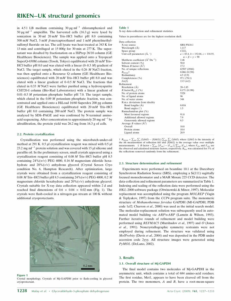
Crystals suitable for X-ray data collection appeared within 2 d and
reached final dimensions of 0.6 � 0.04 � 0.02 mm (Fig. 1). The
crystals were flash-cooled in a nitrogen-gas stream at 100 K without
additional cryoprotectants.
2.3. Structure determination and refinement
Experiments were performed on beamline 10.1 at the Daresbury
Synchrotron Radiation Source (SRS), employing a Si(111) sagittally
focused monochromator and a MAR Mosaic 225 CCD detector. The
data-collection and refinement parameters are summarized in Table 1.
Indexing and scaling of the reflection data were performed using the
HKL-2000 software package (Otwinowski & Minor, 1997). Molecular
replacement was accomplished using the program MOLREP (Vagin
& Teplyakov, 1997) from the CCP4 program suite. The monomeric
structure of Methanothermus fervidus GAPDH (Mf-GAPDH; PDB
code 1cf2; Charron et al., 2000) was used as the initial search model.
The molecular-replacement solution was subsequently used in auto-
mated model building via ARP/wARP (Lamzin & Wilson, 1993).
Further iterative rounds of refinement and model building were
performed using REFMAC5 (Murshudov et al., 1997) and O (Jones
et al., 1991). Noncrystallographic symmetry restraints were not
employed during refinement. The structure was validated using
MolProbity (Davis et al., 2004) and was deposited in the PDB under
accession code 2yyy. All structure images were generated using
PyMOL (DeLano, 2002).
3. Results
3.1. Overall structure of Mj-GAPDH
The final model contains two molecules of Mj-GAPDH in the
asymmetric unit, which contains a total of 684 amino-acid residues.
The first residue, Met1, appears to have been cleaved off from the
protein. The two monomers, A and B, have a root-mean-square
RIKEN–UK structural genomics
1228 Malay et al. � Glyceraldehyde-3-phosphate dehydrogenase Acta Cryst. (2009). F65, 1227–1233
Figure 1Crystal morphology. Crystals of Mj-GAPDH prior to flash-cooling in glycerolcryoprotectant.
Table 1X-ray data-collection and refinement statistics.
Values in parentheses are for the highest resolution shell.
Data collectionX-ray source SRS PX10.1Wavelength (A) 1.117Space group C2221
Unit-cell parameters (A, �) a = 83.4, b = 152.04, c = 118.55,� = � = � = 90
Matthews coefficient (A3 Da�1) 2.46Solvent content (%) 50.0Wilson B factor (A2) 17.2No. of unique reflections 62507 (4944)Rmerge† 0.066 (0.238)Redundancy 4.5 (2.9)Completeness (%) 97.1 (78.1)I/�(I) 13.5 (4.2)
RefinementResolution (A) 20–1.85R factor/Rfree‡ (%) 0.171 (0.198)No. of protein atoms 5328No. of ligand atoms 96No. of water atoms 348R.m.s. deviations from ideality
Bond lengths (A) 0.016Bond angles (�) 1.5
Ramachandran plot (%)Most favoured regions 91.8Additional allowed regions 7.9Generously allowed regions 0.3
Average B values (A2)Overall 18.8Protein atoms 18.4Ligand atoms 20.9
† Rmerge =P
hkl
Pi jIiðhklÞ � hIðhklÞij=
Phkl
Pi IiðhklÞ, where Ii(hkl) is the intensity of
the ith measurement of reflection hkl and hI(hkl)i is the average value over multiplemeasurements. ‡ R factor =
Phkl
��jFobsj � jFcalcj
��=P
hkl jFobsj, where Fobs and Fcalc arethe observed and calculated structure factors, respectively. Rfree was calculated for 5% ofthe reflections removed randomly from the refinement.
deviation (r.m.s.d.) value of 0.11 A for C� positions along the entire
sequence. Although it was not added separately to the sample, the
NADP+ cofactor is clearly visible as electron density within the
putative nucleotide-binding site of each GAPDH monomer. There
are 348 water molecules in the model, with no additional anions at the
known Pi (inorganic phosphate) and Ps (substrate phosphate) binding
sites.
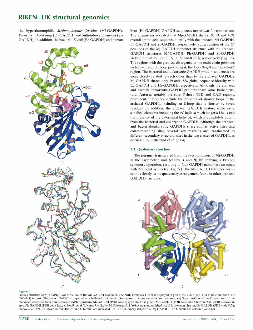
The overall architecture of the Mj-GAPDH monomer is typical of
archaeal GAPDH proteins and comprises three domains, a nucleo-
tide-binding domain (NBD; residues 1–142), a catalytic domain
(CAD; 143–305) and a C-terminal domain (CTD; 306–343), as shown
in Figs. 2 and 3(a). The N-terminal NBD consists of a parallel �-sheet
surrounded by �-helices in a classical Rossmann fold (sheets �A–�F
and helices �A–�G). The CAD is composed of an antiparallel �-sheet
surrounded by �-helices (sheets �1–�8 and helices �1–�40). This
domain contains the active-site residues, as well as the so-called
S-loop between �1 and �2 (residues 173–187). The relatively short
CTD, composed of the two helices �H and �J, is located at the
junction between the NBD and CAD. It should be noted that in
previous studies the C-terminal region has been treated as part of the
NBD; however, for the sake of clarity, we are evaluating the CTD as a
separate domain. The three proline residues, Pro183 in the S-loop and
Pro191 and Pro195 in the loop following �2, are in the cis config-
uration and two of them (Pro183 and Pro195) are conserved across all
GAPDH species.
Ramachandran plot analysis revealed that 91.8% of the residues
are in the most favoured regions and 7.9% are in additional favoured
regions, while 0.3% are in generously allowed regions. One residue,
Lys92, falls into the disallowed region in both monomers. A closer
inspection revealed that this surface residue is constrained owing to
the effects of crystal packing. The average B factor is 18.8 A2 for the
overall structure.
3.2. Comparison with GAPDH structures from other sources
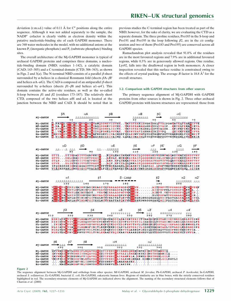
The primary sequence alignment of Mj-GAPDH with GAPDH
proteins from other sources is shown in Fig. 2. Three other archaeal
GAPDH proteins with known structures are represented: those from
RIKEN–UK structural genomics
Acta Cryst. (2009). F65, 1227–1233 Malay et al. � Glyceraldehyde-3-phosphate dehydrogenase 1229
Figure 2The sequence alignment between Mj-GAPDH and orthologs from other species: Mf-GAPDH, archaeal M. fervidus; Ph-GAPDH, archaeal P. horikoshii; Ss-GAPDH,archaeal S. solfataricus; Ec-GAPDH, bacterial E. coli; Hs-GAPDH, eukaryotic human liver. Regions of similarity are in blue boxes, with the strictly conserved residueshighlighted in red. The secondary-structure elements of Mj-GAPDH are indicated above the alignment. The naming of the secondary structural elements follows that ofCharron et al. (2000)
RIKEN–UK structural genomics
1230 Malay et al. � Glyceraldehyde-3-phosphate dehydrogenase Acta Cryst. (2009). F65, 1227–1233
the hyperthermophilic Methanothermus fervidus (Mf-GAPDH),
Pyrococcus horikoshii (Ph-GAPDH) and Sulfolobus solfataricus (Ss-
GAPDH). In addition, the bacterial E. coli (Ec-GAPDH) and human
liver (Hs-GAPDH) GAPDH sequences are shown for comparison.
The alignments revealed that Mj-GAPDH shares 59, 53 and 46%
overall amino-acid sequence identity with the archaeal Mf-GAPDH,
Ph-GAPDH and Ss-GAPDH, respectively. Superposition of the C�
positions of the Mj-GAPDH monomer structure with the archaeal
GAPDH structures Mf-GAPDH, Ph-GAPDH and Ss-GAPDH
yielded r.m.s.d. values of 0.51, 0.72 and 0.82 A, respectively (Fig. 3b).
The regions with the greatest divergence in the main-chain positions
include �C and the loop preceding it, the loop �7–�8 and the �2–�20
region. The bacterial and eukaryotic GAPDH protein sequences are
more closely related to each other than to the archaeal GAPDHs.
Mj-GAPDH shares only 19 and 18% global sequence identity with
Ec-GAPDH and Hs-GAPDH, respectively. Although the archaeal
and bacterial/eukaryotic GAPDH proteins share some basic struc-
tural features, notably the core �-sheet NBD and CAD regions,
prominent differences include the presence of shorter loops in the
archaeal GAPDHs, including an S-loop that is shorter by seven
residues. In addition, the archaeal GAPDHs feature some extra
�-helical elements, including the �C helix, a much longer �4 helix and
the presence of the C-terminal helix �J, which is completely absent
from the bacterial and eukaryotic GAPDHs. Although the archaeal
and bacterial/eukaryotic GAPDHs share similar active sites and
cofactor-binding sites, several key residues are translocated to
different secondary-structural sites in the two classes of GAPDHs, as
discussed by Littlechild et al. (2004).
3.3. Quaternary structure
The tetramer is generated from the two monomers of Mj-GAPDH
in the asymmetric unit (chains A and B) by applying a twofold
symmetry operation, resulting in four GAPDH monomers arranged
with 222 point symmetry (Fig. 3c). The Mj-GAPDH tetramer corre-
sponds closely to the quaternary arrangement found in other archaeal
GAPDH structures.
Figure 3Overall structure of Mj-GAPDH. (a) Structure of the Mj-GAPDH monomer. The NBD (residues 1–142) is displayed in green, the CAD (143–305) in blue and the CTD(306–343) in pink. The bound NADP+ is depicted as a ball-and-stick model. Secondary-structure elements are indicated. (b) Superposition of the C� positions of themonomer structures from four archaeal GAPDH proteins. Mj-GAPDH (PDB code 2yyy) is shown in green, Mf-GAPDH (PDB code 1cf2; Charron et al., 2000) is shown ingrey, Ph-GAPDH (PDB code 2czc; K. Ito, R. Arai, T. Kamo-Uchikubo, M. Shirouzu & S. Yokoyama, unpublished work) is shown in blue and Ss-GAPDH (PDB code 1b7g;Isupov et al., 1999) is shown in red. The N- and C-termini are indicated. (c) The quaternary structure of Mj-GAPDH. The C subunit is coloured as in (a).
The Mj-GAPDH tetramer has three intersubunit interfaces. The
AB interface is composed of the central �-sheets from the catalytic
domains of two adjacent monomers and has a surface area of
approximately 1700 A2 that mostly consists of hydrophobic residues.
The AC interface is largely �-helical in content, with a surface area of
approximately 1400 A2. It has been suggested that the high concen-
tration of electrostatic interactions at this interface is a determinant
of thermal stability in archaeal GAPDH (Isupov et al., 1999). Notably,
two unique hydrogen-bond interactions that occur at the AC inter-
face of Mj-GAPDH are not found in other archaeal GAPDHs: those
between the side chains of Asp179 and Ser263 and between the side
chains of Asp179 and Ser264, both with bond distances of 2.6 A. The
AD interface is composed primarily of the S-loops from the two
opposite subunits (residues 180–185), with a buried surface area of
440 A2. The interactions at the AD interface of Mj-GAPDH are
similar to those observed in other archaeal GAPDH proteins.
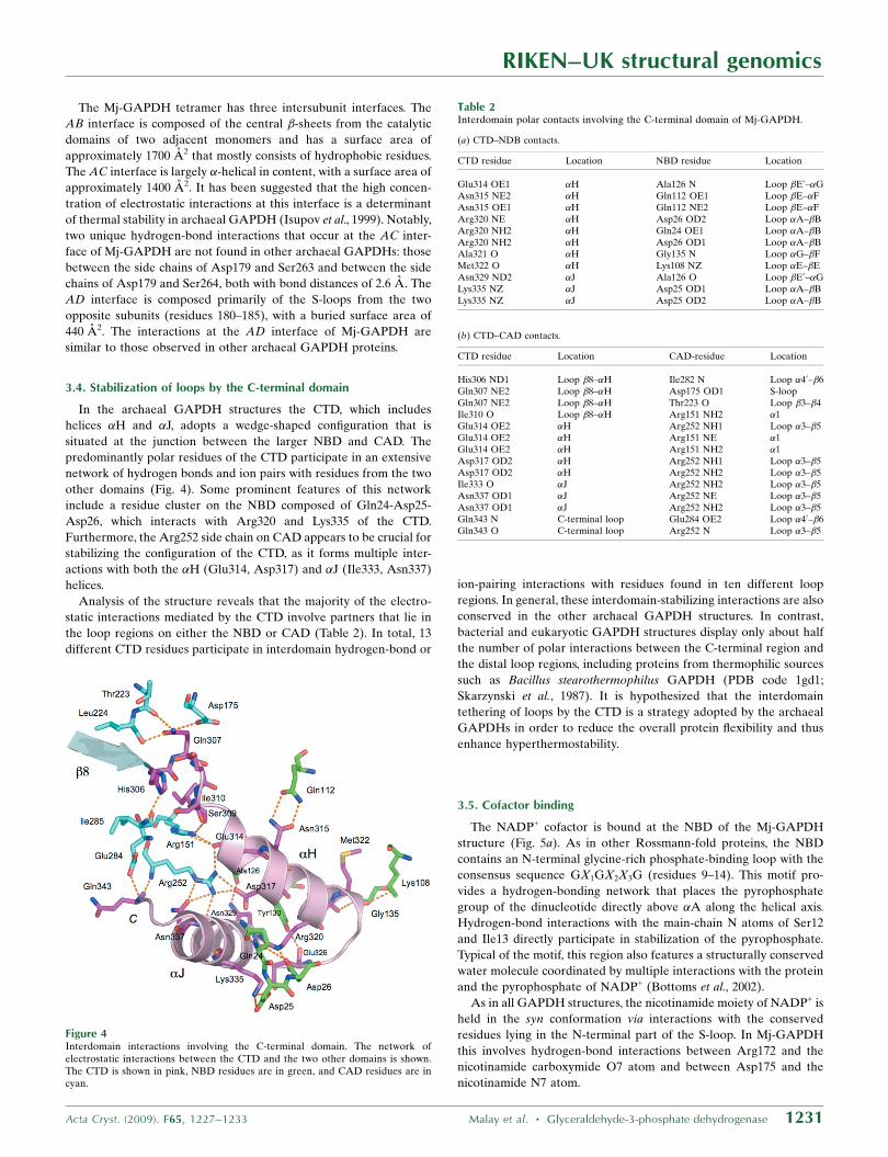
3.4. Stabilization of loops by the C-terminal domain
In the archaeal GAPDH structures the CTD, which includes
helices �H and �J, adopts a wedge-shaped configuration that is
situated at the junction between the larger NBD and CAD. The
predominantly polar residues of the CTD participate in an extensive
network of hydrogen bonds and ion pairs with residues from the two
other domains (Fig. 4). Some prominent features of this network
include a residue cluster on the NBD composed of Gln24-Asp25-
Asp26, which interacts with Arg320 and Lys335 of the CTD.
Furthermore, the Arg252 side chain on CAD appears to be crucial for
stabilizing the configuration of the CTD, as it forms multiple inter-
actions with both the �H (Glu314, Asp317) and �J (Ile333, Asn337)
helices.
Analysis of the structure reveals that the majority of the electro-
static interactions mediated by the CTD involve partners that lie in
the loop regions on either the NBD or CAD (Table 2). In total, 13
different CTD residues participate in interdomain hydrogen-bond or
ion-pairing interactions with residues found in ten different loop
regions. In general, these interdomain-stabilizing interactions are also
conserved in the other archaeal GAPDH structures. In contrast,
bacterial and eukaryotic GAPDH structures display only about half
the number of polar interactions between the C-terminal region and
the distal loop regions, including proteins from thermophilic sources
such as Bacillus stearothermophilus GAPDH (PDB code 1gd1;
Skarzynski et al., 1987). It is hypothesized that the interdomain
tethering of loops by the CTD is a strategy adopted by the archaeal
GAPDHs in order to reduce the overall protein flexibility and thus
enhance hyperthermostability.
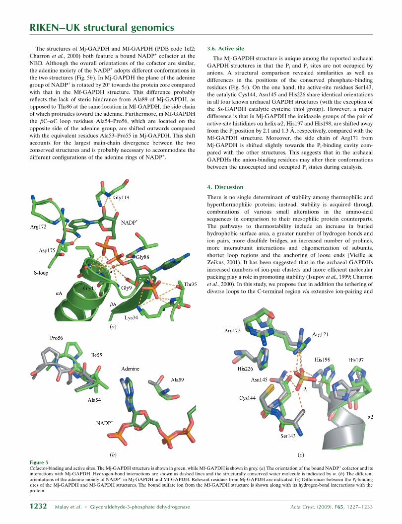
3.5. Cofactor binding
The NADP+ cofactor is bound at the NBD of the Mj-GAPDH
structure (Fig. 5a). As in other Rossmann-fold proteins, the NBD
contains an N-terminal glycine-rich phosphate-binding loop with the
consensus sequence GX1GX2X3G (residues 9–14). This motif pro-
vides a hydrogen-bonding network that places the pyrophosphate
group of the dinucleotide directly above �A along the helical axis.
Hydrogen-bond interactions with the main-chain N atoms of Ser12
and Ile13 directly participate in stabilization of the pyrophosphate.
Typical of the motif, this region also features a structurally conserved
water molecule coordinated by multiple interactions with the protein
and the pyrophosphate of NADP+ (Bottoms et al., 2002).
As in all GAPDH structures, the nicotinamide moiety of NADP+ is
held in the syn conformation via interactions with the conserved
residues lying in the N-terminal part of the S-loop. In Mj-GAPDH
this involves hydrogen-bond interactions between Arg172 and the
nicotinamide carboxymide O7 atom and between Asp175 and the
nicotinamide N7 atom.
RIKEN–UK structural genomics
Acta Cryst. (2009). F65, 1227–1233 Malay et al. � Glyceraldehyde-3-phosphate dehydrogenase 1231
Figure 4Interdomain interactions involving the C-terminal domain. The network ofelectrostatic interactions between the CTD and the two other domains is shown.The CTD is shown in pink, NBD residues are in green, and CAD residues are incyan.
Table 2Interdomain polar contacts involving the C-terminal domain of Mj-GAPDH.
(a) CTD–NDB contacts.
CTD residue Location NBD residue Location
Glu314 OE1 �H Ala126 N Loop �E0–�GAsn315 NE2 �H Gln112 OE1 Loop �E–�FAsn315 OE1 �H Gln112 NE2 Loop �E–�FArg320 NE �H Asp26 OD2 Loop �A–�BArg320 NH2 �H Gln24 OE1 Loop �A–�BArg320 NH2 �H Asp26 OD1 Loop �A–�BAla321 O �H Gly135 N Loop �G–�FMet322 O �H Lys108 NZ Loop �E–�EAsn329 ND2 �J Ala126 O Loop �E0–�GLys335 NZ �J Asp25 OD1 Loop �A–�BLys335 NZ �J Asp25 OD2 Loop �A–�B
(b) CTD–CAD contacts.
CTD residue Location CAD-residue Location
His306 ND1 Loop �8–�H Ile282 N Loop �40–�6Gln307 NE2 Loop �8–�H Asp175 OD1 S-loopGln307 NE2 Loop �8–�H Thr223 O Loop �3–�4Ile310 O Loop �8–�H Arg151 NH2 �1Glu314 OE2 �H Arg252 NH1 Loop �3–�5Glu314 OE2 �H Arg151 NE �1Glu314 OE2 �H Arg151 NH2 �1Asp317 OD2 �H Arg252 NH1 Loop �3–�5Asp317 OD2 �H Arg252 NH2 Loop �3–�5Ile333 O �J Arg252 NH2 Loop �3–�5Asn337 OD1 �J Arg252 NE Loop �3–�5Asn337 OD1 �J Arg252 NH2 Loop �3–�5Gln343 N C-terminal loop Glu284 OE2 Loop �40–�6Gln343 O C-terminal loop Arg252 N Loop �3–�5
RIKEN–UK structural genomics
1232 Malay et al. � Glyceraldehyde-3-phosphate dehydrogenase Acta Cryst. (2009). F65, 1227–1233
The structures of Mj-GAPDH and Mf-GAPDH (PDB code 1cf2;
Charron et al., 2000) both feature a bound NADP+ cofactor at the
NBD. Although the overall orientations of the cofactor are similar,
the adenine moiety of the NADP+ adopts different conformations in
the two structures (Fig. 5b). In Mj-GAPDH the plane of the adenine
group of NADP+ is rotated by 20� towards the protein core compared
with that in the Mf-GAPDH structure. This difference probably
reflects the lack of steric hindrance from Ala89 of Mj-GAPDH, as
opposed to Thr86 at the same location in Mf-GAPDH, the side chain
of which protrudes toward the adenine. Furthermore, in Mf-GAPDH
the �C–�C loop residues Ala54–Pro56, which are located on the
opposite side of the adenine group, are shifted outwards compared
with the equivalent residues Ala53–Pro55 in Mj-GAPDH. This shift
accounts for the largest main-chain divergence between the two
conserved structures and is probably necessary to accommodate the
different configurations of the adenine rings of NADP+.
3.6. Active site
The Mj-GAPDH structure is unique among the reported archaeal
GAPDH structures in that the Pi and Ps sites are not occupied by
anions. A structural comparison revealed similarities as well as
differences in the positions of the conserved phosphate-binding
residues (Fig. 5c). On the one hand, the active-site residues Ser143,
the catalytic Cys144, Asn145 and His226 share identical orientations
in all four known archaeal GAPDH structures (with the exception of
the Ss-GAPDH catalytic cysteine thiol group). However, a major
difference is that in Mj-GAPDH the imidazole groups of the pair of
active-site histidines on helix �2, His197 and His198, are shifted away
from the Pi position by 2.1 and 1.3 A, respectively, compared with the
Mf-GAPDH structure. Moreover, the side chain of Arg171 from
Mj-GAPDH is shifted slightly towards the Pi-binding cavity com-
pared with the other structures. This suggests that in the archaeal
GAPDHs the anion-binding residues may alter their conformations
between the unoccupied and occupied Pi states during catalysis.
4. Discussion
There is no single determinant of stability among thermophilic and
hyperthermophilic proteins; instead, stability is acquired through
combinations of various small alterations in the amino-acid
sequences in comparison to their mesophilic protein counterparts.
The pathways to thermostability include an increase in buried
hydrophobic surface area, a greater number of hydrogen bonds and
ion pairs, more disulfide bridges, an increased number of prolines,
more intersubunit interactions and oligomerization of subunits,
shorter loop regions and the anchoring of loose ends (Vieille &
Zeikus, 2001). It has been suggested that in the archaeal GAPDHs
increased numbers of ion-pair clusters and more efficient molecular
packing play a role in promoting stability (Isupov et al., 1999; Charron
et al., 2000). In this study, we propose that in addition the tethering of
diverse loops to the C-terminal region via extensive ion-pairing and
Figure 5Cofactor-binding and active sites. The Mj-GAPDH structure is shown in green, while Mf-GAPDH is shown in grey. (a) The orientation of the bound NADP+ cofactor and itsinteractions with Mj-GAPDH. Hydrogen-bond interactions are shown as dashed lines and the structurally conserved water molecule is indicated by w. (b) The differentorientations of the adenine moiety of NADP+ in Mj-GAPDH and Mf-GAPDH. Relevant residues from Mj-GAPDH are indicated. (c) Differences between the Pi-bindingsites of the Mj-GAPDH and Mf-GAPDH structures. The bound sulfate ion from the Mf-GAPDH structure is shown along with its hydrogen-bond interactions with theprotein.
hydrogen-bond interactions likewise contributes to increased thermal
stability.
We thank Ms Michiyo Takahara, Mr Hitoshi Iino, Mr Yoshihiro
Agari and Dr Akio Ebihara for their assistance with sample
preparation. We are grateful to Ms Tomoko Nakayama and Ms Azusa
Ishii for their clerical assistance. This work was supported in part by
the RIKEN Structural Genomics/Proteomics Initiative (RSGI), the
National Project on Protein Structural and Functional Analyses,
Ministry of Education, Culture, Sports, Science and Technology of
Japan. This work was supported by the Synchrotron Radiation
Department at the Science and Technology Facilities Council,
Daresbury Laboratory UK and beamline 10.1 at the Synchrotron
Radiation Source, which was supported by Biotechnology and
Biological Sciences Research Council Grant BB/E001971 (to SSH
and RWS).
References
Antonyuk, S. V., Eady, R. R., Strange, R. W. & Hasnain, S. S. (2003). ActaCryst. D59, 835–842.
Arcari, P., Russo, A. D., Ianniciello, G., Gallo, M. & Bocchini, V. (1993).Biochem. Genet. 31, 241–251.
Bottoms, C. A., Smith, P. E. & Tanner, J. J. (2002). Protein Sci. 11, 2125–2137.Charron, C., Talfournier, F., Isupov, M. N., Littlechild, J. A., Branlant, G.,
Vitoux, B. & Aubry, A. (2000). J. Mol. Biol. 297, 481–500.
DeLano, W. L. (2002). The PyMOL Molecular Graphics System. DeLanoScientific, San Carlos, California, USA. http://www.pymol.org.
Davis, I. W., Murray, L. W., Richardson, J. S. & Richardson, D. C. (2004).Nucleic Acids Res. 32, W615–W619.
Duee, E., Olivier-Deyris, L., Fanchon, E., Corbier, C., Branlant, G. &Dideberg, O. (1996). J. Mol. Biol. 257, 814–838.
Harris, J. I. & Waters, M. (1976). The Enzymes, edited by P. D. Boyer, pp. 1–49.New York: Academic Press.
Isupov, M. N., Fleming, T. M., Dalby, A. R., Crowhurst, G. S., Bourne, P. C. &Littlechild, J. A. (1999). J. Mol. Biol. 291, 651–660.
Jones, T. A., Zou, J.-Y., Cowan, S. W. & Kjeldgaard, M. (1991). Acta Cryst.A47, 110–119.
Lamzin, V. S. & Wilson, K. S. (1993). Acta Cryst. D49, 129–147.Littlechild, J. A., Guy, J. E. & Isupov, M. N. (2004). Biochem. Soc. Trans. 32,
255–258.Murshudov, G. N., Vagin, A. A. & Dodson, E. J. (1997). Acta Cryst. D53,
240–255.Murthy, M. R., Garavito, R. M., Johnson, J. E. & Rossmann, M. G. (1980). J.
Mol. Biol. 138, 859–872.Otwinowski, Z. & Minor, W. (1997). Methods Enzymol. 276, 307–326.Sirover, M. A. (1999). Biochim. Biophys. Acta, 1432, 159–184.Sirover, M. A. (2005). J. Cell. Biochem. 95, 45–52.Skarzynski, T., Moody, P. C. & Wonacott, A. J. (1987). J. Mol. Biol. 193,
171–187.Tanner, J. J., Hecht, R. M. & Krause, K. L. (1996). Biochemistry, 35, 2597–
2609.Tisdale, E. J. (2002). J. Biol. Chem. 277, 3334–3341.Vagin, A. & Teplyakov, A. (1997). J. Appl. Cryst. 30, 1022–1025.Vieille, C. & Zeikus, G. J. (2001). Microbiol. Mol. Biol. Rev. 65, 1–43.Zheng, L., Roeder, R. G. & Luo, Y. (2003). Cell, 114, 255–266.
RIKEN–UK structural genomics
Acta Cryst. (2009). F65, 1227–1233 Malay et al. � Glyceraldehyde-3-phosphate dehydrogenase 1233