structure of an engineered porcine phospholipase a2 with enhanced activity at 2·1 Å resolution
TRANSCRIPT
J. Mol. Biol. (1990) 216, 425-439
Structure of an Engineered Porcine Phospholipase A2 withEnhanced Activity at 2·1 A Resolution
Comparison with the Wild-type Porcine and Crotalus atroxPhospholipase A2
Marjolein M· G· M· Thunnissen, Kor H· Kalk, Jan Drenth and Bauke W· Dijkstra
Laboratory of Chemical Physics, University of GroningenNijenborg 16, 9747 AG Groningen, The Netherlands
(Received 6 April 1990; accepted 12 July 1990)
The crystal structure of an engineered phospholipase A2 with enhanced activity has beenrefined to an R-factor of 18·6% at 2·1 A resolution using a combination of moleculardynamics refinement by the GROMOS package and least-squares refinement by TNT· Thismutant phospholipase was obtained previously by deleting residues 62 to 66 in porcinepancreatic phospholipase A2, and changing Asp59 to Ser, Ser60 to Gly and Asn67 to Tyr·The refined structure allowed a detailed comparison with wild-type porcine and Crotalusatrox phospholipase A2· The conformation of the deletion region appears to be intermediatebetween that in those two enzymes· The residues in the active center are virtually the same·An internal hydrophobic area occupied by Phe63 in the wild-type porcine phospholipase A2is kept as conserved as possible by local rearrangement of neighboring atoms· In the mutantstructure, this hydrophobic pocket is now occupied by the disulfide bond between residues61 and 91. A detailed description of the second binding site for a calcium ion in this enzymeis given·
1. Introduction
Phospholipase A2 (PLA2†, phosphatide acyl-hydrolase, E.e.3·1.1.4) catalyzes the hydrolysisof the 2-acylesterbond of 3-sn-glycerophospholipidsin a calcium-dependent reaction (Waite, 1987)· Theenzyme can be found both inside and outside thecell· Extracellular phospholipases A2 occur abun-dantly in mammalian pancreatic tissue and juiceand in snake and bee venoms· The enzymes frommammalian pancreas and snake venom show a highdegree of sequential homology (Waite, 1987;Volwerk & de Haas, 1982), and the intracellularphospholipases A2 appear to be homologous(Mizushima et al., 1989; Seilhamer et al·, 1989;Kramer et al·, 1989)· The pancreatic PLA2 isproduced in the pancreas as a zymogen· Uponsecretion into the duodenum the proenzyme is acti-vated by trypsin, which cleaves off sevenN-terminal amino acid residues· For the snakevenom PLA2s, the existence of a zymogen is notknown (Nieuwenhuizen et al·, 1974).
† Abbreviations used: PLA2, phospholipase A2; Δ62-66PLA2 mutant, porcine PLA2 with residues 62 to 66deleted and additional mutations D59S, S60G andN67Y; r·m.s·, root-mean-square; bis-Tris, bis(2-hydroxy-ethyl )amino- tris(h ydroxymethy I)methane·
One of the intriguing properties of phospholipasesA2 is the fact that they show quite differentbehavior towards monomeric and aggregatedsubstrates· While the mammalian proenzyme andthe mature enzyme have a similar activity on mono-meric substrates, the mature enzyme hydrolyzesaggregated substrates with much higher rates thanit does monomeric substrates· The proenzyme doesnot show this activation by aggregated substrates(Pieterson et al·, 1974)· The much higher rates ofhydrolysis of aggregated substrates imply that theenzyme must recognize lipid/water interfaces, andthat it probably possesses a binding site for aggre-gated substrates· Indeed, several residues have beenidentified by chemical modification to be part of thisrecognition site and were shown to affect theenzyme's properties with respect to the binding ofaggregated substrates· On the basis of these studies,it was concluded that Leu2, Trp3, Arg6, Leu19 andTyr69 are part of the binding site for aggregatedsubstrates (Volwerk & de Haas, 1982). The three-dimensional structure of the bovine pancreaticPLA2 showed that these residues are on one face ofthe protein molecule, around the entrance of theactive site cleft (Dijkstra et al·, 1981). From theseobservations it was concluded that the binding sitefor aggregated substrates is an extended area
4250022-2836/90/220425-15 $03.00/0 © 1990 Academic Press Limited
426 M. M. G. M. Thunnissen et al.
around the active site cleft. Residues 65, 67, 70 and72 were proposed to be part of this binding site(Dijkstra et al., 1981). These latter residues have notbeen further analyzed by chemical modificationmethods.
In the pancreatic phospholipases A2, residues 65to 70 occur in a surface loop. A comparison of thestructures of the mature bovine phospholipase A2and the bovine proenzyme shows that in the pro-enzyme this surface loop (comprising residues 62 to72) and a part of the N-terminal helix are flexible ordisordered, but that they have a well-definedconformation in the mature enzyme (Dijkstra et al.,1982). The same flexibility was observed in thestructure of the N-terminally transaminated bovinephospholipase A2, which has the same kineticproperties as the proenzyme (Dijkstra et al., 1984).An explanation for this difference in mobility inmature and proenzyme can be found in the fact thatthe 62-72 loop is linked via a hydrogen-bondingsystem to the N-terminal α-NH3+group. When thishydrogen-bonding system is disrupted, as is the casein both the proenzyme and the N-terminally trans-aminated enzyme where no free α-NH3+ existsanymore, the 62-72loop and part of the N-terminalhelix become flexible. Because the proenzyme andthe transaminated enzyme do not show any increasein activity when the substrate concentration isabove the critical micelle concentration, it wasconcluded that these enzymes do not have an intactbinding site for aggregated substrates and that the62-72 loop is important for the regulation of theenzyme's activity towards aggregated substrates(Dijkstra et al., 1984).
Interestingly, sequences of snake venom phospho-lipases A2 show a deletion of five to eight residues inthis loop (see Table I). It is one of the most con-spicuous sequence differences (Waite, 1987). Also,the kinetic properties and substrate specificity ofsnake venom phospholipases A2 are rather different.The snake venom phospholipases A2 have in generalhigher turnover numbers and have a higher affinityfor phospholipid molecules aggregated in micellesthan do the pancreatic phospholipases A2. Thepancreatic PLA2s are in general more activetowards negatively charged phospholipids (van Eyket al., 1983; Verhey et al., 1981).
In an attempt to investigate the influence of theseresidues on the catalytic activity, residues 62 to 66in the porcine PLA2 were deleted (Kuipers et al.,
1989a). To increase the sequential homology withelapid snake venom PLA2, a few other mutationswere performed: Asp59Ser, Ser60Gly and Asn67Tyr(see Table I). The glycine at position 60 was intro-duced because it was thought that the enzymeneeded at that position a residue with a largerconformational freedom in order to form the properdisulfide bridge between cysteine 61 and cysteine91. The Tyr at position 67 was introduced becausein many snake venom phospholipases A2 an aro-matic residue is found at this position. Ser at posi-tion 59 is found in many of the elapid snake venomPLA2s (Dennis, 1983). The construction and charac-terization of this mutant phospholipase A2 (Δ62-66mutant) and a preliminary description of its struc-ture were given by Kuipers et al. (1989a). TheΔ62-66 mutant showed a 16-fold increase in activityon micellar (zwitterionic) short chain lecithinscompared to the native porcine PLA2. The hydro-lysis of phospholipid monomers is twice as fast. Thestructure at 2,5 A (I A = 0·1 nm) resolution revealedthat the biggest difference between the wild-typeporcine and the Δ62-66 mutant porcine PLA2 struc-ture is found in the deleted loop area and in thetopology of the binding site for aggregatedsubstrates.
Here, we report the structure of the Δ62-66mutant, refined at 2·1 A resolution to a finalR-factor of 18'6%. Both molecular dynamics refine-ment and conventional least-squares refinementprocedures have been applied. A detailed descrip-tion of the structure and a comparison with thestructure of wild-type phospholipase A2 is given.
2. Materials and Methods(a) Crystallization and data collection
The Δ62-66 PLA2 mutant was generously provided tous by the group of H. M. Verhey, A.J. Slotboom andG. H. de Haas (State University, Utrecht).
The freeze-dried protein was dissolved in 0'1 M-sodiumacetate (pH 4'5) to a final concentration of 10 mg/ml.This solution was dialyzed overnight against100 mM-bis-Tris' HCI (pH 7'0) with 5 mM-CaCI2 added, inorder to prevent rapid precipitation. The enzyme wascrystallized at room temperature by vapor diffusion inhanging drops. The reservoir solution contained I ml of asolution of 50% (v/v) methanol in 100 mM-bis-Tris' HCI(pH 7'0), 5 mM-CaCI2. Drops of 6 µl were formed by equalmixing of 3 µl of protein solution and 3 µl of the reservoir
Table 1Comparison of part of the sequences of the mutant P LA 2 and of 3 native P LA 2S
50 55 60 65 70
(a) H D N C Y R D A K N L D S C K F L V D N P Y T E S Y(b) * * * * * G E * E K I S G * ---W * * I K T *(c) * * C * * G K * T * * * * K * V * *(d) * * * * * * * * * * * S G * - -- -- - - Y * * * * * *
(a) Porcine pancreatic PLA2; (b) Naja melanoleuca PLA2 fraction DE-III; (c) C. atrox PLA2; and(d) mutant Δ62-66 PLA2. The sequence numbering is according to Renetseder et al. (1984). An asterisk(*) denotes homology with the porcine sequence. Absent amino acid residues are indicated by a dash.

Structure of Δ62-66 Porcine Phospholipase A 2 427
Table 2Completeness of data
Before† After†
Resolution (Å) Completeness(%) Rmerge‡ Completeness(%) Rmerge‡
∞~IO·OO 58·8 2·36 58·8 2·361O·00~ 7·07 85·3 3·15 85·3 3·16
7·07~ 5·77 92·5 3·56 92·5 3·565·77~ 5·00 91·0 3·91 91·0 3·935·00- 4·47 91·8 4·43 91·8 4·444·47- 4·08 91·3 4·69 91·3 4·704·08- 3·78 92·0 4·70 92·0 4·703·78~ 3·54 92·2 5·06 92·0 5·073·54- 3·33 93·4 5·48 93·1 5·483·33~ 3·16 94·4 5·83 93·8 5·903·16~ 3·02 93·6 5·82 88·6 5·823·02- 2·89 92·7 6·04 86·5 6·112·89- 2·77 92·8 7·24 82·1 7·152·77~ 2·67 92·3 7·94 82·7 7·612·67~ 2·58 89·0 8·90 74·3 8·592·58~ 2·50 89·6 8·75 77·5 8·452·50- 2·43 87·7 9·20 73·4 8·182·43- 2·36 87·1 12·01 72·8 11·042·36~ 2·29 83·7 11·35 68·1 1O·572·29~ 2·24 82·1 11·29 67·6 1O·952·24- 2·18 77·8 12·09 65·0 11·522·18- 2·13 74·8 12·80 61·1 11·962·13~ 2·09 71·8 15·43 61·1 14·46
Total 87·0 5·54 78·4 5·29
H Before and after removal of strongly deviating reflections with Is-II (see the text forexplanation)·
I I |I(hkl,j)-I(hkl)|
H Rmerge= hkl refLΣΣ I(hkl)hkl refl.
solution· Crystals suitable for X-ray analysis grew within1 week· The crystals are platelets about 0·7 mm x 0·3 mmx 0·15 mm in size· The spacegroup is P21· There are 2molecules of 119 residues each in an asymmetric unit·
Data up to 2·1 A resolution were collected from 1crystal with cell dimensions a = 45·8 Å, b = 73·4 Å,c = 37·3 Å and β = 1O7·4°on an Enraf-Nonius FAST areadetector, equipped with a CAD4 kappa-goniostat, withgraphite monochromatized CuKα radiation from an ElliotGX-21 rotating anode generator· Data collection andreduction were carried out using the program systemMADNES (Pflugrath & Messerschmidt, 1986)· The crystalwas mounted in a glass capillary with its b-axis approxi-mately parallel to the capillary axis· The capillary withthe crystal was positioned such that it was more or lessaligned with the goniostat's φ-axis· The φ-axis was set soas to make an angle of about 36° with the ω-axis of thegoniostat (thus the crystal's b-axis and the ω-axis do notcoincide) · This was done to optimize collection of avirtually complete dataset· Data were collected byrotation about the ω-axis using 2 different φ-settings ofthe crystal 90° apart, one at φ = 1450 and one atφ = 2340
• At the Ist setting, 1800 of oscillation werecollected· However, due to radiation damage, only 130°could be collected at the 2nd setting· The resultingdataset contained 30,069 usable observations, 13,801 ofwhich were unique reflections· The data had Rmerge=0·055 for 12,278 hkl values (for a definition of Rmergeseethe legend to Table 2)·
The overall completeness was 89·7 %· Data weredivided into chunks with Δφ = 10°, scaled (Hamilton et al·,
1965) and merged together into a single dataset usingprograms of the Groningen BIOMOL protein structuredetermination software package· Deviating reflections(reflections not within 2·24 σ from the mean intensity)were omitted· The completeness of the dataset decreasedto 87·0% after this procedure· Because the refinement didnot converge (see below), we reassessed the quality of ourdata at a later stage· After a local scaling procedure itappeared that reflections with I≤ -11 had relatively veryhigh R-factors (up to 80%)· These reflections wereomitted· The completeness decreased thereby to 78·4%·In order to find an explanation for these high R-factors,the whole data collection procedure was checked· It wasobserved that most of these reflections were collected inthe later stages of the data collection· Most probably weunderestimated the effect of (anisotropic) radiation decay·Table 2 gives an overview of the completeness and Rmergeas a function of resolution both before and after theremoval of these deviating reflections·
(b) Refinement
It has been shown that energy refinement combinedwith molecular dynamics simulation can be very powerfulat the initial stages of a refinement procedure (Gros et al·,1989a)· Molecular dynamics techniques allow a molecularsystem to explore a larger conformational space than ispossible with the more conventional methods of least-squares refinement (Brünger, 1988; Fujinaga et al·, 1989;Gros et al·, 1989b)· We started molecular dynamics refine-ment with the GROMOS MDXREF program (Fujinaga et

428 M. M. G. M. Thunnissen et al.
al., 1989). As a starting model we used the partiallyrefined structure at 2'5 A resolution of the Δ62-66mutant without water molecules (Kuipers et al., 1989a). A1st R-factor with data between 6 and 2·1 A was 33 %(R = ~||Fo|-|Fe||/~|Fo|). The water molecules wereomitted in order to avoid problems in the initial moleculardynamics runs carried out at higher temperatures (Gros etal., 1989b).
The 1st refinement cycle was started by energy minimi-zation with X-ray constraints followed by moleculardynamics simulation with X-ray constraints at 600 K, atrelatively low resolution (8'0 to 3'5 Å). After this step, thetemperature was slowly decreased to 300 K and in themean time the resolution was gradually expanded from3'5 to 2·9 Å. This refinement cycle lasted 3'3 ps. At theend of this 1st cycle, the R-factor was 27'8% (8 to 2'9 Åresolution). The model was inspected in a SIGMAAweighted 2Fo - Fe map (Read, 1986), in the later stagesalso OMIT maps were used (Bhat & Cohen, 1984; Bhat,1988). All model building was done on an Evans &Sutherland PS390 system using the program FRODO(Jones, 1978). For the analysis of the results, theprograms WHATIF (Vriend, 1990) and BIOGRAF(BIOGRAF, Biodesign Corporation, Pasadena, U.S.A.)were also used. Only minor rebuilding was done. At thisstage 31 water molecules were added to the model.
After this manual intervention, a 2nd cycle ofmolecular dynamics refinement was performed. The wholecycle was done at a temperature of 300 K during 2·7 ps.While the resolution was gradually expanded to 2'7 Å, theR-factor decreased to 25'6% (8 to 2'7 Å resolution) at theend of this cycle. Because the refinement did not convergeanymore at this stage, neither with the MDXREFprogram nor with the TNT package, we reprocessed ourdata to remove data that suffered too much fromradiation damage (see above).
At this stage we switched to the least-squares refine-ment procedure of TNT (Tronrud et al., 1987) instead ofthe molecular dynamics refinement, because of less severe
central processing unit requirements. Cycles of alternatingTNT refinement and model building were performed. Asummary is given in Table 3. Water molecules werelocated in Fo - Fe maps. Peaks with a density ≥ 2 σ wereidentified and used as possible water positions by theprogram PEKPIK from the XTAL package (Hall &Stewart, 1987). Only water molecules at proper distancesfrom protein atoms were kept. Errors in the structurewere found by careful inspection of Ramachandran plots(Ramakrishnan & Ramachandran, 1965) and the geo-metrical analysis part of TNT. Water molecules thatdeveloped B-factors higher than 55 Å2 were removed fromthe model.
The refined co-ordinates for this PLA2 Δ62-66 mutanthave been deposited with the Protein Databank,Chemistry Department, Brookhaven NationalLaboratory, Upton NY 11973, U.S.A. (reference number3P2P).
3. Results
(a) Refinement
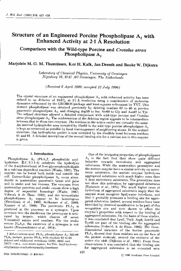
After completion of the refinement, the crystallo-graphic R-factor had decreased to 18·6% for alldata between 6 and 2·1 A resolution. The finalmodel contains 204 water molecules and threecalcium ions: one in the active site of each PLA2molecule and one on a non-crystallographic 2-foldaxis between the two molecules in the asymmetricunit (monomer I, residues 1 to 124; monomer 2,residues 201 to 324)· The density for both moleculesis clear (Fig. 1), except for residues around theβ-wing tip, residues 79 and 80, in both molecules,where the density is weak. Also, some of the residuesat the surface of the molecule have their side-chainsin weak density. The C-terminal end of monomer 2is disordered and no density is visible for residues
Table 3Refinement statistics
A. Progress of refinement
Number of Resolution Start R End R Water molecules andCycle rounds (A) (%) (%) rebuilding
1st 3'3 ps 8'0-3'5 s 33'5 Added 31 water molecules, minorMD 8'0-2'9 e 27·8 rebuilding
2nd 2·7 ps 8·0-2·9 s 27·8 Added 65 water molecules, minorMD 8,0-2,7 e 25·6 rebuilding
1 175 xyz 8,0-2'8 s 25·6 Added 70, removed 20 waterTNT 25 B 8,0-2,1 e 22·6 molecules, rebuilding
2 135 xyz 8,0-2'1 25'6 20·2 Added 80, removed 16 waterTNT 35 B molecules, minor rebuilding
3 70 xyz 6'0-2,1 23·6 19·6 Removed 6 water molecules,TNT lOB rebuilding
4 120 xyz 6·0-2'1 22·1 18'6TNT 28 B
13. Geometry statistics after completion of the refinement
r.m.s. bond length deviations (A) 0·010r.m.s. bond angle deviations (0) 2'59 1r.m.s. trigonal non-planarity deviations (A) 0'006r.m.s. planarity deviations (A) 0'010r.m.s. non-bonded interaction deviations (A) 0·044
MD, molecular dynamics; s, at start of refinement cycle; e, at end of refinement cycle.
Structure of /162-66 Porcine Phospholipase A2 429
Figure 1. Stereo pictures showing (2mFo - DFc), αe (Read, 1986) electron density for parts of the map. The density iscontoured at l·lσ. (a) Trp3 to Arg6 at the N-terminal region of monomer I. (b) Ser72 to Ser76 (main chain) ofmonomer I.
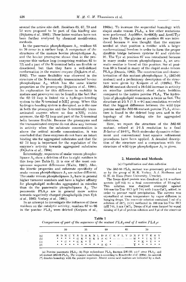
319 to 323. Residue Cys324, which is connected toCys227 by a disulfide bond, is visible again. In bothmonomers the density for the deletion region is clearand could easily be interpreted using OMIT mapsand 2Fo - Fe maps (Fig. 2). From a Luzzati plot, ther.m.s. positional error in the co-ordinates is esti-mated to be 0·25 A (Luzzati, 1952). An independentestimate of the error can be deduced from the r.m.s.difference in positions of the Cα atoms after super-
position of the two monomers. The r.m.s. differencebetween all 119 Cα positions is 0·52 Å. If the mostdeviating atoms are omitted, this differencedecreases to 0·35 A for 101 Cα pairs. Details aboutthe refinement and the quality of the structure canbe found in Table 3·
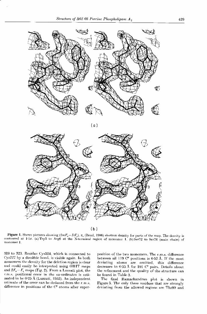
The final Ramachandran plot is shown inFigure 3. The only three residues that are stronglydeviating from the allowed regions are Thr80 and
430 M. M. G. M. Thunnissen et al.
Figure 2. Stereo picture showing the (2mFo - DFc), αc (Read, 1986) electron density for the deletion region (residues 59to 69) in monomer 1. The density is contoured at 1·10".
Figure 3. The φ, ψ plot of both monomers of the L162-66 mutant. The φ, ψ angles of non-glycine residues are indicatedby a cross and those of glycine residues by a square. The continuous lines define the area of fully allowed conformationswith τ(N, Cα, C) = 110° for the non-glycine residues. The broken lines show the regions obtained by relaxing the van derWaals' contact constraints as well as by allowing the bond angle τ at Cα to increase to 115° (Ramakrishnan &Ramachandran, 1965). Strongly deviating residues are Thr80 (φ = 62°, ψ = -29°), Thr280 (φ = 91°, ψ = -32°) andLys317 (φ = 14°, ψ = -89°).

Structure of Δ62-66 Porcine Phospholipase A2 431
Thr280, both located in the β-wing tip, and Lys317located in the disordered C terminus of monomer 2.
(b) Comparison of the two molecules in theasymmetric unit
In the asymmetric unit, two protein molecules arepresent that are related to each other by a non-crystallographic axis (κ = 178'3°). This 2-fold axisconstitutes the most conspicuous contact betweenthe two monomers and it runs through a calcium ionthat is ligated by atoms from both monomers (seebelow). In the wild-type porcine PLA2 structure,this calcium-binding site is also present, but there itis found on a crystallographic 2-fold axis. Theoverall structure of the two monomers in the asym-metric unit is almost the same, including the confor-mation of the deletion region, but three stretcheswith differences occur. All these differences arecaused by different intermolecular contacts (seeFig. 4 and Table 4).
The first of these stretches can be found aroundPro14. A hydrogen bond exists between thecarbonyl oxygen of Pr0214 and the Nηl atom ofArg43, but this hydrogen bond is not presentbetween the carbonyl oxygen of Pro14 and the Nηl
of Arg243, because these atoms are more than 10 Aapart. As a result the C= O of Pro 14 is in a differentposition and, as a consequence, G1y15 has quitedifferent φ, ψ angles in both monomers (G1y15φ = 111'5°, ψ = -22'5°; G1y215 φ = -96'5°,ψ = 156'3°).
The second stretch is around Leu31 and involvesthe first calcium-binding site. In both monomers thecalcium site is formed by the same residues, and theconfiguration around the calcium ion is hardlychanged, but the conformation of Leu31 in bothmonomers is completely different. In monomer 2,Leu231 has hydrophobic interactions with asymmetry-related molecule, but in monomer I thisresidue is totally exposed to the solvent (seeTable 4).
The last deviating stretch is found at theC terminus. In monomer 1, the COO- group of the
Figure 4. (a) Differences in Cα positions (in A) aftersuperimposing monomer I and monomer 2.(b) Temperature factors (A2) of the Cα atoms as a functionof residue number for the 2 monomers: heavy line,monomer I; broken line: monomer 2.
C terminus makes a salt bridge with Arg206 froma crystallographically related molecule. In addition,this Arg206 is hydrogen-bonded to the carbonyl'oxygen of Lys122. Hydrophobic interactions occurbetween Tyr123 and Leu319, and the side-chain ofLys121 is hydrogen-bonded to the carbonyl oxygenof Ser86 of a symmetry-related molecule. Inmonomer 2, none of these interactions occurs, andthis most probably explains why in monomer 2residues 319 to 323 are disordered or flexible.
Table 4Differences in intermolecular contacts between the 2 monomers in the asymmetric
unit
Monomer 1 Monomer 2
Site 1; around Pro14Cα Pro 14-Cβ Leu231 3'8 Å o Pro214-Nη1 Arg43 2'6 ÅCβ ProI4-Cβ, Cδl & C Leu231 3'8 -4'0 ÅC Pro 14-Cβ Leu231 3·8 Å
Site 2; around Leu31No intermolecular interactions Cβ Leu231-Cα, Cβ Pro14 3'8 -4 Å
COl Leu231-Cβ Pro14 3'9 ÅC Leu231-Cβ Pro14 4'0Å
Site 3; around the C terminusNζ Lys121-0 Ser8 6 3'0 Å Nζ Lys322-0 Cys77 3·7 Åo Lys122-Nη2 Arg206 2·9 ÅCβ TyrI23-Cδ1 Leu319 3'5 ÅCOl TyrI23-C' Leu319 3'7 Åo Cys124-Nηl Arg206 2·7 Å

432 M. M. G. M. Thunnissen et al.
Figure 5. Comparison of a Cαbackbone tracing of wild-type (dotted lines) and Δ62-66 mutant PLA2 (heavy lines).
(c) Comparison with wild-type porcine phospholipaseA2
In Figure 5, a comparison is shown between theΔ62-66 mutant and the wild-type porcine PLA2, thestructure of which has been refined at 2·6 A resolu-tion to an R-factor of 24'1 % (Dijkstra et al., 1983).The greater part of these structures is virtually thesame. As expected, the largest differences are foundin the deletion area, residues 59 to 70.
In wild-type porcine PLA2, these residues form ashort surface loop from residue 59 to 66 and thenone and a half turns of 310 helix from residues 66 to70. Specific interactions of this loop with the rest ofthe molecule are made by Cys61, which forms adisulfide bridge with Cys91, and by Phe63, which isin the interior of the protein and fills up a hydro-phobic pocket· This pocket is formed by the side-chains of Pro68, Ala55, Leu58, Ile95, the disulfidebridge between Cys61 and Cys91, and the backboneatoms of Glu71. The 61-91 disulfide bridge is at the
surface of the protein and shields this hydrophobicpocket from the solvent· All the interactions ofthese residues are given in Table 5.
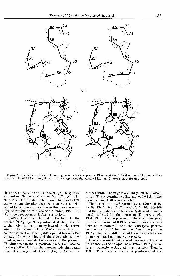
In the Δ62-66 mutant, the loop 59 to 70 becomesshortened and the 310 helix part is not presentanymore. Residues 59 and 60 form a turn, residues61 to 68 have an extended conformation, andresidues 69 and 70 are positioned in another turn(Fig. 6).
The most conspicuous changes occur aroundresidue 63· In the Δ62-66 mutant, Phe63 is deleted,but the hydrophobic pocket occupied by thisresidue still exists· Instead of Phe63, the disulfidebridge between Cys61 and Cys91 moves to theinterior of the protein and fills this pocket. The CαofCys61 thereby changes 4·4 A in position, and theside-chain of Cys91 has to rotate 1800 around theCα-Cβ bond to make a proper disulfide bridge(Fig. 7). The rest of Cys91 does not change. Thepocket itself has hardly changed in conformation.The biggest change here is effected by Pro68 moving
Table 5Interactions of residues 59 to 70 in porcine and Δ62-66 mutant P LA 2
Residues Porcine PLA2 Residues Δ62-66 PLA2 mutant
Asp59 Surface residue, no specific interactions Ser59 Surface residue, no specific interactionsSer60 Surface residue, no specific interactions Gly60 Surface residue, no specific interactionsCys61 Surface residue, disulfide bridge to 91, Cys61 Surface residue, disulfide bridge to 91,
hydrophobic interactions with Phe63 hydrophobic interactions with several residues,C= O interaction with a water molecule
Lys62 Surface residue, Nδ2 interaction to C = 0 Phe63Phe63 Interior residue, phenyl ring, several hydrophobic
interactions, C = 0 interaction with Nδ2 Lys62and N Val65
Leu64 Surface residue, hydrophobic interactions withThr70, C= 0 interaction with N Asn67
Val65 Surface residue, interaction with Asn67, Ninteraction with C = 0 of Phe63
Asp66 Surface residue, C=O interaction with Nζ Lys56Asn67 Surface residue, 061 interaction with 0,1 Thr70, Tyr67 Surface residue, no specific interactions
N interaction with C = 0 Leu64Pro68 Interior residue, hydrophobic interactions with Pro68 Interior residue, hydrophobic interactions with
Phe63 disulfide bridge 61-91Tyr69 Surface residue, no specific interactions Tyr69 Surface residue. no specific interactionsThr70 Interior residue, hydrophobic interactions with Thr70 Interior residue, hydrophobic interactions with
Leu64, 0,1 with C61Asn67, C=O interaction Glu71, Cγ1 with water, C=O interaction withwith N-terminal α-NH3+ N-terminal α-NH3+
Structure of 1162-66 Porcine Phospholipase A2 433
Figure 6. Comparison of the deletion region in wild-type porcine PLA2 and the Δ62-66 mutant. The heavy linesrepresent the Δ62-66 mutant, the dotted lines represent the porcine PLA2. (a) Cα atoms only; (b) all atoms.
closer (0·2to 0·3 Å) to the disulfide bridge· The glycineat position 60 has φ, ψ values (φ = 67°, ψ = 43°)close to the left-handed helix region. In 18 out of 21snake venom phospholipases A2 that have a dele-tion of five amino acid residues in this area there is aglycine residue at this position (Dennis, 1983). Inthe three exceptions it is Arg, Ser or Lys.
Tyr69 is located at the end of the loop. In theporcine PLA2, Tyr69 is positioned at the entranceto the active center, pointing inwards to the activesite of the protein. Since Pro68 has a differentconformation, the Cαof Tyr69 is pulled towards theoutside of the protein, and the side-chain is nowpointing more towards the exterior of the protein.The difference in the oη position is 5 Å. Leu2 movesto the position left by the tyrosine side-chain andfills up the newly created cavity (Fig. 8)· As a result,
the N-terminal helix gets a slightly different orien-tation. The N-terminal α-NH3+moves 1·31 Å in onemonomer and 1·1OÅ in the other.
The active site itself, formed by residues His48,Asp99, Phe5, Ile9, Phe22, Ala102, Ala103, Phe106and the disulfide bridge between Cys29 and Cys45 ishardly affected by the mutation (Dijkstra et al·,1981, 1983). A superposition of these residues givesa r.m.s. difference of 0'43 Å between pairs of atomsbetween monomer 1 and the wild-type porcineenzyme and 0'40 Å for monomer 2 and the porcinePLA2. The r.m.s. difference of these atoms betweenmonomer 1 and monomer 2 is 0'33 Å.
One of the newly introduced residues is tyrosine67. In many of the elapid snake venom PLA2s thereis an aromatic residue at this position (Dennis,1983). This tyrosine residue is positioned at the

434 M. M. G. M. Thunnissen et al.
Fig.7.
surface of the protein and it does not make anyspecific interactions. It might be a part of thebinding site for aggregated substrates.
(d) The second calcium-binding site
The presence of a second calcium-binding site hasbeen reported for porcine phospholipase A2 basedon both biochemical evidence and crystallographicwork (Slotboom et al., 1978; Andersson et al., 1981;Donne-Op den Kelder et al., 1983; Dijkstra et al.,1983). This second calcium-binding pocket has aconsiderably lower affinity for calcium than themajor site. From titration studies, it was found thatGlu71 in the porcine PLA2 is a ligand of thiscalcium ion (Donne-Op den Kelder et al., 1983). Tnsite-directed mutagenesis studies combined withactivity measurements at high pH, Glu71 andAsp66 were identified as possible candidates forligating the calcium ion. According to these studies,Glu92 would not be a ligand (van den Bergh et al.,1989). In the crystal structure of wild-type porcinePLA2, the calcium ion is bound on a crystallo-graphic 2-fold axis and two symmetry-related mole-cules contribute to the binding of the calcium ion.As ligands, the carbonyl oxygen of Ser72, twice, andthe ad atom of Glu92, twice, were identified in thecrystal structure. The two other ligands werethought to be water molecules but, due to thelimited resolution of the porcine PLA2 data, thesewater molecules were not visible in the electrondensity (Dijkstra et al., 1983). Glu71 in the wild-type porcine PLA2 structure is near the secondcalcium-binding site and through a simple rotationabout the CLCO bond, ad or 0ε2 could come into
binding distance to the calcium ion. Residue 66 istoo far away (≥ 10 Å) from the second calcium-binding pocket to be a direct ligand of the calciumion (Dijkstra et al., 1983).
In the crystal structure of the Δ62-66 mutant wealso find this second calcium-binding site. This siteis on the non-crystallographic 2-fold axis betweentwo monomers. The calcium ion is surrounded bysix ligands: the carbonyl oxygen of Ser72, twice, adof Glu92, twice, and Oε1 of Glu71, twice. The side-chain of Glu71 has rotated and it is now clearly aligand of the calcium ion. The ligating residuesmake an optimum number of hydrogen bonds, bothdirect hydrogen bonds between residues fromdifferent monomers as well as hydrogen bondsmediated through water molecules (see Fig. 9).Thus, this calcium-binding site constitutes a regionof strong interactions between the two monomers.
(e) Comparison with C. atrox snake venomphospholipase A 2
The only snake venom PLA2 of which a detailedstructure is available is the one from the Westerndiamondbacked rattle snake Crotalus atrox (Keith etal., 1981; Brunie et al., 1985). This C. atrox enzymehas a deletion of eight amino acid residues in theloop 56 to 70 compared with the pancreatic PLA2s.On the basis of structural and sequential homology,it was concluded that residues 57, 58, 60 and residues62 to 66 were deleted (Renetseder et al., 1984).
Since a detailed comparison between the C. atroxPLA2 and the bovine pancreatic PLA2 has beengiven by Renetseder et al. (1984), we compare onlythe loop region from residues 52 to 70.

Structure of Δ62-66 Porcine Phospholipase A2 435
Figure 7. Comparison of the packing of the residues in the hydrophobic pocket in wild-type porcine PLA2 and theΔ62-66 mutant. (a) Stereo drawing of the difference in conformation of the region around the disulfide bridge betweenresidues 61 and 91. The heavy lines represent the Δ62-66 mutant, the dotted lines the porcine PLA2. (b) The packing ofresidue 63 in the wild-type PLA2. The heavy dots represent the van der Waals' surface of the atoms of Phe63. The otherdots indicate the van der Waals' surface of the atoms of the rest of the protein. (c) The packing of the disulfide bridge61-91 in the Δ62-66 mutant. The heavy dots represent the atoms ofCys61 and Cys91, the other dots indicate the van derWaals' surface of the other atoms of the Δ62-66 mutant structure.

436 M. M. G. M. Thunnissen et al.
Figure 8. Comparison of the N-terminalloop and the loop from 67 to 70 in porcine PLA2 (dotted lines) and in the,1.62-66 mutant (heavy lines), showing the concerted movement of Tyr69 and Leu2.
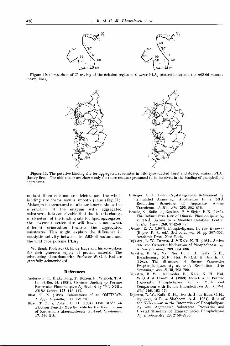
The main conformational difference between theΔ62-66 mutant and the C. atrox PLA2 occurs beforeresidue 61. The difference in structure of residues 61to 70 is minimal. In order to allow the extra deletionof three residues, helix 40-58 is shortened by oneand a half turns in the C. atrox enzyme. This helixterminates in the snake venom PLA2 at position 52.Residues 53 to 55 have an extended conformationand residues 56 and 59 are located in a turn. In themutant, the α-helix terminates at position 58 andthe bend formed by residues 59 and 60 is locatedfurther on the outside of the protein (Fig. 10). Cys61of the Δ62-66 PLA2 mutant is located at almost thesame position (the difference in cα position is 1'1 Å).
4. DiscussionAbove the structure of the Δ62-66 PLA2 mutant
and a comparison with the wild-type porcine PLA2and C. atrox snake venom PLA2 are described. Thebiggest conformational differences occur around thedeletion area, residues 62 to 66. The structure of thisregion in the mutant appears to be intermediatebetween that in wild-type porcine and C. atroxphospholipases A2.
The enzyme has kept an internal hydrophobicarea as conserved as possible by a relatively simplelocal adjustment of its conformation. While in theporcine wild-type PLA2 Phe63 occupies a hydro-phobic pocket, in the Δ62-66 mutant the disulfidebridge between Cys61 and Cys91 fills this hydro-phobic pocket.
Due to a difference in crystallographic contactsthe conformations around Leu31 in the twomonomers are slightly different. In a comparison ofthe structures of bovine and porcine phospholipaseA2 (Dijkstra et al., 1983) also a conformationaldifference in this region was found. Apparently thisregion has some conformational freedom. Also theregions around Pro14 and the C terminus appear tohave some conformational freedom.
The Δ62-66 mutant has a second binding site forcalcium similar to that found in the wild-typeporcine PLA2 structure. This site is on the non-
crystallographic 2-fold axis relating the twomonomers in the asymmetric unit. The side-chainsof residues Glu71 and Glu92 and the carbonyloxygen of residue 72 are ligands for this calcium ion.Although based on site-directed mutagenesis studiescombined with activity measurements at high pH(van den Bergh et al., 1989) Asp66 was suggested tobe a ligand of the calcium ion instead of Glu92, thisis extremely unlikely. In the crystal structure ofwild-type porcine PLA2 the Asp66 side-chain ismore than 10 A from the calcium ion, and in thepresent mutant structure, where Asp66 has beendeleted, we still observe a second calcium ion boundat the same position as in the wild-type structure.
Kinetic studies with the Δ62-66 mutant showthat this enzyme has a fully functional active siteand a kcat/ Km twice that of the wild-type porcinePLA2 (Kuipers et al., 1989a). The slight increase inactivity towards monomeric substrates of thisΔ62-66 mutant compared with wild-type porcinePLA2 could be caused by small alterations in theactive site. In the present 2·1 A structure somechanges around the active site can be detected,although the active site residues proper are, withinerror, identical between mutant and wild-typePLA2. Due to the change in position of Tyr69, Leu2moves to the newly created gap and the N-terminalα-helix changes slightly in orientation (I to 1·3A atthe N terminus). This observation of Leu2 comingtowards Tyr69 has also been reported in filteredmolecular dynamic runs on bovine PLA2 bySessions et al. (1989). In that paper, it was suggestedthat this movement changes the pocket between thetwo residues and that this might be important foradjusting the active site for the binding of mono-meric substrates. Alternatively, the different posi-tion of Tyr69 could be responsible for the observedincrease in activity. Tyr69 has been found to beimportant for the precise positioning of a phospho-lipid in the active site by binding to the phosphategroup of the phospholipid (Kuipers et al., 1989b).Another indication that there are indeed minutechanges in the active site is the observation thatthis Δ62-66 mutant has a somewhat differentbinding constant for the calcium ion in the active

Structure of /162-66 Porcine Phospholipase A2 437
Figure 9. The surroundings of the 2nd calcium-binding site. (a) An overview of the ligands of the calcium ion and thehydrogen-bonding pattern surrounding this calcium-binding site. (b) (2mFo-DFc), αc map (Read, 1986) of the 2ndcalcium-binding site. The density is contoured at 1·10"·
center compared with wild-type porcine PLA2· Thisbinding constant changes from 1·8 mM to 0·8 mM atpH 6 in the /162-66 mutant (Kuipers et al·, 1989a)·
The activity of the /162-66 mutant PLA2 onaggregated short chain lecithins has increased up to16-fold with respect to the wild-type enzyme
(Kuipers et al·, 1989a)· All residues identified tohave interactions with the aggregated substratemolecules lie in a plane around the entrance to theactive site on one face of the protein· Residues 64 to66 in the wild-type porcine PLA2 bulge out of theplane and form a protuberance. In the /162-66 PLA2
438 M. M. G. M. Thunnissen et al.
Figure 10. Comparison of Cα tracing of the deletion region in C. atrox PLA2 (dotted lines) and the Δ62-66 mutant(heavy lines).
Figure 11. The putative binding site for aggregated substrates in wild-type (dotted lines) and Δ62-66 mutant PLA2(heavy lines). The side-chains are shown only for those residues presumed to be involved in the binding of phospholipidaggregates.
mutant these residues are deleted and the wholebinding site forms now a smooth plane (Fig. II).Although no structural details are known about theinteraction of the enzyme with aggregatedsubstrates, it is conceivable that due to this changein structure of the binding site for lipid aggregates,the enzyme's active site will have a somewhatdifferent orientation towards the aggregatedsubstrates. This might explain the difference incatalytic activity between the Δ62-66 mutant andthe wild-type porcine PLA2.
We thank Professor G. H. de Haas and his co-workersfor their generous supply of protein material. Thestimulating discussions with Professor W. G. J. Hoi aregratefully acknowledged.
ReferencesAndersson, T., Drakenberg, T., Forsen, S., Wieloch, T. &
Lindstrom, M. (1981). Calcium Binding to PorcinePancreatic Phospholipase A2 Studied by 43Ca NMR.FEBS Letters, 123, 115-117.
Bhat, T. N. (1988). Calculations of an OMITMAP.J. Appl. Crystallogr. 21, 279-281.
Bhat, T. N. & Cohen, G. H. (1984). OMITMAP: anElectron Density Map Suitable for the Examinationof Errors in a Macromolecule. J. Appl. Crystallogr.17,244-248.
Brünger, A. T. (1988). Crystallographic Refinement bySimulated Annealing: Application to a 2'8 AResolution Structure of Aspartate AminoTransferase. J. Mol. Biol. 203, 803-816.
Brunie, S., Bolin, J., Gewirth, P. & Sigler, P. B. (1985).The Refined Structure of Dimeric Phospholipase A2at 2·5 Å. Access to a Shielded Catalytic Center.J. Biol. Chem. 260, 9742-9747.
Dennis, E. A. (1983). Phospholipases. In The Enzymes(Boyer, P. D., ed.), 3rd edit., vol. 16, pp.307-353,Academic Press, New York.
Dijkstra, B. W., Drenth, J. & Kalk, K. H. (1981). ActiveSite and Catalytic Mechanism of Phospholipase A2·
Nature (London), 289, 604-606.Dijkstra, B. W., Van Nes, G. J. H., Kalk, K. H.,
Brandenburg, N. P., Hoi, W. G. J. & Drenth, J.(1982). The Structure of Bovine PancreaticProphospholipase A2 at 3'0 Å Resolution. ActaCrystallogr. sect. B, 38, 793-799.
Dijkstra, B. W., Renetseder, R., Kalk, K. H., Hoi,W. G. J. & Drenth, J. (1983). Structure of PorcinePancreatic Phospholipase A2 at 2·6 Å andComparison with Bovine Phospholipase A2. J. Mol.Biol. 168, 163-179.
Dijkstra, B. W., Kalk, K. H., Drenth, J., de Haas, G. H.,Egmond, M. R. & Slotboom, A. J. (1984). Role ofthe N-Terminus in the Interaction of PhospholipaseA2 with Aggregated Substrates. Properties andCrystal Structure of Transaminated PhospholipaseA2. Biochemistry, 23, 2759-2766.

Structure of Δ62-66 Porcine Phospholipase A 2 439
Donne-Op den Kelder, G. M., de Haas, G. H. & Egmond,M. R. (1983). Localisation of the Second CalciumBinding Site in Porcine and Equine PhospholipaseA2. Biochemistry, 22, 2470-2478.
Fujinaga, M., Gros, P. & van Gunsteren, W. F. (1989).Testing the Method of Crystallographic Refinementusing Molecular Dynamics. J. Appl. Crystallogr. 22,1-8.
Gros, P., Betzel, C., Dauter, Z., Wilson, K. S. & Hoi,W. G. J. (1989a). Molecular Dynamics Refinement ofa Thermitase-Eglin-C Complex at 1·98 A Resolutionand Comparison of Two Crystal Forms that Differ inCalcium Content. J. Mol. Biol. 210, 347-367.
Gros, P., Fujinaga, M., Dijkstra, B. W., Kalk, K. H. &Hoi, W. G. J. (1989b). Crystallographic Refinementby Incorporation of Molecular Dynamics:Thermostable Serine Protease Thermitase Complexedwith Eglin-C. Acta Crystallogr. sect. B, 45, 488-499.
Hall, S. R. & Stewart, J. M. (1987). Editors of Xtal 2.2User's Manual, Universities of Western Australiaand Maryland.
Hamilton, W. C., Rollet, J. S. & Sparks, R. A. (1965). Onthe Relative Scaling of X-ray Photographs. ActaCrystallogr. 18, 129-130.
Jones, T. A. (1978). A Graphics Modelbuilding andRefinement System for Macromolecules. J. Appl.Crystallogr. 11, 268-272.
Keith, C., Feldman, D. S., Deganello, S., Glick, .I., Ward,K. B., Jones, G. D. & Sigler, P. B. (1981). The 2·5 ACrystal Structure of a Dimeric Phospholipase A2from the Venom of Crotalus atrox. J. Biol. Chem. 256,8602-8607.
Kramer, R. M., Hession, C., Johansen, B., Hayes, G.,McGray, P., Chow, E. P., Tizard, R. & Pepinsky,R. B. (1989). Structure and Properties of a HumanNon-Pancreatic Phospholipase A2. J. Biol. Chem.264, 5768-5775.
Kuipers, O. P., Thunnissen, M. M. G. M., de Geus, P.,Dijkstra, B. W., Drenth, .I., Verhey, H. M. & deHaas, G. H. (1989a). Enhanced Activity and AlteredSpecificity of Phospholipase A2 by Deletion of aSurface Loop. Science, 244, 82-85.
Kuipers, O. P., Dijkman, R., Pals, C. E. G. M., Verheij,H. M. & de Haas, G. H. (1989b). Evidence for theInvolvement of Tyr69 in the Control ofStereospecificity of Porcine Pancreatic PhospholipaseA2. Protein Eng. 2, 467-471.
Luzzati, V. (1952). Traitement Statistique des Erreursdans la Determination des Structures Cristallines.Acta Crystallogr. 5, 802-810.
Mizushima, H., Kudo, 1., Horigome, K., Murakami, M.,Hayakawa, M., Kim, D., Kondo, E., Tomita, M. &Inoue, K. (1989). Purification of Rabbit PlateletSecretory Phospholipase A2 and its Characteristics.J. Biochem. 105, 520-525.
Nieuwenhuizen, W., Dunze, H. & de Haas, G. H. (1974).Phospholipase A2 (Phosphatide AcylhydrolaseE.C.3.1.1.4.), from Porcine Pancreas. MethodsEnzymol. 32b, 147-154.
Pflugrath, J. W. & Messerschmidt, M. (1986). MunichArea Detector NE System, User's Guide, version 5.
Pieterson, W. A., Vidal, J. C., Volwerk, J. J. & de Haas,G. H. (1974). Zymogen Catalyzed Hydrolysis ofMonomeric Substrates and the Presence of aRecognition Site for the Lipid-Water Interfaces onPhospholipase A2. Biochemistry, 13, 1439-1445.
Ramakrishnan, C. & Ramachandran, G. N. (1965).Stereochemical Criteria for Polypeptide and ProteinChain Conformations. II. Allowed Conformations fora Pair of Peptide Units. Biophys. J. 5, 909-933.
Read, R. (1986). Improved Fourier Coefficients for Mapsusing Phases from Partial Structures with Errors.Acta Crystallogr. sect. A, 42, 140-149.
Renetseder, R., Brunie, S., Dijkstra, B. W., Drenth, J. &Sigler, P. B. (1984). A Comparison of the CrystalStructures of Phospholipase A2 from BovinePancreas and Crotalus atrox Venom. J. Biol. Chem.260, 11627-11634.
Seilhamer, J. J., Pruzanski, W., Vadas, P., Plant, S.,Miller, J. A., Kloss, J. & Johnson, L. K. (1989).Cloning and Recombinant Expression ofPhospholipase A2 Present in Rheumatoid ArthriticSynovial Fluid. J. Biol. Chem. 264, 5335-5338.
Sessions, R. B., Dauber-Osguthorpe, P. & Osguthorpe,D. J. (1989). Filtering Molecular DynamicsTrajectories to Reveal Low Frequency CollectiveMotions: Phospholipase A2. .I. Mol. Biol. 209,617-633.
Slotboom, A. J., Jansen, E. H. J. M., Vlijm, H., Pattus,F., Soares de Araujo, P. & de Haas, G. H. (1978).Calcium Binding to Porcine PancreaticPhospholipase A2 and its Function in Enzyme-LipidInteraction. Biochemistry, 17,4593-4600.
Tronrud, D. E., ten Eyk, L. F. & Matthews, B. W. (1987).An Efficient General Purpose Least SquaresRefinement Program for Macromolecular Structures.Acta Crystallogr. sect. A, 43, 489-501.
van den Bergh, C. .I., Bekkers, A., Verhey, H. M. & deHaas, G. H. (1989). Glutamic Acid 71 and AsparticAcid 66 Control the Binding of the Second CalciumIon in Porcine Phospholipase A2. Eur. J. Biochem.182, 307-313.
van Eyk, J. H., Verhey, H. M., Dijkman, R. & de Haas,G. H. (1983). Interaction of Phospholipase A2 fromNaja melanoleuca Snake Venom with MonomericSubstrate Analogs. Activation of the Enzyme byProtein-Protein or Lipid-Protein Interactions? Eur.J. Biochem. 132, 183-188.
Verhey, H. M., Egmond, M. R. & de Haas, G. H. (1981).Chemical Modification of the I1-Aminogroup in SnakeVenom Phospholipases A2. A Comparison of theInteraction of Pancreatic and Venom Phospholipaseswith Lipid-Water Interfaces. Biochemistry, 20,94-99.
Volwerk, J. J. & de Haas, G. H. (1982). In Lipid andProtein Interactions (.lost, P. C. & Griffith, O. H.,eds), vol. I, pp. 69-149, Wiley, New York.
Vriend, G. (1990). WHATIF: A Molecular Modeling andDrug Design Program. J. Mol. Graph. 8, 52-56.
Waite, M. (1987). Handbook of Lipid Research, vol. 5,Plenum, New York.
Edited by R· Huber