structural elements of the streptomyces oric region and their interactions with the dnaa protein
TRANSCRIPT

Microbiology (1998), 144, 1281–1290 Printed in Great Britain
Structural elements of the Streptomyces oriCregion and their interactions with the DnaAprotein
Dagmara Jakimowicz,1 Jerzy Majka,1,2 Walter Messer,2 Christian Speck,2
Marisol Fernandez,3 M. Cruz Martin,3 Jesus Sanchez,3
Florian Schauwecker,4 Ullrich Keller,4 Hildgund Schrempf5
and Jolanta Zakrzewska-Czerwin! ska1
Author for correspondence: Jolanta Zakrzewska-Czerwin! ska. Tel : 48 71 67 90 81. Fax: 48 71 67 91 11.e-mail : zakrzew!immuno.iitd.pan.wroc.pl
1 Ludwik Hirszfeld Instituteof Immunology andExperimental Therapy,Polish Academy ofSciences, ul. Weigla 12,53-114 Wrocław, Poland
2 Max-Planck-Institut fu$ rMolekulare Genetik,Ihnestraße 73, D-14195Berlin-Dahlem, Germany
3 Departamento de BiologiaFuncional e InstitutoUniversitario deBiotecnologia de Asturias,Universidad de Oviedo, J.Claveria 6, Oviedo 33006,Spain
4 Max-Volmer-Institut fu$ rBiophysikalische Chemieund Biochemie, FachgebietBiochemie und MolekulareBiologie, TechnischeUniversita$ t Berlin,Franklinstrasse 29, D-10587Berlin, Germany
5 FachbereichBiologie/Chemie,Universita$ t Osnabru$ ck,Barbarastraße 11, 49069Osnabru$ ck, Germany
Streptomycetes differ from other prokaryotic organisms in their mycelial lifecycle and in possessing a large, linear, GC-rich chromosome. To deducestructural features of the Streptomyces origin of chromosomal replication, theoriC sequences of three Streptomyces species (S. antibioticus, S. chrysomallusand S. lividans) were compared. In Streptomyces, the oriC region contains 19DnaA boxes whose location, orientation and spacing are conserved. Theconsensus sequence of the DnaA box identified within Streptomyces oriC is(T/C)(T/C)(G/A/C)TCCACA (preferred bases underlined). The interactions of DnaAwith DNA fragments containing single, two or three DnaA boxes were studiedusing surface plasmon resonance. The dissociation constant (KD) for specificbinding of individual DnaA boxes varied between 12 and 78 nM. StreptomycesoriC does not contain the three AT-rich 13-mer direct repeats present in the 5Hpart of the Escherichia coli oriC region. However, short AT-rich sequences aredistributed among the DnaA boxes of Streptomyces oriC. Repeated attemptsto unwind Streptomyces oriC have been unsuccessful. It remains to beelucidated whether DnaA interacts with putative accessory proteins whichhelp in unwinding Streptomyces oriC.
Keywords : DnaA box, AT-rich sequence, oriC, Streptomyces
INTRODUCTION
Streptomycetes are Gram-positive soil bacteria thatgrow as substrate mycelia differentiating to aerialmycelia and spores upon depletion of nutrients (Ku$ tzner,1981). These bacteria differ from other prokaryotes notonly in their mycelial life cycle but also in possessing alarge (6–8 Mb), GC-rich chromosome, which has beenfound in linear form (Leblond et al., 1993; Lin et al.,1993; Lezhava et al., 1995).
.................................................................................................................................................
Abbreviations: BD, binding domain; GST, glutathione S-transferase.
The GenBank accession numbers for the sequences reported in this paperare AF026792 (Streptomyces antibioticus), AF027658 (Streptomyces chryso-mallus) and M86491 (Streptomyces lividans).
In bacteria, chromosome replication is initiated at thereplication origin, oriC, and the process is highlyregulated (for review, see Kornberg & Baker, 1992;Messer & Weigel, 1996). The structure of the oriCregion has been analysed within Gram-negative andGram-positive bacteria. The sequences of oriC regionsare conserved only among closely related organisms(Yoshikawa & Ogasawara, 1991). Sequence analyseshave revealed that the origins of various eubacteriacontain short, conserved sequences which are essentialfor oriC function: non-palindromic 9 bp sequences, so-called DnaA boxes and AT-rich regions (Yoshikawa &Ogasawara, 1991; Messer & Weigel, 1996). Theseconserved sequences are separated by spacer regionswhich vary in nucleotide composition and length. A
0002-2203 # 1998 SGM 1281

D. JAKIMOWICZ and OTHERS
Escherichia coli
Coxiella burnetii
Pseudomonas putida
Caulobacter crescentus
Mycoplasma capricolum
Spiroplasma citri
Bacillus subtilis
Micrococcus luteus
Mycobacterium lepraeMycobacterium smegmatisMycobacterium tuberculosis
Streptomyces coelicolorStreptomyces lividans
.................................................................................................................................................................................................................................................................................................................
Fig. 1. Comparison of the genetic organization of the oriC region of different bacteria. DnaA boxes are indicated byarrows. References: Escherichia coli, von Meyenburg & Hansen (1987); Coxiella burnetii, Shuan et al. (1994);Pseudomonas putida, Smith et al. (1991); Caulobacter crescentus, Marczyn! ski & Shapiro (1992); Mycoplasma capricolum,Fujita et al. (1992); Spiroplasma citri, Ye et al. (1994); Bacillus subtilis, Yoshikawa & Wake (1993); Micrococcus luteus,Fujita et al. (1990); Mycobacterium leprae, Mycobacterium smegmatis, Mycobacterium tuberculosis, Salazar et al. (1996);Streptomyces coelicolor, Calcutt & Schmidt (1992); Streptomyces lividans, Zakrzewska-Czerwin! ska & Schrempf (1992),Zakrzewska-Czerwin! ska et al. (1994).
cluster of four or more DnaA boxes is an indication of afunctional chromosomal origin. A putative oriC regionfrom Coxiella burnetii contains only two DnaA boxes
(Fig. 1) (Shuan et al., 1994). However, it remains to beelucidated whether this region is able to promoteautonomous replication. In Bacillus subtilis, two DnaA
1282
Initiation of replication of the Streptomyces chromosome
box clusters are arranged upstream and downstream ofthe dnaA gene and act together as a replication origin(Fig. 1) (Yoshikawa & Wake, 1993). DnaA is the keyprotein in the initiation of DNA replication in bacteria(Kornberg & Baker, 1992; Skarstad & Boye, 1994;Messer & Weigel, 1996) and binds specifically to theDnaA box. The interaction of DnaA with its chromo-somal origin is best understood in Escherichia coli. FiveDnaA boxes are present within the E. coli oriC region.Binding of 10–20 DnaA monomers promotes a localunwinding of the AT-rich region (Bramhill & Kornberg,1988; Messer & Weigel, 1996). The unwound regionprovides the entry site for the DnaB}DnaC helicasecomplex, followed by other proteins required to form areplication fork (Kornberg & Baker, 1992).
Most eubacteria contain a block of genes, dnaA-dnaN-recF-gyrB, encoding DnaA, the β-subunit of the DNApolymerase III holoenzyme, a product for recombinationand the β-subunit of DNA gyrase, respectively (Messer& Weigel, 1996). Within many bacteria, including B.subtilis (Yoshikawa & Wake, 1993), Micrococcus luteus(Fujita et al., 1990), Mycobacterium spp. (Salazar et al.,1996), Mycoplasma capricolum (Fujita et al., 1992) andPseudomonas putida (Smith et al., 1991), the oriC regionis situated close to dnaA (Fig. 1). However, within somebacteria the arrangement is different. The E. coli dnaAregion is located about 40 kb away from oriC. InCoxiella burnetii, dnaA is absent within the putativeoriC region (Fig. 1) (Shuan et al., 1994). The Caulobactercrescentus oriC is situated between hemE (encodinguroporphyrinogen decarboxylase) and rpsT (encoding ahomologue of the ribosomal protein S20) and 2 kb awayfrom dnaA which is separated (150 kb) from the dnaN-recF-gyrB region (Marczyn! ski & Shapiro, 1992; Rizzioet al., 1993; Zweiger & Shapiro, 1994). Sequencesresembling other eubacterial oriC regions have not beendetected in the vicinity of the dnaA genes of Sino-rhizobiummeliloti (Margolin et al., 1995), Synechocystissp. (Richter & Messer, 1995) and Prochlorococcusmarinus (Richter et al., 1998).
As in several other bacteria, the oriC region of Strepto-myces lividans was identified as an autonomouslyreplicating minichromosome (Zakrzewska-Czerwin! ska& Schrempf, 1992; Zakrzewska-Czerwin! ska et al.,1995). It is situated between dnaA and dnaN andcorresponds to the sequenced oriC region (Calcutt &Schmidt, 1992) of the closely related strain S. coelicolorA3(2). Recent discoveries suggest that the chromosomeof S. coelicolor A3(2) replicates bi-directionally (Musi-alowski et al., 1994) from the centrally located oriC(Radenbach et al., 1996) and its linear form is assumedto be patched up at the ends by protein-primedreplication (Chen, 1996).
In this paper we have compared the characteristics of S.lividans oriC with cloned oriC regions from S. anti-bioticus and S. chrysomallus, and we have determinedinteractions of individual DnaA boxes with S. lividansDnaA.
METHODS
Bacterial strains, culture conditions and transformation.Streptomyces and E. coli strains used here are listed in Table1. E. coli strains were grown in Luria–Bertani medium(Sambrook et al., 1989). Streptomyces strains were cultivatedon agar plates containing complete medium until sporulationoccurred (Hopwood et al., 1985). Spores were used toinoculate YEME liquid medium (Hopwood et al., 1985).Propagation and transformation of E. coli and Streptomycesstrains were carried out as described by Hopwood et al. (1985)and Sambrook et al. (1989). Streptomyces spp. were selectedfor resistance to 10 µg thiostrepton ml−".
Plasmids and DNA library. Plasmids are listed in Table 1. TheS. chrysomallus genomic library in cosmid pV34 contains32–34 kb DNA fragments obtained by partial Sau3A digestionof chromosomal DNA and cloned in the BamHI site of thevector (Pahl et al., 1992).
Chemicals, enzymes and oligonucleotides. Standard chemi-cals were obtained from Sigma or Serva. Restriction enzymeswere supplied by Boehringer Mannheim, MBI Fermentas orGibco-BRL. Oligonucleotides used for sequencing and PCRwere chemically synthesized (MWG). For BIAcore studies, 5«biotin-end-labelled oligonucleotides and their non-biotinyl-ated complementary oligonucleotides were annealed bymixing equimolar amounts in 50 mM Tris}HCl, pH 7±5,100 mM NaCl, 0±1 mM EDTA, heating to 85 °C and slowlycooling the samples to room temperature.
DNA isolation and manipulation. Total DNA was isolatedfrom Streptomyces strains as described by Hopwood et al.(1985). Plasmid purification was done using a kit according tothe manufacturer’s protocols (Qiagen). The methods for thepurification of DNA fragments, colony and Southern hybrid-ization, and preparation of DNA probes have been describedby Sambrook et al. (1989).
DNA sequence determination and computer analysis. DNAsequencing was performed using the dideoxy chain-termin-ation method (Sanger et al., 1977) with Sequenase (USB) and[α-$&S]ATP (Amersham). The nucleotide sequence was de-termined on both strands. Computer analysis was done usingthe GCG package programs for ORF identification andsequence alignment.
PCR. Comparison of the amino acid sequences of DnaA andDnaN allowed selection of highly conserved motifs at the Cterminus of DnaA (FGGRDH) and the N terminus of DnaN(MKIRVER). Taking into account the known Streptomycescodon usage, two degenerate primers were deduced. Thenucleotide sequences of the primers are as follows: p
dnaA, 5«
CGCGGATCCTTCGGSGGSCGSGACCAC 3« ; pdnaN
, 5«AACTGCAGSCGCTCSACSCGGATCTTCAT 3« (S¯G orC). Each of them was tailed by a motif for a restriction site.PCR was done with 2±5 U Dynazyme II DNA polymerase(Finnzymes) in 50 µl of the recommended buffer and wasperformed for 40 cycles (10 cycles of 1 min at 95 °C, 1 min at50 °C and 1±5 min at 72 °C, and 30 cycles of 1 min at 95 °C,1 min at 55 °C and 1±5 min at 72 °C). Approximately 300 nggenomic DNA from S. antibioticus ETH 7451 was used forPCR. The amplified products were analysed on a 1% agarosegel, purified with the QIAquick PCR purification kit (Qiagen),digested with restriction enzymes (BamHI, PstI) and thencloned in pUO9090.
Isolation of oriC fragments using affinity chromatography.The DNA-binding domain (BD) of S. lividans DnaA was fused
1283

D. JAKIMOWICZ and OTHERS
Table 1. Bacterial strains and plasmids used in this study
Strain or plasmid Genotype and/or relevant characteristic* Reference
Strains
E. coli AG115 lacX74 galU galK araD139 strA hsdR17 F« lacIq lacZ : :Tn5 Mattern (1992)
E. coli DH5α supE44 hsdR17 recA1 endA1 gyrA96 thi-1 relA1 Sambrook et al. (1989)
E. coli TG1 supE hsd∆5 thi ∆(lac–proAB) F« [traD36 proAB+ lacIq
lacZ∆M15]
Sambrook et al. (1989)
E. coli WM2121 ara ∆(lac–pro) fis : :Km recA56 rpsL srlC300 : :Tn10 thi Koch et al. (1988)
S. antibioticus ETH 7451 Novella et al. (1992)
S. chrysomallus ATCC 11523
S. lividans TK21 SLP2− SLP3− derivative of S. lividans 66 Hopwood et al. (1985)
Plasmids
pBR322 Apr, Tcr Bolivar et al. (1977)
pUC18 Apr Yanisch-Perron et al. (1985)
pUK21 Kmr Vieira & Messing (1991)
pUO9090 pUK21 derivative containing 1±5 kb Amr M. C. Martin (unpublished)
pBluescript II SK() pUC derivative (pMB1 replicon) Short et al. (1988)
pLEX3BT tac promoter, Apr Diederich et al. (1994)
pGEX-3X-6His GST gene fusion vector, Apr Majka et al. (1997a)
pUSA1 pUO9090 derivative containing 934 bp BamHI–PstI fragment of
the oriC region of S. antibioticus
This study
pBSC1 pBR322 derivative containing 4±2 kb BamHI fragment of the S.
chrysomallus dnaA region
This study
pBSC2 pBluescript II SK() derivative containing 1228 bp SacI–BamHI
fragment of the oriC region of S. chrysomallus
This study
pBSL1 pBluescript II SK() derivative containing 1092 bp SphI–BglII
fragment of the oriC region of S. lividans
Zakrzewska-Czerwin! ska et al. (1995)
pGDnaA(BD) pGEX-3X-6His derivative containing 432 bp XhoII–XhoII–SphI
fragment encoding the DNA BD of S. lividans DnaA
Majka et al. (1997a)
pLEXDnaA6xHis pLEXBT derivative containing the entire S. lividans dnaA gene
and (CACCAT)$encoding six histydyl residues at the 3« end of
dnaA gene
Majka et al. (1997b)
*ATCC, American Type Culture Collection, Rockville, MD, USA. Am, apramycin; Ap, ampicillin ; Km, kanamycin; Tc, tetracyclin.
to the C terminus of glutathione S-transferase (GST) asdescribed previously (Majka et al., 1997a). Plasmid pGEX-DnaA(BD), encoding the fusion protein GST-DnaA(BD), wastransformed into E. coli AG115. Cells were grown for 3 h at37 °C to an OD
&&!of 0±6 in the presence of 100 µg ampicillin
ml−" and then induced with 0±5 mM IPTG for 1±5 h. Aftercentrifugation (5000 g, 4 °C, 10 min), cells were resuspendedin lysis buffer (50 mM Tris}HCl, 100 mM NaCl, 1 mMEDTA, 1 mM PMSF, pH 8±0) and degraded by sonification(five times, 30 s each). After centrifugation (30000 g, 4 °C,60 min), the crude extract was treated with 25 µg DNaseI ml−"(37 °C, 30 min). The GST-DnaA(BD) fusion bound directlyfrom the bacterial extract to the glutathione-Sepharose beads.The column was then washed with 10 bed volumes of lysisbuffer. The DNA, digested with restriction endonuclease, wasloaded onto the glutathione-Sepharose-GST-DnaA(BD) in‘ low’ salt buffer (20 mM Tris}HCl, 100 mM NaCl, pH 8±0).After 1 h incubation at room temperature, the column waswashed with three column volumes of ‘medium’ salt buffer(20 mM Tris}HCl, 500 mM NaCl, pH 8±0) to remove DNAthat did not tightly associate with the GST-DnaA(BD) beads.DNA that remained bound to the beads was then eluted with‘high’ salt buffer (20 mM Tris}HCl, 2000 mM NaCl, pH 8±0),followed by 2-propanol precipitation. The DNA was re-
suspended in TE buffer (10 mM Tris}HCl, 1 mM EDTA,pH 8) and analysed on an agarose gel.
Purification of S. lividans DnaA. S. lividans DnaA wasoverexpressed in E. coli WM2121 as a His-tagged protein andthen purified on a Ni#+-NTA-agarose column (Qiagen) asdescribed previously (Majka et al., 1997b).
Surface plasmon resonance. The biotinylated double-stranded oligonucleotides were immobilized on a streptavidin-coated SM 5A chip of the BIAcore apparatus. Usually,100–250 RU (resonance units) of DNA was immobilized.DNA loosely attached to the surface of the chip was removedwith 0±05% SDS. To exclude the effects of mass transfer on thekinetics of protein–DNA interactions, the measurements wereperformed at various protein concentrations (17±4–174 nM)and at three different flow rates (2, 5 and 10 µl min−"). Thecalculated kinetic constants did not differ significantly. Thus,the subsequent measurements were performed at a continuousflow of 2 µl HBS buffer min−" (HBS buffer : 10 mM HEPES,pH 7±4, 150 mM NaCl, 3±4 mM EDTA, 0±005% BIAcoresurfactant P20). At the end of each cycle, bound DnaA wasremoved by washing with 0±05% SDS. The BIAevaluationversion 2.1 program (Pharmacia Biosensor) was utilized fordata analysis.
1284

Initiation of replication of the Streptomyces chromosome
RESULTS AND DISCUSSION
Cloning of the oriC regions of S. antibioticus and S.chrysomallus
Comparisons of the deduced amino acid sequences ofDnaA and DnaN from several bacteria, including S.lividans, showed two highly conserved motifs in therespective C- and N-terminal regions. Two correspond-ing degenerate primers were deduced and used toamplify a DNA fragment (C 900 bp) from total S. anti-bioticus DNA. The fragment was cloned into pUO9090to yield plasmid pUSA1 (Table 1). Hybridization withtotally digested chromosomal DNA proved that thecloned fragment was derived from S. antibioticus (datanot shown).
To isolate the oriC region of S. chrysomallus, a libraryof its genomic DNA in the cosmid vector pV34 (Pahl etal., 1992) was hybridized with a 1±2 kb BamHI–SphIfragment encoding the C terminus of S. lividans DnaA(Zakrzewska-Czerwin! ska et al., 1994). From 12000colonies, eight positive clones were obtained. Withineach of the cosmids a 4±2 kb BamHI fragment hybridizedwith the dnaA probe and cloning of this BamHIfragment in pBR322 gave pBSC1. Sequence analysisallowed the identification of the 3« end of dnaA and the5« end of dnaN. A 1±2 kb BamHI–SacI fragment ofpBSC1 containing the intergenic region between dnaAand dnaN (putative oriC region) was subcloned intopBluescript II SK(), resulting in pBSC2. Hybrid-izations with three selected probes from the oriC regionindicated a moderate homology among the cloned oriCregions (data not shown).
Comparisons of the oriC regions
The nucleotide sequence of the cloned putative oriCfragmentswas determined on both strands and subjectedto computer analysis. The overall GC content of theanalysed regions of S. antibioticus and S. chrysomallusis about 65% and 63%, respectively (Table 2), and isthus, as in the S. lividans oriC region, approximately10% lower than the mean GC content of knownStreptomyces genes. The oriC regions of S. antibioticusand S. chrysomallus were flanked by dnaA and dnaNwhich are separated by 909 and 921 bp, respectively. Asearch for DnaA box motifs whose sequences differ upto 2 nt from the preferred sequence (TTGTCCACA)(Zakrzewska-Czerwin! ska & Schrempf, 1992) allowedidentification of 19 putative DnaA boxes in each
Table 2. Characteristics of the Streptomyces dnaA–dnaN intergenic region (oriC region)
Species No. of DnaA GC content Length of oriC Sequence similarities in the
boxes (mol%) region (bp) oriC region (%)
1 2 3
1. S. antibioticus 19 65±2 909 100 60 59
2. S. chrysomallus 19 63±0 921 60 100 63
3. S. lividans 19 63±2 934 59 63 100
analysed oriC region (Fig. 2). Previously, 17 DnaA boxeshad been identifiedwithin the oriC region of S. coelicolorA3(2) and S. lividans (Zakrzewska-Czerwin! ska &Schrempf, 1992) ; the 9th and 13th DnaA boxes had notbeen found (Fig. 2). The position and orientation of eachof the 19 deduced DnaA boxes are identical in S.antibioticus, S. chrysomallus and S. lividans (Fig. 2).Only the distance between DnaA boxes 9 and 10 in S.antibioticus is shorter (2 bp) than in S. chrysomallus andS. lividans (12 bp). Alteration of the spacing betweenDnaA boxes inactivates replication activity of the E. coliorigin. It is interesting that only mutants with aninsertion or deletion of 10 bp (close to a full helical turn)between the DnaA boxes R2 and R3, or R3 and R4retain a functional oriC (Woelker & Messer, 1993). Incontrast, point mutations in the DnaA boxes of E. colioriC sequence have a comparatively slight effect onreplication (Langer et al., 1996). Thus, the location ofthe DnaA boxes with respect to the helix axis isapparently important. The short stretches of DNAflanking the individual boxes are more variable (about60% identical) in the corresponding regions between thethree Streptomyces species.
The 57 DnaA boxes were used to determine theconsensus sequence (T}C)(T}C)(G}AC)TCCACA (pre-ferred bases in bold) (Table 3). The consensus sequenceof the Streptomyces DnaA box is more variable than inother bacteria, similar to Mycobacterium (Salazar et al.,1996). As in other organisms, bases in positions 4 and 6of the Streptomyces DnaA boxes were found to behighly conserved. C-5, A-7 and C-8 were also wellconserved, in contrast to the third position which is themost variable.
Following the interaction of DnaA with DnaA boxes, alocal unwinding occurs at specific AT-rich sequences,characteristic for a replication origin (Bramhill &Kornberg, 1988; Gille & Messer, 1991; Hsu et al., 1994).The partially unwound oriC then presumably providesthe entry site for the replicative helicase. In E. coli, threeAT-rich 13-mer direct repeats are localized in the 5« partof the oriC region close to the first DnaA box. B. subtilisDnaA-mediated unwinding occurs in an AT-rich 27-meradjacent to the DnaA boxes that are located downstreamof dnaA (Moriya et al., 1994; Krause et al., 1997). Noneof the three Streptomyces oriC regions contains the AT-rich repeats adjacent to clusters of DnaA boxes. How-ever, each of the Streptomyces regions contains five
1285

D. JAKIMOWICZ and OTHERS
Fig
.2.
Alig
nm
ent
of
nu
cleo
tid
ese
qu
ence
so
fth
eSt
rep
tom
yces
dn
aA–d
naN
inte
rgen
icre
gio
n(o
riC
).Th
ep
osi
tio
ns
and
ori
enta
tio
ns
of
the
Dn
aAb
oxe
sar
ein
dic
ated
(bla
ckb
oxe
san
dar
row
s).
Gap
sar
ein
dic
ated
by
do
ts.
Bas
esw
hic
har
eid
enti
cal
intw
oo
rth
ree
of
the
seq
uen
ces
are
sho
wn
inca
pit
alle
tter
s.A
T-ri
chre
gio
ns
are
inb
old
.Th
elig
ht
gre
yb
oxe
sat
the
star
to
fth
ese
qu
ence
sin
dic
ate
the
3«en
do
fd
naA
;th
ed
ark
gre
yb
oxe
sat
the
end
of
the
seq
uen
ces
ind
icat
eth
e5«
end
of
dn
aN.S
.a.,
S.an
tib
ioti
cus;
S.ch
.,S.
chry
som
allu
s;S.
l.,S.
livid
ans;
Co
n.,
con
sen
sus
seq
uen
ce.
1286

Initiation of replication of the Streptomyces chromosome
Table 3. Frequency of nucleotide usage in Streptomyces DnaA boxes
Nucleotide No. of times used in position no. :*
1 2 3 4 5 6 7 8 9
A 1 0 9 1 2 1 55 0 41
C 11 10 13 5 51 56 2 57 6
G 5 1 32 0 4 0 0 0 7
T 40 46 3 51 0 0 0 0 3
Consensus
Streptomyces spp. T}C T}C A/G/C T C C A C A
Mycobacterium spp. T}C T G}A T C C A}C C A
E. coli (a)† T T A}T T N C A C A
E. coli (b)† T T}C A}T T A}C C A C}A A
E. coli (c)† T}C T}C T/A/C T A}C C A}G A/C/T A
*Numbers}letters in bold indicate the preferred sequence.
†Consensus sequences for the E. coli DnaA box were determined by three different methods : (a)binding constants measurements (Schaper & Messer, 1995) ; (b) DNaseI footprinting analysis (Fuller etal., 1984) ; (c) in vivo analysis of the effects of DnaA on transcription termination (Schaefer & Messer,1991).
bpM 1 2 3 4 5 6 M
21761766
12301033
653517394
234
.................................................................................................................................................
Fig. 3. DNA-binding assay – selective binding of an oriC-containing DNA fragment using GST-DnaA(BD) beads. PlasmidDNA (1 µg) digested with SalI was bound to the beads.Fragments were analysed by agarose gel electrophoresis. Lanes:1, 3, 5, DNA loaded on the affinity column; 2, 4, 6, DNAspecifically bound to the fusion protein and released from thebeads by washing with ‘high’ salt buffer. The analysed DNAfragments were from S. antibioticus (lanes 1, 2), S. chrysomallus(3, 4) and S. lividans (5, 6). Lanes M, standard size markers(Boehringer VI).
short AT-rich sequences that are located close to the 5«end of the first DnaA box (positions 191–199), betweenthe 1st and 2nd boxes (230–245), between the 2nd and3rd boxes (269–282), between the 6th and 7th boxes(371–378) and following the 16th box in the 3« flankingsequence (678–684) (Fig. 3). Repeated attempts todetermine DnaA-mediated unwinding of oriC, usingpermanganate footprinting, have been unsuccessful(data not presented). S. lividans DnaA differs from the
corresponding E. coli protein in its acquisition of anadditional stretch of 120 predominantly acidic aminoacids in domain II (Majka et al., 1997b). It remains to beelucidated whether the acidic domain II interacts withputative accessory basic proteins which help in theunwinding of oriC.
The S. chrysomallus minichromosome replicates in S.lividans. However, similar to the minichromosomes ofS. lividans, B. subtilis and Mycobacterium tuberculosis,it is unstable and present only in low copy numbers.This apparent incompatibility could be caused by thecompetition of the same replicons for DnaA. Thus, as inB. subtilis (Moriya et al., 1994), the initiation ofStreptomyces oriC replication appears to be tightlycontrolled.
Interaction of DnaA with DnaA boxes
To test whether the previously characterized S. lividansDnaA protein binds to the cloned Streptomyces oriCfragments, a recently described DNA-binding assay wasperformed (Majka et al., 1997a). The fusion protein,consisting of the BD of DnaA and GST, was fixed toglutathione-Sepharose beads and its specific interactionswith the cloned oriC fragments were tested (Fig. 3). Eachof the cloned DNA fragments of S. antibioticus and S.chrysomallus, as well the previously characterized oriCof S. lividans (control), was selectively bound to theGST-DnaA(BD) fusion protein.
His-tagged S. lividans DnaA (Majka et al., 1997b) wasinvestigated for its binding properties with respect tovarious DnaA boxes. To evaluate the interactions of
1287
D. JAKIMOWICZ and OTHERS
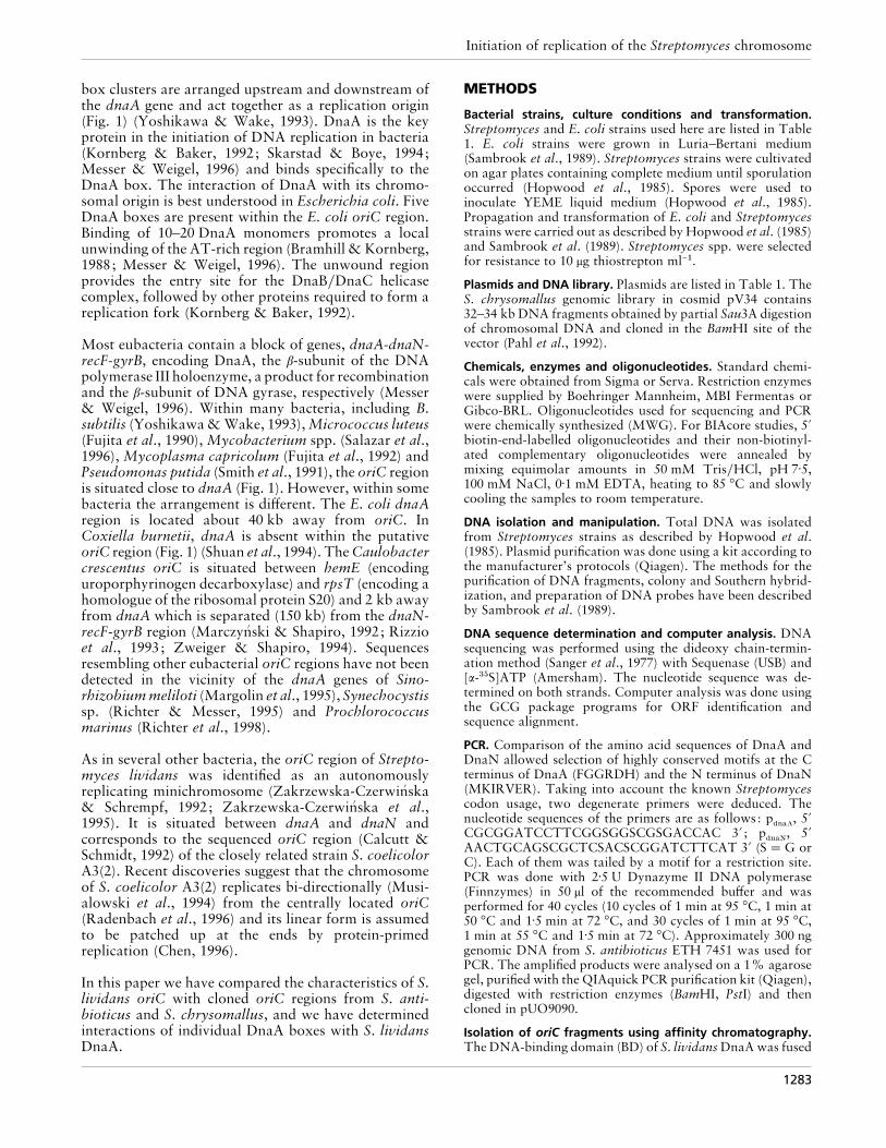
Table 4. Kinetic rate constants for the binding of DnaA to DnaA boxes
DnaA box* Sequence (5«–3«)† Kinetic constants‡
kon (M−1 s−1) koff (s−1) KD (nM)§
1dnaA
¯ 6oriC
GAGACACTTGTCCACACAACTTG 1±2¬10' 1±4¬10−# 12
2dnaA
GAGACACTGGGGACAACAACTTG – – –
2oriC
GTTTTTTCCGTCCACACCTTGGG 5±0¬10& 3±9¬10−# 78
4oriC
GTGGATTCGTGGACGAAGAAATG – – –
5oriC
GTGGATTATCTCCACAAGAAATG 6±3¬10' 2±0¬10−# 32
10oriC
CACCAGCTTCTCCACATGCCTGT – – –
12oriC
GTTGGGCTGTGGGAAACGTGGT – – –
17oriC
AACGAGGTTATCCACGGTATCCA – – –
Nonsense box TTGTGCGATATAGTTCTCCGA – – –
*The numbering of DnaA boxes is derived from the oriC region according to Fig. 3 ; the promoterregion of the S. lividans dnaA gene contains two DnaA boxes: 1
dnaAand 2
dnaA(Zakrzewska-Czerwin! ska
et al., 1994).
†Bold letters indicate the region of specific recognition (DnaA box).
‡ –, non-specific binding (KD" 200 nM).
§KD¯k
off}k
on.
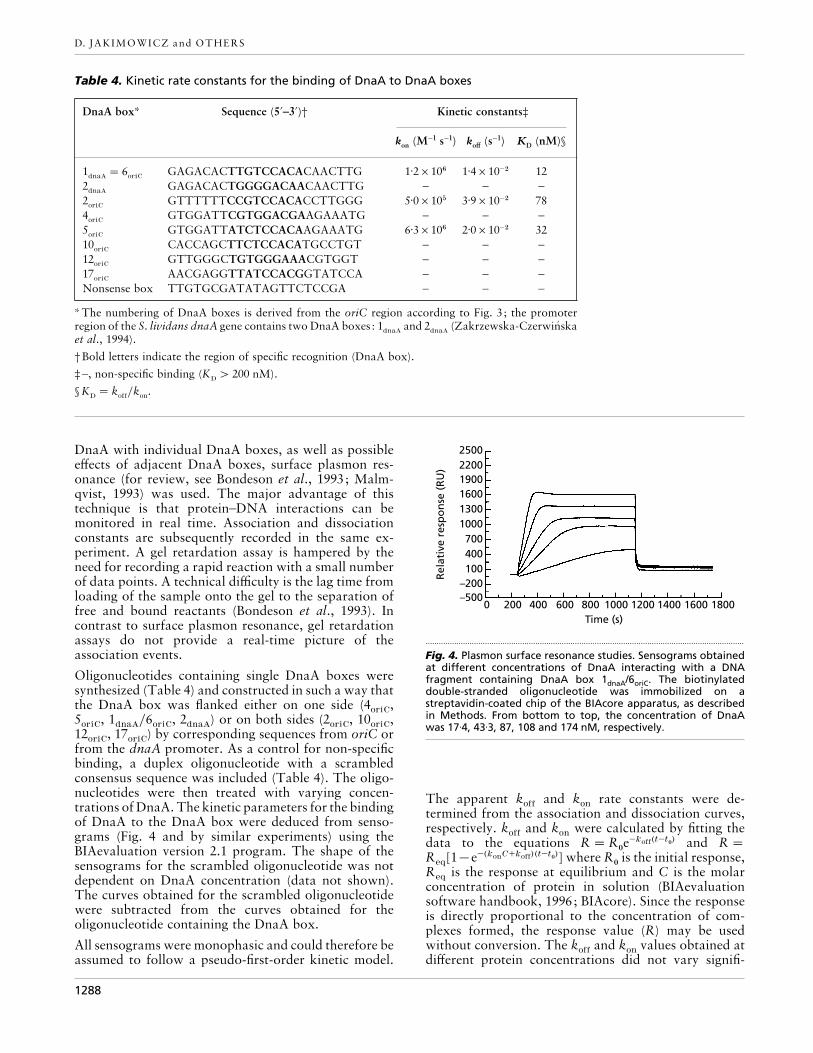
DnaA with individual DnaA boxes, as well as possibleeffects of adjacent DnaA boxes, surface plasmon res-onance (for review, see Bondeson et al., 1993; Malm-qvist, 1993) was used. The major advantage of thistechnique is that protein–DNA interactions can bemonitored in real time. Association and dissociationconstants are subsequently recorded in the same ex-periment. A gel retardation assay is hampered by theneed for recording a rapid reaction with a small numberof data points. A technical difficulty is the lag time fromloading of the sample onto the gel to the separation offree and bound reactants (Bondeson et al., 1993). Incontrast to surface plasmon resonance, gel retardationassays do not provide a real-time picture of theassociation events.
Oligonucleotides containing single DnaA boxes weresynthesized (Table 4) and constructed in such a way thatthe DnaA box was flanked either on one side (4
oriC,
5oriC
, 1dnaA
}6oriC
, 2dnaA
) or on both sides (2oriC
, 10oriC
,12
oriC, 17
oriC) by corresponding sequences from oriC or
from the dnaA promoter. As a control for non-specificbinding, a duplex oligonucleotide with a scrambledconsensus sequence was included (Table 4). The oligo-nucleotides were then treated with varying concen-trations of DnaA. The kinetic parameters for the bindingof DnaA to the DnaA box were deduced from senso-grams (Fig. 4 and by similar experiments) using theBIAevaluation version 2.1 program. The shape of thesensograms for the scrambled oligonucleotide was notdependent on DnaA concentration (data not shown).The curves obtained for the scrambled oligonucleotidewere subtracted from the curves obtained for theoligonucleotide containing the DnaA box.
All sensograms were monophasic and could therefore beassumed to follow a pseudo-first-order kinetic model.
250022001900160013001000700400100
–200–500
Rel
ativ
e re
spo
nse
(R
U)
0 200 400 600 800 1000 1200 1400 1600 1800Time (s)
.................................................................................................................................................
Fig. 4. Plasmon surface resonance studies. Sensograms obtainedat different concentrations of DnaA interacting with a DNAfragment containing DnaA box 1dnaA/6oriC. The biotinylateddouble-stranded oligonucleotide was immobilized on astreptavidin-coated chip of the BIAcore apparatus, as describedin Methods. From bottom to top, the concentration of DnaAwas 17±4, 43±3, 87, 108 and 174 nM, respectively.
The apparent koff
and kon
rate constants were de-termined from the association and dissociation curves,respectively. k
offand k
onwere calculated by fitting the
data to the equations R¯R!e−koff(t−t!) and R¯
Req
[1®e−(konC+koff)(t−t!)] where R!is the initial response,
Req
is the response at equilibrium and C is the molarconcentration of protein in solution (BIAevaluationsoftware handbook, 1996; BIAcore). Since the responseis directly proportional to the concentration of com-plexes formed, the response value (R) may be usedwithout conversion. The k
offand k
onvalues obtained at
different protein concentrations did not vary signifi-
1288

Initiation of replication of the Streptomyces chromosome
cantly. The equilibrium dissociation constant, KD, was
calculated as the ratio between dissociation and as-sociation rate constants (k
off}k
on) (Table 4).
Only three of the eight individual DnaA boxes analysed(2
oriC, 5
oriCand 1
dnaA}6
oriC) exhibited specific binding of
DnaA; the KD
varied between 12 and 78 nM (Table 4).Similar values were obtained by mobility shift assay(data not shown). The DnaA box with the ‘perfect ’sequence (TTGTCCACA) showed the highest affinity toDnaA (12 nM). DnaA boxes 2
dnaA, 4
oriC, 12
oriCand
17oriC
, which differ from the consensus at highlyconserved positions (4, 7 and 9), were not recognizedspecifically by DnaA, i.e. the K
Dwas "200 nM. DnaA
box 10oriC
was not bound specifically by DnaA, despitethe fact that its sequence differs by one base from theconsensus at the most variable third position. Differenttechniques were employed to define the consensussequence of the E. coli DnaA box (Table 3). Havingdetermined the equilibrium binding constant by gelretardation, the consensus sequence TT(A}T)-TNCCACA was stringently defined (Schaper & Messer,1995). It corroborates the fact that the DnaA box 10
oriC(C is at the third position instead of A or T) was notspecifically recognized by DnaA.
Future studies are planned to elucidate the initiation ofreplication of Streptomyces oriC, which is more com-plex than the corresponding process in E. coli.
ACKNOWLEDGEMENTS
This work was supported by grant Me659}5-1 of the DeutscheForschungsgemeinschaft, by the PolishCommittee of ScientificStudies (KBN, grant 0482}P2}93}05) and by grant PB95-1057,Programa Sectorial de PGC, Subdireccio! n General de Form-acio! n Promocio! n del Conocimiento, MEC, Spain. We areespecially grateful to Martina Lemme for help in manuscriptpreparation.
REFERENCES
Bolivar, F., Rodriguez, R. L., Greene, P. J., Betlach, M. C.,Heyneker, H. L., Boyer, H. W., Crosa, J. H. & Falkow, S. (1977).Construction and characterization of new cloning vehicles. II. Amultipurpose cloning system. Gene 2, 95–113.
Bondeson, K., Frostell-Karlsson, A., Fagerstam, L. & Magnusson,G. (1993). Lactose repressor-operator DNA interactions : kineticanalysis by a surface plasmon resonance biosensor. Anal Biochem214, 245–251.
Bramhill, D. & Kornberg, A. (1988). Duplex opening by DnaAprotein of novel sequences in initiation of replication at the originof the E. coli chromosome. Cell 52, 743–755.
Calcutt, M. J. & Schmidt, F. J. (1992). Conserved gene arrangementin the origin region of the Streptomyces coelicolor chromosome.J Bacteriol 174, 3220–3226.
Chen, C. W. (1996). Complications and implications of linearbacterial chromosomes. Trends Genet 12, 192–196.
Diederich, L., Roth, A. & Messer, W. A versatile plasmid vectorsystem for the expression of genes in Escherichia coli. Bio-Techniques 16, 916–923.
Fujita, M. Q., Yoshikawa, H. & Ogasawara, N. (1990). Structure ofthe dnaA region of Micrococcus luteus : conservation andvariations among eubacteria. Gene 93, 73–78.
Fujita, M. Q., Yoshikawa, H. & Ogasawara, N. (1992). Structure ofthe dnaA and DnaA-box region in the Mycoplasma capricolumchromosome: conservation and variations in the course ofevolution. Gene 110, 17–23.
Fuller, R. S., Funnell, B. E. & Kornberg, A. (1984). The DnaAprotein complex with the E. coli chromosomal origin (oriC) andother sites. Cell 38, 889–900.
Gille, H. & Messer, W. (1991). Localized unwinding and structuralperturbations in the origin of replication, oriC, of Escherichia coliin vitro and in vivo. EMBO J 10, 1579–1584.
Hopwood, D. A., Bibb, M. J., Chater, K. F., Kieser, T., Bruton,C. J., Kieser, H. M., Lydiate, D. J., Smith, C. P., Ward, J. M. &Schrempf, H. (1985). Genetic Manipulation of Streptomyces : aLaboratory Manual. Norwich: John Innes Foundation.
Hsu, J., Bramhill, D. & Thompson, Ch. M. (1994). Open complexformation by DnaA initiation protein at the Escherichia colichromosomal origin requires the 13-mers precisely spaced relativeto the 9-mers. Mol Microbiol 11, 903–911.
Koch, C., Vandekerhove, J. & Kahmann, R. (1988). Escherichia colihost factor for site-specific DNA inversion: Cloning and charac-terization of the fis gene. Proc Natl Acad Sci USA 85, 4237–4241.
Kornberg, A. & Baker, T. (1992). DNA Replication, 2nd edn. NewYork: W. H. Freeman.
Krause, M., Ru$ ckert, B., Lurz, R. & Messer, W. (1997). Complexesat the replication origin of Bacillus subtilis with homologous andheterologous DnaA protein. J Mol Biol 274, 365–380.
Ku$ tzner, H. J. (1981). The family Streptomycetaceae. In TheProkaryotes : A Handbook on Habitats, Isolation and Iden-tification of Bacteria, pp. 2028–2090. Edited by M. P. Starr, H.Stolp, H. G. Tru$ per, A. Balows & H. Schlegel. Berlin: Springer.
Langer, U., Richter, S., Roth, A., Weigel, Ch. & Messer, W. (1996).A comprehensive set of DnaA-box mutations in the replicationorigin, oriC, of Escherichia coli. Mol Microbiol 21, 301–311.
Leblond, P., Redenbach, M. & Cullumn, J. (1993). Physical map ofthe Streptomyces lividans 66 genome and comparison with that ofthe related strain Streptomyces coelicolor A3(2). J Bacteriol 175,3422–3429.
Lezhava, A., Mizukami, T., Kajitani, T., Kameoka, D., Redenbach,M., Shinkawa, H., Nimi, O. & Kinashi, H. (1995). Physical map ofthe linear chromosome of Streptomyces griseus. J Bacteriol 177,6492–6498.
Lin, Y.-S., Kieser, H. M., Hopwood, D. A. & Chen, C. W. (1993).The chromosomal DNA of Streptomyces lividans 66 is linear.Mol Microbiol 10, 923–933.
Majka, J., Jakimowicz, D., Zarko-Postawka, M. & Zakrzewska-Czerwin! ska, J. (1997a). Glutathione S-transferase fusion proteinsas an affinity reagent for rapid isolation of specific sequencedirectly from genomic DNA. Nucleic Acids Res 25, 2537–2538.
Majka, J., Messer, W., Schrempf, H. & Zakrzewska-Czerwin! ska, J.(1997b). Purification and characterization of the Streptomyceslividans initiator protein DnaA. J Bacteriol 179, 2426–2432.
Malmqvist, M. (1993). Biospecific interaction analysis usingbiosensor technology. Nature 361, 186–187.
Marczyn! ski, G. T. & Shapiro, L. (1992). Cell-cycle control of acloned chromosomal origin of replication from Caulobactercrescens. J Mol Biol 226, 959–977.
Margolin, W., Bramhill, D. & Long, S. R. (1995). The dnaA gene ofRhizobium meliloti lies within unusual gene arrangement. JBacteriol 177, 2892–2900.
Mattern, S. (1992). Regulation of the galactose operon of
1289
D. JAKIMOWICZ and OTHERS
Streptomyces lividans. PhD Thesis. Universita$ t Osnabru$ ck,Osnabru$ ck, Germany.
Messer, W. & Weigel, C. (1996). Initiation of chromosomereplication. In Escherichia coli and Salmonella typhimurium :Cellular and Molecular Biology, pp. 1579–1601. Edited by F. C.Neidhardt, J. L. Ingraham, K. Brooks Low, B. Magasanik, M.Schaechter & H. E. Umbarger. Washington, DC: AmericanSociety for Microbiology.
von Meyenburg, K. & Hansen, F. G. (1987). Regulation ofchromosome replication. In Escherichia coli and Salmonellatyphimurium : Cellular and Molecular Biology, pp. 1555–1577.Edited by F. C. Neidhardt, J. L. Ingraham, K. Brooks Low, B.Magasanik, M. Schaechter & H. E. Umbarger. Washington, DC:American Society for Microbiology.
Moriya, S., Firshein, W., Yoshikawa, H. & Ogasawara, N. (1994).Replication of a Bacillus subtilis oriC plasmid in vitro. MolMicrobiol 12, 469–478.
Musialowski, M. L., Flett, F., Scott, G. B., Hobbins, G., Smith, C. P.& Oliver, S. G. (1994). Functional evidence that the principal DNAreplication origin of the Streptomyces coelicolor chromosome isclose to the dnaA–gyrB region. J Bacteriol 176, 5123–5125.
Novella, I. S., Barbes, C. & Sanchez, J. (1992). Sporulation ofStreptomyces antibioticus ETHZ 7451 in submerged culture. CanJ Microbiol 38, 769–773.
Pahl, A., U> hlein, M., Bang, H., Schlumbohm, W. & Keller, U.(1992). Streptomycetes possess peptidyl-prolyl cis-trans iso-merases that strongly resemble cyclophilins from eukaryoticorganisms. Mol Microbiol 6, 3551–3558.
Radenbach, M., Kieser, H. M., Denapaite, D., Eicher, A., Cullumn,J., Kinashi, H. & Hopwood, D. A. (1996). A set of ordered cosmidsand detailed genetic and physical map for the 8 Mb Streptomycescoelicolor A3(2) chromosome. Mol Microbiol 21, 77–96.
Richter, S. & Messer, M. (1995). Genetic structure of the dnaAregion of the cyanobacterium Synechocystis sp. strain PCC6803.J Bacteriol 177, 4245–4251.
Richter, S., Hess, W. R., Krause, M. & Messer, W. (1998). Uniqueorganization of the dnaA region from Prochlorococcus marinusCCMP 1375, a marine cyanobacterium. Mol Gen Genet (inpress).
Rizzio, M., Shapiro, L. & Gober, J. W. (1993). Asymmetricexpression of the DNA gyrase B subunit gene from replication-competent chromosome in the Caulobacter predivisional cell. JBacteriol 175, 6970–6981.
Salazar, L., Fsihi, H., de Rossi, E., Riccardi, G., Rios, C., Cole, S. T.& Takiff, H. E. (1996). Organization of the origins of replication ofthe chromosomes of Mycobacterium tuberculosis and isolation ofa functional origin from M. smegmatis. Mol Microbiol 20,283–293.
Sambrook, J., Fritsch, E. F. & Maniatis, T. (1989). MolecularCloning: a Laboratory Manual, 2nd edn. Cold Spring Harbor,NY: Cold Spring Harbor Laboratory.
Sanger, F., Nicklen, S. & Coulson, A. R. (1977). DNA sequencingwith chain-terminating inhibitors. Proc Natl Acad Sci USA 74,5463–5467.
Schaefer, C. & Messer, W. (1991). DnaA protein DNA interaction.Modulation of the recognition sequence. Mol Gen Genet 226,34–40.
Schaper, S. & Messer, W. (1995). Interaction of the initiatorprotein DnaA of Escherichia coli with its DNA target. J BiolChem 270, 17622–17626.
Short, J. M., Fernandez, J. M., Sorge, J. A. & Huse, W. D. (1988). Abacteriophage lambda expression vector with in vivo excisionproperties. Nucleic Acids Res 16, 7583–7600.
Shuan, M., Chen, S.-Y., Thompson, H. A., Hoover, T. A., Hill, A. &Williams, J. C. (1994). Cloning and characterization of an auton-omous replication sequence from Coxiella burnetii. J Bacteriol176, 5233–5243.
Skarstad, K. & Boye, E. (1994). The initiator protein DnaA:Evolution, properties and function. Biochim Biophys Acta 1217,111–130.
Smith, D. W., Yee, T. W., Baird, C. & Krishnapillai, V. (1991).Pseudomonad replication origins : a paradigm for bacterialorigins? Mol Microbiol 5, 2581–2587.
Vieira, J. & Messing, J. (1991). New pUC-derived cloning vectorswith different selectable markers and DNA replication origins.Gene 100, 189–194.
Woelker, B. & Messer, W. (1993). The structure of the initiationcomplex at the replication origin, oriC, of Escherichia coli.Nucleic Acids Res 21, 5025–5033.
Yanisch-Perron, C., Vieira, J. & Messing, J. (1985). ImprovedM13 phage cloning vectors and host strains : nucleotide sequencesof the M13mp18 and pUC19 vectors. Gene 33, 103–119.
Ye, F., Renaudin, J., Bove, J.-M. & Laigret, F. (1994). Cloning andsequencing of the replication origin (oriC) of the Spiroplasma citrichromosome and construction of autonomously replicatingartificial plasmids. Curr Microbiol 29, 23–29.
Yoshikawa, H. & Ogasawara, N. (1991). Structure and function ofDnaA and the DnaA-box in eubacteria : evolutionary relation-ships of bacterial replication origins. Mol Microbiol 5, 2589–2597.
Yoshikawa, H. & Wake, R. G. (1993). Initiation and termination ofchromosome replication. In Bacillus subtilis and Gram-PositiveBacteria : Biochemistry, Physiology and Molecular Genetics, pp.507–528. Edited by A. L. Sonenshein, J. A. Hoch & R. Losick.Washington, DC: American Society for Microbiology.
Zakrzewska-Czerwin! ska, J. & Schrempf, H. (1992). Charac-terization of an autonomously replicating region from Strepto-myces lividans. J Bacteriol 174, 2688–2693.
Zakrzewska-Czerwin! ska, J., Nardmann, J. & Schrempf, H. (1994).Inducible transcription of the dnaA gene from Streptomyceslividans 66. Mol Gen Genet 242, 440–447.
Zakrzewska-Czerwin! ska, J., Majka, J. & Schrempf, H. (1995).Minimal requirements of the Streptomyces lividans 66 oriCregion, and its transcriptional and translational activities. JBacteriol 177, 4765–4771.
Zweiger, G. & Shapiro, L. (1994). Expression of Caulobacter dnaAas a function of the cell cycle. J Bacteriol 176, 401–408.
.................................................................................................................................................
Received 6 October 1997; revised 8 January 1998; accepted 14 January1998.
1290