structural and dielectric properties of yttrium-substituted hydroxyapatites
TRANSCRIPT
Materials Science and Engineering C 47 (2015) 333–338
Contents lists available at ScienceDirect
Materials Science and Engineering C
j ourna l homepage: www.e lsev ie r .com/ locate /msec
Structural and dielectric properties ofyttrium-substituted hydroxyapatites
Omer Kaygili a,⁎, Sergey V. Dorozhkin b, Tankut Ates a, N. Canan Gursoy c, Serhat Keser d,Fahrettin Yakuphanoglu a, A. Birkan Selçuk e
a Department of Physics, Faculty of Science, Firat University, 23119 Elazig, Turkeyb Kudrinskaja sq. 1-155, 123242 Moscow, Russiac Department of Microbiology and Clinic Microbiology, Inonu University, 44280 Malatya, Turkeyd Department of Chemistry, Faculty of Science, Firat University, 23119 Elazig, Turkeye Technology Department, Saraykoy Nuclear Research and Training Centre, 06983 Ankara, Turkey
⁎ Corresponding author.E-mail address: [email protected] (O. Kaygili).
http://dx.doi.org/10.1016/j.msec.2014.11.0390928-4931/© 2014 Elsevier B.V. All rights reserved.
a b s t r a c t
a r t i c l e i n f oArticle history:Received 18 June 2014Received in revised form 2 October 2014Accepted 10 November 2014Available online 11 November 2014
Keywords:HydroxyapatiteYttrium (Y)X-ray diffraction (XRD)Antimicrobial activityDielectric properties
Hydroxyapatite (HAp) samples doped with 0, 2 and 4 at.% of yttrium (Y) were characterized using X-ray diffrac-tion (XRD), Fourier transform infrared (FTIR) spectroscopy, scanning electron microscopy attached with energydispersive X-ray (EDX) spectroscopy, antimicrobial activity tests and dielectric studies. The hydroxyl groups ob-served in FTIR spectra confirmed the formation of HAp phase in the studied samples. The crystallite size, crystal-linity degree and lattice parameters of the sampleswere changedwith Y content. The volume of the unit cell wasgradually decreasedwith the addition of Y. Undoped and Y-containing HAp samples were screened to determinetheir in vitro antimicrobial activities against the standard strains. It was found that no samples have any antimi-crobial effect. The relative dielectric permittivity and dielectric loss are affected by Y content. While the alternat-ing current conductivity increases with increasing frequency, it decreases with increasing Y content.
© 2014 Elsevier B.V. All rights reserved.
1. Introduction
Due to the great similarity with the inorganic components ofhuman bones and teeth, calcium orthophosphates and calciumorthophosphate-based materials have a great interest for biomedicalapplications. Among them, hydroxyapatite (HAp, Ca10(PO4)6(OH)2) isone of the most known implant materials used in several clinical appli-cations (i.e., orthopedics, dentistry, neurosurgery and plastic surgery)due to its superior biological responses (e.g., non-toxicity, high biocom-patibility and osteoconductivity) in the physiological environments[1–6]. The stoichiometric HAp has hexagonal crystal structure withthe lattice parameters a = b = 0.9418 nm, c = 0.6884 nm and theunit cell volume of V = 0.5288 nm3 [7–9].
However, both the composition and properties of the chemicallypureHAp do not fully correspond to those of bones and teeth. Therefore,doping of HAp with various ions has been used to improve itsproperties. Metal ions such as Ag+, Mg2+, Zn2+, Sr2+, Al3+, Ce3+,La3+, Bi3+, Y3+ and Eu3+ can substitute Ca2+ ions in the HApstructure [10–22]. These ionic substitutions affect the crystallinity, lat-tice parameters and morphology of HAp. Since the physical, chemicaland biological properties of HAp directly linkedwith its crystal structure
and composition, the ionic substitutions provide the possibilities to con-trol the characteristic properties of HAp [22–26].
Yttrium (Y) has been extremely used in medical applications. Someapplications given in the literature can be summarized as follows: Y3+
is used in the treatment of hepatocellular carcinoma [27,28]. Thomaset al. [29] reported that 90Y-containingHAp could serve as an alternativetherapy for chronic synovitis because of bleeding disorders. Zhang et al.[30] reported that Y3+ promoted the adipocyte transdifferentiation ofprimary mouse osteoblasts. Liu et al. [31] reported that Y-containingHAp synthesized by hydrothermal method accelerates the human peri-odontal fibroblast growth and restricts slightly the oral bacterialgrowth. The work of Nathanael et al. [32] revealed that the mechanicalperformance of the Y-doped HAp nanorod reinforced high molecularweight polyethylene (HMWPE) composites was higher than those ofthe pure HAp nanorod reinforced HMWPE composites.
The dielectric and electric properties of a biomaterial synthesized forbone substituting or bone repairing applications have a great impor-tance because bone is a dielectricmaterial. Additionally, the electromag-netic fields have been used for bone healing applications, and the effectsof the electrical stimulation have been reported [33–39]. Many authorshave investigated and determined these properties for HAps and calci-um phosphate based samples [40–44].
Even though, in the literature, there are some investigations relatedto Y-containing HAps, the investigation of their antimicrobial and
334 O. Kaygili et al. / Materials Science and Engineering C 47 (2015) 333–338
dielectric properties is new in comparison to the earlier studies. In pres-entwork,we synthesized the pure- andY-dopedHAp samples using theprecipitationmethod and investigated the effects of the addition of Y onthe crystal structure, phase composition, crystallinity, chemical compo-sition, morphology, dielectric properties and antimicrobial activity ofHAp.
2. Materials and method
2.1. Synthesis of the samples
The yttrium doped HAp samples were synthesized for the variousmolar ratios of Y/(Ca + Y):0, 0.02 and 0.04, and were named as Y1, Y2and Y3, respectively. The (Ca + Y)/P molar ratios were adjusted to1.67. Diammoniumhydrogen phosphate ((NH4)2HPO4,Merck)was dis-solved in the distilledwater using amagnetic stirrer and heated to tem-perature of 90 °C. Meanwhile, calcium nitrate tetrahydrate (Ca(NO3)2·4H2O, Merck) and yttrium(III) nitrate hexahydrate (Y(NO3)3·6H2O,Sigma-Aldrich) were also dissolved separately in the distilled waterand poured in one flask and then as-prepared solution was addeddrop by drop to (NH4)2HPO4 solution. During the synthesis, the pH ofthe mixture was adjusted and kept at ~10 with ammonium hydroxide(NH4OH, Sigma-Aldrich). The reaction was performed at 90 °C for 6 h.Afterwards, the suspension was filtrated and the precipitates werewashed out by distilled water and dried in an oven at 110 °C for 22 h.The obtained powders were calcined at 700 °C for 2 h.
2.2. Characterization of the samples
2.2.1. X-ray diffraction (XRD) measurementsX-raydiffraction (XRD) analyseswere performed on a Bruker D8Ad-
vance diffractometer using a CuKα radiation with wavelength of λ-0.15406 nm produced at 40 kV and 40 mA, and the XRD data were col-lected over the 2θ range of 20°–55° at every 0.02° for the scan speed of2° min−1. The crystalline phases were identified by reference to theJoint Committee on Powder Diffraction Standards (JCPDS) files.
The percentage of the formation of the hydroxyapatite phasewas es-timated in the 2θ range of 20°–55° using the following relation:
%HAp phase ¼ Area underpeaks belonging to HAp phaseTotalAreaunder allpeaks
� 100%: ð1Þ
Similarly, the percentage of the beta tricalcium phosphate (β-TCP)was computed. The lattice parameters (a and c) were calculated withthe relation belonging to hexagonal structure [45]:
1d2
¼ 43
h2 þ hkþ k2
a2
!þ l2
c2ð2Þ
where d is the distance for two adjacent planes, and h, k and l are theMiller indices. The volume of the hexagonal unit cell was calculated bythe following relation [45]:
V ¼ 0:866a2c ð3Þ
and Scherrer equation can be used to determine the crystallite size (D)[45]:
D ¼ 0:9λβ cosθ
ð4Þ
where β is the full width at half maximum (FWHM) in radian and θ isthe diffraction angle in degree. The crystallite size of the samples wasevaluated for the perpendicular crystal planes of (002) and (300) asD002 and D300, respectively. The crystallinity degree (XC) was calculated
by the following relation [46]:
XC≈1−V112=300
I300ð5Þ
where V112/300 is the intensity of the hollow between (112) and (300)crystal planes, and I300 is the intensity of the (300) plane.
2.2.2. Fourier transform infrared (FTIR) analysisFourier transform infrared (FTIR) spectra were collected by a
PerkinElmer Spectrum One spectrometer in the region 450–4000 cm−1
using KBr pellets with a spectral resolution of 4 cm−1.
2.2.3. Microstructural observationsThemicrostructure and elemental compositions of the sampleswere
investigated using a scanning electronmicroscope (SEM, ZEISS EVO 50)equipped with an energy dispersive X-ray (EDX, Oxford InstrumentsInca Energy 350) spectrometer operated at 10 kV. The as-synthesizedHAp samples were uniaxially compacted into disks, with a diameter of13 mm and a thickness of 2 mm, using a MTI 24T Desktop HydraulicPressingMachine under pressure of 10MPa. All the samples were coat-edwith a conductive layer of gold for 20 s using a Denton Desk V coater,and then the microstructures were observed.
2.2.4. Antimicrobial activity testsAntimicrobial activities of the pure and Y-containing HAp samples
were determined by using agar dilution procedure recommended bythe Clinical and Laboratory Standards Institute [47,48]. Minimal inhibi-tory concentrations for each compound were investigated against stan-dard bacterial strains; Staphylococcus aureus ATCC 29213, Enterococcusfaecalis ATCC 29212, Escherichia coli ATCC 25922, and PseudomonasaeruginosaATCC 27853were obtained fromAmerican Type Culture Col-lection (Rockville,MD.) and the fungal strains Candida albicans and Can-dida tropicalis were obtained from the Department of Microbiology,Faculty of Medicine, Ege University (Turkey). The inoculum was pre-pared by making a direct broth of isolated colonies selected from an18- to 24-hour blood agar plate. Bacterial strains were subcultured onMuller Hinton Broth (HiMedia Laboratories Pvt. Ltd., Mumbai—India)and fungal strains were also on RPMI 1640 Broth (Sigma-AldrichChemie GmbH, Taufkirchen, Germany). Their turbidities matchedthat of a McFarland no. 0.5 turbidity standard, and the absorbanceshould be 0.08 to 0.13 at 625 nm for this standard [49]. Since the pureand Y-containing HAp samples have limited solubility, the stock solu-tion of all compounds was prepared in dimethyl sulfoxide (DMSO) at800 μg/ml concentration. All of the dilutions were done with distilledwater. 1 ml of this stock solution wasmixed with 9ml of Muller HintonAgar (MHA), sterilized using an autoclave and cooled until 50 °C. ThenMHAs containing the HAp samples were waited until they solidify. Bymaking serial twofold dilutions, the concentrations of the tested com-pounds were 800, 400, 200, 100, 50, 25, 12.5 and 6.25 μg/ml. Ampicillinand ciprofloxacin were used as antibacterial standard drugs, while flu-conazole were used as antifungal standard drugs whose minimum in-hibitory concentration (MIC) values are provided. A loopful (0.01 ml)of the standardized inoculum of the bacteria and yeasts (106 CFU/ml)was spread over the surface of agar plates. All the inoculated plateswere incubated at 35 °C and results were evaluated after 16–20 h of in-cubation for bacteria and 48 h for yeasts. The lowest concentration ofthe compounds that prevented visible growth was considered as theminimal inhibitory concentration (MIC).
2.2.5. Dielectric studiesTo make the dielectric measurements, all the samples were grinded
and uniaxially compacted into disks, with a diameter of 13 mm and athickness of 2 mm under pressure of 10 MPa. The dielectric measure-ments were performed using a HIOKI 3532-50 LCR HiTESTER at roomtemperature. Using Eqs. (6), (7) and (8), the relative permittivity (ε'),
335O. Kaygili et al. / Materials Science and Engineering C 47 (2015) 333–338
dielectric loss (ε″) and alternating current conductivity (σac) were de-termined by the following relations [50]
ε′ ¼ C � lεo � A
ð6Þ
ε″ ¼ tanδ� ε0 ð7Þ
σac ¼l
Z � Að8Þ
where εo is the permittivity of free space, A is the area of the electrode,tan δ is the loss tangent and C, l and Z are the capacitance, thicknessand impedance of the sample, respectively. The areas of thesample and gold electrode used in the dielectric measurements are1.327 × 10−4 and 1.130 × 10⁻⁴ m2, respectively. To determine theconductivity mechanism of the as-prepared HAp samples, the well-known Jonscher relation [51] was used
σac ¼ σdc þ Bωs ð9Þ
where σdc is the direct current conductivity, B is a constant,ω is the an-gular frequency and s is an exponent.
3. Results and discussion
3.1. Phase and crystal structure analyses of the samples
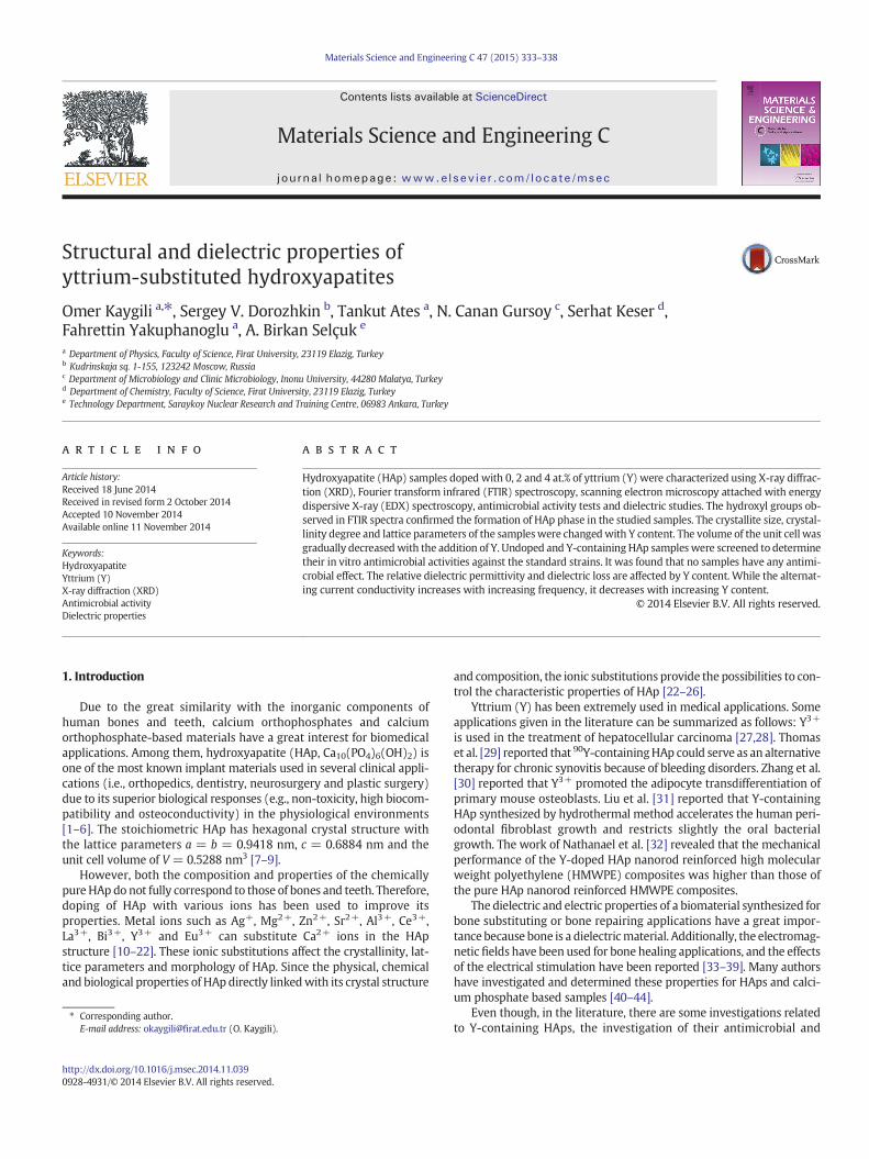
The powder XRD patterns of the prepared samples are shown inFig. 1. The patterns showed that all the peaks are matched perfectlywith the standard HAp (PDF no: 09-432) except for a peak belongingto the β-tricalcium phosphate (β-TCP, PDF no: 09-169) phase detectedat around 2θ = 31.22°. The formation percents of HAp phase are esti-mated to be 98.0, 97.4 and 95.7% for Y1, Y2 and Y3, respectively. Thepercents belonging to the β-TCP phase are found to be 2.0, 2.6 and4.3% for Y1, Y2 and Y3, respectively. The formation of HAp and β-TCPphases is affected by Y content. With increasing amount of Y, the
20 25 30 35 40 45 50 55
(203
)
(311
)
(301
)
Inte
nsity
(a.u
.)
(002
)
(111
)
(200
)
(410
)(3
21)(2
13)
(312
)(222
)
(310
)
(202
)
(300
)(1
12)
(211
)
(210
)
(102
)
(004
)
(402
)
Hydroxyapatite
-TCP
2 ( )
Standard HAp(JCPDS 09-432)
Y3
Y2
Y1
Fig. 1. XRD patterns of the as-synthesized samples.
formation of HAp phase decreases, whereas the amount of β-TCPphase increases. No other peaks, nor additional phases were detectedwith Y addition ranging from 0 to 4 at.%. The relative intensity of thepeak at ~40.12° belonging to (221) crystal plane was gradually de-creased with the addition of Y, and the calculated values were foundto be 0.303, 0.288 and 0.257, respectively. The relative intensity of thepeak at ~32.38° belonging to (112) crystal plane was gradually in-creased with adding of Y, and the calculated values were found to be0.464, 0.506 and 0.680 for Y1, Y2 and Y3, respectively.
The calculated values of the crystallite size, crystallinity degree, lat-tice parameters and the unit cell volume are given in Table 1. All thementioned parameters were significantly affected by the amount of Y.For each sample, the calculated values of D002 and D300 appeared to beclose to each other. The estimated values of the crystallite size for(221) crystal plane were found to be 37.59 nm for Y1, 34.39 nm for Y2and 32.41 nm for Y3. With the addition of Y, the crystallite size wasgradually decreased for (221) crystal plane, whereas the change inthis parameter did not significantly increase or decrease for (002) and(300) crystal planes. The lattice parameter c, volume of the unit celland crystallinity degree were gradually decreased by the addition of Yranging from 0 to 4 at.%, and this result is in good agreement with thereported works in the literature [20,32,52,53]. Since Y3+ ions (ionic ra-dius 0.09 nm) are smaller than Ca2+ ions (ionic radius 0.10 nm), it is ex-pected that Y incorporation might cause the size reduction (shrinkage)of the apatitic structure [20,21,32]. The decrease in the crystallinity de-gree can be related with the presence of β-TCP phase, namely, the rela-tive intensity of the peak at around 2θ = 31.22° was found to increasewith the addition of Y (0.188 for Y1, 0.202 for Y2 and 0.386 for Y3). Asknown, the amount of the β-TCP content in the HAp can affect the crys-tallinity [54].
3.2. Detection of the functional groups of the samples
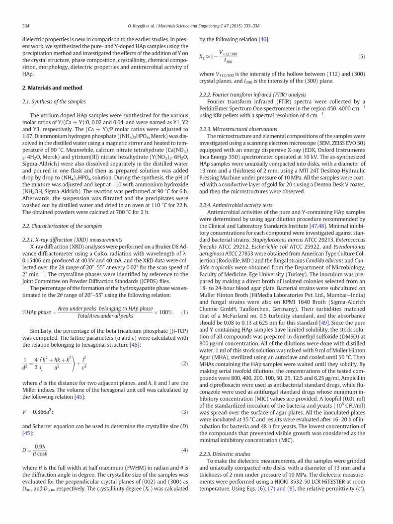
Fig. 2 shows the FTIR spectra of the pure and Y-containing HAp. Theobserved bands and their assignments are given as follows. The bandsdetected at 1037, 601 and 569 cm−1 were assigned to the vibrationalmodes of the phosphate groups. The sharp band at 3571 cm−1 and aweak one at 631 cm−1 were related with the characteristic vibrationalmodes of the hydroxyl group. With addition of Y, while the intensityof the band at 3571 cm−1 increased, the intensities of the bands at1037, 601 and 569 cm−1 decreased. A wide band centered at~3642 cm−1 and a weak band observed at 1635 cm−1 were associatedwith the water, which was absorbed in the samples and/or in the KBrpellet [55]. The doublet at 1458 and 1415 cm−1 was attributed to the vi-brationalmodes of the carbonate groups [56], which incorporated at theprecipitation stage.
3.3. Microstructure and composition
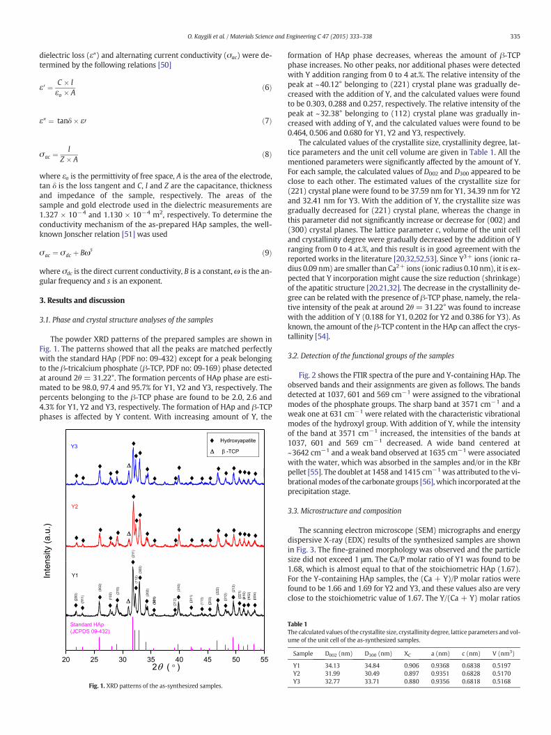
The scanning electron microscope (SEM) micrographs and energydispersive X-ray (EDX) results of the synthesized samples are shownin Fig. 3. The fine-grained morphology was observed and the particlesize did not exceed 1 μm. The Ca/P molar ratio of Y1 was found to be1.68, which is almost equal to that of the stoichiometric HAp (1.67).For the Y-containing HAp samples, the (Ca + Y)/P molar ratios werefound to be 1.66 and 1.69 for Y2 and Y3, and these values also are veryclose to the stoichiometric value of 1.67. The Y/(Ca + Y) molar ratios
Table 1The calculated values of the crystallite size, crystallinity degree, lattice parameters and vol-ume of the unit cell of the as-synthesized samples.
Sample D002 (nm) D300 (nm) XC a (nm) c (nm) V (nm3)
Y1 34.13 34.84 0.906 0.9368 0.6838 0.5197Y2 31.99 30.49 0.897 0.9351 0.6828 0.5170Y3 32.77 33.71 0.880 0.9356 0.6818 0.5168
4000 3600 3200 2800 2400 2000 1600 1200 800 400
OH
PO4
2CO3
H2OH2O
Y3
Y2
Tran
smitt
ance
(a.u
.)
Wavenumber (cm—1)
Y1OH
3PO4
Fig. 2. FTIR spectra for the pure- and yttrium-containing HAp samples.
Fig. 3. SEM images and EDX analysis re
Table 2Minimum inhibitory concentrations (μg/ml) of the tested compounds.
Microorganism Y1 Y2 Y3 Ampicillin Ciprofloxacin Fluconazole
E. coli N800 N800 N800 3.12 1.56 –
S. aureus N800 N800 N800 3.12 0.39 –
E. faecalis N800 N800 N800 1.56 0.78 –
P. aeruginosa N800 N800 N800 – 3.12 –
C. albicans N800 N800 N800 – – 3.12C. tropicalis N800 N800 N800 – – 3.12
336 O. Kaygili et al. / Materials Science and Engineering C 47 (2015) 333–338
for Y2 and Y3 samples were found to be 0.003 and 0.010, respectively.One can see that both values were smaller than the expected values(i.e., 0.02 for Y2 and 0.04 for Y3). Thus, not all amounts of Y were incor-porated into the crystal structure of HAp. Probably, this was due to thecharge imbalance. Moreover, this result is in a very good agreementwith the results reported by Capuccini et al. [57]. Namely, despite the in-corporation of Y riseswith increasing amount of Y, this increase is small-er than that of the as-expected or theoretical value.
sults of the as-prepared samples.
0 1x10⁶ 2x10⁶ 3x10⁶ 4x10⁶ 5x10⁶
8
10
12
14
16
18
Y3
Y2Rel
ativ
e pe
rmitt
ivity
, '
Frequency (Hz)
Y1
Fig. 4. Relative permittivity as a function of frequency plots of the samples.
101 102 103 104 105 106 10710-8
10-7
10-6
10-5
10-4
10-3
10-2
Y1 Y2 Y3
ytivitcudnoctnerrucgnitanretlA
ac (S
m)
Frequency (Hz)
Fig. 6. Alternating current conductivity vs. frequency plots of the as-prepared samples.
337O. Kaygili et al. / Materials Science and Engineering C 47 (2015) 333–338
3.4. Antimicrobial activity of the samples
The antimicrobial activity of the pure- and yttrium-containing HApsamples was tested against standard strains Gram-negative E. coli,P. aeruginosa aswell as Gram-positive E. faecalis, S. aureus and the fungalspecies C. albicans and C. tropicaliswhich are common infection-causingorganisms in human. As seen in Table 2, irrespective of yttrium content,no samples exhibited any antimicrobial effect at any tested concentra-tions (800–6.25 μg/ml). In other words, the addition of yttrium in theHAp structure did not cause any positive effect to improve the antimi-crobial activity of the HAp. This is not a surprising result for the pure-HAp because it does not possess antibacterial properties [58].
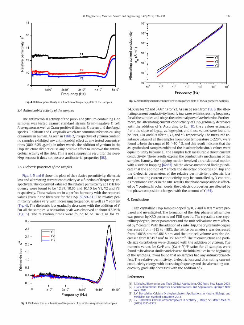
3.5. Dielectric properties of the samples
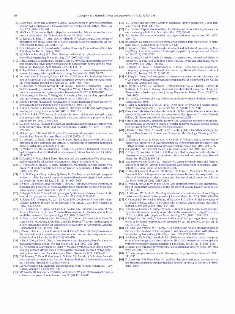
Figs. 4, 5 and 6 show the plots of the relative permittivity, dielectricloss and alternating current conductivity as a function of frequency, re-spectively. The calculated values of the relative permittivity at 1 kHz fre-quency were found to be 12.97, 10.65 and 10.10 for Y1, Y2 and Y3,respectively. These values are in a perfect harmony with the reportedvalues given in the literature for the HAp [50,59–61]. The relative per-mittivity values vary with increasing frequency, as well as Y content(Fig. 4). The dielectric loss gradually decreases with the addition of Y.For all the samples, a relaxation peak was observed at about 4.6 MHz(Fig. 5). The relaxation times were found to be 34.52 ns for Y1,
0 1x10⁶ 2x10⁶ 3x10⁶ 4x10⁶ 5x10⁶
0.0
0.4
0.8
1.2
1.6
2.0
2.4
2.8
3.2
Frequency (Hz)
Y1 Y2 Y3
Die
lect
ric lo
ss, "
Fig. 5. Dielectric loss as a function of frequency plots of the as-synthesized samples.
34.60 ns for Y2 and 34.67 ns for Y3. As can be seen from Fig. 6, the alter-nating current conductivity linearly increaseswith increasing frequencyfor all the samples and obeys theuniversal power lawbehavior. Further-more, the alternating current conductivity of HAp gradually decreaseswith the addition of Y. According to Eq. (8), the s values estimatedfrom the slope of logσac vs. logω plot, and these values were found tobe 0.99, 1.01 and 0.99 for Y1, Y2, and Y3, respectively. Themeasured re-sistance values of all the samples from room temperature to 220 °Cwerefound to be in the range of 1011–1012Ω, and this result indicates that theas-synthesized samples exhibited the insulator behavior. s values wereequal to unity because all the samples lack measurable direct currentconductivity. These results explain the conductivity mechanism of thesamples. Namely, the hopping motion involved a translational motionwith a sudden hopping [62,63]. All the above-mentioned findings indi-cate that the addition of Y affects the dielectric properties of HAp andthe dielectric parameters of the relative permittivity, dielectric lossand alternating current conductivity may be controlled by Y content.Asmentioned earlier in the XRD results, the phase composition is affect-ed by Y content. In other words, the dielectric properties are affected bythe phase composition changed with the amount of Y [64].
4. Conclusions
High crystalline HAp samples doped by 0, 2 and 4 at.% Y were pre-pared and investigated. The formation of the HAp phase in all sampleswas proven by XRD patterns and FTIR spectra. The crystallite size, crys-tallinity degree, lattice parameters and the unit cell volumewere affect-ed by Y content.With the addition of Y into HAp, the crystallinity degreedecreased from ~91% to ~88%, the lattice parameter c was decreasedfrom 0.6838 nm to 0.6818 nm, and the unit cell volume was also de-creased from 0.5197 nm3 to 0.5168 nm3. The microstructure and parti-cle size distribution were changed with the addition of yttrium. Thenumeric values for Ca/P and (Ca + Y)/P ratios for all samples werefound to be almost similar and close to the initial values in the beginningof the synthesis. It was found that no samples had any antimicrobial ef-fect. The relative permittivity, dielectric loss and alternating currentconductivity change with increasing frequency and the alternating con-ductivity gradually decreases with the addition of Y.
References
[1] T. Kokubo, Bioceramics and Their Clinical Applications, CRC Press, Boca Raton, 2008.[2] J. Park, Bioceramics: Properties, Characterizations, and Applications, Springer, New
York, 2008.[3] S.V. Dorozhkin, Calcium Orthophosphates: Applications in Nature, Biology, and
Medicine, Pan Stanford, Singapore, 2012.[4] S.V. Dorozhkin, Calcium orthophosphates in dentistry, J. Mater. Sci. Mater. Med. 24
(2013) 1335–1363.
338 O. Kaygili et al. / Materials Science and Engineering C 47 (2015) 333–338
[5] O. Kaygili, S. Keser, R.H. Al Orainy, T. Ates, F. Yakuphanoglu, In vitro characterizationof polyvinyl alcohol assisted hydroxyapatite derived by sol gel method, Mater. Sci.Eng. C 35 (2014) 239–244.
[6] M. Okada, T. Furuzono, Hydroxylapatite nanoparticles: fabrication methods andmedical applications, Sci. Technol. Adv. Mater. 13 (2012) 1–14.
[7] O. Kaygili, S. Keser, T. Ates, A.A. Al-Ghamdi, F. Yakuphanoglu, Controlling ofdielectrical and optical properties of hydroxyapatite based bioceramics by Cd con-tent, Powder Technol. 245 (2013) 1–6.
[8] D. Shi, Introduction to Biomaterials, Tsinghua University Press and World ScientificPublishing Co. Pte. Ltd, Beijing, 2006.
[9] S.J. Kalita, A. Bhardwaj, H.A. Bhatt, Nanocrystalline calcium phosphate ceramics inbiomedical engineering, Mater. Sci. Eng. C 27 (2007) 441–449.
[10] S. Jadalannagari, K. Deshmukh, S.R. Ramanan, M. Kowshik, Antimicrobial activity ofhemocompatible silver doped hydroxyapatite nanoparticles synthesized by modi-fied sol–gel technique, Appl. Nanosci. 4 (2014) 133–141.
[11] A. Bigi, G. Falini, E. Foresti, A. Ripamonti, M. Gazzano, N. Roveri, Magnesium influ-ence on hydroxyapatite crystallization, J. Inorg. Biochem. 49 (1993) 69–78.
[12] W.L. Suchanek, K. Byrappa, P. Shuk, R.E. Riman, V.F. Janas, K.S. TenHuisen, Prepara-tion of magnesium-substituted hydroxyapatite powders by the mechanochemi-cal–hydrothermal method, Biomaterials 25 (2004) 4647–4657.
[13] D. Laurencin, N. Almora-Barrios, N.H. de Leeuw, C. Gervais, C. Bonhomme, F. Mauri,W. Chrzanowski, J.C. Knowles, R.J. Newport, A. Wong, Z. Gan, M.E. Smith, Magne-sium incorporation into hydroxyapatite, Biomaterials 32 (2011) 1826–1837.
[14] K. Matsunaga, H. Murata, T. Mizoguchi, A. Nakahira, Mechanism of incorporation ofzinc into hydroxyapatite, Acta Biomater. 6 (2010) 2289–2293.
[15] A. Bigi, E. Foresti, M. Gandolfi, M. Grazzano, N. Roveri, Inhibiting effect of zinc on hy-droxylapatite crystallization, J. Inorg. Biochem. 58 (1995) 49–58.
[16] A. Bigi, E. Boanini, C. Capuccini, M. Gazzano, Strontium-substituted hydroxyapatitenanocrystals, Inorg. Chim. Acta 360 (2007) 1009–1016.
[17] N.D. Ravi, R. Balu, T.S.S. Kumar, Strontium-substituted calcium deficient hydroxyap-atite nanoparticles: synthesis, characterization, and antibacterial properties, J. Am.Ceram. Soc. 95 (2012) 2700–2708.
[18] Q.L. Feng, F.Z. Cui, T.N. Kim, J.W. Kim, Ag-substituted hydroxyapatite coatings withboth antimicrobial effects and biocompatibility, J. Mater. Sci. Lett. 18 (1999)559–561.
[19] M.A. Jakupec, P. Unfried, B.K. Keppler, Pharmacological properties of cerium com-pounds, Rev. Physiol. Biochem. Pharmacol. 153 (2005) 101–111.
[20] T.J. Webster, C. Ergun, R.H. Doremus, R. Bizios, Hydroxylapatite with substitutedmagnesium, zinc, cadmium, and yttrium. II. Mechanisms of osteoblast adhesion, J.Biomed. Mater. Res. 59 (2002) 312–317.
[21] T.J. Webster, E.A. Massa-Schlueter, J.L. Smith, E.B. Slamovich, Osteoblast response tohydroxyapatite doped with divalent and trivalent cations, Biomaterials 25 (2004)2111–2121.
[22] O. Kaygili, S.V. Dorozhkin, S. Keser, Synthesis and characterization of Ce-substitutedhydroxyapatite by sol–gel method, Mater. Sci. Eng. C 42 (2014) 78–82.
[23] L. Yingguang, Y. Zhuoru, C. Jiang, Preparation, characterization and antibacterialproperty of cerium substituted hydroxyapatite nanoparticles, J. Rare Earths 25(2007) 452–456.
[24] F. Liu, R. Wang, Y. Cheng, X. Jiang, Q. Zhang, M. Zhu, Polymer grafted hydroxyapatitewhisker as a filler for dental composite resin with enhanced physical and mechan-ical properties, Mater. Sci. Eng. C 33 (2013) 4994–5000.
[25] E. Vasile, L.M. Popescu, R.M. Piticescu, A. Burlacu, T. Buruinan, Physico-chemical andbiocompatible properties of hydroxyapatite based composites prepared by an inno-vative synthesis route, Mater. Lett. 79 (2012) 85–88.
[26] O. Kaygili, S. Keser, T. Ates, F. Yakuphanoglu, Synthesis and characterization of lith-ium calcium phosphate ceramics, Ceram. Int. 39 (2010) 7779–7785.
[27] R. Salem, K.G. Thurston, B.I. Carr, J.E. Goin, J.F.H. Geschwind, Yttrium-90 micro-spheres: radiation therapy for unresectable liver cancer, J. Vasc. Interv. Radiol. 13(2002) S223–S229.
[28] J.F.H. Geschwind, R. Salem, B.I. Carr, M.C. Soulen, K.G. Thurston, K.A. Goin, M. vanBuskirk, C.A. Roberts, J.E. Goin, Yttrium-90 microspheres for the treatment of hepa-tocellular carcinoma, Gastroenterology 127 (2004) S194–S205.
[29] S. Thomas, M.C. Gabriel, S.A.L. De Souza, S.C. Gomes, P.E. Assi, M.L.P. Perri, W.Liberato, C.S. Matushita, B. Gutfilen, L.M.B. Da Fonseca, 90Yttrium–hydroxyapatite:a new therapeutic option for radioactive synovectomy in haemophilic synovitis,Haemophilia 17 (2011) e985–e989.
[30] J. Zhang, C. Liu, Y. Li, J. Sun, P. Wang, K. Di, H. Chen, Y. Zhao, Effect of yttrium ion onthe proliferation, differentiation andmineralization function of primary mouse oste-oblasts in vitro, J. Rare Earths 28 (2010) 466–470.
[31] Y. Liu, R. Zhou, A. Mo, Z. Chen, H. Wu, Synthesis and characterization of yttrium/hy-droxyapatite nanoparticles, Key Eng. Mater. 330–332 (2007) 295–298.
[32] A.J. Nathanael, D. Mangalaraj, S.I. Hong, Y. Masuda, Synthesis and in-depth analysisof highly ordered yttrium doped hydroxyapatite nanorods prepared by hydrother-mal method and its mechanical analysis, Mater. Charact. 62 (2011) 1109–1115.
[33] P.M. Meaney, T. Zhou, D. Goodwin, A. Golnabi, E.A. Attardo, K.D. Paulsen, Bone di-electric property variation as a function of mineralization at microwave frequencies,Int. J. Biomed. Imaging 2012 (2012) 649612.
[34] S.A.W. Pickering, B.E. Scammell, Electromagnetic fields for bone healing, Int. J. LowExtrem. Wounds 1 (2002) 152–160.
[35] S.P. Bowen, J.D. Mancini, V. Fessatidis, M. Grabiner, Why do electromagnetic pulsesenhance bone growth? Ann. Biomed. Eng. 36 (2008) 195–203.
[36] R.O. Becker, The bioelectric factors in amphibian-limb regeneration, J. Bone JointSurg. Am. 43 (1961) 643–656.
[37] G.V. Cochran, Experimental methods for stimulation of bone healing by means ofelectrical energy, Bull. N. Y. Acad. Med. 48 (1972) 899–911.
[38] R.O. Becker, Stimulation of partial limb regeneration in rats, Nature 235 (1972)109–111.
[39] R.O. Becker, J.A. Spadaro, Electrical stimulation of partial limb regeneration in mam-mals, Bull. N. Y. Acad. Med. 48 (1972) 627–641.
[40] O. Kaygili, C. Tatar, F. Yakuphanoglu, Structural and dielectrical properties of Mg3Ca3(PO4)2 bioceramics obtained from hydroxyapatite by sol–gel method, Ceram.Int. 38 (2012) 5713–5722.
[41] K.K. Bamzai, S. Suri, V. Singh, Synthesis, characterization, thermal and dielectricproperties of pure and cadmium doped calcium hydrogen phosphate, Mater.Chem. Phys. 135 (2012) 158–167.
[42] O. Kaygili, C. Tatar, F. Yakuphanoglu, S. Keser, Nano-crystalline aluminum-containing hydroxyapatite based bioceramics: synthesis and characterization, J.Sol-Gel Sci. Technol. 65 (2013) 105–111.
[43] O. Kaygili, C. Tatar, The investigation of some physical properties andmicrostructureof Zn-doped hydroxyapatite bioceramics prepared by sol–gel method, J. Sol-Gel Sci.Technol. 61 (2012) 296–309.
[44] O. Kaygili, S. Keser, S.V. Dorozhkin, F. Yakuphanoglu, A.A. Al-Ghamdi, S. Kirbag, D.Sertkaya, T. Ates, N.C. Gursoy, Structural and dielectrical properties of Ag- andBa-substituted hydroxyapatites, J. Inorg. Organomet. Polym. Mater. 24 (2014)1001–1008.
[45] B.D. Cullity, Elements of X-ray Diffraction, Addison–Wesley Publishing Company,Massachusetts, 1978.
[46] E. Landi, A. Tampieri, G. Celotti, S. Sprio, Densification behaviour and mechanisms ofsynthetic hydroxyapatites, J. Eur. Ceram. Soc. 20 (2000) 2377–2387.
[47] Clinical and Laboratory Standards Institute (CLSI), Method for dilution antimicrobialsusceptibility tests for bacteria that grow aerobically: Approved Standard-SeventhEdition, CLSI Document M7-A7, Wayne, Pennsylvania2006.
[48] Clinical and Laboratory Standards Institute (CLSI), Reference method for broth dilu-tion antifungal susceptibility testing of yeasts: approved standard-second edition,CLSI document M27-A3. Wayne, Pennsylvania2002.
[49] J. Hindler, L. Hochstein, A. Howell, in: H.D. Isenberg (Ed.), Clinical Microbiology Pro-cedures Handbook, vol. 1, American Society for Microbiology, Washington D.C.,1992
[50] O. Kaygili, T. Ates, S. Keser, A.A. Al-Ghamdi, F. Yakuphanoglu, Controlling ofdielectrical properties of hydroxyapatite by ethylenediamine tetraacetic acid(EDTA) for bone healing applications, Spectrochim. Acta A 129 (2014) 268–273.
[51] A.K. Jonscher, The ‘universal’ dielectric response, Nature 267 (1977) 673–679.[52] C. Ergun, T.J. Webster, R. Bizios, R.H. Doremus, Hydroxylapatite with substituted
magnesium, zinc, cadmium, and yttrium. I. Structure and microstructure, J. Biomed.Mater. Res. 59 (2002) 305–311.
[53] M.A. Pogosova, P.E. Kasin, Y.D. Tretyakov, M. Jansen, Synthesis, structural features,and color of calcium–yttrium hydroxyapatite with copper ions in hexagonal chan-nels, Russ. J. Inorg. Chem. 58 (2013) 381–386.
[54] V. Aina, G. Lusvardi, B. Annaz, I.R. Gibson, F.E. Imrie, G. Malavasi, L. Menabue, G.Cerrato, G. Martra, Magnesium- and strontium-co-substituted hydroxyapatite: theeffects of doped-ions on the structure and chemico-physical properties, J. Mater.Sci. Mater. Med. 23 (2012) 2867–2879.
[55] H. Wang, K. Sun, A. Li, W. Wang, P. Chui, Size-controlled synthesis and characteriza-tion of fluorapatite nanocrystals in the presence of gelatin, Powder Technol. 209(2011) 9–14.
[56] I.R. Gibson, W. Bonfield, Novel synthesis and characterization of an AB-typecarbonate-substituted hydroxyapatite, J. Biomed. Mater. Res. A 59 (2002) 697–708.
[57] C. Capuccini, P. Torricelli, E. Boanini, M. Gazzano, R. Giardino, A. Bigi, Interaction ofSr-doped hydroxyapatite nanocrystals with osteoclast and osteoblast-like cells, J.Biomed. Mater. Res. A 360 (2007) 594–600.
[58] B. Singh, A.K. Dubey, S. Kumar, N. Saha, B. Basu, R. Gupta, In vitro biocompatibil-ity and antimicrobial activity of wet chemically prepared Ca10 − xAgx(PO4)6(OH)2(0.0 ≤ x ≤ 0.5) hydroxyapatites, Mater. Sci. Eng. C 31 (2011) 1320–1329.
[59] O. Kaygili, S.V. Dorozhkin, T. Ates, A.A. Al-Ghamdi, F. Yakuphanoglu, Dielectric prop-erties of Fe doped hydroxyapatite prepared by sol–gel method, Ceram. Int. 40(2014) 9395–9402.
[60] C.C. Silva, M.A. Valente, M.P.F. Graça, A.S.B. Sombra, The modulus formalism used inthe dielectric analysis of hydroxyapatite and calcium phosphate with titaniumformed by dry ball milling, J. Non-Cryst. Solids 351 (2005) 2945–2950.
[61] A.K. Dubey, P.K. Mallik, S. Kundu, B. Basu, Dielectric and electrical conductivity prop-erties of multi-stage spark plasma sintered HA–CaTiO3 composites and comparisonwith conventionally sintered materials, J. Eur. Ceram. Soc. 33 (2013) 3445–3453.
[62] J.C. Dyre, T.B. Schrøder, Universality of ac conduction in disordered solids, Rev. Mod.Phys. 72 (2000) 873–892.
[63] K. Funke, Jump relaxation in solid electrolytes, Prog. Solid State Chem. 22 (1993)111–195.
[64] R. Gregorio Jr., E.M. Ueno, Effect of crystalline phase, orientation and temperature onthe dielectric properties of poly(vinylidene fluoride) (PVDF), J. Mater. Sci. 34 (1999)4489–4500.