stroke: high-field magnetic resonance imaging
TRANSCRIPT

This article appeared in a journal published by Elsevier. The attachedcopy is furnished to the author for internal non-commercial researchand education use, including for instruction at the authors institution
and sharing with colleagues.
Other uses, including reproduction and distribution, or selling orlicensing copies, or posting to personal, institutional or third party
websites are prohibited.
In most cases authors are permitted to post their version of thearticle (e.g. in Word or Tex form) to their personal website orinstitutional repository. Authors requiring further information
regarding Elsevier’s archiving and manuscript policies areencouraged to visit:
http://www.elsevier.com/copyright

Author's personal copy
Stroke: High-FieldMagnetic ResonanceImagingKarl-Olof Lövblad, MD*, Sven Haller, MSc, MD,Vitor Mendes Pereira, MSc, MD
In industrialized countries, cerebrovascular dis-ease is 1 of the top 3 causes of morbidity andmortality of the general population, making ita priority for any health care system. Since thearrival of thrombolysis for cerebral stroke, therehas been a complete change in paradigm when itcomes to approaching patients with acute stroke.Indeed, previously, the outcome was bleak formost patients with very little beyond reeducationavailable if they survived the initial event. Followingthe National Institute of Neurological Disordersand Stroke and the European Cooperative AcuteStroke trials,1,2 intravenous thrombolysis estab-lished itself as the treatment modality for patientsbeing recruited within 3 hours after the initial event;following recent studies in Europe, intravenoustherapy can now be applied up to 4.5 hours3 andthere is an evolving understanding that using
alternative therapies it may be possible to gobeyond even this strict time window. Since then,there has been an unprecedented parallel evolu-tion in advances in both imaging and treatmentfor stroke. During the 1990s there was an explo-sion of imaging modalities with the developmentat first of new magnetic resonance (MR) tech-niques, such as diffusion and perfusion, and thenthe improvement of concurrent computed tomog-raphy (CT) techniques, as well as an improvedunderstanding of their interpretation.4–6 Beforethe development of fast echo–planar imagingtechniques, these types of imaging were difficultto perform because of motion artifacts. Theseimaging developments were at the same timeaccompanied by improvements in endovasculartechniques for the treatment of brain ischemia.7
The important improvements in imaging have led
Key Points
� Imaging must be fast and comprehensive (20 mins).
� Exclude hemorrhage with T2*/susceptibility-weighted imaging.
� Demonstrate/exclude ischemia with diffusion-weighted imaging/diffusion tensor imaging.
� Demonstrate tissue at risk with perfusion.
� Demonstrate occlusion with time of flight.
� Demonstrate/exclude proximal carotid disease with neck magnetic resonance angiography.
Dr Lovblad is the recipient of a grant from the Swiss National Science Foundation on high-field stroke imaging(SNF grant 320000-121565). He is a recipient of a research grant on MR from Bayer Schering Pharma.Division of Neuroradiology, Department of Imaging and Medical Informatics, Geneva University HospitalsHUG, 4 rue Gabrielle-Perret-Gentil, 1211 Geneva, Switzerland* Corresponding author.E-mail address: [email protected]
KEYWORDS
� Stroke � Magnetic resonance imaging � Perfusion� Diffusion � High-field imaging
Neuroimag Clin N Am 22 (2012) 191–205doi:10.1016/j.nic.2012.02.0021052-5149/12/$ – see front matter � 2012 Elsevier Inc. All rights reserved. ne
uroimaging.theclinics.com

Author's personal copy
to the development of a model of the ischemicpenumbra, or tissue at risk, that allows to triagepatients for appropriate treatment8; this altogetherhas made available an extremely varied armamen-tarium that can help us investigate these patients.The aims of imaging should be to exclude any kindof other pathology that might simulate a stroke,9
then establish that there is a stroke and not simplythe absence of hemorrhage, demonstrate thepresence of tissue that might undergo infarction,and finally in the acute setting try to demonstratethe thrombus and eventually the cause (cardiac,carotid origin).10 Then finally the method shouldallow for monitoring of treatment by allowingfollow-up. Although most major studies havebeen using CT for the initial screening, the para-digms used clinically are also changing. As statedearlier, the first initial step to be undertaken is toexclude the presence of any kind of other diseasethat might cause acute symptoms that might bemistaken for a stroke. This also applies to the clin-ical examination. Although for more than a decadescanners at 1.5 T were considered the state of theart, continuous developments in hardware andsoftware have rendered the implementation ofhigh-field imaging (3 T or more) possible andcommercially available.11 At the moment in the clin-ical neurosciences, these higher-field scanners arequickly becoming the new standard of imaging.The use of higher magnetic fields has the evidentadvantage of providing more signal that can beused to produce faster imaging, higher resolution,or a combination of both; also before the advent ofparallel imaging techniques,12 many MR imagingartifacts encountered at 3 T were considered aproblem.13
IMAGING TECHNIQUES: CT VERSUS MRIMAGING
Asalreadymentioned, the2mainaimsofneuroimag-ing are excluding hemorrhage and demonstratingischemia.9 CT has shown itself to be very sensitive
to hemorrhage and also capable of detecting signsof early ischemia in well-trained hands.4,5 Using MRimaging, these 2 aims can be attained by usingT2* images for blood detection14 and diffusion-weighted imaging (DWI) for ischemic lesions.15
Although initially there was a heated debate if MRcould fulfill the first imaging criterion (exclusion ofhemorrhage), we have now seen that this is easilyfeasible with MR imaging.14,16 Although in a fewselect situations, suchassubarachnoidhemorrhage,which usually does not mimic stroke clinically,imaging can be done with both MR imaging and CTfor screening of hemorrhage; MR imaging is ideallysuited for the detection of ischemia. MR imagingwith diffusion has been shown to be able to detectstrokes with sensitivities as high as 90% or more.15
The reports by Kidwell and colleagues16 andChalelaand colleagues17 found MR imaging and CT to beequivalently suited for the detection of acute hemor-rhage. Multiple studies comparing CT and MRimaging have shown that diffusion imaging isextremely sensitive to thepresenceof infarction.17–22
Although CT criteria are well established and havebeen used with success to detect early ischemicchanges, MR imaging can much better detect thepresence of small subcortical and cortical infarcts.MR imaging protocols now run for approxi-
mately 20 to 30 minutes, meaning that imagingtime will not interfere to a greater degree anymore.This time allows for performance of diffusionimaging, T2* imaging, and T2 imaging, as well asMR angiography (MRA) and MR perfusion (Box 1).Although being initially very sensitive to lesion
detection,MRprotocols, includingdiffusion imaging,have also shown themselves to provide information
Diagnostic Checklist
When confronted with a patient with acutestroke symptoms:
Rule out other pathology (tumor, inflamma-tion, hemorrhage)
Demonstrate ischemia
Demonstrate cause of stroke
Identify other possible cause of stroke (venous)
Demonstratepotentially viable tissue (penumbra)
Box 1Recommended sequences
Minimal stroke protocol:
Axial diffusion-weighted imaging (b0 and bmax)
Axial T2* images
Intracranial MR angiography (time of flight)
Regular stroke protocol:
Add to above:
Axial T1-weighted images
Axial T2-weighted images
Contrast-enhanced perfusion imaging
Extensive stroke protocol:
Add to above:
Extracranial and intracranial contrast-enhancedMR angiography of the neck
Lovblad et al192

Author's personal copy
Fig. 1. Patientwith righthemispheric stroke. There isa largehyperintensity in the rightMCAterritoryonthediffusionimagewithmaximumb value (A), accompanied by diminished perfusion on theMTTmap (B) and a reducedADC (C).
Fig. 2. Patient with embolic stroke: the diffusion image shows small lesions in the cortex (A). The watershedregion in the right hemisphere is hypoperfused: this is seen both on the contrast-enhanced perfusion maps (B)and on the ASL images (C). No hemorrhage on the SWI images (D).
193Stroke: High-Field MR Imaging

Author's personal copy
about outcome23,24 and lesionprogression,25 aswellas provide information about possible cause ofischemia owing to pattern differences in lesionlocation.26
MR imaging also has established itself as thepost-therapeutic modality of choice. This is be-cause DWI can be performed that is able todemonstrate very small lesions that might occurafter treatment,27 as well as demonstrate reperfu-sion with apparent diffusion coefficient (ADC)mapping.28,29
Considering all these arguments, it is obviouswhy MR imaging is destined to become themethod of choice for stroke management.When considering the guidelines of the American
Heart Association, we can see that CT and MRimaging can be used to exclude pathology anddemonstrate ischemia, that angio-MR imagingcan detect the presence of occlusion as well oralmost as well as other concurrent techniques,such as CT angiography or digital subtraction
angiography, and that perfusion imaging, althoughnot yet fully validated, can demonstrate the pres-ence of hemodynamic changes that correlate wellwith the neurologic changes that will eventuallypredict outcome. Thus, the use of multimodalityimaging is today necessary and this is renderedespecially easy at a high field.
DIFFUSION IMAGING
To the diffusion-imaging techniques belong DWI30
and diffusion tensor imaging.31 DWI is mainly usedfor stroke and consists of a relatively simple modi-fication of a spin-echo sequence that is sensitizedto motion by diffusion gradients. This produces theso-called diffusion-weighted images; at the higherb value, early ischemia will lead to a hyperintensityon imaging that corresponds to stroke. DWIs ata high b value have an inherent strong contrastand any ischemic lesion will appear as a bright sig-nal against a dark background (Fig. 1). Nowadays,
Fig. 3. Distal emboli after thrombolysis. Patient who underwent recanalization for an occlusion of the left MCA(A, B); afterward there are small distal subcortical lesions subcortically in the left hemisphere visible on thediffusion-weighted images (C, D).
Lovblad et al194

Author's personal copy
mostly directly isotropic images are used andproduced32: images without artifacts owing tothe directionality of water motion seen whenthe gradients are used with different directions.Usually DWI is done at various settings of diffusionsensitivity (b values) and these various images are
then used to create the so-called maps of theADC; whereas the DWIs are used for screeningpurposes and detection, the ADC maps providean interesting way to quantify or date the ischemicevent.33 Because of their strong inherent contrast,DWIs are better suited to detect also smallischemic events, such as those caused byhemodynamic compromise or embolism (Fig. 2).Although the diffusion effects themselves are inde-pendent of the magnetic field, the use of a higherfield will improve quality. In a study comparingDWIs at 1.5 and 3 T in 25 patients, Kuhl andcolleagues34 found an increased diagnostic confi-dence provided by the higher field images whencompared with the standard 1.5 T images, despitethe presence of higher susceptibility artifacts butprincipally because of higher signal-to-noise ratio(SNR). Also, diffusion imaging is ideally suited forthe follow-up of patients who have undergone in-terventional procedures (Fig. 3).
Diffusion tensor imaging may at the momenthave a less clearly established role in the ultra-early assessment of patients with stroke; indeeddiffusion images with more directionality havea slightly lesser contrast and are often less usedclinically. The maps of fractional anisotropy, whichreflect directionality, have a rather low imagingresolution so they are rarely used for the diagnosisof acute stroke; however, in the follow-up afterthe ischemic event, the use of tractography asprovided by diffusion tensor imaging could be ofgreat interest, as it allows us to follow the impactof the stroke on connectivity (Fig. 4). Diffusion
Fig. 4. Poststroke degeneration of the white mattertracts: a patient with a left-sided MCA stroke. Thereis a large chronic ischemic lesion visible on the T2image in the left MCA territory. The overlaid recon-structed tractography shows a marked asymmetry ofthe white matter tracts.
Fig. 5. Mismatch at 3 T. Patient with a small insular diffusion abnormality visible on the high b value diffusionimage (A) with a corresponding small ADC decrease (B). The MTT mp at the same level (C) shows a lager areaof hypoperfusion: there is a large mismatch zone corresponding to the functional penumbra.
Stroke: High-Field MR Imaging 195

Author's personal copy
tensor imaging will benefit greatly from the use ofhigher magnetic fields because it will increasethe SNR greatly.
PERFUSION IMAGING
Brainperfusion techniqueswerealsomadepossibleby the development of scanners capable of fast
imaging35,36 and they now play a central role inthe assessment of patients with stroke; indeed,based on the so-called diffusion perfusion mis-match model, a first easy-to-use working modelof the penumbra was created. The core lesion isbelieved to be constituted by the diffusion lesionand this is often surrounded by a greater area ofhypoperfused brain tissue; the difference between
Fig. 6. ASL in stroke: patient with a left MCA lesion visible on the high b value diffusion value (A) with an area ofhyperperfusion on ASL (B), despite a decrease in the ADC (C) and a large area of hypoperfusion on contrast-enhanced perfusion (D).
Lovblad et al196

Author's personal copy
both constitutes the tissue at risk or so-calledpenumbra (Fig. 5). Although this model is some-what different from the traditional model of thepenumbra, which relied on detecting differentlevels of energetic failure, it has proven to bea model that helps in the patient workup. Twomain types of perfusion imaging can be donewith MR techniques. Perfusion using contrastmaterial35,36 and perfusion without contrast.37
Perfusion with contrast or susceptibility-weightedcontrast perfusion relies on T2* images to provideimaging contrast. T1-weighted images could bemore reliable, but because of inherent difficultieshave not been widely implemented.38
Arterial Spin Labeling Perfusion
Arterial spin labeling (ASL) is a noninvasiveimaging technique that allows performance ofperfusion imaging of the brain without the adminis-tration of contrast. It uses flowing blood as aninherent contrast material after an initial tagginghas been performed. This technique has beenaround for more than a decade under various
forms and at first was a single-slice technique39;however, the development of high-field scannersat 3 T and more has allowed it to become a multi-slice product that is easily implemented in the clin-ical setting. Although ASL has been shown todemonstrate hypoperfusion quite well in acutestroke,40 it is also capable in some cases of demon-strating the presence of collateral flow (Fig. 6).41,42
This, in addition to its capacity to demonstrate re-gional territorial flow, is what could provide a greatimpact in the future.43 Also the fact that contrastmaterial is not necessary makes ASL an interestingnew possibility when confrontedwith elderly peoplewhose renal function might be impaired.
Dynamic Susceptibility-Weighted ImagingContrast
Mainly T2* imaging has been used to obtain perfu-sion images of the brain. After intravenous injec-tion of a gadolinium chelate, there is a decreasein signal on images where perfusion is normal; onthe raw MR images an area of hypoperfusion willthus be seen as a focus of relative hyperintensity
Fig. 7. TOF MRAs at 1.5 T and 3 T in the same patient. Note that the distal MCA branches are much more visible at3 T.
Stroke: High-Field MR Imaging 197

Author's personal copy
(less perfusion). The use of higher magnetic fieldswill provide an increase in the T2* effect, thusprovoking a more dramatic demonstration of anykind of altered perfusion. The use of a highermagnetic field should also allow reduction of theamount of injected contrast material, which wasdemonstrated by Manka and colleagues.44
MR ANGIOGRAPHY
The gain in signal strength has been shown toimprove the quality of brain MRA to an important
degree. This leads to an improvement of signal inthe arteries on time-of-flight (TOF) MRA but alsoto a better definition of peripheral branches ofthe intracranial vessels (Fig. 7).45 Willinek andcolleagues46 showed that the addition of sensitivityencoding was able to further improve imaging reso-lution and quality. Sommer and colleagues47 foundmore than 35% increase in SNR in the coronaries at3 T when compared with 1.5 T. In a study of the in-tracranial vessels, Willinek and colleagues45 foundan improvement in the diagnostic accuracy of TOFMRA performed at 3 T over images at 1.5 T.
Fig. 8. Patient with a subacute right-sided MCA infarction: there is slight hyperintensity on T1 (A), hypoin-tensity on T2 (B), and signal drop on T2* images (C). SWI shows an accumulation of blood after stroke evenmore dramatically (D).
Lovblad et al198

Author's personal copy
MRA techniques have also improved to sucha degree that they can reliably follow-up vesselsafter stenting.48 Also, using contrast-enhanced se-quences, MRA of the neck vessels has improvedto a degree that it can be used with confidenceto screen patients.49
SUSCEPTIBILITY-WEIGHTED MR IMAGING
Susceptibility-weighted MR imaging sequencesoffer a very strong T2* contrast but a very lowtissular anatomic differentiation.50 They have been
shown to be sensitive to the presence of smallhemorrhagic lesions; in the presence of intracere-bral hemorrhage this can be helpful in identifyingthe presence of micro-bleeds (Fig. 8). These se-quences are also capable of providing extremelyhigh-resolution anatomic imaging of the intra-cranial vessels; this is mainly true for the veins.Because of its inherent sensitivity to the oxygen-ated/deoxygenated status of the vessels, it is alsocapable of demonstrating the presence of vascu-lar stasis (Fig. 9), which is a known phenomenonin acute stroke and which is a predictor for
Fig. 9. Patient with a left-sided stroke who underwent successful thrombolysis: SWI before and after stroke.Before stroke there is vascular stasis (A) that disappears after thrombolysis (B).
Fig. 10. Chemical shift imaging allows mapping of cerebral metabolites. Patient with a small right-sided MCAinfarction: the lesion is visible in the right temporal lobe on DWI (A) and T2 images (B); on the metabolitemaps there is a decrease of NAA (C) and CR (D).
Stroke: High-Field MR Imaging 199

Author's personal copy
hemorrhagic transformation.51,52 Inastudyusing3Tperfusion imaging, Manka and colleagues44 foundthat the perfusion maps were of good quality andthat the distortions caused by the higher suscepti-bility artifactsat3Tdidnot relevantly affect their clin-ical potential.
FUNCTIONAL MR IMAGING (ACTIVATION)
Although difficult in the early phase of infarction,functional MR imaging is of great interest for thefollow-up of patients with stroke. Functional MRimaging is based on the capacity of T2* imagesto detect changes in local brain oxygenation. It
Fig. 11. Patient with a right-sided carotid stenosis (A) with distal emboli. The distal emboli are located in thewatershed regions (B) as well as in the basal ganglia (C) and in the basal temporal lobe (D).
Lovblad et al200

Author's personal copy
has been shown to be able to reflect functionalreorganization after stroke.53
MR SPECTROSCOPY
Spectroscopy using MR can also provide informa-tion about alterations in brain metabolites. MRspectroscopy is used for brain tumor staging butcan also be used for stroke. In the presence ofischemia there will be a decrease in N-acetyl-aspartate (NAA), which is a known neuronalmarker. There will also be a concurrent decreaseof lactate. Both these metabolites will be alteredwithin the areas of altered diffusion.54 AlthoughMR spectroscopy has shown itself to be an impor-tant tool for the investigation of many diseases andpathologic processes, it has never gained fullacceptance in clinical use for stroke; this is partlybecause of its complexity of use, which oftennecessitates postprocessing when time is critical.There is also the problem of voxel placement.High-field imaging now has made it easier toperform chemical shift imaging or multivoxel spec-troscopy, where one obtains maps of metabolites.Although single-voxel spectroscopy may be moreprecise, multivoxel spectroscopy or chemical shiftimaging has the advantage of allowing mapping ofaffected regions and thus one does not have torely on one single measurement (Fig. 10). Sodiumimaging may provide a further tool to improve visu-alization of ischemic lesions in patients with acutestroke55 but is still under investigation and needsfurther validation.
TYPES OF STROKES
Using MR imaging it is possible to investigate whatis the cause of stroke. Indeed, using T2 imagesand DWI it is now clearly possible to differentiatewhether the source is cardioembolic or carotiddisease (Figs. 11–13). A cardioembolic sourcewill cause multiple emboli in both the posteriorand anterior circulations on both sides, whereasa carotid unilateral source will usually affect onlyone defined territory. This was nicely demon-strated in a study by Baird and colleagues.26
Venous Ischemia
Cerebral venous thrombosis was previouslybelieved to be a rare and severe cause of cerebralischemia. Cerebral venous occlusive disease ismore frequent than believed and, if treated earlyand aggressively, is not necessarily associatedwith a catastrophic outcome. Clinically, the initialpresentation is often atypical and signs can rangefrom typical thunderclap headache to coma witha large variety of clinical signs in-between. Froma physiopathological point of view, it is importantto understand that because of venous obstruction,there is at the beginning simple stasis followed byedema and eventually blood extravasation owingto vascular rupture. Venous edema will tend tobe localized in the white matter and not to corre-spond to the classical arterial vascularization terri-tories. On “conventional”-type MR sequences,one will often see diminished or absent flow, withabsence of flow void on T2-weighted images and
Fig. 12. Hypoperfusion demonstrated with ASL in a case of carotid stenosis. The contrast-enhanced perfusionmap (A) shows a large area of hypoperfusion in the left MCA territory that can also be seen on the correspondingASL map (B). (C) Fractional anisotropy map derived from the diffusion tensor images.
Stroke: High-Field MR Imaging 201
Author's personal copy
hyperintensity on T1-weighted images in the pres-ence of an intravascular occlusion. Then, by usingMR phlebographic sequences, one can see theinterruption of flow in the affected veins; the use
of contrast-enhanced MR phlebography is stronglyrecommended to visualize cortical veins.56 Diffu-sion can sometimes demonstrate the presence ofrestriction in the clot itself.57
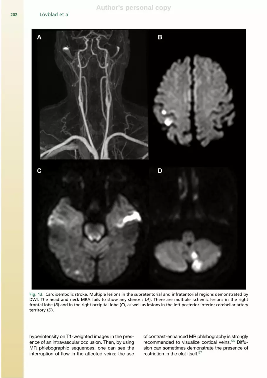
Fig. 13. Cardioembolic stroke. Multiple lesions in the supratentorial and infratentorial regions demonstrated byDWI. The head and neck MRA fails to show any stenosis (A). There are multiple ischemic lesions in the rightfrontal lobe (B) and in the right occipital lobe (C), as well as lesions in the left posterior inferior cerebellar arteryterritory (D).
Lovblad et al202

Author's personal copy
Carotid Disease, Occlusion
One important underlyingcauseof cerebral ischemiais the presence of carotid arteriosclerotic disease.This can be fully investigated by performingcontrast-enhanced MRA of the neck vessels.Contrast-enhanced MRA has replaced the moreconventional TOF method and allows coveragefrom the aortic arch into the cerebral vasculature inmuch less time.
Using additional MRA sequences it is alsopossible to determine if a vascular dissection isunderlying the ischemia.
SUMMARY
MR imaging has made great progress overallthanks to the implementation of high-field scan-ners and high-field imaging protocols; nowhere isthis more true than in the area of stroke imaging.These new high-field units have allowed previouslyexperimental imaging protocols to be implementedclinically. This began with the application of diffu-sion and perfusion techniques more than 10 yearsago that revolutionized the way we visualize acutestroke lesions. Now there is a further improvementby going to higher MR fields than previously.Indeed, there is an overall gain in signal that canprovide either higher resolution or imaging speed;together these 2 advantages have shown to bevery helpful in acute stroke imaging. Becausetime is an extremely central factor in the successof stroke management, even a minimal reductionof scan timewill have an extremely positive impact,we nowadays can perform either a full stroke MRprotocol within 20 minutes that comprises diffu-sion, perfusion, angiography, and T2* images, orif time is even a more important factor, we canperform a minimal MR examination that willdemonstrate or exclude ischemia with diffusion,T2* imaging, and MRA that can be performed infewer than 10 minutes. Now that it is well estab-lished that MR imaging can reliably establish thepresence or absence of hemorrhage and ischemia,the main objectives of MR and any kind of imagingwill be to determine exact tissue outcome and helpin selecting or excluding patients from therapy.Here, the use of perfusion with ASL can probablyhelp because one has seen that ASL can reliablydemonstrate the status of collateral flow and circu-lation, which was previously only reliably demon-strable with digital subtraction angiography. ASLtechniquesmay also demonstrate areas of reperfu-sion or hyperperfusion that occur in the cascadeof events during and after ischemia and thatwere difficult to assess with more conventionalMR imaging methods. In addition, the use of
sequences, such as susceptibility-weighted im-aging, which are extremely sensitive to the pres-ence of even small hemorrhagic deposits, shouldhelp us in assessing when it may or may not besafe to perform treatment using more conventionalthrombolysis.
These developments have had a maximalimpact because of their growth in parallel tomodern therapies, be they pharmacologic or en-dovascular in nature.
REFERENCES
1. The NINDS rt-PA stroke study group tissue plasmin-
ogen activator for acute ischemic stroke. N Engl J
Med 1995;333:1581–7.
2. Hacke W, Kaste M, Fieschi C, et al. Intravenous
thrombolysis with recombinant tissue plasminogen
activator for acute hemispheric stroke. The Euro-
pean Cooperative Acute Stroke Study (ECASS).
JAMA 1995;274(13):1017–25.
3. Hacke W, Kaste M, Bluhmki E, et al. Thrombolysis
with alteplase 3 to 4.5 hours after acute ischemic
stroke. N Engl J Med 2008;359(13):1317–29.
4. Warach S, Chien D, Li W, et al. Fast magnetic reso-
nance diffusion-weighted imaging of acute human
stroke. Neurology 1992;42(9):1717–23.
5. von Kummer R, Allen KL, Holle R, et al. Acute stroke:
usefulness of early CT findings before thrombolytic
therapy. Radiology 1997;205(2):327–33.
6. von Kummer R, Holle R, Gizyska U, et al. Interob-
server agreement in assessing early CT signs of
middle cerebral artery infarction. AJNR Am J Neuro-
radiol 1996;17(9):1743–8.
7. Gonner F, Remonda L, Mattle H, et al. Local intra-
arterial thrombolysis in acute ischemic stroke. Stroke
1998;29(9):1894–900.
8. Schlaug G, Benfield A, Baird AE, et al. The ischemic
penumbra: operationally defined by diffusion and
perfusion MRI. Neurology 1999;53(7):1528–37.
9. Adams HP Jr, del Zoppo G, Alberts MJ, et al,
American Heart Association, American Stroke Asso-
ciation Stroke Council, Clinical Cardiology Council,
Cardiovascular Radiology, Intervention Council,
Atherosclerotic Peripheral Vascular Disease and
Quality of Care Outcomes in Research Interdisci-
plinary Working Groups. Guidelines for the early
management of adults with ischemic stroke: a guide-
line from the American Heart Association/American
Stroke Association Stroke Council, Clinical Cardi-
ology Council, Cardiovascular Radiology and Inter-
vention Council, and the Atherosclerotic Peripheral
Vascular Disease and Quality of Care Outcomes in
Research Interdisciplinary Working Groups: the
American Academy of Neurology affirms the value
of this guideline as an educational tool for neurolo-
gists. Stroke 2007;38(5):1655–711.
Stroke: High-Field MR Imaging 203

Author's personal copy
10. Lovblad KO, Baird AE. Actual diagnostic approach
to the acute stroke patient. Eur Radiol 2006;16(6):
1253–6.
11. Willinek WA, Kuhl CK. 3.0 T neuroimaging: technical
considerations and clinical applications. Neuroi-
maging Clin N Am 2006;16(2):217–28.
12. Heidemann RM, Seiberlich N, Griswold MA, et al.
Perspectives and limitations of parallel MR imaging
at high field strengths. Neuroimaging Clin N Am
2006;16(2):311–20.
13. Vargas MI, Delavelle J, Kohler R, et al. Brain and
spine MRI artifacts at 3 Tesla. J Neuroradiol 2009;
36(2):74–81.
14. PatelMR, EdelmanRR,Warach S.Detection of hyper-
acute primary intraparenchymal hemorrhage by
magnetic resonance imaging. Stroke 1996;27(12):
2321–4.
15. Lovblad KO, Laubach HJ, Baird AE, et al. Clinical
experience with diffusion-weighted MR in patients
with acute stroke. AJNR Am J Neuroradiol 1998;
19(6):1061–6.
16. Kidwell CS, Chalela JA, Saver JL, et al. Magnetic
resonance imaging and computed tomography in
emergency assessment of patients with suspected
acute stroke: a prospective comparison. JAMA
2004;292(15):1823–30.
17. Chalela JA, Kidwell CS, Nentwich LM, et al. Magnetic
resonance imaging and computed tomography in
emergency assessment of patients with suspected
acute stroke: a prospective comparison. Lancet
2007;369(9558):293–8.
18. Lansberg M, Albers G, Beaulieu C, et al. Compar-
ison of diffusion-weighted MRI and CT in acute
stroke. Neurology 2000;54:1557–61.
19. Barber P, Darby D, Desmond P, et al. Identification of
major ischaemic change: diffusion-weighted imaging
versuscomputed tomography.Stroke1999;30:2059–65.
20. Mullins ME, Schafer PW, Sorensen AG, et al. CT and
conventional and diffusion-weighted MR imaging in
acute stroke: study in 691 patients at presentation
to the emergency department. Radiology 2002;
224:353–60.
21. Fiebach JB, Schellinger PD, Jansen O, et al. CT and
diffusion-weighted MR imaging in randomized order.
Diffusion-weighted imaging results in higher accu-
racy and lower interrater variability in the diagnosis
of hyperacute ischemic stroke. Stroke 2002;33:
2206–10.
22. Fiebach J, Jansen O, Schellinger P, et al. Comparison
of CT with diffusion-weighted MRI in patients with
hyperacute stroke. Neuroradiology 2001;43(8):628–32.
23. Lovblad KO, Baird AE, Schlaug G, et al. Ischemic
lesion volumes in acute stroke by diffusion-weighted
magnetic resonance imaging correlate with clinical
outcome. Ann Neurol 1997;42(2):164–70.
24. Baird AE, Lovblad KO, Dashe JF, et al. Clinical corre-
lations of diffusion and perfusion lesion volumes in
acute ischemic stroke. Cerebrovasc Dis 2000;10(6):
441–8.
25. Baird AE, Benfield A, Schlaug G, et al. Enlargement
of human ischemic lesion volumes measured by
diffusion-weighted magnetic resonance imaging.
Ann Neurol 1997;41:581–9.
26. Baird AE, Lovblad KO, Schlaug G, et al. Multiple
acute stroke syndrome: marker of embolic disease?
Neurology 2000;54(3):674–8.
27. Lovblad KO, Pluschke W, Remonda L, et al. Diffu-
sion-weighted MRI for monitoring neurovascular
interventions. Neuroradiology 2000;42(2):134–8.
28. Taleb M, Lovblad KO, El-Koussy M, et al. Reperfusion
demonstrated by ADC mapping after intra-arterial
thrombolysis for human ischemic stroke confirmed by
cerebral angiography. Neuroradiology 2001;43:591–4.
29. Marks MP, Tong DC, Beaulieu C, et al. Evaluation of
early reperfusion and i.v. tPA therapy using diffusion-
and perfusion-weighted MRI. Neurology 1999;52(9):
1792–8.
30. Le Bihan D, Breton E, Lallemand D, et al. MR
imaging of intravoxel incoherent motions: applica-
tion to diffusion and perfusion in neurologic disor-
ders. Radiology 1986;161(2):401–7.
31. Le Bihan D, Mangin JF, Poupon C, et al. Diffusion
tensor imaging: concepts and applications. J Magn
Reson Imaging 2001;13(4):534–46.
32. Warach S, Mosley M, Sorensen AG, et al. Time
course of diffusion imaging abnormalities in human
stroke. Stroke 1996;27(7):1254–6.
33. Schlaug G, Siewert B, Benfield A, et al. Time course
of the apparent diffusion coefficient (ADC) abnor-
mality in human stroke. Neurology 1997;49(1):113–9.
34. KuhlCK,Textor J,GiesekeJ, et al. Acuteandsubacute
ischemic stroke at high-field-strength (3.0-T) diffusion-
weighted MR imaging: intraindividual comparative
study. Radiology 2005;234(2):509–16.
35. Rosen BR, Belliveau JW, Chien D. Perfusion imaging
by nuclear magnetic resonance. Magn Reson Q
1989;5(4):263–81.
36. Lev MH, Rosen BR. Clinical applications of intracra-
nial perfusion MR imaging. Neuroimaging Clin N Am
1999;9(2):309–31.
37. Detre JA, Wang J, Wang Z, et al. Arterial spin-
labeled perfusion MRI in basic and clinical neurosci-
ence. Curr Opin Neurol 2009;22(4):348–55.
38. Heid O. T1-gewichtete MR perfusion [dissertation].
Switzerland: University of Bern; 2000.
39. Edelman RR, Siewert B, Darby DG, et al. Qualitative
mapping of cerebral blood flow and functional local-
ization with echo-planar MR imaging and signal tar-
geting with alternating radio frequency. Radiology
1994;192(2):513–20.
40. ViallonM, Altrichter S, Pereira VM, et al. Combineduse
of pulsed arterial spin-labeling and susceptibility-
weighted imaging in stroke at 3T. Eur Neurol 2010;
64(5):286–96.
Lovblad et al204

Author's personal copy
41. Lim CC, Petersen ET, Ng I, et al. MR regional perfu-
sion imaging: visualizing functional collateral circula-
tion. AJNR Am J Neuroradiol 2007;28(3):447–8.
42. Altrichter S, Kulcsar Z, Sekoranja L, et al. Arterial
spin labeling demonstrates early recanalization after
stroke. J Neuroradiol 2009;36(2):109–11.
43. Hendrikse J, Petersen ET, Chng SM, et al. Distribu-
tion of cerebral blood flow in the nucleus caudatus,
nucleus lentiformis, and thalamus: a study of territo-
rial arterial spin-labeling MR imaging. Radiology
2010;254(3):867–75.
44. Manka C, Traber F, Gieseke J, et al. Three-dimen-
sional dynamic susceptibility-weighted perfusion
MR imaging at 3.0 T: feasibility and contrast agent
dose. Radiology 2005;234(3):869–77.
45. Willinek WA, Born M, Simon B, et al. Time-of-flight
MR angiography: comparison of 3.0-T imaging and
1.5-T imaging—initial experience. Radiology 2003;
229(3):913–20.
46. Willinek WA, Gieseke J, von Falkenhausen M, et al.
Sensitivity encoding (SENSE) for high spatial resolu-
tion time-of-flight MR angiography of the intracranial
arteries at 3.0 T. Rofo 2004;176(1):21–6.
47. Sommer T, Hackenbroch M, Hofer U, et al. Coronary
MR angiography at 3.0 T versus that at 1.5 T: initial
results in patients suspected of having coronary
artery disease. Radiology 2005;234(3):718–25.
48. Lovblad KO, Yilmaz H, Chouiter A, et al. Intracranial
aneurysm stenting: follow-up with MR angiography.
J Magn Reson Imaging 2006;24(2):418–22.
49. Debrey SM, Yu H, Lynch JK, et al. Diagnostic accu-
racy of magnetic resonance angiography for internal
carotid artery disease: a systematic review and
meta-analysis. Stroke 2008;39(8):2237–48. AJNR
Am J Neuroradiol 2007;28(3):447–8.
50. Haacke EM, Mittal S, Wu Z, et al. Susceptibility-
weighted imaging: technical aspects and clinical
applications, part 1. AJNR Am J Neuroradiol 2009;
30(1):19–30.
51. Hermier M, Nighoghossian N, Derex L, et al. Hypo-
intense transcerebral veins at T2*-weighted MRI:
a marker of hemorrhagic transformation risk in
patients treated with intravenous tissue plasminogen
activator. J Cereb Blood Flow Metab 2003;23(11):
1362–70.
52. Hermier M, Nighoghossian N, Derex L, et al. Hypo-
intense leptomeningeal vessels at T2*-weighted
MRI in acute ischemic stroke. Neurology 2005;
65(4):652–3.
53. Cramer SC, Nelles G, Benson RR, et al. A functional
MRI study of subjects recovered from hemiparetic
stroke. Stroke 1997;28(12):2518–27.
54. Mader I, Rauer S, Gall P, et al. (1)H MR spectros-
copy of inflammation, infection and ischemia of the
brain. Eur J Radiol 2008;67(2):250–7.
55. Hussain MS, Stobbe RW, Bhagat YA, et al. Sodium
imaging intensity increases with time after human
ischemic stroke. Ann Neurol 2009;66(1):55–62.
56. Lovblad KO, Schneider J, Bassetti C, et al. Fast
contrast-enhanced MR whole-brain venography.
Neuroradiology 2002;44(8):681–8.
57. Lovblad KO, Bassetti C, Schneider J, et al. Diffusion-
weighted MR in cerebral venous thrombosis. Cere-
brovasc Dis 2001;11(3):169–76.
Stroke: High-Field MR Imaging 205