stride cycle influences on goal-directed head movements made during walking
TRANSCRIPT
Source of Acquisition NASA Johnson Space Center
STRIDE-CYCLE INFLUENCES ON GOAL-DIRECTED HEAD MOVEMENTS MADE
DURING WALKING
Brian T. Richasd E.A. van Emmerik2, and Jacob J. Bloomberg3
Neuroscience Laboratoiies, Wyle Life Sciences, Houston, TX, USA
Department of Exercise Science, University of Massachusetts, Amherst, MA, USA
Neuroscience Laboratoiies, NASA/Johnson Space Center, Houston, TX, USA
Subtitle: Head Movements Made While Walking
Coil-espondence : Brian T. Peters Wyle Life Sciences 1290 Hercules Dr. Suite 120 Houston, TX 77058, USA Phone: (281) 244-6574; Fax: (281) 244-5734 e-mail: bpeters @ems.j sc.nasa.gov
Abstract
Horizontal head movements were studied in six subjects as they made rapid hoi-izontal gaze
adjustments while walking. The aim of the present research was to deteimine if gait-cycle events
alter the head movement response to a visual target acquisition task. Gaze shifts of approximately
40" were elicited by a step change in the position of a visual target from a central location to a
second location in the left or i-ight horizontal pei-ipheiy. The timing of the target position change
was constrained to occur at 25,50,75 and 100% of the stride cycle. The trials were randomly
presented as the subjects walked on a treadmill at their prefeiyed speed (range: 1.25 to 1.48 m / s ,
mean: 1.39 1: 0.09 d s ) . Analyses focused on the movement onset latencies of the head and eyes
and on the peak velocity and saccade amplitude of the head movement response. A comparison of
the group means indicated that the head movement onset lagged the eye onset (262 ms versus 252
ms). The head and eye movement onset latencies were not affected by either the direction of the
target change nor the point in the gait cycle during which the target relocation occm-ed. However,
the presence of an interaction between the gait cycle events and the direction of the visual target shift
indicates that the peak head saccade velocity and head saccade amplitude are affected by the natural
head oscillations that occur while walking.
Key words: head movement, target acquisition, locomotion, eye-head coordination, vision
Introduction
Visual fixation of targets peripheral to the progression of travel is common as we walk, yet gaze
adjustments made duiing locomotion have not been systematically studied. When these gaze
adjustments are sufficiently large, a head rotation must accompany the eye movements to achieve
the goal. Little is known about the interaction between these goal-directed head movements and the
periodic head translations and rotations that naturally accompany locomotion. Repoi-ts vaiy
regarding the magnitude of these peiiodic head movements that occur while walking, but the phase
relationship between these movements and ongoing gait cycle is consistent'-8. In the sagittal plane
for example, the head pitches upward as the body translates down during each step. Yaw head
movements counter the lateral translations of the body in a similar way. Based on studies using
passive rotations of the body, it is believed that these movements are the result largely of reflexive
mechanismsgy lo. Regardless of the control mechanism (Le. reflexive, voluntay, or passive inertial)
these head movements imply the presence of modulating levels of neck muscle activation and
vestibular stimulation.
The effects of this modulation on goal-dliected head movements are unknown. Bent and
colleagues found differences in lower body gait parameters that were dependent on when in the gait
cycle a vestibular stimulus was given12. A head saccade made while walking could impart a similar
vestibular disturbance. The results of a study comparing the effects of voluntaiy and unexpected
head turns while walking, led Vallis and Patla to conclude that the central nervous system partially
nullifies the sensoiy input created by voluntary head t u i ~ ~ s ' ~ . In addition to this proposed use of
efferent copy to minimize the vestibular disturbance, a strategy of triggei-ing head movements to
coincide with phases of the gait cycle where the vestibular infoimation is less impoi-tant could also
be used. Such a strategy would affect the timing of the head movement response. Differences in
the response timing may also result from interactions with other ongoing activities. Lajoie et al.
showed that reaction time was dependent on when in the gait-cycle the stimulus was presented".
The authors attributed this result to varying levels of attentional demand across the gait cycle.
2
In addition to possible changes in the initiation of a response, the process of superimposing
a voluntslly head movement on the naturally occurring motions may affect the dynamics of the head
movement response. The eye-head coordination during gaze re-fixations has .been studied
extensively in seated subjects, but this behavior has not been studied in waking subjects. Changes
in the head movement dynamics could impose limitations on the total response time (i.e. reaction
time plus movement time) and could result in final head positions that leave the eyes at orbital
eccenhicities that are sub-optimal for visual acuity. Through measures of movement onset latency
and head movement kinematics, the goal of the present research was to assess the effects of gait
cycle events on head movements made to acquire targets in the visual pei-ipheiy.
Materials and Methods
Subjects
Six healthy males in the age range from 26 to 35 (mean 3 1 years 24) served as subjects for
this study. None of the subjects had complaints of neck soreness at the time of the test and none
had any history of vestibular disease. The expeiiment protocol was approved by the University of
Massachusetts’ Human Subjects Review Committee and all subjects gave their informed consent
piior to participation.
Testing Conditions
Walking Speed
The subjects, all wearing a similar type jogging shoe, were tested as they walked on a motor-
driven h-eadrnill ( A c c u d P, Pacer Fitness Systems, Inc., Irving, TX) at their prefei-red walking
speed The subject-specific speeds were established using an interactive method defined in Molt et
al l 4 which resulted in speeds ranging fiom 1.25 to 1.48 m / s (mean 1.39 -c 0.09 d s ) .
Visual Targets
Visual targets were presented on a rear-projection screen @a-Lite Screen Company, Inc.,
Warsaw, Indiana) that was placed between the subject and an LCD projector (Sharp, Model: XG-
E670U, Mahwah, New Jersey). The screen was perpendicular to the walking direction at a distance
3
of -1 10 cm from the subject’s nasal bridge. The targets, consisting of alpha-numeiic characters,
were controlled by custom software (LabVIEW; National Instruments Coip., Austin, TX.). When
presented directly in front of the subject, the visual targets subtended a visual angle of -1.3”.
During each of the 14-second walking trials, the visual target was initially presented in the
center of the subject’s field-of-view at a height that was perceived by them to be eye level. The
presented targets were randomly selected from a “pool” that contained 24 upper-case characters
(0 and I were excluded) and 8 numerals (0 and 1 were omitted). With the exception of the first
target to appear in the lateral position, which was visible for 900 ms, the display duration for the
sequentially displayed characters was assigned randomly using the finite set of times that fell at 50
ms intei-vals from 400 to 700 ms.
Targets in the lateral positions were restricted to the pool of 24 letters and presented at the
same height as the central target but at positions that were 1 m to the left or iight of it. A gaze
compensation angle of -r40” was required to visually fixate the lateral targets that continued to be
displayed until the end of the trial. At the conclusion of each trial the subject ‘s recollection of the
number of numerals that appeared in the central position, as well as the first letter seen in the lateral
position, was compared to the actual values. While this information was used to establish an
inclusion criterion for acceptable data trials, the piimary intent of requiring this from the subject was
to motivate them to concentrate on’the task.
The timing of the target change from the central position to the lateral position was one of
the ciitical aspects of this investigation. The stimulus-generating program used a signal produced
by a roller switch secured to the lateral edge of the heel of the iight shoe to control this change.
Measured from the right heel strike, the target position changes were restricted to four phases, or
quadrants, coi-responding to 25,50,75 and 100% of the stride cycle.
The two lateral positions and four stride quadrants created eight possible test conditions,
which were each repeated three times per subject. The order of the twenty-four tiials was
randomized piior to the start of data collection.
4
Standing test
Although a comparison between walking and standing conditions was not the intended
focus of this investigation, relatively long reaction time latencies during the walking task prompted
us to repeat the task during a subsequent test session. Five of the six subjects repeated the gaze re-
fsation task as they stood on the non-moving treadmill belt.
Measurement S y s tems
Kinematic and analog variables were collected at 240 Hz using the Qualisys motion analysis
system (Qualisys, Inc., Glastonbm-y, CT.). The system monitored the positions of seven retro-
reflective markers affixed to a light-weight headband (183 g) and a torso harness. The relative
position of a virtual marker placed on the subject's nasal bridge was determined piioi- to the start of
the data collection. This infoimation allowed the location of that landmark to be calculated
throughout each of the data trials.
The analog signals consisted of two TTL outputs that were used to indicate light heel strike
and the timing of visual target position change and hoiizontal eye movements. The eye movements
were obtained using standard DC Electrooculography (EOG) techniques.
Data Processing
Head Yaw Calculation
A line segment connecting the virtual nasal biidge marker and the marker placed on the
occipital region of the head was used to calculate the yaw movements of the head. Because this line
segment deviated only slightly above and below (- 1-2") the transverse plane while waking any
angle calculation errors introduced by out-of-plane motions were minimal.
Movement Onset Detei-mination
The onset of the saccadic movements for both the head and eyes was considered to be the
point at which their velocity trajectoiies deviated from a 21 SD band around the average per-stride
trajectoiy. A fourth-order, zero-lag Butteiwoi-th filter was applied to the raw position data piior to
5

calculating the velocities with a 3-point central difference ggoiithm. The per-stride average
trajectories and standard deviations (SD) were calculated for each hid using the first six complefe
strides, each of which had been linearly interpolated to 250 points. Because the 5 Hz filter applied
to the data prior to determining the average trajectory significantly attenuated the shai-pness of the
saccadic movement, the epoch containing the saccade was filtered using 15 Hz cut-off. The point at
which this signal deviated from the average trajectoiy was automatically deteimined using custom-
wiitten software (MATLAB; The Mathworks, Inc., Natick, MA). An operator who was sufficiently
blinded from knowing the condition of the trial being displayed, visually inspected each trial and
was able to manually input the appropriate position if the location of the automatically determined
movement onsets was incoirect.
Peak Yaw Head Velocity
The peak yaw head velocity was deteimined to be the maximum value observed in the
filtered (15 Hz cutoff) yaw velocity. This value was also visually inspected from the time series
data.
Yaw Head Saccade Amditude
Variability in the stride-to-stride trajectory of the yaw head position prevented the "offset"
of the saccade from being reliably determined. In lieu of using the offset to deteimine the
amplitude of the head saccade, it was calculated to be the difference between the average yaw
position of the head during the first complete stride after the saccade and the average position of the
last complete stride before the saccade.
Statistical Analysis
Piior to perfoiming any statistical analyses, data from 5 of the 120 trials were excluded
from the data set because the target position change occurred outside a 55% window of the desired
stride position (i.e. 20 - 30% for the 25% condition). Another 19 trials were eliminated because the
subjects did not accurately identify the first letter that appea-ed in the lateral position. This was
usually the result of a response that was sufficiently delayed due to either starting the movement in
the opposite direction or because of a lack of attention. An ANOVA using a split-plot factorial
6
design was used for the statistical analyses. The ANOVA model contained tests for main effect
differences between SUBJECTS, TRIALS , target step DIRECTION, 2nd gait-cycle QUADRANT.
It also included a test for a DIRECTION x QUADRANT interaction that was nested within the
SUBJECTS. The test for TRIALS effects was included to make the analysis complete, but as
would be expected given the randomized presentation of the 24 trials this comparison showed no
effect. Therefore, it is not included in the discussion of the results.
Results
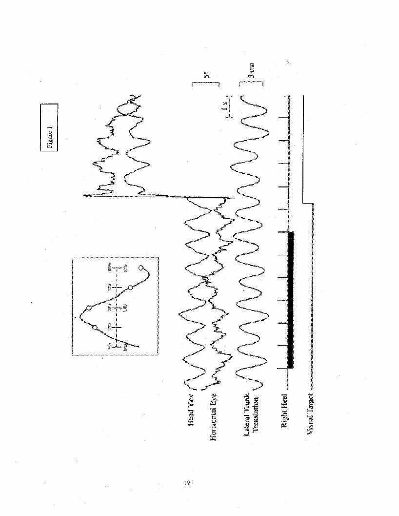
Basic Head and Eye Motions in Yaw
While walking and attending to the visual target, the yaw head motion of all of the subjects
had an oscillatory nature that repeats itself with each stride (i.e. heelsti-ike to heelsti-ike of the same
foot). This oscillation is present during gaze fixation of both the central and lateral target locations
(see Figure 1). The inset in Figure 1 shows the per-stride average head yaw trajectoiy for the
central target portion of the trial. The desired 25,50,75, and 100% points for the possible target
relocation times are depicted on the plot.
........................................................................................... Insert Figure 1 here
...........................................................................................
In all subjects, the medio-lateral position of a marker located near the seventh cervical
vertebra maintained an anti-phase relationship with the head yaw signal. Across all of the data trials
the peak-to-peak amplitude of the lateral trunk translation was 4.9 -+ 1.0 cm (mean -+ 1 SD) (range:
3.1 to 5.6 cm) and the peak-to-peak amplitude of the head yaw rotation was 3.2 2 1.2 degrees
(range: 2.1 to 5.1 degrees). Four of the six subjects also had horizontal eye movements that clearly
showed a similar oscillatoiy pattern that maintained an anti-phase relationship with the head yaw
rotation (see Figure l), indicating that when the head rotated to the left, the eyes rotated to the light.
This type of coordination could be consistent with a natural head oi-ientation projection that crosses
the standing line of sight between the subject and the visual target 15. In this scenario, the counter-
7
rotation of the eyes is necessary because the head rotation over-compensates for the lateral
translation of the hxnk '' 16. Reaction Time Variables
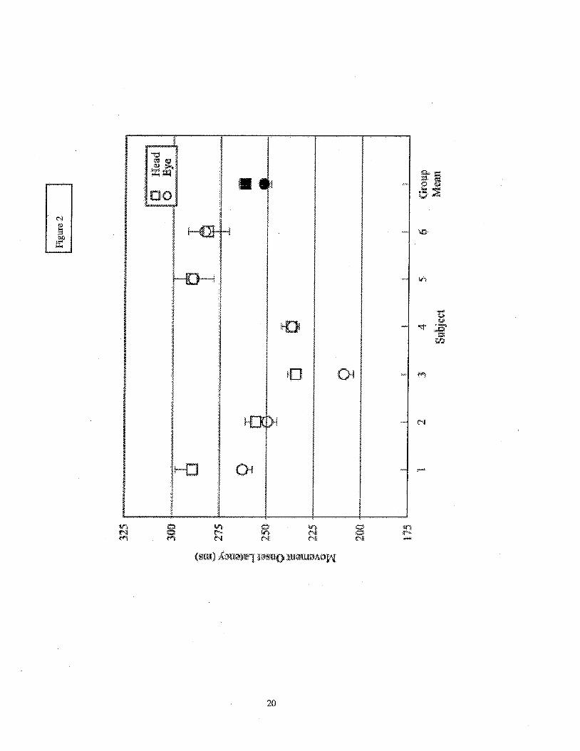
With subject means ranging from 209 to 288 ms (group mean: 252 ms), the horizontal eye
movement onset latencies varied significantly between subjects (p < 0.001). This was also true for
the latency of the onset for the head yaw movement (range: 234 to 290 ms; group mean: 262 ms).
The group means suggest that the onset of the eyes preceded the head movement onset, but this was
only clearly evident for half of the subjects (see Figure 2). A presentation of the p-values obtained
for each of the dependent variables across all experimental manipulations is provided in Table 1. It
is evident from these data that neither of the reaction time variables were affected by the
experimental manipulations
........................................................................................... Insert Figure 2 here
...........................................................................................
........................................................................................... Insert Table 1 here
Head Saccade Dynamics
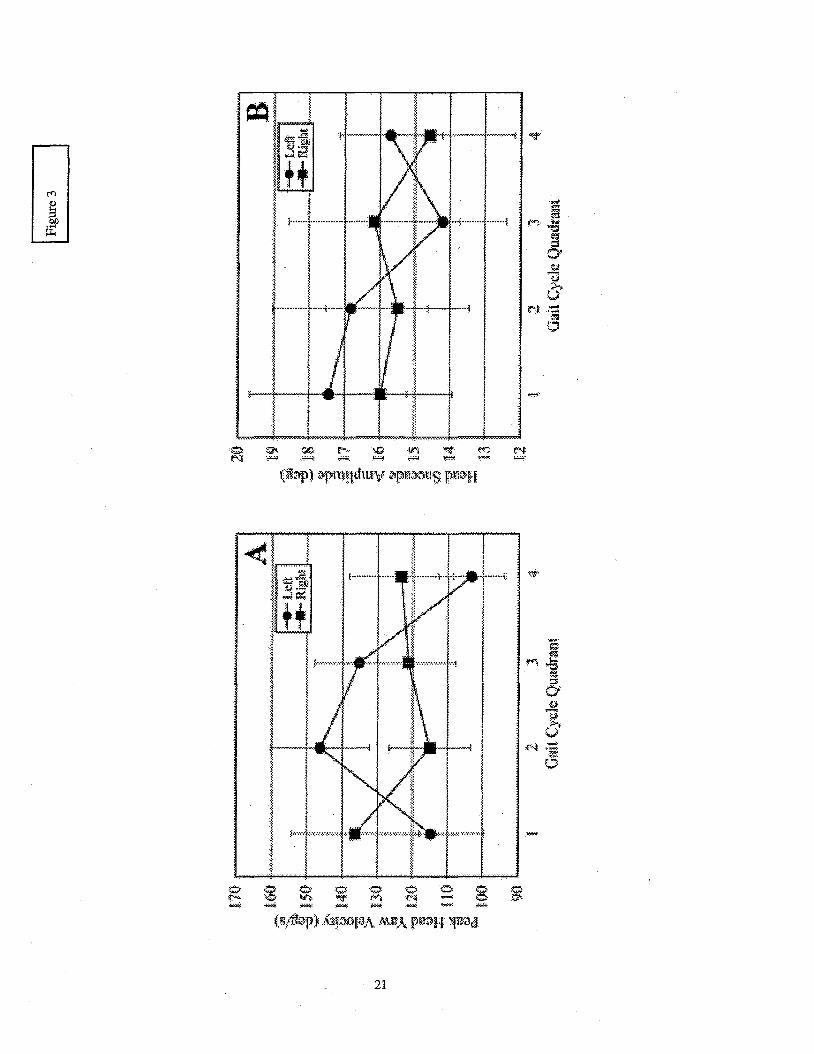
The saccade amplitude and peak velocity of the head yaw signal were measured to provide
an assessment of head saccade dynamics. Across all subjects the mean peak velocity values ranged
from 64 - 172"h and the amplitude of the head saccade ranged from 3-25". Each of these measures
illustrate that the head dynamics of the saccade response differed between subjects (p < 0.001 for
each). A statistically significant interaction between the direction of the required gaze re-fixation
and the point in the gait cycle at which the target changed locations was also observed in each
@=0.015 and p = 0.021 for peak velocity and saccade amplitude, respectively). The presence of
this interaction, clearly visible in the group means presented in Figure 3, indicates that stride cycle
parameters can influence the head movements made to acquire visual targets while walking.
8

........................................................................................... Insert Figure 3 here
...........................................................................................
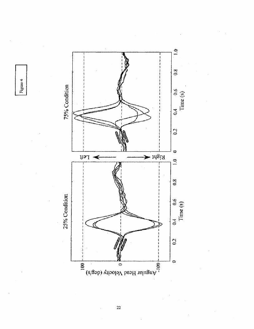
A reason for this interaction becomes apparent when inspecting a subject’s head yaw
velocity signals from the 25 and 75% conditions (see Figure 4). A comparison of the peak velocity
amplitudes between the two conditions shows that movements made to fixate a target in the left
periphery achieve higher magnitudes in the 75% condition than those in the 25% condition. The
opposite is true for rightward saccades. The slope of the head velocity signal just piior to the onset
of the movement shows the direction of the head’s rotational acceleration at the time that the
movement is initiated. An upward slope indicates that the head is accelerating to the left. When the
visual target appears in the periphery to the side that the head is naturally accelerating, the
accompanying head movement saccade achieves a higher peak velocity. When required to stop and
accelerate in the opposite direction the magnitude of the peak velocity of the saccade is lower.
These results indicate that the natural motions of the head that occur while walking affect the
dynamics of the head saccade.
........................................................................................... Insert Figure 4 here
...........................................................................................
Discussion
The results presented here suggest that the head movement responses to visual target
position changes can be influenced by the phase of the gait cycle during which the target relaxtion
takes place. Differences were present in the peak head velocity and head saccade amplitude signals,
but the movement onset latencies of the eye and head movements appeared to be unaffected by the
experimental manipulations.
The fact that the reaction time variables (i.e. the eye and head movement onset latencies)
were unaffected by the experimental conditions of this walking paradigm is in contrast with
previously repoi-ted results of reaction time tasks peiformed while walking ’’* 17, . The previous
9
studies reported longer reaction times when in the single-support phase of walking when compared
to the doublekpport phase. The authors attributed this difference to a modulation in attentional
demands during the stride cycle. The disparate results between these studies and the current one
may be linked to the different expeikental tasks. The subjects in the previous studies were
required to respond verbally to an auditory stimulus whereas a visual stimulus and non-verbal
response were used in the present study. Although the modality of a sensoiy stimulus can affect
reaction times, it seems more likely that the cognitive demand of the required response rather than
the perception of the stimulus is the reason that a similar result was not present in the current
paradigm. A visual task that requires a higher cognitive demand piior to making the response, such
as an anti-saccade visual task m y be able to validate this assumption.
A rapid change in head position accompanies some gaze adjustments and this change
creates afferent vestibular signals. By observing the effects of vestibular stimulation on lower-body
gait parameters, Bent et al. showed that some portions of the gait cycle are more susceptible to
vestibular stimulation than others19. The initiation of the head movement responses in this present
task could have been triggered to avoid these susceptible periods. The similar movement onset
latencies of the gaze response, including the head movement response, observed in the results
presented here provides evidence that the gaze adjustments are not constrained to occur at the
peiiods of lesser vestibular influence. This lack of phase dependency however, does not imply a
lack of coordination between the visual and locomotor tasks. Indeed a study compaing the effects
of voluntay and unexpected head turns during walking provides evidence that the perturbations to
the cyclical vestibular afferents are at least partially nullified by the central nervous system during a
voluntary head tun'3. This nullification minimizes potential threats to gait stability regardless of
when in the gait cycle the movement occurs. It stands to reason that the visual acquisition of targets
not be constrained by gait cycle events. The potential impoi-tance of the infoimation obtained
through a saccadic eye/head re-fixation may be very high. Delays in obtaining this infoimation
could be detrimental.
10

Although a dependency on the phase of the gait cycle was not evident in the onset latencies
of the response, the same cannot be said for the peak head saccade velocity and the head saccade
amplitude. Evidence for a gait-cycle dependent influence on saccadic gaze shifts is found in the
statistical interactions that occw in these variables. The initial static head position has been shown
to influence the dynamics of the head movement response 'O, but the data presented in Figure 4
provide evidence that the direction that it is accelerating can also influence the movement
characteristics. Interesting questions can be raised regarding the interaction between the underlying
neuromuscular mechanisms responsible for these natural movements and those necessary to make
the head saccade. The differences in head saccade dynamics presented here could be the result of
an open-loop neural command that generates the same change in neck muscle activation regardless
of the head motions that are occurring at the time of the movement onset. Such a system would
simplify the effects on the descending neural commands, but may do so at the expense of accurate
gaze oiientation. Empirical studies that have a more direct measure of the visual systems abilities
during locomotion may provide insight into the optimization parameters that govein the interaction
between obtaining visual information while walking and walking itself.
While it is reasonable to assume that walking imposes a different set of constraints on
vision relative to a non-locomotoiy condition, follow-up data collected to adhess methodological
questions in the current protocol indicate that the new limits may not be more restrictive than those
in a static condition. The head movement onset latencies for all of the subjects in the cull-ent
protocol fell either beyond, or in the high range, when compared to data gathered from seated
subjects using a similar target displacement amplitude2'. To i-ule out experimental factors that may
have been the cause of this result, a subset (n = 4) of the oiiginal subjects repeated the visual
manipulations while standing on the non-moving treadmill. Interestingly, all of the subjects had
longer movement onset latencies while standing compared to their walking trials. From this result,
we wese able to conclude that it was likely the cognitive demands of counting and remembering the
number of numerals seen while the target remained in the central field of view that caused the higher
latency times. Most target acquisition studies use point light displays for the visual targets. The
11
appearance of an enhanced ability to perceive a visual target in a new location while walking
requires a protocol that is specifically designed to address the issue, but this preliminary result
indicates that locomotion may not be in all cases a detriment to visual perfoimance. From the data
presented here, future similar studies that focus only on the onset latencies can be conducted
without regard for gait cycle influences. However, those that include investigations of head
movement dynamics and even overall response times should consider the effects of gait events in
their design.
12

Acknowledgement
This research and the preparation of this article were supported by a grant from the NASA
Graduate Student Researchers Program awarded to Brian T. Peters (NGT 5-28932). The results
presented here were also submitted to the Graduate School of the University of Massachusetts in
partial fulfillment of the requirements of the Master of Science degree program of MI-. Peters.
R.E.A. van Emmerik was supported by grant #RG99-0097 from the Whitakei- Foundation and Mr.
Peters and Dr. Bloomberg were both supported through NASA Cooperative Agreement NCC9-58
with the National Space Biomedical Research Institute.
13

BIBLIOGRAPHY
1. Pozzo T, Berthoz A, Lefort L. Head kinematic during various motor tasks in humans. In:
Allum JHJ, Hulliger My eds. Progress in Brain Research. Vol80: Elsevier Science
Publishers; 1989:377-3 83.
2. Pozzo T, Berthoz A, Lefort L. Head stabilization during various locomotor tasks in
humans. I. Normal subjects. Exp Brain Res. 1990;82(1):97-106.
Pozzo T, Berthoz A, Vitte E, Lefort L. Head stabilization during locomotion. Acta
Otolaryngol (Stocklz). 1991;Suppl481:322-327.
Keshner EA, Peterson B W. Multiple Control Mechanisnis Contribute to Functional
Behaviors’of the Head and Neck. In: Berthoz A, Graf W, Vidal PP, eds. The Head-Neck
Sensory System. New York: Oxford University Press; 1992:3 8 1-3 86.
Bloomberg JJ, Reschke MF, Huebner WP, Peters BT. The effects of target distance on
eye and head movement during locomotion. Ann N YAcad Sei. 1992.
Berthoz A, Pozzo T. Head and Body Coordination during Locomotion and Complex
Movements. Interlimb Coordination: Neural, Dynamical, and Cognitive Constraints:
3.
4.
5.
6.
Academic Press, Inc.; 1994.
Hirasaki E, Moore ST, Raphan T, Cohen B. Effects of walking velocity on vertical head 7.
and body movements during locomotion. Exp Brain Res. 1999; 127(2): 117-1130.
Moore ST, Hirasaki E, Cohen By Raphan T. Effect of viewing distance on the generation
of vertical eye movements during locomotion. Exp Bruin Res. 1999;129(3):347-361.
8.
14
9. Keshner EA, Cromwell RL, Peterson BW. Mechanisms controlling human head
10.
11.
12.
13.
14.
15.
16.
17.
stabilization. 11. Head-neck characteristics during random rotations in the vertical plane. J
Newuoplzysiol. 1995;73(6):2302-23 12.
Keshner FA, Peterson B W. Mechanisms controlling human head stabilization. I. Head-
neck dynamics during random rotations in the horizontal plane. J Neurophysiol.
1995;73(6):2293-2301.
Lajoie Y , Teasdale N, Bard Cy Fleury M. Attentional demands for static and dynamic
equilibrium. Exp Brain Res. 1993;97(1): 139-144.
Bent LR, Inglis JT, McFadyen BJ, When is Vestibular Infomiation Important During
Walking? JNeui-oplzysiol. Sep 2004;92(3): 1269-1275.
Vallis LA, Patla AE. Expected and unexpected head yaw movements result in different
modifications of gait and whole body coordination strategies. Exp Brain Res. Jul
2004; 157(1):94-110.
Holt KG, Jeng SF, Ratcliffe R, Hamill J. Energetic cost and stability during human
walking at the preferred stride frequency. JMot Behav. 1995;27(2): 164-178.
Moore ST, Hirasaki E, Raphan T, Cohen B. The human vestibulo-ocular reflex during
linear locomotion. Ann N YAcad Sei. 2001;942: 139-147.
Peters BT, Bloomberg JJ, Layne CS, McDonald PV, Huebner WP. Eye, head, and trunk
phase relationships during treadmill loconiotion while viewing visual targets at different
distances. Paper presented at: Society for Neuroscience, 1996; Washington D.C.
Gage WH, Sleik RJ, Polych MA, McKenzie NC, Brown LA. The allocation of attention
during locomotion is altered by anxiety. Exp Brain Res. Jun 2003; 150(3):385-394.
15

18. Lajoie Y, Teasdale N, Bard C, Fleury M. Upright standing and gait: are there changes in
attentional requirements related to normal aging? Exp Aging Res. Apr-Jun 1996;22(2): 185-
198.
Bent LR, McFadyen BJ, Inglis JT. Visual-vestibular interactions in postural control
during the execution of a dynamic task. Exp Brain Res. Oct 2002;146(4):490-500.
Fuller JH. Head movement propensity. Exp Brain Res. 1992;92(1): 152-164.
Fuller JH. Eye position and target amplitude effects on human visual saccadic latencies.
Exp Brain Res. 1996;109(3):457-466.
19.
20.
21.
16

Table I
Table I p-values for each of the piimary dependent variables.
17
Figure Captions
Figure 1 Time series data for one exemplar trial (left lateral target; 25% quadrant). The solid bar
on the heel-strike event trace represents the strides used to calculate the average
trajectories. The inset shows the average head yaw trajectory for these strides. Circles on
the averaged signal indicate the relative time points duiing which the visual target could
have changed from the central to lateral positions.
Figure 2 Mean Eye (0) and Head (El) movement onset latencies (ms) for each subject. The emor
bars represent the standard deviations across trials. The filled symbols indicate the group
means.
Figure 3 Peak Head Velocity (A) and Head Saccade Amplitude (B) data sepaated by target
direction and gait cycle event. The group mean values for target step directions made to
the left are shown as circles (O), while those made to the right are represented by squares
(I). Ei~or bars represent rtl standard error.
Figure 4 Head movement velocity trajectories from one subject for all of his tiials for the 25 and
75% quadrants. As indicated, positive value represented movements to acquire a target in
the left periphery and negative values represent movements toward targets in the right.
Time zero corresponds to the point at which the visual target disappeared from the central
position and appeared in the periphery. The slopes of the velocity traces preceding the
onset of the head saccade are highlighted by the parallel bars.
18
19
r
4
G laz w
20
...... 3
p ....... 5, T 1
4
........... !<.!.
.....................
21
22