store-independent orai1/3 channels activated by intracrine leukotrienec4: role in neointimal...
TRANSCRIPT

1013
Calcium (Ca2+) is a universal signal that controls a variety of cell functions of major importance in health and dis-
ease. Stimulation of specific phospholipase C (PLC)-coupled receptors generates spatio-temporal Ca2+ signals pivotal for control of cellular responses, such as gene transcription, con-traction, secretion, migration, proliferation, and apoptosis.1,2 These receptor-activated Ca2+ entry pathways comprise the following: (1) store-operated Ca2+ entry (SOCE) channels
activated by inositol 1,4,5-trisphosphate (IP3)-mediated deple-
tion of Ca2+ from the endoplasmic reticulum (ER) stores3 and (2) store-independent Ca2+ entry (non-SOCE) channels acti-vated by largely unknown mechanisms that do not depend on the state of filling of ER stores.4 Whether SOCE and store-independent Ca2+ channels are concomitantly activated by the same agonist or whether specific agonists selectively activate specific Ca2+ channels remain unknown.
Original received October 3, 2012; revision received January 17, 2013; accepted January 24, 2013. In December 2012, the average time from submission to first decision for all original research papers submitted to Circulation Research was 14.5 days.
From the Center for Cardiovascular Sciences, Albany Medical College, Albany NY (J.C.G-C., X.Z., W.Z., B.R., R.K.M., A.M.S., J.M.B., A.V.S., H.A.S., M.B., M.T.); Institute of Biophysics, Johannes Kepler University Linz, Linz, Austria (R.S., M.M., M.F., C.R.); Department of Physiology, Hypertension and Renal Center of Excellence, Tulane University, New Orleans, LA (K.M.); Nanobioscience Constellation, College of Nanoscale Science and Engineering, University at Albany, State University of New York, Albany, NY (J.C.G-C., X.Z., W.Z., M.T.); and Department of Physiology, Eastern Virginia School of Medicine, Norfolk, VA (K.M.).
*These authors contributed equally to this work.The online-only Data Supplement is available with this article at http://circres.ahajournals.org/lookup/suppl/doi:10.1161/CIRCRESAHA.111.
300220/-/DC1.Correspondence to Mohamed Trebak, College of Nanoscale Science and Engineering, NFE4417, University at Albany, State University of New York,
257 Fuller Rd, Albany, NY 12203. E-mail [email protected]© 2013 American Heart Association, Inc.
Circulation Research is available at http://circres.ahajournals.org DOI: 10.1161/CIRCRESAHA.111.300220
RES
Circulation Research
0009-7330
10.1161/CIRCRESAHA.111.300220
201603
LTC4, Orai3, and Neointimal Hyperplasia
González-Cobos et al
29
March
2013
112
112
00
00
20120310
17012013
24012013
© 2013 American Heart Association, Inc.
Rationale: Through largely unknown mechanisms, Ca2+ signaling plays important roles in vascular smooth muscle cell (VSMC) remodeling. Orai1-encoded store-operated Ca2+ entry has recently emerged as an important player in VSMC remodeling. However, the role of the exclusively mammalian Orai3 protein in native VSMC Ca2+ entry pathways, its upregulation during VSMC remodeling, and its contribution to neointima formation remain unknown.
Objective: The goal of this study was to determine the agonist-evoked Ca2+ entry pathway contributed by Orai3; Orai3 potential upregulation and role during neointima formation after balloon injury of rat carotid arteries.
Methods and Results: Ca2+ imaging and patch-clamp recordings showed that although the platelet-derived growth factor activates the canonical Ca2+ release-activated Ca2+ channels via store depletion in VSMC, the pathophysiological agonist thrombin activates a distinct Ca2+-selective channel contributed by Orai1, Orai3, and stromal interacting molecule1 in the same cells. Unexpectedly, Ca2+ store depletion is not required for activation of Orai1/3 channel by thrombin. Rather, the signal for Orai1/3 channel activation is cytosolic leukotrieneC4 produced downstream thrombin receptor stimulation through the catalytic activity of leukotrieneC4 synthase. Importantly, Orai3 is upregulated in an animal model of VSMC neointimal remodeling, and in vivo Orai3 knockdown inhibits neointima formation.
Conclusions: These results demonstrate that distinct native Ca2+-selective Orai channels are activated by different agonists/pathways and uncover a mechanism whereby leukotrieneC4 acts through hitherto unknown intracrine mode to elicit store-independent Ca2+ signaling that promotes vascular occlusive disease. Orai3 and Orai3-containing channels provide novel targets for control of VSMC remodeling during vascular injury or disease. (Circ Res. 2013;112:1013-1025.)
Key Words: calcium signaling ■ ion channel ■ neointima formation ■ Orai1 ■ Orai3 ■ STIM1 ■ vascular smooth muscle
Store-Independent Orai1/3 Channels Activated by Intracrine LeukotrieneC4
Role in Neointimal Hyperplasia
José C. González-Cobos,* Xuexin Zhang,* Wei Zhang,* Brian Ruhle, Rajender K. Motiani, Rainer Schindl, Martin Muik, Amy M. Spinelli, Jonathan M. Bisaillon, Arti V. Shinde, Marc Fahrner,
Harold A. Singer, Khalid Matrougui, Margarida Barroso, Christoph Romanin, Mohamed Trebak
Nancy I
130,152,162
Circ Res
Cellular Biology

1014 Circulation Research March 29, 2013
In This Issue, see p 977 Editorial, see p 983
SOCE channels mediate the highly Ca2+-selective, Ca2+ release-activated Ca2+ (CRAC) current.5 The past several years have brought about remarkable advances in our understanding of the SOCE pathway with the identification of stromal interacting molecule1 (STIM1) as the ER Ca2+ sensor6,7 and Orai1 as the pore-forming unit of CRAC channels at the plasma membrane (PM).8–10 Depletion of ER Ca2+ stores causes oligomerization of STIM1 and its accumulation in punctuate structures within regions of close contacts (10–25 nm) between the ER and PM.11 Direct STIM1–Orai1 interaction involving the binding of a minimal C-terminal 100 amino acid domain of STIM1 (called STIM Orai activating region/CRAC activating domain, spanning the coil-coiled region) to the C-and N-termini of Orai1 activates local Ca2+ entry through CRAC channels.12–15 CRAC currents were originally measured in cell lines, such as rat basophilic leukemia mast cells and Jurkat T cells; In rat basophilic leukemia cells, for example, CRAC current density is 1 to 2 pA/pF at −100 mV.5 The paucity of electrophysiological recordings of native store-operated Ca2+-selective conductances in response to physiological agonists in primary cell types, such as vascular smooth muscle cells (VSMCs), is largely attributable to the tiny single-channel conductance of CRAC channels (≈15 femto-Siemens)2 coupled to the low-expression levels of STIM1 and Orai1 proteins in these primary cells; CRAC current density is 0.1 to 0.3 pA/pF at −100 mV in cultured VSMCs.16
Although it is generally accepted that Orai1 homologs, Orai2, and Orai3 mediate SOCE and CRAC currents when coexpressed with STIM1 in HEK293 cells, a very interesting finding from a large number of mammalian cell types and tis-sues studied so far is that native SOCE is exclusively medi-ated by Orai1, despite concomitant and abundant expression of Orai2 and Orai3 proteins in these systems17; the exception is an instance where Orai3 encodes SOCE in breast cancer cell lines that expresses estrogen receptors.18,19 This raises the interesting possibility that homo- and hetero-multimers contributed by Orai2 and Orai3 might encode Ca2+-selective
channels distinct from CRAC that are activated by alterna-tive store-independent mechanisms that would enhance the diversity of cellular Ca2+-selective conductances. In fact, although a great deal of attention has been focused on the SOCE pathway, there is increasing evidence for several non-SOCE pathways in various cell types.4 The likely signal for activation of store-independent Ca2+ entry pathways is second messengers-generated downstream PLC activity. However, (1) the molecular identity, (2) the exact mechanisms of second messenger action, (3) the specific agonist requirement, (4) the ionic currents mediating these pathways, and (5) the physi-ological and pathophysiological functions controlled by these pathways remain largely obscure. One exception is the store-independent Ca2+-selective channel mediating the arachidonic acid (AA)-regulated Ca2+ (ARC) current.20 ARC channels have been shown to be activated by AA in HEK293 cells and to be contributed by both Orai1 and Orai3 subunits21 and regu-lated by a specific pool of PM-resident STIM1.22 Members of the transient receptor potential canonical (TRPC) family are either activated by diacylglycerol-produced downstream of PLC (TRPC3/6/7)23 or by store depletion through STIM1 (TRPC1/3/4/5/6).24 However, TRPCs are nonselective cation channels carrying mainly Na+ ions and have protein structures that are different from Orai channels, suggesting that TRPCs’ and Orais’ contributions to native Ca2+ entry pathways and cell functions are likely nonredundant.
VSMCs are one of the major cell types in blood vessels and play major roles in vessel integrity and control of blood pres-sure.25 Unlike cardiac and skeletal muscles that are terminally differentiated, VSMCs are plastic in nature and retain the abil-ity to switch in vivo from a contractile excitable phenotype to a proliferative migratory nonexcitable phenotype (also called synthetic),25 a condition that can be recapitulated in culture. This VSMC phenotype modulation is essential for vascular de-velopment, angiogenesis, and repair. However, its dysfunction contributes to vascular diseases, such as atherosclerosis, hyper-tension, restenosis, and leiomyosarcomas. VSMC phenotypic modulation is characterized by downregulation of L-type Ca2+ channels and upregulation of STIM1 and Orai1.16,25,26 STIM1 and Orai1 were shown to be required for VSMC phenotypic switch in vitro and neointima formation in animal models of vascular injury,16,26–30 supporting a role for agonist-activated Ca2+ entry pathways in driving VSMC phenotypic modulation during disease. Indeed, mitogenic migratory and inflammatory agonists, such as the platelet-derived growth factor (PDGF) and thrombin, are major contributors to vascular remodeling that are heavily produced and see the expression of their recep-tors increased during vascular injury.31–33
In this study, we apply improved low-noise high-resistance (>16 GΩ) whole-cell patch-clamp recordings, amplify and measure reliable, tiny, highly Ca2+-selective SOCE and non-SOCE currents from primary synthetic VSMCs in response to PDGF and thrombin. We show that PDGF and thrombin, 2 established VSMC pathophysiological agonists, activate dis-tinct Ca2+-selective channels involving distinct mechanisms; whereas PDGF activates classical store-dependent CRAC currents mediated by Orai1, thrombin activates a store-independent current encoded by both Orai1 and Orai3 that
Nonstandard Abbreviations and Acronyms
AA arachidonic acid
CRAC calcium release-activated calcium
ER endoplasmic reticulum
LTC4 leukotrieneC4
PDGF platelet-derived growth factor
PLC phospholipase C
PM plasma membrane
SOCE store-operated calcium entry
STIM1 stromal interacting molecule1
SOAR/CAD STIM Orai activating region/CRAC activating domain
TRPC channels transient receptor potential canonical channels
VSMC vascular smooth muscle cell

González-Cobos et al LTC4, Orai3, and Neointimal Hyperplasia 1015
requires intracellular leukotrieneC4 (LTC
4)-produced down-
stream receptor activation. Finally, VSMC Orai3 and LTC4-
activated currents are upregulated in vivo in an animal model of carotid vessel injury, and Orai3 knockdown using specific shRNA-encoding lentiviruses inhibits these currents as well as VSMC remodeling and neointima formation.
MethodsList of reagents used throughout the study methods and detailed experimental procedures on VSMC dispersion and culture, cell transfections, Ca2+ measurements, patch-clamp electrophysiology,
Förster resonance energy transfer microscopy, and balloon injury of rat carotid arteries are provided in the Online Data Supplement.
ResultsThrombin-Activated Ca2+ Entry and Currents Are Distinct From SOCE and CRAC CurrentsPrevious studies from our group showed that the proprolif-erative agonist PDGF activates Ca2+ entry through classical SOCE in rat synthetic VSMCs.30 Interestingly, Fura2 Ca2+ imaging protocols with agonist stimulation in a nominally
Figure 1. Thrombin-activated Ca2+ entry and currents are additive to store-operated calcium entry and calcium release-activated calcium (CRAC) and are store independent. A, Ca2+ imaging experiments showing additivity between thrombin- (100 nmol/L) and platelet-derived growth factor (PDGF)-activated (100 ng/mL) Ca2+ entry pathways. B, Whole-cell patch-clamp electrophysiology shows additivity of CRAC currents (activated by dialysis of 20 mmol/L BAPTA through the patch pipette for 6 min) and thrombin-activated currents (Na+ current/voltage [I/V] depicted in C and statistics in D). E, Endoplasmic reticulum (ER)-Ca2+ levels were measured using the ER-targeted Förster resonance energy transfer (FRET) sensor Cameleon D1ER, before and after stimulation with maximal concentrations of thapsigargin (TG, 4 µmol/L; n=12) and thrombin (Th, 500 nmol/L; n=9); only thapsigargin caused a significant decrease in ER Ca2+ levels. Whole-cell patch-clamp electrophysiology showing the development of PDGF-activated CRAC currents with typical depotentiation in divalent-free (DVF) solutions (PDGF, 100 ng/mL; F). Heparin (3 mg/mL) dialysis through the patch pipette for 6 minutes completely abrogated PDGF-activated CRAC (G, H). Heparin dialysis failed to inhibit the development of thrombin-activated currents (I, K). Na+ I/V relationships taken from traces (F, G, I) indicated by the color-coded asterisks are depicted in J. CFP indicates cyan fluorescent protein.

1016 Circulation Research March 29, 2013
Table 1. Statistical Analyses of All Patch-Clamp Experiments Performed in the Study
Figure Experiment Stimulus I[Ca2+], pA/pF N I[Na+], pA/pF n P Value
1
b Additivity BAPTA (first) 0.107±0.038 5 0.387±0.057 5 First vs second; P=0
First→second Th (second) 0.224±0.080 5 0.851±0.125 5
Heparin dialysis
f No heparin PDGF 0.118±0.032 4 0.418±0.058 4
g Heparin PDGF 0.013±0.016 4 0.015±0.009 4 f vs g; P=0
i Heparin Th 0.109±0.032 4 0.406±0.073 4 Control vs i; P=0.60017
2
e Additivity AA (first) 0.102±0.038 6 0.381±0.082 5
First→second Th (second) 0.103±0.042 5 0.391±0.040 4 First vs second; P=0.64243
3
a Additivity LTC4 (first) 0.097±0.031 5 0.401±0.046 5
First→second Th (second) 0.108±0.034 4 0.398±0.061 4 First vs second; P=0.85552
shLTC4S knockdown
h shNT LTC4 in pip. nd nd 0.347±0.060 3
i shLTC4S LTC
4 in pip. nd nd 0.330±0.143 3 h vs i; P=0.72379
4
STIM1 knockdown
a shLuc LTC4 in pip. 0.097±0.026 3 0.403±0.061 3
b shSTIM1 LTC4 in pip. 0.016±0.013 5 0.017±0.012 4 a vs b; P=0
5 Orai1 knockdown
a shLuc LTC4 in pip. 0.104±0.023 4 0.384±0.085 4
b shOrai1 LTC4 in pip. 0.016±0.014 5 0.024±0.047 3 a vs b; P=0.00004
Orai3 knockdown
i shNT LTC4 in pip. 0.100±0.027 3 0.394±0.053 3
j shOrai3 LTC4 in pip. 0.021±0.041 5 0.028±0.024 5 i vs j; P=0.00002
6 Freshly isolated cells
a Noninjured LTC4 in pip. 0.020±0.004 4 0.038±0.011 4
b Injured media LTC4 in pip. 0.094±0.007 5 0.366±0.018 5 a vs b; P=0.00004
c Injured neointima LTC4 in pip. 0.092±0.009 5 0.376±0.019 5 a vs c; P=0.00001
7 Freshly isolated cells
Injured media
a shNT LTC4 in pip. 0.090±0.009 4 0.368±0.017 4
b shOrai3 LTC4 in pip. 0.038±0.006 5 0.162±0.020 5 a vs b; P=0.00013
Injured neointima
c shNT LTC4 in pip. 0.102±0.011 4 0.355±0.026 4
d shOrai3 LTC4 in pip. 0.047±0.004 5 0.184±0.021 5 c vs d; P=0.00016
II
e Additivity Th (first) 0.099±0.035 6 0.385±0.058 5
First→second AA (second) 0.101±0.045 5 0.399±0.062 5 First vs second; P=0.41261
III
a Additivity LTB4 (first) 0.015±0.017 5 0.015±0.016 5
First→second Th (second) 0.103±0.023 5 0.402±0.051 5 First vs second; P=0
d LTC4 in bath sol. LTC
4 in bath 0.012±0.002 5 0.015±0.003 5
siLTC4S knockdown
h siControl Thrombin 0.108±0.017 3 0.375±0.077 3
i siLTC4S Thrombin 0.047±0.016 5 0.158±0.081 5 h vs i; P=0.00003
shLTC4S knockdown
(Continued)

González-Cobos et al LTC4, Orai3, and Neointimal Hyperplasia 1017
Ca2+-free solution followed by restoration of Ca2+ (2 mmol/L) to the extracellular milieu revealed that thrombin (at maximal concentrations, 100 nmol/L) activates a Ca2+ en-try pathway in primary synthetic rat aortic VSMCs that is additive to the Ca2+ entry activated by PDGF (100 ng/mL; Figure 1a), suggesting that these 2 Ca2+ entry pathways are distinct. Whole-cell patch-clamp recordings using a pipette solution containing 20 mmol/L of the chelator BAPTA (to cause maximal store depletion) demonstrated the activation of an inwardly rectifying Ca2+-selective CRAC current (sam-pled at −100 mV; Figure 1b). Subsequent addition of throm-bin (100 nmol/L) to the same cells consistently activated an additional current (Figure 1b), suggesting that thrombin mediates Ca2+ entry through a CRAC-independent pathway. CRAC and thrombin-activated currents were recorded in Ca2+-containing (20 mmol/L) bath solutions and amplified in divalent-free (DVF; Na+ is the charge carrier) solutions. Both these currents have reversal potentials of around +60 mV (Online Table I). Figure 1c shows the current/voltage (I/V) relationships of CRAC and thrombin-activated currents and their additivity. Positions on traces where I/V curves for currents are taken in this figure and all subsequent figures are indicated by the color-coded signs (eg, asterisks). Statistics on whole-cell current densities measured in DVF solutions on store depletion (first) and subsequent addition of throm-bin (second) are shown in Figure 1d. Statistical analyses of patch-clamp data (mean±range, n) and probability values for comparisons done from this figure and all subsequent figures are reported in Table 1.
Thrombin-Activated Ca2+ Entry and Currents Are Store IndependentBecause thrombin-activated Ca2+ entry and membrane currents are additive to SOCE and CRAC currents activated by either passive store depletion or physiologically by PDGF, 2 fol-lowing major questions arise: (1) does thrombin, by virtue of activating PLCβ and producing IP
3, cause store depletion?
and (2) is store depletion required for activation of Ca2+ entry and currents in response to thrombin? Surprisingly, using 3 different approaches, detailed below, we found that thrombin does not cause sustained store depletion, only reversible Ca2+ release, and the Ca2+ entry and currents it activates do not require store depletion:
1. Ca2+ imaging experiments with sequential addition of PDGF followed by thrombin and vice versa at maxi-mally saturating concentrations (500 nmol/L thrombin and 500 ng/mL PDGF) in nominally Ca2+-free solutions showed that while PDGF caused store depletion, throm-bin did not. Using the following protocol, PDGF was added first in nominally Ca2+ free followed by extracel-lular Ca2+ restoration for 6 minutes (to prevent excessive Ca2+ leak from ER) in the continuous presence of PDGF (to maintain IP
3 production), followed by thrombin addi-
tion in nominally Ca2+ free, thrombin failed to cause Ca2+ release. However, when thrombin was added first fol-lowed by PDGF using the same protocol, PDGF, though added second, caused comparable Ca2+ release to when it was added first (Online Figure Ia–d).
2. The use of the ER-targeted Ca2+ dye Cameleon D1ER coupled to Förster resonance energy transfer fluorescence
Table 1. Continued
Figure Experiment Stimulus I[Ca2+], pA/pF N I[Na+], pA/pF n P Value
l shNT Thrombin Nd Nd 0.378±0.213 3
m shLTC4S Thrombin Nd Nd 0.120±0.103 3 l vs m; P=0.02491
IV
STIM1 knockdown
a shLuc BAPTA 0.106±0.041 3 0.396±0.053 3
b shSTIM1 BAPTA 0.014±0.015 6 0.018±0.014 5 a vs b; P=0
V Orai1 knockdown
a shLuc BAPTA 0.110±0.025 4 0.401±0.058 4
b shOrai1 BAPTA 0.013±0.016 5 0.032±0.040 5 a vs b; P=0
Orai3 knockdown
e shNT BAPTA 0.102±0.028 3 0.410±0.062 3
f shOrai3 BAPTA 0.094±0.023 3 0.408±0.059 3 e vs f; P=0.70322
VII
Freshly isolated cells
a Noninjured BAPTA 0.018±0.005 4 0.080±0.015 4
b Injured media BAPTA 0.098±0.007 5 0.400±0.029 5 a vs b; P=0
c Injured neointima BAPTA 0.094±0.007 5 0.404±0.021 5 a vs c; P=0
The study was organized by figure and figure panels, showing mean±range of Ca2+ and Na+ currents, corresponding n number and probability values. Group comparisons are done on recordings performed the same day. Please note that when considering recordings performed on separate days, the n number is higher than reported (eg, recordings with LTC
4 on VSMC infected with control shRNA).
AA indicates arachidonic acid; LTC4, leukotrieneC
4; Luc, luciferase; Nd, not determined; PDGF, platelet-derived growth factor; STIM1, stromal interacting molecule1;
and Th, thrombin.Probability values obtained from data not included in this table are also listed in Online Table II.

1018 Circulation Research March 29, 2013
microscopy showed that although thapsigargin was very effective at causing ER Ca2+ store depletion, maximal concentrations of thrombin had only a small and tran-sient effect (Figure 1e). Similar experiments showed that PDGF also caused store depletion but at a faster rate and to a lesser extent than thapsigargin (Online Figure Ie).
3. Whole-cell patch-clamp recordings using a pipette solu-tion where free Ca2+ was buffered to 150 nmol/L with BAPTA showed that PDGF-activated a Ca2+-selective
current reminiscent of CRAC that showed the typical de-potentiation in DVF solutions34 (Figure 1f). Inclusion of heparin in the patch pipette, destined to inhibit IP
3 recep-
tors, completely abrogated CRAC currents activated by PDGF as would be expected for a store-dependent cur-rent (Figure 1g). However, thrombin-activated currents did not depotentiate in DVF solutions and were normally activated in the presence of heparin (Figure 1i). The I/V curves taken indicated in traces by color-coded asterisks
Figure 2. Thrombin-activated Ca2+ entry is mediated by stromal interacting molecule1 (STIM1), Orai1, and Orai3. Efficiency of STIM1, Orai1, and Orai3 protein knockdown after shRNA infection was documented by Western blot (A). Representative Ca2+ imaging traces in response to 100 nmol/L thrombin (Th) in vascular smooth muscle cells (VSMCs) infected for 7 days with lentivirus-encoding a nontargeting shRNA (shNT, control for shOrai3) or an shRNA targeting fly luciferase (shLuc, control for shSTIM1 and shOrai1; see online Methods for details), or shRNA targeting either Orai1, Orai3, or STIM1 (B); VSMCs were also transfected (and assayed after 36 hours) with either pore mutants of Orai1 (O1-E106Q), Orai3 (O3-E81Q), or green fluorescent protein (GFP) vectors (control) (B). All control traces (shNT, shLuc, GFP) show comparable Ca2+ entry to wild-type cells and only one trace, shLuc, is shown. A representative image showing membrane expression of Orai1-E106Q construct is shown in inset D1. Representative Ca2+ imaging traces in response to 100 nmol/L thrombin in VSMCs transfected with siRNA sequences targeting Orai2, Orai3, and STIM1 and nontargeting control siRNA (C). D, Statistical summary on several independent Ca2+ imaging experiments, including experiments described in B and C. Statistics on Ca2+ imaging experiments evaluating the contributions of TRPC1/4/6 channels (the only isoforms expressed in rat VSMCs) to thrombin-activated Ca2+ entry are also shown (D). Whole-cell patch-clamp electrophysiological recordings showing no additivity between arachidonic acid (AA; 8 µmol/L) and thrombin (100 nmol/L; E–G). For experiments depicted in E, current/voltage (I/V) relationships are also shown (F). Origins of I/V curves on the current traces are indicated by the color-coded asterisks. The statistical summary is also included in G.

González-Cobos et al LTC4, Orai3, and Neointimal Hyperplasia 1019
are represented in Figure 1j and statistics for PDGF and thrombin are shown in Figure 1h and 1k, respectively. Statistical analyses of patch-clamp data from each figure (mean±range, n) and probability values for group com-parisons with control are reported in Table 1.
Thrombin Activates a Ca2+-Selective Entry Pathway Mediated by STIM1, Orai1, and Orai3To determine the molecular identity of thrombin-activated Ca2+ entry pathway, we used an unbiased molecular knock-down approach targeting all Orai and TRPC isoforms ex-pressed in synthetic VSMCs. We used the following: (1) infection with specific short hairpin (shRNA)-encoding len-tiviruses; (2) transfection with specific small interference RNA (siRNA) sequences (Online Table III); and (3) trans-fection with dominant negative Orai constructs (Figure 2d, image 1) showed that thrombin-mediated Ca2+ entry requires STIM1, Orai1, and Orai3, but was independent of Orai2, and the 3 TRPC1/4/6 isoforms found expressed in rat synthetic VSMCs16 (Figure 2b–2d). Knockdown of STIM1, Orai1, and Orai3 is shown in Figure 2a, whereas knockdown of Orai2 is documented in Online Figure IIa. Statistical analyses on the extent of Ca2+ entry is shown in Figure 2d. Please note
throughout that the representative Ca2+ imaging traces rep-resent averages from several cells on the same coverslip as indicated by n. For statistical analysis, the numbers between parentheses next to each column of bar graphs (x,y) represent x=number of independent runs and y=total number of cells from all these runs. All probability values for comparisons are listed in Online Table II. The unexpected involvement of Orai3 in this pathway prompted us to use yet an additional siRNA sequence against Orai3 and demonstrate that Orai3 protein knockdown inhibits thrombin-activated Ca2+ en-try without effecting Orai1 and STIM1 protein expression (Online Figure IIb–d).
Thrombin-Activated Ca2+ Entry and Currents Require Cytosolic LTC4Because store depletion is not required for thrombin-activated Ca2+ entry and currents, we systematically evaluated the role of second messengers-produced downstream thrombin recep-tor in the activation of this pathway. IP
3 dialysis through the
patch pipette exclusively activated CRAC currents recognized by their pharmacology and their depotentiation in DVF so-lutions and addition of a diacylglycerol analog (1-oleoyl-2-acetyl-sn-glycerol; OAG) to the bath solution activated
Figure 3. Thrombin (Th)-activated Ca2+ entry and currents are mediated through leukotrieneC4 (LTC4) production. Whole-cell patch-clamp electrophysiological recordings testing for additivity between LTC4 and thrombin (A–C). Current/voltage (I/V) relationships and statistical summary are shown in B and C, respectively. D, Competitive enzyme-linked immunosorbent assay measurements of LTC4 concentrations from vascular smooth muscle cell (VSMC) cultures of either control cells (t=0) or cells stimulated with 500 nmol/L thrombin for a duration of 5 or 15 minutes. E, Cells transfected with siRNA against LTC4S show significant knockdown of LTC4S protein and abrogation of thrombin-activated Ca2+ entry (F, G). F, Representative Ca2+ imaging traces and statistical analysis (G) from cells transfected with either control siRNA or LTC4S siRNA and stimulated with thrombin. Whole-cell patch-clamp electrophysiology in VSMCs infected with lentivirus carrying either nontargeting control (shNT) or LTC4S shRNA (shLTC4S) showed that dialysis of LTC4 (100 nmol/L) through the pipette was able to activate currents indistinguishable from thrombin-activated currents in control cells and LTC4S-depleted cells (H, I). Na+ I/V relationships (I) and statistical analysis (K) are shown for LTC4-activated currents. Origins of I/V curves on the current traces are indicated by the color-coded asterisks. DVF indicates divalent-free.

1020 Circulation Research March 29, 2013
nonselective currents mediated by TRPC6 in VSMCs (not shown). Given the molecular similarity between the throm-bin-activated Ca2+ entry and arachidonic acid-regulated Ca2+ channels (dependence on STIM1, Orai1, and Orai3), we ap-plied exogenous arachidonic acid (AA; 8 µmol/L) to VSMCs which consistently activated a Ca2+-selective current that did not depotentiate in DVF solutions and was not additive to cur-rents activated by thrombin (Figure 2e–2g). The addition of thrombin first followed by AA confirmed this lack of additiv-ity (Online Figure IIe–IIg).
The use of pharmacological reagents targeting the down-stream AA metabolism pathway ruled out the requirement of cyclooxygenases (COX1/2) and leukotrieneA
4 hydrolase but
suggested requirement of leukotrieneC4 synthase (LTC
4S) in
thrombin-mediated Ca2+ entry (not shown). Therefore, we tested whether introduction of LTC
4 directly into the cytosol
through the patch pipette could activate a current reminiscent of thrombin and AA-activated currents. We used a concen-tration of LTC
4 (100 nmol/L) shown to be physiologically
relevant.35 LTC4 dialyzed through the patch pipette activated
a Ca2+-selective current in VSMCs under Ca2+-containing and DVF solutions, and this current had a reversal potential of +60 mV (Online Table I), did not depotentiate in DVF solutions, and more importantly, was not additive to thrombin-activated currents (Figure 3a–3c). However, a closer leukotriene, LTB
4
failed to activate whole-cell currents when dialyzed through the patch pipette, whereas subsequent addition of thrombin to the same cells consistently activated Ca2+-selective cur-rents (Online Figure IIIa–IIIc). Furthermore, addition of LTC
4 to the bath solution did not activate any current, sug-
gesting that LTC4 acts from the inside and not via its specific
PM G-protein–coupled receptors (Online Figure IIId, IIIe). Stimulation of VSMCs with thrombin lead to an increase of LTC
4 production after either 5 or 15 minutes in the pres-
ence of thrombin as measured using competitive enzyme-linked immunosorbent assay (Figure 3d; probability values for comparisons are provided in Online Table II). Molecular knockdown of LTC
4S with either siRNA transfection (with
2 independent siRNA sequences) or shRNA infection (en-coding a third sequence) reduced LTC
4S protein expres-
sion (Figure 3e) and inhibited thrombin-activated Ca2+ entry (Figure 3f and 3g) and membrane currents (Online Figure IIIh–IIIk for siRNA and III l–o for shRNA). Control experi-ments showed that SOCE activated by thapsigargin was in-sensitive to LTC
4S knockdown (Online Figure IIIf and IIIg).
Significantly, currents activated by direct introduction of LTC
4 into cells were insensitive to LTC
4S protein knockdown
(Figure 3h–3k).
LTC4-Regulated Ca2+ (LRC) Currents Require STIM1, Orai1, and Orai3Knockdown experiments using shRNA-encoding lentivirus-es showed that STIM1 was required for current activation by direct application of LTC
4 in the patch pipette (Figure
4a–4d), arguing that STIM1 is downstream of LTC4 action.
Control experiments demonstrating that STIM1 knockdown also abrogates CRAC currents in the same cells are shown (Online Figure IV). We showed that Orai1 and Orai3 are both required for thrombin-activated Ca2+ entry (Figure 2). Here, we show that LTC
4 delivered through the patch pi-
pette into VSMCs also requires both Orai1 (Figure 5a–5d) and Orai3 (Figure 5i–5l) as demonstrated with knockdown using shRNA-encoding lentiviruses; these results further strengthen the idea that LTC
4 and thrombin activate the same
Ca2+ entry pathway in VSMC. Control experiments showed that, in the same cells, store-dependent CRAC currents re-quire Orai1 (Online Figure Va–Vd), but not Orai3 (Online Figure Ve–Vh). Infection with shRNA-encoding lentiviruses against Orai3 (shOrai3) caused downregulation of Orai3 protein levels with no effect on its closest homolog, Orai1 (Figure 5e–5h), thus establishing the specificity of shOrai3 for subsequent in vivo studies.
Orai3 and LRC Currents Are Upregulated in Medial and Neointima VSMC After Vascular InjuryCultured synthetic VSMCs reminiscent of vascular occlusive disease used so far in this study have upregulated protein lev-els of Orai3 by comparison with quiescent freshly isolated VSMCs that are reminiscent of healthy vessels (Online Figure VIa; please note that β-actin is also known to be upregulated in proliferative VSMCs in vitro and in vivo). Given the es-tablished importance of thrombin in vascular injury and vas-cular occlusive disease31,33,36 and the specific involvement of
Figure 4. Stromal interacting molecule1 (STIM1) is required for leukotrieneC4 (LTC4)-activated currents. Whole-cell patch-clamp recordings of LTC4-activated currents (A–D) in vascular smooth muscle cells infected with lentiviral vectors encoding either control shRNA against luciferase (shLuc) or STIM1 shRNA (shSTIM1). Depletion of STIM1 completely abrogated currents activated by inclusion of LTC4 in the patch pipette (B). Na+ current/voltage (I/V) relationships are shown for LTC4-activated currents (C). Statistics on current data are shown in D. Na+ I/V relationships are taken from current traces indicated by the color-coded asterisks. DVF indicates divalent-free.

González-Cobos et al LTC4, Orai3, and Neointimal Hyperplasia 1021
Orai3 in the thrombin-activated Ca2+ entry pathway, we tested whether Orai3 is also upregulated in vivo in a model of vascu-lar remodeling and neointima formation after balloon injury of rat carotids. The validated shOrai3 lentiviruses, along with lentiviruses encoding nontargeting control shRNA (shNT), were used in vivo in this injury model to determine whether preventing Orai3 upregulation after injury could have inhibi-tory effects on vascular remodeling and neointima formation. ShOrai3 achieved significant Orai3 knockdown in cultured VSMCs (Figure 5g and 5h), and infection with shOrai3 and shNT lentiviruses led to essentially 100% infection of VSMCs in vitro as visualized using green fluorescent protein encoded by the lentiviruses (Online Figure VIb).
Injury of rat left carotid arteries leads to the apparition of LTC
4-regulated Ca2+ (LRC) currents in medial and neointimal
VSMC acutely isolated from injured arteries; medial VSMC from noninjured arteries showed no significant LRC currents on dialysis of LTC
4 in the cells (Figure 6a–6c). The I/V re-
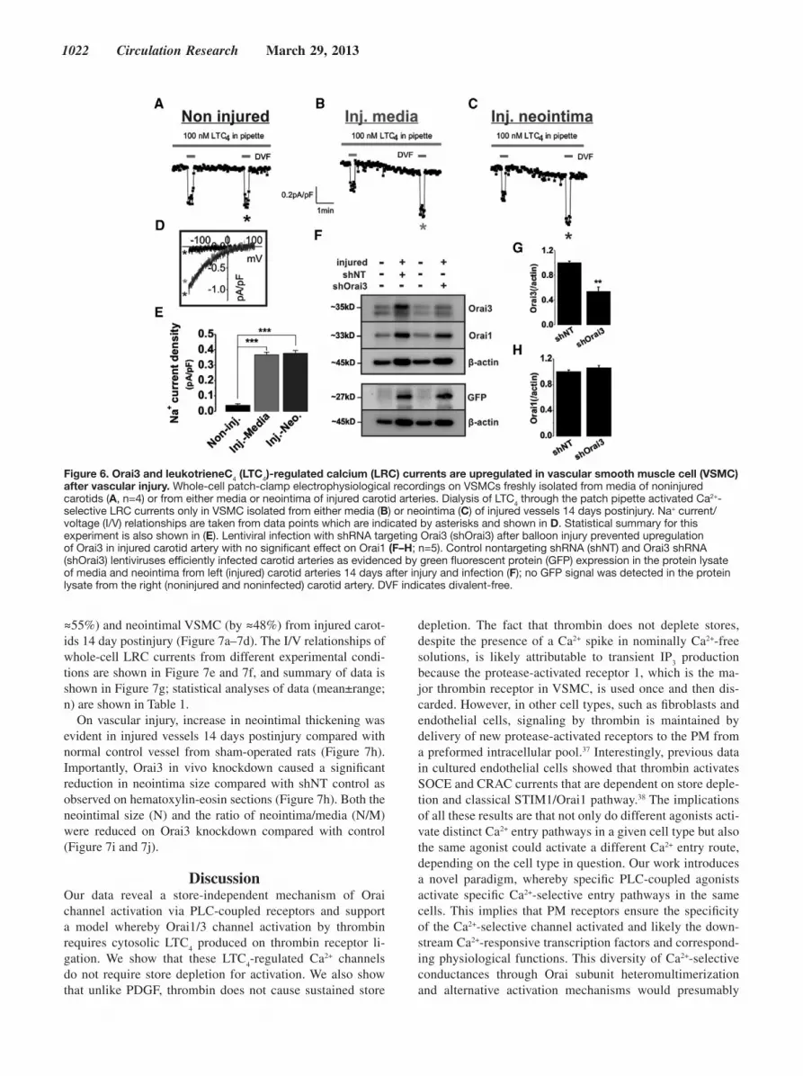
lationships of whole-cell LRC currents from these different types of cells are shown in Figure 6d, and summary of data is shown in Figure 6e; statistical analyses of data (mean±range; n) are depicted in Table 1. Similarly, we found that CRAC cur-rents, activated by store depletion with dialysis of 20 mmol/L BAPTA through the patch pipette, were evident in medial and
neointimal VSMC acutely isolated from injured carotid arter-ies, whereas medial VSMC from noninjured vessels showed no detectable CRAC currents (Online Figure VII). These re-sults are consistent with previous data reporting upregulation of Orai1 and SOCE on vascular injury.16,29,30
Injury of left rat carotid arteries also caused a significant upregulation of Orai3 proteins in lysates of medial and neo-intimal VSMCs on day 14 postinjury (Figure 6f) as well as that of Orai1 proteins as previously shown.29 Efficient vessel infection by shOrai3 or shNT-encoding lentiviruses was docu-mented by the expression of lentivirus-encoded green fluo-rescent protein (driven by a separate promoter) in medial and neointimal VSMCs from injured and infected left carotids 14 days postinjury (Figure 6f).
Orai3 Is Required for LRC Currents and Neointima Formation In VivoSurvival surgery involving transduction of injured carotid arteries of anesthetized animals with shOrai3 lentiviruses caused a significant attenuation of Orai3 protein upregula-tion on day 14 postinjury with no significant effect on Orai1 expression (Figure 6f–6h) by comparison with control shNT. This in vivo Orai3 knockdown corresponded functionally to a decrease in whole-cell LRC current densities in medial (by
Figure 5. Orai1 and Orai3 are required for leukotrieneC4 (LTC4)-regulated Ca2+ (LRC) currents. Whole-cell patch-clamp electrophysiology in vascular smooth muscle cells (VSMCs) infected with lentivirus carrying either shRNA against luciferase (shLuc), or shRNA targeting Orai1 (shOrai1). Orai1 knockdown completely abrogated LTC4-activated Na+ currents (B) as compared with control (A). Na+ current/voltage (I/V) relationships (C) confirm the requirement of Orai1 for LTC4-activated currents in VSMCs. Statistical analysis is shown in D. Representative Western blots showing that shRNA targeting Orai3 does not affect Orai1 protein levels (E, F), while significantly abrogating Orai3 protein expression (G, H). Whole-cell patch-clamp electrophysiology in VSMCs infected with lentivirus-encoding either nontargeting control shRNA (shNT) or shRNA targeting Orai3 (shOrai3). Orai3 knockdown completely abrogated LTC4-activated currents (J) as compared with control (I). The I/V relationships are shown in K. Statistical analysis of patch-clamp data is shown in L. Values for current densities represented as mean±range and number of independent recordings for shRNA Control, shRNA Orai1, and shRNA Orai3 are reported in Table 1. DVF indicates divalent-free.
1022 Circulation Research March 29, 2013
≈55%) and neointimal VSMC (by ≈48%) from injured carot-ids 14 day postinjury (Figure 7a–7d). The I/V relationships of whole-cell LRC currents from different experimental condi-tions are shown in Figure 7e and 7f, and summary of data is shown in Figure 7g; statistical analyses of data (mean±range; n) are shown in Table 1.
On vascular injury, increase in neointimal thickening was evident in injured vessels 14 days postinjury compared with normal control vessel from sham-operated rats (Figure 7h). Importantly, Orai3 in vivo knockdown caused a significant reduction in neointima size compared with shNT control as observed on hematoxylin-eosin sections (Figure 7h). Both the neointimal size (N) and the ratio of neointima/media (N/M) were reduced on Orai3 knockdown compared with control (Figure 7i and 7j).
DiscussionOur data reveal a store-independent mechanism of Orai channel activation via PLC-coupled receptors and support a model whereby Orai1/3 channel activation by thrombin requires cytosolic LTC
4 produced on thrombin receptor li-
gation. We show that these LTC4-regulated Ca2+ channels
do not require store depletion for activation. We also show that unlike PDGF, thrombin does not cause sustained store
depletion. The fact that thrombin does not deplete stores, despite the presence of a Ca2+ spike in nominally Ca2+-free solutions, is likely attributable to transient IP
3 production
because the protease-activated receptor 1, which is the ma-jor thrombin receptor in VSMC, is used once and then dis-carded. However, in other cell types, such as fibroblasts and endothelial cells, signaling by thrombin is maintained by delivery of new protease-activated receptors to the PM from a preformed intracellular pool.37 Interestingly, previous data in cultured endothelial cells showed that thrombin activates SOCE and CRAC currents that are dependent on store deple-tion and classical STIM1/Orai1 pathway.38 The implications of all these results are that not only do different agonists acti-vate distinct Ca2+ entry pathways in a given cell type but also the same agonist could activate a different Ca2+ entry route, depending on the cell type in question. Our work introduces a novel paradigm, whereby specific PLC-coupled agonists activate specific Ca2+-selective entry pathways in the same cells. This implies that PM receptors ensure the specificity of the Ca2+-selective channel activated and likely the down-stream Ca2+-responsive transcription factors and correspond-ing physiological functions. This diversity of Ca2+-selective conductances through Orai subunit heteromultimerization and alternative activation mechanisms would presumably
Figure 6. Orai3 and leukotrieneC4 (LTC4)-regulated calcium (LRC) currents are upregulated in vascular smooth muscle cell (VSMC) after vascular injury. Whole-cell patch-clamp electrophysiological recordings on VSMCs freshly isolated from media of noninjured carotids (A, n=4) or from either media or neointima of injured carotid arteries. Dialysis of LTC4 through the patch pipette activated Ca2+-selective LRC currents only in VSMC isolated from either media (B) or neointima (C) of injured vessels 14 days postinjury. Na+ current/voltage (I/V) relationships are taken from data points which are indicated by asterisks and shown in D. Statistical summary for this experiment is also shown in (E). Lentiviral infection with shRNA targeting Orai3 (shOrai3) after balloon injury prevented upregulation of Orai3 in injured carotid artery with no significant effect on Orai1 (F–H; n=5). Control nontargeting shRNA (shNT) and Orai3 shRNA (shOrai3) lentiviruses efficiently infected carotid arteries as evidenced by green fluorescent protein (GFP) expression in the protein lysate of media and neointima from left (injured) carotid arteries 14 days after injury and infection (F); no GFP signal was detected in the protein lysate from the right (noninjured and noninfected) carotid artery. DVF indicates divalent-free.

González-Cobos et al LTC4, Orai3, and Neointimal Hyperplasia 1023
help increase the repertoire of spatial cellular Ca2+ microdo-mains for the purpose of selective Ca2+ signaling in complex mammalian organisms.
Thrombin has been shown to have multiple pleiotropic effects: it impacts on VSMC contractility and proliferation and is a major contributor to vascular remodeling. Thrombin is produced massively after vascular injury and during de-velopment of atherosclerosis.39 The thrombin receptors protease-activated receptors are also upregulated in VSMCs during injury. In this study, we showed that Orai3, the unique component of thrombin-activated Orai1/3 channels, is up-regulated in synthetic VSMCs in vitro and in medial and neo-intimal VSMCs in vivo in a rat model of vascular injury. We also show that preventing Orai3 upregulation during carotid
injury using lentiviral particles encoding shRNA reverses the increase in Orai3 protein levels 14 days postinjury, in-hibits LTC
4-regulated calcium channel activation in medial
and neointimal VSMC, and inhibits neointima formation. We and others previously showed that STIM1 and Orai1 are important mediators in neointima formation.16,26–29 Orai3 or Orai3-containing channels could represent a potential target for treatment of VSMC remodeling during vascular occlu-sive diseases and might represent a better target than Orai1 or STIM1 because STIM1/Orai1-mediated CRAC is ubiqui-tous and prominently functional in many tissues, including immune cells and skeletal muscle; the major defects in Orai1-deficient patients and mice are severe immunodeficiency and skeletal muscle hypotonia.40
Figure 7. In vivo knockdown of Orai3 inhibits leukotrieneC4 (LTC)-regulated calcium (LRC) currents and neointima formation. Whole-cell patch-clamp electrophysiological recordings of vascular smooth muscle cells (VSMCs) freshly isolated from the media (A, B) or neointima (C, D) of injured carotid arteries 2 weeks postinjury and transduction treatment with viral particles carrying either a control shRNA sequence (shNT) or a sequence targeting Orai3 (shOrai3). As shown in Figure 6 for injured vessels, carotid vessels injured and treated with shNT are characterized by the emergence of a Ca2+-selective LRC current activated by intracytoplasmic LTC4 in both medial (A, N=4) and neointimal VSMCs (C, N=4). This Ca2+-selective LRC current was reduced on Orai3 knockdown with shRNA by ≈55% in medial cells (B, N=5) and ≈48% in neointimal cells (D, N=5). Na+ current/voltage (I/V) relationships are taken from traces, which are indicated by the color-coded signs and are shown in (E, F). Statistical summary is shown in (G). H, Hematoxylin and eosin staining on vessel cross-sections from control left carotid isolated from a sham-operated rat and from injured left carotid arteries infected with either control shNT or shOrai3 (scale bar =200 µm). Fourteen days after injury, neointimal growth was evident in left carotids injured and infected with shNT compared with left control noninjured vessels from sham-operated animals. This neointima (N) was visibly inhibited by shOrai3 as compared with shNT. I, The neointimal (N), intimal (I), and medial (M) areas of the carotid cross-sections measured from left-injured and virus-treated carotids and from right noninjured and noninfected carotids from the same animals using Image J software and statistical analyses on areas (mm2) are shown. The media/neointima (N/M) ratios for left-injured and virus-treated carotids or intima/media (I/M) ratios of right noninjured and noninfected right carotids from the same animals (I) from 5 independent rats per condition are shown. Statistics on Western blots of medial and neointimal VSMCs and quantification of neointima were performed on 5 rats per condition. DVF indicates divalent-free.

1024 Circulation Research March 29, 2013
We show that STIM1 is required downstream of LTC4 ac-
tion during the activation of this novel LTC4-regulated cal-
cium channel. This fact, along with the high Ca2+ selectivity of this channel, suggests that STIM1 might be a component of the Orai1/3 channel complex. Indeed, recent data from the Prakriya group showed that STIM1 endows, otherwise nonse-lective, Orai Ca2+ channels with high Ca2+ selectivity.41 Several questions remain to be answered by future structural studies as follows: how LTC
4 triggers Orai1/3 channel activation and
whether this is a direct action? If it is through direct action, what are the domains in STIM/Orai that are involved in this interaction? What is the exact stoichiometry of LTC
4-activated
channels in VSMC? The answer to these questions and others will likely help in the targeting of this channel for the purpose of therapy.
Sources of FundingThis work was mainly supported by grant HL097111 from National Institutes of Health (NIH) to M. Trebak, in part by NIH grant HL095566 to K. Matrougui, and Austrian Science Fund (FWF) grants P22747 to R. Schindl and P22565 to C. Romanin.
DisclosuresNone.
References 1. Berridge MJ. Calcium microdomains: organization and function. Cell
Calcium. 2006;40:405–412. 2. Clapham DE. Calcium signaling. Cell. 2007;131:1047–1058. 3. Putney JW Jr. A model for receptor-regulated calcium entry. Cell
Calcium. 1986;7:1–12. 4. Bird GS, Aziz O, Lievremont JP, Wedel BJ, Trebak M, Vazquez G,
Putney JW Jr. Mechanisms of phospholipase C-regulated calcium entry. Curr Mol Med. 2004;4:291–301.
5. Hoth M, Penner R. Depletion of intracellular calcium stores activates a calcium current in mast cells. Nature. 1992;355:353–356.
6. Roos J, DiGregorio PJ, Yeromin AV, Ohlsen K, Lioudyno M, Zhang S, Safrina O, Kozak JA, Wagner SL, Cahalan MD, Veliçelebi G, Stauderman KA. STIM1, an essential and conserved component of store-operated Ca2+ channel function. J Cell Biol. 2005;169:435–445.
7. Liou J, Kim ML, Heo WD, Jones JT, Myers JW, Ferrell JE Jr, Meyer T. STIM is a Ca2+ sensor essential for Ca2+-store-depletion-triggered Ca2+ influx. Curr Biol. 2005;15:1235–1241.
8. Feske S, Gwack Y, Prakriya M, Srikanth S, Puppel SH, Tanasa B, Hogan PG, Lewis RS, Daly M, Rao A. A mutation in Orai1 causes immune deficiency by abrogating CRAC channel function. Nature. 2006;441:179–185.
9. Vig M, Peinelt C, Beck A, Koomoa DL, Rabah D, Koblan-Huberson M, Kraft S, Turner H, Fleig A, Penner R, Kinet JP. CRACM1 is a plasma membrane protein essential for store-operated Ca2+ entry. Science. 2006;312:1220–1223.
10. Prakriya M, Feske S, Gwack Y, Srikanth S, Rao A, Hogan PG. Orai1 is an essential pore subunit of the CRAC channel. Nature. 2006;443:230–233.
11. Soboloff J, Madesh M, Gill DL. Sensing cellular stress through STIM proteins. Nat Chem Biol. 2011;7:488–492.
12. Park CY, Hoover PJ, Mullins FM, Bachhawat P, Covington ED, Raunser S, Walz T, Garcia KC, Dolmetsch RE, Lewis RS. STIM1 clusters and activates CRAC channels via direct binding of a cytosolic domain to Orai1. Cell. 2009;136:876–890.
13. Yuan JP, Zeng W, Dorwart MR, Choi YJ, Worley PF, Muallem S. SOAR and the polybasic STIM1 domains gate and regulate Orai channels. Nat Cell Biol. 2009;11:337–343.
14. Muik M, Fahrner M, Derler I, Schindl R, Bergsmann J, Frischauf I, Groschner K, Romanin C. A cytosolic homomerization and a modulatory
domain within stim1 c-terminus determine coupling to orai1 channels. The Journal of biological chemistry. 2009
15. Kawasaki T, Lange I, Feske S. A minimal regulatory domain in the C terminus of STIM1 binds to and activates ORAI1 CRAC channels. Biochem Biophys Res Commun. 2009;385:49–54.
16. Potier M, Gonzalez JC, Motiani RK, Abdullaev IF, Bisaillon JM, Singer HA, Trebak M. Evidence for STIM1- and Orai1-dependent store-operat-ed calcium influx through ICRAC in vascular smooth muscle cells: role in proliferation and migration. FASEB J. 2009;23:2425–2437.
17. Trebak M. PLC: Johnny-come-lately to ORAI and the ups and downs of calcium signalling. J Physiol (Lond). 2011;589:5337–5338.
18. Motiani RK, Abdullaev IF, Trebak M. A novel native store-operated cal-cium channel encoded by Orai3: selective requirement of Orai3 versus Orai1 in estrogen receptor-positive versus estrogen receptor-negative breast cancer cells. J Biol Chem. 2010;285:19173–19183.
19. Motiani RK, Zhang X, Harmon KE, Keller RS, Matrougui K, Bennett JA, Trebak M. Orai3 is an estrogen receptor alpha-regulated Ca2+ chan-nel that promotes tumorigenesis. FASEB Journal: Official Publication of the Federation of American Societies for Experimental Biology. 2013;27:63–75.
20. Shuttleworth TJ. Arachidonic acid, ARC channels, and Orai proteins. Cell Calcium. 2009;45:602–610.
21. Mignen O, Thompson JL, Shuttleworth TJ. Both Orai1 and Orai3 are es-sential components of the arachidonate-regulated Ca2+-selective (ARC) channels. J Physiol (Lond). 2008;586:185–195.
22. Mignen O, Thompson JL, Shuttleworth TJ. STIM1 regulates Ca2+ entry via arachidonate-regulated Ca2+-selective (ARC) channels without store depletion or translocation to the plasma membrane. J Physiol (Lond). 2007;579:703–715.
23. Trebak M, Vazquez G, Bird GS, Putney JW Jr. The TRPC3/6/7 subfam-ily of cation channels. Cell Calcium. 2003;33:451–461.
24. Yuan JP, Zeng W, Huang GN, Worley PF, Muallem S. STIM1 heteromul-timerizes TRPC channels to determine their function as store-operated channels. Nat Cell Biol. 2007;9:636–645.
25. House SJ, Potier M, Bisaillon J, Singer HA, Trebak M. The non-excit-able smooth muscle: calcium signaling and phenotypic switching during vascular disease. Pflugers Arch. 2008;456:769–785.
26. Berra-Romani R, Mazzocco-Spezzia A, Pulina MV, Golovina VA. Ca2+ handling is altered when arterial myocytes progress from a contrac-tile to a proliferative phenotype in culture. Am J Physiol Cell Physiol. 2008;295:C779–C790.
27. Guo RW, Wang H, Gao P, Li MQ, Zeng CY, Yu Y, Chen JF, Song MB, Shi YK, Huang L. An essential role for stim1 in neointima formation following arterial injury. Cardiovasc Res. 2009;81:660–668.
28. Aubart FC, Sassi Y, Coulombe A, Mougenot N, Vrignaud C, Leprince P, Lechat P, Lompré AM, Hulot JS. RNA interference targeting STIM1 suppresses vascular smooth muscle cell proliferation and neointima for-mation in the rat. Mol Ther. 2009;17:455–462.
29. Zhang W, Halligan KE, Zhang X, Bisaillon JM, Gonzalez-Cobos JC, Motiani RK, Hu G, Vincent PA, Zhou J, Barroso M, Singer HA, Matrougui K, Trebak M. Orai1-mediated I (CRAC) is es-sential for neointima formation after vascular injury. Circ Res. 2011;109:534–542.
30. Bisaillon JM, Motiani RK, Gonzalez-Cobos JC, Potier M, Halligan KE, Alzawahra WF, Barroso M, Singer HA, Jourd’heuil D, Trebak M. Essential role for STIM1/Orai1-mediated calcium influx in PDGF-induced smooth muscle migration. Am J Physiol Cell Physiol. 2010;298:C993–1005.
31. Martorell L, Martínez-González J, Rodríguez C, Gentile M, Calvayrac O, Badimon L. Thrombin and protease-activated receptors (PARs) in atherothrombosis. Thromb Haemost. 2008;99:305–315.
32. Raines EW. PDGF and cardiovascular disease. Cytokine Growth Factor Rev. 2004;15:237–254.
33. Hirano K. The roles of proteinase-activated receptors in the vascu-lar physiology and pathophysiology. Arterioscler Thromb Vasc Biol. 2007;27:27–36.
34. Prakriya M, Lewis RS. Separation and characterization of currents through store-operated CRAC channels and Mg2+-inhibited cation (MIC) channels. J Gen Physiol. 2002;119:487–507.
35. McIntyre TM, Zimmerman GA, Prescott SM. Leukotrienes C4 and D4 stimulate human endothelial cells to synthesize
González-Cobos et al LTC4, Orai3, and Neointimal Hyperplasia 1025
platelet-activating factor and bind neutrophils. Proc Natl Acad Sci USA. 1986;83:2204–2208.
36. Coughlin SR. Protease-activated receptors in hemostasis, thrombosis and vascular biology. J Thromb Haemost. 2005;3:1800–1814.
37. Coughlin SR. Thrombin signalling and protease-activated receptors. Nature. 2000;407:258–264.
38. Abdullaev IF, Bisaillon JM, Potier M, Gonzalez JC, Motiani RK, Trebak M. Stim1 and Orai1 mediate CRAC currents and store-operated calcium entry important for endothelial cell proliferation. Circ Res. 2008;103:1289–1299.
39. Schrör K, Bretschneider E, Fischer K, Fischer JW, Pape R, Rauch BH, Rosenkranz AC, Weber AA. Thrombin receptors in vascular smooth muscle cells - function and regulation by vasodilatory prostaglandins. Thromb Haemost. 2010;103:884–890.
40. Feske S. Calcium signalling in lymphocyte activation and disease. Nat Rev Immunol. 2007;7:690–702.
41. McNally BA, Somasundaram A, Yamashita M, Prakriya M. Gated regulation of CRAC channel ion selectivity by STIM1. Nature. 2012;482:241–245.
What Is Known?
• The ubiquitous store-operated Orai1 Ca2+ channels show low expres-sion in healthy quiescent vascular smooth muscle cells (VSMC), but their expression is increased during VSMC remodeling in a proliferative migratory phenotype.
• Activation of VSMC with the platelet-derived growth factor, a VSMC mitogen, activates Orai1-mediated Ca2+ entry through a mechanism involving endoplasmic reticulum Ca2+ store depletion and subsequent interaction of the Ca2+ sensor stromal interacting molecule1 with Orai1.
• Molecular knockdown of Orai1 inhibits neointima formation in re-sponse to balloon injury in rat carotid arteries.
What New Information Does This Article Contribute?
• VSMC activation by thrombin activates a novel Ca2+ entry pathway that requires both Orai1 and its homolog Orai3. This pathway requires stromal interacting molecule1 but does not depend on endoplasmic reticulum Ca2+ store depletion.
• This novel Ca2+ entry pathway in VSMC is mediated by the intracrine actions of leukotriene C
4 (LTC
4) produced downstream of thrombin re-
ceptor stimulation.• Balloon injury of rat carotid arteries increased Orai3 expression and
manifestation of Ca2+ currents activated by LTC4 dialysis in medial
and neointimal VSMC; healthy medial VSMC show no LTC4-activated
currents.
• Knockdown of Orai3 in balloon-injured carotid arteries using lentivirus-encoding shRNA prevents Orai3 upregulation, inhibits LTC
4-activated
currents, and decreases neointima formation.
Orai1, a store-operated Ca2+ channel activated by ligation of phospholipase C-coupled receptors, is required for neointima formation on vascular injury. However, Orai1 is functional in many cell types and tissues, which could complicate its use as a spe-cific target in VSMC-related pathologies. Orai1 has 2 homologs Orai2 and Orai3; Orai2 is expressed exclusively in vertebrates, whereas Orai3 is expressed exclusively in mammals. The role of Orai2 and Orai3 in the vascular system remained unknown. We describe a new role of Orai3 in VSMC Ca2+ signaling and remodeling. We show that Orai3 contributes to a novel hetero-meric Orai1/3 Ca2+ entry channel in thrombin-activated VSMC. We found that Orai1/3 channel activation is store independent and mediated by cytosolic LTC
4 produced downstream throm-
bin receptor. Furthermore, Orai3 expression and LTC4-activated
channel activity increase in VSMC on rat carotid artery injury, whereas knockdown of Orai3 in injured carotids inhibits Orai3 upregulation, LTC
4-activated channels, and neointima formation.
These findings suggest that Orai3 represents a novel drug target for controlling VSMC remodeling during vascular injury or dis-ease, and that Orai3 may be a better target than the ubiquitous Orai1 channel.
Novelty and Significance