stereotactic high dose fraction radiation therapy of extracranial tumors using an accelerator:...
TRANSCRIPT

Acta Oncologica Vol. 34, No. 6, pp. 861-870, 1995
STEREOTACTIC HIGH DOSE FRACTION RADIATION THERAPY OF
EXTRACRANIAL TUMORS USING AN ACCELERATOR
Clinical experience of the first thirty-one patients
HENRIC BLOMGREN, INGMAR LAX, INGEMAR NASLUND and RUT SVANSTROM
A stereotactic body frame with a fixation device has been developed for stereotactic radiation therapy of extracranial targets, a precision localization and positioning system in analogy with the stereotactic head frames used for intracranial targets. Results of the first 42 treated tumors in 31 patients are presented. Most of the patients had solitary tumors in liver, lung or retroperitoneal space. Clinical target volumes ranged from 2 to 622 cm3 (mean 78 cm3) and minimum doses to the planning target volumes (PTV) of 7.7-30 Gy/fraction (mean 14.2 Gy) were given on 1-4 occasions to a total minimum dose to the PTVs of 7.7-45 Gy (mean 30.2 Gy) to the periphery of the PTV and total mean doses to the PTVs of 8-66 Gy (mean 41 Gy). The central part of the tumor was usually given about 50% higher dose compared to that of the periphery of the PTV by a planned inhomogeneous dose distribution. Some of the patients received stereotactic radiation therapy concomitantly to more than one target, in others new metastases were also treated which appeared during the follow-up period. We observed a local rate of no progressive disease of 80% during a follow-up period of 1.5-38 months. Fifty percent of the tumors decreased in size or disappeared.
Stereotactic radiation therapy (SRT) of brain metastases with high single doses was first given in 1975 at the Karolinska Hospital using a multi-cobalt-60 unit (gamma knife) (1). At present several thousand patients with in- tracranial metastases have been treated using this tech- nique in different centers of the world ( 1-4). Local tumor control rate is around 90%. This SRT technique can frequently be used for treatment of inoperable tumors and those which are considered to be resistant to convention- ally fractionated radiation. There is no other known treat- ment modality for solitary brain metastases which seems to
Accepted 21 June 1995. From the Department of Oncology (H. Blomgren, I. Naslund), Department of Hospital Physics (I. Lax) and Department of Diagnostic Radiology (R. Svanstrom) Karolinska Hospital, Stockholm, Sweden. Correspondence to: Henric Blomgren, Department of Oncology, Karolinska Hospital, S-171 76 Stockholm, Sweden.
be able to compete with stereotactic high single dose radiation therapy with respect to tumor control rate, sur- vival and side-effects.
A prerequisite for gamma knife treatment is a stereotac- tic frame, firmly attached to the skull of the patient, which allows exact localization of the lesion and use during set-up in the multi-cobalt unit. Similar stereotactic systems have been used for SRT of intracranial lesions using accelerators (5-7). From the results reported, this treat- ment seems to be as effective as gamma knife treatment of intracranial metastases (8- 1 1). A stereotactic body frame has now been developed for SRT of extracranial targets using an accelerator. This stereotactic system allows multi- ple non-coplanar beams to be used for the treatment. Details of the system and examples of treatment tech- niques and dose distributions have been described previ- ously ( 12).
In the present article we give a detailed presentation of the results of the first 31 patients treated with this new technique.
0 Scandinavian University Press 1995. ISSN 0284-186X 86 1
Act
a O
ncol
Dow
nloa
ded
from
info
rmah
ealth
care
.com
by
117.
165.
19.1
58 o
n 05
/20/
14Fo
r pe
rson
al u
se o
nly.

862 H. BLOMGREN ET AL. Acta Oncologica 34 (1995)
Material and Methods
Patients. The material to be presented represents the first consecutive 3 1 patients who received stereotactic high-dose radiation therapy for extracranial lesions using an accelera- tor. The first treatment was given in September 1991 and the last treatment in this patient population in February 1995. The patients have been numbered in chronological order (patient No. 1-31) and each treated tumor of a given patient has been defined by a letter (A, B, C, D, E).
The majority of the patients had previously received treatment for various types of primary tumors and there- after developed distant metastases which were treated with SRT. In some patients the primary tumor was treated. At the time of treatment most patients had one known lesion which had increased in size as assessed by repeated chest and abdominal computerized tomography (CT) scans. Biopsies for histopathological examinations were obtained from some patients during open surgery and from others by means of needle aspirations. The majority of the irradi- ated lesions were considered so typical for metastatic
spread that attempts were not made to verify the diagnosis morphologically.
Age and sex of the patients as well as type of primary tumor, site, volume of the treated tumor, details concerning dosage and tumor responses are presented in Tables 1-4. Patient No. 14, who was a 72-year-old woman with a breast cancer metastasis to the liver, is not presented, due to the fact that the clinical effects of the irradiation could not be evaluated since chemotherapy was started shortly after treatment.
Radiation therapy Stereotactic body-frame. The stereotactic body-frame
(marketed by Precision Therapy International) was de- scribed in detail in a previous paper (12). In short it consists of a stereotactic frame that covers the body in- cluding the head in cranial direction and to the thighs in the caudal direction. There are indicators mounted on the frame that are visible on CT images, thus defining the stereotactic system. The indicators can be changed to magnetic resonance (MR) indicators as well. The fixation
Table 1
Primary intrahepatic
Patient No.'
5 7A 7B 8 10
Gender (M/F) Age (years) Primary tumorZ
Site of irradiated tumor
Volume of clinical target (cm')
Minimum dose in PTV (Gy) Mean dose in PTV (Gy) ICRU reference dose (Gy)/
Number of fractions Treatment period (d)/
Size of tumor after
Time period after
Follow-up period of
Reason for discontinuation
maximum dose in PTV (Gy)
time between fractions (d)
irradiation
irradiation s month^)^
irradiated tumor (months)
of follow-up of irradiated tumor
(months) Survival after irradiation
M 70 HCC
Left lobe
229
30142
48/51
1 1 lo
Patient died 2 days after irradiation 0
F 62 HCC
Ventral Left liver edge liver edge Concomitant treatment 15 5
30/34 15/16
34/35 17/17
2 1 44/44 1 lo
Disappeared Disappeared
22-38 3-38
38 38
39 +
M 65 HCC
Left lobe
57
45/66
69/71
3 34/ 17
Formation of ascitic fluid. Deterioration 1.5
' A, B, C, D represent different irradiated tumors in the same patient. ' HCC = hepatocellular cancer.
Time period during which the change of tumor size, as indicated above, was observed. + =the patient is still alive.
F 61 Bile duct cancer Hilus
67
45/63
79/82
3 301 15
Unchanged
1.5-11
1 1
Multiple new liver tumors 14
Act
a O
ncol
Dow
nloa
ded
from
info
rmah
ealth
care
.com
by
117.
165.
19.1
58 o
n 05
/20/
14Fo
r pe
rson
al u
se o
nly.
Acta Oncologica 34 (1 995) STEREOTACTIC RADIOTHERAPY 863
of the patient to the frame is obtained by a vacuum pillow or a foam. For set-up of isocenter coordinates in the treatment room, scales mounted on the frame, correspond- ing to the CT/MR indicators are used. Due to mechanical imperfections of the treatment tables of most accelerators it is important to be able to level the frame to the horizontal plane. For this purpose a level mechanism has been added to the stereotactic frame. The size of the frame allows for use in CT, MR and positron emission tomogra- phy (PET) scanners with an aperture of 55 cm or more.
For targets affected by the diaphragmatic motions due to breathing, a device was developed to minimize the motions by applying a pressure on the abdomen. The diaphragmatic motions were checked by fluoroscopy be- fore and after the diaphragmatic control device was ap- plied. For the patients in the present study the diaphragmatic motions were 1 cm or less (i.e. f 5 mm from the average position), at normal breathing.
The reproducible position of the patient in the frame is obtained by the vacuum pillow and two markers; chest marker and tibia marker. These markers are set to pre-
defined stereotactic coordinates, and at these coordinates the markers should correspond to tattoo-points on the skin over sternum and tibia which are used to ensure a high reproducibility in the cranial-caudal direction. For the patients in the present study, 72 separate CT examinations have been performed with the patient in treatment position to determine the reproducibility of the target in the stereotactic system. This check thus includes the internal motion of the tumor within the body as well as the differences in position of the body (e.g. skeleton) in the stereotactic system. For the total number of CT examina- tions the mean deviation of the tumor compared with the first CT (before dose planning) was 3.7 mm (95% within 5 mm) in the transversal plane, and 5.7 mm in mean (89% within 8 mm) in the cranial-caudal direction. The slice thickness of the CT images was generally IOmm, thus differences less than around 5mm have been difficult to measure. Based on the measurements of the first patients a margin around the tumor, visible on CT and MR images, was added for set-up errors and internal target motion of 5 mm in the transversal plane and 10 mm in the cranial-
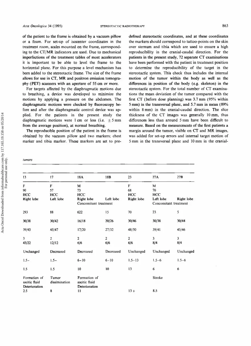
tumors
13 17 18A 18B 23 27A 27B
F 90 HCC Right lobe
F 57 HCC Left lobe
M 73 HCC Right lobe Left lobe Concomitant treatment
F 68 HCC Right lobe
M 70 HCC Left lobe Right lobe Concomitant treatment
293 88 622 15 70 23 5
30138 30142 16/18 20126 30146 30138 30144
39/43 45/47 17/20 27/32 48/50 39/41 45/46
3 43/22
2 12/12
2 616
3 3 814 814
Unchanged
1.5-
Decreased Decreased Decreased Unchanged Unchanged Unchanged
1.5- 6- 10 6- 10 1.5-13 1.5-6 1.5-6
1.5 1.5 10 10 13 6 6
Formation of ascitic fluid Deterioration 2.5
Tumor dissimination
Formation of ascitic fluid Deterioration I 1
Stroke
8 13 + 8.5
Act
a O
ncol
Dow
nloa
ded
from
info
rmah
ealth
care
.com
by
117.
165.
19.1
58 o
n 05
/20/
14Fo
r pe
rson
al u
se o
nly.

864 H. BLOMGREN ET AL. Acta Oncologica 34 (1995)
Table 2 Liver
Patient No.
1A IB 2 3 4 I I 12 15
Gender (M/F) M M F F F F F Age (years) 53 70 51 51 49 56 80 Primary tumor Colonic Colonic Colonic Colonic Basaloid cancer Ovarian Colonic
Site of irradiated Right lobe Hilus Right lobe Right lobe Left lobe Right lobe Right lobe Right lobe cancer cancer cancer cancer of the anal canal cancer cancer
tumor Concomitant treatment
Volume of clinical I56 target (cm3)
Minimum dose in PTV (Gy)/ mean dose in PTV (Gy)
ICRU Reference dose (Gy)/ maximum dose in PTV (Gy)
Number of fractions 1 Treatment period (d)/ 1 10
lOjl0.5
l0.5jl I
time between fractions (d)
irradiation (months)
irradiation (months)
irradiated tumor (months)
of follow-up of irradiated dissimination tumor
(months)
Size of tumor after Increased
Time period after 4-
Follow-up period of 4
Reason for discontinuation Tumor
Survival after irradiation 6
258
1.718
819
1 1 10
Increased
4-
4
88
2012 I
2 1/22
1 I 10
Increased
1.5-9
9
Tumor dissimination
16
184
16/22
27/28
1 1 I0
Increased
1.5-6
6
Tumor dissimination
12
181
40157
65/68
2 72/72
Increased
4-9
9
Tumor dissimination
24.5
29
40156
62/64
2 11/11
Unchanged
1.5-9
9
Pelvic recurrence
17
13 11
4 / 5 3 @/m
56/61 64/66
2 2 15/15 21/21
Disappeared Disappeared
3-23 5-11
23 1 1
Traumatic femure fracture
26 + 13
Table 3 Tumors metastatic to the thoracic cavity
Patient No.
6 9 16C* 21 24A 248
Gender (M/F) Age (years) Primary tumor
Site of irradiated tumor
Volume of clinical target (cm’)
Minimum dose in PTV (Gy)/ mean dose in PTV (Gy)
ICRU reference dose (Gy)/ maximum dose in PTV (Gy)
Number of fractions Treatment period (d) /
Size of tumor after
Time period after
Follow-up period of
Reason for discontinuation
time between fractions (d)
irradiation
irradiation (months)
irradiated tumor (months)
of follow-up of irradiated tumor
(months) Survival after irradiation
F 75 Thyroid cancer Right lung
76
20127
30132
1 1 10
Unchanged
1.5-3.5
3.5
Tumor dissimination
I
F 69 HCC
Right lung
3
20/26
26/26
2 22/22
Decreased
1.5-9
9
Tumor dissimination
I 1
F 80 Colonic cancer Left lung
82
30141
42/44
2 414
Decreased
1.5-12
12
I3 +
M 70 Kidney cancer Right lung
11
40160
64/70
2 14/14
Decreased
4-7
7
Tumor dissimination
I
M 50 Colonic cancer Right lung
198
30140
39/42
3 1417
Decreased
1-11
11
I2 +
Mediastinal tumor at 6 months 15
20128
3013 I
2 4/4
Decreased
1.5-5
5
* Treatment was given 5 months after primary irradiation of hepatic and retroperitoneal tumors.
Act
a O
ncol
Dow
nloa
ded
from
info
rmah
ealth
care
.com
by
117.
165.
19.1
58 o
n 05
/20/
14Fo
r pe
rson
al u
se o
nly.
Acta Oncologica 34 ( 1995)
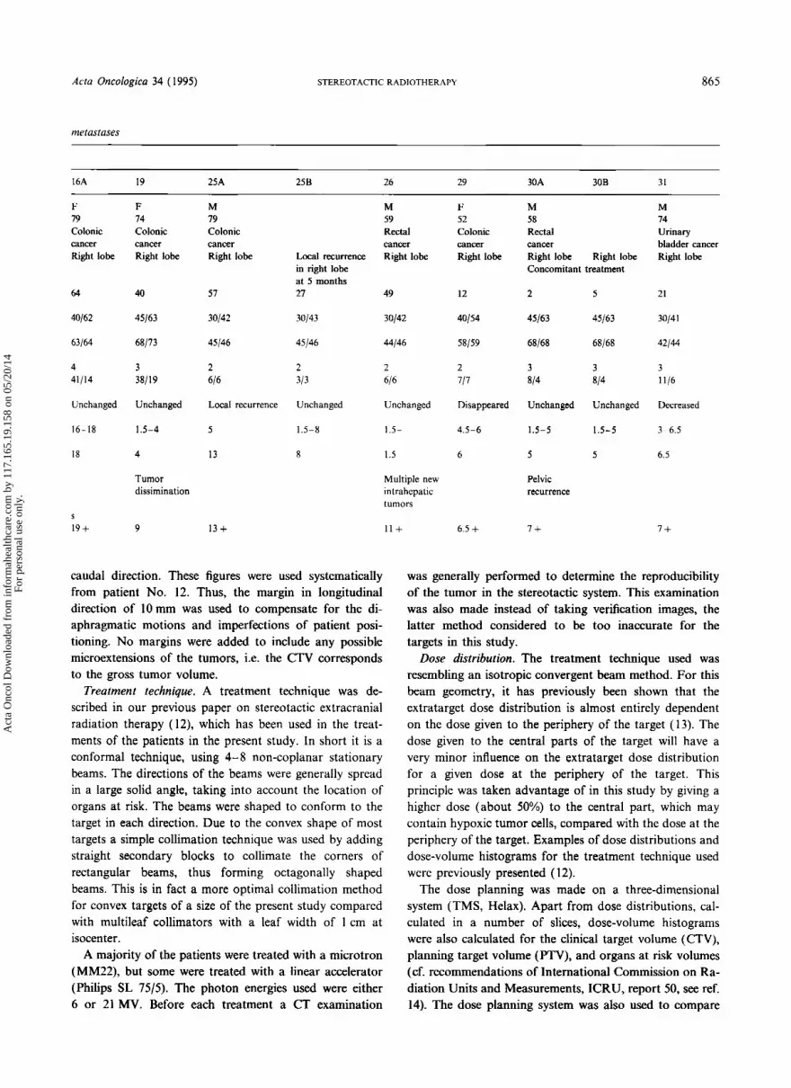
metastases
STEREOTACTIC RADIOTHERAPY 865
16A 19 25A 258 26 29 30A 30B 31
F 19 Colonic cancer Right lobe
64
40162
63/64
4 41/14
Unchanged
16-18
18
S
I 9 +
F 14 Colonic cancer Right lobe
40
45/63
68/13
3 38/19
Unchanged
1.5-4
4
Tumor dissirnination
9
M 19 Colonic cancer Right lobe
51
30142
45/46
2 616
Local recurrence
5
13
I 3 +
Local recurrence in right lobe at 5 months 21
30143
45/46
2 313
Unchanged
1.5-8
8
caudal direction. These figures were used systematically from patient No. 12. Thus, the margin in longitudinal direction of 10mm was used to compensate for the di- aphragmatic motions and imperfections of patient posi- tioning. No margins were added to include any possible microextensions of the tumors, i.e. the CTV corresponds to the gross tumor volume.
Treatment technique. A treatment technique was de- scribed in our previous paper on stereotactic extracranial radiation therapy ( 12), which has been used in the treat- ments of the patients in the present study. In short it is a conformal technique, using 4-8 non-coplanar stationary beams. The directions of the beams were generally spread in a large solid angle, taking into account the location of organs at risk. The beams were shaped to conform to the target in each direction. Due to the convex shape of most targets a simple collimation technique was used by adding straight secondary blocks to collimate the corners of rectangular beams, thus forming octagonally shaped beams. This is in fact a more optimal collimation method for convex targets of a size of the present study compared with multileaf collimators with a leaf width of 1 cm at isocenter.
A majority of the patients were treated with a microtron (MM22), but some were treated with a linear accelerator (Philips SL 75/5). The photon energies used were either 6 or 21 MV. Before each treatment a CT examination
M 59 Rectal cancer Right lobe
49
30142
44/46
2 616
Unchanged
1.5-
1.5
Multiple new intrahepatic tumors
I I +
F 52 Colonic cancer Right lobe
12
40154
58/59
2 717
Disappeared
4.5-6
6
6.5 +
M 58 Rectal cancer Right lobe Right lobe Concomitant treatment
2
45/63
68/68
3 814
Unchanged
1.5-5
5
Pelvic recurrence
I +
5
45/63
68/68
3 814
Unchanged
1.5-5
5
M 14 Urinary bladder cancer Right lobe
21
30141
42/44
3 1116
Decreased
3-6.5
6.5
l +
was generally performed to determine the reproducibility of the tumor in the stereotactic system. This examination was also made instead of taking verification images, the latter method considered to be too inaccurate for the targets in this study.
Dose distribution. The treatment technique used was resembling an isotropic convergent beam method. For this beam geometry, it has previously been shown that the extratarget dose distribution is almost entirely dependent on the dose given to the periphery of the target (13). The dose given to the central parts of the target will have a very minor influence on the extratarget dose distribution for a given dose at the periphery of the target. This principle was taken advantage of in this study by giving a higher dose (about 50%) to the central part, which may contain hypoxic tumor cells, compared with the dose at the periphery of the target. Examples of dose distributions and dose-volume histograms for the treatment technique used were previously presented (12).
The dose planning was made on a three-dimensional system (TMS, Helax). Apart from dose distributions, cal- culated in a number of slices, dose-volume histograms were also calculated for the clinical target volume (CTV), planning target volume (FTV), and organs at risk volumes (cf. recommendations of International Commission on Ra- diation Units and Measurements, ICRU, report 50, see ref. 14). The dose planning system was also used to compare
Act
a O
ncol
Dow
nloa
ded
from
info
rmah
ealth
care
.com
by
117.
165.
19.1
58 o
n 05
/20/
14Fo
r pe
rson
al u
se o
nly.

866 H BLOMGREN ET AL.
Table 4 Retroperitoneul and skeletal metastases
Actn Oncologicu 34 ( 1995)
Patient No
7 c * 16B** 16D 16E 20 22 28
Gender (M/F) Age (years) Primary tumor
Site of irradiated tumor
Volume of clinical target (cm’)
Minimum dose in PTV (Gy)/ mean dose in PTV (Gy)
ICRU reference dose (Gy)/ maximum dose in PTV (Gy)
Number of fractions Treatment period (d ) /
Sire of tumor after
Time period after
Follow-up period of
Reason for discontinuation
time between fractions (d)
irradiation
irradiation (months)
irradiated tumor (months)
of follow-up of irradiated tumor
F 62 HCC
Lymph node at truncus coeliacus
51
30/48
54/54
2 44/44
Disappeared
21 ~ 38
38
Survival after irradiation 39 t (months)
F 79 Colonic cancer Right kidney Hilus
25
30/44
44/48
3 23/14
Disappeared
1.5- 18
18
I9 +
New tumor in left adrenal gland 8 months after first irradiation 5
30/4 I
45/46
3 714
Disappeared
3.5 I0
10
* Two tumors in the liver were irradiated concomitantly (see Table I ) . ** One tumor in the liver was irradiated concomitantly (see Table 2).
different CT examinations to determine the reproducibility of the target in the stereotactic system (12).
Treatments were given in 1-4 fractions, with a high dose to the PTV in each fraction. This temporal pattern of dose delivery is not optimal with regard to biological effects expected from alp-ratios of most tumors and those of normal tissues, especially late reacting ones. However, the steep dose gradient outside the tumor makes the dose/frac- tion to the surrounding tissues acceptable.
Several factors were taken into consideration when de- termining the dose/fraction and interval between the irra- diations. These include size of the CTV, dose distribution with dose-volume histograms and radiation sensitivity of surrounding normal tissues. The treatment method was somewhat modified as this study progressed. For instance, we have changed from one single fraction towards two or usually three fractions, the total treatment period has been shortened. Also the margin due to the internal target motion and set-up errors have been determined and ap- plied systematically during the study.
Follow -up after treatment and dejinition of response. CT-scans were performed at 6-9-week intervals following
M 55 Rectal cancer
New tumor at Sacral abdominal aorta vertebrae 17 months after SILS3 first irradiation
34 226
30/42 30/32
45/45 32/33
3 3 7/4 14/7
Decreased Local
1.5- 5 recurrence
1.5 9
Tumor dissimination
12 t
F F 39 78 Malignant Thyroid melanoma cancer Paraaortal Lumbosacral lymph node
27 78
30/37 30141
39/40 42/45
2 3 6/6 7/4
Decreased Unchanged
1.5-4 1.5-9
5 9
Progression of irradiated tumor at 5 months. Tumor dissimination 10 9 1
SRT. The follow-up was discontinued in patients who deteriorated or developed generalized disease which re- quired chemotherapy. Only changes of the size of a tumor were used to describe its response to the stereotactic radia- tion therapy. They were grossly classified as the product of the two largest perpendicular tumor diameters and com- pared with the pretreatment value. Increased = more than 120% of the pretreatment value, unchanged = 120-50% of pretreatment value, decreased = < 50% of pretreatment value but the tumor had not disappeared, disap- peared =the tumor can no longer be visualized on CT- scans. Local recurrence has been used to indicate the appearance of new tumors in the immediate vicinity of the irradiated target or at its border. Although local recur- rences may represent new metastases they have been classified as local failures.
The time period after the last radiation session when a tumor was observed to have decreased in size, as well as its duration, are indicated in the tables. This time period has not been indicated for tumors which disappeared after irradiation but only when this event was first noted and its duration.
Act
a O
ncol
Dow
nloa
ded
from
info
rmah
ealth
care
.com
by
117.
165.
19.1
58 o
n 05
/20/
14Fo
r pe
rson
al u
se o
nly.

Acta Oncologica 34 ( 1995) STEREOTACTIC RADIOTHERAPY 867
Results
Radiation therapy ofprimary intrahepatic tumors (Table 1 )
Eight patients with morphologically proven hepatocellu- lar cancer (HCC) and one with an intrahepatic bile duct cancer were treated. Six of the patients had solitary lesions, two had two separate lesions in the liver and one patient had two hepatic and one extrahepatic tumor which were irradiated concomitantly (patient No. 7). The total mini- mum doses to the PTVs varied from 15 to 45 Gy, and the total mean doses to the PTVs 16-66 Gy, delivered during 1-3 sessions with 8-30 Gy/fraction (minimum dose to the PTV). The total isocenter doses (ICRU reference dose) varied from 17 to 33 Gy and the total maximum doses to the PTVs 17-82 Gy. The longest period between the first and the last irradiation was 44 days.
Tumor responses. Responses could not be assessed in two of the patients (patients Nos. 5 and 8). In four patients (Nos. 10, 13, 23, 27) the size of the irradiated tumors (five lesions) were unchanged during follow-up periods ranging from 1.5 to 11 months. In two patients (Nos. 17 and 18) volume reductions (three lesions) were first noted 1.5 and 6 months after irradiation. Both lesions of one patient (No. 7) had disappeared 3 and 22 months after treatment and have not recurred during a total observation period of 38 months. Two of the patients with HCC are still alive 12 and 39 months after irradiation.
Complications. All patients developed fever (up to 38.5"C) and nausea for a few hours after a treatment. These symptoms could be partially prevented by pretreat- ment with paracetamol and antiemetics. Patient No. 5 died 2 days after a single minimum dose to the PTV of 30 Gy and isocenter dose of 48 Gy in a 229 cm3 (CTV) large HCC in the left liver lobe. The patient, who had liver cirrhosis on the basis of hemochromatosis, was icteric and had ascitic fluid at the time of treatment. Autopsy was not performed. Patient No. 8, who was a 65-year-old man with a history of liver cirrhosis and a hepatitis C infection, developed ascitic fluid within 20 days after completion of SRT of a 5 7 m 3 large HCC and died 6 weeks after the treatment. The total mean dose in the liver volume, exclud- ing the PTV, was generally 1-8 Gy and as a maximum 18 Gy (patient No. 10).
Patient No. 13, who was a 90-year-old woman with a 293 cm3 large HCC, developed ascitic fluid within 6 weeks after treatment and died shortly thereafter. Patient No. 18, a 73-year-old man with Gilbert's disease, received SRT directed to one very large HCC (622 cm3) of the right liver lobe and a smaller one (15cm3) of the left lobe. Both tumors extended to the anterior surface of the liver. Two weeks after treatment the patient complained of pain in the right upper abdomen and a CT-scan showed a subcapsular bleed and material from the large tumor, which had a necrotic appearance, also seemed to pour into the subcap sular region of the liver.
Radiation therapy of liver metastases (Table 2)
Fourteen patients with liver metastases were treated, eleven of whom were of colorectal cancer origin, one urinary bladder cancer, one ovarian cancer and one origi- nated from a basaloid cancer of the anal canal. Two of the patients received concomitant SRT to two tumor le- sions in the liver (patients Nos. 1 and 30) and one (pa- tient No. 16) received concomitant treatment of one liver metastasis and one extrahepatic metastasis at the right kidney (see Table 4). One patient (No. 25) received a second treatment 5 months after the initial one. The total minimum doses in the PTVs varied from 7.7 to 45Gy, and the total mean doses in the PTVs 8-63Gy. delivered during 1-4 sessions with 7.7-20 Gy/fraction (minimum dose in the PTV). The total isocenter doses varied from 8 to 68 Gy and the total maximum doses in the PTVs 9-73 Gy. The longest period between the first and the last radiation treatment was 72 days. The total mean dose in the liver volume, excluding the PTV, was generally 3-6 Gy and as a maximum 16 Gy (patient No. 16).
Tumor responses. Five of the lesions increased in size after SRT. This tumor progression was first noted 1.5-4 months after treatment (patients Nos. 1-4). Three of the patients (Nos. 1-3) were given single fraction doses with comparatively low total minimum doses in the PTVs of 7.7-20 Gy and patient No. 4 received two treatments 72 days apart with a minimum dose in the PTV of 20 Gy in each treatment. In one patient (No. 25) a new tumor appeared closely adjacent to the one which was treated 5 months earlier. These five tumors are classified as local failures. Six of the irradiated tumors (the possible recur- rence of patient No. 25 included) remained unchanged in size during follow-up periods, ranging from 1.5 to 13 months. In one patient (No. 31) there was a clear reduc- tion of tumor size and four tumors had disappeared com- pletely 3- 16 months after stereotactic irradiation (Nos. 12, 15, 16, 29). One of these tumors was a metastasis from an ovarian cancer and the others were of colonic cancer origin. Three of the tumors, which were relatively small ( 1 1 - 13 an3) had disappeared completely 3 - 5 months after SRT whereas it took 16 months for a larger tumor (64 cm3) to disappear.
Complications. Apart from nausea and fever a few hours after radiation therapy very few, if any, complica- tions could be ascribed to the SRT. One patient (No. 29) experienced epigastralgias for a few weeks after irradia- tion. Gastroscopy showed a picture of hemorrhagic gas- tritis. Calculated dose distributions indicated that 30% or less of the ventricular wall had been exposed to 7 Gy during each of the two radiotherapy sessions. Although the patient had a known premorbid history of gastritis it cannot be excluded that the hemorrhagic gastritis was a side-effect of the irradiation.
Act
a O
ncol
Dow
nloa
ded
from
info
rmah
ealth
care
.com
by
117.
165.
19.1
58 o
n 05
/20/
14Fo
r pe
rson
al u
se o
nly.

868 H. BLOMGREN ET AL. Acta Oncologica 34 (1995)
Radiation therapy of metastases to the thoracic cavity (Table 3)
Five patients with metastases in the lungs were treated. Two of them were metastatic to a colonic cancer, one Hiirtel cell cancer of the thyroid, one HCC and one kidney cancer. One of the patients (No. 16) had received stereotac- tic radiation therapy 5 months earlier to a hepatic and a retroperitoneal mass. Another patient (No. 24) was first treated for a large tumor in the right lung and six months later for a newly developed mediastinal lesion adjacent to the aortic arch. Total minimum doses in the PTVs varied from 20 to 40Gy, and total mean doses to the PTVs 20-60 Gy, delivered during 1-3 sessions with 10-20 Gy/ fraction (minimum dose to the PTV). The total isocenter doses varied from 26 to 64 Gy and the total maximum doses in the PTVs from 26 to 70 Gy. The longest period between the first and the last irradiation was 22 days.
Tumor responses. All but one of the irradiated tumors showed definite signs of regression. Total disappearance of the tumors in the lungs could not be stated because of the development of peritumoral infiltrations which made inter- pretations of the CT-scans difficult.
Complications. One of the patients (No. 24) who was treated for a large tumor developed fever and discomfort in the right part of the thorax 2-3 months after treatment coinciding with the formation of a peritumoral lung infiltration. Two months later a segment of the lung lobe became atelectatic. Similar roentgenologic changes, which however were asymptomatic, were seen in all patients.
Radiation therapy of retroperitoneal and skeletal metas- tases (Table 4)
Three patients were treated for retroperitoneal metas- tases. One of them (No. 22) had a metastases secondary to a malignant melanoma and one (No. 7) had a metastatic lesion on the ventral side of the abdominal aorta a t the truncus coeliacus. The latter patient received concomitant SRT to two HCC masses in the liver. The third patient, with a colonic cancer (No. 16), received her first SRT to a metastatic lymph node at the hilus of the right kidney concomitantly with a metastasis in the liver. Eight months later she received stereotactic radiation therapy for a new tumor in the left adrenal gland and at 17 months for a new lesion at the ventral side of the abdominal aorta at the a. mesenterica superior.
One patient (No. 28) had a metastasis from a follicular thyroid cancer growing as a solid tumor in the 5th lumbar vertebra and sacrum. Another patient (No. 20) with a primary rectal cancer had a metastatic lesion in the S1-S3 region of the sacrum.
The total minimum dose in the PTV of each tumor was 30Gy, and total mean doses in the PTVs 32-4XGy, delivered during 2-3 sessions with 10- 15 Gy/fraction (minimum dose in the PTV). The total isocenter dose
varied from 32 to 54 Gy and the total maximum doses in the PTVs 33-54 Gy. The longest period between the first and the last fraction was 44 days.
Tumor responses. The melanoma metastasis decreased in size 1.5-4 months after treatment, followed by a rapid local progression at 5 months (patient No. 22). Three retroperitoneal metastases disappeared completely 1.5 -21 months after SRT (patients Nos. 7 and 16) and one had diminished in size a t 1.5 months (patient No. 16).
The size of the lumbosacral thyroid cancer metastasis remained unchanged during an observation period of 7 months (patient No. 28). The irradiated rectal cancer metastases in the sacrum remained unchanged for 4 months. At 5 months, however, there was tumor growth in the S4-S5 region, i.e. a t the lower border of the irradiated target. This is classified as a local failure (patient No. 20).
Complications. One patient experienced epigastralgias after stereotactic irradiation for a metastasis located be- hind the ventricle (patient No. 16, tumor E). These symp- toms are likely to be caused by a damage to the ventricular mucosa. Studies of the planned dose distribution indicate that a few cm3 of the ventricular wall may have been included within the 100% isodose.
Discussion
A total number of 43 tumors were treated with stereotactic high-dose radiation therapy using an accelera- tor. Three of them could not be evaluated for response. Local tumor control (no progressive disease), i.e. the tu- mors did not increase in size or no growth adjacent to the target was observed in 32 of the 40 evaluable tumors (XOo/,). Eleven of the tumors decreased in size (27.5%) and 9 showed complete roentgenologic disappearance (22.5%). This gives a tumor regression rate of 50%. Corresponding values, excluding the first four patients (Nos. l-4), was 89% (32/36) in local tumor control rate and 56% (20/36) in tumor regression rate. When the first four patients were treated there was a lack of knowledge about the radiation sensitivity of the liver for high-dose SRT, consequently comparatively low minimum doses in the PTVs were given, or the time interval between two treatments was very long (72 days, patient No. 4). It is likely that the minimum dose in the PTV was too low or the treatment time too long, for the first patients. Furthermore, the methodology was less developed at that time with regard to the equipment used and the set-up margin needed for the reproducibility of the tumor in the stereotactic reference system.
The above tumor regression rates are probably biologi- cal underestimations since our response criteria were en- tirely based on the size of the irradiated lesions as judged on CT-scans. For instance, the fact that several tumors exhibited central necrosis following SRT (the center of a tumor was usually planned to be given a 50% higher dose than the periphery of the PTV) was not taken into consid-
Act
a O
ncol
Dow
nloa
ded
from
info
rmah
ealth
care
.com
by
117.
165.
19.1
58 o
n 05
/20/
14Fo
r pe
rson
al u
se o
nly.

Acra Oncologica 34 (1995) STEREOTACTIC RADIOTHERAPY 869
eration. Moreover, some of the lung tumors may have regressed completely, but due to the formation of local lung fibrosis and segmental atelectasis this conclusion was not drawn. Radiation responses of tumors in the liver were also difficult to interpret in several cases. Typically, a zone of edema first developed around the tumor which remained relatively unchanged for several months. In one patient with an HCC (patient No. 7) a needle aspiration biopsy was done from a tumor at the ventral liver edge which had been exposed to a single fraction with a mini- mum dose in the FTV of 15 Gy three weeks earlier. Although this biopsy only showed necrotic material it took 22 months to a complete roentgenological regres- sion. In contrast, needle aspiration biopsy from a liver metastasis of colonic cancer origin (patient No. 25) who had been given a total minimum dose of 30Gy in two fractions in the PTV 5 months earlier, showed a com- pletely viable tumor cell population without any signs of necrosis. The cell population obtained from this irradi- ated tumor, which remained as a low attenuating lesion, could be compared with that of a neighboring tumor which was classified as a local recurrence. A large propor- tion of the cells of the irradiated tumor had enlarged nuclei and the tumor cells showed signs of reduced adhe- siveness to each other. In contrast, large aggregates of cells, with much less nuclear polymorphism, were ob- tained from the unirradiated tumor. These observations may suggest that high doses may cause a long-lasting mitotic block of some tumor cells without killing them. Further support for the view that measurements of tumor size underestimate the biological radiation effects comes from studies on the serum thyroglobulin levels of patient No. 28 who received a total minimum target dose of 30Gy in three fractions in a thyroid cancer metastasis. Before treatment the level of this ‘marker’ was 4 160 pg/l and 8 months later it had gradually been reduced to 298 pg/l in spite of the fact that the roentgenological size of the tumor was unchanged.
It is our impression that a fraction of the patients benefit from this new type of radiation therapy technique both with respect to palliation and, perhaps, also survival. These patients represent those who do not develop multi- ple new metastases shortly after treatment of the first lesion. Solitary tumors which appear during follow-up may, however, also be suitable for treatment with this SRT technique. For instance, one 80-year-old woman who had undergone surgery for a tumor on the transverse colon in 1991 (patient No. 16) received concomitant SRT two years later for one metastatic lesion in the liver and one in the right kidney. During a follow-up period of approximately 18 months she has received SRT to an- other three metastatic lesions.
The treatment policy, which has emerged during the course of the present study, has moved towards larger margins around the tumors to be irradiated in the liver
due to local failures, and shortening of the intervals be- tween the fractions. So far, we have given as high radia- tion doses as possible in each fraction. However, intense fractionation may be questioned for patients with intra- hepatic HCC whose posttreatment period could be dra- matic. This may be due to the fact that some of these tumors seem to undergo massive necrosis shortly after irradiation and that the liver function of these patients may be highly compromized due to liver cirrhosis.
Our impression at present is that SRT with high-dose fractions may not only be of clinical value for patients with intracranial metastases but also for some patients with extracranial metastases. The treatment is convenient for the patients because of the limited number of frac- tions and comparatively mild side-effects in most cases. We believe that this new technique may also be used for delivering boost doses with a high precision after conven- tional radiation therapy.
ACKNOWLEDGEMENTS The authors wish to thank Mrs Karin Lundh for the prepara-
tion of the manuscript, and the staff at the departments of Radiotherapy and Hospital Physics who made this study possible.
REFERENCES 1. Kihlstrom L, Karlsson B, Lindqvist C. Gamma knife surgery
for cerebral metastases. Implications for survival based on 16 years’ experience. Stereotact Funct Neurosurg 1993; 61 (Suppl I ) : 45-50.
2. Coffey RJ, Flickinger JC, Bisonette DJ, Lunsford LD. Radio- surgery for solitary brain metastases using the cobalt-60 gamma unit: Methods and results in 24 patients. Int J Radiat Oncol Biol Phys 1991; 20: 1287-95.
3. Coffey RJ, Flickinger JC, Lunsford LD, Bisonette DJ. Solitary brain metastases: Radiosurgery in lieu of microsurgery in 32 patients. Acta Neurochir 1991; (Suppl 52): 90-2.
4. Flickinger JC, Kondziolka D, Lunsford LD, et al. A multi-in- stitutional experience with stereotactic radiosurgery for solitary brain metastases. Int J Radiat Oncol Biol Phys 1994; 28: 797-802.
5 . Colombo F, Benedetti A, Pozza F, et al. External stereotactic irradiation by linear accelerator. Neurosurgery 1985; 16: 154- 60.
6 . Greitz T, Lax I, Bergstrom M, et al. Stereotactic radiation therapy of intracranial lesions. Methodologic aspects. Acta Radio1 Oncol 1986; 25: 81-9.
7. Lutz W, Winston K, Maleki N. A system for stereotactic radiosurgery with linear accelerator. Int J Radiat Oncol Biol Phys 1988; 14: 373-81.
8. Sturm V, Kober B, Hover K-H, et al. Stereotactic percuta- neous single dose irradiation of brain metastases with a linear accelerator. Int J Radiat Oncol Biol Phys 1987: 13: 279-82.
9. Loeffler JS, Kooy HM, Wen PY, et al. The treatment of recurrent brain metastases with stereotactic radiosurgery. J Clin Oncol 1990; 8: 576-82.
10. Fuller BG, Kaplan ID, Adler J, et a]. Stereotactic radiosurgery for brain metastases: The importance of adjuvant whole brain irradiation. Int J Radiat Oncol Biol Phys 1992; 23: 413-8.
Act
a O
ncol
Dow
nloa
ded
from
info
rmah
ealth
care
.com
by
117.
165.
19.1
58 o
n 05
/20/
14Fo
r pe
rson
al u
se o
nly.

870 H. BLOMGREN ET AL. Acta Oncologica 34 ( 1995)
11. Adler JR, Cox RS. Kaplan I, et al. Stereotactic radiosurgery treatment of brain metastases. J Neurosurg 1992; 76: 444-9.
12. LAX I, Blomgren H, Naslund I, Svanstrom R. Stereotactic radiotherapy of malignancies in the abdomen, Methodological aspects. Acta Oncol 1994; 33: 677-83.
13. Lax I. Target dose versus extratarget dose in stereotactic radiosurgery. Acta Oncol 1993; 32: 453-7.
14. ICRU. International Commission on Radiation Units and Measurements. Prescribing, recording and reporting photon beam therapy. Bethesda, 1993.
Act
a O
ncol
Dow
nloa
ded
from
info
rmah
ealth
care
.com
by
117.
165.
19.1
58 o
n 05
/20/
14Fo
r pe
rson
al u
se o
nly.