judged successful. thirty-one procedures resulted in ... - ncbi
TRANSCRIPT
SCAR REMODELING AFTER STRABISMUS SURGERY*
BY Irene H. Ludwig, MD
ABSTRACT
Purpose: Patients with overcorrected strabismus (and several patients withundercorrection after extraocular muscle resection) underwent explo-ration of previously operated muscles, with the intention of advancingtheir tendons to prevent the need for surgery on additional muscles.Unexpectedly, it was found that, in many cases, an elongated scar segmentof variable length was interposed between the muscle and its insertion siteon the sclera. Laboratory investigations were carried out to elucidate theunderlying mechanism(s) and to create an animal model of the disorder.
Methods: Lengthened scars were repaired on 198 muscles during 134 pro-cedures performed on 123 patients. The scars consisted of amorphous con-nective tissue interposed between the globe and normal tendon. Repairwas accomplished by excision of the scar and reattachment of the muscle tosclera, using absorbable sutures in 64 cases and nonabsorbable sutures in70 cases. Histopathologic examination was performed on 82 clinical spec-imens, and tissue culture studies were performed on 7 specimens.
To develop an animal model, 10 New Zealand white rabbits under-went bilateral superior rectus resection. Half of the eyes received sub-Tenon's injections of collagenase over the operative site during weeks 2, 3,5, and 6 postoperatively; the other half received saline solution injectionson the same schedule. At 10 weeks, half the sites were studied histologi-cally, and the other half underwent collagen creep analysis. In a secondstudy, the use of absorbable versus nonabsorbable sutures was comparedin the rabbit model.
Results: In the clinical cases, the mean length of the elongated scar seg-ments was 4.2 mm. A total of 105 of the 134 repair procedures werejudged successful. Thirty-one procedures resulted in recurrence of theoriginal overcorrection; 7 of these had documented restretches. Factorsthat distinguished patients with stretched scars from patients with classic
'From the Department of Ophthalmology, LSU Eye Center, Loluisiana State UniversityHealth Sciences Center, New Orleanis.
TR. ANi. OPOITII. SOC. VOL. XCVII, 1999
slipped muscles included minimal or no limitation of versions, less separa-tion of the tendons from sclera, and thicker appearance of the scar seg-ments. The use of nonabsorbable sutures in the repair procedure reducedthe recurrence rate. Histologic examination of the clinical stretched scarspecimens showed dense connective tissue that was less well organizedcompared with normal tendon. In the tissue cultuire studies, cells culturedfrom the stretched scar specimens grew rapidly and were irregularlyshaped. A high-molecular-weight protein was identified in the culturemedium. By contrast, cells cuiltured from normal tendon (controls) grewmore slowly and regularly, stopped growing at 4 days, and produced lesstotal protein than cultured stretched scar specimens.
In the animal model studies, the collagenase-treated sites showedelongated scars with increased collagen between the muscle and the scle-ra, as well as increased collagen creep rates, compared with the saline-treated controls. The use of nonabsorbable sutures in collagenase-treatedanimal model surgery sites was associated with shorter, thicker scars com-pared with similar sites sutured with absorbable sutures.
Conclusiotis: A lengthened or stretched, remodeled scar between an oper-ated mulscle tendon and sclera is a common occurrence and is a factor con-tributing to the variability of outcome after strabismus repair, even yearslater. This abnormality mLay be revealed by careftil exploration of previ-ouisly operated muscles. Definitive repair requires firm reattachment oftendon to sclera with nonabsorbable suture support.
INTRODUCTION
Variable results after strabismus surgery have long been reported.Overcorrection rates of 2% to 8% occur following uncomplicated surgery,often despite good initial postoperative alignment.' The true lifetime inci-dence of overcorrection (or undercorrection following muscle resection)may actually be higher than has been recognized, in that alignment canchange many years postoperatively, unbeknownst to the original surgeon.
Pioneering strabismuis surgeons experienced difficulty in correctingovercorrected strabismus by readvancing previously recessed mnuscles.They noticed that the amount of muscle advancement that was requiredto obtain improvement was greater than expected. Cooper, in his 1961paper on secondary exotropia,' reported improved results obtained byrecessing the antagonist lateral recti. He recommended basing the surgicalapproach to secondary exotropia on the clinical findings at the time, ratherthan trying to undo the previous procedure. Cooper's dictum has beenwidely adopted by strabismologists, who usually operate on fresh antagonist
Ltidwuig584
Scar Remodeling After Strabismuns Surgery
muscles without exploring the originally recessed ones, unless version lim-itation or strabismus incomitance suggests lost or slipped muscles.3
During the author's 12 years of clinical strabismus practice, allpatients with secondary strabismus after previous strabismus surgery (bythe author or by other surgeons) underwent exploration of previouslyoperated muscles. In the early years, the expectation was that the muscleswould be found normally healed at their original surgical attachment sitesand that repositioning of the insertions would repair the deviations. Inmany cases, however, a segment of amorphous scar tissue was found sep-arating the tendon from its attachment site on the sclera.
At first, it was believed that improper techlnique during the original stir-gery was the cause of the scar segment complication, and that excision of thescar with secure reattachment of tendon to sclera with absorbable sutureswould ensure correction of the defect. Over time, howevei; several fuirtherrecurrences were documented, despite whalt had seemed to be appropriaterepair technique. These observations led to a new idea as to the cause of theproblem, as well as a change in the approach to repair and investigation intopossible mechanisms that might uinderlie this phenomenon.
In this thesis, the previously unreported observation of an elongatedsegment of scar tissue interposed between the muscle tendoon and scleralattachment site in 134 reoperation cases, as well as related histochemicaland tissuie culture studies and the development of an anim-al model, isdescribed. On the basis of the clinical and laboratory evidence, it is suig-gested that the elongated scars may have lengthened or stretched despiteadequiate initial surgical reattac,hment of' tendon to sclera, and thalt thiselongation is a fuinction of the stresses of this particular type of surgicalproceduire combined with individual metabolic and/or wound healingcharacteristics that differ from patient to patient. The phenomenon of scarlengthening in the extraocular muscles is shown to be simnilar to knownpathophysiology in healing of other tissuies under tension. The literatureconcerning recent advances in wound healing and surgical research isreviewed in terms of new insight into the miiechanisms of scar stretclh afterstrabismus surgery (see "DiscussionD"). Fincally, new approaches to reducethe inciden-ce of this complication, as well as to increase the rate of stuccessof repair, are suggested.
METHODS
CLINICAL STUDIES
Patietnt CharacteristicsOnly cases with lengthened scar segmeints interposedI between extraocularmuscle tendons are inclucded in this thesis. These cases represent a subsetof secondary strabismus reoperation cases, encompassing perhaps as much
585
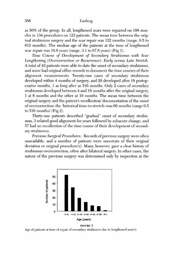
as 50% of the group. In all, lengthened scars were repaired on 198 mus-cles in 134 procedures on 123 patients. The mean time between the orig-inal strabismus surgery and the scar repair was 122 months (range, 0.5 to612 months). The median age of the patients at the time of lengthenedscar repair was 18.8 years (range, 3.1 to 67.8 years) (Fig 1).
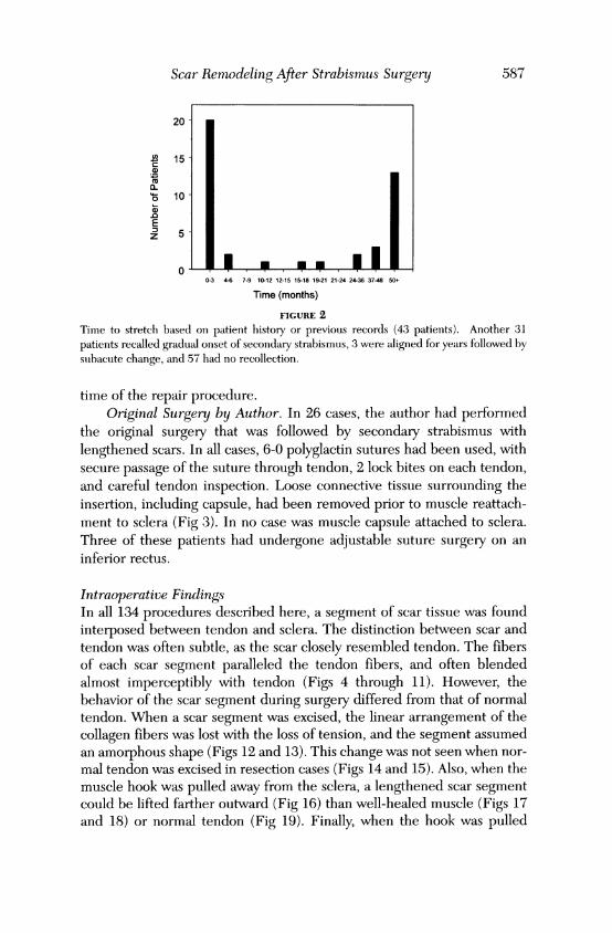
Time Course of Development of Secondary Strabismus with ScarLengthening (Overcorrection or Recurrence): Early versus Late Stretch.A total of 43 patients were able to date the onset of secondary strabismus,and some had original office records to document the time courses of theiralignment measurements. Twenty-one cases of secondary strabismusdeveloped within 4 months of surgery, and 20 developed after 18 postop-erative months, 1 as long after as 516 months. Only 2 cases of secondarystrabismus developed between 4 and 18 months after the original surgery,1 at 6 months and the other at 10 months. The mean time between theoriginal surgery and the patient's recollection/ documentation of the onsetof overcorrection-the historical time-to-stretch-was 60 months (range 0.5to 516 months) (Fig 2).
Thirty-one patients described "gradual" onset of secondary strabis-mus, 3 related good alignment for years followed by subacute change, and57 had no recollection of the time course of their development of second-ary strabismus.
Previous Surgical Procedlures. Records of previous surgery were oftenunavailable, and a number of patients were uncertain of their originaldeviation or original procedure(s). Many, however, gave a clear history ofstrabismus overcorrection, often after bilateral surgery. In other cases, thenature of the previous surgery was determined only by inspection at the
50
, 40
0l.W_ 30~ 0
E 20 II Iz
10
00-10 11-20 21-30 31-40 41-50 51-60 61+
Age (years)
FIGURE 1
Age of patients at time of repair of secondary strabismnols due to lengthened scar(s).
Ludwig586
Scar Remodeling After Strabismus Surgery
4i 15
0 10-
.0~E I0-3 4-6 7-9 10-12 12-15 15-18 19-21 21-24 24-36 37-48 50+
Time (months)
FIGURE 2
Time to stretch based on patient history or previous records (43 patients). Another 31patients recalled gradual onset of secondary strabismus, 3 were aligned for years followed bysubacute change, and 57 had no recollection.
time of the repair procedure.Original Surgery by Author. In 26 cases, the author had performed
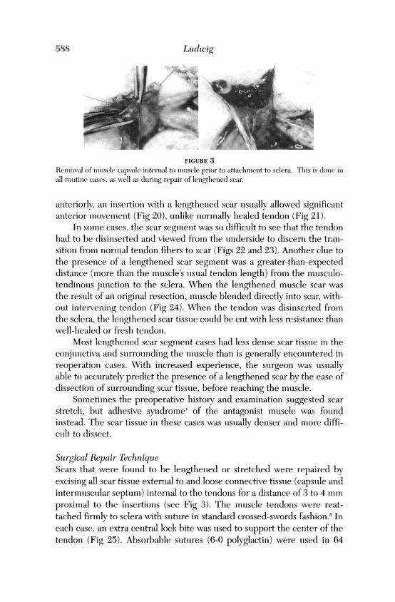
the original surgery that was followed by secondary strabismus withlengthened scars. In all cases, 6-0 polyglactin sutures had been used, withsecure passage of the suture through tendon, 2 lock bites on each tendon,and careful tendon inspection. Loose connective tissue surrounding theinsertion, including capsule, had been removed prior to muscle reattach-ment to sclera (Fig 3). In no case was muscle capsule attached to sclera.Three of these patients had undergone adjustable suture surgery on aninferior rectus.
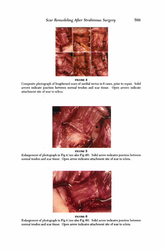
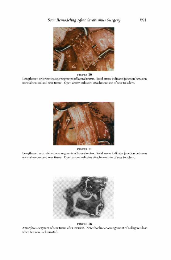
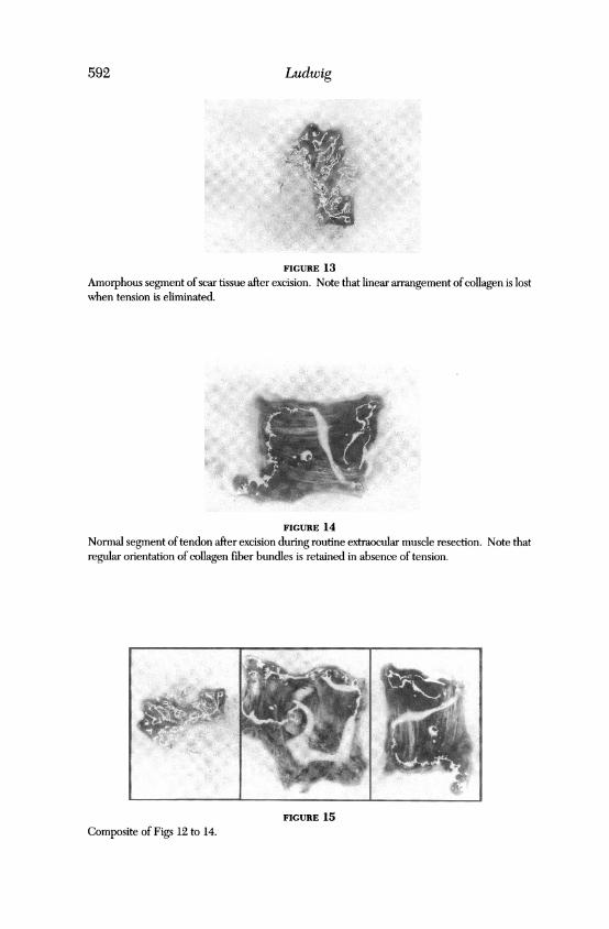
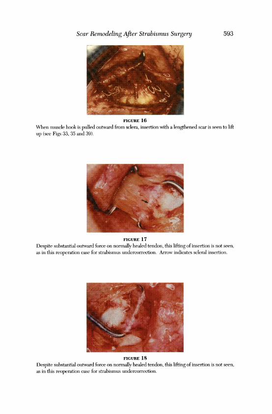
Intraoperative FindingsIn all 134 procedures described here, a segment of scar tissue was foundinterposed between tendon and sclera. The distinction between scar andtendon was often subtle, as the scar closely resembled tendon. The fibersof each scar segment paralleled the tendon fibers, and often blendedalmost imperceptibly with tendon (Figs 4 through 11). However, thebehavior of the scar segment during surgery differed from that of normaltendon. When a scar segment was excised, the linear arrangement of thecollagen fibers was lost with the loss of tension, and the segment assumedan amorphous shape (Figs 12 and 13). This change was not seen when nor-mal tendon was excised in resection cases (Figs 14 and 15). Also, when themuscle hook was pulled away from the sclera, a lengthened scar segmentcould be lifted farther outward (Fig 16) than well-healed muscle (Figs 17and 18) or normal tendon (Fig 19). Finally, when the hook was pulled
587
Luidwig
FIGURE 3
Removal of muscle capsule internial to imuscle prior to attatchment to sclera. This is dlonie inall routinie cases, as well as dturing repair of lengtlhenied sear.
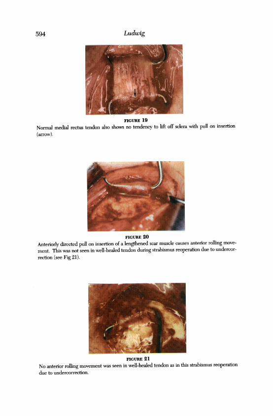
anteriorly, anI insertion with a lengthened scar usually allowed significantanterior movement (Fig 20), unlike normally healed tendon (Fig 21).
In some cases, the scar segment was so difficult to see that the tendonhad to be dlisinserted and viewed from the underside to discern the tran-sition from normal tendon fibers to scar (Figs 22 and 23). Another clue tothe presence of a lengthened scar segment was a greater-than-expecteddistance (more than the muscle's usual tendon length) from the musculo-tendinous junction to the sclera. When the lengthened muscle scar wasthe result of an original resection, muscle blended directly into scar, with-out intervening tendon (Fig 24). When the tendon was disinserted fromthe sclera, the lengthened scar tissue could be cut with less resistance thanwell-healed or fresh tendon.
Most lengthened scar segment cases had less dense scar tissue in theconjunctiva and surrounding the muscle than is generally encountered inreoperation cases. With increased experience, the surgeon was usuallyable to accurately predict the presence of a lengthened scar by the ease ofdissection of surrounding scar tissue, before reaching the muscle.
Sometimes the preoperative history and examination suggested scarstretch, but adhesive syndrome4 of the antagonist muscle was foundinstead. The scar tissue in these cases was usually denser and more diffi-cult to dissect.
Surgical Repair TechniquieScars that were found to be lengthened or stretched were repaired byexcising all scar tissue external to and loose connective tissue (capsule andintermuscular septum) internal to the tendons for a distance of 3 to 4 mmproximal to the insertions (see Fig 3). The muscle tendons were reat-tached firmly to sclera with suiture in standard crossed-swords fashion.5 Ineach case, an extra central lock bite was used to support the center of thetendon (Fig 25). Absorbable sutures (6-0 polyglactin) were used in 64
588
Scar Remodeling After Strabismus Surgery
FIGURE 4
Composite photograph of lengthened scars of medial rectus in 6 cases, prior to repair. Solidarrows indicate junction between normal tendon and scar tissue. Open arrows indicateattachment site of scar to sclera.
FIGURE 5Enlargement of photograph in Fig 4 (see also Fig 40). Solid arrow indicates junction betweennormal tendon and scar tissue. Open arrow indicates attachment site of scar to sclera.
FIGURE 6Enlargement of photograph in Fig 4 (see also Fig 40). Solid arrow indicates junction betweennormal tendon and scar tissue. Open arrow indicates attachment site of scar to sclera.
589
Ludwig
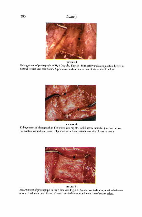
FIGURE 7Enlargement of photograph in Fig 4 (see also Fig 40). Solid arrow indicates junction betweennormal tendon and scar tissue. Open arrow indicates attachment site of scar to sclera.
FIGURE 8Enlargement of photograph in Fig 4 (see also Fig 40). Solid arrow indicates junction betweennormal tendon and scar tissue. Open arrow indicates attachment site of scar to sclera.
FIGURE 9Enlargement of photograph in Fig 4 (see also Fig 40). Solid arrow indicates junction betweennormal tendon and scar tissue. Open arrow indicates attachment site of scar to sclera.
590
Scar Remrwdeling After Strabismus Surgery 591
FIGURE 10
Lengthenied or stretchled scar segmeints of lateral rectus. Solid arrow indicates jtunc'tioIn betweeinnormal teindloni and scar tissue. Openi arrow indicates attsachmiient site of scar to sclera.
FIGURE 11
Lengthened or stretchied scar segmeints of later-al rectus. Solid arrowv inclicates juinctioni hetweennormal tendon and scar tissoe. Openi arrow indicates attachmiient site of scar to sciera.
FIGURE 12Amorphous segmiienit of scar tisstue after excision. Note that liniear arrangement of collagen is lostwhen tension is eliminiated.
Ludwig
FIGURE 13
Amorphous segment of scar tissue after excision. Note that linear arrangement of collagen is lostwhen tension is eliminated.
FIGURE 14Normal segment oftendon after excision during routine extraocular muscle resection. Note thatregular orientation of collagen fiber bundles is retained in absence of tension.
FIGURE 15Composite of Figs 12 to 14.
592
Scar Remodeling After Strabismus Surgery
FIGURE 16When muscle hook is pulled outward from sclera, insertion with a lengthened scar is seen to liftup (see Figs 33, 35 and 39).
FIGURE 17Despite substantial outward force on normally healed tendon, this lifting of insertion is not seen,as in this reoperation case for strabismus undercorrection. Arrow indicates scleral insertion.
FIGURE 18Despite substantial outward force on normally healed tendon, this lifting of insertion is not seen,as in this reoperation case for strabismus undercorrection.
593
Ludwig
FIGURE 19
Normal medial rectus tendon also shows no tendency to lift off sclera with pull on insertion
(arrow).
FIGURE 20
Anteriorly directed pull on insertion of a lengthened scar muscle causes anterior rolling move-ment. This was not seen in well-healed tendon during strabismus reoperation due to undercor-rection (see Fig 21).
FIGURE 21
No anterior rolling movement was seen in well-healed tendon as in this strabismus reoperationdue to undercorrection.
594
Scar Rernodelinig After Strabismus Surgeeny
FIGURE 22
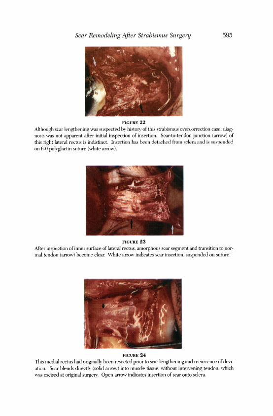
Although scar lengthening was suspected by history of this strabismus overcorrection case, diag-nosis was not apparenit after initial inspection of insertion. Scar-to-tendon junction (arrowv) ofthis right lateral rectus is indistinct. Insertion has been detached from sclera and is suspendedon 6-0 polyglactin suture (white arrow).
FIGURE 23After inspection of inner surface of lateral rectus, amorphous scar segment and transition to nor-mal tendon (arrox) become clear. WVhite arrowN indicates scar insertion, suspended on sutuire.
FIGURE 24This medial rectus had originally been resected prior to scar lengthening and recurrence of devi-ation. Scar blends directly (solid arrowv) into muscle tissue, without intervening tendon, whichw%as excised at original surgerv Open arroN indicates insertion of scar onto sclera.
595
cases. Several recurrences were observed following repair witlh excision ofthe scar segment and firm reattachment of muscle to sclera, and it was feltthat prolonged support of the attachment site was needed. Beginning in1995, repairs were performed with nonabsorbable sutures: 17 with 6-0braided polyester (Fig 26), 1 with 6-0 clear polypropylene alone, and 52with combined tandem 6-0 clear polypropylene and 6-0 polyglactin (Fig27). For all cases sutured with polypropylene, after the knot was tied onthe muscle, the sutures were passed under the muscle belly and retied 7to 8 mm posteriorly, to prevent suture end erosion through the conjuncti-va (Fig 28). The use of polypropylene sutures in combination withpolyglactin sutures was necessary because polypropylene sutures alone aretoo slippery to tie. Clear polypropylene sutures were used for anteriorlypositioned muscles to prevent these sutures from showing through theconjunctiva. Polyester sutures were used for posteriorly attached musclesand inferior and superior recti, which are hidden by the eye lids.
The amount of muscle advancement was determined by preoperativemeasurements and routine surgical tables (MM Parks, Lancaster Course,Colby College, Waterville, Maine, August, 1985), but the plan was oftenadjusted intraoperatively to account for factors such as muscle contracture.The goal was a physical centering of the eye intraoperatively as judged byforced ductions, the spring-back test,6 and corneal light reflexes.
One hundred twenty-four procedures were performed by the author, whoused no postoperative corticosteroids, and 10 procedures were performed byanother surgeon, who used topical corticosteroids for 10 days postoperatively.
Pathologic examinations were performed in specimens from 82 cases.An additional 7 specimens were used in tissue culture studies.
TISSUE CULTURE STUDIES
Cells from 7 excised scar specimens and 3 normal tendon resection speci-mens (controls) were cultured in Dulbecco minimal Eagle medium(DMEM) with 10% fetal bovine serum. The cells were passaged andmedium was collected from each culture after 7 days. The activity ofmatrix metalloproteinases (MMPs) in the medium was assayed using gel-atin and casein zymography. ' Western blot was used for the identificationof MMP1, MMP2, and MMP9 in the medium. Levels of MMPs andinhibitors (tissue inhibitors of metalloproteinases, ie, TIMP1 and TIMP2)were determined in cells and tissue extracts by Western blot.
ANIMAL STUDIES
Development ofan Animal ModelIn an attempt to create an animal model of scar lengthening after strabismus
Ludwig596
Scar Remodeling After Strabismus Surgery
.1
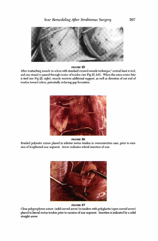
FIGURE 25After reattaching muscle to sclera with standard crossed-swords technique,5 central knot is tied,and one strand is passed through center of tendon (see Fig 25, left). When this extra center biteis tied (see Fig 25, right), muscle receives additional support, as well as direction of cut end oftendon toward sclera, potentially reducing gap formation.
r:
FIGURE 26Braided polyester suture placed in inferior rectus tendon in overcorrection case, prior to exci-sion of lengthened scar segment. Arrow indicates scleral insertion of scar.
FIGURE 27Clear polyproplyene suture (solid curved arrow) in tandem with polyglactin (open curved arrow)placed in lateral rectus tendon prior to excision of scar segment. Insertion is indicated by a solidstraight arrow.
597
Ludwig
FIGURE 28
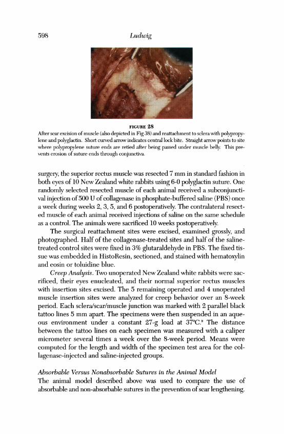
After scar excision of muscle (also depicted in Fig 38) and reattachment to sclera with polypropy-lene and polyglactin. Short curved arrow indicates central lock bite. Straight arrow points to sitewhere polypropylene suture ends are retied after being passed under muscle belly. This pre-vents erosion of suture ends through conjunctiva.
surgery, the superior rectus muscle was resected 7 mm in standard fashion inboth eyes of 10 New Zealand white rabbits using 6-0 polyglactin suture. Onerandomly selected resected muscle of each animal received a subconjuncti-val injection of500 U ofcollagenase in phosphate-buffered saline (PBS) oncea week during weeks 2, 3, 5, and 6 postoperatively. The contralateral resect-ed muscle of each animal received injections of saline on the same scheduleas a control. The animals were sacrificed 10 weeks postoperatively.
The surgical reattachment sites were excised, examined grossly, andphotographed. Half of the collagenase-treated sites and half of the saline-treated control sites were fixed in 3% glutaraldehyde in PBS. The fixed tis-sue was embedded in HistoResin, sectioned, and stained with hematoxylinand eosin or toluidine blue.
Creep Analysis. Two unoperated New Zealand white rabbits were sac-rificed, their eyes enucleated, and their normal superior rectus muscleswith insertion sites excised. The 5 remaining operated and 4 unoperatedmuscle insertion sites were analyzed for creep behavior over an 8-weekperiod. Each sclera/scar/muscle junction was marked with 2 parallel blacktattoo lines 5 mm apart. The specimens were then suspended in an aque-ous environment under a constant 27-g load at 370C.9 The distancebetween the tattoo lines on each specimen was measured with a calipermicrometer several times a week over the 8-week period. Means werecomputed for the length and width of the specimen test area for the col-lagenase-injected and saline-injected groups.
Absorbable Versus Nonabsorbable Sutures in the Animal ModelThe animal model described above was used to compare the use ofabsorbable and non-absorbable sutures in the prevention of scar lengthening.
598
Scar Remodeling After Strabismus Surgeny
The superior rectus muscles were resected bilaterally in 2 rabbits; 1 musclewas attached with 6-0 polyglactin suture and the contralateral muscle with6-0 braided polyester suture. Collagenase was injected subconjunctivallyover each reattachment site once a week during weeks 2, 3, 5, and 6 post-operatively. At 10 weeks, the animals were sacrificed and the attachmentsites dissected, examined grossly, and prepared for creep analysis.
RESULTS
CLINICAL STUDIES
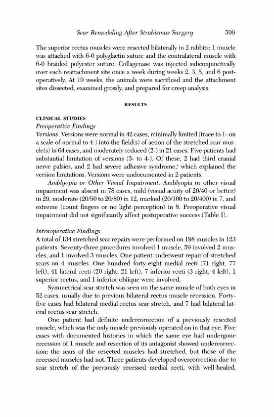
Preoperative FindingsVersions. Versions were normal in 42 cases, minimally limited (trace to 1- ona scale of normal to 4-) into the field(s) of action of the stretched scar mus-cle(s) in 64 cases, and moderately reduced (2-) in 21 cases. Five patients hadsubstantial limitation of versions (3- to 4-). Of these, 2 had third cranialnerve palsies, and 2 had severe adhesive syndrome,4 which explained theversion limitations. Versions were undocumented in 2 patients.
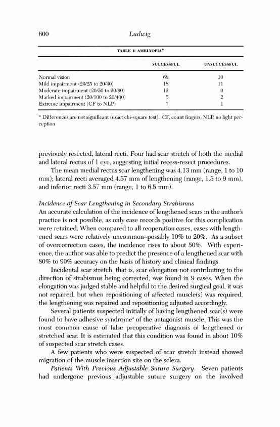
Amblyopia or Other Visual Impairnent. Amblyopia or other visualimpairment was absent in 78 cases, mild (visual acuity of 20/40 or better)in 29, moderate (20/50 to 20/80) in 12, marked (20/100 to 20/400) in 7, andextreme (count fingers or no light perception) in 8. Preoperative visualimpairment did not significantly affect postoperative success (Table I).
Intraoperative FindingsA total of 134 stretched scar repairs were performed on 198 muscles in 123patients. Seventy-three procedures involved 1 muscle, 59 involved 2 mus-cles, and 1 involved 3 muscles. One patient underwent repair of stretchedscars on 4 muscles. One hundred forty-eight medial recti (71 right, 77left), 41 lateral recti (20 right, 21 left), 7 inferior recti (3 right, 4 left), 1superior rectus, and 1 inferior oblique were involved.
Symmetrical scar stretch was seen on the same muscle of both eyes in52 cases, usually due to previous bilateral rectus muscle recession. Forty-five cases had bilateral medial rectus scar stretch, and 7 had bilateral lat-eral rectus scar stretch.
One patient had definite undercorrection of a previously resectedmuscle, which was the only muscle previously operated on in that eye. Fivecases with documented histories in which the same eye had undergonerecession of 1 muscle and resection of its antagonist showed undercorrec-tion; the scars of the resected muscles had stretched, but those of therecessed muscles had not. Three patients developed overcorrection due toscar stretch of the previously recessed medial recti, with well-healed,
599
600 Ludwig
TABLE I: AMBLYOPIA*
SUCCESSFUL UNSUCCESSFUL
Normal vision 68 10Mild impairment (20/25 to 20/40) 18 11Moderate impairment (20/50 to 20/80) 12 0Marked impairmenit (20/100 to 20/400) 5 2Extreme impairmnent (CF to NLP) 1
4 Differences are not significant (exact chi-square test). CF, count fingers; NLP, no light per-ception
previously resected, lateral recti. Four had scar stretch of both the medialand lateral rectus of 1 eye, suggesting initial recess-resect procedures.
The mean medial rectus scar lengthening was 4.13 mm (range, 1 to 10mm); lateral recti averaged 4.57 mm of lengthening (range, 1.5 to 9 mm),and inferior recti 3.57 mm (range, 1 to 6.5 mm).
Incidence of Scar Lengthening in Secondary StrabismusAn accurate calculation of the incidence of lengthened scars in the author'spractice is not possible, as only case records positive for this complicationwere retained. When compared to all reoperation cases, cases with length-ened scars were relatively uncommon-possibly 10% to 20%. As a subsetof overcorrection cases, the incidence rises to about 50%. With experi-ence, the author was able to predict the presence of a lengthened scar with80% to 90% accuracy on the basis of history and clinical findings.
Incidental scar stretch, that is, scar elongation not contributing to thedirection of strabismus being corrected, was found in 9 cases. When theelongation was judged stable and helpful to the desired surgical goal, it wasnot repaired, but when repositioning of affected muscle(s) was required,the lengthening was repaired and repositioning adjusted accordingly.
Several patients suspected initially of having lengthened scar(s) werefound to have adhesive syndrome4 of the antagonist muscle. This was themost common cause of false preoperative diagnosis of lengthened orstretched scar. It is estimated that this condition was found in about 10%of suspected scar stretch cases.
A few patients who were suspected of scar stretch instead showedmigration of the muscle insertion site on the sclera.
Patients With Previous Adjustable Suture Surgery. Seven patientshad undergone previous adjustable suture surgery on the involved
Scar Remitodleling After Strabismus Surgery
muscle(s). They had lengthened scars that were clinically indistinguishablefrom the other stretched scar cases. These scars were not pseudotendonsbetween the tendon and original insertion. Scar tissue was attached to thesclera well behind the original insertion in each case.
One of these patients was especially interesting, as she had undergone2 previous strabismus procedures. In the first procedure, both medial rectiwere recessed, with direct suturing of tendons to sclera. During the secondprocedure, the left medial rectus was advanced with direct suturing, but theleft lateral and right superior recti were recessed on adjustable sutures. Shegradually developed esotropia and a deficit of upgaze on the right. When themuscles were explored during repair surgery, the directly sutured medialrecti showed no signs of scar lengthening, but each of the previously adjust-ed muscles showed lengthened scars of 6 mm (see Figs 22 and 23).
Follow-upPostoperative follow-up after stretched scar repair averaged 11.4 months forall cases (range, 1 to 69 months). Mean follow-up was 14.8 months for theabsorbable suture group and 8.6 months for the nonabsorbable suture group.
ComplicationsConjunctival Scar Tissue. Occasionally, thickened scar tissue of the con-junctiva and Tenon's capsule overlying the repaired post-scar-stretch mus-cle created a cosmetic problem. This problem was usually the result ofadhesive syndrome from the original surgery, with penetration of Tenon'scapsule and prolapse of orbital fat. Conjunctival recession was performedat the time of stretched scar repair in 10 patients because of this compli-cation from previous surgery. Two other patients chose to undergo sec-ondary conjunctival recession at a later date.
Unrecognized Scar Stretch. Two patients remained exotropic after unilat-eral repair of a stretched medial rectus. Each was later found to have a stretchof the contralateral medial rectus, which was repaired in a second procedure.
Secondary Scar Stretch of Antagonist Muscle. During stretched scarrepair, the antagonist of the muscle being repaired was occasionallyrecessed to relieve contracture. Although nonabsorbable suture was usedfor the stretched scar repair, it was not initially used for other muscles. In2 cases, the recessed antagonist muscles later developed stretches, requir-ing re-repair. The repaired stretched scar muscles did not restretch.
Pulled-in-two Syndrome. Two stretched scar patients developedrupture of a medial rectus at its musculotendinous junction intraopera-tively. A third patient developed a partial separation that did not pullapart completely. These 3 patients were 6, 14, and 19 years old. In the
601
Ludwig
literature, this complication is considered rare and usually seen onlyin the elderly.',"'
Absorbable Versus Nonabsorbable Sutures in Cases of Suspected RestretchSuccessful repair was defined in terms of the patient noting substantialcorrection of the preoperative deviation and being satisfied with the out-come. These patients achieved undercorrection of their preoperative devi-ations to within 10 prism diopters (A ) or overcorrection to 14A.
The first 64 stretched scar cases were repaired with absorbablesuture. By 1995, however, it became evident that late recurrence was com-mon despite good initial postoperative alignment, and the use of nonab-sorbable suture was implemented (Table II).
Absorbable Sutures. In the absorbable suture group, 43 of 62 proce-dures with 1 month or more of follow-up (69%) and 21 of 28 (75%) with12 months or more of follow-up were judged successful by the patient andthe surgeon. One of these patients was an exception to the definition ofsuccess as given above; she had an undercorrection of 18A, but the preop-erative deviation was so large that she was satisfied with the improvement.
Restretch was suspected when there was postoperative drift of 6A ormore, with the return of the original deviation. Twenty-six cases (41%)had suspected restretch after absorbable suture repair. Six of these werethen documented (see below).
Two other patients underwent re-exploration during surgery on addi-tional muscles 1 year after absorbable suture stretched scar repair andwere found to have firm re-attachments of the previously stretched mus-cles (2 medial and 2 lateral recti).
Nonabsorbable Sutures. In the nonabsorbable suture group, 62 of 68procedures with 1 or more months of follow-up (91%) and all 19 with 12 ormore months of follow-up (100%) were judged successful. One was over-corrected, 4 showed postoperative drift that indicated possible restretch,and 1 of these 4 had restretch documented (see below). Three underwentrepeat surgery for overcorrection of the stretched scar deficit or on othermuscles, which permitted documentation of the success of the repair, (ie,no recurrence of scar lengthening). Five cases in which restretch occurredafter absorbable suture repair responded well to repair with nonabsorbablesutures (see below). One patient considered to be a success had an over-correction of 20A, which required repeat surgery on the same muscles toachieve orthotropia, but showed no recurrence of stretch.
Stability of Postoperative Alignment. Good postoperative alignmentis an imperfect measure of the success of repair of the lengthened scarabnormality, because a successfully repaired muscle may have been
602
Scar Remodeling After Strabismus Surgery60
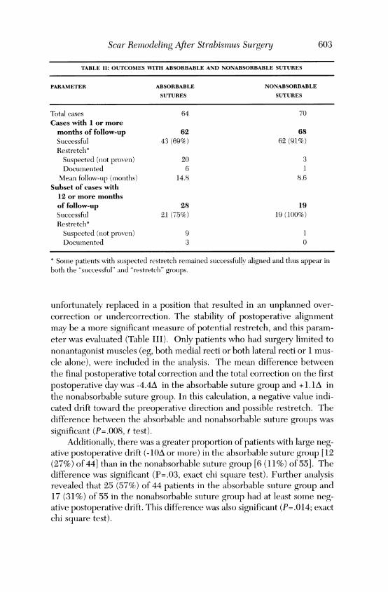
TABLE II: OUTCOMIES WITH ABSORBABLE AND NONABSORBABLE SUTURES
PARAMETER ABSORBABLE NONABSORBABLE
SUTURES SUTURES
Total cases 64 70Cases with 1 or moremonths of follow-up 62 68Successftil 43 (69%) 62 (91%)Restretch°SuspectedI (niot proven) 20 3Documenited 6 1Mean follow-op (molntlhs) 14.8 8.6
Subset of cases with12 or more monthsof follow-up 28 19Successful 21 (75%) 19 (1(0%)Restretch°Suspected (niot proven) 9 1Documenited 3 0
Some patients with suspected restretch remained successfully aligned anid thuts appear inbotlh the "successfutl" anid "restretch'" groutps.
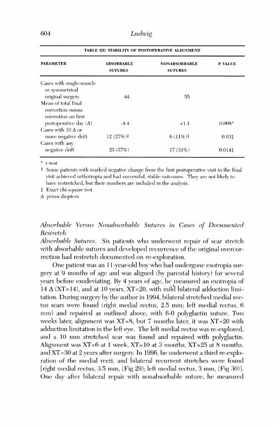
unfortunately replaced in a position that resulted in an unplanned over-correction or undercorrection. The stability of postoperative alignmentmay be a more significant measure of potential restretch, and this param-eter was evaluated (Table III). Only patients who had surgery limited tononantagonist muscles (eg, both medial recti or both lateral recti or 1 mus-cle alone), were included in the analysis. The mean difference betweenthe final postoperative total correction and the total correction on the firstpostoperative day was -4.4A in the absorbable suture group and + 1.1A inthe nonabsorbable suture group. In this calculation, a negative value indi-cated drift toward the preoperative direction and possible restretch. Thedifference between the absorbable and nonabsorbable suture groups wassignificant (P=.008, t test).
Additionally, there was a greater proportion of patients with large neg-ative postoperative drift (-10A or more) in the absorbable suture group [12(27%) of 44] than in the nonabsorbable suture group [6 (11%) of 55]. Thedifference was significant (P=.03, exact chi squiare test). Further analysisrevealed that 25 (57%) of 44 patients in the absorbable suture group and17 (31%) of 55 in the nonabsorbable suture grouip had at least some neg-ative postoperative drift. This difference was also significant (P=.014; exactchi square test).
603
Ludwig
TABLE III: STABILITY OF POSTOPERATIVIE ALIGNMENT
PARAMETER ABSORBABLE NONABSORBABLE P VALUE
SUTURES SUTURES
Cases wNith single-muiiiscleor symmetricaloriginal surgery 44 55
Mean of total finalcorrection minuiscorrection on firstpostoperative day (A) -4.4 +1.1 0.008*
Casescwith 10 A ormore negative drift 12 (27%)t 6 (11%)t 0.03++
Cases wNitlh anynegative drift 25 (57%) 17 (31%) 0.0144
t-testf Somiie patients with miarked negative chlange fromn the first postoperativ7e visit to the final
visit achieved orthotropia and had successful, stable outcomes. They lare not likely tohave restretched, btot their numbers are included in the anialysis.
+ Exact chi-square testA prism diopters
Absorbable Versus Nonabsorbable Sutures in Cases of DocumnentedRestretchAbsorbable Sutures. Six patients who underwent repair of scar stretchwith absorbable sutures and developed recurrence of the original overcor-rection had restretch documented on re-exploration.
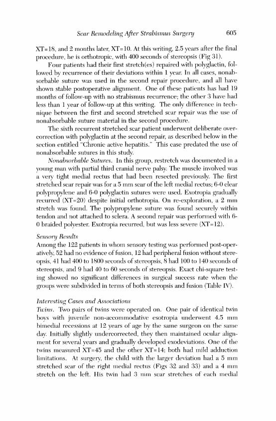
One patient was an 1 1-year-old boy who had undergone esotropia sur-gery at 9 months of age and was aligned (by parental history) for severalyears before exodeviating. By 4 years of age, he measured an exotropia of14 A (XT=14), and at 10 years, XT=20, with miOl bilateral adduction limi-tation. During surgery by the author in 1994, bilateral stretched medial rec-tus scars were found (right medial rectus, 2.5 mm; left medial rectus, 6mm) and repaired as outlined above, with 6-0 polyglactin suture. Twoweeks later, alignment was XT=8, but 7 months later, it was XT=20 withadduction limitation in the left eye. The left medial rectus was re-explored,and a 10 mm stretched scar was found and repaired with polyglactin.Alignment was XT=6 at 1 week, XT=10 at 3 months, XT=25 at 8 months,and XT=30 at 2 years after suirgery. In 1996, he underwent a third re-explo-ration of the medial recti, and bilateral recurrent stretches were found[right medial rectus, 3.5 mm, (Fig 29); left medial rectus, 3 mm, (Fig 30)].One day after bilateral repair with nonabsorbable suture, he measured
604
Scar Remodeling After Strabi.smus Sn rgery
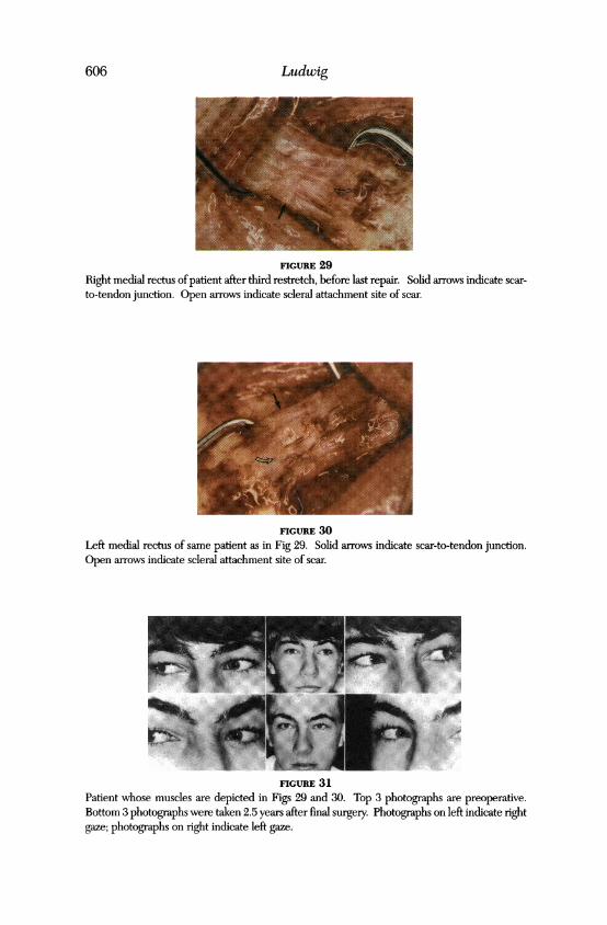
XT= 18, and 2 months later, XT= 10. At this writing, 2.5 years after the finalprocedure, he is orthotropic, with 400 seconds of stereopsis (Fig 31).
Four patients had their first stretch(es) repaired with polyglactin, fol-lowed by recurrence of their deviations within 1 year. In all cases, nonab-sorbable suture was used in the second repair procedure, and all haveshown stable postoperative alignment. One of these patients has had 19months of follow-uip with no strabismus recurrence; the other 3 have hadless than 1 year of follow-up at this writing. The only difference in tech-nique between the first and second stretched scar repair was the use ofnonabsorbable suture material in the second procedure.
The sixth recurrent stretched scar patient underwent deliberate over-correction with polyglactin at the second repair, as described below in thesection entitled "Chronic active hepatitis." This case predated the use ofnonabsorbable sutures in this study.
Nonabsorbable Sutures. In this group, restretch was documented in ayoung man with partial third cranial nerve palsy. The muscle involved wasa very tight medial rectus that had been resected previously. The firststretched scar repair was for a 5 mm scar of the left medial rectus; 6-0 clearpolypropylene and 6-0 polyglactin sutures were used. Exotropia graduallyrecurred (XT=20) despite initial orthotropia. On re-exploration, a 2 mmstretch was found. The polypropylene suture was found securely withintendon and not attached to sclera. A second repair was performed with 6-0 braided polyester. Exotropia recurred, but was less severe (XT= 12).
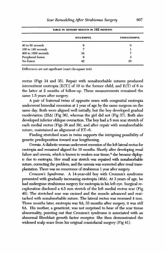
Sensony ResultsAmong the 122 patients in whom sensory testing was performed post-oper-atively, 52 had no evidence of fusion, 12 had peripheral fusion without stere-opsis, 41 had 400 to 1800 seconds of stereopsis, 8 had 100 to 140 seconds ofstereopsis, and 9 had 40 to 60 seconds of stereopsis. Exact chi-square test-ing showed no significant differences in suirgical suiccess rate when thegroups were subdivided in terms of both stereopsis and fusion (Table IV).
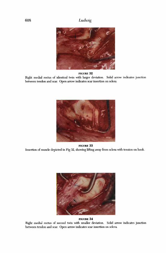
Interesting Cases and AssociationsTwtins. Two pairs of twins were operated on. One pair of identical twinboys with juvenile non-accommodative esotropia underwent 4.5 mmbimedial recessions at 12 years of age by the same surgeon on the sameday. Initially slightly undercorrected, they then maintained ocular align-ment for several years and gradually developed exodeviations. One of thetwins measured XT=45 and the other XT=14; both had mild adduictionlimitations. At surgery, the child with the larger deviation hadl a 5 mmstretched scar of the right medial rectus (Figs 32 and 33) and a 4 mmstretch on the left. His twin had 3 mm scar stretches of each medial
605
Ludwig
FIGURE 29
Right medial rectus ofpatient after third restretch, before last repair. Solid arrows indicate scar-to-tendon junction. Open arrows indicate scleral attachment site of scar.
FIGURE 30Left medial rectus of same patient as in Fig 29. Solid arrows indicate scar-to-tendon junction.Open arrows indicate scleral attachment site of scar.
FIGURE 31Patient whose muscles are depicted in Figs 29 and 30. Top 3 photographs are preoperative.Bottom 3 photographs were taken 2.5 years after final surgery. Photographs on left indicate rightgaze; photographs on right indicate left gaze.
606
Scar Remodeling After Strabismus Surgery
TABLE IV: SENSORY RESULTS IN 122 PATIENTS
SUCCESSFUL UNSUCCESSFUL
40 to 60 seconds 9 0100 to 140 seconds 7 1400 to 1800 seconds 34 7Peripheral fusion 9 3No fusion 42 10
Differences are not significant (exact chi-square test).
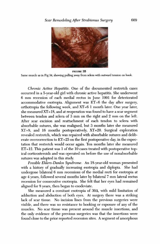
rectus (Figs 34 and 35). Repair with nonabsorbable sutures producedintermittent esotropia [E(T)] of 10 in the former child, and E(T) of 6 inthe latter at 2 months of follow-up. These measurements remained thesame 1.5 years after surgery.
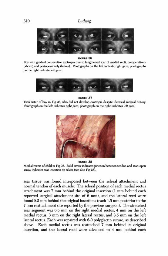
A pair of fraternal twins of opposite sexes with congenital esotropiaunderwent bimedial recession at 1 year of age by the same surgeon on thesame day. Both were aligned well initially, but the boy developed gradualexodeviation (25A) (Fig 36), whereas the girl did not (Fig 37). Both alsodeveloped inferior oblique overaction. The boy had a 5 mm scar stretch ofeach medial rectus (Figs 38 and 39), and after repair with nonabsorbablesuture, maintained an alignment of ET=6.
Finding stretched scars in twins supports the intriguing possibility ofgenetic predisposition toward scar lengthening.
Uremia. A diabetic woman underwent recession ofthe left lateral rectus forexotropia and remained aligned for 10 months. Shortly after developing renalfailure and uremia, which is known to weaken scar tissue,u2 she became diplop-ic due to esotropia. Her small scar stretch was repaired with nonabsorbablesuture, correcting the problem, and the uremia was corrected after renal trans-plantation. There was no recurrence of strabismus 1 year after surgery.
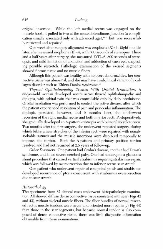
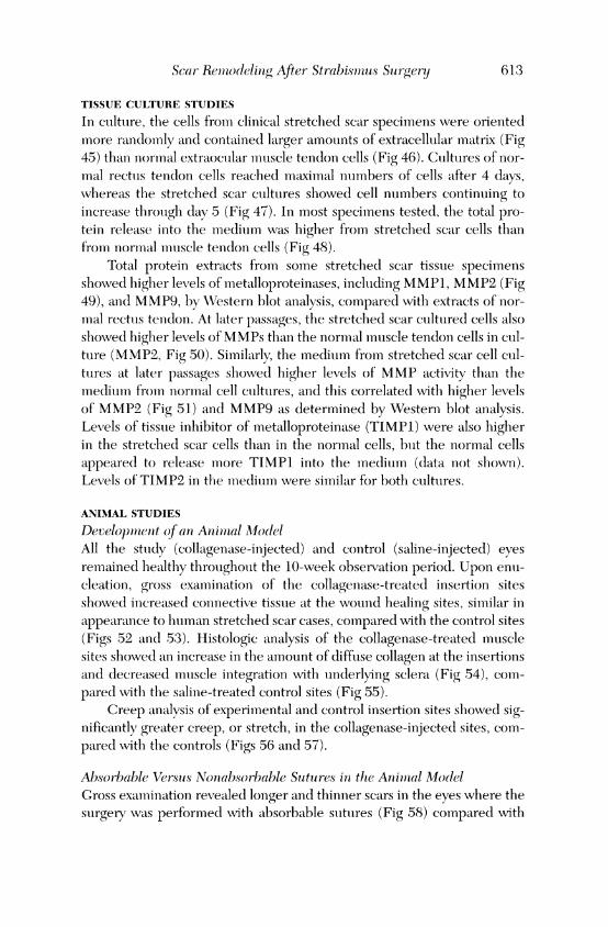
Crouzon's Syndrome. A 14-year-old boy with Crouzon's syndromepresented with gradually increasing exotropia (40A). At 3 years of age, hehad undergone strabismus surgery for esotropia in his left eye. Surgical re-exploration disclosed a 4.5 mm stretch of the left medial rectus scar (Fig40). The stretched scar was excised and the muscle advanced and reat-tached with nonabsorbable suture. The lateral rectus was recessed 4 mm.Three months later, exotropia was 8A; 33 months after surgery, it was still8A. His mother, a geneticist, was not surprised to hear of the scar tissueabnormality, pointing out that Crouzon's syndrome is associated with anabnormal fibroblast growth factor receptor. She then demonstrated thewidened scalp scars from his original craniofacial surgery (Fig 41).
607
Ludwig
FIGURE 32
Right medial rectus of identical twin with larger deviation. Solid arrow indicates junctionbetween tendon and scar. Open arrow indicates scar insertion on sclera
FIGURE 33Insertion of muscle depicted in Fig 32, showing lifting away from sclera with tension on hook.
FIGURE 34Right medial rectus of second twin with smaller deviation. Solid arrow indicates junctionbetween tendon and scar. Open arrow indicates scar insertion on sclera.
608
Scar Remodeling After Strabismus Surgery
FIGURE 35
Same muscle as in Fig 34, showing pulling away from sclera with outward tension on hook.
Chronic Active Hepatitis. One of the documented restretch casesoccurred in a 5-year-old girl with chronic active hepatitis. She underwent6 mm recession of each medial rectus in June 1991 for deterioratedaccommodative esotropia. Alignment was ET=6 the day after surgery,orthotropia the following week, and XT=6 1 month later. One year later,she measured XT= 18, and at reoperation was found to have a scar segmentbetween tendon and sclera of 3 mm on the right and 2 mm on the left.After scar excision and reattachment of each tendon to sclera withabsorbable sutures, she was realigned, but 3 months later she measuredXT=8, and 16 months postoperatively, XT=20. Surgical explorationrevealed restretch, which was repaired with absorbable sutures and delib-erate overcorrection to ET=25 on the first postoperative day, in the expec-tation that restretch would occur again. Ten months later she measuredET= 12. This patient was 1 of the 10 cases treated with postoperative top-ical corticosteroids and was operated on before the use of nonabsorbablesutures was adopted in this study.
Possible Ehlers-Danlos Syndrome. An 18-year-old woman presentedwith a history of gradually increasing esotropia and diplopia. She hadundergone bilateral 6 mm recessions of the medial recti for esotropia atage 4 years, followed several months later by bilateral 7 mm lateral rectusrecession for consecutive exotropia. She felt that her eyes had remainedaligned for 8 years, then began to esodeviate.
She measured a comitant esotropia of 30A, with mild limitation ofadduction and abduction of both eyes. At surgery there was a strikinglack of scar tissue. No incision lines from the previous surgeries werevisible, and there was no resistance to hooking or exposure of any of themuscles. No scar tissue was present around the muscle insertions, andthe only evidence of the previous surgeries was that the insertions werefound close to the prior reported recession sites. A segment of amorphous
609
Ludwig
FIGURE 36
Boy with gradual consecutive exotropia due to lengthened scar of medial recti, preoperatively(above) and postoperatively (below). Photographs on the left indicate right gaze; photographson the right indicate left gaze.
FIGuRE 37Twin sister of boy in Fig 36, who did not develop exotropia despite identical surgical history.Photograph on the left indicates right gaze; photograph on the right indicates left gaze.
FIGURE 38Medial rectus of child in Fig 36. Sohd arrow indicates junction between tendon and scar; openarrow indicates scar insertion on sclera (see also Fig 28).
scar tissue was found interposed between the scleral attachment andnormal tendon of each muscle. The scleral position of each medial rectusattachment was 7 mm behind the original insertion (1 mm behind eachreported surgical attachment site of 6 mm), and the lateral recti werefound 8.5 mm behind the original insertions (each 1.5 mm posterior to the7 mm reattachment site reported by the previous surgeon). The stretchedscar segment was 6.5 mm on the right medial rectus, 4 mm on the leftmedial rectus, 3 mm on the right lateral rectus, and 3.5 mm on the leftlateral rectus. Each was repaired with 6-0 polyglactin suture, as describedabove. Each medial rectus was reattached 7 mm behind its originalinsertion, and the lateral recti were advanced to 4 mm behind each
610
Scar Remodeling After Strabismus Surgery
FIGURE 39
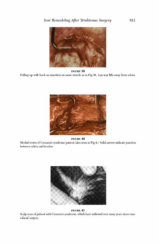
Poilling tip with hook on insertion on same mtiscle as in Fig 38. Lax scar lifts away from sclera.
FIGURE 40Medi-al rectuis of Crouzon's syndrome patient (a-lso seen in Fig 4.) Solid arrows indicate junctionbetween sclera and tendon.
FIGURE 41
Scalp scars of patient with Crouzon's syndrome, which have widened over many years since cran-iofacial surgery.
611
original insertion. While the left medial rectus was engaged on themuscle hook, it pulled in two at the musculotendinous junction (a compli-cation usuially associated only with advanced age),'"" but was sutccessfuil-ly retrieved and repaired.
One week after surgery, alignment was exophoria (X)=4. Eight monthslater, she measuired esophoria (E)=4, with 800 seconds of stereopsis. Threeand a half years after surgery, she measured E(T)=6, 800 seconds of stere-opsis, and mild limitation of abduction and adduction of each eye, suiggest-ing possible restretch. Pathologic examination of the excised segmentsshowed fibrous tissuie and no muscle fibers.
Although this patient was healthy with no overt abnormalities, her con-nective tissue was abnormal, and she may halve a subclinical variant of a col-lagen disorder such as Ehlers-Danlos syndrome."'
Thyroid Ophthalmopathy Treated WVith Orbital Irrasdiationr. A53-year-old woman developed severe active thyroid ophthalmnopathy anddiplopia, with orbital pain that was controllable only by daily prednisone.Orbital irradiation was performed to control the active disease, after whichthe patient experienced resolution of pain and periocular inflammulation. Thediplopia persisted, however, and 9 months later, she underwentrecession of the right medial rectus and both inferior recti. Postoperatively,she gradually developed an A-pattern exotropia with bilateral incyclotorsion.Two months after the first surgery, she underwent repeated surgery, duringwhicb bilateral scar stretches of the inferior recti were repaired withl nonab-sorbable sutures and the muscle insertions were displaced temporally toimprove the torsion. Both the A-pattern and primary position torsionresolved and had not returned at 2.5 years of follow-uip.
Other Disorders. One patient had Crohn's disease, another had Down'ssyndrome, and 3 had severe cerebral palsy. One had undergone a glaucomashunt procedure that caused vertical strabismus requiring strabismus repair,which was followed by overcorrection due to inferior rectus scar stretch.
One patient who underwent repair of congenital ptosis and strabismusdeveloped recurrence of ptosis concurrent with strabismus overcorrectiondue to scar stretch.
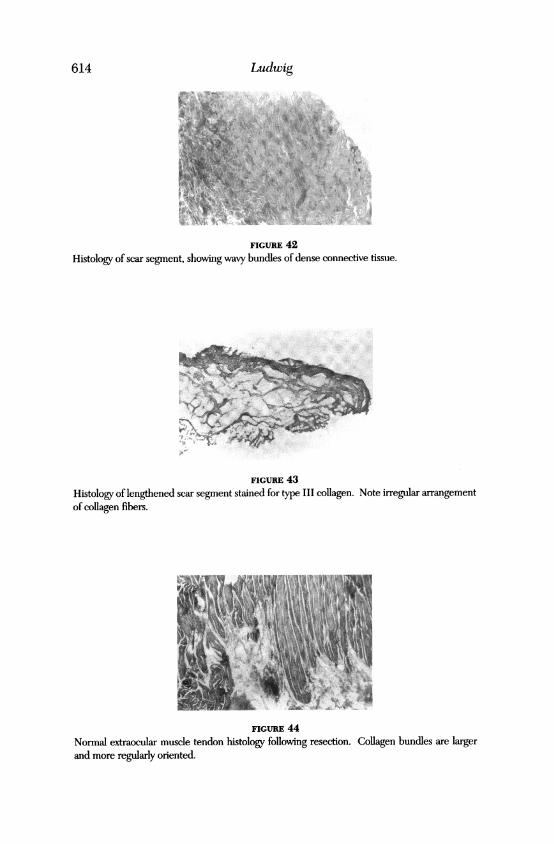
HistopatholoogyThe specimens from 82 clinical cases un-derwent histopathologic examina-tion. All showed diffuse dense connective tissue consistent with scar (Figs 42and 43), without skeletal muscle fibers. The fiber bundles of normal resect-ed rectuis mutscle tendons were larger and oriented more regularly (Fig 44)than those in the scar segments, but because normal tendon is also com-posed of dense connective tissue, there was little diagnostic informationobtainable from these examinations.
612 Ludwig
Scar Remoodeling After Strabi.snims St rgery1
TISSUE CULTURE STUDIES
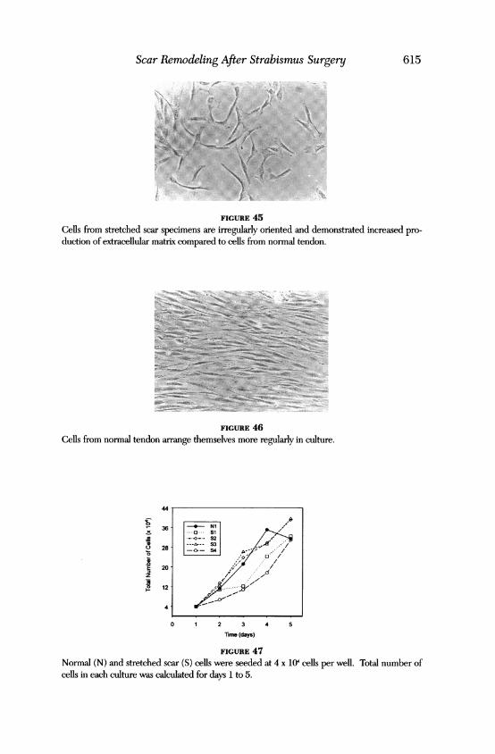
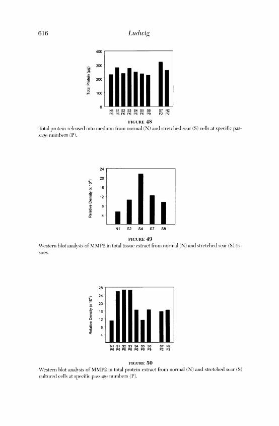
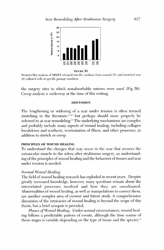
In cuilture, the cells from clinical stretched scar specimens were orientedmore randomuly and contained larger amounts of extracelluilar imiatrix (Fig45) than normnal extraocular mulscle tendon cells (Fig 46). Cultures of nor-mal rectus tendon cells reached maximal numbers of cells after 4 days,wlhereas the stretched scar cultures showed cell numbers continuing toincrease througli day 5 (Fig 47). In most specimens tested, the total pro-tein release into the medium was higher from stretched scar cells thanfrom normial imuscle tendon cells (Fig 48).
Total protein extracts from some stretclhed scar tissuie specimensshowed hiiglher levels of metalloproteinases, incluiding MMP1, MMP2 (Fig49), and MMP9, by WVestern blot analysis, compared with extracts of nor-mal rectus tendlon. At later passages, the stretclhed scar cultured cells alsoshowed higher levels of MMPs than the normlal muscle tendon cells in cul-ture (MMP2, Fig 50). Similarly, the mediumn from stretched scar cell cul-ttires at later passages showed higher levels of MMP activity than themedium fromi normal cell cuiltures, and this correlated with higher levelsof MMP2 (Fig 51) and MMP9 as determined by Western blot analysis.Levels of tissuie inhibitor of metalloproteinase (TIMPI) were also higherin the stretched scar cells than in the normlal cells, but the normal cellsappeared to release miiore TIMPI into the medium (data not shown).Levels of TIMP2 in the medium were similar for both cultures.
ANIMAL STUDIES
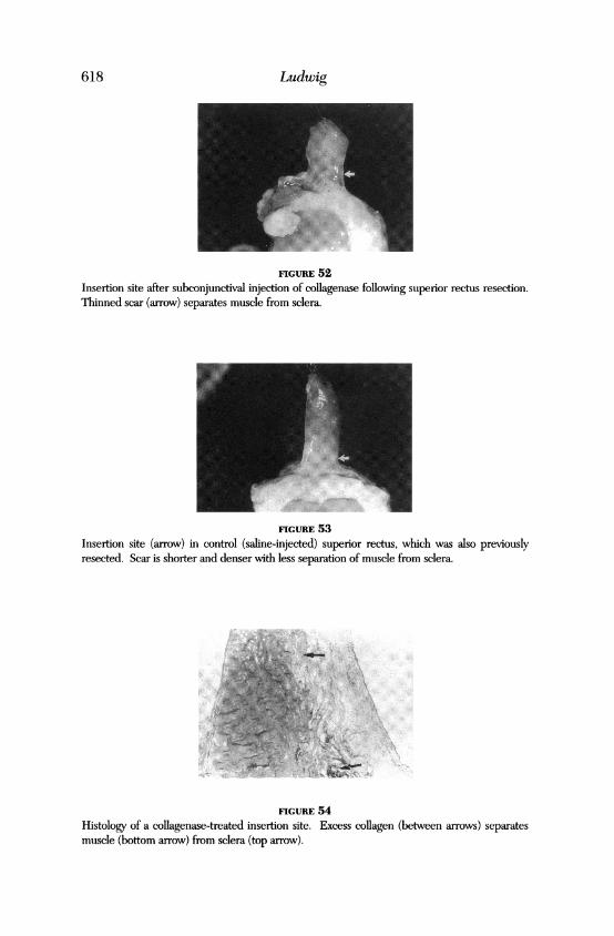
Developtment of ani Animilal ModelAll the study (collagenase-injected) and control (saline-injected) eyesremained healtlhy throuighout the 10-week observation period. Upon en-cleation, gross examination of the collageniase-treated insertion sitesshowed increased connective tissue at the wounid healing sites, similar inappearanice to luman stretclhed scar cases, compared with the control sites(Figs 52 and 53). Histologic analysis of the collagenase-treated musclesites showed an increase in the amount of diffuse collagen at the insertionsand decreased muscle integration with underlying sclera (Fig 54), com-pared with the saline-treated control sites (Fig 55).
Creep analysis of experimental and control insertion sites showed sig-nificantly greater creep, or stretch, in the collagenase-in-jected sites, com-pared with the controls (Figs 56 and 57).
Absorbable Versvs Nonabsorbable Suitures in the Animal MoaelGross examiniation revealed longer and thinner scars in the eyes where thesurgey \Nvas perforimled with absorbable sutuires (Fig 58) compared with
613
Ludwig
FIGURE 42
Histology of scar segment, showing wavy bundles of dense connective tissue.
FIGURE 43
Histology of lengthened scar segment stained for type III collagen. Note irregular arrangementof collagen fibers.
FIGURE 44Normal extraocular muscle tendon histology following resection. Collagen bundles are largerand more regularly oriented.
614
Scar Remodeling After Strabismus Surgery 615
FIGURE 45Cells from stretched scar specimens are irregularly oriented and demonstrated increased pro-duction of extracellular matrix compared to cells from normal tendon.
FIGURE 46Cells from normal tendon arrange themselves more regularly in culture.
36
28
E 20z
12-
4.
0 1 2 3 4 5
Time (days)
FIGURE 47
Normal (N) and stretched scar (S) cells were seeded at 4 x 10⁴ cells per well. Total number ofcells in each culture was calculated for days 1 to 5.
-4- NI I-..1.o ... SiX2
,,0A---S3...
I>.- S4 A /
616 Ludtwig
400
300
200
~- 100
0Nl S1 S2 S3 S4 S5 S6 S7 N2P6 P6 P6 P6 P6 P6 P9 P2 P2
FIGURE 48Total proteili r-eleased illto miiedliium fromii oiorosial (N) aoid stretchel( scar (S) cells at specific pas-sage iouiosbers (P).
24
20
16
12
> 8
4
Nl S2 S4 S7 S8
FIGURE 49WN7estenr blot aoalysis of NIMP2 io total tissue extract fromii iiolrllial (N) and stretched scar (S) tis-sties.
28
- 24
20
a 16c
a 12
a)8
4
Ni Si S2 S3 S4 S5 S6P6 P6 P6 P6 P6 P6 P9
S7 N2P2 P2
FIGURE 50
Westerm blot aoalvsis of MMMP2 io total proteiii extract froiry oormal (N) acld stretched scar (S)cultuired cells at specific passage oombers (P).
IL
Scar Rem)o(deling After Strabismunns Suirgery
2824
,,20
Z 16
O 12
a8
Nl S1 S2 S3 S4 S5 S6 S7 N2P6 P6 P6 P6 P6 P6 P9 P2 P2
FIGURE 51
WVestern blot analysis of MMP2 release(l inito the imiedtiumi fromn niormnal (N) and stretched sear(S) cuiltuired cells at specific passage niumhers.
the surgery sites in which nonabsorbable sututres were uised (Fig 59).Creep analysis is underway at the time of this writing.
DISCUSSION
The lengthening or widening of a scar under tension is often termedstretching in the literature,'4-' but perhaps should more properly bereferred to as scar remodeling."9 The underlying mechanisms are complexand probably include many aspects of wound healing, including collagenbreakdown and synthesis, reorientation of fibers, and other processes, inaddition to stretch or creep.
PRINCIPLES OF WOUND HEALING
To understand the changes that may occur in the scar that secuires theextraocular muscle to the sclera after strabismuts surgery, an understand-ing of the principles ofwound healing and the behaviors of tissues and scarunder tension is needed.
Normal WVounld HealingThe field ofwound healing research has exploded in recent years. Despitegreatly increased knowledge, however, many questions remain about theinterrelated processes involved and how they are coordinated.Abnormalities ofwound healing, as well as manipulations to correct them,are another complex area of current and future study. A comprehensivediscussion of the intricacies of wound healing is beyond the scope of thisthesis, but a brief synopsis is provided.
Phases ofWound Healing. Under normnal circumstances, wound heal-ing follows a predictable pattern of events, although the time course ofthese stages is variable depending on the type of tissue and the species."
617
Ludwig
FIGURE 52Insertion site after subconjunctival injection of collagenase following superior rectus resection.Thinned scar (arrow) separates muscle from sclera.
FIGURE 53Insertion site (arrow) in control (saline-injected) superior rectus, which was also previouslyresected. Scar is shorter and denser with less separation of muscle from sclera.
FIGURE 54Histology of a collagenase-treated insertion site. Excess collagen (between arrows) separatesmuscle (bottom arrow) from sclera (top arrow).
618
Scar Remodeling After Strabismus Surgery 619
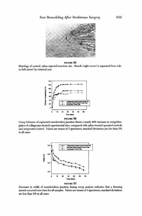
FIGURE 55
Histology of control, saline-injected insertion site. Muscle (right arrow) is separated from scle-ra (left arrow) by minimal scar.
*
Ii0
120
100
80 r°60
40 l -- Collarmg4snas.l.. Eopwlmnt.l SNb0 Opwrald Saoe-bmamdContro SN
20 [ -q- UnopratsdContra She
0
0 10 20 30 40 50 60
Trne (das)
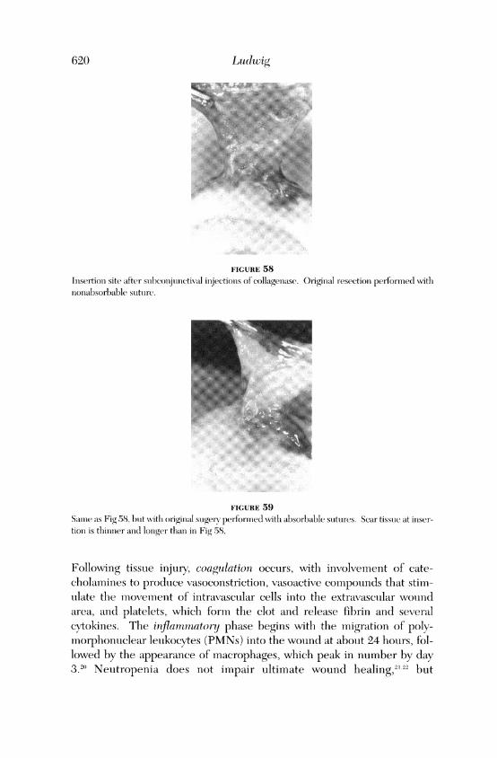
FIGURE 56Creep behavior of explanted muscle/sclera junction shows a nearly 40% increase in creep/elon-gation of collagenase-treated experimental sites, compared with saline-treated operated controlsand unoperated control. Values are means of3 specimens; standard deviations are less than 5%in all cases.
0.6
0.5
0.4 -
0.3
0 10 20 30 40 50 60
nrme (days)
FIGURE 57
Decrease in width of muscle/sclera junction during creep analysis indicates that a thinningstretch occurred over time for all samples. Values are means of3 specimens; standard deviationsare less than 5% in all cases.
C--Cob- asatred Exakont Sb..0.. Oparals Sabe-teala Control Sb
\-P- Unopeaed Control SibIfss<
Ltudwig
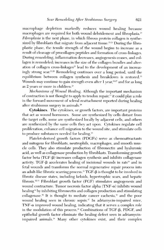
FIGURE 58Insertion site after slub)onjunctival ioijectioois of collagenase. Original resection performed withnonabsorbable suture.
FIGURE 59Samiie as Fig 58, but with original sugerv performned wNith absorbable sutures. Sear tissioe at iniser-tiooi is thliooller aind loniger thao io Fig 58.
Following tissne injury, coagulation occurs, with involvement of cate-cholamines to produce vasoconstriction, vasoactive compounds that stim-ulate the miovemnent of intravascular cells into the extravasclilar woundarea, and platelets, which form the clot and release fibrin and severalcytokines. The itnflatmmatorj phase begins with the migration of poly-morphonuclear leuikocytes (PMNs) into the wound at about 24 hours, fol-lowed by the appearance of macrophages, which peak in number by day3.2 Neutropenia does not impair ultimate wound healing,222 but
620
........
Scar Remitoclelitng After Strabis7mn.s Szirgery
macrophage depletion markedly reduces wound( healing becausemacrophages are re(luired for both wound debridement and fibroplasia.2"Fibroplasia is the next phase, in which fibrous protein collagen is synthe-sized by fibroblasts that migrate from adjacent tissue.-3-4 During the fibro-plastic phase, the tensile strength of the wound begins to increase as aresult of cleavage of procollagen peptides and formation of cross-linkages.Duiring remiiodelinig, inflamnmation (lecreases, angiogenesis ceases, and col-lagen is remodeled; increases in the size of the collagen buindles and alter-ation of collagen cross-linkages25 lead to the development of an increas-ingly strong scar.'32' Remodeling continiues over a long period, until theequilibrium between collagen synthesis and breakdown is restored."3Wounds may continue to gain strength even after 1 year,"'27 and for as longas 2 years or more in children.2s
Mechanisns of XVound Healing. Althouigh the important mechanismnof contraction is not thouight to apply to tendon repair," it could play a rolein the forward movement of scleral reattachment reported during healingafter strabismus surgery in animals.
Cytokines. The cytokines, or growth factors, are important proteinsthat act as wound hormones. Some are syinthesized by cells distant fromthe target cells, some are synthesized locally by adjacent cells, and othersare synthesized by the same cells they act uipon. Cytokines regulate cellproliferation, enhance cell migration to the wound site, and stimulate cellsto produce substances needed for healing."
Platelet-derived growth factors (PDGFs) serve as chemoattractantsand mitogens for fibroblasts, neutrophils, macrophages, and smootlh mus-cle cells. They also stimulate production of fibronectin and hyaluronicacid, as well as collagenase production by fibroblasts. Transforming growthfactor beta (TGF-P) increases collagen synthesis and inhibits collagenaseactivity. TGF-f3 accelerates healing of incisional wounds in rats3' and infetal wounds and transforms the normal regenerative repair process intoan aduilt-like fibrotic scarring process.:" TGF-4 is thought to be involved infibrotic disease states, including keloids, hypertrophic scars, and hepaticfibrosis.3'2" Fibroblast growth factor (FGF) stimulates angiogenesis andwoun(l contracture. Tumor necrosis factor alpha (TNF-oa) inhibits woundhealing34 by inhibitiing fibronectin and collagen produ-ction and stimulatingcollagenase." It is thought to mediate cancer cachexia,'6 and the poorwound healing seen in chronic sepsis.3' In adriamycin-impaired mice,TNF-ot improved wound healing, indicating that it serves a complex rolein the moldulation of this process.:" Combinations of TGF-1, PDGF, andepithelial growth factor eliminate the healing defect seen in adriamyciin-impaired animals.'" Many other cytokines exist, and their complex
621
Ludwig
interactions are still being uncovered. Perhaps cytokines may be used inthe future to manipulate wound healing.'3'26'33
The effect of increased TGF-4 on extraocular muscle healing couldpossibly increase scar formation and therefore lengthen the scar segment.A deficiency in TGF-1 could cause a weak scar, which would then beprone to stretch. The effect of this cytokine on scar lengthening is there-fore difficult to predict.
Extracellular matrix. Fibronectin is an adhesive protein that is thefirst substance to be laid down in a wound.26 It acts as scaffolding for cellmigration and collagen matrix deposition and is essential for wound heal-ing. Addition of fibronectin may enhance wound healing.
Glycosaminoglycans and proteoglycans are important components ofwound and scar extracellular matrix. Hyaluronic acid, a glycosaminogly-can, is thought to contribute to early cell migration into a wound and isfound in large quantities in fetal wounds, which heal by regeneration.4
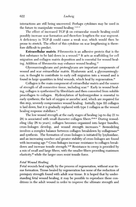
Collagen is the main component of extracellular matrix and the sourceof strength of all connective tissue, including scar.'3 Early in wound heal-ing, collagen is synthesized by fibroblasts and then converted from solubleprocollagen to collagen. Hydroxylation of hydroxyproline is essential toearly synthesis; the lack of oxygen or ascorbic acid, which are required forthis step, severely compromises wound healing. Initially, type III collagenis laid down, but it is gradually replaced with type I collagen as the woundhealing response stabilizes.4'
The low wound strength at the early stages of healing (up to day 21 to28) is associated with small diameter collagen fibers.25,4243 During remod-eling (day 28 to years), collagen becomes organized into larger bundles,cross-linkages develop, and wound strength increases.'2 Remodelinginvolves a complex balance between collagen breakdown by collagenase44and synthesis. The formation of cross-linkages is initiated by lysyloxidase,and an increasing number and greater stability of cross-linkages are foundwith increasing age.45 Cross-linkages increase resistance to collagen break-down and increase tensile strength.'2 ' Resistance to creep is provided bya mix of small and large fibers, with the smaller fibers providing maximumelasticity,46 while the larger ones resist tensile force.
Fetal Wound HealingFetal wounds heal rapidly by the process of regeneration, without scar tis-sue formation. Tissue healed by regeneration has none of the reduction ofpreinjury strength found with adult scar tissue. It is hoped that by under-standing fetal wound healing, it may be possible to reproduce those con-ditions in the adult wound in order to improve the ultimate strength and
622
Scar Remnocdeling After Strabismus Surgery6
function of the repaired tissue.'3'6 The potential ability to create a fetalwound-healing response could prove useful in the prevention or repair ofscar stretch after strabismus surgery.
Healing ofNormal Tendo)nDuring healing of normal tendon and ligament in the extremities, scar tissueorganizes parallel to tendon fibers. Biochemical differences between normaltendon and its scar tissue in terms of collagen type, arrangement of collagenfibrils, water content, DNA, and glycosamine content persist indefinitelyafter healing. When stress is applied to tendons, microscopic failures occurin collagen fibrils before gross separation, causing lengthening.47 If thesemicrobreaks then fill in with scar tissue, the lengthening would be expectedto become permanent. A similar mechanism could occur in scar lengthen-ing in strabismus surgery.
During tendon and ligament repair, gap formation should be kept to aminimum, as longer scars are weaker.4849 Promising prosthetic materials fortendon repair include polyester textiles and expanded polytetrafluoroethyl-ene fiber material. Electrical stimulation enhances ultimate tendon healing.Nonsteroidal anti-inflammatory drugs may improve the quality of healing.Fibronectin coating may enhance fibrosis to prosthetic devices such as poly-ester mesh.4
Factors That Influence and Impair Wound HealingManipulation of factors that influence wound healing may reduce the inci-dence of scar lengthening as well as improve repair results in stretched scarcases.
Nutrition. Nutritional factors that influence wound healing are numer-ous. Protein deficiency markedly impairs healing.5""', Glucose; essential fattyacids; vitamins A, B complex, and C; iron; zinc; calcium; copper; magne-sium; and essential fatty acids are all required for normal healing.'3'26,5'Vitamin C is especially important, as it is required for collagen cross-linking.
Excess vitamin E delays wound healing and increases postoperativeadhesions." Vitamin A reverses the effect of cortisone465" on wound healingand also increases collagen accumulation and postsurgical intra-abdominaladhesions in rats.52 Vitamin D deficiency causes collagen cross-linkingabnormalities.'
Oxygen. Oxygen is required for hydroxylation of proline and lysine incollagen formation as well as for the energy needed for all the cellular activ-ities. Hypoxia, anemia, and poor tissue perfuision all impair healing and canbe improved by oxygen administration.'3'26
Chronic Disease. Chronic diseases, including diabetes, uremia, cancer
623
(as well as its treatment with chemotherapeutic agents), sepsis, and radia-tion, impair wound healing.'9 Smoking, advanced age, jaundice, and alco-holism are also factors in impaired healing.53 Uremia interferes with colla-gen cross-linking.2
Chronic Inflamemation. Chronic inflammation leads to pathologic tis-sue destruction and impaired wound healing.19'54
Corticosteroids. Corticosteroids markedly reduce wound and ultimatescar strength by affecting all components of healing, in particular byinhibiting collagen synthesis.'9'2746'53
Hormones. Hypothyroidism leads to poor wound healing, whichimproves with thyroid replacement.52 Growth hormone increases woundstrength. Growth hormone has been shown to inhibit the reduction inwound healing caused by glucocorticoids.55
Abnormalities of Collagen. As collagen synthesis requires many com-plex steps, there are numerous potential sites where genetic abnormalitiesmay interfere with collagen production. Ehlers-Danlos syndrome is agroup of collagen disorders in which poor wound healing is common.45Although clinically diagnosed patients may have severe connective tissuedisease, there are probably subclinical variants that will become betterknown as collagen biochemistry techniques improve.'3
Marfan's syndrome and osteogenesis imperfecta are associated withdeficient collagen cross-linking.'2
PROPERTIES OF SCAR TISSUE
Strength ofNormal Scar TissuieMost scar tissue never achieves the original strength of native collagen.2The strength of the healed tissue varies relative to the strength of the orig-inal, unimpaired tissue, depending on the type of tissue and factors affect-ing wound healing.
The tensile strength of a healed tendon is about the same as thestrength of the musculotendinous junction, or about 8% to 10% of normaltendon strength.i Healed medial collateral ligaments of the knee achieve amaximum strength that is only 40% of normal.56 A comeal scar may neverrestore the original strength of the injured tissue.5 Rat skin wounds achieve80% of normal skin strength52 and guinea pig skin wounds reach only 25%of normal.5" Abdominal fascial wounds in rabbits ranged from 50% to 80%of normal strength at 1 year, but occasionally reach 90%.59 Stomach and duo-denal wounds generally do achieve the strength of normal tissue.6'
Metabolisml of Mature Scar TissueScar tissue is metabolically active, with equilibriuim existing between
624 Ludwvig
Scar Remnodeling After Strabisinus Surgery
collagen breakdown by collagenase and active collagen synthesis.The disruption of healed, mature scars in vitamin C deficiency was
noted centuries ago, when it was observed in sailors with scurvy. It is knownthat ascorbate deficiency results in a decrease in collagen synthesis."Collagenase activity in normal scars of healthy patients was found to bemarkedly greater than its activity in normal skin. The investigators postulat-ed that the equilibrium between normal collagenolysis and collagen synthe-sis in scar tissue becomes imbalanced in scurvy, causing scar weakening.6
Vitamin C deficiency any time after strabismus repair could cause scarweakening and lengthening, which would result in the clinical picture of astretched scar case.
EFFECTS OF TENSION
No studies of the effects of chronic tension on the extraocular muscle scarare found in the literature. However, extensive knowledge exists concern-ing the effects of tension on materials, cells, tissues, and scar tissues else-where in the body. These principles should have general applicability tothe extraocular muscles.
Material Response to Tension (Creep)From an engineering and materials science standpoint, catastrophic fail-ure or failure by fracture occurs when high stress is applied to a materialover a short period of time. Creep, on the other hand, is characterized bythe deformation of material resulting from continuous lower levels ofstress applied over relatively long periods of time.6263
All previous studies of wound and suture strength in strabismus sur-gery have been studies of the biologic equivalent of failure by fracture-rupture strength.'9"4'5 The behavior of strabismus wounds under lowerlevels of continuous stress, which is more indicative of the true environ-ment of the strabismus scar, has not been evaluated prior to this study.
Viscoelastic studies of human connective tissue show complex behav-iors.6"7 Collagen, as a complex macromolecular structure, is subject tocreep. The alternating fiber diameters in the arrangement of collagen ofnormal tendon increases resistance to creep,4" but this structure is lost inscar tissue, which, therefore, may be substantially less creep-resistant.
Cellular Response to TensionA study of cultured fibroblasts from chick embryos demonstrated thatmechanical stretching induced an increase in cell division and DNA syn-thesis."" Cultured rabbit aortic medial cells subjected to cyclic stretchingshowed twofold to fourfold increases in synthesis of types I and III colla-gen as compared to controls of agitated, but unstretched, cultured cells.
625
Hyaluronate and chondroitin 6-sulfate synthesis was also increased.69 Intissue cultures of fibroblasts on 3-dimensional collagen lattices, freely con-tracting lattices showed down-regulation of production of collagens I andIII. When the matrices were maintained under tension by fixing the rimsof the gel, contraction was inhibited and fibroblasts continued to replicateand to synthesize collagen without down-regulation.70
Epithelial cells cultured from porcine periodontal ligament signifi-cantly increased their synthesis of DNA within 30 minutes of being sub-jected to stretching forces. This effect was seen both during active stretch-ing as well as 2 hours after stretching had been discontinued. Stretchedcells showed an increased number of desmosomes and greater volumefraction of filamentous structures."' Cultured bone cells also showedincreased DNA synthesis along with increased protein synthesis after 2hours of applied stretch.7-
Prostaglandin synthesis was shown to increase when cultured bonecells were subjected to stretch.73 Cells cultured from rabbit coronal suturesresponded to stretch by increasing their synthesis of collagenase, 2 neutralmetalloproteinases, and collagenase inhibitor. Additionally, protein syn-thesis increased and collagen production doubled.74 Both collagen and theenzymes that degrade it, as well as the inhibitors that control degradation,are necessary for scar formation and remodeling.
Externally applied mechanical tension was also shown to change thearrangement of occluding junctions of cultured mammary epithelial cells.75
These tissue culture studies all demonstrated increases in cellular activ-ities when cells were specifically subjected to stretch, but not with the appli-cation of other types of mechanical stress. The findings in these studies sug-gest that the lengthened scars seen in strabismus could resuilt from mecha-nisms far more complex than mere stretching and may actually involveincreased scar tissue produiction. Such a hypothesis is in agreement with ourclinical impression that stretched scars are not just thinned and that agreater total volume of scar tissue is usually present in stretched scar cases.
Tissue Response to TensionSkin under tension is known to stretch, and this property is used intraop-eratively by plastic surgeons when closing large defects. Skin expanderprostheses may be used preoperatively to generate increased tissue for usein reconstructive surgery. A silicone bag is implanted under the skin to bestretched, and saline solution is injected weekly (beginning 2 weeks post-operatively) to gradually expand the bag and stretch the skin. Thestretched dermis shows fibroplasia, increased collagen prodluction, colla-gen fiber realignment, and increased vascularization.3
In a study of the biomnechanical properties of skin, load deformation
626 Ludwig
Scar Remodeling After Strabismus Surgery
curves were obtained by applying tension to skin.79 The data were difficultto explain mathematically until it was shown that several mechanisms wereinvolved because skin is not homogeneous.79 Histologic analysis showedthat the collagen fibers of relaxed dermis were arranged haphazardly, butthat when skin was held stretched during fixation, the fibers oriented alongthe line of stretch.79"" These results explained the ease of stretching relaxedskin, in contrast to the increasing difficulty of achieving more stretch oncethe fibers had aligned themselves. Gibson and Kenedi described the phe-nomenon of stress-relaxation, in which the force required to hold astretched piece of skin in the same position decreased over time.79 Theyalso found that creep occurred, in that a constant load applied to skincaused the skin extension to increase over time. They reported that con-ditions that cause gradual skin stretch, including lymphedema or obesity,can cause a permanent fourfold increase in skin length. Perhaps the verygradual stretching in these conditions is accompanied by collagen synthe-sis (in addition to creep), as when expander prostheses are used.
When mouse skin was subjected to externally applied stretch forces,an increase in the cellular mitotic index was seen, compared to controls.Initially, the epidermis thinned under tension (day 1), but at 4 days hyper-plasia was seen, with increased thickness of the cellular layer compared tounstretched controls.8' In another study, increased numbers of desmo-somes were found in stratified squamous epithelium subjected to severemechanical stress, especially in the epidermis of the bovine muzzle, whichis repeatedly stretched.82
Increased tension on vascular walls under conditions of experimentalhypertension was found to increase the aortic diameter, wall thickness, andcross-sectional area. Increased deposition of collagen in the rat aorticmedia was demonstrated.!3
Effects ofTension on Early Wound HealingWound strength early in healing is dependent on suture strength as well as thestrength of the tissues anchoring the sutures at the wound margin. A study ofearly healing and wound margin strength in rats showed that tissue within 1.5mm of an incision was markedly weakened all along the wound, not only atsuture placement sites. Tissue strength was not decreased 3mm from the inci-sion, except in incisions sutured under stretch, which had wider zonesofweakening. Midline rat laparotomywounds closed under tension showeda 77% decrease in wound breaking strength at 72 hours, even when thestretch was removed and the wound was resutured prior to testing. This earlydecrease in postoperative wound strength was reduced by oxygen, free radicalscavengers, and collagenase inhibitors, and was eliminated by induced neu-tropenia and a proteinase inhibitor (soybean trypsin inhibitor). The author felt
627
that these factors may therefore play a role in the stretch-induced reductionof early wound strength.22 Collagen content did not change during early heal-ing phases. Late gain in strength during the fibroplasia phase of wound heal-ing was unaffected by these early manipulations. 2`4
Perhaps antioxidants or antineutrophilic agents may have potential toprevent scar lengthening after strabismus surgery.
Effects of Tension on Scar TissueIn scars under tension, collagen fibers remodel and reorient themselves alongthe lines of tension.'79 When tension is equal in several directions, the resultis a hypertrophic scar, and when the tension is applied mainly in 1 direction,a stretched scar results.85
Histologic analysis of skin scars showed that with stretched scars, colla-gen fibers at the periphery of each scar were aligned longitudinally and cen-tral fibers were oriented transversely. The scars without tension remainednarrow, and all the fibers were oriented longitudinally. The authors hypothe-sized that the first fibers laid down during healing orient along a scar. Whentension across the scar is present, after external or suture support is removed,the scar under tension responds by laying down new collagen centrally, andthese fibers orient along the line of tension, across the scar.'8 The alignmentofcollagen fibers along tension lines explains the tendon-like appear-ance of lengthened scar segments and the frequent difficulty in dis-tinguishing the exact demarcation between normal tendon and scar.
Increased fibroplasia as well as greater collagen phagocytosis by fibrob-lasts occurs when the tension across a healing wound is increased.86 It isthought that this may provide a mechanism for the reorientation of fiber bun-dle direction seen in tension-stressed scar tissue. It appears that collagenremodeling, as well as increased collagen cross-linking, contributes to theprogressive increase in tensile strength measured in normal scars over thefirst year."7 A study in rats demonstrated that high closing tension impairedwound healing and strength in the abdominal wall.88
SURGICAL SCAR STRETCH/REMODELING IN OTHER TISSUES
Wherever scar tissue is subject to tension, "stretching," widening, or length-ening has been reported. The findings ofthe studies described below supportthe use of nonabsorbable sutures and materials to prevent or repair thesecomplications. Extrapolation of these findings to similar conditions in theextraocular muscle scar would predict improved results with nonabsorbablesutures.
SkinScar stretch is described in plastic surgery, often in wounds without apparenttension. Late stretching of nonfacial scars is frequently seen." Stretch is a
628 Ludwig
Scar Remodeling After Strabisnmus Surgery62
common cause of recurrence of brow ptosis after repair.'Three studies have been performed of skin wounds closed under ten-
sion, mostly after tattoo excision. In each study, a randomly selected half ofeach incision received a test suture method, with the other half serving as thecontrol. Each study showed decreased scar stretch with more prolongedsuture support.
In the first study, scar halves supported for 3 weeks by nonabsorbable,nylon subcuticular suture were 73% narrower 1 year later than control halvessupported by interrupted silk sutures removed at 10 days. Wounds closedwith deep dermal catgut were not different from the silk suture "controls."Wounds supported for 3 months only by superficial tape closure initially hadnarrower scars, which later widened to the same dimensions as the silk "con-trol" scars by 6 months. Although final scar widths were variable, the rate ofstretch was similar in all groups, with about 90% of stretch occurring in thefirst 6 months.'8
A similarly designed study compared 2 absorbable sutures, polyglycolicacid and polydioxanone; polydioxanone suture is known to retains its tensilestrength twice as long as polyglycolic acid suture. When the 2 types of suturewere placed subcuticularly in skin wounds under tension, the polyglycolicacid-sutured scars were 33% wider than the polydioxanone-sutured scars.'7
The third and most extensive study compared 6 months of subcuticular,nonabsorbable, polypropylene suture support to interrupted percutaneousnylon sutures removed at 12 days (controls). Variations tested included 3weeks of subcuticular polypropylene or polyglycolic acid suture support. Thescars supported for 6 months with nonabsorbable polypropylene suturesstretched 37.5% less than those with 12 days of nylon suture support and15.7% less than the scars with 3 weeks of nonabsorbable suture support. Theabsorbable sutures produced no difference in scar widths as compared to thenylon "controls.""
ScalpIn a study of scalp reduction surgery in 13 patients, in which midline scalpwas excised and the wounds were closed under tension with absorbablesuture, the rate of stretch-back was accurately measured over time, usingthe distances between 4 pairs of tattoos placed at surgery. It was shown thatmost stretch occurred in the wound area and not in adjacent skin. Stretchingwas clinically complete by 12 weeks, and averaged 34% to 52% of the origi-nal excision width.1'5 A similarly designed study compared postoperativestretch-back in wounds closed with permanent subcutaneous nonabsorbablepolypropylene suture in 8 patients versus absorbable polyglycolic acid suturein 13 patients. The wounds closed with absorbable sutures had scars thatwere on average 60% to 76% wider than the scars for wounds closed with
629
nonabsorbable sutures; the absorbable suture closures were also associatedwith significantly greater average total wound area and significantly greaterscar depression, compared with the nonabsorbable wound closures. Again,the greatest stretch was observed in the first 12 weeks, after which no changewas seen. The author felt that equilibrium was reached at 12 weeks, by whichtime the scars had achieved sufficient strength to resist stretching.'4 In thisstudy, measurements were made for only 5 months, and the possibility oflater stretch is not discussed. Additionally, the surgical procedure includedextensive undermining of scalp. It is likely, therefore, that these woundswould ultimately be supported by scalp-to-galea adhesions, which mayreduce tension across the midline scars. This is not the case in properly per-formed strabismus surgery, in which the entire tension of muscle pull isbrought to bear on the single scar between tendon and sclera.
FasciaAn incisional hernia develops after abdominal surgery in 3% to 11% ofcases.:"- It may develop long after seemingly successful healing has takenplace, sometimes years after the original surgery.','," Midline incisions,which are subject to higher tension across the wound, have higher rates ofincisional hernia than transverse abdominal incisions.92 Predisposing factorsare those which increase tension across the wound (obesity, pulmonary dis-ease with coughing, abdominal distension) or decrease the initial inherentwound strength (wound infection, use of steroids, jaundice, increased age,malnutrition, chronic illness).9-' One group of investigators found thatabsorbable polyglycolic acid-sutured wounds had a 12.5% failure rate ascompared to a 4.7% failure rate with nylon sutures.9"' A study of the longer-acting absorbable polydioxanone suture showed the development of inci-sional hernias in about 3% of cases by 1 year."3 Incorporation of absorbablemeshes did not provide the long-term tensile strength needed to preventincisional hernias in predisposed patients."x
Recurrence rates following primary repair of incisional hernias are high(30% to 50%).9297I0 2 The use of permanent, nonabsorbable polypropylenemesh for additional support was associated with reduced recurrence rates of0% to 10%.92911,101,H13'M Nonabsorbable internal retention sutures may alsoreduce recurrence of incisional hernias by eliminating the tension on themidline sutures.92"'5
The late-developing incisional hernia is less well understood. It seems todevelop independently of known predisposing factors.9 "06"17 Nonabsorbablesutures did not reduce the risk of late incisional hernia, although they marked-ly lowered the incidence of early ones.'(w Mudge and Hughes96 found that seri-ous disease, such as cancer, predisposed to the late development of incisional
Lud(wig630
Scar Renodeling After Strabisnius Surgerj6
hernia. An initially wider scar appears to increase the risk of late hernia.9Y"8Ellis and colleagues'°6 found no associated clinical factors to explain late-devel-oping incisional hernias, and felt they were due to stretching of mature scartissue. Harding and associates"', hypothesized that either scars were stretchingor scar tissue may be more metabolically active than has been recognized, andthat endogenous patient factors could disrupt the equilibrium of continuousresorption and laying down of new collagen. Urschel and coworkersN" notedthat scar tissue continues to undergo active remodeling, including productionof collagenase, at well after 1 year. They suggested that chronic mechanicalstress, as is associated with heavy physical exertion or chronic constipation,may play a role in late development of abdominal hernias.
The distinction between early and late developing incisional hernias sug-gests a parallel to the early and late development of secondary strabismuswith stretched scars of the extraocular muscles (see "Methods" and "StudyConclusions" sections).
Tendon and LigamentStretching has been described after both tendon-8 and ligament5 repairs. Astudy of factors affecting the strength of canine flexor tendon repairs con-cluded that the braided nonabsorbable suture Supramid produced the great-est final tensile strength. A suture technique that prevented gap formation atthe repair site was crucial. The best technique was found to be one that pre-vented tissue damage at the cut end of a tendon by suturing along the lengthof the tendon at its periphery. Sutures that traumatized the cut end of thetendon caused tip necrosis and a wider scar. Wider scars were found to bemore prone to stretch. An initially minimal gap between tendon ends filledmore quickly with collagen and allowed the scar to mature more rapidly, pro-ducing a stronger union. Active tension across the site in the early postoper-ative period markedly weakened the final result, presumably by creating alarger gap and longer scar. It was also demonstrated that tendon microcircu-lation was compromised by tension on the tendon during healing.Triamcinolone injections at the time of repair significantly reduced woundstrength at 6 weeks.' However, passive mobilization during healing increasedtendon scar strength, compared with immobilized tendons.'0
The principles of tendon repair advanced by these studies could proveapplicable to strabismus surgery.
Blood VesselsMicrovascular anastomoses in rats were seen to stretch with progressiveseparation of the ends of the vessels and pseudoaneurysm formation.Sutures were found within each scar and no longer through the vesselmedial layers by 10 days, and the gaps between vessel ends were bridged
631
Luclwig
by scar. One group of investigators suggested that the sutures had pulledthrough the vessel ends,"" but another group believed that the portions ofvessel wall contained within the loops of suture had become necrotic, suchthat the sutures were no longer attached to firm tissue, thereby allowingthe separation."'
Eyelid (Ptosis)The recurrence rate of ptosis after levator aponeurosis dehiscence repairwith long-acting absorbable polydioxanone sutures was significantlygreater than when nonabsorbable polypropylene sutures were used."12
PRINCIPLES OF SURGERY AND SUTURE TECHNIQUES
Injury is the stimulus for wound healing, and successful healing requires theapposition of wounded tissue. The healing of cut tendon to intact tissue,such as the intact sclera of strabismus surgery, is a situation not encounteredelsewhere in general surgery. Nevertheless, the direct suturing of tendon tosclera may induce enough scleral injury to allow proper tendon-to-sclerascar formation (E.E. Peacock, Jr., MD, oral communication).
Surgical technique and the choice of suture strongly influence theoutcome of wound healing. Electrocautery causes tissue necrosis thatinterferes with healing, and sutures tied too tightly cause necrosis in theincorporated tissue.26"'3 Tying knots too tightly has been shown to repro-ducibly reduce wound-breaking strength in rats.84 Irregular tissue injuryduring incision, as well as excessive needle-induced trauma during sutureplacement, hinders healing."3 In wounds closed under tension, sutures canbe seen to pull through their supporting tissues.22
Sutures that cause reactive inflammation, such as absorbable sutures,predispose to infection,"3 which in turn reduces wound strength.Absorbable polyglactin suture loses almost 50% of its strength by 2 weeks,has negligible strength at 4 weeks when collagen cross-linking is begin-ning, and causes an increasing chronic inflammatory response from days10 to 28 after placement."14 Absorbable monofilament polydioxanonesuture retains 70% of its strength after 28 days and produces little inflam-matory response."3 "4 Among the nonabsorbable sutures are polyester,which is a stable braided multifilament, and polypropylene, which is a low-reactivity and long-lasting monofilament."'
During tendon repair, the need to prevent gap formation is critical. Toprevent tissue necrosis at the cut ends of tendon, a lateral trap48 or endweave49 suture is used, which places the suture along the tendon periph-ery, widely distributing the tension.
Incorporation of these surgical and suture technique principles intostrabismus surgery may improve results.
632
Scar Remtodeling After Strabi.smus Surgery6
EXTRAOCULAR MUSCLE SURGERY: A REVIEW OF THE STRABISMUS LITERA-TURE, CONSIDERING THE POSSIBILITY OF STRETCH
Overcorrection After Strabismus SurgeryVariable long-term results after strabismus surgery have been reported bystrabismologists. Early reports of consecutive exotropia following esotropiarepair often lumped together different types of surgery, including medialrectus recessions and lateral rectus resection procedures, in the same studypopulation, leading to difficulty in interpreting the data with regard to scarstretch."6120 Overcorrection rates of 5% to 41% were reported.2"'612'2' Arecent pure study of bilateral medial rectus recessions showed an overcor-rection rate of 12%, with a mean follow-up of 27 months.'22
Many surgeons have reported that correction of secondary exotropia byundoing the original esotropia surgery often required more extensive surgerythan was done initially.2 1"'1931l25`2 This observation suggests that scar stretch-ing may have been involved, with reoperation performed on scar tissue ratherthan tendon. Readvancement in the presence of a stretch would require agreater distance of surgery to accommodate the length of the stretch.
Interestingly, Coopere noted that consecutive exotropia usually devel-oped following medial rectus recessions but was rarely seen after bilaterallateral rectus resection alone. Scar stretch following a resection procedurewould be expected to cause undercorrection, and such a case would not beincluded in an investigation of overcorrections. The effect of scar stretch onthe results of an initial uniocular recess-resect procedure could be variable:none if the stretches were balanced, or an undercorrection or overcorrec-tion, depending on which scar was stronger and which was subject to greatermuscle pull. This variability of effect seems to be borne out by the lowerreported incidence of overcorrection following recess-resect procedures.2"'7
Progressive overcorrection after inferior rectus recession was report-ed to be common, especially after adjustable suture surgery. It was mostcommon (50%), and difficult to treat, in patients with thyroid disease. Theauthors described the possibility that in some cases the muscle hadslipped, but mentioned that the transition between tendon and musclewas difficult to distinguish and that diagnosis of slip was difficult.'26 Theirobservations are not unlike the stretched scars described in this thesis.
The contribution of adhesive syndrome to variable results after stra-bismus surgery is frequently mentioned," 127 but not systematically analyzedin most case series of strabismus overcorrections. In our series, excessivescarring and adhesions of antagonist muscle(s) was the most commoncause of false preoperative suspicion of scar lengthening.
Normal and Elevated Forces of the Extraocular MusclesDuring unrestrained eye movements, tension in the horizontal rectus
633
muscles was reported to be 8 to 12 g at 15 degrees away from each mus-cle's field of action, and 40 g at maximal gaze into the field of action, instrabismic patients undergoing surgery.'28 In another study of 29 normalhuman subjects, maximum medial rectus force during active contractionwas 74.8 g, with 1 subject achieving 103 g. Mean maximum active lateralrectus force was 59.1 g, with a high of 92 g in 1 individual. In normal sub-jects, average lateral rectus active force was 40% lower than average medi-al rectus force.'29 Superior rectus tension exceeding 90 g has been meas-ured in thyroid ophthalmopathy with inferior rectus restriction.'31
Normal and Postoperative Extraocular Muscle Disinsertion ForcesDisinsertion forces of the musculoscleral junction have been reported inrabbit studies. After superior rectus recessions and resections with 6-0 silk,the tension required to disinsert each muscle was measured between days2 and 9. By day 4, average disinsertion tension was less than 50 g, but byday 5, it was 100 g, and on day 8, 300 g. The investigators concluded thatsuture support was no longer needed after day 5, as a 100 g disinsertiontension was probably sufficient to prevent rupture.64
A second study evaluated disinsertion forces over a longer postopera-tive period (weeks 1 through 5) and compared conventional inferior rec-tus recession to suspension inferior rectus recession. The ipsilateral supe-rior rectus of each eye underwent resection; 6-0 polyglactin sutures wereused in all cases. The disinsertion forces of normal unoperated controlsuperior recti and inferior recti ranged from 420 to 450 g. Resected supe-rior recti showed close to normal disinsertion force at each of weeks 1 to5, and conventionally recessed inferior recti achieved a disinsertion forceclose to 300 g by 1 week. Suspension-recessed muscles had a rupturestrength of less than 50 g at 1 week and 200 g at 2 weeks. The investiga-tors felt that the reduced rupture strength of the suspended muscle wasnot clinically significant in that normal rotational forces averaged 30 g.29
In the animal studies described above (see "Methods" and "Results"sections), significant scar lengthening occurred postoperatively with a con-stant 27 g load in both the collagenase-treated and saline-treated controlsuperior recti in rabbits. It appears that scar lengthening during remodel-ing develops at lower tensions than those required for rupture, well with-in normal extraocular muscle tensions. In pathologic states such as thyroidophthalmopathy, extraocular muscle tensions may greatly exceed normal,thus further predisposing to scar stretch.
Wound Healing in StrabismusWound healing after strabismus surgery in animals has been studied.29'3"132A detailed study of histologic changes after eye muscle surgery in rhesus
Ludwig634
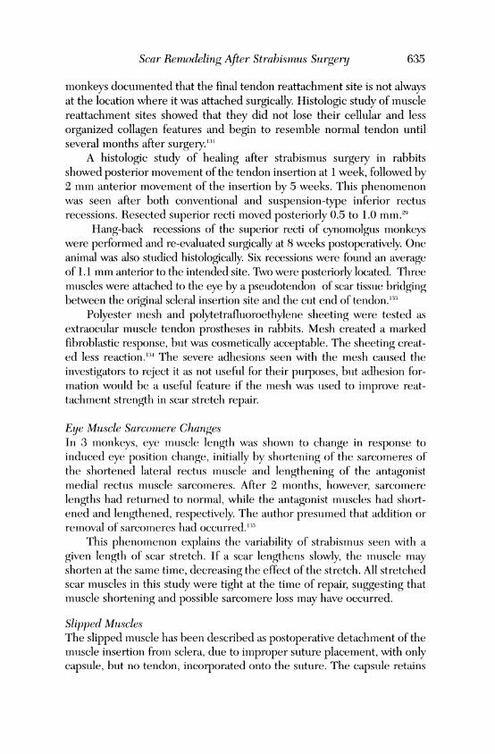
Scar Remwdeling After Strabisinus Surgery
monkeys documented that the final tendon reattachment site is not alwaysat the location where it was attached surgically. Histologic study of musclereattachment sites showed that they did not lose their cellular and lessorganized collagen features and begin to resemble normal tendon untilseveral months after surgery."'
A histologic study of healing after strabismus surgery in rabbitsshowed posterior movement of the tendon insertion at 1 week, followed by2 mm anterior movement of the insertion by 5 weeks. This phenomenonwas seen after both conventional and suspension-type inferior rectusrecessions. Resected superior recti moved posteriorly 0.5 to 1.0 mm.29
Hang-back recessions of the superior recti of cynomolgus monkeyswere performed and re-evaluated surgically at 8 weeks postoperatively. Oneanimal was also studied histologically. Six recessions were found an averageof 1.1 mm anterior to the intended site. Two were posteriorly located. Threemuscles were attached to the eye by a pseudotendon of scar tissue bridgingbetween the original scleral insertion site and the cut end of tendon.'33
Polyester mesh and polytetrafluoroethylene sheeting were tested asextraocular muscle tendon prostheses in rabbits. Mesh created a markedfibroblastic response, but was cosmetically acceptable. The sheeting creat-ed less reaction.'34 The severe adhesions seen with the mesh caused theinvestigators to reject it as not usefuil for their purposes, but adhesion for-mation would be a useful feature if the mesh was used to improve reat-tachment strength in scar stretch repair.
Eye Muscle Sarconiere ChangesIn 3 monkeys, eye muscle length was shown to change in response toinduced eye position change, initially by shortening of the sarcomeres ofthe shortened lateral rectus muscle and lengthening of the antagonistmedial rectus muscle sarcomeres. After 2 months, however, sarcomerelengths had returned to normal, while the antagonist muscles had short-ened and lengthened, respectively. The author presumed that addition orremoval of sarcomeres had occurred.135
This phenomenon explains the variability of strabismus seen with agiven length of scar stretch. If a scar lengthens slowly, the muscle mayshorten at the same time, decreasing the effect of the stretch. All stretchedscar muscles in this study were tight at the time of repair, suggesting thatmuscle shortening and possible sarcomere loss may have occurred.
Slipped MusclesThe slipped muscle has been (lescribed as postoperative detachment of themuscle insertion from sclera, due to improper suture placemnent, with onlycapsule, but no tendon, incorporated onto the suture. The capsuile retains
635
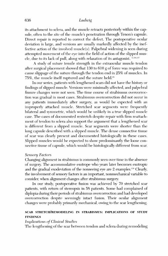
its attachment to sclera, and the muscle retracts posteriorly within the cap-sule, often to the site of the muscle's penetration through Tenon's capsule.Direct repair is reported to correct the defect. The postoperative oculardeviation is large, and versions are usually markedly affected by the inef-fective action of the involved muscle(s). Palpebral widening is seen duringattempted movement of the eye into the field of action of the slipped mus-cle, due to its lack of pull, along with relaxation of its antagonist. 3136137
A study of suture tensile strength in the extraocular muscle tendonafter surgical placement showed that 130 to 610 g of force was required tocause slippage of the suture through the tendon end in 25% of muiscles. In75%, the muscle itself ruiptuired and the suture held.35
In our series, patients with lengthened scars did not have the history orfindings of slipped muscle. Versions were minimally affected, and palpebralfissure changes were not seen. The time course of strabismus overcorrec-tion was gradual in most cases. Strabismus overcorrection did not occur inany patients immediately after surgery, as would be expected with animproperly attached muscle. Stretched scar segments were frequentlybilateral and symmetric, which would be unlikely in a true slipped musclecase. The cases of documented restretch despite repair with firm reattach-ment of tendon to sclera also suipport the argument that a lengthened scaris different from a slipped muscle. Scar segments were shorter than thelong capsule described with a slipped muscle. The dense connective tissueof scar was clearly present and documented histologically in these cases.Slipped muscles would be expected to show predominantly the loose con-nective tissue of capsule, wlhich would be histologically different from scar.
Sensory FactorsChanging alignment in strabismus is commonly seen over time in the absenceof surgery. The accommodative esotrope who years later becomes exotropicand the gradual exodeviation of the nonseeing eye are 2 examples.'38 Clearly,the involvement of sensory factors is an important, nonmechanical variable toconsider, when alignment changes after strabismus surgery.
In our study, postoperative fusion was achieved by 70 stretched scarpatients, with return of stereopsis in 58 patients. Some had complained ofdiplopia during their periods of strabismus overcorrection and had developedovercorrection despite seemingly intact fusion. Their ocular alignmentchanges were probably primarily mechanical, owing to the scar lengthening.
SCAR STRETCH/REMODELING IN STRABISMUS: IMPLICATIONS OF STUDY
FINDINGS
Imiiplications of Clinical StudiesThe lengthening of the scar between tendon and sclera during remodeling
636 Luidwig
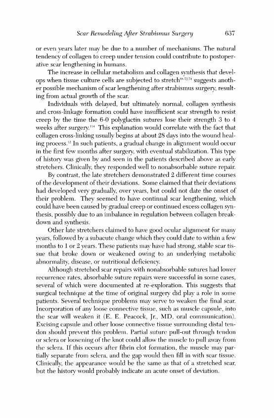
Scar Renodeling After Strabismuts Surgery6
or even years later may be due to a number of mechanisms. The naturaltendency of collagen to creep under tension could contribute to postoper-ative scar lengthening in humans.
The increase in cellular metabolism and collagen synthesis that devel-ops when tissue culture cells are subjected to stretch68-7274 suggests anoth-er possible mechanism of scar lengthening after strabismus surgery, result-ing from actual growth of the scar.
Individuals with delayed, but ultimately normal, collagen synthesisand cross-linkage formation could have insufficient scar strength to resistcreep by the time the 6-0 polyglactin sutures lose their strength 3 to 4weeks after surgery."'4 This explanation would correlate with the fact thatcollagen cross-linking usually begins at about 28 days into the wound heal-ing process.'- In such patients, a gradual change in alignment would occurin the first few months after surgery, with eventual stabilization. This typeof history was given by and seen in the patients described above as earlystretchers. Clinically, they responded well to nonabsorbable suture repair.
By contrast, the late stretchers demonstrated 2 different time coursesof the development of their deviations. Some claimed that their deviationshad developed very gradually, over years, but could not date the onset oftheir problem. They seemed to have continual scar lengthening, whichcould have been caused by gradual creep or continued excess collagen syn-thesis, possibly due to an imbalance in regulation between collagen break-down and synthesis.
Other late stretchers claimed to have good ocular alignment for manyyears, followed by a subacute change which they could date to within a fewmonths to 1 or 2 years. These patients may have had strong, stable scar tis-sue that broke down or weakened owing to an underlying metabolicabnormality, disease, or nutritional deficiency.
Although stretched scar repairs with nonabsorbable sutures had lowerrecurrence rates, absorbable suture repairs were successfuil in some cases,several of which were documented at re-exploration. This suggests thatsurgical technique at the time of original surgery did play a role in somepatients. Several technique problems may serve to weaken the final scar.Incorporation of any loose connective tissue, such as muscle capsule, intothe scar will weaken it (E. E. Peacock, Jr., MD, oral communication).Excising capsule and other loose connective tissue surrounding distal ten-don should prevent this problem. Partial sututre pull-out through tendonor sclera or loosening of the knot could allow the muscle to pull away fromthe sclera. If this occurs after fibrin clot formation, the muscle may par-tially separate from sclera, and the gap would then fill in with scar tissue.Clinically, the appearance would be the same as that of a stretched scar,but the history wouild probably indicate an acute onset of deviation.
637
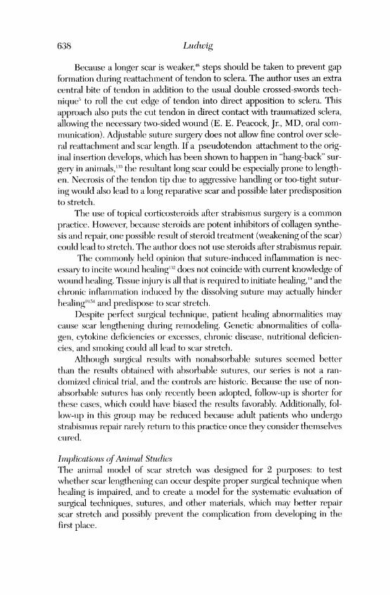
Because a longer scar is weaker,43 steps should be taken to prevent gapformation during reattachment of tendon to sclera. The author uses an extracentral bite of tendon in addition to the usual double crossed-swords tech-nique5 to roll the cut edge of tendon into direct apposition to sclera. Thisapproach also puts the cut tendon in direct contact with traumatized sclera,allowing the necessary two-sided wound (E. E. Peacock, Jr., MD, oral com-munication). Adjustable suture surgery does not allow fine control over scle-ral reattachment and scar length. If a pseudotendon attachment to the orig-inal insertion develops, which has been shown to happen in "hang-back" sur-gery in animals,'33 the resultant long scar could be especially prone to length-en. Necrosis of the tendon tip due to aggressive handling or too-tight sutur-ing would also lead to a long reparative scar and possible later predispositionto stretch.
The use of topical corticosteroids after strabismus surgery is a commonpractice. However, because steroids are potent inhibitors of collagen synthe-sis and repair, one possible result of steroid treatment (weakening of the scar)could lead to stretch. The author does not use steroids after strabismus repair.
The commonly held opinion that suture-induced inflammation is nec-essary to incite wound healing'32 does not coincide with current knowledge ofwound healing. Tissuie injury is all that is required to initiate healing,'9 and thechronic inflammation induced by the dissolving suture may actually hinderhealing"'54 and predispose to scar stretch.
Despite perfect surgical technique, patient healing abnormalities maycause scar lengthening during remodeling. Genetic abnormalities of colla-gen, cytokine deficiencies or excesses, chronic disease, nutritional deficien-cies, and smoking could all lead to scar stretch.
Althouigh surgical results with nonabsorbable sutures seemed betterthan the results obtained with absorbable sutures, our series is not a ran-domized clinical trial, and the controls are historic. Because the use of non-absorbable sutures has only recently been adopted, follow-up is shorter forthese cases, which could have biased the results favorably. Additionally, fol-low-up in this group may be reduced because adult patients who undergostrabismus repair rarely return to this practice once they consider themselvescured.
Imp)licationms ofAnimaull StudiesThe animal model of scar stretch was designed for 2 purposes: to testwhether scar lengthening can occur despite proper surgical technique whenhealing is impaired, and to create a model for the systematic evaluation ofsurgical techniques, sutures, and other materials, which may better repairscar stretch and possibly prevent the complication from developing in thefirst place.
Ludwig638
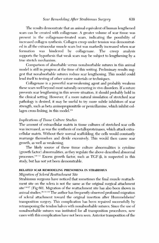
Scar Remsodeling After Strabisin.s Surger6
The resuilts demonstrate that an animal equiv-alent ofhuman lengthenedscars can be created with collagenase. A greater volume of scar tissue waspresent in the collagenase-treated scars, indicating the possibility ofincreased collagen synthesis. Collagen creep under tension was demonstrat-ed in all the extraocular muscle scars but was miarkedly increased when scarformation Wcas hindered by collagenase. The creep analysissupports the lhypothesis that weak scars may be subject to lengthening by atrue stretch mechanism.
Comparison of absorbable versus nonabsorbable sutures in this animalmodel is still in progress at the time of this writing. Preliminary results sug-gest that nonabsorbable sutures reduce scar lengthening. This model couldlend itself to testing of other suture materials or techniques.
Collagenase is a powverful scar-weakening agent and probably weakensthese scars well beyond most ncaturally occurring in vivo disorders. If a sutureprevents scar lengthening in this severe situation, it should probably hold inthe clinical setting. However, if a more natural simulation of stretched scarpathology is desired, it may be useful to try more subtle inhibitors of scarstrength, such as beta aminopropionitrile or penicillamine, which inhibit col-lagen cross-linldng, in this model."9
Imp)lications of Tissue Cultulre StudiesThe amount of extracellular matrix in tissue cultures of stretched scar cellswas increased, as was the synthesis of metalloproteinases, which attack extra-cellular matrix. WVithout their normal scaffolding, the cells would constantlyrearrange them-selves and divide excessively. This would then catuse scargrowth, as well as weakening.
The likely source of these tissue culture abnormalities is cytokine(growth factor) abnormalities, as they regulate the above-described abnormalprocesses.'4'1'-2 Excess growth f:actor, such as TGF-,B, is suspected in thisstudy, but hals not yet been demonstrable.
RELATED SCAR REMODELING PHENOMENA IN STRABISMUS
Migration of Scleral Reattaclhmtent SiteStrabismus surgeons have noticed that sometimnes the final muscle reattach-ment site on the sclera is not the same as the original surgical attachmnentsite' l-27 (Fig 60). Migration of the reattachment site has also been shown inanimal studies.-''-' The author has frequently observed profound migrationof scleral attachment towvard the original insertion after Hummelsheim5transposition surgery. This comuplication has been repaired suceessfuilly byretransposing the tendon halves with nonabsorbable sutures. Since the use ofnonabsorbable sutures was instituted for all transposition procedures, newcases with this complicatioin have not been seen. Anterior transposition of the
639
inferior oblique has also occasionally failed graduially, owing to posteriormigration of the scleral reattachment site during remodeling, and nonab-sorbable sutures may prevent this. The mechanism of scleral site migration isunknown, buit it could be hypothesized to be related to greater tension on themore anterior fibers of the scar, with gradual lengthening, as well as increas-ing scar strength of the shorter, less-stressed posterior fibers. Because thereis no corresponding scleral wound to direct the scar position, collagen fiberswill attach themselves anywhere, and vill adhere better where they are underless tension. As remodeling progresses, the position gradually shifts posteri-orly. Nonabsorbable suture should reduce tension on the scar, reducing posi-tion shift.
Faden and Jensen PnmcedtresTwo patients who had undergone previous Faden sututre placements else-where underwent reoperation for recurrent strabismus. All the operatedmuscles were intact, in their normal anatomic positions, with no adhlerenceof the muscle bellies to sclera. Similar observations have been made byanother strabismologist (D. R. Stager, MD, oral communication).
Another patient underwent a Jensen procedure for ciabetic third nervepalsy. Afterward, postoperative alignment was excellent, but the originalexodeviation recurred over several months. On reoperation, the superior,inferior, and medial recti were found to have returned to their normalanatomic positions; although the muscles were surrounded by scar tissue,they themselves bore no evidence of the original surgery. The nonabsorbablesutures were found embedded in connective tissue at the sites where theyand the muscle halves had originally been placed, midway between the nor-mal muscle positions. A simnilar case was described to the author by anothercolleague (E. C. Price, MD, oral communication).
The practice of suturing into muscle tissue is common in strabismus sur-gery, but not in other (isciplines. Muscle must be repaired by repairing itsconnective tissue, as one canniot suture globular protein (E. E. Peacock, Jr.,MD, oral communication).
During natural remodeling of the surgical site, sutured muscle fibers canpull out of their sutuires, and the muscles may returm to their norm-al positionsand repair their defects. If anl adhesion forms between sclera and the muscle,the desired surgical effect may persist, but the sutuire alone is unlikely to holdits position under tension in muscle.
WOUND HEALING IN STRABISMUS
With the miarked expansion of research and understanding of mechanismsin wound healing, clinical application of these insights should translate to
640 Liiclwig
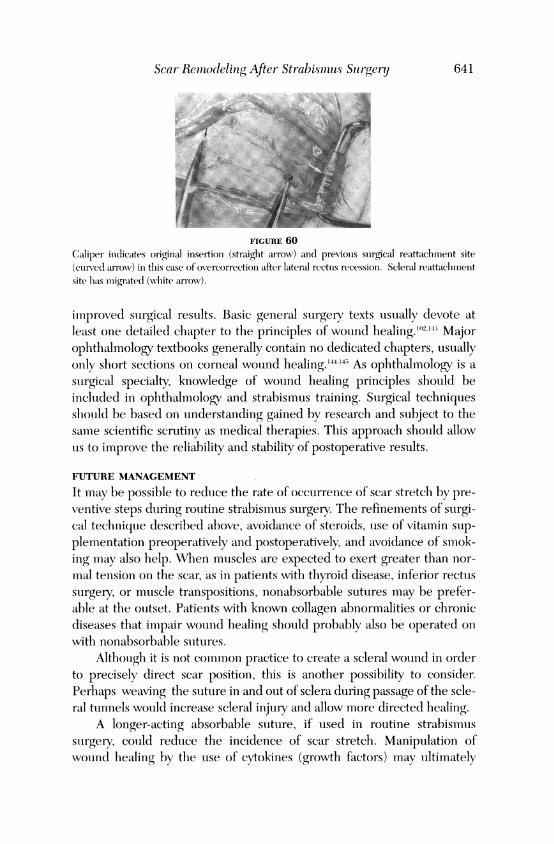
Scar Remo(leling After Strabismlus Suirgery6
FIGURE 60Caliper indicates original inisertion (straight arrow) and prexious sturgicall reattachlimenit site(cunred arrow) in this case of overcorrection after lateral rectus recession. Scleral reattachmehntsite las Iiligrated (white arrow).
imuproved surgical results. Basic general surgery texts usually devote atleast one detailed clhapter to the principles of wounid healing.'-1243 Majorophthalmology textbooks generally contain no dedicated chapters, usuallyonly short sections on corneal wound healing. 44'15 As ophthalmology is asurgical specialty, knowledge of wound healing principles should beincluded in ophthalmuology and strabismus training. Surgical techniquesshouild be based on understanding gained by research and subject to thesame scientific scrutiny as medical therapies. This approach should allowuis to imnprove the reliability and stability of postoperative results.
FUTURE MANAGEMENT
It may be possible to reduce the rate of occurrence of scar stretch by pre-venitive steps during routine strabismnus surgery. The refinements of surgi-cal techniquie described above, avoidance of steroids, use of vitamin sup-plementation preoperatively and postoperatively, and avoidance of smok-ing may also help. When muscles are expected to exert greater than nor-mal tension on the scar, as in patients with thyroid disease, inferior rectussuirgery, or muscle transpositions, nonabsorbable sutures may be prefer-able at the ouitset. Patients with known collagen abnormalities or chronicdiseases that impair wound healing should probably also be operated onwith nonabsorbable suitures.
Although it is not common practice to create a scleral wound in orderto precisely direct scar position, this is another possibility to consider.Perhaps weaving the suture in and out of sclera during passage of the scle-ral tunnels would increase scleral injury and allow more directed healing.
A longer-acting absorbable suture, if used in routine strabismussurgery, could reduce the incidence of scar stretch. Manipuilation ofwouind healing by the use of cytokines (growth factors) may ultimately
641
allow regenerative healing anid better scar strength.Under tension, nonabsorbable sutures can pull out of tissue, and in
some situiations, a broader surface area of nonabsorbable support isrequired. Because nonabsorbable meshes are known to reduce the recuir-rence of incisional hernia, polyester surgical mesb reinforcemenit was uisedin superior rectus surgery in rabbit eyes to determine whether the mesbwould be tolerated in the subcoinjunctival space (Fig 61), in the hope thatthis m-aterial could be used to strengthen the muscle-to-tendon adhesionin cases of severe stretched scar. The mesh was well tolerated for a year(Fig 62), with integration of the mesh and the underlying sclera and mus-cle (Fig 63). With the development of finer-gauge meshes and improvedsurgical teclhnique, mesh reinforcement may halve future utility in humanstretched scar cases that recuir after nonabsorbable suture repair.
CONCLUSIONS
This thesis has detailed the intraoperative finding of a segment of length-ened scar tissuie between extraocular muscle and sclera in a number ofstrabismus reoperation cases. The frequency of occurrence of lengthenedscar is especially high in strabismuts overcorrection cases.
The scar segment can be difficult to see despite carefuil dissection andinspection of the muscle, as the scar often blends indistinctly into tendon.Feattures that suggest scar stretch include laxity at the insertion site, theamorphous natuire of the scar as compared to tendon, and longer-than-expected tendon. Inspection of the internal aspect of the mullscle afterdisinsertion can be helpful in confirming the diagnosis.
On the basis of the clinical and animal studies and a literatuire reviewof similar phenomena in other surgical situations, it is concluded that:
* The scar segment comnplication can develop despite properly per-formed strabismus surgery, and the time course of development ofsecondary strabismus caused by lengthened scars is variable, withmany occurring during the first few months after surgery, and othersyears later. Physiologic processes must therefore contribute to scarlengthening or stretching.
* The stresses to which these operated muscles are subject contributeto the development of this complication in predisposed patients.Studies demonstrating the effect of tension on cells, tissues, and thewound healing process suggest a high probability that scars tindertension may actually grow by synthesizinig extra collageni, and alsostretclh due to creep imeclhanisms when a scar is weak.
642 Ltidtvig

Scar Remodeling After Strabismulls Sn rgery 643
FIGURE 61
Polyester mlesh is surgically implanted over superior rectois ao(lI sclera of rabbit. Stoperior rectoLswas disioiserted aiod reattachied.
FIGURE 62Soiperior coojunctiva of ralbbit in Fig 61 after 1 year. Edges of mnesh indicated bsy arrows.
FIGURE 63After enieleation, mnesh is seen to be completely covered by conIjunctiva, wNithout erosion. Messhouttlines are irldicated bv arrows.
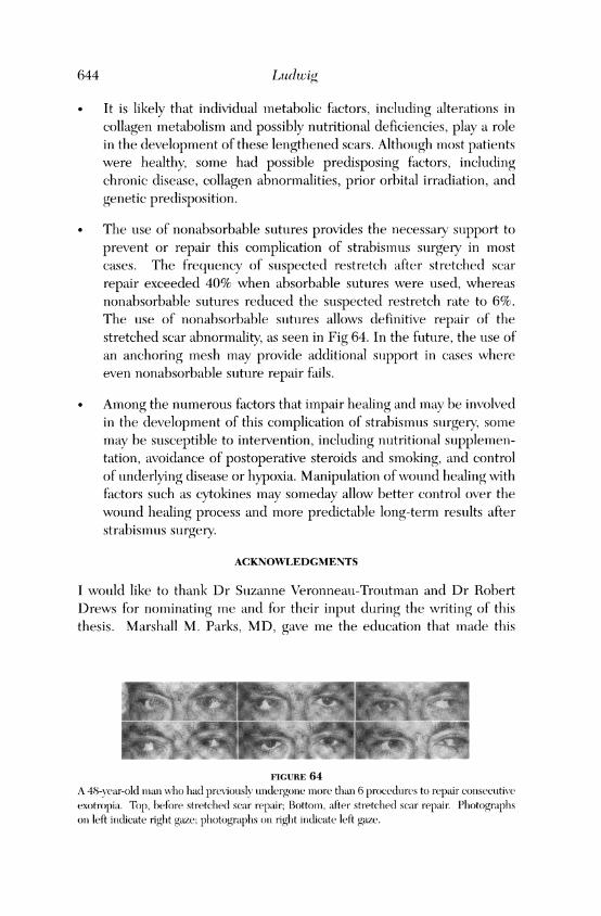
* It is likely that individual metabolic factors, including alterations incollagen metabolism and possibly nutritional deficiencies, play a rolein the development of these lengthened scars. Although most patientswere healthy, some had possible predisposing factors, includingchronic disease, collagen abnormalities, prior orbital irradiation, andgenetic predisposition.
* The use of nonabsorbable sutures provides the necessary support toprevent or repair this complication of strabismus surgery in mostcases. The frequency of suspected restretch after stretched scarrepair exceeded 40% when absorbable sutures were used, whereasnonabsorbable sutures reduced the suspected restretch rate to 6%.The use of nonabsorbable sutures allows definitive repair of thestretched scar abnormality, as seen in Fig 64. In the future, the use ofan anchoring mesh may provide additional support in cases whereeven nonabsorbable suture repair fails.
* Among the numerous factors that impair healing and may be involvedin the development of this complication of strabismus surgery, somemay be susceptible to intervention, including nutritional supplemen-tation, avoidance of postoperative steroids and smoking, and controlof underlying disease or hypoxia. Manipulation ofwound healing withfactors such as cytokines mLay someday allow better control over thewound healing process and more predictable long-term results afterstrabismus surgery.
ACKNOWLEDGMENTS
I would like to thank Dr Suzanne Veronneau-Troutman and Dr RobertDrews for nominating me and for their input during the writing of thisthesis. Marshall M. Parks, MD, gave me the education that made this
FIGURE 64A 48-vear-old iiiato w ho ia(l preNiously ondergone more than 6 procedures to repair coosecutiveexotropia. Top, before stretched scar repair; Bottom, after stretched scar repairn Photographsoni left ioidicate right gaze; photographs on right iihiccate left gaze.
644 Luidwig
Scar Reniodeliug After Strabismann,s Sn rgery
work possible. Alan Y. Chowv, MD, who had independently recognized thestretched scar pbenomenion before the first of our many discussions, con-tributed his ovni case records, which are inecluded in this series. DrHerbert E. Katufimiani enecouraged and provided the resources for thiswork. Jean T. Jacob, PhD, and her associates performed the rabbit care,creep studies, and rabbit Ihistopathology. Roger W. Beuerman, PhD andDoan Nguyen, PhD, performed cellular studies. Alma Baird, PaulFalkenstein, and the suture development team at Ethicon Corporationcreated and provided sutures, as well as supplies and financial support.Most of the intraoperative photographs were taken by anesthesiologists H.Sharron Carson III, MD, and Ralph Fillmore, MD, and the histopatho-logic interpretation and photographs of human tissues were contribuitedby pathologists Arthur R. Summerlin III, MD, and John Rodenbough,MD. Hilary W. Thompson, PhD, provided statistical analyses, and MaxineHaslauer created the photographiic layouts. Patila Gebhardt provided edi-torial services, and Lisa Curole spent many houirs in the library searchingfor references.
I would also like to thank my loyal patients, who made imiany follow-up visits, and most of all, my husband, Jim Cox, and my sons, Keith andPeter Minor, who worked hard to give me the many quiet evenings need-ed to accomplish this project.
REFERENCES
1. voi Noorleni CGK. Bitnocdlar Visioni anid OcuIlair Alotility. Tlheori anld Aalaagcllieft ofStrabisnims. 5th dcl. St Louis: Mosbv; 1996;299-340.
2. Cooper EL. The surgical muaniagemiienit of secondary exotropia. Tranis Aoii AcadOphlitalmol Otolaryinol 1961;65:59.5f607.
3. Parks MM, Bloonii JN. The "slipped imutscle. Ophltlialiiolo(gy 1979;86:1389-1396.4. Parks MM. The overacting iiiferior obliqiie-muscle. The XXXVI deSchweinitz lectuire.
An, J Op/lialinli0ol 1974;77:787-797.5. Parks MM. Atlas of Strabisimii.s Suirgety Philadelphiia, Harper & Rowr? 1983;11:3.6. von Noordei CGK. Binioctular- V7isioii anid Ocutlair Alotility. Theorij aiild Alnagclliiemt of
Strabisinus. 5th edl. St Louiis: Moshv; 1996;526-583.7. Kleiner D, StetlerfStevensoni WNI Quantitative zvInography. Detection of picograim
quianitities of gelatiniases. Anial Biocliciii 1994;218:325-329.8. WN'iinrel) RN, Kashivagi J. Kashiwvagi F. et al. Pr-ostagland(inls inicreases miiatrix inietallo-
proteiiuase release fromii hluisiaIn ciliarv smiiooth muiiiscle cells. Iiic.st Oplithlalmol Vi.s Sci1997:;38:2772-2780.
9. Jacol JT, Lini Jj Mikal SP. Syn-thetic scleral reiniforcemiienit miiaterials. III. Changes insurface anid blulk physical properties. J Bioioed Alater Rc.s 1997;37:525-533.
1(). Creenwald MJ, Ticho Bl.l Enigel JM. Extr-aoctilar muitiscle surgery. In: Kirupini T,Kolker AE, eels. Atlas of Coinplication.s in Opltalmalic Suirgcrj. LonldoI, Wolfe/Mosby-Yearbook; 199:3;9.2-9.25.
1 1. Dtubar JA, lender GT. Intraoperative delhisceuice of' a rectuis mulscle: Report of 2
645
646 Ludwig
cases. J Ani Assoc Pediatr Ophlthalmol Strbisnsins 1i997;1: I 75-1 77.12. Yamanchi M, Mechanic (L. Cross-liinkinig of collaagen. In: Nimnli fME, ed(. Collaogco.
A'oloiiiie 1 Biochleoi.strl Boca Raton, Fla: C'RC Press, Inco: 1988;6:157-172.13. Coheni IK, DiegelmacInn RF, Yager DR, et al. \\Nonilll care an(I wOnd(l healing. In:
Schwartz SI, Shires GT, Spenicer- FC, et al, ecds. Principles (of Surgetryi. TtlI ed. New York:McGraw-Hill; 1999;263-295.
14. Nor-dstrm fREA. "Stretch-back' in scalp redtuctionis for iimale patter-ni baldiiess. Pla.stReconstr Sirg, 1984;73:422-426.
15. Nordstrom REA, Nordcstimil RNI. Ahsorhahle verstus nonabsorhable stitures to preventpostoperativ,e stretching of wound area. Plast Reron st- Sn r-g 1986:;78:186-190.
16. Elliot ID, Mahaffey PJ. The stretclhedl scar: The l)enefit of prolonigel dermiial stnpport. Br-JPla.st Snrg 1989;42:74-78.
17. Chanitar-asak NI). Milner lI 1. A comllpar-isoni of scar (lualitv ill woun1114ds closed nuitder tent-sion with PCA (Dexoni) and polvdcioxianonse (PDS). Br^j Plast Sm-g 1989;42:687-691.
18. Sommiiiierlad BC, Creasev JM. The stretclhed scar: A cliniical and histological stndv. BrJPlast Surg 1978: 31:34-45.
19. Peacock EE Jr. WN'oundicl Repair. 3rd edlitioni. Philadelphlial: WTB. Sauniiders; 1984:102-140.20. Liehovich SJ, Ross R. TIse role of tlse uniacroplhage in wound repair. A study' with hvdro-
cortisone annd anitimiacr-ophalge ser-uimll. Ami1 J Pathlol 1975;78:71--1()0.21. Simipsoni DNI, Ross R. The nietitrophilic leikocvte in wound repair. A study with aniti-
nieutroplhil serLumii.J Clitn Incest 1972;51:2009-2023.22. Il0gstr6m TI. Meclhaniismiis and( prevention of' (lecrease in wounel imargini strenigtlh in
initestinial aniastomiioses anid laparotomy wounds. Thesis. Lundi(I University, Malm6i,Sweelen, 198 7.
23. Morgani CJ, Pledger WNI. Fibroblast proliferationi. In: Coheni IK, lDiege'lmann ',R,Liidblald N'j, eeds. XlVound Hcaliua. Biochemnical oe!d Cliniral Aspects. Philae(lclhlial:WN7B. Saumders; 1992;6:3-76.
24. lRoss R. The fibrohlast adel wounld repair. Biol RHe 1968;43:51-96.25. Clark RAF. XNoiuidI repair. Ov-eriew and general coinsicleratiomis. In: Clark IRAF, ed. The
Molecular antid C/ellndt- Bioloay of \ouIInd RHepair. 2ned ed. Nes York: Plenitumii Prc'ss;1996;3-501.
26. Aclzick NS. \V'onloid heallinig. Biologic' and cliniical featuires. In: Sabistoni DC ji', ed.Textbook of Surgery: The Biologic Basis of \Modern Suirgical Practice. 15th eld.Plilaldelpliia: \WB. Saunders; 1997:;207-22t).
27. Van \VNinkle \V7 Jr. The tenisile strength of wouincl anid factors that influence' it. SnurgC|ynecol Ob'stet 1969;129:819-842.
28. Marks NIMW,Marks C. Fulnda,nicnital.s of Plastic Sturger7y. PPhiladelphliia: WB. Saunders;1997;1-18.
29. He1-tle 11W7, Jamiies NI, Farher MG. Iniser-tioni site dynaics histology in a rahhitImlodel after conventional or suispenisioni recttis recession comhinecl \ith ipsilaterIalaintadgonlist resectioni. J Pediaitr Opithlalmol Strabi.snuns 1993;:30:184-191.
30. Miustoce TA, Pierce GF. Thoiatsomi A, et al. Acceler-ated healina c)f incisional woundls inrats iniuclt'ed hv- transforning grosw'th fac'tor-P. ScielOc( 1987;237:1333-1336.
31. Kruiiueumiel TNi, Michlna BA, Tlhomlas BL, et a]. Transforming growth factor eta (TCGF-P) ineldtces fibrosis in a fetal wound miiodlel. ] Ped/iat- Suerg 1988:;23:647-652.
32. Bor(ler \WA, Nohle NA. TrIansforming grrowth factor P in tissue' fibrosis. NV Enigl J Med1994;331:1286-1292.
33. Mar-tin CR, Peacock EE Jr. Ceurreuit perspectives in wos el hiealinig. In: Coheln IK,Diegelmiiaunii RF. Lindlhlacl WJ, ecls. llWiund Healing. Biochemic'ii(il an}d Clinical Aspects.Philaldelphia: WN.B. Saunders; 1992;1-4.
Scar Remlodeling After Strabism2-us Surgery
34. Rapala K, Laato M, Niiniikoski J, et al. Ttumiior niecrosis factor alpha inhibits wound heal-ing in the rat. Eur Suirg Res 1991;23:261-268.
35. Regan MC, Kirk SJ, Huirsoni M, et al. Ttiumior niecrosis factor-ot inihibits in vivo collageIsynthesis. Suirgery 1993;113:173-177.
36. Mooney DP, O'Reilly M, Gamelli RL. Tumiiior necrosis factor and wound healinlg. AnnrSuirg 1990;211:124-129.
37. Cooney R, locono J, Maish G, et al. Tulmor necrosis factor miediates imapaired woundhealing in chroniic abdomninal sepsis. J Traum7Ia 1997;42:415-420.
38. Grotenidorst GR. Chemoattractants and growth factors. In: Coheni IK, Diegelmlanii RF,Lindblad XVJ, eds. XVotuuid Healinig. Biochiemlical and Clinical Aspects. Philadelphia:WN'B. Saumsders; 1992;237-246.
39. Grininell F. Cell adlhesioni. In: Colheni IK, Diegelmann IIF, Lindblad WJ, eds. WVoui2ndHealing. Biochemizical and Clinical Aspects. Philadelphia: NV.B. Sauinders; 1992;209-222.
40. Weitzhandler M, Beruifield MR. Proteoglycan glycoconjugates. In: Cohen IK,Diegelmann RF, Lindblad WNJ, eds. XVouncl Healing. Biochemiiical and Cliniical Aspects.Philadelphia: W1B. Saunii(ders; 1992;195-208.
41. GCallo RL, Bernfield M. Proteoglycans anid their role in wound repair. In: Clark RAF,ed. The AMolecuilar and Celltular Biology of WVound Repair. 2nd ed. NeW York: PlenumPress; 1996;475-492.
42. Doilloin CJ, Dunn MC, Bender E, et al. Collageni fiber formiiation in repair tissule:Developmnent of strenigth and totuglhniess. Coll Relat Res 1985;5:481-492.
43. Marks MW Marks C. Funtaclmtiienitals of Plastic Surgery. Philadelphia: W.B. Sauinders;1997;101-111.
44. Jeffrey JJ. Collagen degradation. In: Cohen IK, Diegelmann RF, Lindblad WNJ, eds.XVountid Healing. Biochemttical anid Cliniical Aspects. Philadelphia: N1B. Satuniders;1992;177-194.
45. Phillips C, WNenstruLp RJ. Biosynthetic and genietic disorders of collagen. In: Coheni IK,Diegelmann RF, Lindblad XV7J, eds. WVotunid Healing. Biochemrlical atnd Cliniical Aspects.Philadelplhia: W1B. Sauinders; 1992;152-176.
46. Nimni ME, Harkness RD. Molecular structure and fiunctions of collagen. In: NimniiiME, ed. Collagen. Vlolumjiie I: Biochemistry. Boca Raton, Fla: CRC Press; 1988;1-77.
47. Amiiadio PC. Tendon anid ligamnent. In: Coheni IK, Diegelmann RF, Lindblad WNJ, eds.\Votutnd Healina. Biochemtiical and(1 Cliniical Aspects. Philadelphia: W1B. Salunders;1992;384-395.
48. Ketchuin LD, Martin NL, Kappel DA. Experimental evaluation of factors affecting thestrength of tendon repairs. Plast Reconistr Suirg 1977;59:708-719.
49. Peacock EE Jr. WVoutnd Repair. 3rd ed. Philadelplhia: WV.B. Sauniiders; 1984;263-331.50. Leveinsoni SM, Demetriou AA. Metabolic factors. In: Colsen IK, Diegelinann RF,
Lindblad XVJ, eds. WVoundi(l Healing. Biochemttical and Cliniical Aspects. Philadelphia:WV.B. Sauin(ders; 1992;248-273.
51. WN'imidsor JA, Kniight GS, Hill GL. WN'ouind healinig response in suirgical patienits: Recenitfood intake is imore important than nutritionial status. BrJ Soirg 1988;75:135-137.
52. Levensoni SM, Geever EF, Crowley LM, et al. The lhealinig of rat skin wounds. Atnnt Suirg1965: 161:293-308.
53. Lawrenice 'NNT. Cliniical mainaagemilenit of ioilihealinig wounds. In: Coh1eIn IK, DiegelmunaiI1RF, Lindblad NVJ, eds. XVoultid Healinga. Biochliemiical anid Cliniical Aspects. Philadelplhia:\V.B. Saunders; 1992;541-561.
54. Wlahl LM, W;\all SM. Iinflaminationi. In: Cohleni IK, Diegelmilauini RF, Lindblad NVJ, eds.Wunnd Healing. Biochlemtiical atnd Clinical Aspects. Philadelphia: W1B. Sauunders; 1992;40-62.
647
648 Ltudtwig
55. Kellev SF, Felix ANI, Elirliclh HP The antagonism of gllCocorticoi(d inhibition ofwound buld-inog in rats bvgrowtb lormone-releasinc faictor. Pr)c Soc Exp Biol Mled 1991d94:320-326.
56. FranIk C, Am11iel D, W7oo SL-Y. et 11. NormIal ligam1eint properties an1d ligami1en1t hecalingC/lin Ortlhopt 1985; 196:15-25.
57. Dlavison PF, Galba-vv EJ. Connective tissuie remi(odeliig in corineal an(l scleral wvonnds.Invest Ophlthlalm7otl Vis Sci 198627:147(8-1484.
58. Donglas DM. The healing of apon1euirotic inlcisionls. B1rJ Snrg, 1952:40:79-84.59. I)ouelas DM, Forrester JC Ogilvrie RR. Physical characteristics of collageIn in the later
stages of wound healinig. BtrJ Stnrg 1969;56:219-222.60. Gottrup F. Healinjg of incisioiial wou(ns in stoIm1ach1 aIn(I (lIio(denlmiii: Iniflutence of loig-
terimi lhealing oni miechianiical strengthl anid collagen distribtitioii. Actai Chiir Scanl1983; 149:57-62.
61. CohIenI IK, Keiser HR. DisruptioIi of hiealed scars in scur-vy-thie result of a disequilibri-UIi1 in collagen imetabolisimi. Pla.st Recon.str Snirg 1976;57:213-215.
62. Rovlance D. MNechaIIics of Materials. New York, Jolin Wilev & SoIns; 1996.63. IRilev WF, Stuirges LD, NMoi-ris DfI. Statics an,d Mechlanics of Materials: Ant Integrated
Approaclh. New York: Joh1I1 Wiley aind SoIns; 1995;66-126.64. Apt L, Cafnev- WL. Dorat AF. ExperiiimenItal suture sttudies in strabismiius surgery. I.
IReattachm111en1t rate of extraoctilarI imiuscles after recession anlCI resection1 operatiois.Grafe.s Archl KJini Exp) Ophthilmol/ 1976;201:11-17.
65. CIristiaiisein SP, Rettelc GA, Soulslbv ME, et al. Tensile strenIgth of iInsertioInal sutuiretechnIiquies in strabisimutis surgery. J Peliatr Ophlthlal/ol Strabi.smu.s 1996;33:93-97.
66. Dunn111 MG, Silver FH. \'iscoelastic behavior of human connective tisslues: relative coI-tribution of viscous anId elastic coImlpoIneInts. Connlfect Tissuse Res 1983;12:59-70.
67. FulIlg YCB. Ela.sticitv of soft tissIues in similple clonigation). Atl J Phl,.siol 1967;213:1532-1544.68. Cur1Itis ASG, Seeliar G.M. The cointrol of cell division by tensioIn or diffusioni. ,Natuire
1978;274:52-53.69. LeunIig DYM, Glagov S, Mathews MIB. Cyclic stretchinig stinsutilates synthesis of miatrix
comiponents by- arterial sml-ooth muiiiscle cells in \itro. Scienice 1976;191:475-477.710. Eckes B, Aumaillev NI, Krieg T. Collagenis anid the reestablishmienit of dermial integrity.
In: Clark RAF. ed. The Alo/ecuiar andlS Celliiluir Biology of ll)ountd Rep)air. 2Ind ed. NewYork: Pleniuim1 Press; 1996;493-512.
71. Brtuinette DMf. Meelicanical stretchiiig iincreases the number of epithelial cells svnthie-sizinig DNA in culture. J Cell Sci 1984;69:35-45.
72. Hasegawa S, Sato S, Saito S, et al. Mechlainical stretchineg iniereases the number of'cul-ttired bonie cells synthesizing DNA aiid alters their patterni of proteiin synthesis. CalcifTissuie Iit 1085;37:431-436.
7:3. I larell A, Dekel S, Binidlerimiain I. Biochemilical effect of milechainiical stress oni cuiltuiredbone cells. Calcif Tisstue Res 1977;22:202-207.
74. NMeikle NIC, Sellers A, Reynolds JJ. Effect of tenisile mileclhanical stress on the synthesisof miletallopr-oteiniases by r-abbit coronial stituires in vitro. Calcif Tiss Itmt 1980;30:77-82.
75. Pitelka DR, Tauggart BN. Meclhatniicatl tenisioin indtuces lateral movement of initraiieini-brlane comIlponlenlts of the tighit junicttionl: sttuldies onl moiotise mammary cells in ciultiure. JCell Biol 1983;96:606-611.
76. Hirshowitz B, Linidenibaiuimi E, Har-Shai Y. A skini-stretchinig dexice fo)r the harnessingof the viscoelastic properties of skini. Plaist Recoistr- Stira 1993;92:260-270.
77. Liaing MID, B-iggs P, Heckler FR, et al. Presuitti-iia-a new techniiquie for closinig largeskiildefects: Clinlical aiid cxperimilenItal stuldics. Plast Recon.nst- Surg, 1988;81:694-70)2.
7-8. Tremilolada C. Blaiidinii D, Berettat NI, et al. The rouind block" purse-str-inig suituire: A
Scar Remodeling After Strabismus Surgery
simple method to close skin defects with minimal scarring. Plast Reconistr Suirg1997; 100: 126-131.
79. Gibson T, Kenedi RM. Biomechanical properties of skin. Surg CliGu North Am1967;47(2):279-294.
80. Kenedi RM, Gibson T, Daly CH, et al. Biomechanical characteristics ofhuman skin andcostal cartilage. Fed Proc 1966;25(Part 2):1084-1087.
81. Squier CA. The stretching of mouse skin in vivo: effect on epidermal proliferation andthickness. J Invest Denratol 1980;74:68-71.
82. Fawcett DW The Cell. 2nd ed. Philadelphia: WB. Saunders; 1981;160.83. Wolinsky H. Response of the rat aortic media to hypertension. Morphological and
chemical studies. Circ Res 1970;26(XXVI):507-522.84. Jonsson T, Hogstr6m H. Neutrophil-dependent decrease in early wound margin
strength. Arch Surg 1991;126:1423-1426.85. Meyer M, McGrouther DA. A study relating wound tension to scar morphology in the
pre-sternal scar using Langers technique. BrJ Plast Surg 1991;44:291-294.86. McGaw WT, Ten Cate AR. A role for collagen phagocytosis by fibroblasts in scar
remodeling: An ultrastructural stereologic study. J Invest Dermatol 1983;81:375-378.87. MeCaw W`T. The effect of tension on collagen remodelling by fibroblasts: A stereolog-
ical ultrastructural study. Connect Tissue Res 1986;14:229-235.88. Sauter E, Thibodeaux K, Myers B. Effect of high tension and relaxing incisions on
wound healing in rats. South MedJ 1985;78:1451-1453.89. McGregor IA. Fundamental Techniques of Plastic Surgenr and their Surgical
Applications. 8th ed. Edinburgh: Churchill Livingstone; 1989;3-4,17-18,44.90. Ramirez OM: The anchor subperiosteal forehead hft. Plast Reconstr Surg 1995;95:993-1003.91. Bucknall TE, Cox PJ, Ellis H. Burst abdomen and incisional hernia: A prospective study
of 1129 major laparotomies. Br MedJ 1982;284:931-933.92. Santora TA, Roslyn JJ. Incisional hernia. Surg Clin North Am 1993;73:557-570.93. Schoetz DJ, Coller JA, Veidenheimer MC. Closure of abdominal wounds with
Polydioxanone: A prospective study. Arch Surg 1988;123:72-74.94. Urschel JD, Scott PG, Williams HTG. Etiology of late developing incisional hernias the
possible role of mechanical stress. Med Hypotheses 1988;25:31-34.95. Pollock AV. Laparotomy. J R Soc Med 1981;74:480-489.96. Mudge M, Hughes LE. Incision-al hernia. A 10 year prospective study of incidence and
attitudes. BrJ Surg 1985;72:70-71.97. Moossa AR, Hart ME, Easter DW Surgical complications. In: Sabiston DC Jr, ed.
Textbook of Surgeny: The Biologic Basis of Modern Surgical Practice. 15th ed.Philadelphia: WB. Saunders; 1997;341-359.
98. Adloff M, Arnaud J-P. Surgical management of large incisional hernias by an intraperi-toneal Mersilene mesh and an aponeurotic graft. Surg Gynecol Obstet 1987;165:204-206.
99. Bucknall TE, Ellis H. Abdominal wound closure-a comparison of monofilament nylonand polyglycolic acid. Surgeny 1981;89:672-677.
100. Tyrell J, Silberman H, Chandrasoma P, et al. Absorbable versus perm-anent mesh inabdominal operations. Surg Gynecol Obstet 1989;168:227-232.
101. Fischer JD, Turner FW. Abdominal incisional hernias. A ten-year review. Can J Sturg1974;17:202-204.
102. Schwartz SI, Shires GT, Spencer FC, et al, eds. Principles of Surgeny. 7th ed. New York:McGraw-Hill; 1999.
103. Wantz GE. Incisional hernioplasty with Mersilene. Surg Gynecol Obstet 1991;172:129-137.104. Wantz GE. Abdominal wall hernias. In: Schwartz SI, Shires GT, Spencer FC, et al, eds.
Principles of Surgery. 7th ed. New York: McGraw-Hill; 1999;1585-1611.
649
650 Ludwig
105. Sitzmuanii JV7 McFaddeni DNV The internal retenitioni repair of massive ventral henlia.Amii Soirg 1989;55:719-7-23.
106. Ellis II, Gajraj H, George CD. Incisional herniias: N'hen (lo they oCcir? B-J Soirg1983;70:290-291.
107. Har(dinig KG, Mud(lge M, Leinster SJ, et al. Late development of inicisionial herniia: Annnjirecognlized problelm. Br- AedJ 1983;286:519-520.
108. Playforth MJ, Sauven PD, Evans M, et al. The predictioni of inicisionial herniias by radio-opa(teicmarkers. Annii R Coll Sulrg Fuel 1986;68:82-84.
109. WN0oo SL-Y, Gelherm-nan Rfl, Cobb NG, et al. The imiiportance of controlled passivemuobilizationi oni flexor tendon healing. Acta Orthlopt Scand 1981;52:615-622.
110. Acland RD, Trachtenhberg L. The histopathologv off small arteries folloNwinig experimiieni-tal microvascular aniastomosis. Pla.st Reconistr St rg 1977;59:868-875.
111. Tlhomnsoni SR, Gregory MA, Mars M, et al. Morphological aspects of microarterial anias-tomoses: A comparisoni of nylon wNitlh polydioxanone. BrJ Pla.st Storg 1995;48:165-171.
112. Lemiike BN. Lonig-actinig, absorbable, polvdioxancone suituire in levator palpaebrae stipe--ioris surgerv. Presented at the annuitial mneetinig of the Amnerican Society for OphthalmiiicPlastic anlld Reconstructive Surgery, Las V7egas, Nev, October 7, 1988.
113. Edlich IRF, Rodeheaver GT, Thacker JG. Sturgical devices in wvound healing mallnage-osieint. In: Cohen IK, Diegelmuanoii RF, Lindblad W;1, e(ds. XVoootid Healitig. Biochiemdicalanid Clinlical Aspects. Philadelphia: WNB. Sautnders; 1992;581-600.
114. Sanz LE, Pattersoni JA, Kamiiatlh R, et al. Compparisoni of Maxoni stutture wNith Vicryl, chromiiiccatgut, anid PDS sutures in fltseial closuire in rats. Obstct Gyniecol 1988;71:418-422.
115. Drews RC. Polypropylene ini the lhuman eve. Ami Ihtra-ocular Imtplanit Soc J1983;9:137-412.
116. Windsor CE. Surgicallv overcorrected esotropia: A study of its cauises, sensory anilomi-alies, fuisioinal resuilts, and(I miianiageml-enit. Ani Orthiopt J 1966;16:8-15.
117. von Noordeni GK, Isaza A, Parks ME. Surgical treatmenit of conigeniital esotropia. TratisAmii Acad Ophtlhalmtiol Otolarlingol 1972;76:1465-1478.
118. Duinninicgtoni JH, \Vheeler MC. Operative results in twvo huniiidred anid eleven cases ofconvergent strabismults. Arch Ophthlalmtiol 1942;28:1-11.
119. Dtnninigtoni JH, Regani EF. Factors influienicinig the postoperative resuilts in conicomlli-tanUt convergent strahismus. Archi Ophlthlalmtiol 1950;44:813-822.
120. Shautlxv Y, Prager TC, Mazow ML. Cliniical chlaracteristics anid loing-termii postoperativeresults of infantile esotropia. Am]iJ Ophthlalmitol 1994;117:183-189.
121. Tour RL, Asbury T. Overcorrection of esotropia following lilateral 5 mm1 miiedial rectusrecessioni. Am,l J Ophlthlalmitol 1958;45:644-653.
122. Weakley DR, Parks MM. Resuilts fromii 7 mm11 bilateral recessionis of the imiedial r-ectuismuiiiscles for conigeniital esotropia. Ophthalmnic Surg 1990;21:827-830).
123. Ly,le TK, Bridgen-tan GJO. Wkorth anid Chavasse's Squint. Thlc Binioctular Reflexes an7d thcTreatmtetit (f Stralbismuiiis. 9th ed. Londoin: Balliere, Tinidall & Cox; 1959;356-374.
124. Calahdan A. Stirgcnj oftlhe Eye-Diseases. Sprinigfield, Ill: Clharles C. Thomas; 1956;316-329.125. Brown H\N, Finik WH, Drewson WVE, et al. Panel discuissioni oni ocuilar motility.
Tranisactionis of the Newv York Academy of Medicine, Sectioni of Ophtlhallmology aindNew York Society for Clinical Ophthalmology, January 21, 1957. Archi Ophthlmaol1957;58:290-305.
126. Sprtuiiger DT, Helveston EM. Progressive overcorrection after iniferior rectuis reces-sioll. J Pediatr Ophlthalm7l:)ol Strabi.smu.s 1993;30: 145-148.
127. Gifford SR. Positioni of muiiiscles after operation for strabismus. Arch Ophlthlalmtiol1942;27:443-459.
Scar Ren2deling After Strabismus Sn rgery 651
128. Collinis CC, O'Meara D, Scott AB. Mtuscle tenisioni dutrinig unllrestrainied htllall evemovements. J Plhysiol 1975;245:351-369.
129. Collinis CC, Carlsoni MR, Scott AB, et a]. Extraoctular muitiscle forces in normiial lhuanisuljects. Incest OphlthalmolV1is Sci 1981;20:652-664.
130. Scott AB, Collinis CC, O'Meara DNI. A forceps to miieasure strabisimutis forces. Arc/iOphlthlalmtiol 1972;88:330-333.
131. Inigram RM. WN'ouind healing after operationis onl the extra-ocular miiuseles of monkeys.BrJ Ophlthalmsol 1966;50:186-208.
132. Repka MX. Inisertioni site dynlamics anid histology in a rabbit muiodel alfter conventionalor suispenisioni rectuis recessioni comt)ined with ipsilateral antagonist resectioni: disculs-sioms. J Pedliatr Ophthlalmitol Strabismuis 1993;30: 192-193.
133. Repka MX, Fishlmlani PJ, GCvton DL. The site of reattaclhmllenlt of the extraociilar Imulls-cle following hang-hack recession. J Pedliatr Ophthalmlol Strabi.smus 1990;27:286-290.
134. Aggarwal RK, Willshaw LIE, Townsend P. New miiaterials for recttus muwtscle tenidonextenisioni in strabismuss surgery. Ey1e 1993;7:40-42.
135. Scott AB. Chanige of eye muitiscle sarcomneres accordinig to eye positioll. J PecliatrOph'thalomol Strabisimus 1994;31 :86-88.
136. Plager DA, Parks MM. Recogniition and repair of tlse slipped rectus mluscle. J PccliatrOplhtlialmzol Strabisimus 1988;25:270-274.
137. Blooml JN, Parks MM. The etiologx, treatmiieint and prevention of the "slipped miiusele.'J Pediatr Ophlthlalmtiol Strabisimus 1981;18:6-1 1.
138. von Noorden GK. Biniocuilar Visioni and Ocular 1lotilitiy. Tleor1al(I lWallalMnagelet ofStrabismt.s. 5tls ed. St Louis: Mosby; 1996;341-359.
139. Peacock EE Jr. WVounld Repair. 3rd ed. Philadelpliia: WB. Sauniiders; 1984;56-101.140. Gomez DE, Aloniso DF, Yoshiji H, et al. Tissue inliibitors of issetalloproteiniases:
Str-uictuire, regutlationi anid biological funcietionis. EturJ Cell Biol 1997;74:111-122.141. Plaintner JJ, Smine A, Quinln TA. Maltrix metalloproteinases and metalloproteinase
ilihibitors in hulimanl interphotoreceptor Ilaitrix anddxitreous. CturrrEye Res 1997;17:132-140.142. Kennev MC, Chwva M, Alha A, et al. Localization of TIMP-1, TIMP-2, TIMP-3, gelati-
nase A and gelatiimase B in patlhological hlumani cornieas. Clurr Eiye Res 1998;17:238-246.143. Sabistoni DC Jr, ed. Textbook of Surgery. The Biologic Basis of Mllodlent Sutrgical
Practice. 15th ed. Phiiladelphia: WN'B. Saunders; 1997.144. Albert DM, Jakobiec FA. Principles anid Practice of Ophthalmnologm. Basic Sciences.
Philadelphia: WVB. Sauinders; 1994.145. Duiauies Cliniical Oplhthlallology. Rexised ed. Philadeplhia: Lippincott WN'illiamiis and(I
WN7ilkinis; 1998.