state of new hampshire patient care protocols
TRANSCRIPT

State of New HampshirePatient Care Protocols
Version 8.1
Approved by the NH Medical Control Board
EMR EMT AEMT PARAMEDIC EXTENDED

E
2013
New Hampshire Department of Safety
Division of Fire Standards and Training and Emergency Medical Services
Patient Care Protocols – Version 8.1
Legend Definition
Emergency Medical Responder (EMR)
Emergency Medical Technician (EMT)
Advanced Emergency Medical Technician (AEMT)
Paramedic
Extended Care Protocol
CAUTION – Red Flag topic
Telephone Medical Control
Pediatric
Blue underline – text formatted as a hyperlink
This document is the Patient Care Protocols for New Hampshire Prehospital Medical Providers – Version 8.1.
These protocols are a “living document” developed and drafted by the Protocol Committee of the New Hampshire Emergency Medical Services Medical Control Board. At the option of the Bureau of EMS and the Medical Control Board, they can be edited and updated at any time. However, they are formally reviewed, edited, and released every two years.
These NH EMS Patient Care Protocols, Version 8.1 were reviewed, edited, and unanimously approved of by the NH EMS Medical Control Board.
These are New Hampshire State Patient Care Protocols; they have been written and approved of by the NH EMS Medical Control Board, under authority granted by RSA 153-A;2.
Please Note: For visual clarity, trademark and registered symbols have not been included with drug, product, or equipment names.
Questions and comments should be directed to:
Bureau of Emergency Medical Services33 Hazen DriveConcord, NH 03305603-223-4200
Copyright 2005, renewed 2007, 2009, 2011, 2013, 2015, 2018, 2020, 2022 New Hampshire Bureau of Emergency Medical Services.
This document may not be amended or altered; however, it may be reproduced and distributed for free to NH EMS Providers without permission.
DISCLAIMER: Although the authors of this document have made great efforts to ensure that all the information is accurate, there may be errors. The authors cannot be held responsible for any such errors. For the latest corrections to theses protocols, see the New Hampshire EMS website at: https://www.nh.gov/safety/divisions/fstems/ems/advlifesup/patientcare.html.
20132013
A
P
EMR
X

NH Patient Care Protocols 8.1 Updates and Corrections
March 2022: Critical Care Transport: New prerequisite protocol Leave – Behind Naloxone New prerequisite protocol Anaphylaxis/Allergic Reaction – Pediatric Changed epinephrine infusion dose Benzodiazepines Doses Benzodiazepines doses have been modified and standardized throughout the protocols Immunization: Changed who can add immunization in accordance with the recommendations of the Centers for Disease Control and Prevention and the New Hampshire Department of Health and Human Services Scope of Practice: Added intravenous pump to AEMT in the Adult section NH Approved EMS Medication (by Provider Level): Corrected Lidocaine under the AEMT section to include Pediatric for IO

New Hampshire Patient Care Protocols Version 8.0 – Table of Contents
(Alphabetical order by section) Page
Dedication………………………………………………………………………………….vi Preface……………………………………………………………………………………..vii
SECTION 1 – General Patient Care
Routine Patient Care…………………………………………………………………….. 1.0 Exception Protocol……………………………………………………………………….. 1.1 Extended Care Guidelines………………………………………………………………. 1.2
SECTION 2 – Medical Protocols
Abdominal Pain – Adult/Pediatric………………………………………………………..2.0A Adrenal Insufficiency - Adult/Pediatric………………………………………………… 2.1 Allergic Reaction/Anaphylaxis – Adult…………………………………………………. 2.2A Allergic Reaction/Anaphylaxis – Pediatric…………………………………………….. 2.2P Asthma/COPD/RAD – Adult…………………………………………………………….. 2.3A Asthma/Bronchiolitis/Croup – Pediatric Respiratory Distress………………………..2.3P Behavioral Emergencies – Adult/Pediatric…………………………………………......2.4 BRUE (Brief Resolved Unexplained Event……………………………………………..2.5 Childbirth…………………………………………………………………………………...2.6 Hyperglycemia – Adult & Pediatric...…………………………..………………………..2.7 Hyperthermia (Environmental) – Adult & Pediatric…………………………………… 2.8 Hypoglycemia – Adult……………...……………………………………………………..2.9A Hypoglycemia – Pediatric………………………………………………………………...2.9P Hypothermia (Environmental) – Adult & Pediatric……………………………………. 2.10 Nausea/Vomiting – Adult & Pediatric…………………………………………………... 2.11 Nerve Agent/Organophosphate Poisoning – Adult…………………………………… 2.12A Nerve Agent/Organophosphate Poisoning – Pediatric………………………………. 2.12P Newborn Resuscitation…………………………………………………………………. 2.13 Obstetrical Emergencies…………………..……………………………………………. 2.14 Opioid Overdose – Adult…………………………………………………………………2.15A Opioid Overdose – Pediatric…………………………………………………………….2.15P Pain Management – Adult……………………………………………………………….2.16A Pain Management – Pediatric…………………………………………………………... 2.16P Poisoning/Substance Abuse/Overdose – Adult………………………………………. 2.17A Poisoning/Substance Abuse/Overdose – Pediatric………………………………….. 2.17P Seizures – Adult…………………………………………………………………………...2.18A Seizures – Pediatric……………………………………………………………………… 2.18P Sepsis – Adult…………………………………………………………………………….. 2.19A Sepsis – Pediatric…………………………………………………………………………2.19P Shock – Non-traumatic – Adult/Pediatric……………………………………………….2.20 Smoke Inhalation – Adult……………………………………………………………….. 2.21A Smoke Inhalation – Pediatric…………………………………………………………….2.21P Stroke – Adult & Pediatric……………………………………………………………….. 2.22 Syncope………………………………………………………………………………........2.23

New Hampshire Patient Care Protocols Version 8.0 – Table of Contents
(Alphabetical order by section) Page
Section 3 – Cardiac Emergencies
Acute Coronary Syndrome – Adult……………………………………………………...3.0 Bradycardia – Adult ………………………………………………………………........3.1A Bradycardia – Pediatric…………………………………………………………………...3.1P Cardiac Arrest – Adult…………………………………………………………………… 3.2A Cardiac Arrest – Pediatric………………………………………………………………. 3.2P Congestive Heart Failure (Pulmonary Edema)……………………………………….. 3.3 Post Resuscitative Care………………………………………………………………… 3.4 Tachycardia – Adult …………………………………………………………………… 3.5A Tachycardia – Pediatric…………………………………………………………………..3.5P Team Focused CPR……………………………………………………….……………..3.6
Section 4 – Traumatic Emergencies Burns/Electrocution/Lightening – Adult & Pediatric………...………………………….4.0 Crush Injury – Adult……………………………………………………………………….4.1A Crush Injury – Pediatric…………………………………………………………………..4.1P Drowning/Submersion Injuries – Adult & Pediatric…………………………………….4.2 Eye & Dental Injuries – Adult & Pediatric……………………………………………… 4.3 Hemorrhage Control………………………………………………..…………………….4.4 Musculoskeletal Injuries – Adult & Pediatric……………………………………...…… 4.5 Shock – Traumatic Adult & Pediatric……………………………………………………4.6 Spinal Injury – Adult & Pediatric..…………………………………………………..……4.7 Thoracic Injuries – Adult & Pediatric………………………………………………........4.8 Traumatic Brain Injury – Adult & Pediatric……………………………………………...4.9 Traumatic Cardiac Arrest……………………………………………...…………………4.10
Section 5 – Airway Protocols & Procedures
Airway Management Procedure………………………………………………………... 5.0 Airway Management Protocol – Adult…………………………………………………. 5.1A Airway Management Protocol – Pediatric……………………………………………...5.1P Analgesia & Sedation for Invasive Airway Device…………………………………….5.2 Bi-Level Positive Airway Pressure (BiPAP)……………………………………………5.3 Continuous Positive Airway Pressure (CPAP)..……………………………………....5.4 Cricothyrotomy – Percutaneous………………..……………………………………….5.5 Endotracheal Tube Introducer…………………………………………………………. 5.6 Nasotracheal Intubation…………………………………………………………………. 5.7 Orotracheal Intubation…………………………………………………………………… 5.8 Suction (Advanced)……………………………………………………………………… 5.9 Supraglottic Airways……………………………………………………………………...5.10 Tracheostomy Care……………………………………………………………………… 5.11 Ventilators………………………………………………………………………………… 5.12

New Hampshire Patient Care Protocols Version 8.0 – Table of Contents
(Alphabetical order by section) Page
Section 6 – Other Medical Procedures
12 Lead Acquisition……………………………………………………………………… 6.0 Capnography……………………..………………………………………………………..6.1 Double Sequential Defibrillation………………………………………………………....6.2 Gastric Tube Insertion……………………………………………………………............6.3 Intraosseous Access…………………………………………………………………….. 6.4
Restraints…………………………………………………………………………………. 6.5 Tasers…………………………………………………………………………………….. 6.6
Vascular Access via Central Catheter – Adult & Pediatric………………………….. 6.7
Section 7 – Prerequisite Protocols
Advanced Sepsis………………………………………………………………………….7.0 Critical Care ……………………………………………………………………………….7.1 Immunizations……………………………………………..………………………….….. 7.2 Interfacility Transfer……………………………………………………………………… 7.3 Leave – Behind Naloxone………………………………………………………………..7.4 Mobile Integrated Healthcare……………………………………………………...........7.5 Rapid Sequence Intubations……………………………………………………………. 7.6
Surgical Cricothyrotomy – Bougie Assisted…………………………………………....7.7
Section 8 – Medical Policies
Air Medical Transport……………………………………………………………………. 8.0 Baby Safe Haven………………………………………………………………………….8.1 Bariatric Triage, Care, and Transport……………………………………………………8.2 Communications…………………………………………………………………………. 8.3 Communications Failure………………………………………………………………… 8.4 Consent for Treatment of a Minor……………………………………………………… 8.5 Continuity of Care………………………………………………………………………....8.6 Crime Scene/Preservation of Evidence……………………………………………….. 8.7 DNR, POLST and Advanced Directive………………………………………………….8.8 Hospice……………………………………………………………………………………..8.9 Infection Control…………………………………………………………………………...8.10 On-Scene Medical Personnel…………………………………………………………... 8.11 Patient Acuity………………………………………………………………………………8.12 Pediatric Transportation………………………………………..……………………….. 8.13 Police Custody…………………………………………………………………….……... 8.14 Refusal of Care…………………………………………………………………………... 8.15
Resuscitation Initiation and Termination……...……………………………………….. 8.16 Strangulation……………………………………………………………………………....8.17 Trauma Triage and Transport Decision……………………………………………….. 8.18
Ventricular Assist Device (VAD).………………………………………………………..8.19 Victims of Violence……………..………………………………………………….……..8.20

New Hampshire Patient Care Protocols Version 8.0 – Table of Contents (Alphabetical order by section) Page
Section 9 – Hazmat & MCI Hazardous Material Exposure………………………………………………………….. 9.0
Mass/Multiple Casualty Triage…………………………………………………………. 9.1 Radiation Injuries – MCI………………………………………………………………… 9.2
Appendices Approved Medication by Provider Level………………..……………………………....A1 EMS Adult Formulary…………………………………………..…………………………A2 Pediatric Color Coded Appendix………..……………………………………………….A3 Pediatric Drip Rate Reference…………………………………………………………...A4 Adult Drip Rate Reference………………………………………………………………..A5
Scope of Practice……………………………………………………………………….....A6 POLST……………………………………………………………………………………...A5

ACKNOWLEDGMENTS
Protocol Committee Members
Jim Suozzi, DO, Cheshire Medical Center, Chairman Russell Bardsley, RPh, Catholic Medical Center Christine Beres, Conway Fire Department Vicki Blanchard, NH Bureau of EMS Tyler Boucher, DiLuzio Ambulance Gerard Christian, Stewart’s Ambulance Craig Clough, Concord Hospital/Loudon Fire Department Jeff Dropkin, Spear Memorial Hospital, Waterville Valley Jeanne Erickson, Speare Memorial Hospital Michael Flynn, RPh, Monadnock Community Hospital; Walpole FD/EMS Bette Fredrickson, Sutton Rescue Squad Matt Fulton, Lebanon Fire Department Bruce Goldthwaite, Franklin Fire Department Chuck Hemeon, Derry Fire Department Eric Jaeger, Exeter Hospital Paramedic Intercept Michael Kelley, Elliot Hospital/Concord Fire Department Dennis Ketner, Exeter Hospital Paramedic Intercept Josh Morrison, Lakes Region General Hospital Brian Nicholson, Wentworth Douglass Hospital Clay Odell, New London Hospital Joe Stalker, Hooksett Fire Department Joey Scollan, DO,Elliot Hospital Jon Snow, Bedford Fire Department Anna Sessa, NH EMS for Children Tim Redding, EEC Medical Justin Romanello, NH Bureau of EMS Tom Trimarco, MD, Dartmouth-Hitchcock Medical Center Laurie Warnock, New England Poison Control Center
Members of the Medical Control Board Robert Rix, MD, Chair [email protected] Michelle Nathan, MD, Vice Chair, [email protected] John Freese, MD [email protected] Marc Grossman, MD [email protected] Frank Hubbell, DO, [email protected] Patrick Lee, MD, [email protected] Joshua Morrison, DO, [email protected] Joey Scollan, DO, [email protected] Andrew Seefeld, MD [email protected] James Suozzi, DO, [email protected] Brian Sweeney, DO, [email protected] Tom Trimarco, MD, [email protected] Harry Wallus, DO, [email protected]

20132013
Susanna "Susie" W. AyersApril 26, 1960 – January 25, 2020 The New Hampshire Emergency Medical Services (EMS) Patient Care Protocols Version 8 is dedicated to Susanna (Susie) Ayers, whom passed away suddenly from complications of cancer in January of 2020.Susie served for seven years on the NH EMS Coordinating Board, as a representative of the American Red Cross. In her role on the Coordinating Board, she was a strong proponent of EMS and the important role it plays in people’s lives. Susie served decades with the American Red Cross (ARC) teaching CPR and first aid classes as well as advocating strongly for the life-saving mission of the ARC. People who knew Susie well recall two key aspects of the passion she brought both to EMS and to the ARC. · Mission driven. If she saw a need, she would be an advocate. Not just for herself or a specific
organization, but for truly addressing the perceived need. As one person who knew her well noted “If we had twelve Susie’s out there, we could have accomplished almost anything!”
· Kindness and compassion for others. She always took time to work through a situation to a favorable solution with her sweet disposition and overall fondness for every person.
Mrs. Ayers was dedicated to fostering the relationships between the ARC and the EMS community as well as sought opportunities to advocate for the missions of both. Susie increased efforts to expand educational opportunities that benefited the EMS community while promoting the ARC. She also was passionate and became a conduit for communicating the needs of EMS. For example, as the opioid epidemic grew and became a focus of EMS, she reinforced the need for the ARC to offer and promote its opioid training program here in Northern New England. Susie’s dedication and passion extended far beyond her professional life. She is survived by her loving husband of four decades, Mr. Kenneth Ayers. Upon getting married on December 14, 1979 in Washington, DC; Susie became a “dependent wife” (a term she hated) as she followed her husband on multiple assignments domestically and abroad in his twenty-four year career in the United States Air Force. Over the course of multiple deployments, Susie earned two Bachelor degrees (Anthropology and English) all while she continued to volunteer and work in multiple capacities in health services. After her husband’s retirement, she went on to earn a third Bachelor’s degree in the Sciences and Mathematics from Keene State and continued to teach health and safety courses for the ARC. Mrs. Ayers soon became known to the Rindge Fire department as the manikin lady because of all the CPR training manikins stored in the garage. Putting into practice what she taught in the classroom Susie volunteered as a member of Roger’s Rangers, providing medical support for the Cheshire Fair Grounds until 2018. Susie served as station chief on several occasions and during the last two years of the organization’s existence was on the Board of Directors. Susie also developed and delivered the Keene Dartmouth-Hitchcock Clinic/Hospital, “Keeping Baby Safe” program.

2013
Pre
fac
e v
ii
New Hampshire Department of Safety, Division of Fire Standards and Training & Emergency Medical Services 2020
PrefaceWelcome to the NH EMS Patient Care Protocols Version 8. Using the best available data, we continue to improve and enhance each version of the protocols to drive exceptional patient care in the Granite State.
New Hampshire is continuing to work collaboratively with other New England states to explore the concept of standardizing our EMS protocols based on evidence-supported best practices. In this protocol edition we have once again partnered with Vermont and Maine to create an updated “Northern New England Stroke Protocol,” this time incorporating the FAST-ED scoring tool.
Historically, we have released our protocols on a two-year cycle and referenced them by the year of the release. Last cycle, we changed the referencing of the protocols to “versions”; this edition of the protocols will be Version 8, as it is the 8th time we have released a statewide protocol set. If there is a protocol change mid-cycle of a complete review of the protocol set, we will reference the update as Version 8.01, etc. When the complete set has been reviewed again we will then update the reference to Version 9.
We have added several protocols to this edition:· Opioid Overdose was removed from the general Poisoning/Substance Abuse/Overdose protocol
and is now a stand-alone protocol· Traumatic Cardiac Arrest which focuses on early airway interventions and addressing possible
causes rapidly and aggressively· Crush Injuries defines compression syndrome, compartment syndrome and crush syndrome and
discusses recommended evaluation and treatment · Hemorrhage Control is an updated version of what was previously our “Tourniquet” protocol· Sedation for Invasive Airway Devices serves as a mechanism to provide ongoing analgesia and
sedation after placement of devices including an oral or nasal endotracheal tube, a supraglottic device or a surgical airway
You will also find several other fairly significant changes throughout version 8 including, but not limited to, the addition of push dose pressors for shock, bradycardia, post-resuscitative care and advanced sepsis care; the addition of lactated ringers as a IV fluid option; benzodiazepines to treat anxiety; the removal of transcutaneous nitroglycerin for ACS; CPAP at the EMT level amongst others.
While our protocols continue to evolve, we have also kept many concepts from the past – some bear repeating: All licensed providers functioning within the New Hampshire EMS system are required to be familiar with the contents of this document pertinent to their level of training.
· It is understood that emergency medical care begins when a patient accesses the system. Telecommunications Specialists at the Bureau of Emergency Communications are integral to delivering effective care by notifying, in a timely manner, the appropriate local dispatcher, as well as by initial instructions offered via Emergency Medical Dispatch (EMD) algorithms. Information will be offered via the Medical Priority Dispatch System including dispatch determinant descriptors (i.e., Omega, Alpha, Bravo, Charlie, Delta, Echo) to local dispatchers. With local medical director approval, each EMS agency may choose what resources and type of response (i.e., lights and siren versus flow of traffic) for each dispatch determinant.
· Emergency Medical Responders will function under the EMT standing orders up to the training outlined by the United States Department of Transportation (DOT) Emergency Medical Responder curriculum.
Preface Continues

Pre
fac
e v
iii
New Hampshire Department of Safety, Division of Fire Standards and Training & Emergency Medical Services 2020
Preface
· It is assumed that the Paramedic standing orders include those of the EMT and AEMT, likewise AEMT standing orders include all those orders listed under EMT. The sequence of orders in these protocols is not necessarily the order in which they might be executed.
· Standing orders listed in this document are not orders that must be carried out. They are orders that may be carried out at the discretion of the EMS provider without the need for on-line medical control. EMS providers at any level of training are encouraged to contact on-line medical control in cases where they feel that additional treatment is warranted beyond standing orders, cases where there is uncertainty regarding treatment (e.g., age or size appropriateness for a pediatric patient procedure), or in cases involving medico legal or jurisdictional issues.
· Emergency Medical Responders and EMT’s are encouraged to consider timely ALS involvement.
· When transferring care from one provider to another, the transfer must be to a provider of equal or higher level unless the patient’s condition and reasonably anticipated complications can be effectively managed by a lower level provider’s scope of practice. For example, a paramedic who is a member of a non-transporting agency may transfer care of a patient with an uncomplicated ankle injury to an EMT for transport. On the other hand, a patient who is treated on the scene by a paramedic for active seizures shall only have care transferred to another paramedic.
· While medical control may have some variation from facility to facility, on-line medical control should not direct providers to practice outside their usual scope of practice, and likewise, providers should not ask to perform procedures or administer medications outside their scope of practice as defined within these protocols.
· Multiple medications are sometimes listed to provide options for treatment. While the first medication listed is considered the “preferred agent”, the list is intended to provide latitude to medical directors and medical resource hospitals to choose which medications an EMS agency under its direction may carry. It will also help us deal with ongoing medication shortages. There is no intent that all listed medications should be carried.
We will be using the New Hampshire EMS and Fire Distance Learning Environment (NHOODLE)for the protocol rollout again this year. Providers can complete this at their convenience, but the rollout module must be completed prior to utilizing the new protocols.I would like to thank the members of the Protocol Subcommittee, The Medical Control Board and Bureau of EMS staff for their ongoing dedication and tireless efforts towards the development and revising of these protocols to enable the best possible care of our residents.Finally, and most importantly, I would like to thank each of our EMS providers across the state. The work that you do is both physically and emotionally demanding, but each day you strive to provide excellent care with pride, skill & compassion. The dedication that you continuously display is admirable and commendable. Please remember, what you do matters!
Sincerely,
Joey Scollan, DO, FACEP, FAAP Medical DirectorNH Bureau EMS
Preface Continued

Ge
ne
ral P
atie
nt C
are
1.0
New Hampshire Department of Safety, Division of Fire Standards and Training & Emergency Medical Services 2020
Emergency Medical DispatchEmergency Medical Care begins when 911 or a dispatch center is called. Telecommunications Specialists that are certified in Emergency Medical Dispatch (EMD) with the New Hampshire Bureau of Emergency Communications serve as the “First, First Responders” and are an integral part of the EMS system. They are the first-activated professional link in the chain of survival for cardiac arrest care and provide vital interim care pending EMS arrival. New Hampshire currently uses the Medical Priority Dispatch System (MPDS). Some of the Telecommunication Specialists’ functions include: · Timely notification to local dispatch centers. · Systematized caller interrogation and pre-arrival instructions using scripted protocols. · Triage emergency medical calls by level of medical acuity and provide dispatch centers with
standardized dispatch determinants (i.e., Omega, Alpha, Bravo, Charlie, Delta, Echo). · With local medical director approval, each EMS agency may choose what resources and type of
response (i.e., lights and siren versus flow of traffic) for each dispatch determinant.Respond to Scene in a Safe Manner· Review dispatch information. · Use lights and sirens and/or pre-emptive devices when responding as appropriate per
emergency medical dispatch information and local guidelines. · Use Incident Management/Command System (IM/CS) for all responses and scene management. Scene Arrival and Size-up Universal precautions, scene safety, environmental hazards assessment, number of patients, need for additional resources, and bystander safety. Initiate Mass Casualty Incident procedures as necessary.Patient Approach
· Determine mechanism of injury / nature of illness.· Determine if pediatric protocols apply. “Pediatric Patient” is defined as a child who fits on
a length-based resuscitation tape up to 36 kg (79 lbs) or 145 cm (57 in). · Establish responsiveness. · General Impression.
· Determine if DNR/Comfort Care protocol applies (DNR Policy). Airway and Breathing
· Airwayo Assess the patient for a patent airway.o Open the airway using a head-tilt/chin-lift, or a jaw thrust if suspicious of cervical spine
injury.o Suction the airway as needed.o Treat foreign body obstruction in accordance with current guidelines.o Consider an oropharyngeal or nasopharyngeal airway.o Consider advanced airway interventions as appropriate and as trained and credentialed
to perform. · Assess breathing: rate, effort, tidal volume, and breath sounds.
o If breathing is ineffective, ventilate with 100% oxygen using Bag-Valve-Mask.o If breathing is effective, but patient’s oxygen saturation remains ≤ 94% (≤ 90% for COPD
patient) or short of breath, administer oxygen.§ Both skin signs and pulse oximetry are important in assessing potential hypoxia.
o For patients with an SpO2 of 100%, consider titrating oxygen lower while maintaining SpO2 ≥ 94% - 98%.
o Consider capnography (EtCO2) and/or CO-oximetry, if available.o Assess lung sounds and chest. Protocol Continues
Routine Patient Care
Appearance Work of Breathing Circulation to Skin
Adult
PediatricMuscle tone,
interactiveness, consolability, gaze/look,
speech/cry
Awake, speaking, eye opening, agitated, limp,
unresponsiveAirway sounds, body
position, head bobbing, chest wall retractions,
nasal flaring
Labored, noisy, fast, slow, equal chest rise
Pallor, mottling, cyanosis
Pink, flushed, pale, ashen, cyanosis
1.0

Ge
ne
ral P
atie
nt C
are
1.0
2020
Circulation Assessment· Assess patient’s pulse, noting rate, rhythm, and quality. · Control active bleeding using direct pressure, pressure bandages, tourniquets, or hemostatic
bandages. o Hemostatic powders or granules are not approved.
· Assess patient’s skin color, capillary refill, temperature, and moisture.· Assess blood pressure.· Provide IV access and fluid resuscitation as appropriate for the patient’s condition.
o For adult patients, administer fluids to maintain systolic blood pressure per the Shock Protocols 2.19A, 2.20, 4.6.
o For pediatric patients, administer fluids based on physiological signs and therapeutic end-points per the Shock Protocol 2.19P, 2.20, 4.6.
o For adult patients with suspected dehydration without shock administer IV fluids as indicated in increments of 250 mL 0.9% NaCl or Lactated Ringers.
o Consider obtaining a blood sample, per receiving hospital’s preference.NOTE: An IV for the purposes of these protocols is a saline lock or line with 0.9% NaCl (normal saline) or Lactated ringers, unless otherwise specified in an individual protocol. Routes of medication administration when written as “IV” can also include “IO”. Disability Assessment · Assess level of consciousness appropriate for age; use Glasgow Coma Scale for trauma.· Spinal motion restriction by collaring patient, placing flat on cot and securing, if indicated by
Spinal Injury Protocol 4.7.· If a child requires spinal motion restriction, transport in a child safety seat (See Spinal Trauma
4.7 and Pediatric Transportation 8.13).Transport· The destination hospital and mode of transport are determined by the prehospital provider with
the highest medical level providing patient care; it should not be determined by fire, police or bystanders.
· Refer to the Trauma Triage and Transport Decision 8.18 and Air Medical Transport 8.0 policies as necessary.
· Notify receiving facility as early as possible.· The majority of patients do not medically require transport with lights and sirens. Lights and
sirens should be justified by the need for immediate medical intervention that is beyond the capabilities of the ambulance crew using available supplies and equipment, (e.g. STEMI, acute stroke, multi-system trauma). Use of lights and sirens should be documented in the patient care report.
· Non emergent medical transports from home or a medical facility with self or caretaker managed devices is an EMT level skill. The caretaker must travel with the patient if it is not a self managed device. See Continuity of Care Policy 8.6.
Secondary/Focused Assessment and Treatment· Obtain chief complaint, history of present illness, and prior medical history.· Complete a physical assessment as appropriate for the patient’s presentation.· Determine level of pain.· Consider field diagnostic tests including: cardiac monitoring, blood glucose, temperature, stroke
assessment, pulse oximetry, capnography, etc.· Dress and bandage lacerations and abrasions.· Cover evisceration with an occlusive dressing and cover to prevent heat loss.· Stabilize impaled objects. Do not remove an impaled object unless it interferes with CPR or
your ability to maintain the patient’s airway.· Monitor vital signs approximately every 15 minutes (more frequently if the patient is unstable).
Protocol Continues
New Hampshire Department of Safety, Division of Fire Standards and Training & Emergency Medical Services
Protocol Continues
Routine Patient Care 1.0
2020
For more information on hospital services click on this LINK

Ge
ne
ral P
atie
nt C
are
* Ventilation rates should be titrated to goal EtCO2, if available, or patient conditions (e.g., severe asthma, aspirin overdose, traumatic brain injury)
Note: In children, pulse oximetry may identify clinically significant hypoxia that may be missed through evaluation of skin signs alone.
Protocol Continues
Percent O2 Saturation
94% – 100 %
Ranges
Normal
General Patient CareUsually indicate adequate oxygenation; validate with clinical assessment (see below)
90% – 93% Mild hypoxia Consider O2 to maintain saturation ≥ 94 - 98%. Caution in COPD patients
Less than 90% Moderate to severe hypoxia Give oxygen to maintain saturation ≥ 94 - 98%, as needed
Notes:· If pulse oximeter’s heart rate is not the same as ECG monitor’s heart rate, oxygen saturation reading may not be reliable. · If patient is profoundly anemic or dehydrated, oxygen saturation may be 100%, but patient may be hypoxemic.· False pulse oximetry readings may occur in the following: hypothermia, hypoperfusion, carbon monoxide poisoning,
hemoglobin abnormality (sickle cell anemia), vasoconstriction, and nail polish.
Normal
EtCO2 Reading
35 mmHg – 45 mmHg
Ranges General Patient CareUsually indicate adequate ventilation; validate with clinical assessment (see below)
Greater than 45 mmHg Hypercarbia Consider increasing ventilatory rate, assess adjuncts for occlusions
Less than 35 mmHg Hypocarbia Consider slowing ventilatory rate
Pediatric Respiratory FailurePediatric Respiratory Distress
· Able to maintain adequate oxygenation by using extra effort to move air.
· Signs include increased respiratory rate, sniffing position, nasal flaring, abnormal breath sounds, head bobbing, intercostal retractions, mild tachycardia.
· Hallmarks of respiratory failure are respiratory rate less than 20 breaths per minute for children <6 years old; less than 12 breaths per minute for children <16 years old; and >60 breaths per minutes for any child; cyanosis, marked tachycardia or bradycardia, poor peripheral perfusion, decreased muscle tone, and depressed mental status.
Respiratory distress in children and infants must be promptly recognized and aggressively treated as patient may rapidly decompensate.
Patient
Adult
Child
Infant
Basic Airway
12 – 20 breaths per minute
12 – 20 breaths per minute
20 – 30 breaths per minute
Supraglottic/ETT*
8 – 10 breaths per minute
8 – 10 breaths per minute
8 – 10 breaths per minute
Ventilation Rates
Glasgow Coma Scale
Motor Response Score
Obeys commands/spontaneousLocalizes painWithdraws to painDecorticate flexionDecerebrate extensionNo response
654321
Verbal Response Score
Oriented and alertDisorientedInappropriate wordsMoans, unintelligibleNo response
54321
Eye Response Score
OpenTo voiceTo Pain
No response
4321
BabblesIrritableCries to painMoansNo response
Verbal - Infants
When a child tires and is unable to maintain adequate oxygenation, respiratory failure occurs and may lead to cardiac arrest.
Ge
ne
ral P
atie
nt C
are
1.0
Routine Patient Care
New Hampshire Department of Safety, Division of Fire Standards and Training & Emergency Medical Services 2020
1.0

Ge
ne
ral P
atie
nt C
are
1.1
2013
“Exception Principle” of the Protocols
· The Statewide Patient Care Protocols represent the best efforts of the EMS physicians and pre-hospital providers of New Hampshire to reflect the current state of out-of-hospital emergency medical care, and as such should serve as the basis for such treatment.
· For situations covered by existing protocols, providers are expected to operate under those protocols. This exception protocol may not be used to circumvent protocols or directives of the Medical Control Board (e.g., Medication Assisted Intubation). We recognize, though, that on rare occasion good medical practice and the needs of patient care may require actions not otherwise authorized by these protocols, as no protocol can anticipate every clinical situation. In those circumstances, under this Exception Principle, EMS personnel are authorized to take actions not otherwise explicitly authorized under these protocols provided that:
1. Such action is within their current EMS certification, licensure level, and scope of practice, AND
2. They have obtained the approval of online medical control.
· This exception is intended only to be used when unanticipated clinical situations arise. This Exception Principle is not intended to cover advancements in medical science or emerging changes or improvements to existing protocols. These advancements should be evaluated based on the best available evidence under our existing process for protocol review. For example, providers who believe that intra-cardiac arrest cooling has beneficial effects may not implement that action under the Exception Principle. They should instead submit their desire to see the existing protocol modified in the next protocol cycle to the protocol subcommittee of the Medical Control Board.
· Where a patient has a medical condition that cannot be appropriately treated under the existing protocols, and has provided the provider with a written treatment plan prepared by the patient’s physician and approved by the provider’s medical control physician, the provider may perform the treatments prescribed in the treatment plan provided they are within their level and scope of practice. This specific instance would not require online medical control.
· Actions taken under this policy are considered to be appropriate and within the scope of the protocols. The EMS provider shall provide a written notification pertaining to the action taken describing the events including the patient’s condition and treatment given, and referencing the EMS Incident Report. This report must be filed with the Medical Resource Hospital’s EMS Medical Director, Hospital EMS Coordinator, and Bureau of EMS within 48 hours of the event. Use of this protocol must be documented under “Protocols Used” in the Patient Care Report.
2013
Exception Protocol
New Hampshire Department of Safety, Division of Fire Standards and Training & Emergency Medical Services 2020
1.1

Ge
ne
ral P
atie
nt C
are
1.2
Extended Care Guidelines
New Hampshire Department of Safety, Division of Fire Standards and Training & Emergency Medical Services 2020
When NH EMS providers treat patients in remote or difficult environments and ambulance transport to hospital care is significantly delayed, it may be necessary to provide extended patient care. Extended care applies to any low resource setting where access to definitive care is delayed or impossible. This may be due to a remote location or infrastructure destruction, (e.g., extreme weather conditions or extended mass casualty with active shooter incidents).
Extended care patients may require repeat administration of medications beyond what is specified in regular protocols or assistance with administration of the patient’s prescribed medication. Patients may also require some treatments and procedures that clearly exceed the scope of NH EMS providers licensed at the EMT, Advanced and Paramedic levels.
In an extended care environment, EMS providers will follow the following guidelines:
1. Every effort should be made to contact medical control for guidance.
2. If medical control is unavailable, it is reasonable to administer repeat medication dosing at the same intervals as prescribed in protocol or as prescribed for patient’s own medications. Caution must be used due to cumulative effects that may result in over-sedation, hypotension, respiratory depression, etc.
3. If changes to regular protocol are necessary for medication use in extended care situations, these changes appear in the specific protocol under a separate Extended Care Section denoted by an .
4. Any other treatment or procedure outside the provider’s normal scope of practice requires additional levels of training and certification from nationally recognized courses as deemed appropriate per the NH Bureau of EMS. (An example of a procedure that would require additional training and certification would be the reduction of dislocations).
Special circumstances to consider in an extended care environment:· Protecting patient from the environment while awaiting extrication and/or transport. This may
require an improvised shelter and insulation to protect the patient and providers from rain, snow and wind.
· Requesting additional resources/personnel early if an extended care call is suspected. Resources to consider but are not limited to:o NH Fish and Gameo Rescue organizationso Technical Climberso Snowmobile, ATV or boato Helicopterso Tracking dogso Swift water technicians
· Oral fluids to maintain a patient’s hydration and high energy foods to maintain caloric requirements, if the patient is conscious and able to swallow.
· Limited resources due to difficulty accessing patient and/or transporting equipment to the patient’s location. These resources may include:o Oxygeno Suctiono Cardiac Monitor/AEDo Pulse Oximetryo Capnographyo Glucose Metero BP Cuff and Stethoscopeo Intravenous accesso Medicationso Communication with online medical control
X
1.2

Me
dic
al P
roto
co
l 2.0
A
· Routine Patient Care.· Consider acquiring and transmitting a 12-Lead ECG for upper abdominal or
epigastric pain, see 12-Lead Acquisition Protocol 6.0.· Vaginal bleeding or suspected pregnancy see, Obstetrical Emergencies Protocol
2.14.
PEARLS:· Common causes of acute abdominal pain may be appendicitis, cholecystitis, bowel
perforation, diverticulitis, abdominal aortic aneurysm, ectopic pregnancy, pelvic inflammatory disease and pancreatitis.
· It is important to remember that abdominal pain can be caused by a number of different disease processes. Pain may originate from the esophagus, stomach, intestinal tract, liver, gall bladder, pancreas, spleen, kidneys, male or female reproductive organs or urinary bladder. Referred pain from the chest may involve the heart, lungs and pleura.
· Lower abdominal pain in women of child bearing age should be treated as an ectopic pregnancy until proven otherwise.
· Myocardial infarction can present with upper abdominal pain especially in the diabetic and elderly.
· DKA may present with abdominal pain, nausea and vomiting.· The diagnosis of abdominal aneurysm should be considered in patients over 50 years old.
· See Pain Management Protocol 2.16A.· See Nausea/Vomiting Protocol 2.11.P
· If patient is hypotensive, consider fluid per Shock – Non-traumatic Protocol 2.20.A
E
EMT STANDING ORDERS
ADVANCED EMT STANDING ORDERS
PARAMEDIC STANDING ORDERS
2013
Abdominal Pain (Non Traumatic) Adult
New Hampshire Department of Safety, Division of Fire Standards and Training & Emergency Medical Services 2020
2.0A

Me
dic
al P
roto
co
l 2.1
PEARLS:Adrenal insufficiency results when the body does not produce the essential life-sustaining hormones cortisol and aldosterone. These are vital to maintaining blood pressure, cardiac contractility, water, and salt balance.Chronic adrenal insufficiency can be caused by a number of conditions:
· Congenital or acquired disorders of the adrenal
gland and/or the pituitary gland
· Long-term use of steroids (COPD, asthma,
rheumatoid arthritis, and transplant patients)Acute adrenal insufficiency can result in refractory shock or death in patients on a maintenance dose of hydrocortisone (SoluCortef)/prednisone who experience illness or trauma and are not given a stress dose and, as necessary, supplemental doses
of hydrocortisone.
E
A
· Routine Patient Care. · Identify and treat the underlying condition.· Consider paramedic intercept.
· Assist the patient/caregiver in giving the patient his or her own medications, as prescribed.
PStress Dose:· Adult: History of adrenal insufficiency; administer hydrocortisone 100 mg IV/IM. · Pediatric: History of adrenal insufficiency; administer hydrocortisone 2 mg/kg, to a
maximum of 100 mg IV/IM.
PEARLS:A “stress dose” of hydrocortisone should be given to patients with known chronic adrenal insufficiency who have the following illnesses/injuries:
· Shock (any cause)
· Fever >100.4°F and ill-appearing
· Multi-system trauma
· Drowning
· Environmental hyperthermia or hypothermia
· Multiple long-bone fractures
· Vomiting/diarrhea accompanied by
dehydration
· Respiratory distress
· 2nd or 3rd degree burns >5% BSA
· RSI
X
· After the initial hydrocortisone (100 mg IV/IM), give hydrocortisone 50 mg IV bolus administered every 6 hours until stabilization of vital signs and capacity to eat and take medication orally.o Pediatric: 2 mg/kg IV/IM every 6 hours to a maximum single dose of 100
mg.· In patients with the following signs and symptoms consider the need for repeat
stress dosing:o Nausea, vomiting, weakness, dizziness, abdominal pain, muscle pain,
dehydration, hypotension, tachycardia, fever, mental status changes.· Additional Considerations:
o Aggressive volume replacement therapy.o Vasopressors may be needed to treat refractory hypotension, see Shock –
Non-Traumatic Protocol 2.20.o Treat for hypoglycemia, see Hypoglycemia Protocol 2.9A or 2.9P.o Normalize body temperature.
EMT STANDING ORDERS – ADULT & PEDIATRIC
ADVANCED EMT STANDING ORDERS - ADULT & PEDIATRIC
PARAMEDIC STANDING ORDER – ADULT & PEDIATRIC
PARAMEDIC EXTENDED CARE ORDERS- ADULT & PEDIATRIC
2013
Adrenal InsufficiencyAdult & Pediatric
New Hampshire Department of Safety, Division of Fire Standards and Training & Emergency Medical Services 2020
2.1
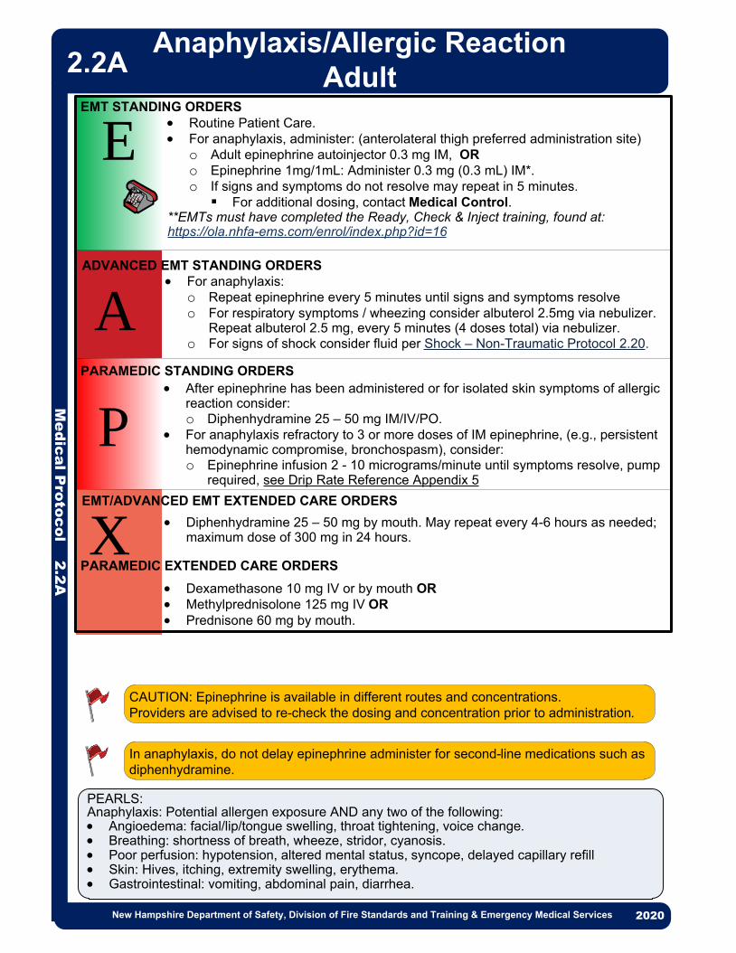
· Routine Patient Care.· For anaphylaxis, administer: (anterolateral thigh preferred administration site)
o Adult epinephrine autoinjector 0.3 mg IM, ORo Epinephrine 1mg/1mL: Administer 0.3 mg (0.3 mL) IM*.o If signs and symptoms do not resolve may repeat in 5 minutes.§ For additional dosing, contact Medical Control.
**EMTs must have completed the Ready, Check & Inject training, found at:https://ola.nhfa-ems.com/enrol/index.php?id=16
· After epinephrine has been administered or for isolated skin symptoms of allergic reaction consider:o Diphenhydramine 25 – 50 mg IM/IV/PO.
· For anaphylaxis refractory to 3 or more doses of IM epinephrine, (e.g., persistent hemodynamic compromise, bronchospasm), consider:o Epinephrine infusion 2 - 10 micrograms/minute until symptoms resolve, pump
required, see Drip Rate Reference Appendix 5
CAUTION: Epinephrine is available in different routes and concentrations. Providers are advised to re-check the dosing and concentration prior to administration.
P
· For anaphylaxis:o Repeat epinephrine every 5 minutes until signs and symptoms resolveo For respiratory symptoms / wheezing consider albuterol 2.5mg via nebulizer.
Repeat albuterol 2.5 mg, every 5 minutes (4 doses total) via nebulizer. o For signs of shock consider fluid per Shock – Non-Traumatic Protocol 2.20.
A
E
Me
dic
al P
roto
co
l 2.2
A
X
EMT STANDING ORDERS
ADVANCED EMT STANDING ORDERS
PARAMEDIC STANDING ORDERS
· Diphenhydramine 25 – 50 mg by mouth. May repeat every 4-6 hours as needed; maximum dose of 300 mg in 24 hours.
· Dexamethasone 10 mg IV or by mouth OR· Methylprednisolone 125 mg IV OR· Prednisone 60 mg by mouth.
EMT/ADVANCED EMT EXTENDED CARE ORDERS
2013
Anaphylaxis/Allergic ReactionAdult
New Hampshire Department of Safety, Division of Fire Standards and Training & Emergency Medical Services 2020
2.2A
In anaphylaxis, do not delay epinephrine administer for second-line medications such as diphenhydramine.
PARAMEDIC EXTENDED CARE ORDERS
PEARLS:Anaphylaxis: Potential allergen exposure AND any two of the following:· Angioedema: facial/lip/tongue swelling, throat tightening, voice change.· Breathing: shortness of breath, wheeze, stridor, cyanosis.· Poor perfusion: hypotension, altered mental status, syncope, delayed capillary refill· Skin: Hives, itching, extremity swelling, erythema.· Gastrointestinal: vomiting, abdominal pain, diarrhea.

1. READY
2. CHECK
3. INJECT
Remove syringe and dispose in proper sharps container.
Attach needle to syringe. Remove cap from epinephrine vial.
Wipe top of vial with alcohol prep pad.
Remove cap from needle and then carefully insert needle into top of epinephrine vial.
Withdraw correct amount of medication by pulling back on syringe plunger. Refer to numbers on side of syringe.
Pediatric Dose: 0.15 mL
Identify patient’s mid-thigh.Expose skin if possible.
Flatten skin of mid-thigh with thumb and forefinger. Insert needle quickly at a 90° angle to mid-thigh.
Depress plunger slowly to inject entire dose of medication.
Check volume of medication in syringe by comparing to diagram below. (Diagram to scale) Use Cross Check, with another person if possible, to confirm the proper dose. (SEE REVERSE)
* Inject medication intramuscularly only.
A B
C D E
A B
C D
• Don gloves.• Use 1mL syringe
with 1” needle.• Use only
1mg/1mL epinephrine.
Align plunger here
Adult Dose: 0.3 mL
0.1
0.2
0.4
0.3
0.5
1mL
0.6
0.8
0.7
0.9
Align plunger here
0.1
0.2
0.4
0.3
0.5
1mL
0.6
0.8
0.7
0.9
* Adult Dose: 0.3 mL** Pediatric Dose (<55lbs/<25kg): 0.15 mL
Epi-Ready-Check-Inject_Card.indd 1 2/22/17 10:00 AM
• Wear gloves • Use 1mL syringe with 1” needle • Use only 1mg/1mL epinephrine
1. READY
2. CHECK
3. INJECT
Remove syringe and dispose in proper sharps container.
Attach needle to syringe. Remove cap from epinephrine vial.
Wipe top of vial with alcohol prep pad.
Remove cap from needle and then carefully insert needle into top of epinephrine vial.
Withdraw correct amount of medication by pulling back on syringe plunger. Refer to numbers on side of syringe.
Pediatric Dose: 0.15 mL
Identify patient’s mid-thigh.Expose skin if possible.
Flatten skin of mid-thigh with thumb and forefinger. Insert needle quickly at a 90° angle to mid-thigh.
Depress plunger slowly to inject entire dose of medication.
Check volume of medication in syringe by comparing to diagram below. (Diagram to scale) Use Cross Check, with another person if possible, to confirm the proper dose. (SEE REVERSE)
* Inject medication intramuscularly only.
A B
C D E
A B
C D
• Don gloves.• Use 1mL syringe
with 1” needle.• Use only
1mg/1mL epinephrine.
Align plunger here
Adult Dose: 0.3 mL
0.1
0.2
0.4
0.3
0.5
1mL
0.6
0.8
0.7
0.9
Align plunger here
0.1
0.2
0.4
0.3
0.5
1mL
0.6
0.8
0.7
0.9
* Adult Dose: 0.3 mL** Pediatric Dose (<55lbs/<25kg): 0.15 mL
Epi-Ready-Check-Inject_Card.indd 1 2/22/17 10:00 AM
1. READY
2. CHECK
3. INJECT
Remove syringe and dispose in proper sharps container.
Attach needle to syringe. Remove cap from epinephrine vial.
Wipe top of vial with alcohol prep pad.
Remove cap from needle and then carefully insert needle into top of epinephrine vial.
Withdraw correct amount of medication by pulling back on syringe plunger. Refer to numbers on side of syringe.
Pediatric Dose: 0.15 mL
Identify patient’s mid-thigh.Expose skin if possible.
Flatten skin of mid-thigh with
needle quickly at a 90° angle
Depress plunger slowly to inject entire dose of medication.
Check volume of medication in syringe by comparing to diagram below. (Diagram to scale) Use Cross Check, with another person if possible, to confirm the proper dose. (SEE REVERSE)
* Inject medication intramuscularly only.
A B
C D E
A B
C D
• Don gloves.• Use 1mL syringe
with 1” needle.• Use only
1mg/1mL epinephrine.
Align plunger here
Adult Dose: 0.3 mL
0.1
0.2
0.4
0.3
0.5
1mL
0.6
0.8
0.7
0.9
Align plunger here
0.1
0.2
0.4
0.3
0.5
1mL
0.6
0.8
0.7
0.9
* Adult Dose: 0.3 mL** Pediatric Dose (<55lbs/<25kg): 0.15 mL
Epi-Ready-Check-Inject_Card.indd 1 2/22/17 10:00 AM
thumb and forefinger. Insert
to mid-thigh.
Correct Dose: Adult 0.3 mL Pediatric 0.15 mL
READY, CHECK, INJECT CROSS CHECK
• This protocol is designed to be performed by two people. • If a second person is not available, pause at each step, think and
confirm before moving to next step.• All steps should be confirmed prior to administering the injection.
PERSON 1PERSON 2
INJECTIntramuscular training video and more info at: healthvermont.gov/
emergency-preparedness-ems/emergency-medical-services/education
IF CONFIRMED
Visually inspects vial Confirms correct medication
Confirms correct dose
Inspect Syringe: Compare against visual reference (SEE REVERSE)
Visually inspects and confirms correct volume
CONFIRM
Correct Medication: Epinephrine 1 mg / 1 mL
CONFIRM
IF CONFIRMED
CONFIRM
IF CONFIRMATION COMPLETE
READY, CHECK, INJECTProtocol for Administering Epinephrine
for Acute Anaphylaxis
Epi-Ready-Check-Inject_Card.indd 22/22/17 10:00 AM
1. READY
2. CHECK
3. INJECT
Remove syringe and dispose in proper sharps container.
Attach needle to syringe. Remove cap from epinephrine vial.
Wipe top of vial with alcohol prep pad.
Remove cap from needle and then carefully insert needle into top of epinephrine vial.
Withdraw correct amount of medication by pulling back on syringe plunger. Refer to numbers on side of syringe.
Pediatric Dose: 0.15 mL
Identify patient’s mid-thigh.Expose skin if possible.
Flatten skin of mid-thigh with thumb and forefinger. Insert needle quickly at a 90° angle to mid-thigh.
Depress plunger slowly to inject entire dose of medication.
Check volume of medication in syringe by comparing to diagram below. (Diagram to scale) Use Cross Check, with another person if possible, to confirm the proper dose. (SEE REVERSE)
* Inject medication intramuscularly only.
A B
C D E
A B
C D
• Don gloves.• Use 1mL syringe
with 1” needle.• Use only
1mg/1mL epinephrine.
Align plunger here
Adult Dose: 0.3 mL
0.1
0.2
0.4
0.3
0.5
1mL
0.6
0.8
0.7
0.9
Align plunger here
0.1
0.2
0.4
0.3
0.5
1mL
0.6
0.8
0.7
0.9
* Adult Dose: 0.3 mL** Pediatric Dose (<55lbs/<25kg): 0.15 mL
Epi-Ready-Check-Inject_Card.indd 1 2/22/17 10:00 AM
Correct Dose: Adult 0.3 mL Pediatric 0.15 mL
READY, CHECK, INJECT CROSS CHECK
• This protocol is designed to be performed by two people. • If a second person is not available, pause at each step, think and
confirm before moving to next step.• All steps should be confirmed prior to administering the injection.
PERSON 1PERSON 2
INJECTIntramuscular training video and more info at: healthvermont.gov/
emergency-preparedness-ems/emergency-medical-services/education
IF CONFIRMED
Visually inspects vial Confirms correct medication
Confirms correct dose
Inspect Syringe: Compare against visual reference (SEE REVERSE)
Visually inspects and confirms correct volume
CONFIRM
Correct Medication: Epinephrine 1 mg / 1 mL
CONFIRM
IF CONFIRMED
CONFIRM
IF CONFIRMATION COMPLETE
READY, CHECK, INJECTProtocol for Administering Epinephrine
for Acute Anaphylaxis
Epi-Ready-Check-Inject_Card.indd 22/22/17 10:00 AM

Correct Dose: Adult 0.3 mL Pediatric 0.15 mL
READY, CHECK, INJECT CROSS CHECK
• This protocol is designed to be performed by two people. • If a second person is not available, pause at each step, think and
confirm before moving to next step.• All steps should be confirmed prior to administering the injection.
PERSON 1 PERSON 2
INJECTIntramuscular training video and more info at: healthvermont.gov/
emergency-preparedness-ems/emergency-medical-services/education
IF CONFIRMED
Visually inspects vial Confirms correct medication
Confirms correct dose
Inspect Syringe: Compare against visual reference (SEE REVERSE)
Visually inspects and confirms correct volume
CONFIRM
Correct Medication: Epinephrine 1 mg / 1 mL
CONFIRM
IF CONFIRMED
CONFIRM
IF CONFIRMATION COMPLETE
READY, CHECK, INJECTProtocol for Administering Epinephrine
for Acute Anaphylaxis
Epi-Ready-Check-Inject_Card.indd 2 2/22/17 10:00 AM

2013
· Routine Patient Care.· For anaphylaxis administer: (anterolateral thigh preferred administration site)
o Pediatric epinephrine autoinjector (EpiPen Jr) 0.15 mg IM for < 25 kg, o Adult epinephrine autoinjector (EpiPen) 0.3 mg IM if > 25 kg ORo If < 25 kg, epinephrine (1 mg/mL) 0.15 mg (0.15 mL) IM*,o If > 25 kg, epinephrine (1 mg/mL) 0.3 mg (0.3 mL)IM*. o If signs and symptoms do not resolve may repeat in 5 minutes.§ For additional dosing, contact Medical Control
*EMTs must have completed the Ready, Check & Inject training, found at:https://ola.nhfa-ems.com/enrol/index.php?id=16
· After epinephrine has been administered or for isolated skin symptoms of allergic reaction consider:o Diphenhydramine 1.25 mg/kg PO ORo Diphenhydramine 1 mg/kg IV/IM (maximum dose 50 mg).
· For anaphylaxis refractory to 3 or more doses of IM epinephrine, (e.g., persistent hemodynamic compromise, bronchospasm) consider:o Epinephrine Infusion 0.1 - 1 micrograms/kg/minute (maximum 10 micrograms/
min) via pump until symptoms resolve, see Pediatric Drip Rate Appendix 4
P
X
EMT STANDING ORDERS
PARAMEDIC STANDING ORDERS
· Diphenhydramine: o Ages 2 to 5 years: 6.25 mg by mouth. May repeat every 4-6 hours as needed;
maximum dose of 37.5 mg in 24 hours.o Ages 6 to 11 years: 12.5 – 25 mg by mouth. May repeat every 4-6 hours as
needed; maximum dose of 150 mg in 24 hours.
o Dexamethasone 0.6 mg/kg PO/IM/IV (PO preferred) maximum 10 mg ORo Methylprednisolone 1 -2 mg/kg IV (maximum dose 125 mg).
EMT/ADVANCED EMT EXTENDED CARE ORDERS
2013
Me
dic
al P
roto
co
l 2.2
P
Anaphylaxis/Allergic ReactionPediatric
New Hampshire Department of Safety, Division of Fire Standards and Training & Emergency Medical Services 2020
CAUTION: Epinephrine is available in different routes and concentrations. Providers are advised to re-check the dosing and concentration prior to administration.
2.2P
PARAMEDIC EXTENDED CARE ORDERS
In anaphylaxis, do not delay epinephrine administer for second-line medications such as diphenhydramine.
E
AADVANCED EMT STANDING ORDERS
· For anaphylaxis: o Repeat epinephrine every 5 minutes until signs and symptoms resolve.o For respiratory symptoms / wheezing consider albuterol 2.5 mg via nebulizer.
Repeat albuterol 2.5 mg, every 5 minutes (4 doses total) via nebulizer.o For signs of shock consider fluid per Shock – Non-Traumatic Protocol 2.20.
PEARLS:Anaphylaxis: Potential allergen exposure AND any two of the following:· Angioedema: facial/lip/tongue swelling, throat tightening, voice change.· Breathing: shortness of breath, wheeze, stridor, cyanosis.· Poor perfusion: hypotension, altered mental status, syncope, delayed capillary refill· Skin: Hives, itching, extremity swelling, erythema.· Gastrointestinal: vomiting, abdominal pain, diarrhea.

· Routine Patient Care.· Attempt to keep oxygen saturation between 94 - 98% (90% in COPD); increase
the oxygen rate with caution and observe for fatigue, decreased mentation, and respiratory failure.
· Assist the patient with his/her metered dose inhaler (MDI): 4 - 6 puffs.o May repeat every 5 minutes, as needed.o MDI containing either albuterol, levalbuterol, or a combination of albuterol/
ipratropium bromideo For patients in severe respiratory distress consider use of CPAP. See CPAP
Procedure 5.4.
Consider:· Methylprednisolone 125 mg IV/IM OR· Dexamethasone 10 mg PO/IM/IV For patients who do not respond to treatments, or for impending respiratory failure, consider:· BiPAP, (See BiPAP Procedure 5.3)· Magnesium sulfate 2 grams in 100 ml NS given IV over 10 minutes. · Epinephrine (1 mg/mL) 0.3 mg (0.3 mL) IM should only be administered for
impending respiratory failure as adjunctive therapy when there are no clinical signs of improvement
P
· Consider DuoNeb unit dose OR albuterol 2.5 mg and ipratropium bromide 0.5 mg via nebulizer.o Consider additional DuoNeb, may repeat every 5 minutes (3 doses total).
· Consider albuterol 2.5 mg via nebulizer every 5 minutes, as needed A
E
EMT STANDING ORDERS
ADVANCED EMT STANDING ORDERS
PARAMEDIC STANDING ORDERS
2013
Me
dic
al P
roto
co
l 2.3
A
Asthma, COPD, RAD – Adult
New Hampshire Department of Safety, Division of Fire Standards and Training & Emergency Medical Services 2020
PEARLS:· Chronic obstructive pulmonary disease (COPD) refers to a group of lung diseases that block
airflow and make breathing difficult. Emphysema and chronic bronchitis are the two most common conditions that make up COPD.
· Reactive Airway Disease (RAD) refers to a group of conditions that include reversible airway narrowing due to external stimulation.
· Beware of patients with a “silent chest” as this may indicate severe bronchospasm and impending respiratory failure
2.3A

Consider differential diagnosis:· Asthma· Pneumonia (CPAP for respiratory failure)· Bronchiolitis· Anaphylaxis (Anaphylaxis Protocol 2.)
· Routine Patient Care.· Attempt to keep oxygen saturation between 94% - 98%· Observe for fatigue, decreased mentation, and respiratory failure.· Assist the patient with his/her metered dose inhaler (MDI): 4 - 6 puffs.
o May repeat every 5 minutes, as needed.o MDI containing either albuterol, levalbuterol, or a combination of albuterol/
ipratropium bromide.· For patients ≤ 2 who present with increased work of breathing and rhinorrhea, provide
nasal suctioning with saline drops and bulb syringe; no more than 2 attempts.· For pediatrics in severe respiratory distress due to asthma consider use of CPAP. See
CPAP Procedure 5.4.
· Consider Unit dose DuoNeb OR albuterol 2.5 mg and ipratropium bromide 0.5 mg via nebulizer.o Consider additional DuoNeb, may repeat every 5 minutes
(3 doses total).· Consider albuterol 2.5 mg via nebulizer every 5 minutes, as
needed.
P
Me
dic
al P
roto
co
l 2.3
P
ASTHMA - PARAMEDIC STANDING ORDERS
2013
Pediatric Respiratory DistressAsthma, Bronchiolitis, Croup
New Hampshire Department of Safety, Division of Fire Standards and Training & Emergency Medical Services 2020
2.3P
A
E
ASTHMA, BRONCHIOLITIS, CROUP - EMT STANDING ORDERS
ASTHMA - ADVANCED EMT STANDING ORDERS
Consider:· Dexamethasone 0.6 mg/kg PO/IM/IV (PO preferred), maximum 10 mg OR· Methylprednisolone 1 - 2 mg/kg IV/IM, maximum 125 mg.For patients who do not respond to treatment or for impending respiratory failure consider:· Magnesium sulfate 40 mg/kg in 100ml 0.9% NaCl IV over 20 minutes.· Epinephrine:o If < 25 kg, epinephrine (1 mg/mL) 0.15 mg IM, lateral thigh
preferred.o If > 25 kg, epinephrine (1 mg/mL) 0.3 mg IM, lateral thigh
preferred
PBRONCHIOLITIS - PARAMEDIC STANDING ORDERS
P
CROUP - PARAMEDIC STANDING ORDERS
For patients who do not respond to suctioning or for impending respiratory failure consider:· Nebulized epinephrine (1 mg/mL) 3 mg (3 mL) in 3 mL 0.9% NaCl.
Wheezing ≥ 2 years or history of asthma
YES
NO
History of stridor or
barky cough
Wheezing ˂ 2 years
old
NO
YES
YES
· Respiratory distress in children must be promptly recognized and treated. Respiratory arrest is the most common cause of cardiac arrest in children.
· Child with a “silent chest” may have severe bronchospasm with impending respiratory failure.· In patients with suspected croup or stridor, provide necessary interventions while attempting to
minimize noxious stimuli that may induce agitation.
Consider:· Dexamethasone 0.6 mg/kg PO/IM/IV (PO preferred) maximum 10
mg. Croup with stridor at rest:· Nebulized epinephrine (1 mg/mL) 3 mg (3 mL) in 3 mL 0.9% NaCl,
repeat in 20 minute as needed OR· Nebulized racemic epinephrine (2.25% solution) 0.5 mL in 2.5 mL
0.9% NaCl, may repeat in 20 minutes as needed
· Croup· Sepsis (Sepsis Protocol) · Foreign body airway
obstruction
Protocol Continues

Me
dic
al P
roto
co
l 2.3
P
Pediatric Respiratory DistressAsthma, Bronchiolitis, Croup
New Hampshire Department of Safety, Division of Fire Standards and Training & Emergency Medical Services 2020
2.3P
PEARLS· The IV formulation of dexamethasone may be given by mouth.· For suspected epiglottitis, transport the patient in an upright position and limit your assessment
and interventions.Bronchiolitis· Incidence peaks in 2-6 month old infants. · History of low-grade fever, runny nose, and sneezing.· Signs and symptoms include: tachypnea, rhinorrhea, wheezes and / or crackles.Croup · Incidence peaks in children over age 6 months.· Signs and symptoms include: hoarseness, barking cough, inspiratory stridor, signs of
respiratory distress.· Avoid procedures that will distress child with severe croup and stridor at rest.Pneumonia· Signs and symptoms include: tachypnea, fever, intercostal retractions, cough, hypoxia and
chest pain.Tachypnea in children is defined as:· < 2 months: 60 bpm· 2-12 months: 50 bpm· 1-5 years: 40 bpm· >5 years: 20 bpm
Protocol Continued

Maintain Scene Safety· Request law enforcement support, consider staging away until law enforcement has cleared
scene.· Maintain situational awareness, focus on crew safety.· Observe and record the patient’s behavior and living conditions.
Consider Causes & Determine Capacity· Consider causes (e.g., hypoxia, hypoglycemia, alcohol or drug intoxication, excited delirium,
stroke and brain trauma) · Ask patient directly if they have considered harming self or others.Refusal & Police Assistance· Consider requesting law enforcement upon dispatch· If patient lacks capacity or is determined to be a danger to self or others, they MAY NOT
refuse care.○ Contact law enforcement if unable to convince patient to be transported. (Refer to Police Custody Policy 8.14, Refusal of Care Policy 8.15)
Me
dic
al P
roto
co
l 2.4
2013
Anxiety Management (Anxious, apprehensive, but not aggressive)● Approach patient with the SAFER method.● Provide calm emotional support and medical care as required.● Minimize external stimuli (e.g., loud noises, lights).● Encourage patient to be evaluated by a mental health professional.● For significant anxiety that cannot be managed with BLS interventions, consider paramedic intercept for pharmacological intervention.
Resistant or Aggressive Management (Resisting necessary treatment/interventions)● Attempt verbal de-escalation.● Consider paramedic intercept for pharmacological intervention, see Restraints Protocol.
Violent and/or Excited Delirium Management (Immediate danger to self/others)● Attempt verbal de-escalation.● Consider physical restraints as a last resort if the patient is in immediate danger to self or others see Restraints Protocol.Request Paramedic intercept, if available, for pharmacological intervention, see Restraints Protocol.
Anxiety Management (Anxious, apprehensive, but not aggressive)Goal is safe and compliant, contact Medical Control to consider:· Midazolam 2.5 mg IV, may repeat once in 5 minutes, OR
· *Midazolam 5 mg IM/IN, may repeat once in 5 minutes, OR
· Lorazepam 1 mg IV, may repeat once in 5 minutes, OR
· Diazepam 5 mg IV, may repeat once in 5 minutes.
Behavioral Emergencies
Adult & Pediatric
New Hampshire Department of Safety, Division of Fire Standards and Training & Emergency Medical Services 2020
2.4
EMT/ ADVANCED EMT STANDING ORDERS – ADULT & PEDIATRIC
EA/
SAFER Model
S Stabilize the situation by lowering stimuli, including voice.
A Assess and acknowledge crisis by validating patient’s feelings and not minimizing them.
F Facilitate identification and activation of resources (clergy, family, friends, or police).
E Encourage patient to use resources and take actions in his/her best interest.
R Recovery/referral - leave patient in the care of a responsible person, professional or transport to
appropriate medical facility. Do not leave the patient alone when EMS clears the scene.
PARAMEDIC MEDICAL CONTROL ORDERS - ADULT
P*For IN administration of midazolam use a 5 mg/mL concentration.

PEARLS:· There is an increased risk of apnea with >2 doses of benzodiazepines. · Causes of combativeness may be due to comorbid medical conditions or due to hypoxia,
hypoglycemia, drug and/or alcohol intoxication, drug overdose, brain trauma.· Verbal de-escalation is the safest method and should be delivered in an honest, straightforward,
friendly tone avoiding direct eye contact and encroachment of personal space.
EMT/ ADVANCED EMT STANDING ORDERS
2013
Pro
ce
du
re 6
.5
Restraints
New Hampshire Department of Safety, Division of Fire Standards and Training & Emergency Medical Services 2020
INDICATIONSPatients who are a potential harm to themselves or others, or interfere with their own care and lack the ability to refuse care under the Refusal of Care Protocol may be restrained to prevent injury to the patient or crew and facilitate necessary medical care. Restraining must be performed in a humane manner and used only as a last resort.
PROCEDURE1. Request law enforcement assistance, as necessary.2. When appropriate, attempt less restrictive means of managing the patient, including
verbal de-escalation.3. Ensure that there are sufficient personnel available to physically restrain the patient
safely.4. Restrain the patient in a lateral or supine position. No devices such as backboards,
splints, or other devices may be placed on top of the patient. Never hog-tie a patient. In order to gain control, the patient may need to be in a prone position, but must be moved to supine or lateral position as soon as possible.
5. The patient must be under constant observation by the EMS crew at all times. This includes direct visualization of the patient as well as cardiac, pulse oximetry, and quantitative waveform capnography monitoring, if available.6. The extremities that are restrained should have a circulation check at least every 15
minutes. The first of these checks should occur as soon possible after restraints are placed.
7. Documentation should include the reason for the use of restraints, the type of restraints used, the time restraints were placed, and circulation checks.
8. If a patient is restrained by law enforcement personnel with handcuffs or other devices that EMS personnel cannot remove, a law enforcement officer should accompany the patient to the hospital in the transporting ambulance. If this is not feasible, the officer MUST follow directly behind the transporting ambulance to the receiving hospital.
Resistant or Aggressive Management (Resisting necessary treatment/interventions)
Goal is alert and calm, consider:
· Midazolam 2.5 mg IV, may repeat once in 5 minutes, OR · *Midazolam 5 mg IM/IN, may repeat once in 5 minutes (*for IN use 5 mg/mL
concentration), OR· Lorazepam 1 mg IV, may repeat once in 5 minutes, OR· Diazepam 5 mg IV, may repeat once in 5 minutes.
6.5
EA/
Protocol Continues
Continued patient struggling against restraints may lead to hyperkalemia, rhabdomyolysis, and/or cardiac arrest. Chemical restraint may be necessary to prevent continued forceful struggling by the patient.
The Restraints Procedure was placed after Behavorial Emergencies for ease of finding. It is also listed under procedures.
The patient must be under constant observation by the EMS crew at all times. This includes direct visualization of the patient as well as cardiac, pulse oximetry, and quantitative waveform capnography monitoring, if available.
PARAMEDIC STANDING ORDERS - ADULT
P

Pro
ce
du
re 6
.5Restraints
New Hampshire Department of Safety, Division of Fire Standards and Training & Emergency Medical Services
For patients with suspected Excited/Agitated Delirium (Immediate danger to self/others) OR extreme agitation OR ineffective control with benzodiazepines. Goal is safe and compliant:· **Ketamine: 4 mg/kg IM rounded to nearest 50 mg, maximum dose 500 mg, repeat 100
mg IM in 5 – 10 minutes. OR· Benzodiazepines:
o Midazolam 5 mg IV, repeat every 5 minutes as needed OR o *Midazolam 10 mg IM/IN, repeat every 5 minutes as needed ORo Lorazepam 2 - 4 mg IV, repeat every 5 minutes as needed ORo Diazepam 10 mg IV, repeat every 5 minutes as needed
· Consider in addition to benzodiazepines:o ***Haloperidol 10 mg IM; may repeat once in 10 minutes.
Contact Medical Control for additional doses.
After chemical restraint, re-evaluate whether the patient continues to meet criteria for physical restraint and remove if they are no longer necessary to ensure the safety of the patient, providers or both, taking into account transport times, the depth of sedation and the need to transfer the patient at destination.
· If cardiac arrest occurs with suspected excited delirium, consider early administration of: fluid bolus, sodium bicarbonate, calcium chloride/gluconate, see Cardiac Arrest Protocol 3.2A.
For acute dystonic reaction to haloperidol:· Diphenhydramine 25 – 50 mg IV/IM.
P
PARAMEDIC STANDING ORDERS - ADULT
· Excited/Agitated Delirium is characterized by extreme restlessness, irritability, and/or high fever. Patients exhibiting these signs are at high risk for sudden death.
· Medications should be administered cautiously in frail or debilitated patients; lower doses should be considered.
· Administer haloperidol with caution to patients who are already on psychotropic medications which may precipitate serotonin syndrome or malignant hyperthermia.
· Placing a patient in prone position creates a severe risk of airway and ventilation compromise and death.
Protocol Continued
2020
6.5
*For IN administration of midazolam use a 5 mg/mL concentration.
**For ketamine use 100 mg/mL concentration
***Administer haloperidol with caution to patients who are already on psychotropic medication
which may precipitate serotonin syndrome or malignant hyperthermia.
Resistant or Aggressive Management (Resisting necessary treatment/interventions)Contact Medical Control, to discuss treatment options
Violent and/or Excited Delirium Management(Immediate danger to self/others)Target Goal is safe and compliant.Contact Medical Control and consider:· **Ketamine 4 mg/kg IM rounded to nearest 25 mg, maximum dose 250 mg, repeat x 1
in 5-10 minutes OR· Benzodiazepines:
o *Midazolam 0.2 mg/kg IM/IN (single maximum dose 10 mg) repeat every 5 minutes as needed, OR
o Midazolam 0.1 mg/kg IV (single maximum dose 5 mg) repeat every 5 minutes as needed, OR
o Lorazepam 0.1 mg/kg IV (single maximum dose 4 mg) repeat every 5 minutes as needed, OR
o Diazepam 0.2 mg/kg IV (single maximum dose 10 mg IV) repeat every 5 minutes as needed.
· If cardiac arrest occurs with suspected excited delirium, consider early administration of: fluid bolus, sodium bicarbonate, calcium chloride/gluconate, see Cardiac Arrest Protocol
PARAMEDIC STANDING ORDERS - PEDIATRIC

Me
dic
al P
roto
co
l 2.5
PEARLS· BRUE is not a disease, but a constellation of symptoms. Potential etiologies include central
apnea (immature respiratory center), obstructive apnea (structural), gastroesophageal reflux (laryngospasm, choking, gagging), respiratory (pertussis, RSV), cardiac (congenital heart disease, arrhythmia), seizures.
· Always consider non-accidental trauma in any infant who presents with BRUE, see Victims of Violence Protocol 8.20.
2013
· Routine Patient Care.· Perform blood glucose analysis and manage per Hypo/Hyperglycemia Protocols
2.7 & 2.9P.· Obtain history of event with particular attention to:
o Activity at onset and history of the evento State during the event (cyanosis, apnea, coughing, gagging, vomiting)o End of the event (duration, gradual or abrupt cessation, treatment provided)o Infant’s condition after the event (normal, not normal)o Recent history (illness, injuries, exposure to others with illness, use of OTC
medications, recent immunizations, new or different formula).o Past medical history (gestational age, pre-/perinatal history, GERD, seizures,
previous BRUE).o Family history (sudden unexplained deaths, prolonged QT, arrhythmias).o Medications present in the residenceo Sleeping positiono Co-sleeping with parent in the same bed.
· Transport patient to the hospital.
EMT/ADVANCED/PARAMEDIC STANDING ORDERS
EA
P
//
Brief Resolved Unexplained Event (BRUE)
New Hampshire Department of Safety, Division of Fire Standards and Training & Emergency Medical Services 2020
2.5
Brief Resolved Unexplained Event (BRUE) – formerly known as ALTE
An event occurring in an infant < 1 year old when the observer reports a sudden, brief and now resolved episode of 1 or more of the following:· Cyanosis or pallor · Absent, decreased or irregular breathing · Marked change in tone (hyper or hypotonia) · Altered level of responsiveness.
Although children who experience BRUE have a normal physical exam upon assessment by prehospital personnel, they should be transported to the emergency department for further assessment and treatment as they often have a serious underlying condition. Assume history provided by the family/witness is accurate.

EMR/EMT/ADVANCED EMT STANDING ORDERS· Routine Patient care. · Obtain obstetrical (OB) history.· Expose patient and determine if signs of imminent delivery are present.
o Do not digitally examine or insert anything into the vagina.o If obstetrical complication is present, consider contacting Medical Control and
transport to nearest appropriate hospital per local OB Diversion Protocol. (See Obstetrical Emergencies Protocol 2.14)
· If delivery is not imminent place mother in left-lateral recumbent position and transport to a hospital with OB capability.
· If delivery is imminent, assist in newborn’s delivery. o With palm of hand, apply gentle perineal pressure for a slow, controlled
delivery.o As the baby’s head begins to emerge support the head as it turns. Do not pull
on head.o If membranes are intact after head emerges, tear membrane with fingers to
permit escape of fluid.o If umbilical cord is wrapped around newborn’s neck, slip the cord over head
prior to delivery. If after multiple attempts you are unable to slip cord off the neck, clamp and cut the cord between the clamps.
o Guide the baby’s head downward to allow delivery of the upper shoulder.o Then guide the baby’s head upward to allow delivery of the lower shoulders. o Delivery of trunk and legs occurs quickly; be prepared to support infant as it
emerges. · For newborns requiring resuscitation, see Newborn Resuscitation Protocol 2.13.· Prevent heat loss by rapidly drying and warming:
o Remove wet lineno For stable newborn and mother, place newborn skin-to-skin on the mother’s
chest or abdomen.§ Cover newborn’s head, wrap newborn and mother in blankets, silver
swaddler/space blanket or commercially available infant warming device. Do not use hot packs.
· Assess airway by positioning and clearing secretions (only if needed):o Place the newborn on back or side with head in a neutral or slightly extended
position.o Routine suctioning is discouraged even in the presence of meconium-stained
amniotic fluid. Suction oropharynx then nares only if the patient exhibits respiratory depression and/or obstruction, see Newborn Resuscitation Protocol 2.13.
· Assess breathing by providing tactile stimulation:o Flick soles of feet and/or rub the newborn’s back.o If newborn is apneic or has gasping respirations, nasal flaring, or grunting,
proceed to Newborn Resuscitation Protocol 2.13.· Asses circulation, heart rate, and skin color:
o Evaluate heart rate by one of several methods:§ Auscultate apical beat with a stethoscope.§ Palpate the pulse by lightly grasping the base of the umbilical cord.
o If the pulse is <100 bpm and not increasing, proceed to Newborn Resuscitation Protocol 2.13.
o Assess skin color: examine trunk, face and mucous membranes.o Assess temperatureo Record APGAR score at 1 minute and 5 minutes (see chart).
· See Pediatric Color Coded Appendix A3 for vital signs.
2013
EA/
Me
dic
al P
roto
co
l 2.6
Childbirth & Newborn Care
New Hampshire Department of Safety, Division of Fire Standards and Training & Emergency Medical Services 2020
2.6
Protocol Continues

Me
dic
al P
roto
co
l 2.6
Childbirth & Newborn Care
New Hampshire Department of Safety, Division of Fire Standards and Training & Emergency Medical Services 2020
Active Movement
Over 100 bpm
Cry, sneeze, cough, active movement
Completely pink
Vigorous cryFull breaths
Feature EvaluatedActivity
(Muscle Tone)Pulse
Grimace(Irritability/reflexes)Appearance(Skin Color)Respiration
2 Points 1 Point
Arms and legs flexed(Weak, some movement)
Below 100 bpm
Grimace (some flexion of extremities)
Body pink, Extremities blue
Slow, irregular, or gasping breaths, weak
cry
0 Points
Limp or flaccid
Absent
No reflexes
Blue, pale
Absent
APGAR Scale
PPARAMEDIC STANDING ORDERS
· Active seizures—see Seizures Protocol 2.18A.· After delivery:
o Administer oxytocin 10 Units IM to the mother. Note: In multiple pregnancy, do not give until all placentas are delivered.
EA/
PEARLS:
· Newborn infants are prone to hypothermia which may lead to hypoglycemia, hypoxia and lethargy. Aggressive warming techniques should be initiated including drying, swaddling, and warm blankets covering body and head.
· Raise temperature in ambulance patient compartment.
Consider Medical Control for:· Prepartum hemorrhage· Postpartum hemorrhage· Breech presentation· Limb presentation· Nuchal cord· Prolapsed cord
Signs of imminent delivery:· Urge to move bowels· Urge to push· Crowning· Contractions less than 2
minutes apart
OB Assessment:· Length of pregnancy· Number of pregnancies· Number of viable births· Last menstrual period· Due date· Prenatal care· Number of expected babies· Drug use
Protocol Continues
EMR/EMT/ADVANCED EMT STANDING ORDERS· Clamp and cut the umbilical cord:
o After initial assessment and after the cord stops pulsating.o Leave a minimum of 6 inches of cord.
· Allow spontaneous delivery of placenta: o Do not pull on umbilical cord.o Do not delay transport for delivery of the placenta. o Massage abdominal wall overlying uterine fundus.o If placenta delivers, package for hospital staff.
· Monitor maternal blood loss and perfusion. (See Obstetrical Emergencies Protocol 2.14). Note that normal pregnancy is accompanied by higher heart rate and lower blood pressure.
· For transport:o Ensure newborn remains warmo Turn heat to maximum in ambulance compartmento Consider commercial warming device (do not put heat packs directly on skin)o When possible, transport newborn in child safety seat.
2.6

· Routine Patient Care.· Obtain glucose reading.
2013
· ADULT: Administer 1000 ml IV bolus of IV fluid,o May repeat 500 mL fluid bolus, as needed.
· PEDIATRIC: Administer 10 - 20 mL/kg IV bolus of IV fluid.o May repeat fluid bolus two times for a total of 3 fluid boluses, not to exceed
30 mL/kg.
E
PEARLS:· Diabetic Ketoacidosis (DKA) is a life threatening emergency defined as uncontrolled
hyperglycemia with the signs and symptoms of ketoacidosis.· Signs and symptoms of DKA include uncontrolled blood glucose greater than or equal to 250
mg/dL, weakness, altered mental status, abdominal pain, nausea, vomiting, polyuria (excessive urination), polydipsia (excessive thirst), a fruity odor on the breath (from ketones), and tachypnea (Kussmaul respirations).
· Common causes of DKA include infection, acute coronary syndrome, and medication non-compliance.
· Hyperglycemic Hyperosmolar Nonketotic Syndrome (HHNS) is characterized by blood glucose levels greater than 600 mg/dL and profound dehydration without significant ketoacidosis. Most patients present with severe dehydration and focal or global neurologic deficits e.g., coma, altered mental status.
· Hyperglycemia may be detrimental to patients at risk for cerebral ischemia such as victims of stroke, cardiac arrest, and head trauma.
EMT STANDING ORDERS – ADULT & PEDIATRIC
ADVANCED EMT/PARAMEDIC STANDING ORDERS – ADULT & PEDIATRIC
2013
A/P
Me
dic
al P
roto
co
l 2.7
Hyperglycemia – Adult & Pediatric
New Hampshire Department of Safety, Division of Fire Standards and Training & Emergency Medical Services 2020
2.7
XEMT/ADVANCED EMT/PARAMEDIC EXTENDED CARE ORDERS
· Oral fluids: if the patient is not vomiting, provide oral hydration with water. o Patient must be alert enough to swallow and protect airway.
Hyperglycemia is defined as blood glucose greater than or equal to 250 mg/dL. Patient with associated signs and symptoms such as altered mental status, increased respiratory rate, or dehydration may require treatment.

2013
E
· Routine Patient Care.
· Move victim to a cool area and shield from the sun or any external heat source.
· Remove as much clothing as is practical and loosen any restrictive garments.
· If alert and oriented, give small sips of cool liquids.
· Monitor and record vital signs and level of consciousness.
· Obtain temperature – rectal temperature preferred as appropriate.
· If temperature is 40o C (>104
o F) or if altered mental status is present, begin
active cooling by:
o Continually misting the exposed skin with tepid water while fanning the
patient (most effective).
o Truncal ice packs and wet towels/sheets may be used, but are less effective
than evaporation.
o Discontinue active cooling when the patient reaches 38.5o C (101.5
o F), or if
shivering occurs and cannot be managed by paramedics (see below).
· ADULT: Consider 500 ml IV fluid bolus for dehydration even if vital signs are
normal.
· PEDIATRIC: Consider 10 – 20 ml/kg IV fluid bolus for dehydration even if vital
signs are normal.
P· If uncontrolled shivering occurs during cooling:
o Midazolam 2.5 mg IV, may repeat once in 5 minutes, OR
o *Midazolam 5 mg IM/IN may repeat once in 5 minutes, OR
o Lorazepam 1 mg IV, may repeat once in 5 minutes, OR
o Diazepam 5 mg IV, may repeat once in 5 minutes.
· If uncontrolled shivering occurs during cooling:
o Midazolam 0.05 mg/kg IV (single maximum dose 2.5 mg), may repeat once in
5 minutes, OR
o *Midazolam 0.1 mg IM/IN (single maximum dose 5 mg) may repeat once in 5
minutes, OR
o Lorazepam 0.05 mg/kg IV (maximum dose 1 mg); may repeat once in 5
minutes, OR
o Diazepam 0.1 mg/kg IV (maximum dose 5 mg); may repeat once in 5 minutes.
A
EMT STANDING ORDERS- ADULT & PEDIATRIC
ADVANCED EMT STANDING ORDERS – ADULT & PEDIATRIC
PARAMEDIC STANDING ORDERS- ADULT
PARAMEDIC STANDING ORDERS- PEDIATRIC
2013
Me
dic
al P
roto
co
l 2.8
Hyperthermia – Adult & Pediatric
New Hampshire Department of Safety, Division of Fire Standards and Training & Emergency Medical Services 2020
2.8
PEARLS:· Exertional hyperthermic patients may be significantly dehydrated, and may require repeat fluid
boluses.· Immersion cooling is the most effective method to lower core body temperature if proper
resources are available.
Indications: Elevated temperature due to environmental exposure, over exertion,
pharmacological agents or excited/agitated delirium.
Contraindications: Fever associated with likely infectious illness.
*For IN administration of midazolam use a 5 mg/mL concentration.

· Routine Patient Care.· Obtain glucose reading:· Administer 15 – 30 gram commercially prepared glucose gel or equivalent.
o Hypoglycemic patients must be alert enough to swallow and protect airway.
· If intranasal glucagon has been prescribed by the patient’s physician, assist the patient or care giver with the administration in accordance with the physician’s instructions.
· For patients with an insulin pump who are hypoglycemic with associated altered mental status:o Stop the pump or remove catheter at insertion site if patient cannot ingest
oral glucose or ALS is not available.o Leave the pump connected and running if able to ingest oral glucose or
receive ALS interventions.
2013
· Administer dextrose 10% IV via premixed infusion bag (preferred) or prefilled syringe until mental status returns to baseline and glucose level is greater than 60 mg/dL. IV pump not required.
· If unable to establish IV access, administer glucagon 1 mg IMo Recheck glucose 15 minutes after administration of glucagon.o May repeat glucagon 1 mg IM if glucose level is < 60 mg/dl with continued
altered mental status.
E
PEARLS:· Causes of hypoglycemia include medication misuse or overdose, missed meal,
infection, cardiovascular insults (e.g., myocardial infarction, arrhythmia), or changes in activity (e.g., exercise).
· Sulfonylureas (e.g., glyburide, glipizide) have long half-lives ranging from 12 - 60 hours. Patients with corrected hypoglycemia who are taking these agents are at particular risk for recurrent symptoms and frequently require hospital admission.
· Oral glucose equivalents include 3 - 4 glucose tablets, 4 oz. fruit juice (e.g. orange juice), non-diet soda, 1 tablespoon of pure New Hampshire maple syrup, sugar, or honey.
· Encourage patients who refuse transport after improvement of GCS and are back to baseline to consume complex carbohydrates (15 grams) and protein (12 – 15 grams) such as peanut butter toast, mixed nuts, milk or cheese to stabilize blood sugar.
· Hypoglycemia may be detrimental to patients at risk for cerebral ischemia, such as victims of stroke, cardiac arrest, and head trauma.
Intraosseous (IO) administration of dextrose should be reserved for hypoglycemic patients with severe altered mental status or active seizures and IV access cannot be obtained.
EMT STANDING ORDERS
ADVANCED EMT/PARAMEDIC STANDING ORDERS
2013
A/P
Me
dic
al P
roto
co
l 2.9
AHypoglycemia – Adult
New Hampshire Department of Safety, Division of Fire Standards and Training & Emergency Medical Services 2020
2.9A
Hypoglycemic emergency is defined as glucose <60 mg/dl with associated altered mental status..
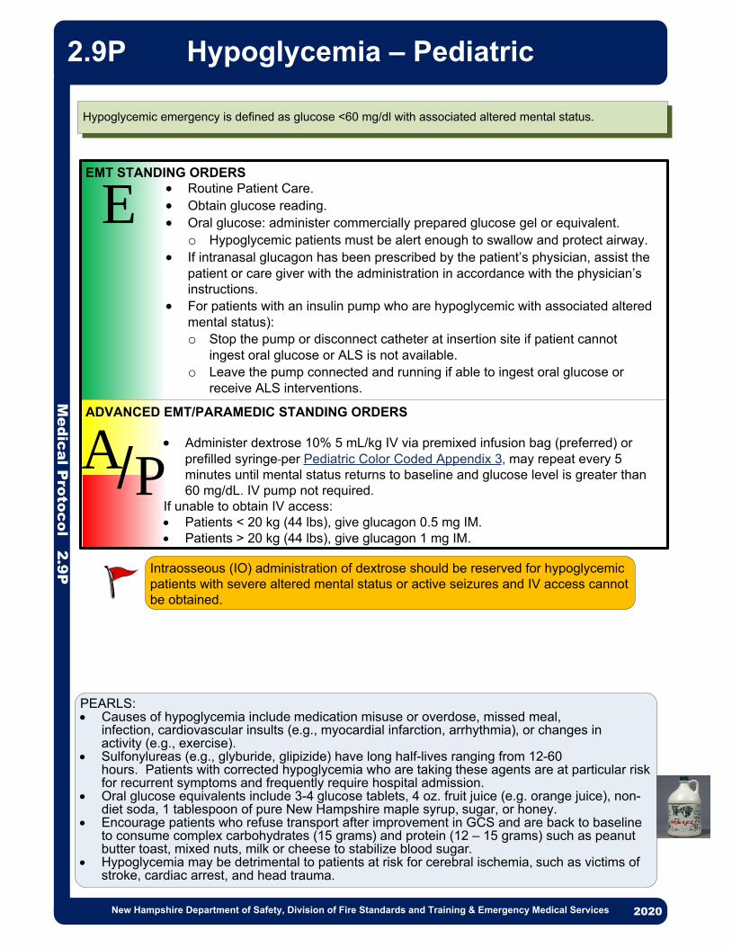
· Routine Patient Care.· Obtain glucose reading.· Oral glucose: administer commercially prepared glucose gel or equivalent.
o Hypoglycemic patients must be alert enough to swallow and protect airway.· If intranasal glucagon has been prescribed by the patient’s physician, assist the
patient or care giver with the administration in accordance with the physician’s instructions.
· For patients with an insulin pump who are hypoglycemic with associated altered mental status):o Stop the pump or disconnect catheter at insertion site if patient cannot
ingest oral glucose or ALS is not available.o Leave the pump connected and running if able to ingest oral glucose or
receive ALS interventions.
2013
E
PEARLS:· Causes of hypoglycemia include medication misuse or overdose, missed meal,
infection, cardiovascular insults (e.g., myocardial infarction, arrhythmia), or changes in activity (e.g., exercise).
· Sulfonylureas (e.g., glyburide, glipizide) have long half-lives ranging from 12-60 hours. Patients with corrected hypoglycemia who are taking these agents are at particular risk for recurrent symptoms and frequently require hospital admission.
· Oral glucose equivalents include 3-4 glucose tablets, 4 oz. fruit juice (e.g. orange juice), non-diet soda, 1 tablespoon of pure New Hampshire maple syrup, sugar, or honey.
· Encourage patients who refuse transport after improvement in GCS and are back to baseline to consume complex carbohydrates (15 grams) and protein (12 – 15 grams) such as peanut butter toast, mixed nuts, milk or cheese to stabilize blood sugar.
· Hypoglycemia may be detrimental to patients at risk for cerebral ischemia, such as victims of stroke, cardiac arrest, and head trauma.
EMT STANDING ORDERS
ADVANCED EMT/PARAMEDIC STANDING ORDERS
2013
Me
dic
al P
roto
co
l 2.9
P
Hypoglycemia – Pediatric
New Hampshire Department of Safety, Division of Fire Standards and Training & Emergency Medical Services 2020
2.9P
· Administer dextrose 10% 5 mL/kg IV via premixed infusion bag (preferred) or prefilled syringe per Pediatric Color Coded Appendix 3, may repeat every 5 minutes until mental status returns to baseline and glucose level is greater than 60 mg/dL. IV pump not required.
If unable to obtain IV access:· Patients < 20 kg (44 lbs), give glucagon 0.5 mg IM.· Patients > 20 kg (44 lbs), give glucagon 1 mg IM.
Intraosseous (IO) administration of dextrose should be reserved for hypoglycemic patients with severe altered mental status or active seizures and IV access cannot be obtained.
A/P
Hypoglycemic emergency is defined as glucose <60 mg/dl with associated altered mental status.

2013
E
· Routine Patient Care· Handle gently. Avoid rough movement and excess activity.· Prevent further heat loss:
o Insulate from the ground and shield from wind/water.o Move to a warm environment.o Gently remove any wet clothing and dry patient.o Cover with warm blankets including the head and neck.o Consider use of heat reflective emergency blanket.o Apply truncal warm packs.o Patients with moderate to severe hypothermia require active external
rewarming with chemical, electrical, or forced hot-air heating packs or blankets.
· Classify hypothermia clinically on the basis of vital signs, level of consciousness and intensity of shivering.
· Core Temperature, if available, provides additional treatment information (see chart)
· Obtain blood glucose. · Support shivering with calorie replacement if alert and able to swallow.· Mildly hypothermic patients should not be allowed to stand or walk for 20 minutes,
while being kept as warm as possible with calorie replacement and shelter.· A minimum of 60 second assessment of respirations and pulse is necessary to
confirm respiratory arrest or cardiac arrest.· If pulse and breathing are absent, start CPR unless contraindications exist
o Contraindications to CPR in the hypothermic patient include:§ Obvious signs of irreversible death§ Chest wall not compressible as whole body is frozen solid§ A valid DNR order§ Avalanche burial > 35 minutes AND airway packed with snow§ Rescuers exhausted or in danger.
*Rigor mortis or fixed and dilated pupils are NOT a contraindication for CPR in hypothermia· Hypothermic patients without contraindications to CPR should have continuous
CPR and should not be considered for Termination of Resuscitation until the core temperature is above 32oC (90oF) without ROSC.o Prolonged CPR is not indicated in patients who are thought to have
experienced cardiac arrest prior to cooling (temperature is thought to have been above 32oC (90oF) at the time of cardiac arrest)
o Causes of cardiac arrest before cooling include major trauma, witnessed normothermic arrest and avalanche burial > 35 minutes AND snow or ice packed airway
· Patients whose torsos are cool to the touch are likely to have cardiac instability [systolic blood pressure < 90 mmHg or ventricular arrhythmias, core temperature < 28oC (82oF)]
· When feasible, patients in cardiac arrest with continuing CPR should be transported directly to a center capable of providing cardiopulmonary bypass (CPB) or extracorporeal membrane oxygenation (ECMO).
· CPR may be delayed or performed intermittently if necessary to accomplish evacuation to an ECMO or CPB center.
EMT STANDING ORDERS - ADULT & PEDIATRIC
2013
Me
dic
al P
roto
co
l 2.1
0Hypothermia – Adult & Pediatric
New Hampshire Department of Safety, Division of Fire Standards and Training & Emergency Medical Services 2020
2.10
Policy Continues

STAGE: I Conscious, shivering Core Temp 35 to 32°C (95 to 89.6°F)Treatment: Warm environment and clothing, warm sweet drinks, and active movement (if possible).
STAGE: II Impaired consciousness, not shivering Core Temp <32 to 28°C (<89.6 to 82.4°F)Treatment: Cardiac monitoring, minimal and cautious movements to avoid arrhythmias, horizontal
position and immobilization, full-body insulation, active external and minimally invasive rewarming techniques (warm environment; chemical, electrical, or forced- air heating packs or blankets; warm parenteral fluids).
STAGE: III Unconscious, not shivering, vital signs presentCore Temp <28 to 24°C (<82.4 to 75.2°F)Treatment: HT II management plus airway management as required; ECMO or CPB in cases with
cardiac instability that is refractory to medical management.
STAGE: IV No vital signsCore Temp <24°C (<75.2°F)Treatment: HT II and III management plus CPR and up to three doses of epinephrine (at an intravenous
or intraosseous dose of 1 mg) and defibrillation, with further dosing guided by clinical response; rewarming with ECMO or CPB (if available) or CPR with active external and alternative internal rewarming.
Me
dic
al P
roto
co
l 2.1
0
Hypothermia – Adult & Pediatric
New Hampshire Department of Safety, Division of Fire Standards and Training & Emergency Medical Services 2020
· Warm IV 0.9% NaCl should be used.· If pulse and breathing are absent and esophageal or rectal temperature core
temperature is < 32o C (89.6o F):o Continue CPR. o Give IV medications based on dysrhythmia (consider limiting and/or
increasing the dosing time).o Defibrillation as indicated.
ADVANCED EMT - ADULT ONLYPARAMEDIC STANDING ORDERS – ADULT & PEDIATRIC
A/P
Protocol Continues
2.10
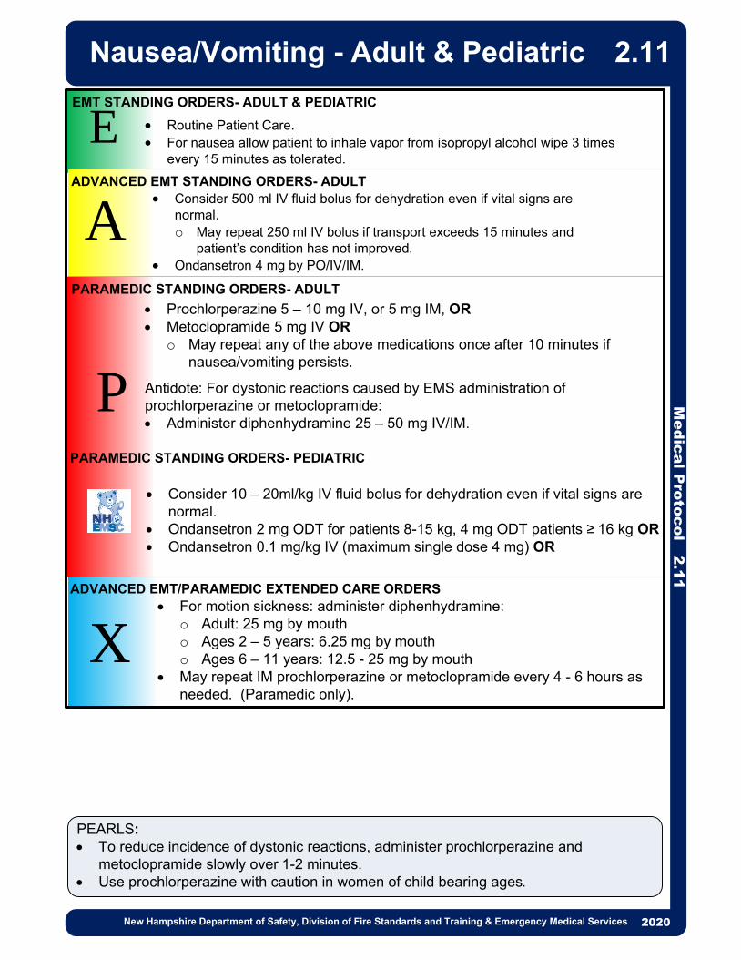
· Routine Patient Care.· For nausea allow patient to inhale vapor from isopropyl alcohol wipe 3 times
every 15 minutes as tolerated.
PEARLS:· To reduce incidence of dystonic reactions, administer prochlorperazine and
metoclopramide slowly over 1-2 minutes. · Use prochlorperazine with caution in women of child bearing ages.
2013
P
E
X
EMT STANDING ORDERS- ADULT & PEDIATRIC
PARAMEDIC STANDING ORDERS- ADULT
ADVANCED EMT STANDING ORDERS- ADULT
A· Consider 500 ml IV fluid bolus for dehydration even if vital signs are
normal. o May repeat 250 ml IV bolus if transport exceeds 15 minutes and
patient’s condition has not improved.· Ondansetron 4 mg by PO/IV/IM.
· Prochlorperazine 5 – 10 mg IV, or 5 mg IM, OR· Metoclopramide 5 mg IV OR
o May repeat any of the above medications once after 10 minutes if nausea/vomiting persists.
Antidote: For dystonic reactions caused by EMS administration of prochlorperazine or metoclopramide:· Administer diphenhydramine 25 – 50 mg IV/IM.
PARAMEDIC STANDING ORDERS- PEDIATRIC
· Consider 10 – 20ml/kg IV fluid bolus for dehydration even if vital signs are normal.
· Ondansetron 2 mg ODT for patients 8-15 kg, 4 mg ODT patients ≥ 16 kg OR· Ondansetron 0.1 mg/kg IV (maximum single dose 4 mg) OR
· For motion sickness: administer diphenhydramine:o Adult: 25 mg by moutho Ages 2 – 5 years: 6.25 mg by mouth o Ages 6 – 11 years: 12.5 - 25 mg by mouth
· May repeat IM prochlorperazine or metoclopramide every 4 - 6 hours as needed. (Paramedic only).
2013
ADVANCED EMT/PARAMEDIC EXTENDED CARE ORDERS
Me
dic
al P
roto
co
l 2.1
1 Nausea/Vomiting - Adult & Pediatric
New Hampshire Department of Safety, Division of Fire Standards and Training & Emergency Medical Services 2020
2.11

· Routine Patient Care.
· Assess for SLUDGEM [Salivation, Lacrimation, Urination, Defecation, Gastric
upset, Emesis, Muscle twitching/Miosis (constricted pupils) and KILLER Bs
(Bradycardia, Bronchorrhea, Bronchospasm).
· Remove to cold zone after decontamination and monitor for symptoms.
· Antidotal therapy should be started as soon as symptoms appear.
· Antidote auto-injections must be administered IM.
Determine dosing according to the following symptom assessment and guidelines.
· If multiple patients consider activation of local CHEMPACK, per regional plan.
2013
P
EMERGENCY MEDICAL RESPONDER/EMT/ADVANCED EMT STANDING ORDERS
PARAMEDIC STANDING ORDERS
2013
EA/
3 Atropine/pralidoxime auto-
injectors AND
1 diazepam (10 mg) auto-
injector
· If field conditions permit, initiate cardiac monitoring and consider the administration of IV medications.
· If symptoms persist after the administration of 3 atroprine/pralidoxime auto-injectors or if atroprine/pralidoxime auto-injectors are not available:o Atropine 2 mg IV; repeat every 5 minutes until secretions clear.o Pralidoxime:§ 1 – 2 grams in 50 - 250 mL of 0.9% NaCl, over 15 – 30 minutes (pump not
required), may repeat within 1 hour if muscle weakness and fasciculations are not relieved. Additional doses may be needed every 3 – 8 hours, if signs or symptoms recur.
o Diazepam 10 mg IV every 5 minutes; or diazepam auto-injector (10 mg) every 10 minutes, as needed. Instead of diazepam, may use either: § Lorazepam 2 - 4 mg IV; repeat every 5 minutes as needed, OR§ Midazolam 5 mg IV every 5 minutes as needed, OR § *Midazolam 10 mg IM/IN every 5 minutes as needed.
Praxlidoxime maintenance infusion:· Pralidoxime: Initial dose 1 – 2 gram followed by a continuous infusion at 500 mg/hr.
Tag ColorSigns & Symptoms of
SLUDGEM
Maintenance
Dose
Autoinjector Dose and
Monitoring Interval
Apnea
Convulsions
Unconsciousness
Flaccid paralysisDyspnea
Twitching
Nausea, vomiting
Sweating, anxiety
Confusion, headache
Constricted pupils, eye
pain, visual impairment
Restlessness, weakness
Asymptomatic
None
1 Atropine/pralidoxime auto-
injector AND
Monitor every 10 minutes for
changes in signs and
symptoms
Monitor every 10 – 15 minutes for evidence of
exposure.
1 Atropine/
pralidoxime auto-
injector every hour
for 3 hours
RED
YELLOW
GREEN
PARAMEDIC MEDICAL CONTROL – MAY CONSIDER:
Me
dic
al P
roto
co
l 2.1
2A
Nerve Agents
Organophosphate Poisoning - Adult
New Hampshire Department of Safety, Division of Fire Standards and Training & Emergency Medical Services 2020
2.12A
*For IN administration of midazolam use a 5 mg/mL concentration.

· Routine Patient Care. · Assess for SLUDGEM [Salivation, Lacrimation, Urination, Defecation, Gastric
upset, Emesis, Muscle twitching/Miosis (constricted pupils) and KILLER Bs (Bradycardia, Bronchorrhea, Bronchospasm).
· Remove to cold zone after decontamination and monitor for symptoms.· Antidotal therapy should be started as soon as symptoms appear.· Antidote auto-injections must be administered IM.Determine dosing according to the following symptom assessment and guidelines.· If multiple patients consider activation of local CHEMPACK, per regional plan.
2013
P
EMT/ADVANCED EMT STANDING ORDERS
PARAMEDIC STANDING ORDERS
2013
EA/
· If field conditions permit, initate cardiac monitoring and consider the administration of IV medications.
· If symptoms persist after the administration of 3 atroprine/pralidoxime auto-injectors or if atroprine/pralidoxime auto-injector are not available:o Atropine 0.05 – 0.1 mg/kg IV (preferred) or IM (minimum dose of 0.1 mg, maximum
single dose 5 mg); repeat every 2 – 5 minutes as needed.o Pralidoxime:§ Infuse 20 – 50 mg/kg (maximum 2 grams) in 50 - 250 mL of 0.9% NaCl, over 30
minutes (pump not required) may repeat in 1 hour if muscle weakness and fasciculations are not relieved. Additional doses may be needed every 10 – 12 hours, if signs and symptoms recur.
o Diazepam 0.2 mg/kg IV (maximum single dose 10 mg), repeat every 5 minutes as needed.Instead of diazepam, may use either:§ Lorazepam 0.1 mg/kg IV (single maximum dose of 4 mg), repeat every 5 minutes as
needed, OR§ Midazolam 0.1 mg/kg IV, (single maximum dose 5 mg),repeat every 5 minutes as
needed, OR§ *Midazolam 0.2 mg IM/IN (single maximum dose 10 mg), repeat every 5 minutes as
needed.
Pralidoxime maintenance infusion:· Initial does of 20 – 50 mg/kg, to a maximum dose of 1gm, followed by continuous infusion at
10 – 20 mg/kg/hr.
PARAMEDIC MEDICAL CONTROL – MAY CONSIDER:
* Atroprine/pralidoxime auto-injectors may be used for pediatric patients < 1 year old in a life-threatening situation with exposure symptoms when no pediatric doses of atropine or pralidoxime chloride are available.
Me
dic
al P
roto
co
l 2.1
2A
Nerve Agents Organophosphate
Poisoning – Pediatric
New Hampshire Department of Safety, Division of Fire Standards and Training & Emergency Medical Services 2020
2.12P
*For IN administration of midazolam use a 5 mg/mL concentration.
ApneaConvulsionsUnconsciousnessFlaccid paralysisDyspneaTwitchingNausea, vomitingSweating, anxietyConfusionConstricted pupils, Restlessness, weakness
1 Peds Atropine Auto-Injector
(0.5mg) *
Monitor every 3 minutes for
changes in signs and symptoms
Tag ColorSigns &
Symptoms of
SLUDGEM
Maintenance
DoseAutoinjector Dose and Monitoring Interval
GREEN
(Pediatric)Asymptomatic
None
1 Atroprine/pralidoxime auto-
injector
Monitor every 3 minutes for
changes in signs and symptoms
None
Monitor every 10 minutes for evidence of exposure.
1 Atropine Auto-
Injector (0.5mg)
every 3 – 5
minutes, as
needed.RED
(Pediatric)
Age < 1
year
Age > 1
year

2013
P
EMT/ADVANCED EMT STANDING ORDERS
PARAMEDIC STANDING ORDERS
· If there is airway or ventilatory compromise due to meconium or other airway obstruction consider endotracheal suctioning using meconium aspirator and/or endotracheal intubation.
· If bag valve mask ventilation is inadequate or chest compressions are indicated, consider intubation using a 3.0 mm or 4.0 mm endotracheal tube. (For an infant born before 28 weeks gestation, a 2.5mm endotracheal tube should be used.)o Heart rate and EtCO2 are the best indicators of whether the tube is properly
placed in the trachea.· Establish IV/IO. Obtain blood sample if possible.
o If hypovolemia is suspected, administer 10 ml/kg bolus over 5 – 10 minutes.o If the heart rate fails to improve with chest compressions, administer
epinephrine (0.1 mg/mL concentration) 0.01 – 0.03 mg/kg IV (0.1 – 0.3 ml/kg).o IV is preferred route for epinephrine (0.1 mg/mL concentration) — if there is a
delay in establishing access, may administer via ETT 0.05 to 0.1 mg/kg .o If glucose level is <60 mg/dl:§ Administer dextrose per Pediatric Color Coded Appendix A3.
· Routine Patient Care—initial steps identified in Childbirth & Newborn Care Protocol 2.6.
· Continue warming techniques during resuscitation efforts. · If the mouth or nose is obstructed or heavy secretions are present, suction
oropharynx then nares using a bulb syringe or mechanical suction using the lowest pressure that effectively removes the secretions, not to exceed 100 mmHg.
· If ventilations are inadequate, or if the chest fails to rise, or the heart rate is less than 100, initiate positive pressure (bag-valve-mask) ventilations at 40 – 60 breaths per minute using room air.o Inflation pressures should be individualized to achieve chest rise with each
breath. Be aware that bag-valve-mask pop-off valves may deliver inconsistent results.
· After 30 seconds of ventilations, assess heart rate:o Auscultate apical beat with a stethoscope or palpate the pulse by lightly
grasping the base of the umbilical cord.· For heart rate <100, reassess ventilatory technique and continue ventilations.· For heart rate <60 after attempts to correct ventilations:
o Initiate CPR at a 3:1 ratio (for a rate of 90 compression/minute and 30 ventilations/minute). Minimize interruptions. Reassess every 60 seconds; if not improving, continue CPR with 100% oxygen until recovery of a normal heart rate, then resume room air.
o When newborn is stabilized see Childbirth & Newborn Care Protocol 2.6.
PEARLS:· ALS NOTES: Flush all meds with 0.5 to 1.0 ml 0.9% NaCl and follow all ETT meds with
positive-pressure ventilation.
2013
EA/
Me
dic
al P
roto
co
l 2.1
3
Newborn Resuscitation
New Hampshire Department of Safety, Division of Fire Standards and Training & Emergency Medical Services 2020
2.13

20132013
Me
dic
al P
roto
co
l 2.1
4Obstetrical Emergencies
New Hampshire Department of Safety, Division of Fire Standards and Training & Emergency Medical Services 2020
2.14Recognition:· 3rd trimester bleeding: vaginal bleeding occurring ≥ 28 weeks of gestation. · Preterm labor: onset of labor/contractions prior to the 37th week of gestation· Malpresentation: presentation of the fetal buttocks or limbs. · Prolapsed umbilical cord: umbilical cord precedes the fetus. · Shoulder dystocia: failure of the fetal shoulder to deliver shortly after delivery of the head.· Postpartum hemorrhage: Active bleeding after uterine message and oxytocin administration.· Pre-eclampsia/Eclampsia: BP> 160/100, severe headache, visual disturbances, edema, RUQ
pain, seizures
· Discourage pushing by the mother· Place mother in knee-chest or Trendelenburg position.· Place a gloved hand into the mother’s vagina and decompress the umbilical
cord by elevating the presenting fetal part off of the cord. · Wrap cord in warm, sterile saline soaked dressing.
· Place mother in knee-chest or Trendelenburg position.· Do not attempt delivery; transport emergently as surgery is likely.
For breech birth (presentation of buttock):· Do not pull on newborn. Support newborn and allow delivery to proceed
normally.· If the legs have delivered, gently elevate the trunk and legs to aid delivery of
the head.· If the head is not delivered within 30 seconds of the legs, place two fingers into
the vagina to locate the infant’s mouth. Press the vaginal wall away from the infant’s mouth to maintain the fetal airway.
For third trimester bleeding· Suspect placenta previa (placenta is implanted in the lower uterine segment)· Suspect placental abruption (placenta is separated from the uterine wall before
delivery); because hemorrhage may occur into the pelvic cavity, shock can develop despite relatively little vaginal bleeding.
· Do not perform digital examination· Place patient in the left lateral position· Monitor hemodynamic stability (see Shock Protocol 2.20)
· Routine Patient Care· Do not delay transport for patients with obstetrical emergencies, provide early
notification to the receiving facility.· If gestational age is known to be < 20 weeks, transport to closest hospital.· If gestational age is known to be > 20 weeks or fundus is palpable at or above
the umbilicus, contact Medical Control and follow local OB diversion protocol, if available.
EMR & EMT STANDING ORDERS
E
For shoulder dystocia:· Suspect if newborn’s head delivers normally and then retracts back into
perineum because shoulders are trapped.· Discourage pushing by the mother· Support the baby’s head, do not pull on it. · Suction the nasopharynx and oropharynx, as needed· Position mother with buttocks dropped off end of stretcher and thighs flexed
upward. Apply firm pressure with an open hand immediately above pubic symphysis (McRobert’s maneuver).
· If the above method is unsuccessful, consider rolling the patient to the all fours position. Policy Continues
For limb presentation:
For prolapsed cord:

PRE-ECLAMPSIA / ECLAMPSIAPre-eclampsia/Eclampsia is most commonly seen in the last 10 weeks of gestation, during labor, or up to 48 hours post-partum. It also may occur up to several weeks post-partum.
PEARL:The amount of bleeding is difficult to estimate. Menstrual pad holds between 5 - 15 mL depending on type of pad. Maternity pad holds 100 mL when completely saturated. Chux pad holds 500 mL. Estimate the amount of bleeding by number of saturated pads in last 6 hours.Consider transporting the soiled linen to the hospital to help estimate blood loss.
Me
dic
al P
roto
co
l 2.1
4
Obstetrical Emergencies
New Hampshire Department of Safety, Division of Fire Standards and Training & Emergency Medical Services 2020
EMR & EMT STANDING ORDERS
For cardiac arrest in the pregnant patient (regardless of etiology)· For patient ≥ 20 week gestation or if the fundus is palpable at or above the level
of the umbilicus, apply left lateral uterine displacement (LUD) with the patient in the supine position to decrease aortocaval compression. LUD should be maintained during CPR. If ROSC is achieved, the patient should be placed in the left lateral position. Transport to nearest emergency department.
· See Cardiac Arrest Protocol 3.2A
AADVANCED EMT STANDING ORDERS
· Establish IV access.· For preterm labor:
o 20 mL/kg IV fluid may repeat once
EMT/ADVANCED EMT STANDING ORDERS· Routine Patient Care.· Ensure quiet environment / dim lights / limited use of siren.· If pregnant, place patient in left lateral recumbent position.
PARAMEDIC STANDING ORDERS
EAP
ADVANCED EMT STANDING ORDERS· Establish IV access.
For patients in the third trimester of pregnancy or post-partum who are seizing or who are post-ictal:· Magnesium sulfate, 4 grams IV (mix in 100 mL 0.9% NaCl) bolus over 10
minutes, then consider 1 gram/hr continuous infusion see Seizure Protocol 2.18A.
· Contact Medical Control and follow local OB Diversion Protocol.
For postpartum hemorrhage:· Vigorously massage fundus until uterus is firm.· If possible initiate breast feeding newborn.
Policy Continued
E
PARAMEDIC STANDING ORDERS
P
· After delivery: o Oxytocin 10 Units IM. o Note: In multiple pregnancy, do not give until all placentas are delivered.
· Ongoing bleeding after uterine massage and oxytocin administration, consider Tranexamic Acid (TXA):o Mix 1 gram of TXA in 50 - 100 ml of 0.9% NaCl; infuse over approximately
10 minutes IV or IO
2.14

· Initial treatment is BLS airway management.· Routine Patient Care· Naloxone should be administered to those with objective signs of hypoventilation
from opioid intoxication, as follows:o Naloxone 1 mg (1 mL) per nostril (IN) via prefilled syringe and atomizer for a
total of 2 mg ORo Naloxone 4 mg (0.5 mL) commercially prepared nasal sprayo Repeat every 5 - 10 minutes (maximum 10 mg) until respiratory depression
resolves and not necessarily until return of consciousness. o Monitor the patient for recurrent respiratory depression and decreased mental
statusNOTE: Must complete First Responder Narcan Rollout before using naloxone, see: https://ola.nhfa-ems.com/enrol/index.php?id=42
EEMR & EMT STANDING ORDERS
ADVANCED EMT/PARAMEDIC STANDING ORDERS· Naloxone 0.4 – 2.0 mg IV, repeat every 3 - 5 minutes (maximum 10 mg) until
respiratory depression resolves and not necessarily until return of consciousness.
· Naloxone 0.4 – 2.0 mg IM, repeat every 5 - 10 minutes (maximum 10 mg) until respiratory depression resolves and not necessarily until return of consciousness.
Me
dic
al P
roto
co
l 2.1
5A
Opioid Overdose – Adult
New Hampshire Department of Safety, Division of Fire Standards and Training & Emergency Medical Services 2020
2.15A
A/P
PEARLS:· Capnography may be helpful for monitoring respiratory status and titrating to lowest effective
naloxone dose. See Capnography Procedure 6.1.· The clinical opioid reversal effect of naloxone is limited and may end within an hour whereas
some opioids may have extended release and therefore may have longer durations, (e.g., methadone).
· Contraindications to naloxone: normal respiratory effort and/or no indication of opiate use or access.
· Patient may become agitated or violent following naloxone administration due to opioid withdrawal/hypoxia.
· Patient may have used more than one type of substance use and reversal of the opiate may unmask the effects of other substances which could lead to violence or other signs and symptoms.
· The primary intervention for opioid induced respiratory depression is basic airway maneuvers and bag-valve-mask ventilation
· Signs and symptoms can include respiratory depression, apnea, altered mental status and/or pinpoint pupils.
· Determine and document if bystander naloxone was given.· Intranasal naloxone may take up to 10 minutes to have effect. Repeat dosing should only be
considered after an adequate amount of time has passed for medication effects to be seen. · If you suspect a poisoning or overdose by any other substance than an opioid see the
Poisoning and Overdose Protocol 2.17
New Hampshire StatewideAddiction Crisis Line
211

· Primary treatment should be BLS airway management skills. · Routine Patient Care · For suspected opioid overdose with severe respiratory depression consider:
o Infant & Toddler: Naloxone* 0.5 mg (0.5 mL) per nostril for a total of 1mg.o Small Child and larger: Naloxone 1 mg (1 mL) per nostril for a total of 2 mg.o Monitor the patient for recurrent respiratory depression and decreased
mental statuso For additional doses call Medical Control.NOTE: Must complete First Responder Narcan Rollout before using naloxone, see: https://ola.nhfa-ems.com/enrol/index.php?id=42
* The administration of the initial dose or subsequent doses should be incrementally titrated until respiratory depression is reversed and not necessarily return of consciousness.
2013
EMT/AEMT STANDING ORDERS
Me
dic
al P
roto
co
l 2.1
5P
Opioid Overdose – Pediatric
New Hampshire Department of Safety, Division of Fire Standards and Training & Emergency Medical Services 2020
2.15P
· Naloxone 0.1 mg/kg IN/IV refer to Pediatric Color Coded Appendix 3, Repeat naloxone* dosing as above, every 5 minutes to a total of 10 mg
· Naloxone 0.1 mg/kg IM refer to Pediatric Color Coded Appendix 3, Repeat naloxone* dosing as above, every 5 – 10 minutes to a total of 10 mg
E
ADVANCED EMT/PARAMEDIC STANDING ORDERS
A/P
PEARLS:· Capnography may be helpful for monitoring respiratory status and titrating to lowest effective
naloxone dose. See Capnography Procedure 6.1.· The clinical opioid reversal effect of naloxone is limited and may end within an hour whereas
some opioids may have extended release and therefore may have longer durations (e.g., methadone).
Indication: Patients with access to opioids and/or suspected opioid use or abuse.Contraindication: Mildly altered mental status with normal respiratory effort and/or no indication of opiate use or access.Signs and symptoms can include respiratory depression, apnea, altered mental status and/or pinpoint pupils. If you suspect a poisoning or overdose by any other substance than an opioid see the Poisoning and Overdose Protocol.
New Hampshire StatewideAddiction Crisis Line
211
· Patient may become agitated or violent following naloxone administration due to opioid withdrawal/hypoxia.
· Patient may have used more than one type of substance use and reversal of the opiate may unmask the effects of other substances which could lead to violence or other signs and symptoms.

2013
P
EMT STANDING ORDERS
PARAMEDIC STANDING ORDERS
Nitrous oxide: The patient must be able to self-administer this medication. Nitrous oxide is contraindicated in patients with abdominal pain, blunt chest trauma,
head-injured, or diving-emergency patients.o Note: Nitrous oxide may only be used if patient has not received an opiate or
ketamine.
For mild or moderate pain consider: Ketorolac 15 mg IV/IM (no repeat)
o Consider as first line in renal colic. For severe pain or pain refractory to above, consider one of the following opiates: Fentanyl:
o 25 – 100 micrograms IV, every 2 – 5 minutes to a total of 300 micrograms titrated to pain relief;
o 50 – 100 micrograms IM/IN, every 5 minutes to a total of 300 micrograms titrated to pain relief, OR
Hydromorphoneo 0.5 – 1 mg IV, every 10 minutes to a total of 4 mg titrated to pain relief and if
systolic BP is >100 mmHg, o 1 – 2 mg IM every 20 minutes to a total of 4 mg titrated to pain relief and if
systolic BP is greater than 100, OR Morphine:
o 2 – 10 mg IV/IM every 10 minutes to a total of 20 mg titrated to pain relief and if systolic BP is >100 mmHg.
Antidote: For hypoventilation from opiate administration by EMS personnel, assist ventilations and administer naloxone as directed in the Opioid Overdose Protocol 2.15A.AND/OR Ketamine:
o 10 – 20 mg IV diluted in 50 – 100 mL 0.9% NaCl or D5W over 10 minutes (no IV pump needed) may repeat every 5 minutes to a total of 40 mg, as tolerated, OR
o 25 – 50 mg IM may repeat every 30 minutes, as tolerated To minimize chance of dysphoric reaction consider starting at lower doses
and increasing if needed for analgesia.Antidote: For dysphoria (emergence reaction) caused by ketamine administer midazolam 2.5 mg IV or *5 mg IM/IN (*5 mg/mL concentration), may repeat once in 5 minutes.
Routine Patient Care. Use ample padding when splinting musculoskeletal injuries. Consider the application of a cold pack for 30 minutes. Have the patient rate his/her pain from 0 to 10, or use another appropriate pain
scale. If there is a language barrier, use self report scale, see Pain – Pediatric Protocol 2.16P.
If not contraindicated, consider :o Acetaminophen 325 – 1000 mg PO, no repeat OR
o Ibuprofen 400 mg PO, no repeat For moderate to severe pain consider paramedic intercept
2013
Me
dic
al P
roto
col 2
.16A
Pain Management – Adult
New Hampshire Department of Safety, Division of Fire Standards and Training & Emergency Medical Services 2020
2.16A
Contraindication of acetaminophen: Hypersensitive to acetaminophen or any component of the formulation;
severe hepatic impairment or severe active liver disease. Do not use with other drug products containing acetaminophen.
Contraindication of ibuprofen: Hypersensitive to ibuprofen; cerebrovasular bleeding or other bleeding
disorders; active gastric bleeding.
E
AAEMT STANDING ORDERS
Protocol Continues

Me
dic
al P
roto
col 2
.15A
Pain Management – Adult
New Hampshire Department of Safety, Division of Fire Standards and Training & Emergency Medical Services 2020
PEARLS: Ketamine should be considered in patients with severe pain, hemodynamic compromise,
pain refractory to opiates, patients on chronic opiate treatment (e.g., Methadone, Buprenophine), and patients with history of substance use disorder.
Ketamine may cause appearance of intoxication at higher doses. Dysphoria (emergence reaction) may occur as the medication effects wear off.
Place the patient in a position of comfort, if possible. Avoid coaching the patient; simply ask them to rate his/her pain on a scale from 0 – 10,
where 0 is no pain at all and 10 is the worst pain they have ever experienced. Reassess the patient’s pain level and vital signs every 5 minutes. Narcotics are not recommended for first line treatment of headache and should be reserved
for severe headaches only.
Avoid acetaminophen in patients who have taken medications containing acetaminophen within the past 4 hours.
Avoid acetaminophen in patients with hepatic disease. Use with caution in patients with history of alcohol abuse.
Medications should be administered cautiously in frail, debilitated, or patients over 65 years of age; lower doses should be considered.
Use caution for altered mental status, hypoventilation, or hypotension. A ventilation fan should be used while administering nitrous oxide
Avoid ketorolac in patients with NSAID allergy, aspirin-sensitive asthma, renal insufficiency, pregnancy, or known peptic ulcer disease.
2.16A
PARAMEDIC STANDING ORDERS
For nausea: see Nausea/Vomiting 2.11 Protocol.Contact Medical Control for guidance in patients with:
Altered mental status OR Additional doses of a medication, OR Benzodiazepine administration in conjunction with narcotic administration for
patients with musculoskeletal spasms.
P
Ketamine is contraindicated in patients unable to tolerate hyperdynamic states such as those with known or suspected aortic dissection, myocardial infarction, and aortic aneurysm.
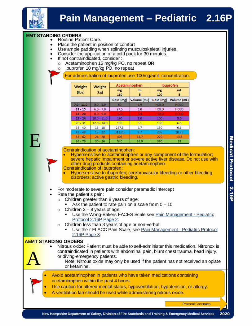
Avoid acetaminophen in patients who have taken medications containing acetaminophen within the past 4 hours.
Use caution for altered mental status, hypoventilation, hypotension, or allergy. A ventilation fan should be used while administering nitrous oxide.
2013
EMT STANDING ORDERS Routine Patient Care. Place the patient in position of comfort Use ample padding when splinting musculoskeletal injuries. Consider the application of a cold pack for 30 minutes. If not contraindicated, consider :
o Acetaminophen 15 mg/kg PO, no repeat ORo Ibuprofen 10 mg/kg PO, no repeat
2013
Me
dic
al P
roto
col 2
.16P
Pain Management – Pediatric
New Hampshire Department of Safety, Division of Fire Standards and Training & Emergency Medical Services 2020
2.16P
For moderate to severe pain consider paramedic intercept Rate the patient’s pain:
o Children greater than 8 years of age: Ask the patient to rate pain on a scale from 0 – 10
o Children 3 – 8 years of age: Use the Wong-Bakers FACES Scale see Pain Management - Pediatric
Protocol 2.16P Page 2.o Children less than 3 years of age or non-verbal: Use the r-FLACC Pain Scale, see Pain Management - Pediatric Protocol
2.16P Page 3.
Nitrous oxide: Patient must be able to self-administer this medication. Nitronox is contraindicated in patients with abdominal pain, blunt chest trauma, head injury, or diving-emergency patients.
Note: Nitrous oxide may only be used if the patient has not received an opiate or ketamine.
Protocol Continues
E
AAEMT STANDING ORDERS
For adminsitration of ibuprofen use 100mg/5mL concentration.
Contraindication of acetaminophen: Hypersensitive to acetaminophen or any component of the formulation;
severe hepatic impairment or severe active liver disease. Do not use with other drug products containing acetaminophen.
Contraindication of ibuprofen: Hypersensitive to ibuprofen; cerebrovasular bleeding or other bleeding
disorders; active gastric bleeding.

Pain Management - Pediatric
Me
dic
al P
roto
col 2
.16P
2.16P
New Hampshire Department of Safety, Division of Fire Standards and Training & Emergency Medical Services 2020
Ketamine is contraindicated in patients unable to tolerate hyperdynamic states such as those with known or suspected aortic dissection, myocardial infarction, and aortic aneurysm.
Avoid ketorolac in patients with NSAID allergy, aspirin-sensitive asthma, renal insufficiency, pregnancy, or known peptic ulcer disease.
PPARAMEDIC STANDING ORDERS
For severe pain or pain consider one of the following for pain control: Fentanyl 1.0 micrograms/kg IV/IM/IN (maximum dose 100 micrograms) may repeat
0.5 micrograms/kg (Maximum dose 50 micrograms) every 5 minutes. May be repeated to a total of 3 doses, OR
Hydromorphone 0.01 – 0.02 mg/kg (maximum dose 1 mg) IV every 10 minutes to a total of 4 mg titrated to pain relief and if systolic BP is greater than 100.
Morphine 0.1 mg/kg IV (maximum dose 5 mg) may repeat 0.05 mg/kg (maximum dose 2.5 mg) every 5 minutes. May be repeated to a total of 3 doses.
Antidote: For hypoventilation from opiate administration by EMS personnel, assist ventilations and administer as directed in the Opioid Overdose Protocol 2.15P.
AND/OR
Ketamine for patient > 3 months:o 0.5 – 1 mg/kg (maximum dose 50 mg) IN OR
o 0.1 – 0.25 mg/kg (maximum dose 20 mg) IV diluted in 50 mL 0.9% NaCl or D5W over 10 minutes (no IV pump needed) To minimize chance of dysphoric reaction consider starting at lower doses and
increasing if needed for analgesia.Antidote: For dysphoria (emergence reaction) caused by ketamine administer midazolam 0.05 mg/kg IV (single maximum dose of 2.5 mg) or *0.1 mg/kg IM/IN (single maximum dose 5 mg), may repeat once in 5 minutes. For nausea: See Nausea/Vomiting 2.11 Protocol Contact Medical Control for guidance regarding:
o Altered mental status or o Requests to provide additional doses of a medication.
Protocol Continues
*For IN administration of midazolam use a 5 mg/mL concentration.

Pain Management - PediatricM
edic
al P
roto
col 2
.16P
Each of the five categories (F) Face; (L) Legs; (A) Activity; (C) Cry; (C) Consolability is scored from 0-2, which results in a total score between zero and ten. Patients who are awake: Observe for at least 1-2 minutes. Observe legs and body uncovered. Reposition patient or observe activity, assess body for tenseness and tone. Initiate consoling interventions if needed Patients who are asleep: Observe for at least 2 minutes or longer. Observe body and legs uncovered. If possible reposition the patient. Touch the body and assess for tenseness and tone. The revised-FLACC can be used for all non-verbal children. The additional descriptors (in bold) are descriptors validated in children with cognitive impairment. The nurse can review with parents the descriptors within each category. Ask them if there are additional behaviors that are better indicators of pain in their child. Add these behaviors to the tool in the appropriate category. © 2002, The Regents of the University of Michigan. All Rights Reserved 09-09-2009
2.16P
PEARLS: Ketamine should be considered in patients with severe pain, hemodynamic compromise, pain
refractory to opiates, patients on chronic opiate treatment. Ketamine dosing is based on Pediatric Color Coded Appendix Ketamine may cause appearance of intoxication at higher doses. Dysphoria may occur as the
medication effects wear off. Avoid coaching the patient; simply ask him/her to rate his/her pain on a scale from 0 – 10,
where 0 is no pain at all and 10 is the worst pain the patient has ever experienced. Give reassurance, psychological support, and distraction. Reassess the patient’s pain level and vital signs every 5 minutes.
New Hampshire Department of Safety, Division of Fire Standards and Training & Emergency Medical Services 2020

· Routine Patient Care.· Prior to calling Poison Control attempt to identify substance, quantity, time/route
of exposure and patient information (weight, medications, history, intentional, accidental).
· Contact Poison Control at (800) 222-1222 as soon as practical.· Ingested Poison:
o Consider activated charcoal 25 – 50 grams by mouth if recommended by Poison Control or Medical Control.
· For suspected opiate overdose with severe respiratory depression, see Opioid Overdose Protocol 2.15A.
· For suspected isolated cyanide poisoning, see Smoke Inhalation Protocol 2.21A.· For decontamination/hazardous materials exposure, see Hazardous Materials
9.0.· For hypoglycemia, see Hypoglycemia Emergencies Protocol 2.9A.· For seizures, see Seizure Protocol 2.18A.
2013
P
EMR & EMT STANDING ORDERS
PARAMEDIC STANDING ORDERS
Suggested Treatments
· Beta Blocker and Calcium Channel Blocker refer to Bradycardia Protocol 3.1A .
· Dystonic Reaction:
o Diphenhydramine 25 – 50 mg IV/IM.
· Organophosphates, see Nerve Agent/Organophosphate Protocol 2.12A.
· Suspected Sympathomimetic/Stimulant:
o Midazolam 2.5 mg IV, may repeat once in 5 minutes, OR
o *Midazolam 5 mg IM/IN, may repeat once in 5 minutes, OR
o Lorazepam 1 mg IV, may repeat once in 5 minutes, OR
o Diazepam 5 mg IV, may repeat once in 5 minutes.
· Tricyclic with symptomatic dysrhythmias, (e.g., tachycardia and wide QRS > 100 milliseconds):
o Sodium bicarbonate 1 - 2 mEq/kg IV.
This protocol is designed to provide general guidelines for treatment. Specific
treatments or antidotes may be appropriate as directed by on-line medical control
or in consultation with Poison Control.
PEARLS:
· If possible, bring container/bottles and/or contents.
· Pulse oximetry may NOT be accurate for toxic inhalational patients.
· Capnography may be helpful for monitoring respiratory status and titrating to lowest effective
naloxone dose. See Capnography Procedure 6.1.
Me
dic
al P
roto
co
l 2.1
7A
Poisoning/Overdose – Adult
New Hampshire Department of Safety, Division of Fire Standards and Training & Emergency Medical Services 2020
2.17A
Protocol Continues
*For IN administration of midazolam use a 5 mg/mL concentration.
EA/

Signs & Symptoms, which may or may not be present:
· Acetaminophen: initially no sign/symptoms or nausea/vomiting. If not detected and treated,
may cause irreversible liver failure.
· Akathisia: May consist of feelings of anxiety, agitation, and jitteriness, as well as inability to sit
still / pacing. This may be induced by antipsychotics, such as haloperidol, or anti-emetics such
as prochlorperazine or metoclopramide.
· Anticholinergic: tachycardia, fever, dilated pupils, mental status changes. Blind as a bat
(blurred vision). Dry as a bone (dry mouth). Red as a beet (flushing). Mad as a hatter
(confusion). Hot as a hare (hyperthermia).
· Aspirin: abdominal pain, vomiting, tachypnea, fever and/or altered mental status. Renal
dysfunction, liver failure, and or cerebral edema among other things can take place later.
· Cardiac Medications: dysrhythmias, altered mental status, hypotension, hypoglycemia.
· Depressants: bradycardia, hypotension, decreased temperature, decreased respirations,
non-specific pupils.
· Dystonic Reaction: Neurological movement disorder, in which sustained muscle contractions
cause twisting and repetitive movements or abnormal postures. This may be induced by
antipsychotics, such as haloperidol, or anti-emetics such as prochlorperazine or
metoclopramide.
· Opiate: Respiratory depression or arrest, pinpoint pupils, decreased mental states. See
Opioid Overdose Protocol 2.15A.
· Organophosphates: bradycardia, increased secretions, nausea, vomiting, diarrhea, pinpoint
pupils.
· Solvents: nausea, coughing, vomiting, mental status change and arrhythmias. Patient with
significant solvent exposure, must be handled gently to reduce the incident of arrhythmia and/
or subsequent cardiac arrest.
· Sympathomimetic/Stimulants: tachycardia, hypertension, seizures, agitation, increased
temperature, dilated pupils, anxiety, paranoia, diaphoresis. Examples are bath salts, cocaine,
methamphetamine, ecstasy, ADHD drugs, thyroid meds (rarely), salbutamol.
· Tricyclic: seizures, dysrhythmias, hypotension, decreased mental status or coma.
Me
dic
al P
roto
co
l 2.1
7A
Poisoning/Overdose – Adult
New Hampshire Department of Safety, Division of Fire Standards and Training & Emergency Medical Services 2020
2.17A
Protocol Continued

· Routine Patient Care.
· Prior to calling Poison Control attempt to identify substance, quantity, time/route
of exposure and patient information (weight, medications, history, intentional,
accidental).
· Contact Poison Control at (800) 222-1222 as soon as practical.
· For suspected opioid overdose with severe respiratory depression, see Opioid
Overdose Protocol 2.15P.
· For suspected isolated cyanide poisoning, see Smoke Inhalation Protocol
2.21P.
· For decontamination/hazardous materials exposure: refer to Hazardous
Materials 9.0.
· For hypoglycemia, see Hypoglycemia Emergencies 2.9P.
· For seizures, see Seizures Protocol 2.18P.
2013
P
EMT/AEMT STANDING ORDERS
PARAMEDIC STANDING ORDERSSuggested Treatments
· Beta Blocker and Calcium Channel Blocker, see Bradycardia Protocol 3.1P.
· Dystonic Reaction:
o Diphenhydramine 1mg/kg IV/IM up to 50 mg.
· Organophosphates, see Nerve Agent/Organophosphate Protocol 2.12P.
· Suspected Sympathomimetic/Stimulant:
o Midazolam 0.05 mg/kg IV (single maximum dose 2.5 mg, may repeat once in 5 minutes, OR
o *Midazolam 0.1 mg/kg mg IM/IN (single maximum dose 5 mg), may repeat once in 5 minutes, OR
o Lorazepam 0.05 mg/kg mg IV (single maximum dose 1 mg), may repeat once in 5 minutes, OR
· Diazepam 0.1 mg/kg IV (single maximum dose 5 mg), may repeat once in 5
minutes.
· Tricyclic with symptomatic dysrhythmias, (e.g., tachycardia and wide QRS > 100
milliseconds):
o Sodium bicarbonate 1 - 2 mEq/kg IV.
This protocol is designed to provide general guidelines for treatment. Specific
treatments or antidotes may be appropriate as directed by on-line medical control or in
consultation with Poison Control.
Me
dic
al P
roto
co
l 2.1
7P
Poisoning/Overdose – Pediatric
New Hampshire Department of Safety, Division of Fire Standards and Training & Emergency Medical Services 2020
2.17P
````
PEARLS:
· If possible, bring container/bottles, and/or contents.
· Pulse oximetry may NOT be accurate for toxic inhalational patients.
· Capnography may be helpful for monitoring respiratory status and titrating to lowest effective
naloxone dose. See Capnography Procedure 6.1.
Protocol Continues
EA/
*For IN administration of midazolam use a 5 mg/mL concentration.

Protocol Continued
Me
dic
al P
roto
co
l 2.1
7P
Poisoning/Overdose – Pediatric
New Hampshire Department of Safety, Division of Fire Standards and Training & Emergency Medical Services 2020
2.17P
Signs & Symptoms, which may or may not be present:
· Acetaminophen: initially no signs/symptoms or nausea/vomiting. If not detected and treated,
may cause irreversible liver failure.
· Akathisia: May consist of feelings of anxiety, agitation, and jitteriness, as well as inability to sit
still / pacing. This may be induced by antipsychotics, such as haloperidol, or anti-emetics such
as prochlorperazine or metoclopramide.
· Anticholinergic: tachycardia, fever, dilated pupils, mental status changes. Blind as a bat
(blurred vision). Dry as a bone (dry mouth). Red as a beet (flushing). Mad as a hatter
(confusion). Hot as a hare (hyperthermia).
· Aspirin: abdominal pain, vomiting, tachypnea, fever and/or altered mental status. Renal
dysfunction, liver failure, and or cerebral edema among other things can take place later.
· Cardiac Medications: dysrhythmias, altered mental status, hypotension, hypoglycemia.
· Depressants: bradycardia, hypotension, decreased temperature, decreased respirations,
non-specific pupils.
· Dystonic Reaction: Neurological movement disorder, in which sustained muscle contractions
cause twisting and repetitive movements or abnormal postures. This may be induced by
antipsychotics, such as haloperidol, or anti-emetics such as prochlorperazine or
metoclopramide.
· Opiate: Respiratory depression or arrest, pinpoint pupils, decreased mental states. See
Opioid Overdose Protocol 2.15P Organophosphates: bradycardia, increased secretions,
nausea, vomiting, diarrhea, pinpoint pupils.
· Solvents: nausea, coughing, vomiting, mental status change and arrhythmias. Patient with
significant solvent exposure, must be handled gently to reduce the incident of arrhythmia and/
or subsequent cardiac arrest.
· Sympathomimetic/Stimulants: tachycardia, hypertension, seizures, agitation, increased
temperature, dilated pupils, anxiety, paranoia, diaphoresis. Examples are bath salts, cocaine,
methamphetamine, ecstasy, ADHD drugs, thyroid meds (rarely), salbutamol.
· Tricyclic: seizures, dysrhythmias, hypotension, decreased mental status or coma.

· Routine Patient Care.
· If the blood glucose reading is <60 mg/dL, see Hypoglycemia Protocol 2.9A.
· If midazolam intranasal or diazepam rectal gel (Diastat) has been
prescribed by the patient’s physician, assist the patient or care giver with
the administration in accordance with the physician’s instructions.
· If the patient has an implanted vagus nerve stimulator (VNS), suggest that
family use the VNS magnet to activate the VNS and assist if required.
o Swipe the VNS magnet over the stimulator, located in the left chest
area, for one second, counting one-one thousand while it’s swiped over
the chest.
o Note: do not delay medication administration.
2013
P
EMT/ADVANCED EMT STANDING ORDERS
PARAMEDIC STANDING ORDERS
While seizure activity is present, consider:· Midazolam 5 mg IV, repeat every 5 minutes as needed, OR · *Midazolam 10 mg IM/IN, repeat every 5 minutes as needed, OR· Lorazepam 2 - 4 mg IV, repeat every 5 minutes as needed, OR
· Diazepam 10 mg IV, repeat every 5 minutes as needed.
For patients in the third trimester of pregnancy or post-partum who are seizing or
who are post-ictal:
· Magnesium sulfate, 4 grams IV (mix in 100 mL 0.9% NaCl) bolus over 10
minutes, then consider 1 gram/hr continuous infusion.
EA/
Me
dic
al P
roto
co
l 2.1
8A
Seizures – Adult
New Hampshire Department of Safety, Division of Fire Standards and Training & Emergency Medical Services 2020
2.18A
*For IN administration of midazolam use a 5 mg/mL concentration.
PEARLS:
· Do not attempt to restrain the patient; protect them from injury.
· History preceding a seizure is very important. Find out what precipitated the seizure (e.g.,
medication non-compliance, active infection, trauma, hypoglycemia, poisoning).
· Status epilepticus is defined as any generalized seizures lasting more than 5 minutes.
This is a true emergency requiring rapid airway control, treatment (including
benzodiazepines), and transport.
· IM/IN is the preferred route for midazolam where an IV has not been previously
established.
· IM midazolam should be administered to the lateral thigh.
· Diazepam and lorazepam are not well absorbed IM and should be given IV.
· There is an increased risk of apnea with >2 doses of benzodiazepines.
Do NOT routinely place an IV/IO for the actively seizing patient
(unless needed for other reasons).

· Routine Patient Care.
· If the blood glucose reading is <60 mg/dL, see Hypoglycemia Protocol 2.9P.
· If midazolam intranasal or diazepam rectal gel (Diastat) has been prescribed
by the patient’s physician, assist the patient or care giver with the
administration in accordance with the physician’s instructions.
· If the patient has an implanted vagus nerve stimulator (VNS), suggest that
family use the VNS magnet to activate the VNS and assist if required.
o Swipe the VNS magnet over the stimulator, located in the left chest area, for
one second, counting one-one thousand while it’s swiped over the chest.
o Note: do not delay medication administration.
P
EMT/ADVANCED EMT STANDING ORDERS
PARAMEDIC STANDING ORDERS
While seizure activity is present, consider:· *Midazolam 0.2 mg/kg IM/IN (single maximum dose 10 mg) repeat every 5
minutes as needed, OR· Midazolam 0.1 mg/kg IV (single maximum dose 5 mg) repeat every 5 minutes
as needed, OR· Lorazepam 0.1 mg/kg IV (single maximum dose 4 mg) repeat every 5 minutes
as needed, OR· Diazepam 0.2 mg/kg IV (single maximum dose 10 mg IV) repeat every 5
minutes as needed.
PEARLS:
· Do not attempt to restrain the patient; protect them from injury.
· History preceding a seizure is very important. Find out what precipitated the seizure (e.g.,
medication non-compliance, active infection, trauma, hypoglycemia, poisoning).
· Status epilepticus is defined as any generalized seizures lasting more than 5 minutes.
This is a true emergency requiring rapid airway control, treatment (including
benzodiazepines), and transport.
· IM/IN is the preferred route for midazolam where an IV has not been previously
established.
· IM midazolam should be administered to the lateral thigh.
· Diazepam and lorazepam are not well absorbed IM and should be given IV.
· There is an increased risk of apnea with >2 doses of benzodiazepines.
EA/
Me
dic
al P
roto
co
l 2.1
8P
Seizures – Pediatric
New Hampshire Department of Safety, Division of Fire Standards and Training & Emergency Medical Services 2020
2.18P
Do NOT routinely place an IV/IO for the actively seizing patient
(unless needed for other reasons).
*For IN administration of midazolam use a 5 mg/mL concentration.

Sepsis – Adult
P
EEMT STANDING ORDERS - ADULT
PARAMEDIC STANDING ORDERS - ADULT
ADVANCED EMT STANDING ORDERS - ADULT
A · Rapidly administer IV fluid, 30 mL/kg bolus to maintain MAP > 65 mmHg (systolic blood pressure >90 mmHg).
· Patients should be reassessed frequently, with special attention given to the lung examination to ensure volume overload does not occur.
· Refer to Advanced Sepsis Protocol, if prerequisites have been met.· Obtain serum lactate level (if available and trained)· If there is no adequate hemodynamic response after initial bolus consider:
o Epinephrine by push dose (dilute boluses) prepare 10 mcg/mL by adding 1 mL 0.1 mg/mL Epinephrine to 9 mL normal saline, then administer 10-20 mcg boluses (1 – 2 mL) every 2 minutes (where feasible, switch to infusion as soon as practical), AND/OR
o Epinephrine 2 -10 micrograms/minute via pump, ORo Norepinephrine 1 - 30 micrograms/minute via pumpo Continue maintenance fluid concurrently with pressor administration, titrate to
MAP ≥ 65 mmHg (systolic blood pressure > 90 mmHg).
· Routine Patient Care.· Do not delay transport.· If positive sepsis screen, notify receiving facility of a “Sepsis Alert”.
Me
dic
al P
roto
co
ls 2
.19
A
New Hampshire Department of Safety, Division of Fire Standards and Training & Emergency Medical Services 2020
IDENTIFICATION OF POSSIBLE SEPSIS · Suspected infection – YES· Evidence of sepsis criteria – YES (2 or more):
o Temperature < 96.8 °F or > 101°Fo Heart rate > 90 bpmo Respiratory rate > 20 bpmo Mean Arterial Pressure (MAP) <65mmHg (systolic blood pressure < 90 mmHg) o New onset altered mental status OR increasing mental status change with previously
altered mental statuso Serum lactate level >2 mmol/Lo ETCO2 < 25 mmHg
2.19A
PEARLS:· Sepsis is life-threatening organ dysfunction due to a dysregulated host response to infection· Septic shock is a subset of sepsis in which underlying circulatory and cellular/metabolic
abnormalities are profound enough to substantially increase mortality.· Sepsis Alert - Notifies receiving facility that patient may require resuscitation and/or more
resource intensive management, may assist with predicting pts who will have poor outcomes without appropriate and timely treatment.
· Provide receiving facility with written documentation that includes time of initial bolus, time of completion of bolus, total volume infused and rate.
· Septic shock has a high mortality and is one of the leading causes of pediatric deaths.
P
EEMT STANDING ORDERS - PEDIATRIC
PARAMEDIC STANDING ORDERS - PEDIATRIC
ADVANCED EMT STANDING ORDERS - PEDIATRIC
A
IV fluids should be titrated to attain normal capillary refill, peripheral pulses, and level of consciousness.· Administer fluid bolus of 10 - 20 mL/kg of IV fluid by syringe push method;
reassess patient immediately after completion of bolus and repeat 2 times (max 60 mL/kg), if inadequate response to boluses.
Note: Reassess patient between each bolus for improving clinical signs and signs of fluid overload (rales, increased work of breathing, or increased oxygen requirements).
· Obtain finger stick lactate level (if available and trained).· If there is no response after 3 fluid boluses, contact Medical Control to consider:
o Additional fluidso Norepinephrine (preferred) 0.05 – 0.1 mcg/kg/min, titrated to effect to a
maximum dose 2 mcg/kg/min, via pump, see appendix 4 ORo Epinephrine 0.1 – 1.0 mcg/kg/min, via pump, titrated to effect see appendix 4.
· Routine Patient Care.· Monitor and maintain airway and breathing as these may change precipitously. · Administer oxygen and continue regardless of oxygen saturation levels.· Obtain blood glucose reading.· Do not delay transport.
Me
dic
al P
roto
co
ls 2
.19
PSepsis – Pediatric
New Hampshire Department of Safety, Division of Fire Standards and Training & Emergency Medical Services 2020
IDENTIFICATION OF POSSIBLE SEPSIS:· Suspected Infection – YES· Temperature > 101o F or < 96.8 · Heart rate or respiratory rate greater than normal limit for
age(heart rate may not be elevated in septic hypothermic patients) AND at least one of the following indications of altered organ function: o Altered mental status o Capillary refill time <1 second (flash) or > 3 secondso Mottled cool extremities o Finger stick lactate level >2mmol/L o ETCO2 < 25 mmHg
Note: Consider early consultation with Medical Control for suspected pediatric septic shock patients.
2.19P

New Hampshire Department of Safety, Division of Fire Standards and Training & Emergency Medical Services
Me
dic
al P
roto
co
l 2.2
0
Non-Traumatic ShockAdult & Pediatric
2020
SHOCKInadequate tissue
perfusion that impairs cellular metabolism
Recognize Compensated Shock- Adult· Anxiety· Tachycardia· Tachypnea· Diaphoresis
NO YES
CARDIOGENIC SHOCK
DISTRIBUTIVE SHOCK
HYPOVOLEMIC SHOCK
OBSTRUCTIVE SHOCK
· Norepinephrine infusion 1 – 30 microgram/minute (preferred) via pump, OR
· Epinephrine infusion 2 – 10 micrograms/minute, via pump*For pediatric cardiogenic shock administer fluid bolus of 10mL/kg of 0.9% saline by syringe push method. Repeat bolus per Medical Control.
Abdominal pain with vaginal bleeding see Obstetric Protocol 2.14.Nausea and vomiting see Nausea Vomiting Protocol 2.11.For GI bleeding see Abdominal Pain Protocol 2.0.Heat exposure, see Hyperthermia Protocol 2.8.
For cardiac tamponade, rapid transport, treat arrhythmias per Cardiac Protocols 3.0 – 3.6.For spontaneous pneumothorax: consider needle decompression per Thoracic Injury Protocol 4.8.For pulmonary embolism: rapid transport
Known history of adrenal insufficiency or recent illness, see Adrenal Insufficiency Protocol 2.1Systemic response to an allergen, see Anaphylaxis/Allergic Reaction Protocol 2.2A&POverwhelming response to an infection, see Sepsis Protocol 2.19 A&P
Primary pump failureDecreased
cardiac output
Insufficient circulating volume.
Obstruction of blood flow outside the
heart
Inadequate blood volume distribution.
Trauma Involved? See Shock – Traumatic Protocol 4.6
Consider
Consider
Consider
Consider
Recognize Compensated Shock - Pediatric:· Delayed capillary refill· Decreased or bounding peripheral pulses· Palpable central pulse, decreased distal
pulse· Cool extremities· Altered mental status· Mild tachypnea
2.20
EEMT STANDING ORDERS - ADULT & PEDIATRIC:
P
PARAMEDIC STANDING ORDERS – ADULT & PEDIATRIC
ADVANCED EMT STANDING ORDERS – ADULT & PEDIATRIC
A· ADULT: Administer IV fluid in 250 mL boluses to return the patient to a
coherent mental status or palpable radial pulse, not to exceed 2000 mL without consultation with Medical Control.
· PEDIATRIC: Administer fluid bolus of 10 - 20 mL/kg of IV fluid by syringe push method (may repeat to a maximum 60 mL/kg) to improve clinical condition (capillary refill time ≤ 2 seconds, equal peripheral and distal pulses, improved mental status, normal breathing.
· Obtain finger stick lactate level (if available and trained)o ETCO2 < 25 mmHg OR lactate > 2 mmol/L may indicate poor
perfusion/shock
ADULT: If there is no adequate hemodynamic response after 2,000 ml IV fluid infused consider: · Epinephrine by push dose (dilute boluses) prepare 10 mcg/mL by adding 1
mL 0.1 mg/mL Epinephrine to 9 mL normal saline, then administer 10-20 mcg boluses (1 – 2 mL) every 2 minutes (where feasible, switch to infusion as soon as practical), AND/OR
· Norepinephrine infusion 1 – 30 microgram/minute (preferred) via pump, OR· Epinephrine infusion 2 – 10 micrograms/minute, via pumpPEDIATRIC: If there is no adequate hemodynamic response after 60 mL/kg IV fluid infused contact Medical Control
Consider

· Routine Patient Care.· Oxygen 100% via non-rebreather mask or BVM.· Decontamination concurrent with initial resuscitation.· If a carbon monoxide (CO) oximeter (e.g., Rad-57) is available, obtain
carbon monoxide levels.· If a measuring device is available, obtain atmospheric levels of carbon
monoxide (CO) and cyanide (CN).
2013
E
EMT STANDING ORDERS
ADVANCED EMT/PARAMEDIC STANDING ORDERSFor a history of smoke exposure with an altered level of consciousness and/or hemodynamic or respiratory compromise, administer, if available:· Hydroxocobalamin via use of Cyanokit:
o Depending on clinical response, a second dose may be required.
· Oxygen saturation may be inaccurate in patients exposed to carbon monoxide or cyanide.
· CO oximeter devices may yield inaccurate low/normal results for patients with CO poisoning. All patients with probable or suspected CO poisoning should be transported to the nearest appropriate hospital, based on their presenting signs and symptoms.
· Do not administer other drugs concurrently in same IV as hydroxocobalamin.
Symptoms: headache, confusion, dyspnea, chest tightness, nausea.Signs: soot in the nose or mouth, change in level of consciousness, seizure, dilated pupils, coughing, tachypnea and hypertension (early), bradypnea and hypotension (late), shock, vomiting.
A/P
Me
dic
al P
roto
co
l 2.2
1A
Smoke Inhalation/Carbon Monoxide Poisoning – Adult
New Hampshire Department of Safety, Division of Fire Standards and Training & Emergency Medical Services 2020
2.21A
PEARLS:· Smoke is a combination of many dangerous toxins produced by incomplete combustion.
Patients exposed to smoke should be considered for carbon monoxide (CO) and hydrogen cyanide (HCN) poisoning.

· Routine Patient Care.· Oxygen 100% via non-rebreather mask or BVM.· Decontamination concurrent with initial resuscitation.· If a carbon monoxide (CO) oximeter (e.g., Rad-57) is available, obtain carbon
monoxide levels.· If a measuring device is available, obtain atmospheric levels of carbon
monoxide (CO) and cyanide (CN).
E
EMT STANDING ORDERS
ADVANCED EMT/PARAMEDIC STANDING ORDERSFor a history of smoke exposure with an altered level of consciousness and/or hemodynamic or respiratory compromise, administer, if available:· Hydroxocobalamin via use of Cyanokit:
o Using vented intravenous tubing, infuse per Pediatric Color Coded Appendix 3 over 7.5 minutes for 100 mL vial set or 15 minutes for 200 mL vial set.
o Depending on clinical response, a second dose may be required.
Symptoms: headache, confusion, dyspnea, chest tightness, nausea.Signs: soot in the nose or mouth, change in level of consciousness, seizure, dilated pupils, coughing, tachypnea and hypertension (early), bradypnea and hypotension (late), shock, vomiting.
A/P
Me
dic
al P
roto
co
l 2.2
1P
Smoke Inhalation/Carbon Monoxide Poisoning – Pediatric
New Hampshire Department of Safety, Division of Fire Standards and Training & Emergency Medical Services 2020
2.21P
· Oxygen saturation may be inaccurate in patients exposed to carbon monoxide or cyanide.
· CO oximeter devices may yield inaccurate low/normal results for patients with CO poisoning. All patients with probable or suspected CO poisoning should be transported to the nearest appropriate hospital, based on their presenting signs and symptoms.
· Do not administer other drugs concurrently in same IV as hydroxocobalamin.
PEARLS:· Smoke is a combination of many dangerous toxins produced by incomplete combustion.
Patients exposed to smoke should be considered for carbon monoxide (CO) and hydrogen cyanide (HCN) poisoning.

20132013
Me
dic
al P
roto
co
l 2.2
2Northern New England Unified Guideline
Stroke – Adult
New Hampshire Department of Safety, Division of Fire Standards and Training & Emergency Medical Services 2020
2.22
· Routine Patient Care.· Complete the Prehospital Stroke Screening Tool
o If Prehospital Stroke screen is positive, complete stroke severity score (e.g., FAST-ED) to determine probability of a large vessel occlusion (LVO)
· Establish Stroke Alert Criteria and notify receiving hospital of “Stroke Alert” with findings from the screening tools, thrombolytic checklist and time last known well (TLKW).
· For symptomatic:o Administer oxygen to maintain SPO2 between 94% - 98%o Elevate head of stretcher to 30 ° (unless patient requires spinal motion restriction);o Minimize on-scene time; do not delay for ALS intercept;o Acquire and transmit 12-lead ECG, if available;o Correct glucose if < 60 mg/dL. See Hypoglycemia Protocol 2.8A or 2.8P.o Rapid transport to the most appropriate facility based on the destination guidance
utilizing your local stroke plan.
EMT STANDING ORDERS
AEMT & PARAMEDIC STANDING ORDERS
A/P· Establish IV (18 gauge catheter & right AC preferred site) and administer 250 mL IV fluid.
E
Protocol Continues
SUSPECT STROKE: with any of the following new or sudden symptoms and/or complaints:· Unilateral motor weakness or paralysis to face, limb or side of body, including facial droop· Unilateral numbness· Dizziness/vertigo· Acute visual disturbance, loss of vision in one eye or one side of vision· Difficulty with balance or uncoordinated movements of a limb, gait disturbance· Difficulty with speech understanding or production (slurred or inappropriate use of words)· Severe headache for no obvious reason· Altered mental state
Arm Drift: Have the patient close their eyes and hold arms extended for 10 seconds.Normal: Both arms move the same, or both arms don’t move at all.Abnormal: One arm doesn’t move, or one arm drifts down compared to the other.
Prehospital Stroke Scale Examination Please check: Normal Abnormal
Witness: Best contact number for witness:
Facial Droop: Have the patient smile and show teeth.Normal: Both sides of the face move equally well..Abnormal: One side of the face does not move as well as the other.
Normal
Blood Glucose:
Prehospital Stroke Screening Tool
Time Last Known Well: (If patient awoke with symptoms, time last known to be at baseline)
Speech: Ask the patient to repeat a phrase such as, “You can’t teach an old dog new tricks”.Normal: Patient says the correct words without slurring.Abnormal: Patient slurs words, says the wrong word, or is unable to speak.
Normal Abnormal
Normal Abnormal
Abnormal
( ) -
Stroke screen is positive if any abnormal finding in facial droop, arm drift or speech.

Me
dic
al P
roto
co
l 2.2
2
New Hampshire Department of Safety, Division of Fire Standards and Training & Emergency Medical Services 2020
2.21Protocol Continued
If stroke screening scale is positive calculate stroke severity score using FAST-ED
Stroke Alert Criteria – Please check Yes or No:Blood glucose is or has been corrected to greater than 60 mg/dL?Deficit unlikely due to head trauma or other identifiable causes?Positive Prehospital Stroke Screen:
- and time last known well is less than 4.5 hours OR - FAST-ED score is greater than or equal to 4 AND time last known well is less than 24 hours
Stroke Alert Criteria – If yes to all criteria determine appropriate destination, contact receiving hospital and report a STROKE ALERT with time last known well, FAST-ED score
& thrombolytic checklist results
Establish Stroke Alert CriteriaYes No
Northern New England Unified Guideline Stroke – Adult2.22

Me
dic
al P
roto
co
l 2.2
2Northern New England Unified Guideline
Stroke – Adult
New Hampshire Department of Safety, Division of Fire Standards and Training & Emergency Medical Services 2020
PEARLS for Anticoagulants:· Patients may recognize anticoagulants as "blood thinners". Ask about anticoagulants including
warfarin (Coumadin or Jantoven), Heparin (IV/IM - including Lovenox), dabigatran (Pradaxa), rivaroxaban (Xarelto), apixaban (Eliquis), betrixaban (Bevyxxa) or edoxaban (Savaysa) and immediately communicate to hospital staff.
· Please note, medication manufacturers are producing new anticoagulants frequently.
PrehospitalStroke
Screening ToolPositive
Positive LVO Screen
Negative LVO Screen
FAST-ED ≥ 4
FAST-ED˂ 4
TLKW˃ 4.5 & < 24 hours
TLKW˂ 4.5 or > 24 hours
If feasible, transport to
Endovascular Center, per your local stroke plan
Nearest Appropriate Hospital (including
Endovascular Center if this is the closest hospital)
DestinationTime Last Known Well (TLKW)Identification of Possible LVO
Destination Guidance for Possible LVO Stroke Patients
Work with your regional endovascular center when developing your local stroke plan.
Thrombolytic Checklist for patients eligible for thrombolytics (t-PA), try to complete the following:
YES NO Has the patient had any of the following:
1. Severe head trauma or intracranial or spinal surgery within the past 3 months?
2. Major non-cranial surgery or trauma within 14 days with uncontrolled bleeding (e.g.; internal organs)?
3. Spontaneous (non-traumatic) intracranial hemorrhage at any time in the past?
4. Is the patient taking any anticoagulants, including oral or injectable medications? If yes, clarify when last dose was taken (see PEARLS below)
PEARLS: · Stroke requires time sensitive interventions. Time = Brain· Every minutes of acute stroke = about 2 million neurons lost.· Transport witness, family or caregiver or obtain witness best phone number for hospital staff to
verify time of symptom onset or Time Last Known Well (TLKW).· TLKW is the last time patient known to be at their neurological baseline. If patient awakes with
symptoms, TLKW is time patient was last known to be at their neurological baseline – Ask if patient got up during the night and was at baseline!
· Consider stroke mimics including: migraine, hypoglycemia, seizures, intoxication, sepsis cerebral infectious process, toxic ingestion, neuropathies (Bell’s palsy), neoplasms, hypertensive encephalopathy.
2.22
Me
dic
al P
roto
co
l 2.2
2
New Hampshire Department of Safety, Division of Fire Standards and Training & Emergency Medical Services 2020
This page left blank to insert your local Stroke agreement
plan.
Northern New England Unified Guideline Stroke – Adult2.22

· Routine Patient Care.· Maintain oxygen saturation 94 - 98%.· Attempt to determine the cause of syncope.· Perform cardiac monitoring; obtain 12-Lead EKG, if available. If acute coronary
syndrome is suspected, refer to Acute Coronary Syndrome Protocol 3.0.· Obtain blood glucose analysis; refer to Hyperglycemia 2.7 A&P or Hypoglycemia
2.9 A&P Protocols, if indicated.· Assess for trauma either as the cause of the syncope or as a consequence of the
syncopal event assess for trauma; refer to Spinal Injury Protocol 4.7 if indicated.· Prevent and treat for shock; see Shock- Non-traumatic 2.20 or Shock - Traumatic
Protocol 4.6.· Consider ALS intercept.
PEARLS:· Syncope is defined as a loss of consciousness accompanied by a loss of postural tone with
spontaneous recovery.· Consider all syncope to be of cardiac origin until proven otherwise.· While often thought as benign, syncope can be the sign of more serious medical emergency. · Syncope that occurs during exercise often indicates an ominous cardiac cause. Patients
should be evaluated at the ED. Syncope that occurs following exercise is almost always vasovagal and benign.
· Prolonged QTc (generally >500ms) and Brugada Syndrome (incomplete RBBB pattern in V1/V2 with ST segment elevation) should be considered in all patients.
· There is no evidence that supports acquiring orthostatic vital signs.· Syncope can be indicative of many medical emergencies including:
· Observe for and treat dysrhythmias as indicated.P
· Consider fluids per Shock – Non-traumatic Protocol 2.20.A
E
EMT STANDING ORDERS
ADVANCED EMT STANDING ORDERS
PARAMEDIC STANDING ORDERS
2013
Me
dic
al P
roto
co
l 2.2
3Syncope Adult & Pediatric
New Hampshire Department of Safety, Division of Fire Standards and Training & Emergency Medical Services 2020
2.23
o Myocardial infarctiono Pulmonary embolismo Cardiac arrhythmias,o Vaso-vagal reflexeso Diabetic emergencies
o Poisoning/drug effectso Dehydrationo Hypovolemiao Seizureso Ectopic pregnancy
Burgada sample

Ca
rdia
c P
roto
co
l 3.0
E
EMT STANDING ORDERS - ADULT
ADVANCED EMT STANDING ORDERS - ADULT
A· Establish IV (if feasible, avoid right wrist)· IV must be established before administration of nitroglycerin. · Nitroglycerin 0.4 mg SL every 3 – 5 minutes while symptoms persist and if
systolic BP remains >100 mmHg.
· Routine Patient Care.· Obtain 12-lead ECG with baseline vitals within 10 minutes if available and
practical; and transmit per local guidelines. See Protocol 6.0 12-Lead EKG Acquisition· If 12-lead ECG indicates a STEMI transport patient to the most appropriate
facility in accordance with local STEMI guidelines/agreements. Notify receiving facility of a “STEMI Alert”.
· Administer oxygen only to patients with dyspnea, hypoxia (O2 sat <94%), or signs of heart failure at a rate to keep O2 saturation ≥ 94 - 98%.
· Administer aspirin 324 mg by mouth (chewable), unless patient self administered 324 mg within the last 30 minutes.
· Facilitate administration of the patient’s own nitroglycerin every 3 – 5 minutes while symptoms persist and systolic BP remains >100 mmHg, to a total of 3 doses.
Not all patients with complaint of chest pain should automatically be treated with aspirin and nitrates. Consider the likelihood of ACS based on the nature of the symptoms, the patient’s age, cardiac risk factors, past medical history, etc.
Acute Coronary SyndromeAdult
New Hampshire Department of Safety, Division of Fire Standards and Training & Emergency Medical Services 2020
P
PARAMEDIC STANDING ORDERS - ADULT· Consider IV nitroglycerin at 10 micrograms/minute if symptoms persist after 3rd
SL nitroglycerin (it is recommended two (2) IV lines in place and the IV nitroglycerin must be on an infusion pump).
· Increase IV nitroglycerin by 10 micrograms/minute every 5 minutes while symptoms persist and systolic BP remains >100 mmHg.
· Consider fentanyl 25 – 100 micrograms slow IV push every five minutes up to 300 micrograms and systolic BP remains >100 mmHg OR
· Consider morphine 2 – 5 mg IV/IM every 5 minutes to a maximum of 15 mg titrated to pain and systolic BP remains >100 mmHg.
If STEMI without uncontrolled bleeding or known thrombocytopenia consider: · Heparin 60 unit/kg to a maximum of 4000 unit IV bolus.
PARAMEDIC MEDICAL CONTROL – MAY CONSIDER
Acute Coronary Syndrome - Adult3.0
· Avoid nitroglycerin in any patient who has used a phosphodiesterase inhibitor such as: sildenafil (Viagra, Revatio), vardenafil (Levitra, Staxyn), tadalafil (Cialis, Adcirca) which are used for erectile dysfunction and pulmonary hypertension. Also avoid use in patients receiving intravenous epoprostenol (Flolan) which is used for pulmonary hypertension.
· Administer nitrates with extreme caution, if at all, to patients with inferior-wall STEMI or suspected right ventricular (RV) involvement because these patients require adequate RV preload.
Protocol Continues

2015
Protocol Continued
PEARLS:· Transmission of 12-lead ECG is critical to the activation of a STEMI system. Transmit any
12-lead ECG that states “Acute MI”, “Meets ST Elevation MI Criteria” or anything similar, or where the interpretation is unclear.
· Early administration of aspirin has been shown to decrease mortality in Acute Coronary Syndrome.
· Administer aspirin to every patient with suspected acute coronary syndrome unless they have:o History of anaphylaxis to aspirin, NSAIDs, oro Evidence of active gastrointestinal bleeding
· Patients with acute coronary syndrome (especially women and the elderly) may present with signs and symptoms other than chest pain including shortness of breath, weakness, syncope and nausea.
Acute Coronary Syndrome – Adult 3.0C
ard
iac
Pro
toc
ol 3
.0
New Hampshire Department of Safety, Division of Fire Standards and Training & Emergency Medical Services 2020
Protocol Continues

Acute Coronary Syndrome – Adult
This page left blank to insert your local STEMI agreement
plan.
Acute Coronary Syndrome – Adult 3.0Acute Coronary Syndrome Adult
Ca
rdia
c P
roto
co
l 3.0
Acute Coronary Syndrome - Adult3.0
New Hampshire Department of Safety, Division of Fire Standards and Training & Emergency Medical Services 2020
Protocol Continued

Ca
rdia
c P
roto
co
l 3.1
A
· Routine Patient Care.
· Consider the underlying causes of bradycardia (e.g., acute coronary
syndrome, hyperkalemia, hypoxia, hypothermia).
· 12-lead ECG if available.
Bradycardia – Adult
New Hampshire Department of Safety, Division of Fire Standards and Training & Emergency Medical Services 2020
P
EMT/ADVANCED EMT STANDING ORDERS
PARAMEDIC STANDING ORDERS
For symptomatic bradycardia:If hemodynamically unstable:
· Consider atropine 0.5 mg IV every 3 – 5 minutes to a maximum of 3 mg.
· Consider transcutaneous pacing.
· Administer procedural sedation prior to or during transcutaneous pacing, if feasible:
o Midazolam 2.5 mg IV, may repeat once in 5 minutes, OR
o *Midazloam 5 mg IM/IN, may repeat once in 5 minutes, OR
o Lorazepam 1 mg IV, may repeat once in 5 minute, OR
o Diazepam 5 mg IV, may repeat once in 5 minutes.
· Consider vasopressor:
o Epinephrine by push dose (dilute boluses) prepare 10 mcg/mL by
adding 1 mL 0.1 mg/mL Epinephrine to 9 mL normal saline, then
administer 10-20 mcg boluses (1 – 2 mL) every 2 minutes (where
feasible, switch to infusion as soon as practical), AND/OR
o Epinephrine 2 -10 micrograms/minute via pump, OR
o Norepinephrine 1 - 30 micrograms/minute via pump
· Contact Medical Control for expert consultation.
Other Causes:
· For symptomatic beta blocker overdose, consider glucagon 5 mg IV over 3 – 5 minutes.
· For suspected hyperkalemia with ECG changes or symptomatic calcium channel blocker/beta blocker overdose consider:
o Calcium gluconate (10% solution) 2 grams IV over 10 minutes, with
continuous cardiac monitoring, may repeat in 10 minutes if clinical indication persists OR
o Calcium chloride (10% solution) 1 gram IV over 10 minutes, with continuous cardiac monitoring. May repeat in 10 minutes if clinical indication persists.
EA/
3.1A
PEARLS:
· Hyperkalemia should be suspected in dialysis or renal failure patients with ECG changes
such as tall peaked T waves, loss of P waves, QRS widening and bradycardia.
· When pushed too quickly, glucagon can cause nausea and vomiting.
*For IN administration of midazolam use a 5 mg/mL concentration.
For calcium chloride administration, ensure IV patency and do not exceed 1 mL per
minute.
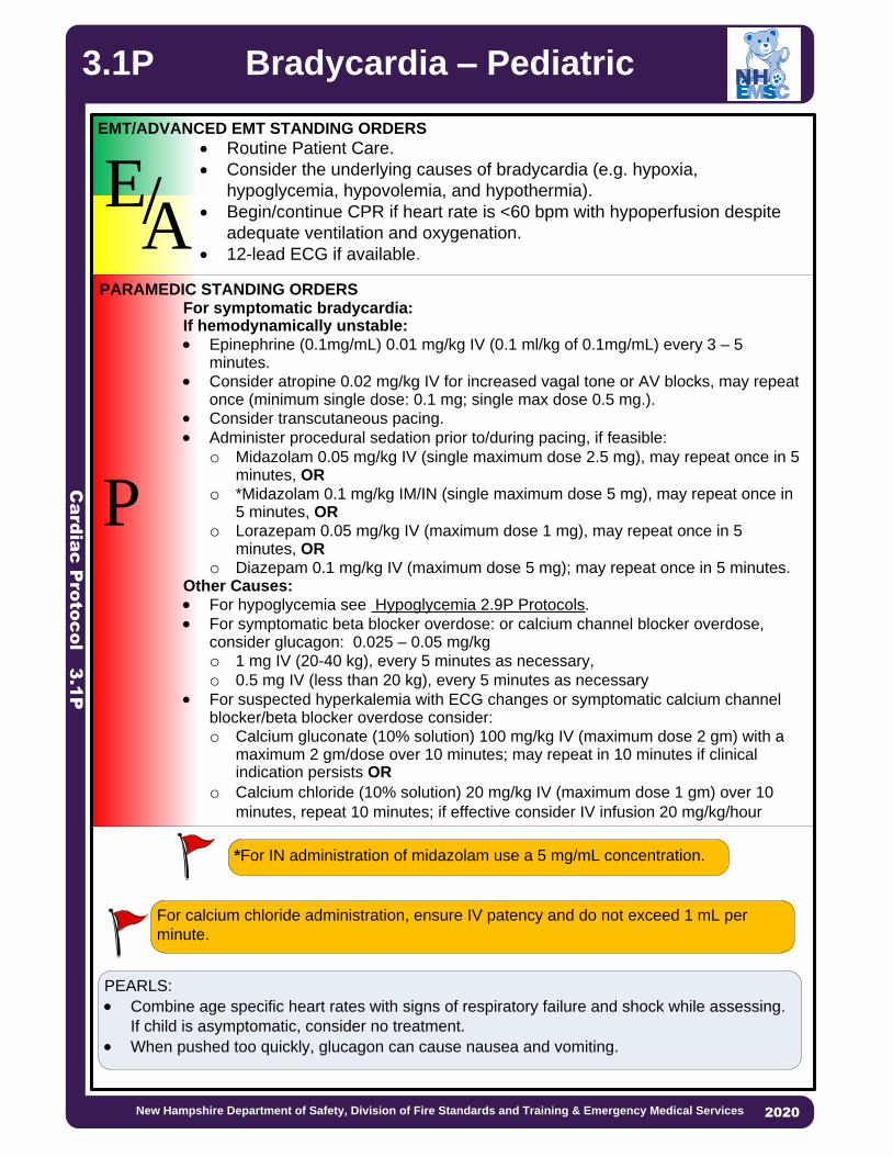
Ca
rdia
c P
roto
co
l 3.1
P
· Routine Patient Care.
· Consider the underlying causes of bradycardia (e.g. hypoxia,
hypoglycemia, hypovolemia, and hypothermia).
· Begin/continue CPR if heart rate is <60 bpm with hypoperfusion despite
adequate ventilation and oxygenation.
· 12-lead ECG if available.
2013
P
EMT/ADVANCED EMT STANDING ORDERS
PARAMEDIC STANDING ORDERSFor symptomatic bradycardia:If hemodynamically unstable:
· Epinephrine (0.1mg/mL) 0.01 mg/kg IV (0.1 ml/kg of 0.1mg/mL) every 3 – 5 minutes.
· Consider atropine 0.02 mg/kg IV for increased vagal tone or AV blocks, may repeat once (minimum single dose: 0.1 mg; single max dose 0.5 mg.).
· Consider transcutaneous pacing.
· Administer procedural sedation prior to/during pacing, if feasible:
o Midazolam 0.05 mg/kg IV (single maximum dose 2.5 mg), may repeat once in 5 minutes, OR
o *Midazolam 0.1 mg/kg IM/IN (single maximum dose 5 mg), may repeat once in 5 minutes, OR
o Lorazepam 0.05 mg/kg IV (maximum dose 1 mg), may repeat once in 5 minutes, OR
o Diazepam 0.1 mg/kg IV (maximum dose 5 mg); may repeat once in 5 minutes.Other Causes:
· For hypoglycemia see Hypoglycemia 2.9P Protocols.
· For symptomatic beta blocker overdose: or calcium channel blocker overdose, consider glucagon: 0.025 – 0.05 mg/kg
o 1 mg IV (20-40 kg), every 5 minutes as necessary,
o 0.5 mg IV (less than 20 kg), every 5 minutes as necessary
· For suspected hyperkalemia with ECG changes or symptomatic calcium channel blocker/beta blocker overdose consider:
o Calcium gluconate (10% solution) 100 mg/kg IV (maximum dose 2 gm) with a maximum 2 gm/dose over 10 minutes; may repeat in 10 minutes if clinical indication persists OR
o Calcium chloride (10% solution) 20 mg/kg IV (maximum dose 1 gm) over 10
minutes, repeat 10 minutes; if effective consider IV infusion 20 mg/kg/hour
For calcium chloride administration, ensure IV patency and do not exceed 1 mL per
minute.
PEARLS:
· Combine age specific heart rates with signs of respiratory failure and shock while assessing.
If child is asymptomatic, consider no treatment.
· When pushed too quickly, glucagon can cause nausea and vomiting.
EA/
Bradycardia – Pediatric
New Hampshire Department of Safety, Division of Fire Standards and Training & Emergency Medical Services 2020
3.1P
*For IN administration of midazolam use a 5 mg/mL concentration.

E
· Routine Patient Care—with focus on high performance CPR· Immediate chest compressions.· Use AED as soon as possible, with minimal interruption of chest compressions.· Continue 2 minute cycles of uninterrupted chest compressions followed by AED
analysis and shock for 4 cycles (8 minutes).· Place an oral or nasal airway.· Ventilation / oxygenation options during 4 cycles (8 minutes):
o Apply high flow oxygen via NRB, ORo BVM ventilation 1 breath every 10 chest compressions without interrupting
compressions Consider advanced airway only if airway patency cannot be maintained using basic maneuvers and adjuncts.
· If using a BVM, monitor capnography, if available, throughout resuscitation to assess high performance CPR quality and to monitor for signs of Return of Spontaneous Circulation (ROSC).
· After 4 cycles (8 minutes):o Continue 2 minute cycles of uninterrupted chest compressions.o If passive insufflation was used, switch to BVM ventilation.o Consider placement of a supraglottic airway without interrupting chest
compressions.· Consider treatable causes: hypoxia, overdose/poisoning, hypothermia,
hypoglycemia, and hypovolemia—treat as per specific protocol.· If ROSC occurs see Post Resuscitative Care Protocol 3.4.· Consider termination of efforts or not attempting resuscitation (see DNR, POLST
& Advanced Directives Protocol 8.8 and/or Resuscitation Initiation & Termination Protocol 8.16
Ca
rdia
c P
roto
co
l 3.2
A
New Hampshire Department of Safety, Division of Fire Standards and Training & Emergency Medical Services 2020
Cardiac Arrest – Adult 3.2A· Perform 2 minute cycles of uninterrupted chest compressions.· Interrupt chest compressions only for rhythm/pulse check and defibrillation. · Ventilation / Oxygenation options:
o Apply high flow oxygen via non-rebreather mask (NRB) for passive ventilation ORo BVM ventilation 1 breath every 10 chest compressions without interrupting compressions. § For arrests of non-cardiac etiology, including respiratory and trauma, use BVM ventilation.
EMT STANDING ORDERS - ADULT
Policy Continues
E
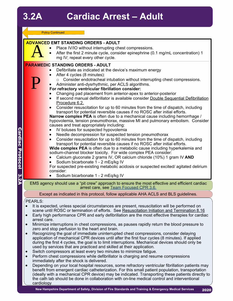
Ca
rdia
c P
roto
co
l 3.2
A
New Hampshire Department of Safety, Division of Fire Standards and Training & Emergency Medical Services 2020
Cardiac Arrest – Adult
P
PARAMEDIC STANDING ORDERS - ADULT
ADVANCED EMT STANDING ORDERS - ADULT
A · Place IV/IO without interrupting chest compressions.· After the first 2 minute cycle, consider epinephrine (0.1 mg/mL concentration) 1
mg IV; repeat every other cycle.
· Defibrillate as indicated at the device’s maximum energy· After 4 cycles (8 minutes):
o Consider endotracheal intubation without interrupting chest compressions.· Administer anti-dysrhythmic, per ACLS algorithms.For refractory ventricular fibrillation consider:· Changing pad placement from anterior-apex to anterior-posterior· If second manual defibrillator is available consider Double Sequential Defibrillation
Procedure 6.2.· Consider resuscitation for up to 60 minutes from the time of dispatch, including
transport for potential reversible causes if no ROSC after initial efforts.Narrow complex PEA is often due to a mechanical cause including hemorrhage / hypovolemia, tension pneumothorax, massive MI and pulmonary embolism. Consider causes and treat appropriately including: · IV boluses for suspected hypovolemia· Needle decompression for suspected tension pneumothorax· Consider resuscitation for up to 60 minutes from the time of dispatch, including
transport for potential reversible causes if no ROSC after initial efforts.Wide complex PEA is often due to a metabolic cause including hyperkalemia and sodium-channel blocker toxicity. For wide complex PEA consider:· Calcium gluconate 2 grams IV, OR calcium chloride (10%) 1 gram IV AND· Sodium bicarbonate 1 - 2 mEq/kg IVFor suspected pre-existing metabolic acidosis or suspected excited/ agitated delirium consider:· Sodium bicarbonate 1 - 2 mEq/kg IV
Policy Continued
3.2A
PEARLS:· It is expected, unless special circumstances are present, resuscitation will be performed on
scene until ROSC or termination of efforts. See Resuscitation Initiation and Termination 8.16· Early high performance CPR and early defibrillation are the most effective therapies for cardiac
arrest care.· Minimize interruptions in chest compressions, as pauses rapidly return the blood pressure to
zero and stop perfusion to the heart and brain.· Recognizing the goal of immediate uninterrupted chest compressions, consider delaying
application of mechanical CPR devices until after the first four cycles (8 minutes). If applied during the first 4 cycles, the goal is to limit interruptions. Mechanical devices should only be used by services that are practiced and skilled at their application.
· Switch compressors at least every two minutes to minimize fatigue.· Perform chest compressions while defibrillator is charging and resume compressions
immediately after the shock is delivered.· Depending on your local hospital resources, some refractory ventricular fibrillation patients may
benefit from emergent cardiac catheterization. For this small patient population, transportation (ideally with a mechanical CPR device) may be indicated. Transporting these patients directly to the cath lab should be done in collaboration with on-line medical control and interventional cardiology
EMS agency should use a “pit crew” approach to ensure the most effective and efficient cardiac arrest care, see Team Focused CPR 3.6.
Except as indicated in this protocol, follow applicable AHA ACLS and BLS guidelines.

Ca
rdia
c P
roto
co
l 3.2
P
P
EMT/ADVANCED EMT STANDING ORDERS
PARAMEDIC STANDING ORDERS· If Return of Spontaneous Circulation occurs see Post Resuscitative Care
Protocol 3.4. · If ventilation is adequate with BVM, routine placement of advanced airway can be
deferred.· Placement of an advanced airway during cardiac arrest should not interrupt chest
compressions. In this setting, supraglottic airways and ETTs can be considered equivalent.
· For suspected metabolic acidosis, suspected or known hyperkalemia (dialysis patient), or known tricyclic antidepressant overdose, consider sodium bicarbonate 1 - 2 mEq/kg IV.
For Ventricular Fibrillation (VF)/Pulseless Ventricular Tachycardia (VT): · Defibrillate at 2 J/kg; perform CPR for 2 minutes and recheck rhythm; if still a
shockable rhythm, defibrillate at 4 J/kg; perform CPR for 2 minutes; reassess every 2 minutes. Subsequent shocks at ≥ 4 J/Kg, maximum 10 J/Kg or adult dose.
· If no response after first defibrillation, administer:o Epinephrine (0.1 mg/mL concentration) 0.01 mg/kg (0.1 ml/kg) IV ORo Epinephrine (1 mg/mL concentration) 0.1 mg/kg (0.1 ml/kg) via ETT.§ Repeat every 3 – 5 minutes.
· If no response after second defibrillation, consider:o Amiodarone 5 mg/kg (maximum 300 mg) IV, ORo Lidocaine 1 mg/kg (maximum 100 mg), o For Torsades de Pointes: magnesium sulfate 25 – 50 mg/kg (maximum 2
grams) IV over 1 – 2 minutes. For Asystole or Pulseless Electrical Activity (PEA):· Epinephrine (0.1 mg/mL concentration) 0.01 mg/kg (0.1 ml/kg) IV OR · Epinephrine (1 mg/mL concentration) 0.1 mg/kg (1ml/kg) via ETT
o Repeat every 3 – 5 minutes. · Give 2 minutes of CPR, then check rhythm:
o If asystole or PEA, continue epinephrine and 2 minutes of CPR until:o Pulse obtained, ORo Shockable rhythm obtained, ORo Decision made to discontinue further efforts.
· Routine patient Care—with focus on CPR· Immediate chest compressions.· Apply AED and use as soon as possible (with minimum interruption of chest
compressions).From birth to age 8 years use pediatric AED pads.o If pediatric AED pads are unavailable, providers may use adult AED pads,
provided the pads do not overlap. · Monitor capnography, if available, throughout resuscitation to assess and
monitor airway placement, CPR quality and to monitor for signs of Return of Spontaneous Circulation.
· Consider termination of efforts or not attempting resuscitation, see DNR, POLST & Advanced Directives Policy 8.8 and/or Resuscitation Initiation & Termination 8.16.
· Consider treatable causes: hypoxia, overdose/poisoning, hypoglycemia, hypothermia, and hypovolemia (treat as per specific protocol).
Cardiac Arrest – Pediatric
New Hampshire Department of Safety, Division of Fire Standards and Training & Emergency Medical Services 2020
EA/
3.2P

Ca
rdia
c P
roto
co
l 3.3
2020
P
E
Titrate until symptomatic improvement or systolic BP of 140 mmHg.Consider nitroglycerine infusion:· For systolic BP of 140 - 160 mmHg: IV nitroglycerin start at 50 micrograms/minute.· For systolic BP of 160 - 200 mmHg: IV nitroglycerin start at 100 micrograms/minute.· For systolic BP > 200 mmHg: IV nitroglycerin start at 200 micrograms/minute.Note: Two (2) IV lines are recommended when giving IV nitroglycerin infusions; IV nitroglycerin infusions must be administered using an infusion pump.
EMT STANDING ORDERS - ADULT
ADVANCED EMT STANDING ORDERS - ADULT
A· Establish IV access.· For patient’s with known history of congestive heart failure, consider:
o For systolic BP of 140 - 160 mmHg: nitroglycerin 0.4 mg SL. o For systolic BP of 160 - 200 mmHg: nitroglycerin 0.8 mg SL (2 tabs/sprays). o For systolic BP > 200 mmHg: nitroglycerin 1.2 mg SL (3 tabs/sprays).o The above doses may be repeated every 5 minutes until symptomatic
improvement or systolic BP of 140 mmHg.· Assess blood pressure every 3 – 5 minutes during nitroglycerin administration.
· Routine Patient Care.· Place the patient in a semi-sitting or full sitting position.· Facilitate administration of the patient’s own nitroglycerin every 5 minutes while
symptoms persist and systolic BP is >140 mmHg.· Consider Continuous Positive Airway Pressure (CPAP) with maximum 15
cmH2O pressure support. See CPAP Procedure 5.4· 12-lead ECG, if available.
PARAMEDIC STANDING ORDERS - ADULT
PEARLS:· If patient has taken their own nitroglycerin without relief, consider loss of potency due to age.· Allow the patient to be in their position of comfort to maximize their breathing effort.
Congestive Heart Failure(Pulmonary Edema)
· Avoid nitroglycerin in any patient who has used a phosphodiesterase inhibitor such as: sildenafil (Viagra, Revatio), vardenafil (Levitra, Staxyn), tadalafil (Cialis, Adcirca) which are used for erectile dysfunction and pulmonary hypertension. Also avoid use in patients receiving intravenous epoprostenol (Flolan) which is also used for pulmonary hypertension.
· Administer nitrates with extreme caution, if at all, to patients with inferior-wall STEMI or suspected right ventricular (RV) involvement because these patients require adequate RV preload.
New Hampshire Department of Safety, Division of Fire Standards and Training & Emergency Medical Services 2020
3.3

Ca
rdia
c P
roto
co
l 3.4
P
EMT/ADVANCED EMT STANDING ORDERS – ADULT & PEDIATRIC
PARAMEDIC STANDING ORDERS - ADULT
· For post resuscitation hypotension:· Adult: Maintain systolic blood pressure of >90 mmHg OR MAP ≥ 65 mmHg.
o Administer IV fluid in 250 mL boluses not to exceed 2000 mL.· Pediatric: 1 – 10 years of age: Maintain systolic blood pressure 70 mmHg + (2 x age)
o Administer fluid bolus of 10 - 20 mL/kg of 0.9% NaCl by syringe push method (may repeat to a maximum 60 mL/kg)
· If feasible, acquire and transmit a 12-lead EKG. · Initial ventilation rate of 10 - 12 BPM for adults and 12 – 20 bpm for pediatric, then
titrate to capnography of 35 to 40 mm Hg, if available.· Titrate oxygen levels to between 94 – 98 % SaO2
Post Resuscitative CareAdult & Pediatric
New Hampshire Department of Safety, Division of Fire Standards and Training & Emergency Medical Services 2020
PEARLS:· Avoid hyperventilation as it increases intrathoracic pressures, potentially worsening
hemodynamic instability.
For Post-Resuscitation Hypotension:· Consider: (An infusion pump is required for the use of these vasopressors)
o Norepinephrine infusion 0.1 – 2 micrograms/kg/min (maximum dose 30 micrograms/min) titrated to effect, OR
o Epinephrine 0.1 – 1 micrograms/kg/min (maximum dose 10 micrograms/min) titrated to effect.
PARAMEDIC STANDING ORDERS - PEDIATRIC
3.4
· For patients with return of spontaneous circulation after cardiac arrest not related to trauma or hemorrhage who are comatose without purposeful movement, consider transporting to a receiving facility capable of starting induced therapeutic hypothermia.
· If patient meets STEMI criteria transport per your STEMI guidelines/agreements. Notify receiving facility of a “STEMI Alert”.
ADVANCED EMT STANDING ORDERS – ADULT & PEDIATRIC
A
E
· Consider vasopressor:o Epinephrine by push dose (dilute boluses) prepare 10 mcg/mL by adding 1 mL
0.1 mg/mL Epinephrine to 9 mL normal saline, then administer 10 - 20 mcg boluses (1 – 2 mL) every 2 minutes (where feasible, switch to infusion as soon as practical), AND/OR
o Norepinephrine infusion 1 – 30 micrograms/min, via pump ORo Epinephrine infusion 2 – 10 micrograms/minute, via pump, titrated to effect.
· Consider nasogastric or orogastric tube for the intubated patient.

Ca
rdia
c P
roto
co
l 3.5
A
· Routine Patient Care.
· 12-lead ECG if available.
2013
EMT/ADVANCED EMT STANDING ORDERS
P
For symptomatic tachyarrhythmias (other than sinus tachycardia):
If hemodynamically unstable:
· Synchronized cardioversion:
Use the following initial energy doses, then escalate to the next higher energy
level if no conversion. Biphasic devices: follow manufacturer’s
recommendations for dosing.
o For narrow regular rhythm: 50 – 100J biphasic or 200J monophasic.
o For narrow irregular rhythm: 120 – 200J biphasic or 200J monophasic.
o For wide regular rhythm: 100J biphasic or monophasic.
o For wide irregular/polymorphic VT: 120 – 200J biphasic or 360 monophasic,
using unsynchronized defibrillation doses if unable to sync.
· Administer procedural sedation prior to or during cardioversion, if feasible:
o Midazolam 2.5 mg IV, may repeat once in 5 minutes, OR
o *Midazolam 5 mg IM/IN may repeat once in 5 minutes, OR
o Lorazepam 1 mg IV, may repeat once in 5 minutes OR
o Diazepam 5 mg IV, may repeat once in 5 minutes.
If hemodynamically stable:
For narrow complex tachycardia consider:
· For regular rhythms greater than 150 bpm, perform vagal maneuvers.
o Adenosine 6 mg rapid IVP, may repeat at dose of 12 mg in 1 – 2 minutes if
no conversion.
o May repeat successful dose if rhythm recurs after conversion.
· Diltiazem 0.25 mg/kg IV (maximum dose 20 mg) over 2 minutes.
o May repeat dose in 15 minutes at 0.35 mg/kg (maximum dose 20 mg), if
necessary.
o Consider maintenance infusion at 5 – 15 mg/hour, OR
· Metoprolol 5 mg IV over 2 – 5 minutes.
o May repeat every five minutes to a maximum of 15 mg as needed to
achieve a ventricular rate of 90 – 100.
PARAMEDIC STANDING ORDERS
· Diltiazem, metoprolol, amiodarone, and adenosine are contraindicated in patients with atrial fibrillation and a history of or suspected Wolff-Parkinson-White (WPW) syndrome.
· Medications should be administered cautiously in frail or debilitated patients; lower doses should be considered.
EA/
Tachycardia – Adult
New Hampshire Department of Safety, Division of Fire Standards and Training & Emergency Medical Services 2020
3.5A
Protocol Continues
*For IN administration of midazolam use a 5 mg/mL concentration.
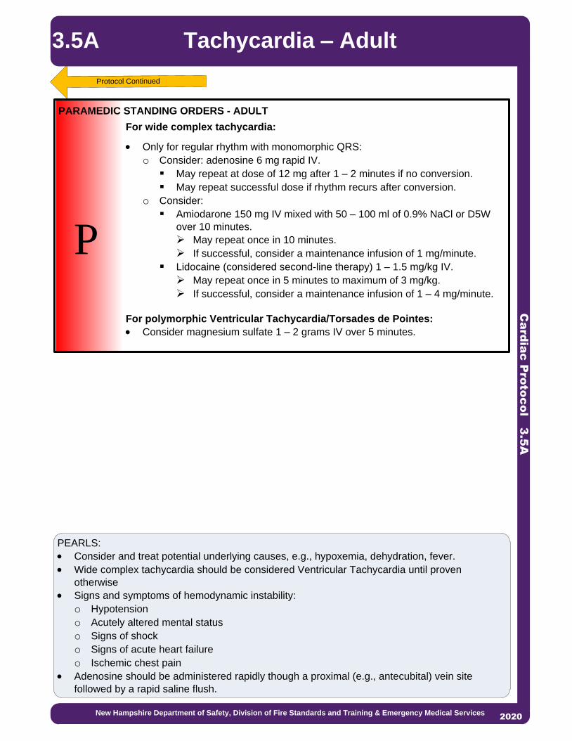
Ca
rdia
c P
roto
co
l 3.5
A
PEARLS:
· Consider and treat potential underlying causes, e.g., hypoxemia, dehydration, fever.
· Wide complex tachycardia should be considered Ventricular Tachycardia until proven
otherwise
· Signs and symptoms of hemodynamic instability:
o Hypotension
o Acutely altered mental status
o Signs of shock
o Signs of acute heart failure
o Ischemic chest pain
· Adenosine should be administered rapidly though a proximal (e.g., antecubital) vein site
followed by a rapid saline flush.
Protocol Continued
P
For wide complex tachycardia:
· Only for regular rhythm with monomorphic QRS:
o Consider: adenosine 6 mg rapid IV.
§ May repeat at dose of 12 mg after 1 – 2 minutes if no conversion.
§ May repeat successful dose if rhythm recurs after conversion.
o Consider:
§ Amiodarone 150 mg IV mixed with 50 – 100 ml of 0.9% NaCl or D5W
over 10 minutes.
Ø May repeat once in 10 minutes.
Ø If successful, consider a maintenance infusion of 1 mg/minute.
§ Lidocaine (considered second-line therapy) 1 – 1.5 mg/kg IV.
Ø May repeat once in 5 minutes to maximum of 3 mg/kg.
Ø If successful, consider a maintenance infusion of 1 – 4 mg/minute.
For polymorphic Ventricular Tachycardia/Torsades de Pointes:
· Consider magnesium sulfate 1 – 2 grams IV over 5 minutes.
PARAMEDIC STANDING ORDERS - ADULT
2013
Tachycardia – Adult
New Hampshire Department of Safety, Division of Fire Standards and Training & Emergency Medical Services2020
3.5A

Ca
rdia
c P
roto
co
l 3.5
P
· Routine Patient Care.
· 12-lead ECG if available.
EMT/ADVANCED EMT STANDING ORDERS
P
If hemodynamically unstable:
For narrow complex/probable SVT:· Synchronized cardioversion:
o 0.5 – 1 J/kg, if unsuccessful, increase to 2 J/kg· Administer procedural sedation prior to/during cardioversion, if feasible:
o Midazolam 0.05 mg/kg IV (single maximum dose 2.5 mg), may repeat once in 5 minutes, OR
o *Midazolam 0.1 mg/kg IM/IN (single maximum dose 5 mg), may repeat once in 5 minutes, OR
o Lorazepam 0.05 mg/kg IV (single maximum dose 1 mg) may repeat once in 5 minutes, OR
o Diazepam 0.1 mg/kg IV (single maximum dose 5 mg); may repeat once in 5 minutes
· Adenosine 0.1 mg/kg IV (maximum dose 6 mg).o Repeat once at 0.2 mg/kg (maximum dose 12 mg).
· If adenosine is ineffective or for wide complex, perform synchronized cardioversion:o 0.5 – 1 J/kg; if unsuccessful, increase to 2 J/kg.
If hemodynamically stable:
For narrow complex, probable supraventricular tachycardia, or regular wide complex tachycardia (monomorphic QRS ONLY):· Consider vagal maneuvers.· Adenosine 0.1 mg/kg IV (maximum dose 6 mg).
o May repeat once at 0.2 mg/kg IV (maximum dose 12 mg).For wide complex:· Contact online Medical Control for consideration of amiodarone 5 mg/kg IV
(maximum: 300 mg) over 20-60 minutes.
PARAMEDIC STANDING ORDERS
PEARLS:· Consider and treat potential underlying causes, e.g., hypoxemia, dehydration, fever.· Signs and symptoms of hemodynamic instability:
o Hypotensiono Acutely altered mental statuso Signs of shock
· Probable Sinus Tachycardia:o Compatible history consistent with known causeo P waves are present and normalo Variable R-R and constant P-R intervalo Infants: rate usually <220/mino Children: rate usually <180/min
· Probable Supraventricular Tachycardia:o Compatible history (vague, nonspecific); history of abrupt onset / rate changeso P waves absent / abnormalo Heart-rate is NOT variableo Infants: rate usually >220/mino Children: rate usually >180/min§ Adenosine should be administered rapidly though a proximal (e.g., antecubital) vein
site followed by a rapid saline flush
EA/
Tachycardia – Pediatric
New Hampshire Department of Safety, Division of Fire Standards and Training & Emergency Medical Services 2020
3.5P
*For IN administration of midazolam use a 5 mg/mL concentration.

POSITION #1-Compressor 1 (right side of patient): · Initiates 1 minute of chest compressions at rate of 100-120 / min· Assists Position 3 with ventilations in off cycle
POSITION #2-Compressor 2 (left side of patient): · Sets up defibrillator· Alternates 1 minute of chest compressions with Position 1· Assists Position 3 with ventilations in off cyclePOSITION #3-Airway (At patient’s head): · Opens airway and inserts OPA· Assembles NRB or BVM· If using BVM, provide 2 handed mask seal· Inserts advanced airway after 8 minutes/4 cycles. POSITION #4-Team Leader (Outside CPR triangle):· Coaches the metrics· Calls for compressor change every one minute· Calls for rhythm analysis every 2 minutes, immediate shock if indicated· Monitor CPR quality and use of metronome at 100-120 bpm· Assumes duties of Position 5/6 if limited to four rescuers throughout
resuscitation.
POSITION #5-Vascular/Meds (Outside CPR triangle):· Initiates IV/IO access· Administers medications per protocol
POSITION #6-Code Commander (Outside CPR triangle):· Ideally highest level provider· Communicates/interfaces with CPR Team Leader· Coordinates patient treatment decisions· Communicates with family/loved ones · Completes Cardiac Arrest Check List
Ca
rdia
c P
roto
co
l 3.6
New Hampshire Department of Safety, Division of Fire Standards and Training & Emergency Medical Services 2020
Team Focused CPR – Adult 3.6
Policy Continues
EMS agency should use a “pit crew” approach when using this protocol to ensure the most effective and efficient cardiac arrest care. Training should include teamwork simulations integrating BLS, and ALS crew members who regularly work together. EMS systems should practice teamwork using “pit crew” techniques with predefined roles and crew resource management principles. One Example is a follows:
Link to HP CPR training

Ca
rdia
c P
roto
co
l 3.6
New Hampshire Department of Safety, Division of Fire Standards and Training & Emergency Medical Services 2020
Team FocusedCPR – Adult
Policy Continued
· If feasible and the scene is safe, immediately upon arrival, one member of the crew should rapidly enter the scene without equipment (other than gloves) to begin chest compressions.
· Clear some space to optimize your working environment. Move furniture or get the patient in a position that will allow a rescuer space to kneel on both sides of them, and where there is sufficient room at the head. Effectiveness of chest compressions decrease during patient movement. Therefore resuscitate the patient as close to the scene as operationally feasible.
· Position 1 and 2 are ideally set up on opposite sides of patient’s chest and perform continuous chest compressions, alternating after minute to avoid fatigue.
· REMEMBER: Effective chest compressions are one of the most important therapies for the pulseless patient. Effective is defined as:o A rate of at least 100 and less than 120 compressions/minute - Use of metronome or
CPR feedback device is essential. (e.g., built into monitor or smart phone app)o A depth of 2 - 2.4 incheso Allow for complete chest recoil (avoid leaning on chest)o Do not interrupt compressions to obtain IV access or perform airway management.o Do not hyperventilate as it increases intrathoracic pressure and decreases blood return to
the heart. Ventilate 1 breath every 10 compressions without interrupting chest compressions.
· Chest compressions should only be interrupted during rhythm check (AED analysis or manual) and defibrillation shocks. Continue compressions when AED/ defibrillator is charging, if device allows.
· During interruptions compressor’s hands should hover over chest. · Perform pulse check simultaneously with rhythm check.· With the goal of immediate uninterrupted chest compressions, if a mechanical device is used,
it should not lead to delay or interruption in chest compressions; consider delayed applications.
· Pre-charge manual defibrillators prior to rhythm check to ensure rapid defibrillation if a shockable rhythm is present. If no shock is indicated, disarm the device (dump the charge)
· Utilize ETCO2 to assess CPR quality and monitor for signs of ROSC.· Use of a CPR checklist to ensure that all best practices are followed during CPR.
Example Cardiac Arrest Check Listq Code commander and pit crew roles identifiedq Chest compression interruptions minimizedq Compressors rotated at minimum every 2 minutesq Metronome set between 100 and 120 beats per minute q AED/defibrillator appliedq OPA/NPA placedq O2 flowing and attached to NRB/BVMq ETCO2 waveform presentq IV/IO access establishedq Possible causes consideredq Gastric insufflation limited and gastric decompression consideredq Family present and ongoing communication provided
Consider possible causesHypovolemiaHypoxiaHydrogen Ions (acidosis)HypothermiaHyper/hypokalemiaHypoglycemia
Tablets/toxinsTamponadeTension pneumothoraxThrombosis (MI)Thrombosis (PE)Trauma
3.6

· Routine Patient Care.· Assess for evidence of smoke inhalation or burns; soot around mouth or nostrils,
singed hair, carbonaceous sputum. · If the patient has respiratory difficulty, altered level of consciousness and /or
hemodynamic compromise, see Airway Management Protocols 5.1 and Smoke Inhalation/Carbon Monoxide Poisoning Protocols 2.21.
Thermal· Stop burning process with water or normal saline· Remove non-adherent clothing and jewelry. Do not remove skin or tissue.· To protect from infection, cover burns with clean dry sterile dressing or sheets. · Keep patient warm and prevent hypothermia due to large thermal injuries. Chemical· Identify agent(s) and consider HAZMAT intervention, if indicated. See Hazardous
Material Exposure Protocol 9.0· Consider contacting Poison Control at 800-222-1222.· Decontaminate the patient as appropriate.
o Brush off dry powders if present, before washing.o Scrape viscous material off with rigid device, e.g., tongue depressor o Flush with copious amounts of clean water or sterile saline for 10 – 15 minutes,
unless contraindicated by type of chemical agent (e.g., sodium, potassium or dry lime and/or phenols).
Electrical/Lightning· Ensure your own safety; disconnect power source, if feasible.· Place patient on a cardiac monitor.· Consider spinal motion restriction for burns due to electric flow across the body.Assess Extent of Burn· Determine extent of the burn using Rules of Nine (see next page).· Determine depth of injury.· Do not include 1st degree burns in burn surface area (BSA) percentage. Pain Control· If a partial thickness burn, 2nd degree is < 10% body surface area:· Apply room-temperature water or room-temperature wet towels to burned area of a
maximum of 15 minutes. Prolonged cooling may result in hypothermia.
2013
E
EMT STANDING ORDERS
2013
AADVANCED EMT STANDING ORDERS - PEDIATRIC
· Transport time less than 1 hour:o BSA > 20%: 20 mL/kg warm IV fluids*, over 10 – 30 minutes.(Does not
need to be on a pump)o BSA < 20%: 10 mL/kg warm IV fluids*, over 10 – 30 minutes.
· Consult Medical Control:o Transport time greater than 1 hour and/oro Patient has signs of shock
* An IO device can be inserted through burned skin as long as the underlying bone has not been compromised.
Tra
um
a P
roto
co
l 4.0
Burns/Electrocution/Lightning Adult & Pediatric
New Hampshire Department of Safety, Division of Fire Standards and Training & Emergency Medical Services 2020
4.0
Protocol Continues
ADVANCED EMT STANDING ORDER - ADULT· Transport time less than 1 hour:
o Administer warm IV fluids* at 500 mL/Hour.· Transport time greater than 1 hour:
o Administer warm fluids* at 1 – 2 mL/kg x % burn/8 = hourly rate x first 8 hours.

Tra
um
a P
roto
co
l 4.0
Burns/Electrocution/Lightning Adult & Pediatric
New Hampshire Department of Safety, Division of Fire Standards and Training & Emergency Medical Services 2020
Air Medical Transport Considerations:§ Major burns with greater than 20% BSA and/or inhalation injury with risk of airway
compromise.§ Electrocution injuries with loss of consciousness, arrhythmia, or any respiratory abnormality.
PPARAMEDIC STANDING ORDERS
· Refer to Pain Management Protocol 2.16.
Rule of Nines
PEARLS:· Electrocution/Lightning burns can occur anywhere along the path a current travels through the body.
Evident surface burns may only comprise a small portion of the overall burn injury, and an injury’s full extent may not be immediately apparent.
· Chemical burns: If 0.9% NaCl or sterile water is not readily available, do not delay, use tap water for flushing the affected area. Flush the area as soon as possible with the cleanest readily available water using copious amounts of water.
4.0
Protocol Continues
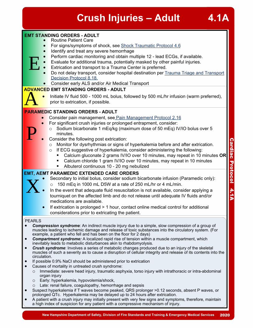
Ca
rdia
c P
roto
co
l 4.1
A
E
EMT STANDING ORDERS - ADULT
ADVANCED EMT STANDING ORDERS - ADULT
A · Initiate IV fluid 500 - 1000 mL bolus, followed by 500 mL/hr infusion (warm preferred), prior to extrication, if possible.
· Routine Patient Care· For signs/symptoms of shock, see Shock Traumatic Protocol 4.6· Identify and treat any severe hemorrhage· Perform cardiac monitoring and obtain multiple 12 - lead ECGs, if available.· Evaluate for additional trauma, potentially masked by other painful injuries.· Extrication and transport to a Trauma Center is preferred. · Do not delay transport, consider hospital destination per Trauma Triage and Transport
Decision Protocol 8.18.· Consider early ALS and/or Air Medical Transport
New Hampshire Department of Safety, Division of Fire Standards and Training & Emergency Medical Services 2020
Crush Injuries – Adult 4.1A
P
PARAMEDIC STANDING ORDERS - ADULT· Consider pain management, see Pain Management Protocol 2.16· For significant crush injuries or prolonged entrapment, consider:
o Sodium bicarbonate 1 mEq/kg (maximum dose of 50 mEq) IV/IO bolus over 5 minutes.
· Consider the following post extrication:o Monitor for dysrhythmias or signs of hyperkalemia before and after extrication.o If ECG suggestive of hyperkalemia, consider administering the following:§ Calcium gluconate 2 grams IV/IO over 10 minutes, may repeat in 10 minutes OR§ Calcium chloride 1 gram IV/IO over 10 minutes, may repeat in 10 minutes § Albuterol continuous 10 - 20 mg nebulized
EMT, AEMT PARAMEDIC EXTENDED CARE ORDERS· Secondary to initial bolus, consider sodium bicarbonate infusion (Paramedic only):
o 150 mEq in 1000 mL D5W at a rate of 250 mL/hr or 4 mL/min.· In the event that adequate fluid resuscitation is not available, consider applying a
tourniquet on the affected limb and do not release until adequate IV fluids and/or medications are available.
· If extrication is prolonged > 1 hour, contact online medical control for additional considerations prior to extricating the patient.
X
PEARLS· Compression syndrome: An indirect muscle injury due to a simple, slow compression of a group of
muscles leading to ischemic damage and release of toxic substances into the circulatory system. (For example, a patient who fell and has been on the floor for 2 days)
· Compartment syndrome: A localized rapid rise of tension within a muscle compartment, which inevitably leads to metabolic disturbances akin to rhabdomyolysis.
· Crush syndrome: Involves a series of metabolic changes produced due to an injury of the skeletal muscles of such a severity as to cause a disruption of cellular integrity and release of its contents into the circulation.
· If possible 0.9% NaCl should be administered prior to extrication· Causes of mortality in untreated crush syndrome:
o Immediate: severe head injury, traumatic asphyxia, torso injury with intrathoracic or intra-abdominal organ injury
o Early: hyperkalemia, hypovolemia/shock, o Late: renal failure, coagulopathy, hemorrhage and sepsis
· Suspect hyperkalemia if T waves become peaked, QRS prolonger >0.12 seconds, absent P waves, or prolonged QTc. Hyperkalemia may be delayed up to 24 hours after extrication.
· A patient with a crush injury may initially present with very few signs and symptoms, therefore, maintain a high index of suspicion for any patient with a compressive mechanism of injury.

Ca
rdia
c P
roto
co
l 4.1
P
E
EMT STANDING ORDERS - PEDIATRIC
ADVANCED EMT STANDING ORDERS - PEDIATRIC
A · Initiate 10-20 mL/kg IV fluid bolus, followed by 10 mL/kg/hr infusion (warm preferred), prior to extrication, if possible.
· Routine Patient Care· For signs/symptoms of shock, see Shock Traumatic Protocol 4.6· Identify and treat any severe hemorrhage· Perform cardiac monitoring and obtain multiple 12-lead ECGs, if available.· Evaluate for additional trauma, potentially masked by other painful injuries.· Extrication and transport to a Trauma Center is preferred. · Do not delay transport, consider hospital destination per Trauma Triage and Transport
Decision Protocol 8.18.· Consider early ALS and/or Air Medical Transport
New Hampshire Department of Safety, Division of Fire Standards and Training & Emergency Medical Services 2020
Crush Injuries – Pediatric4.1P
P
PARAMEDIC STANDING ORDERS - PEDIATRIC· Consider pain management, see Pain Management Protocol 2.16· For significant crush injuries or prolonged entrapment, consider:
o Sodium bicarbonate 1 mEq/kg (maximum dose of 50 mEq) IV/IO bolus over 5 minutes.
· Consider the following post extrication:o Monitor for dysrhythmias or signs of hyperkalemia before and after extrication.o If ECG suggestive of hyperkalemia, consider administering the following:§ Calcium gluconate 100 mg/kg IV/IO with a maximum of 2 gm/dose, over 5
minutes; may repeat in 10 minutes OR§ Calcium chloride 20 mg/kg IV/IO with a maximum of 1 gm/dose over, 5
minutes; may repeat in 10 minutes § Albuterol per chart:
EMT, AEMT & PARAMEDIC EXTENDED CARE ORDERS· In the event that adequate fluid resuscitation is not available, consider applying a
tourniquet on the affected limb and do not release until adequate IV fluids and/or medications are available.
· If extrication is prolonged > 1 hour, contact online medical control for additional considerations prior to extricating the patient.
XPEARLS· Compression syndrome: An indirect muscle injury due to a simple, slow compression of a group of
muscles leading to ischemic damage and thus causing crush substances to enter the blood. (For example, a patient who fell and has been on the floor for 2 days)
· Compartment syndrome: A localized rapid rise of tension within a muscle compartment, which inevitably leads to metabolic disturbances akin to rhabdomyolysis.
· Crush syndrome: Also termed rhabdomyolysis, involves a series of metabolic changes produced due to an injury of the skeletal muscles of such a severity as to cause a disruption of cellular integrity and release of its contents into the circulation.
· Causes of mortality in untreated crush syndrome: o Immediate: severe head injury, traumatic asphyxia, torso injury with intrathoracic or intra-
abdominal organ injuryo Early: hyperkalemia, hypovolemia/shock, o Late: renal failure, coagulopathy, hemorrhage and sepsis
· Suspect hyperkalemia if T waves become peaked, QRS prolonger >0.12 seconds, absent P waves, or prolonged QTc. Hyperkalemia may be delayed up to 24 hours after extrication.
· A patient with a crush injury may initially present with very few signs and symptoms, therefore, maintain a high index of suspicion for any patient with a compressive mechanism of injury.

SUBMERSION: When a patient goes under the water immediately, has a hypoxic cardiac arrest and then cools down. Prognosis considered dismal.
IMMERSION: Patients are in the water with head above water and they continue to breathe while they cool down before they eventually arrest. Prognosis can be good with patients surviving after prolonged CPR.
· Routine Patient Care.· Victims with only respiratory arrest usually respond after a few artificial breaths
are given.o Give a few breaths and check for a pulse.§ Anticipate vomiting.
· For patients in cardiac arrest, provide immediate CPR.o Utilize the sequence ABC, not CAB, i.e. start with airway and breathing before
compressions.· Routine use of spinal motion restriction in the absence of circumstances that
suggest a spinal injury is not recommended.· Assess temperature, if unresponsive, obtain esophageal or rectal temperature. · Due to extremely poor prognosis, providers may consider withholding or
terminating resuscitation efforts when:o A clear history of prolonged submersion (without prior prolonged immersion),
greater than 20 minutes (children may survive despite extended submersion) OR
o Esophageal or rectal temperature is greater than 32oC (89.6o F) with asystole documented in 2 leads OR
o Meets Termination of Resuscitation Criteria, see Resuscitation Initiation and Termination Protocol 8.16.
· Consider hypothermia, see Hypothermia Protocol 2.10.o Do not delay urgent procedures such as airway management and IV access.
Although hypothermic patients may exhibit cardiac irritability, do not delay necessary interventions.
· Conscious patients who survive any form of drowning are at risk of deterioration and should be transported to the hospital.
· Consider CPAP to supplement the patient’s own respiratory effort.
EMT/AEMT STANDING ORDERS
· For unconscious patients in distress, consider early intubation.
2013
Tra
um
a P
roto
co
l 4.2
Drowning/Submersion InjuriesAdult & Pediatric
New Hampshire Department of Safety, Division of Fire Standards and Training & Emergency Medical Services 2020
4.2
Protocol Continues
PARAMEDIC STANDING ORDERS
EA/
P

HYPOTHERMIA CHART
STAGE: I Conscious, shivering Core Temp 35 to 32°CTreatment: Warm environment and clothing, warm sweet drinks, and active movement (if possible)
STAGE: II Impaired consciousness, not shivering Core Temp <32 to 28°CTreatment: Cardiac monitoring, minimal and cautious movements to avoid arrhythmias, horizontal
position and immobilization, full-body insulation, active external and minimally invasive rewarming techniques (warm environment; chemical, electrical, or forced- air heating packs or blankets; warm parenteral fluids)
STAGE: III Unconscious, not shivering, vital signs presentCore Temp <28 to 24°CTreatment: Stage II management plus airway management as required; ECMO or CPB in cases with
cardiac instability that is refractory to medical management
STAGE: IV No vital signsCore Temp <24°CTreatment: Stage II and III management plus CPR and up to three doses of epinephrine (at an
intravenous or intraosseous dose of 1 mg) and defibrillation, with further dosing guided by clinical response; rewarming with ECMO or CPB (if available) or CPR with active external and alternative internal rewarming
PEARLS· Patients with Stage III or IV hypothermia may benefit from treatment at a facility capable of
ExtraCorporeal Membrane Oxygenation (ECMO) or CardioPulmonary Bypass (CPB). Consider air medical transport.
· In hypothermic patients, low levels of ETCO2 may not be a useful predictor of outcome, due to reduced metabolism.
· Oral and tympanic thermometers do not yield an accurate core temperature for severely hypothermic patients.
· Cold water offers enhanced survival only where the patient becomes cold prior to cardiac arrest.
· There is no need to clear the airway of aspirated water; only a modest amount of water is aspirated by most drowning victims, and aspirated water is rapidly absorbed into the central circulation.
· Unnecessary cervical spine immobilization can impede adequate opening of the airway and delay delivery of rescue breaths.
Tra
um
a P
roto
co
l 4.2
Drowning/Submersion InjuriesAdult & Pediatric
New Hampshire Department of Safety, Division of Fire Standards and Training & Emergency Medical Services 2020
4.2Protocol Continues

· Routine Patient Care.· Obtain visual history (e.g., use of corrective lenses, surgeries, use of protective
equipment).· Obtain visual acuity, if possible.· Assist patient with the removal of contact lens, if applicable.· Chemical irritants, including pepper spray: flush with copious amounts of water,
or 0.9% NaCl.· Thermal burns to eyelids: patch both eyes with cool saline compress.· Impaled object: immobilize object and patch both eyes.· Puncture wound: place rigid protective device over both eyes (e.g., eye shield).
Do not apply pressure.· If the patient cannot close their eyelids, keep their eye moist with a sterile saline
dressing.
2013
· Proparacaine or tetracaine:o Apply 2 drops to affected eye; repeat every 5 minutes as needed.
· Consider use of Morgan lens for irrigation.· Refer to Pain Management Protocol 2.16.
P
PEARLS:· Handle the tooth carefully. Avoid touching the root of the tooth (the part of the tooth that was
embedded in the gum) because it can be damaged easily.· Significant eye injury may be present despite normal vision and minimal symptoms.· Any chemical or thermal burn to the face/eyes should raise suspicion of respiratory insult. · Vomiting in connection with blunt or penetrating eye trauma significantly increases intraocular
pressure and should be avoided.
X
EYE – EMT STANDING ORDERS
EYE - PARAMEDIC STANDING ORDERS
2013
EMT/ADVANCED EMT/PARAMEDIC EXTENDED CARE ORDERS· If definitive treatment is expected to be greater than 4 hours, an attempt to
reinsert the avulsed tooth in its socket should be considered, after rinsing tooth in water or normal saline. If multiple teeth require reinsertion, use the shape and size of dentition on the opposing side to guide you in proper placement.
· Routine Patient Care.· Dental avulsions should be placed in an obviously labeled container with saline-
soaked dressing, milk or hanks solution
DENTAL AVULSION – EMT/ADVANCED EMT/PARAMEDIC STANDING ORDERS
E
EA
P/
/
Tra
um
a P
roto
co
l 4.3
Eye & Dental InjuriesAdult & Pediatric
New Hampshire Department of Safety, Division of Fire Standards and Training & Emergency Medical Services 2020
4.3
AEYE - ADVANCED EMT STANDING ORDER
· An anti-emetic is strongly recommended for penetrating or blunt eye trauma, consider Nausea Protocol 2.11.

2013
· Routine Patient Care· Apply direct pressure, using manual control and/or pressure bandage.· Apply limb tourniquet, if direct pressure is ineffective or impractical and for any
traumatic amputation. o Apply directly to the skin 2-3 inches above the bleeding site. If bleeding is not
controlled with the first tourniquet, apply a second tourniquet side-by-side with the first.
o Document time of tourniquet application and communicate this clearly with receiving facility.
· Pack wounds of groin, neck or axillary injuries not amenable to limb tourniquet. o Utilize hemostatic dressing or, if not available, gauze dressing.
· Junctional tourniqueto If the bleeding site is amenable to use of a junctional tourniquet, immediately
apply device following manufacture’s guidelines, if available.
EMT/ADVANCED EMT/PARAMEDIC STANDING ORDERS– ADULT & PEDIATRIC
Pro
ce
du
re 4
.4
Hemorrhage Control
New Hampshire Department of Safety, Division of Fire Standards and Training & Emergency Medical Services 2020
4.4INDICATIONS:· Serious or life threatening extremity hemorrhage in the face of operational considerations that
prevent the use of less aggressive hemorrhage control techniques.
E
· Administer fluids per Shock – Traumatic Protocol 4.6 · Assess pain level and consider pain control measures, see Pain Management
Protocol 2.16
ADVANCED EMT STANDING ORDERS – ADULT & PEDIATRIC
· Administer tranexamic acid (TXA):o Mix 1 gram of TXA in 50 - 100 ml of 0.9% NaCl; infuse over approximately 10
minutes IV or IO.o Notify receiving facility of TXA administration prior to arriving.
APARAMEDIC STANDING ORDERS – ADULT
PTXA Indications
· Evidence of significant trauma AND · Evidence or concern for severe external
and/or internal hemorrhage AND· Presence of one or more markers of
hemodynamic instability.o Sustained systolic blood pressure < 90
mmHg.o Sustained heart rate > 110 after pain
adequately treated AND· Injury occurred within past 3 hours
Protocol Continues
TXA Contraindications· < 15 years of age· Previous allergic reaction to TXA· Isolated head injury· Patients who have received or will receive
prothrombin complex concentrate (PCCs), factor VIIa, or factor IX complex concentrates.
· Women who are known or suspected to be pregnant with a fetus of viable gestational age ( > 24 weeks).

XEMT/ADVANCED EMT/PARAMEDIC STANDING ORDERS
Pro
ce
du
re 4
.4Hemorrhage Control
New Hampshire Department of Safety, Division of Fire Standards and Training & Emergency Medical Services 2020
· Consult Medical Control, if feasible.· If tourniquet has been in place for greater than 6 hours, do not remove. · If less then 6 hours consider Tourniquet Reassessment and Tourniquet
Conversion Algorithms
Protocol Continued
Tourniquet Reassessment Algorithm Tourniquet Conversion Algorithm
Patient in circulatory shock?
Unstable clinical situation?
Limited personnel/resources?
Proceed to conversion algorithm
Further significant bleeding
Is there significant bleeding?
Amputated Extremity?
Leave tourniquet on and transport patient to the
nearest trauma center, if feasible.
Leave tourniquet on and transport patient to the
nearest trauma
center, if feasible
Tighten tourniquet and begin
transporting patient to nearest
trauma center
Continue to assess for bleeding and transport to nearest trauma
center.
Follow wound packing guidance and apply pressure dressing. Loosen tourniquet (leave it in
place)
NO
NO
NO
NO
NO
NO
YES
YES
YES
YES
YES
YES
4.4
In the absence of a commercial tourniquet (preferred), an improvised device e.g., cravat with windlass, blood pressure cuff could be used. The device must be a minimum of 2 inches wide, otherwise it can cut through the skin.
In the event of diminished scene safety (indirect threat, warm zone etc.), limb tourniquets should be placed as high on the limb as possible and over clothing.
Amputations: Rinse severed part briefly and gently with sterile saline to remove debris then wrap severed part in sterile saline gauze, moisten with sterile saline (do not soak) and place in water-tight container. Place container on ice (do not use dry ice). Do not put part directly on ice. If necessary, use ice packs to provide some level of cooling.
PEARLS:· Tourniquets applied prior to EMS arrival should be evaluated for effectiveness and
appropriateness. If tourniquet can be safely removed, remove the tourniquet and apply pressure dressing.
· Do not apply tourniquet over joints. · Reassess for re-bleeding frequently, especially after any patient movement.· Delay in placement of a tourniquet for life threatening hemorrhage significantly increases
mortality. Do not wait for hemodynamic compromise to apply a tourniquet.· If feasible, transport patients directly to a Level 1 or Level 2 trauma center and provide earliest
possible notification / trauma alert.· Damage to the limb from tourniquet application is unlikely if removed in several hours.
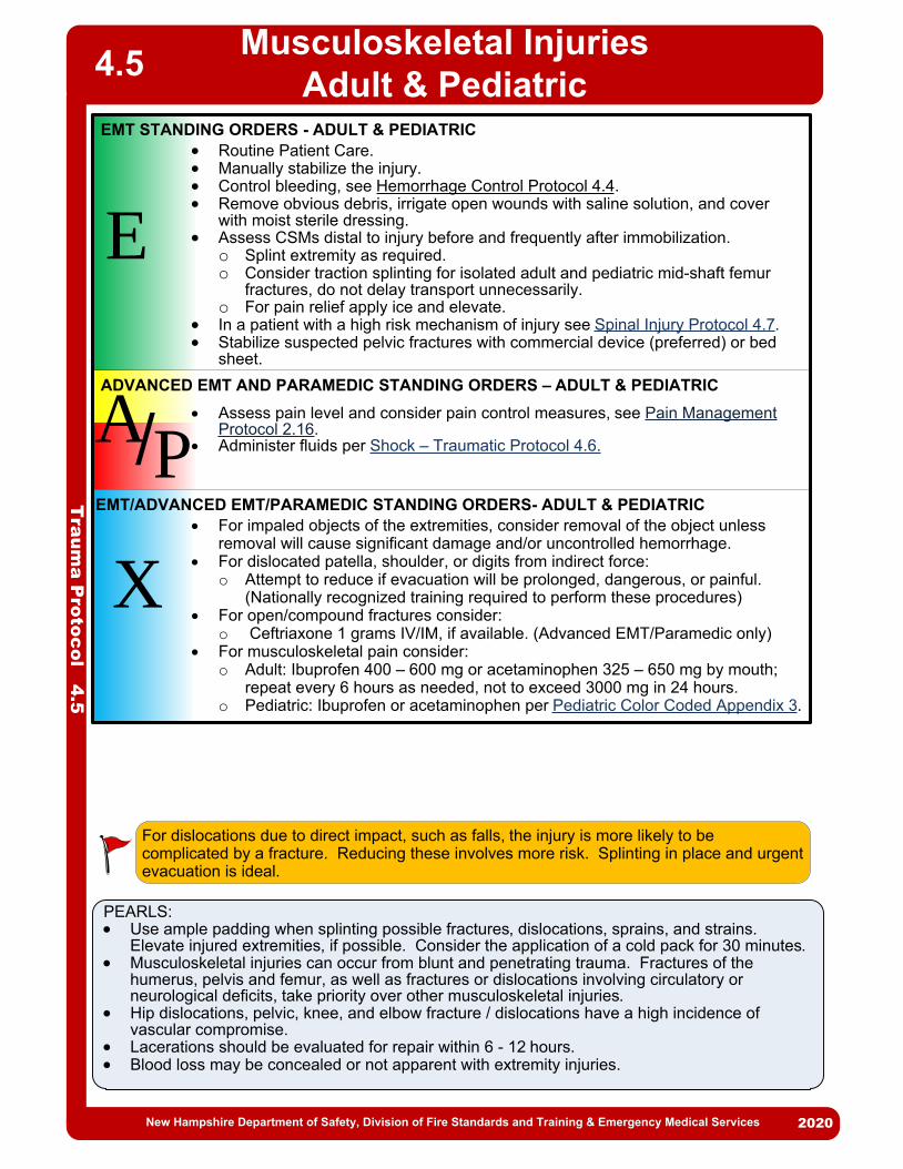
· Routine Patient Care.· Manually stabilize the injury.· Control bleeding, see Hemorrhage Control Protocol 4.4. · Remove obvious debris, irrigate open wounds with saline solution, and cover
with moist sterile dressing.· Assess CSMs distal to injury before and frequently after immobilization.
o Splint extremity as required.o Consider traction splinting for isolated adult and pediatric mid-shaft femur
fractures, do not delay transport unnecessarily.o For pain relief apply ice and elevate.
· In a patient with a high risk mechanism of injury see Spinal Injury Protocol 4.7.· Stabilize suspected pelvic fractures with commercial device (preferred) or bed
sheet.
Musculoskeletal InjuriesAdult & Pediatric
PEARLS:· Use ample padding when splinting possible fractures, dislocations, sprains, and strains.
Elevate injured extremities, if possible. Consider the application of a cold pack for 30 minutes.· Musculoskeletal injuries can occur from blunt and penetrating trauma. Fractures of the
humerus, pelvis and femur, as well as fractures or dislocations involving circulatory or neurological deficits, take priority over other musculoskeletal injuries.
· Hip dislocations, pelvic, knee, and elbow fracture / dislocations have a high incidence of vascular compromise.
· Lacerations should be evaluated for repair within 6 - 12 hours.· Blood loss may be concealed or not apparent with extremity injuries.
X
EMT STANDING ORDERS - ADULT & PEDIATRIC
· For impaled objects of the extremities, consider removal of the object unless removal will cause significant damage and/or uncontrolled hemorrhage.
· For dislocated patella, shoulder, or digits from indirect force:o Attempt to reduce if evacuation will be prolonged, dangerous, or painful.
(Nationally recognized training required to perform these procedures)· For open/compound fractures consider:
o Ceftriaxone 1 grams IV/IM, if available. (Advanced EMT/Paramedic only)· For musculoskeletal pain consider:
o Adult: Ibuprofen 400 – 600 mg or acetaminophen 325 – 650 mg by mouth; repeat every 6 hours as needed, not to exceed 3000 mg in 24 hours.
o Pediatric: Ibuprofen or acetaminophen per Pediatric Color Coded Appendix 3.
For dislocations due to direct impact, such as falls, the injury is more likely to be complicated by a fracture. Reducing these involves more risk. Splinting in place and urgent evacuation is ideal.
Tra
um
a P
roto
co
l 4.5
EMT/ADVANCED EMT/PARAMEDIC STANDING ORDERS- ADULT & PEDIATRIC
New Hampshire Department of Safety, Division of Fire Standards and Training & Emergency Medical Services 2020
4.5
· Assess pain level and consider pain control measures, see Pain Management Protocol 2.16.
· Administer fluids per Shock – Traumatic Protocol 4.6.
ADVANCED EMT AND PARAMEDIC STANDING ORDERS – ADULT & PEDIATRIC
E
A/P
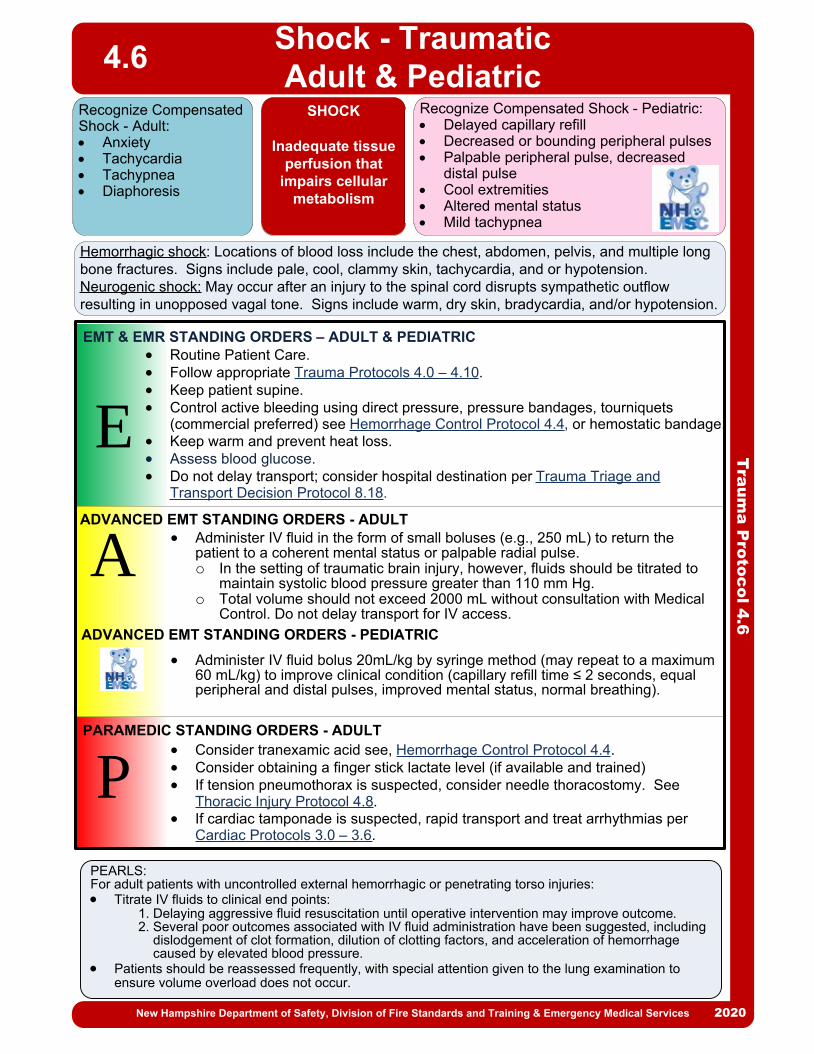
PEARLS:For adult patients with uncontrolled external hemorrhagic or penetrating torso injuries:· Titrate IV fluids to clinical end points:
1. Delaying aggressive fluid resuscitation until operative intervention may improve outcome.2. Several poor outcomes associated with IV fluid administration have been suggested, including dislodgement of clot formation, dilution of clotting factors, and acceleration of hemorrhage caused by elevated blood pressure.
· Patients should be reassessed frequently, with special attention given to the lung examination to ensure volume overload does not occur.
P
E
EMT & EMR STANDING ORDERS – ADULT & PEDIATRIC
PARAMEDIC STANDING ORDERS - ADULT
ADVANCED EMT STANDING ORDERS - ADULT
A· Administer IV fluid in the form of small boluses (e.g., 250 mL) to return the
patient to a coherent mental status or palpable radial pulse. o In the setting of traumatic brain injury, however, fluids should be titrated to
maintain systolic blood pressure greater than 110 mm Hg.o Total volume should not exceed 2000 mL without consultation with Medical
Control. Do not delay transport for IV access.
· Administer IV fluid bolus 20mL/kg by syringe method (may repeat to a maximum 60 mL/kg) to improve clinical condition (capillary refill time ≤ 2 seconds, equal peripheral and distal pulses, improved mental status, normal breathing).
· Consider tranexamic acid see, Hemorrhage Control Protocol 4.4.· Consider obtaining a finger stick lactate level (if available and trained)· If tension pneumothorax is suspected, consider needle thoracostomy. See
Thoracic Injury Protocol 4.8.· If cardiac tamponade is suspected, rapid transport and treat arrhythmias per
Cardiac Protocols 3.0 – 3.6.
· Routine Patient Care. · Follow appropriate Trauma Protocols 4.0 – 4.10.· Keep patient supine.· Control active bleeding using direct pressure, pressure bandages, tourniquets
(commercial preferred) see Hemorrhage Control Protocol 4.4, or hemostatic bandage.· Keep warm and prevent heat loss.· Assess blood glucose.· Do not delay transport; consider hospital destination per Trauma Triage and
Transport Decision Protocol 8.18.
Tra
um
a P
roto
co
l 4.6
Shock - TraumaticAdult & Pediatric
New Hampshire Department of Safety, Division of Fire Standards and Training & Emergency Medical Services 2020
ADVANCED EMT STANDING ORDERS - PEDIATRIC
Recognize Compensated Shock - Adult:· Anxiety· Tachycardia· Tachypnea· Diaphoresis
Recognize Compensated Shock - Pediatric:· Delayed capillary refill· Decreased or bounding peripheral pulses· Palpable peripheral pulse, decreased
distal pulse· Cool extremities· Altered mental status· Mild tachypnea
SHOCK
Inadequate tissue perfusion that
impairs cellular metabolism
Hemorrhagic shock: Locations of blood loss include the chest, abdomen, pelvis, and multiple long bone fractures. Signs include pale, cool, clammy skin, tachycardia, and or hypotension.Neurogenic shock: May occur after an injury to the spinal cord disrupts sympathetic outflow resulting in unopposed vagal tone. Signs include warm, dry skin, bradycardia, and/or hypotension.
4.6

2013
2013
Pro
ce
du
re 4
.7
Spinal Trauma
New Hampshire Department of Safety, Division of Fire Standards and Training & Emergency Medical Services 2020
PURPOSE: This protocol provides guidance regarding the assessment and care of patients who have a possible spinal injury.
Patients who have experienced a mechanism of spinal injury (esp. high risk mechanisms. See Red Flag Box.) require spinal motion restriction (as described further on) and protection of the injury site if they exhibit:· Midline spinal pain or tenderness with palpation.· Abnormal (i.e. not baseline) neurological function or motor strength in any
extremity.· Numbness or tingling (paresthesia).· Sensation is not intact and symmetrical (or baseline for patient).· Cervical flexion, extension and/or rotation elicits midline spinal pain.OR If they cannot competently participate in the assessment due to one of the following:· Altered mental status (e.g., dementia, preexisting brain injury, developmental delay,
psychosis).· Alcohol or drug intoxication.· Unable to participate in assessment (e.g., distracted by significant injuries to self or
others).· Insurmountable communication barriers (e.g., deafness, or hard of hearing,
language barrier).
Patients without any of the above findings should generally be transported without the use of a cervical collar or other means to restrict spinal motion. Utilize spinal motion restriction only where, in the professional judgment of the provider, the patient is at high risk for spinal injury as described above or with clear clinical indications of injury (e.g., midline spinal pain or deformity of the spine).
EMT/ADVANCED EMT/PARAMEDIC STANDING ORDERS
EA
P
//
4.7
PEARLS:· Secondary injury to the spine often arises from increased pressure (e.g. swelling, edema,
hemorrhage) or from hypoperfusion or hypoxia (e.g., vascular injury). While the optimal treatment for secondary injury has not been established, providers should protect the injury site. Protecting the injury site from pressure may be as important as reducing spinal movement.
· In some circumstances, extrication of a patient using traditional spinal immobilization techniques may result in greater spinal movement or may dangerously delay extrication.
· Patients with penetrating trauma DO NOT require spinal motion restriction. All patients who have suffered possible spinal trauma should be handled gently and spinal motion should be minimized.
· Even with neurologic deficits caused by transection of the spinal cord, additional movement will not worsen an already catastrophic injury. Emphasis should be on airway and breathing management, treatment of shock, and rapid transport to a Level 1 or 2 trauma center.
· Caution should be exercised in older patients (e.g., 65 years or older) and in very young patients (e.g., less than 3 years of age), as spinal assessment may be less sensitive in discerning spinal fractures in these populations.
Long backboards do not have a role for patients being transported between facilities. If the sending facility has the patient on a long backboard or is asking EMS to use a long backboard for transport, EMS providers should discuss not using a long backboard with the sending facility physician before transporting a patient. If a long backboard is used, it should be padded to minimize patient discomfort.
Protocol Continues

Pro
ce
du
re 4
.7Spinal Trauma
New Hampshire Department of Safety, Division of Fire Standards and Training & Emergency Medical Services 2020
EMT/ADVANCED EMT/PARAMEDIC STANDING ORDERS
EA
P
//
· Routine Patient Care.· Maintain manual in-line stabilization during assessment.· Minimize spinal movement during assessment and extrication.· Self-extrication by patient is allowable if patient is capable.· A long backboard, scoop stretcher, vacuum mattress, or other appropriate full
length extrication device may be used for extrication if needed. Do not use short board or KED device.
· Apply adequate padding to prevent tissue ischemia, minimize discomfort and maintain spinal neutrality after removing helmet or pads
If patient requires spinal motion restriction:· Apply a rigid cervical collar.
o If collar does not fit properly or patient poorly tolerates collar (e.g., due to anxiety, shortness of breath, torticollis), apply soft collar, towel roll and/or padding.
· Allow ambulatory patients to sit on stretcher and then lie flat. Position backboarded patient on stretcher then remove backboard.
· Situations or treatment priorities may require patient to remain on rigid vacuum mattress or backboard including the combative patient, elevated intracranial pressure see Traumatic Brain Injury 4.9 or rapid transport of unstable patient.
· With patient lying flat, secure patient firmly with all stretcher straps and leave collar in place. Instruct patient to avoid moving head or neck as much as possible.
· Elevate stretcher back only if necessary for patient compliance, respiratory function, or other significant treatment priority.
· Patients with nausea or vomiting may be placed in a lateral recumbent position. Maintain neutral head position with manual stabilization, padding/pillows, and/or patient’s arm.
Pediatric Patients Requiring a Child Safety SeatIf child requires spinal motion restriction, transport in a child safety seat/device see Pediatric Transportation Policy 8.13.· Apply cervical collar. Use rolled towels/padding if infant/child will not tolerate collar.· Patient may remain in own safety seat after motor vehicle crash if it meets the 5
criteria listed in Pediatric Transportation Policy 8.13· If required treatment (e.g., airway management) cannot be performed in a safety
seat, secure patient directly to stretcher using padding and pediatric-sized restraints.
RED FLAG: Mechanisms that indicate a high risk for spinal injury include:· Motor vehicle crash >60 mph, rollover, ejection (low-speed, rear-end can usually be
excluded).· Falls >3 feet/5 stairs (patient standing with feet 3’ above floor).· Axial load to head/neck (e.g., diving accident, heavy object falling onto head, contact sports).· Significant injury or mechanism of injury above the clavicle.· Injuries involving motorized recreational vehicles.· Bicycle struck/collision.
4.7
Protocol Continues
Protocol Continues

Pro
ce
du
re 4
.7
Spinal Trauma
New Hampshire Department of Safety, Division of Fire Standards and Training & Emergency Medical Services 2020
High Risk or Questionable Injury Mechanism
Midline spinal pain or tenderness with palpation
Abnormal (i.e. not baseline) neurological function or motor
strength in any extremity.
Numbness or tingling (paresthesia)
Sensation is diminished and/or asymmetrical (or baseline for
patient)
Cervical flexion, extension and/or rotation elicits midline spinal
pain
NO
NO
NO
NO
YES
Spinal Motion Restriction Unnecessary
Spinal Motion Restriction required.Collar patient, place flat on the cot
and secure
YES
YES
YES
YES
YES
NO
4.7
Patient is incapable of
participating in assessment
NO
Protocol Continues

· Routine Patient Care.· If in shock, see Shock Traumatic Protocol 4.6.· Impaled Objects:
o Secure in place with a bulky dressing.· Open chest wound:
o Cover with an occlusive dressing or use a commercial device; if the patient’s condition deteriorates, remove the dressing momentarily, then reapply.
· Flail segment with paradoxical movement and in respiratory distress:o Consider positive-pressure ventilation.o Do not splint the chest.
· Consider Air Medical Transport.
2013
· Consider pain management, see Pain Management Protocols 2.16. · In presence of tension pneumothorax*, perform needle decompression using 10 – 16 gauge ≥ 3.00 inch angiocath or any other commercially available device intended for needle decompression. Repeat decompression may be necessary with returned signs of tension pneumothorax.
· Consider pain management, see Pain Management Protocols 2.16. · In presence of tension pneumothorax*, perform needle decompression using 14 – 16 gauge ≥ 2.00 inch angiocath or any other commercially available device intended for pediatric needle decompression. Repeat decompression may be necessary with returned signs of tension pneumothorax.
P
*Signs and symptoms of Tension Pneumothorax: · Asymmetric or absent unilateral breath sounds· Increasing respiratory distress or hypoxia· Increasing signs of shock including tachycardia and hypotension· JVD· Possible tracheal deviation above the sternal notch (late sign)
EMT/ADVANCED EMT STANDING ORDERS
PARAMEDIC STANDING ORDERS - ADULT
2013
EA/
Tra
um
a P
roto
co
l 4.8
Thoracic InjuriesAdult & Pediatric
New Hampshire Department of Safety, Division of Fire Standards and Training & Emergency Medical Services 2020
4.8
PEARLS:Needle decompression sites, as trained:· 2nd intercostal mid clavicular.· 4th to 5th intercostal anterior axillary.
PARAMEDIC STANDING ORDERS - PEDIATRIC

Tra
um
a P
roto
co
l 4.9
Traumatic Brain Injury (TBI)
Adult & Pediatric
New Hampshire Department of Safety, Division of Fire Standards and Training & Emergency Medical Services 2020
E
EMT STANDING ORDERS - ADULT
ADVANCED EMT STANDING ORDERS - ADULT
A· Administer 0.9% NaCl (in the form of small boluses, i.e., 250 mL) to maintain
systolic blood pressure greater than 110 mm Hg.
o Total volume should not exceed 2000 mL without consultation with Medical
Control. Do not delay transport for IV access.
· Routine Patient Care.
· If breathing is inadequate, ventilate with 100% oxygen utilizing normal ventilation
parameters, maintaining SpO2 >90%.
· If capnography is available:
o Ventilate to maintain a capnography of 35 – 40mmHg.
o Do not hyperventilate unless clear signs of cerebral herniation are present.
o If signs of cerebral herniation are present, maintain capnography of 30 – 35
mmHg. If capnography is not available, ventilate at the following rates:
§ Adult: 20 breaths per minute.
§ Child: 25 breaths per minute.
§ Infant: 30 breaths per minute.
o Discontinue hyperventilation when signs/symptoms improve.
· Assess and document pupillary response and Glasgow Coma Scale every 5
minutes.
· Check blood glucose; if hypoglycemic, see Hypoglycemia Protocol 2.9.
· For moderate to severe TBI, utilize long backboard for spinal motion restriction
and elevate patient’s head to help control intracranial pressure (ICP).
Protocol Continues
P
PARAMEDIC STANDING ORDERS - ADULT
· Consider intubation if GCS is <8.
· Consider sedation for patients that are combative and may cause further harm to
self and others:
o Midazolam 2.5 mg IV may repeat once in 5 minutes, OR
o *Midazolam 5 mg IM/IN may repeat once in 5 minutes, OR
o Lorazepam 1 mg IV, may repeat once in 5 minutes, OR
o Diazepam 5 mg IV; may repeat once in 5 minutes.
4.9
*For IN administration of midazolam use a 5 mg/mL concentration.

Tra
um
a P
roto
co
l 4.9
Traumatic Brain Injury
Adult & Pediatric
· Administer fluid bolus 20 mL/kg; may repeat x2 (maximum total 60 ml/kg) to
improve clinical condition (capillary refill time ≤ 2 seconds, equal peripheral and
distal pulses, improved mental status, normal breathing).
· Administer fluid in a pediatric patient with normal systolic blood pressure and who
has other signs of decreased perfusion including tachycardia, loss of peripheral
pulses, and delayed capillary filling time of >2 seconds.
· Consider sedation for patients that are combative and may cause further harm to
self and others:
o Midazolam 0.05 mg/kg IV (single maximum dose 2.5 mg) may repeat once in 5
minutes, OR
o *Midazolam 0.1 mg/kg IM/IN (single maximum dose 5 mg) may repeat once in
5 minutes, OR
o Lorazepam 0.05 mg/kg IV (single maximum dose 1 mg); may repeat once in 5
minutes, OR
o Diazepam 0.1 mg/kg IV (maximum dose 5 mg); may repeat once in 5 minutes.
New Hampshire Department of Safety, Division of Fire Standards and Training & Emergency Medical Services 2020
PEARLS:
· If intubation or other advanced airway management is requred, use extreme caution to
avoid hypoxia. Intubation has been assocated with worsened outcomes for TBI
patients in the prehospital setting.
SIGNS OF HERNIATION (2 or More)
· Extensor posturing, lack of motor response to noxious stimuli.
· Asymmetric, dilated, or non-reactive pupils.
· Decrease in the GCS >2 points from a patient’s best score, in a patient with an
initial GCS <9.
4.9
PARAMEDIC STANDING ORDERS - PEDIATRIC
P
Protocol Continues
*For IN administration of midazolam use a 5 mg/mL concentration.

Tra
um
a P
roto
co
l 4.9
Traumatic Brain Injury (TBI)Adult & Pediatric
New Hampshire Department of Safety, Division of Fire Standards and Training & Emergency Medical Services 2020
E
EMT STANDING ORDERS - ADULT
ADVANCED EMT STANDING ORDERS - ADULT
A· Administer 0.9% NaCl (in the form of small boluses, i.e., 250 mL) to maintain
systolic blood pressure greater than 110 mm Hg.o Total volume should not exceed 2000 mL without consultation with Medical
Control. Do not delay transport for IV access.
· Routine Patient Care.· If breathing is inadequate, ventilate with 100% oxygen utilizing normal ventilation
parameters, maintaining SpO2 >90%.· If capnography is available:
o Ventilate to maintain a capnography of 35 – 40mmHg.o Do not hyperventilate unless clear signs of cerebral herniation are present. o If signs of cerebral herniation are present, maintain capnography of 30 – 35
mmHg. If capnography is not available, ventilate at the following rates:§ Adult: 20 breaths per minute.§ Child: 25 breaths per minute.§ Infant: 30 breaths per minute.
o Discontinue hyperventilation when signs/symptoms improve. · Assess and document pupillary response and Glasgow Coma Scale every 5
minutes.· Check blood glucose; if hypoglycemic, see Hypoglycemia Protocol 2.9.· For moderate to severe TBI, utilize long backboard for spinal motion restriction
and elevate patient’s head to help control intracranial pressure (ICP).
Protocol Continues
P
PARAMEDIC STANDING ORDERS - ADULT· Consider intubation if GCS is <8.· Consider sedation for patients that are combative and may cause further harm to
self and others.o *Midazolam 2.5 mg IV/IN may repeat once in 5 minutes or; 5 mg IM may repeat
once in 10 minutes, ORo Lorazepam 1 mg IV, may repeat once in 5 minutes or; 2 mg IM may repeat
once in 10 minutes, ORo Diazepam 2 mg IV; may repeat once in 5 minutes.
4.9
*For IN administration of midazolam use a 5 mg/mL concentration.

Tra
um
a P
roto
co
l 4.9
Traumatic Brain InjuryAdult & Pediatric
· Administer fluid bolus 20 mL/kg; may repeat x2 (maximum total 60 ml/kg) to improve clinical condition (capillary refill time ≤ 2 seconds, equal peripheral and distal pulses, improved mental status, normal breathing).
· Administer fluid in a pediatric patient with normal systolic blood pressure and who has other signs of decreased perfusion including tachycardia, loss of peripheral pulses, and delayed capillary filling time of >2 seconds.
· Consider sedation for patients that are combative and may cause further harm to self and others.o *Midazolam 0.05 mg/kg IV/IM or 0.1 mg/kg IN (maximum dose 3 mg); may
repeat once in 5 minutes, ORo Lorazepam 0.05 mg/kg IV/IM (maximum dose 1 mg); may repeat once in 5
minutes, ORo Diazepam 0.1 mg/kg IV (maximum dose 5 mg); may repeat once in 5 minutes.
New Hampshire Department of Safety, Division of Fire Standards and Training & Emergency Medical Services 2020
PEARLS:· If intubation or other advanced airway management is requred, use extreme caution to
avoid hypoxia. Intubation has been assocated with worsened outcomes for TBI patients in the prehospital setting.
SIGNS OF HERNIATION (2 or More)· Extensor posturing, lack of motor response to noxious stimuli.· Asymmetric, dilated, or non-reactive pupils.· Decrease in the GCS >2 points from a patient’s best score, in a patient with an
initial GCS <9.
4.9
PARAMEDIC STANDING ORDERS - PEDIATRIC
P
Protocol Continues
*For IN administration of midazolam use a 5 mg/mL concentration.

Ca
rdia
c P
roto
col 4
.10
E
EMT STANDING ORDERS – ADULT & PEDIATRIC
ADVANCED EMT STANDING ORDERS - ADULT
A Place IV/IO without interrupting chest compressions Administer 500 mL – 1000 mL of IV fluid, repeat as needed. Epinephrine is NOT recommended in traumatic cardiac arrest
Routine Patient Care with focus on continuous manual chest compressions and AED useo Ventilate with BVM, 1 breath every 10 compressions, ensure quality of ventilation
with capnography, if available Provide early airway intervention using oral and/or nasal airways and suction Aggressively attempt to control internal and external hemorrhage.
o See Shock Traumatic Protocol 4.6 and Hemorrhage Control Protocol 4.4o Apply pelvic bindero Align long bone fractures, splint
Attempt to maintain spinal motion restriction by minimizing head movement. Do not apply a cervical collar before ROSC.
Place a supraglottic airway device, see Supraglottic Airway 5.10 If ROSC occurs, see Post Resuscitative Care Protocol 3.4 and transport to a Level I
or Level II trauma center, if feasible. Consider not initiating resuscitation or early termination of efforts if there are obvious
signs of death, injuries that are not compatible with life, or if there has been a prolonged downtime. See Resuscitation & Termination Protocol 8.16
Traumatic cardiac arrest requires specific interventions that vary from medical cardiac arrest. Priorities are different and standard treatments may not be helpful or may be harmful. One primary difference is that, in trauma, there is a focus on early airway intervention. The goal is to address possible causes rapidly and aggressively try to regain spontaneous circulation. If the underlying cause of arrest is not reversed, the likelihood of survival is minimal. Additionally, if downtime has been prolonged, survival is not likely. If the arrest is recent or witnessed, probability of survival is higher with meaningful interventions.
New Hampshire Department of Safety, Division of Fire Standards and Training & Emergency Medical Services 2020
Traumatic Cardiac Arrest
Adult & Pediatric4.10
PPARAMEDIC STANDING ORDERS – ADULT & PEDIATRIC
Consider early placement of an endotracheal tube without interrupting chest compression. See Airway Management 5.0, Orotracheal Intubation 5.8, Cricothyrotomy-Percutaneous 5.5, or Surgical Cricothyrotomy 7.6o Consider leaving supraglottic airway in place, if effective. Monitor placement with
capnography. Perform bilateral needle chest decompression. See Thoracic Injuries 4.8 If ROSC consider Tranexamic Acid see Hemorrhage Control Protocol 4.4
Epinephrine and antidysrhythmics are not recommended in traumatic cardiac arrest
PEARLS If arrest is caused by traumatic brain injury, survival is unlikely and early termination of efforts
should be considered. Impact brain apnea is a phenomenon that occurs after head trauma causing lack of spontaneous
respirations. It may or may not be associated with severe underlying brain injury. This can lead to death if ventilation is not rapidly restored.
Always remember that a medical cardiac arrest can lead to trauma. For example, a cardiac arrest while driving causing a crash.
ADVANCED EMT STANDING ORDERS - PEDIATRIC
Administer fluid bolus 20mL/kg of 0.9% NaCl by syringe method (may repeat to a maximum 60 mL/kg) to improve clinical condition (capillary refill time ≤ 2 seconds, equal peripheral and distal pulses, improved mental status, normal breathing).
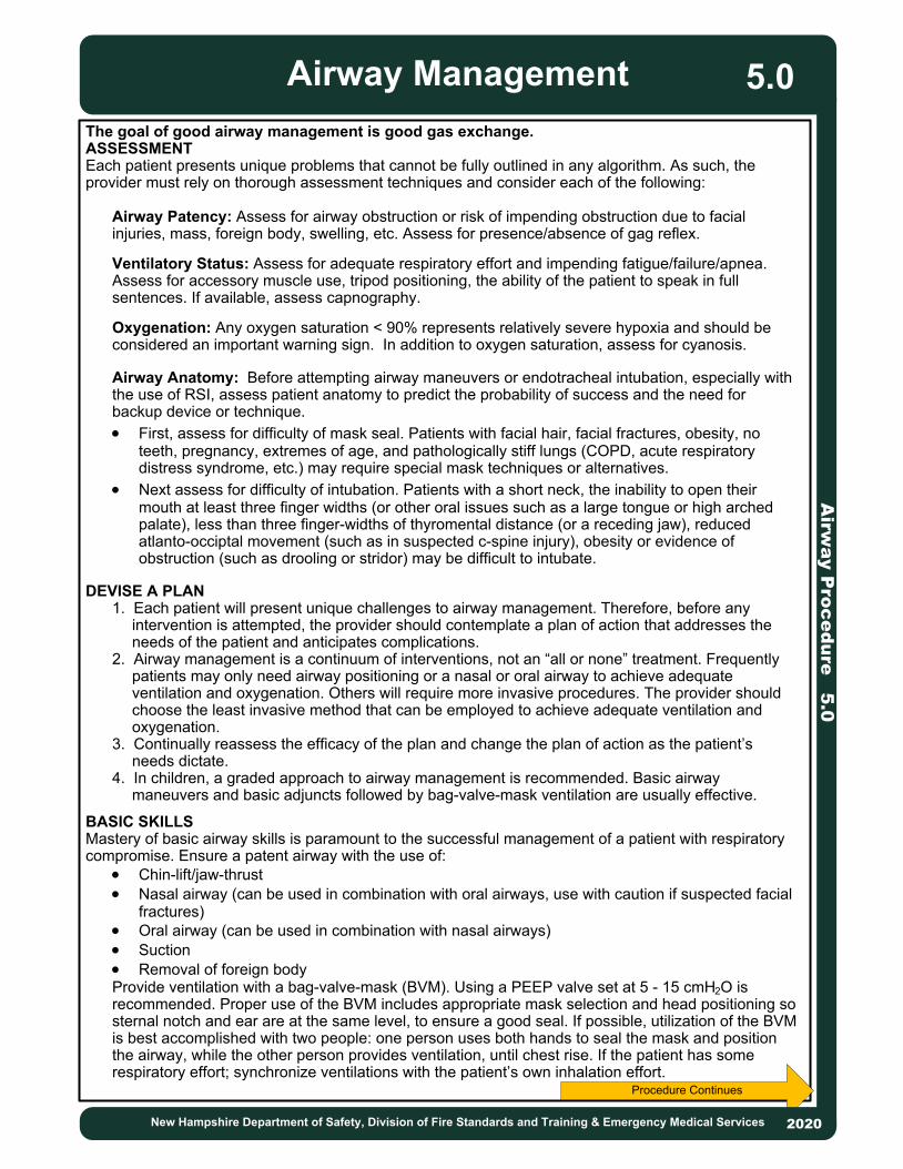
2013
2013
Procedure Continues
The goal of good airway management is good gas exchange.ASSESSMENTEach patient presents unique problems that cannot be fully outlined in any algorithm. As such, the provider must rely on thorough assessment techniques and consider each of the following:
Airway Patency: Assess for airway obstruction or risk of impending obstruction due to facial injuries, mass, foreign body, swelling, etc. Assess for presence/absence of gag reflex.
Ventilatory Status: Assess for adequate respiratory effort and impending fatigue/failure/apnea. Assess for accessory muscle use, tripod positioning, the ability of the patient to speak in full sentences. If available, assess capnography.
Oxygenation: Any oxygen saturation < 90% represents relatively severe hypoxia and should be considered an important warning sign. In addition to oxygen saturation, assess for cyanosis.
Airway Anatomy: Before attempting airway maneuvers or endotracheal intubation, especially with the use of RSI, assess patient anatomy to predict the probability of success and the need for backup device or technique. · First, assess for difficulty of mask seal. Patients with facial hair, facial fractures, obesity, no
teeth, pregnancy, extremes of age, and pathologically stiff lungs (COPD, acute respiratory distress syndrome, etc.) may require special mask techniques or alternatives.
· Next assess for difficulty of intubation. Patients with a short neck, the inability to open their mouth at least three finger widths (or other oral issues such as a large tongue or high arched palate), less than three finger-widths of thyromental distance (or a receding jaw), reduced atlanto-occiptal movement (such as in suspected c-spine injury), obesity or evidence of obstruction (such as drooling or stridor) may be difficult to intubate.
DEVISE A PLAN1. Each patient will present unique challenges to airway management. Therefore, before any
intervention is attempted, the provider should contemplate a plan of action that addresses the needs of the patient and anticipates complications.
2. Airway management is a continuum of interventions, not an “all or none” treatment. Frequently patients may only need airway positioning or a nasal or oral airway to achieve adequate ventilation and oxygenation. Others will require more invasive procedures. The provider should choose the least invasive method that can be employed to achieve adequate ventilation and oxygenation.
3. Continually reassess the efficacy of the plan and change the plan of action as the patient’s needs dictate.
4. In children, a graded approach to airway management is recommended. Basic airway maneuvers and basic adjuncts followed by bag-valve-mask ventilation are usually effective.
BASIC SKILLSMastery of basic airway skills is paramount to the successful management of a patient with respiratory compromise. Ensure a patent airway with the use of:
· Chin-lift/jaw-thrust· Nasal airway (can be used in combination with oral airways, use with caution if suspected facial
fractures)· Oral airway (can be used in combination with nasal airways)· Suction· Removal of foreign body Provide ventilation with a bag-valve-mask (BVM). Using a PEEP valve set at 5 - 15 cmH2O is recommended. Proper use of the BVM includes appropriate mask selection and head positioning so sternal notch and ear are at the same level, to ensure a good seal. If possible, utilization of the BVM is best accomplished with two people: one person uses both hands to seal the mask and position the airway, while the other person provides ventilation, until chest rise. If the patient has some respiratory effort; synchronize ventilations with the patient’s own inhalation effort.
Airw
ay P
roc
ed
ure
5.0
Airway Management
New Hampshire Department of Safety, Division of Fire Standards and Training & Emergency Medical Services 2020
5.0

ADVANCED AIRWAY SKILLSThe appropriate method of airway management should be determined based on patient condition. If basic procedures are deemed inappropriate or have proven to be inadequate then more advanced methods should be used. Procedures documenting the use of each device/technique listed below are found elsewhere in this manual.
CPAP/BiPAP: Continuous positive airway pressure (CPAP) or bilevel positive airway pressure (BiPAP) have been shown to be effective in eliminating the need for intubation and in decreasing mortality in properly-selected patients with acute respiratory distress. Supraglottic Airways (SGA): Utilization of supraglottic airways is an acceptable alternative to endotracheal intubation as both a primary device or a back-up device when previous attempt(s) at ETT placement have failed. Each device has its own set of advantages/disadvantages and requires a unique insertion technique. Providers should have access to, and intimate knowledge of, at least one supraglottic airway.ETT: The endotracheal tube is considered the optimal method of securing the airway in patients with significant respiratory distress and/or airway compromise. However, the incidence of complications is unacceptably high when intubation is performed by inexperienced providers or monitoring of tube placement is inadequate. The optimal method for managing an airway will, therefore, vary based on provider experience, emergency medical services (EMS) or healthcare system characteristics, and the patient’s condition. Use capnography continuously for placement and CO2 monitoring. Use video laryngoscopy, if available and trained.ETT Introducer – “Bougie”: All providers who attempt ETT placement should become intimately familiar with the use of a Bougie. It is the device used most often by anesthesiologists and emergency physicians for helping guide placement when a difficult airway is encountered. Bougie must be available for all intubations performed.Cricothyrotomy: This procedure is indicated only when all other measures fail or you are presented with a situation in which intubation is contraindicated or in which you cannot intubate or otherwise ventilate the patient. Examples include:· Massive facial trauma· Upper airway obstruction due to edema, mass or foreign body
DOCUMENTATIONAll efforts toward airway management should be clearly documented and, at the minimum, should include the following:· Pre/post intervention vital signs including oxygen saturation as well as capnography (if available).· Procedures performed/attempted, including number of failed attempts and who performed each
attempt/procedure.· Size of device(s) placed, depth of placement (if applicable). · Placement confirmation: methods should include auscultation, condensation in the ETT,
symmetrical chest wall rise, as well as capnography.
Procedure Continued
Airw
ay P
roc
ed
ure
5.0
Airway Management
New Hampshire Department of Safety, Division of Fire Standards and Training & Emergency Medical Services 2020
5.0
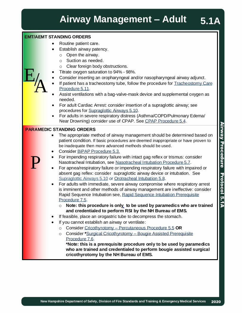
P
EMT/AEMT STANDING ORDERS
PARAMEDIC STANDING ORDERS
2013
Routine patient care. Establish airway patency.
o Open the airway.o Suction as needed.o Clear foreign body obstructions.
Titrate oxygen saturation to 94% - 98%. Consider inserting an oropharyngeal and/or nasopharyngeal airway adjunct. If patient has a tracheostomy tube, follow the procedure for Tracheostomy Care
Procedure 5.11. Assist ventilations with a bag-valve-mask device and supplemental oxygen as
needed. For adult Cardiac Arrest: consider insertion of a supraglottic airway; see
procedures for Supraglottic Airways 5.10. For adults in severe respiratory distress (Asthma/COPD/Pulmonary Edema/
Near Drowning) consider use of CPAP. See CPAP Procedure 5.4.
The appropriate method of airway management should be determined based on patient condition. If basic procedures are deemed inappropriate or have proven to be inadequate then more advanced methods should be used.
Consider BiPAP Procedure 5.3. For impending respiratory failure with intact gag reflex or trismus: consider
Nasotracheal Intubation, see Nasotracheal Intubation Procedure 5.7. For apnea/respiratory failure or impending respiratory failure with impaired or
absent gag reflex: consider supraglottic airway device or intubation. See Supraglottic Airways 5.10 or Orotracheal Intubation 5.8.
For adults with immediate, severe airway compromise where respiratory arrest is imminent and other methods of airway management are ineffective: consider Rapid Sequence Intubation see, Rapid Sequence Intubation Prerequisite Procedure 7.5.o Note: this procedure is only to be used by paramedics who are trained
and credentialed to perform RSI by the NH Bureau of EMS.
If feasible, place an orogastric tube to decompress the stomach. If you cannot establish an airway or ventilate:
o Consider Cricothyrotomy – Percutaneous Procedure 5.5 OR
o Consider *Surgical Cricothyrotomy – Bougie Assisted Prerequisite Procedure 7.6.*Note: this is a prerequisite procedure only to be used by paramedics
who are trained and credentialed to perform bougie assisted surgical
cricothyrotomy by the NH Bureau of EMS.
Airw
ay P
roc
ed
ure
Pro
toco
l 5.1
AAirway Management – Adult
New Hampshire Department of Safety, Division of Fire Standards and Training & Emergency Medical Services 2020
5.1A
EA/

· Hallmarks of respiratory failure are respiratory rate less than 20 breaths per minute for children <6 years old; less than 12 breaths per minute for children <16 years old; and >60 breaths per minutes for any child; cyanosis, marked tachycardia or bradycardia, poor peripheral perfusion, decreased muscle tone, and depressed mental status.
2013
P
EMT/AEMT STANDING ORDERS
PARAMEDIC STANDING ORDERS
2013
· Routine patient care.· Establish airway patency.
o Open Airway.§ Consider patient positioning by placing padding under shoulders to ensure
sternal notch and ear are at the same levelo Suction as needed.o Clear foreign body obstructions.
· If patient has a tracheostomy tube see Tracheostomy Care 5.11.· Consider additional help.· For respiratory distress:
o Administer high concentration oxygen (preferably humidified) via mask positioned on face or if child resists, held near face.
o Titrate oxygen saturation to 94% - 98%; observe for fatigue, decreased mentation, and respiratory failure.
o For children with chronic lung disease or congenital heart disease, maintain or increase home oxygen level to patient’s target saturations.Note: Pulse oximetry is difficult to obtain in children. Do not rely exclusively on pulse oximetry. If child continues to exhibit signs of respiratory distress despite high oxygen saturation levels, continue oxygen administration.
· For respiratory failure or for distress that does not improve with oxygen administration:o Assist ventilations at rate appropriate for child’s age. Reference Pediatric
Color Coded Appendix A3. o If unable to maintain an open airway through positioning, consider placing an
oropharyngeal and/or nasopharyngeal airway.· Determine if child’s respiratory distress/failure is caused by a preexisting condition
o For Allergic Reaction/Anaphylaxis, refer to the Allergic Reaction/Anaphylaxis Protocol 2.2P.
o For Asthma/Reactive Airway Disease/Croup, refer to the Asthma/Bronchiolitis/Croup Protocol 2.3P.
· For Pediatric Cardiac Arrest: consider insertion of a supraglottic airway; see procedures for Supraglottic Airways 5.10
· For pediatrics in severe respiratory distress due to asthma consider use of CPAP. See CPAP Procedure 5.4.
· The appropriate method of airway management should be determined based on patient condition. If basic procedures are deemed inappropriate or have proven to be inadequate then more advanced methods should be used.
· If feasible, place an orogastric tube to decompress the stomach.· If you cannot establish an airway or ventilate, see Cricothyrotomy Percutaneous
Procedure 5.5.
Airw
ay P
roto
co
l Pro
ce
du
re 5
.1P
Airway Management – Pediatric
New Hampshire Department of Safety, Division of Fire Standards and Training & Emergency Medical Services 2020
5.1P
· Child is able to maintain adequate oxygenation by using extra effort to move air.
· Signs include increased respiratory rate, sniffing position, nasal flaring, abnormal breath sounds, head bobbing, intercostal retractions, mild tachycardia.
Pediatric Respiratory Failure
Respiratory distress in children and infants must be promptly recognized and aggressively treated as patient may rapidly decompensate.
EA/
Pediatric Respiratory Distress

Airw
ay P
roc
ed
ure
5.2
2013
2013
P
After placement of an advanced airway device patients may require analgesia and sedation. This includes an oral or nasal endotracheal tube, a supraglottic device, or a surgical airway.
PARAMEDIC STANDING ORDERS – ADULT
Analgesia and Sedation for
Invasive Airway Device
New Hampshire Department of Safety, Division of Fire Standards and Training & Emergency Medical Services2020
5.2
Option 1:· Ketamine 1 mg/kg IV bolus every 5 - 15 minutes as needed, with a target
RASS -2 to -4.o Infusion: 1 mg/kg IV bolus followed by 1 - 5 mg/kg/hour via IV pump titrated to
effect.
Option 2:· Fentanyl 50 - 100 mcg IV every 5 - 10 minutes as needed.AND· Midazolam 5 mg IV, repeat every 5 minutes as needed.
o Infusion: 2.5 - 5 mg IV bolus followed by 5 - 30 mg/hour via IV pump titrated to effect.
OR· Lorazepam 2 - 4 mg every 5 minutes as needed.
PARAMEDIC STANDING ORDERS – PEDIATRICOption 1:· Ketamine 1 mg/kg IV every 5 - 15 minutes, as needed.
Option 2:· Fentanyl 2 - 3 mcg/kg IV every 5 - 10 minutes as needed. AND· Midazolam 0.1 mg/kg IV (single maximum dose 5 mg) every 5 minutes as needed,
OR· Lorazepam 0.1 mg/kg IV (single maximum dose 4 mg) every 5 minutes as needed.
PEARLS· Analgesia and sedation should be considered in all patients with advanced airways in
place, but especially any time a patient shows signs of distress due to their airway device or there is difficulty providing appropriate ventilation due to the patient’s respiratory effort.
· Addressing pain first by administering fentanyl has been shown to decrease the amount of benzodiazepine needed and increases patient comfort.
· Ketamine has analgesic properties and therefore does not require fentanyl to be co-administered.
· Ketamine IV boluses should be pushed slowly, over at least 30-60 seconds.· Sedation can be guided by the RASS scale shown above.· Patients usually require more sedation in the prehospital environment than in-hospital due
to increased external stimuli.· Lower doses should be considered in the setting of hemodynamic compromise.

2013
INDICATIONS Spontaneously breathing patient in severe respiratory distress due to Asthma/COPD,
Congestive Heart Failure / Pulmonary Edema, Pneumonia or Drowning.ABSOLUTE CONTRAINDICATIONS (Do not use) Cardiac/Respiratory arrest. Agonal respirations. Unable to maintain their own airway. Vomiting and/or active upper GI bleed. Respiratory distress secondary to trauma. Suspicion of pneumothorax. Not having a ventilator that is capable of delivering NPPV.RELATIVE CONTRAINDICATIONS (Use cautiously). Unable to follow commands. Agitated or combative behavior.PROCEDURE1. Ensure adequate oxygen supply for the BiPAP device.2. Explain the procedure to the patient. Be prepared to coach the patient for claustrophobia or anxiety.3. Place the patient in an upright position.4. Monitor the patients SpO2, Capnography, ECG and blood pressure.5. Choose the appropriate sized mask for the patient.6. Set the ventilator to the patient appropriate setting. 7. IPAP: Set pressure to 10 cm H2O and titrate to work of breathing not to exceed 20 cmH2O (See chart below).8. EPAP: Set to 5cmH2O and titrate of SpO2 of 94% - 98%; not to exceed 14 cmH2O.9. Pressure support to be no less than 5 cmH2O (Difference between IPAP/EPAP).10. Set back-up ventilatory rate of no less than 8 BPM.11. Set FiO2 to appropriate level to maintain an SpO2 of 94 – 99%. 12. Recheck the mask for leaks and adjust as needed.13. If the patient deteriorates and meets one or more of the contraindications above then discontinue the use BiPAP.Consider Supraglottic Airway 5.10, Intubation 5.7/5.8Consider Rapid Sequence Intubation 7.5 (if trained and credentialed)Consider administering anxiolytic:
Midazolam 2.5 mg IV may repeat once in 5 minutes, OR *Midazolam 5 mg IM/IN may repeat once in 5 minutes, OR Lorazepam 1 mg IV may repeat once in 5 minutes, OR Diazepam 5 mg IV, mahy repeat once in 5 minutes.
PARAMEDIC STANDING ORDERS
Airw
ay P
roc
ed
ure
5.3
Bilevel Positive Airway Pressure
(BiPAP) - ADULT
New Hampshire Department of Safety, Division of Fire Standards and Training & Emergency Medical Services 2020
5.3
Administer benzodiazepines with caution in elderly patients or those with signs of hypercarbia or respiratory fatigue.
Keep in mind BiPAP uses large volumes of oxygen.
P
*For IN administration of midazolam use a 5 mg/mL concentration.

2013
INDICATIONS
Spontaneously breathing patient in severe respiratory distress due to Asthma/COPD, Congestive Heart Failure / Pulmonary Edema, Pneumonia or Drowning.
ABSOLUTE CONTRAINDICATIONS (Do not use)
Cardiac/Respiratory arrest. Agonal respirations. Unable to maintain their own airway. Vomiting and/or active upper GI bleed. Respiratory distress secondary to trauma. Suspicion of pneumothorax. Pediatric patient who is too small for the mask sizes available.
RELATIVE CONTRAINDICATIONS (Use cautiously)
Unable to follow commands. Agitated or combative behavior.
PROCEDURE
1. Ensure adequate oxygen supply for CPAP device.
2. Managing patient anxiety is extremely important. Reduce patient anxiety by coaching and minimize external stimuli as much as possible.
3. Place patient in upright position. Apply pulse oximetry, capnography nasal capture device and ECG as available and trained.
4. Choose appropriate sized device mask for patient, assemble the CPAP device, attach to oxygen supply and insure oxygen is flowing (follow manufacturer’s directions for preparation for your particular device).
5. Place mask over face and secure with straps until minimal air leak.6. Adjust Positive End Expiratory Pressure (PEEP) to 5 - 15 cmH2O to effect for patient
condition.7. If device allows, titrate oxygen level to oxygen saturation of 94 – 98%.8. Recheck mask for leaks and adjust straps as needed to minimize air leaks.9. Reassure anxious patient. 10. Monitor pulse oximetry, capnography and ECG as available and trained.11. If patient stabilizes, maintain CPAP for duration of transport and notify receiving hospital to prepare for a CPAP patient.12. If patient begins to deteriorate, discontinue CPAP and assist respirations by BVM
with PEEP valve. 13. Document CPAP procedure, including time and provider. Document serial pulse
oximetry and capnography readings to demonstrate effects.
If a commercial device is not available you may consider using a BVM with PEEP valve:1. Apply nasal cannula at 15 lpm.2. Attach PEEP valve to BVM at desired PEEP level (5 – 15 cmH2O).3. Attach oxygen to BVM at least 15 lpm and ensure flow.4. Maintain continuous mask seal on patient to deliver CPAP.
EMT/AEMT STANDING ORDERS
Airw
ay P
roc
ed
ure
5.4
Continuous Positive Airway Pressure
(CPAP)
New Hampshire Department of Safety, Division of Fire Standards and Training & Emergency Medical Services 2020
5.4
Keep in mind CPAP uses large volumes of oxygen.
EA/
Protocol Continues

Airw
ay P
roc
ed
ure
5.4
Continuous Positive Airway Pressure
(CPAP)
New Hampshire Department of Safety, Division of Fire Standards and Training & Emergency Medical Services
5.4
PARAMEDIC STANDING ORDERS - ADULT
PARAMEDIC STANDING ORDERS - PEDIATRIC
Administer benzodiazepines with caution in elderly patients or those with signs of hypercarbia or respiratory fatigue.
*For IN administration of midazolam use a 5 mg/mL concentration.
P Consider Supraglottic Airway 5.10 or Intubation 5.7/5.8. Consider Rapid Sequence Intubation 7.5 (if trained and credentialed). Consider administering anxiolytic:
o Midazolam 2.5 mg IV, may repeat once in 5 minutes, OR o *Midazolam 5 mg IM/IN, may repeat once in 5 minutes, ORo Lorazepam mg IV, may repeat once in 5 minutes, ORo Diazepam 5 mg IV, may repeat once in 5 minutes.
Consider Supraglottic Airway 5.10 or Intubation 5.7/5.8. Consider Rapid Sequence Intubation 7.5 (if trained and credentialed). Consider administering anxiolytic:
o Midazolam 0.05 mg/kg IV (single max dose 2.5 mg), may repeat once in 5 minutes, OR
o *Midazolam 0.1 mg/kg IM/IN (single max dose 5 mg), may repeat once in 5 minutes, OR
o Lorazepam 0.05 mg/kg IV (single max dose 1 mg), may repeat once in 5 minutes, OR
o Diazepam 0.1 mg/kg (single max dose 5 mg), may repeat once in 5 minutes.
Protocol Continued
2020

2013
This protocol is intended for the use of commercially prepared rapid cricothyrotomy devices. Devices requiring use of a guide wire may not be used. Approved devices have a plastic cannula preloaded onto a metal introducer (e.g., Rusch QuickTrach).
INDICATIONS:Inability to adequately oxygenate and ventilate using less invasive methods including BVM, supraglottic airways and endotracheal intubation.EQUIPMENT:· Commercially prepared percutaneous cricothyrotomy device.· Chlorhexadine wipes.· Bag-valve-mask.· Quantitative Waveform ETCO2.
PROCEDURE:(May vary slightly with different devices)1. Position the patient supine and extend the neck as needed to improve anatomic view.2. Prepare neck with Chlorhexidine.3. Using non-dominant hand, stabilize larynx and locate the following landmarks: thyroid cartilage (Adam’s apple) and cricoid cartilage (solid ring below the thyroid cartilage). The cricothyroid membrane lies between these cartilages.4. Insert needle bevel through soft tissue and cricothyroid membrane at 90- degree angle while aspirating with syringe.5. As soon as air is freely aspirated stop advancing the needle as this indicates entry into the trachea.6. Direct the needle tip inferiorly by modifying angle to 60-degrees from the patient’s head. Advance the assembly until the stopper is in contact with the skin. (Note: If air is not freely aspirated and the stopper has contacted the skin the stopper may need to be removed in order to reach the trachea. Be aware that if the stopper is removed there is increased risk of perforating the posterior aspect of the trachea.)7. Remove the stopper while holding assembly firmly in place.8. Hold the needle firmly in place and advance only the plastic cannula off the needle into the trachea until the flange rests on the neck. Carefully remove the needle and syringe.9. Secure cannula in place with neck strap. 10. Inflate cuff if one is present.11. Apply BVM with waveform ETCO2 and ventilate the patient.12. Confirm placement by assessing for bilateral lung sounds and presence of quantitative and qualitative ETCO2.13. Frequently reassess placement and continuously monitor ETCO2.
PARAMEDIC STANDING ORDERS
Airw
ay P
roc
ed
ure
5.5
Cricothyrotomy - Percutaneous
New Hampshire Department of Safety, Division of Fire Standards and Training & Emergency Medical Services 2020
5.5
P
· Devices may be utilized on patients of any age for which they are designed and appropriate sizes are available.
· If anatomical landmarks cannot be identified the procedure should not be performed.
cricothyroid membrane
This procedure cannot be performed until the provider has received training from their EMS unit on the commercial device selected and is deemed competent. The device and training must be approved by the EMS unit’s Medical Director. Written notification will be provided to the Medical Resource Hospital’s EMS Medical Director, Hospital EMS Coordinator, and Bureau of EMS within 48 hours of an event. Use of this procedure documented under “Procedures Used” in the Patient Care Report constitutes notification of the Bureau of EMS.

2013
2013
P
INDICATIONS· Unable to fully visualize vocal cords during an intubation attempt.· To facilitate routine placement of endotracheal tube.
LIMITATIONS· Adult Bougies should not be used on less than 6.0 ETT.
PROCEDURE1. Endotracheal tube may be preloaded on bougie if provider is familiar with technique being used. Always lubricate cuff of endotracheal tube with water- based lubricant.2. Using techniques described in the Orotracheal Intubation Protocol 5.8 attempt to visualize the vocal cords. Always use all techniques necessary to optimize laryngeal view before trying to pass the bougie.3. If the vocal cords are partially visualized, pass the bougie through the cords while attempting to feel signs of tracheal placement (see below). Gently advance bougie until holdup is felt. If the bougie does not stop advancing the bougie is likely in the esophagus.4. If the vocal cords are not visualized, pass the bougie behind the epiglottis, guiding the tip of the bougie anteriorly toward the trachea and assess for signs of tracheal placement (see below). Do not attempt to pass the bougie if the epiglottis is not visualized. Gently advance bougie until holdup is felt. If the bougie does not stop advancing the bougie is likely in the esophagus.5. With laryngoscope still in place, advance preloaded tube off bougie or have assistant load the tube onto the bougie and advance it to the lip line.6. Advance the ETT over the Bougie, rotating the ETT about 1/4 turn counterclockwise
so that the bevel is oriented vertically as the ETT passes through the vocal cords. This maneuver allows the bevel to gently spread the arytenoids with a minimum of force, thus avoiding injury. If resistance is felt, withdraw the ETT, rotating it in a slightly more counterclockwise direction, and advance the tube again. Advance the tube to a lip-line of 24 cm in an adult male, and 22 cm in an adult female or until cuff is seen passing through cords.
7. Holding the ETT firmly in place, have an assistant remove the Bougie. 8. Remove the laryngoscope.9. Inflate the cuff with 5 – 10 ml of air. 10. Follow the procedures outlined in Procedure: Orotracheal Intubation 5.8 to confirm
placement, secure the ETT, monitor and document placement of the ETT. SIGNS OF TRACHEAL PLACEMENT· The Bougie is felt to “hold up” as the airway narrows and is unable to be advanced
further. This is the most reliable sign of proper Bougie placement. If the Bougie enters the esophagus, it will continue to advance without resistance.
· It may be possible to feel the tactile sensation of “clicking” as the Bougie tip is advanced downward over the rigid cartilaginous tracheal rings.
· The Bougie may rotate as it enters a mainstem bronchus. Usually it is a clockwise rotation as the Bougie enters the right mainstem bronchus, but occasionally it will rotate counterclockwise if the Bougie enters the left mainstem bronchus.
· If the patient is not paralyzed, he/she may cough.
PARAMEDIC STANDING ORDERS – ADULT
Airw
ay P
roc
ed
ure
5.6
Endotracheal Tube Introducer “Bougie” – Adult
New Hampshire Department of Safety, Division of Fire Standards and Training & Emergency Medical Services 2020
5.6

2013
2013
P
INDICATIONS· Impending respiratory failure with intact gag reflex or if jaw is clenched and unable to be opened. · Inadequate ventilation/oxygenation with basic airway procedures.o The appropriate method of airway management should be determined based
on patient condition. If basic procedures are deemed inappropriate or have proven to be inadequate then more advanced methods should be used.
CONTRAINDICATIONS· Apnea.· Nasal obstruction.· Suspected basilar skull fracture.· Patient fits on a pediatric length-based resuscitation tape (e.g., Broselow Tape).
PROCEDURE1. Pre-medicate nasal mucosa with 2% lidocaine jelly and nasal decongestant spray, if
available.
2. Pre-oxygenate the patient.
3. Select the largest and least obstructed nostril and insert a lubricated nasal airway to help dilate the nasal passage.
4. Lubricate the ETT with water-based lubricant.
5. Remove the nasal airway and gently insert the ETT with continuous capnography monitoring, keeping the bevel toward the septum (a gentle rotation movement may be necessary at the turbinates).
6. Continue to advance the ETT while listening for maximum air movement and watching for capnography wave form.
7. At the point of maximum air movement, indicating proximity to the level of the glottis, gently and evenly advance the tube through the glottic opening on inspiration.
If resistance is encountered, the tube may have become lodged into the pyriform sinus and you may note tenting of the skin on either side of the thyroid cartilage. If this happens, slightly withdraw the ETT and rotate it toward the midline and attempt to advance tube again with the next inspiration.
8. Upon entering the trachea, the tube may cause the patient to cough, buck, strain, or gag. This is normal. Do not remove the ETT. Be prepared to control the cervical spine and the patient, and be alert for vomiting.
9. Placement depth from the nares to the tip of the tube should be approximately 28cm in males and 26 cm in females.
10. Inflate cuff with 5 – 10ml of air.
11. Confirm appropriate placement by capnography, symmetrical chest-wall rise, auscultation of equal breath sounds over the chest and a lack of epigastric sounds with bagging, and condensation in the ETT.
12. Secure the ETT, consider applying a cervical-collar.
Airw
ay P
roc
ed
ure
5.7
Nasotracheal Intubation
New Hampshire Department of Safety, Division of Fire Standards and Training & Emergency Medical Services 2020
PARAMEDIC STANDING ORDERS - ADULT
Protocol Continues
5.7

Airw
ay P
roc
ed
ure
5.7
Nasotracheal Intubation
New Hampshire Department of Safety, Division of Fire Standards and Training & Emergency Medical Services 2020
P
13. Ongoing monitoring of ETT placement and ventilation status using capnography is required for all patients.
14. Document each attempt as a separate procedure so it can be time stamped in the ePCR. An attempt is defined as placement of the tube into the patient’s nare. For each attempt, document the time, provider, placement success, pre-oxygenation, airway grade, ETT size, placement depth, placement landmark (e.g. cm at the patient’s nare), and confirmation of tube placement including chest rise, bilateral, equal breath sounds, absence of epigastric sounds and end-tidal CO2 readings.
If continued intubation attempts are unsuccessful (maximum of 2 attempts) consider Cricothyrotomy. See Cricothyrotomy Procedures 5.5 OR 7.5.
POST TUBE PLACEMENT CARE – ADULT · See Procedure: Analgesia and Sedation for Invasive Airway Device 5.2
PARAMEDIC STANDING ORDERS
Protocol Continued
5.7

Airw
ay P
roc
ed
ure
5.8
2013
2013
P
INDICATIONS· Apnea/respiratory failure, impending respiratory failure, impaired or absent gag
reflex.· Inadequate ventilation/oxygenation with basic airway procedures.
o The appropriate method of airway management should be determined based on patient condition. If basic procedures are deemed inappropriate or have proven to be inadequate then more advanced methods should be used.
CONTRAINDICATION· Epiglottitis.· Facial or neck injuries that prohibit visualization of airway anatomy (relative).
PROCEDUREDirect Laryngoscopy or Direct Video Laryngoscopy:1. Place patient in ear to sternal notch position and elevate head to 30° if possible. Ensure all preparation and planning steps are complete.2. Insertion: Open the mouth fully and insert the tip of the blade into the mouth to the right and sweeping the tongue to midline. The laryngoscope should be gripped lightly as no significant force is needed until later steps. It is helpful, especially if there are substantial secretions, to lead with the suction catheter and suction as the laryngoscope is advanced.3. Epiglottoscopy: SLOWLY advance the blade down the tongue at the midline until the epiglottis is seen. Be sure to control the tongue leaving space to the right for tube delivery. Keep the tip of the blade along the tongue and avoid allowing the laryngoscope to fall posterior.4. Valleculoscopy: Gradually advance the blade until it is seated in the vallecula. The blade must engage the hypoepiglottic ligament in order to adequately lift the epiglottis. The ligament lies directly within the vallecula. If using a Miller blade pass tip of blade under the epiglottis to control it directly.5. Laryngoscopy: Once the tip of Mac blade is seated in the vallecula or tip of Miller blade has passed the epiglottis lifting force should be applied forward and upward without rotating the handle backward. The epiglottis will lift or be displaced and the larynx will be exposed6. If using bougie: Once an optimal view is obtained pass the bougie through the cords. Tracheal rings may be felt if the coude tip remains pointing upright. Advance the bougie slowly until it lodges in the proximal bronchi. Be careful not to advance with too much force as tracheobronchial trauma may occur. If the bougie does not stop advancing this is suggestive of esophageal placement. Advance the lubricated endotracheal tube over the bougie without removing the laryngoscope. If the tube cannot be advanced through the cords rotate it 60° counterclockwise. Visualize the tube passing through the cords if possible and stop advancing once the cuff is past the cords. Remove the laryngoscope, hold tube firmly, and remove the bougie.7. If using stylette: Ensure stylette is bent in “straight-to-cuff” fashion with 30° bend angle and tube cuff is lubricated. Once an optimal view is obtained pass the tube to the right and below the line-of-sight to the cords. The tube must be visualized passing through the cords. Advance tube until the cuff is seen passing through the cords. If resistance is felt rotate the tube clockwise. Once the tube is in place hold it firmly and remove the stylette.8. Inflate ETT cuff with 5 - 10 mL of air and adjust inflation pressure if necessary. The pilot balloon should feel inflated but be easily compressible and not too hard.9. Confirm tube placement via continued waveform capnography, presence of bilateral lung sounds, and absence of epigastric sounds.
PARAMEDIC STANDING ORDERS – ADULT & PEDIATRIC
Orotracheal Intubation
New Hampshire Department of Safety, Division of Fire Standards and Training & Emergency Medical Services 2020
Protocol Continues
5.8

Airw
ay P
roc
ed
ure
5.8
Orotracheal Intubation
New Hampshire Department of Safety, Division of Fire Standards and Training & Emergency Medical Services 2020
10. Secure ETT and continue to monitor waveform capnography. Frequently reassess tube placement.Indirect Video Laryngoscopy(Devices such as Glidescope and King Vision that cannot be used for direct laryngoscopy)
1. Place patient in ear to sternal notch position and elevate head to 30° if possible. Ensure all preparation and planning steps are complete.
2. Insertion: Open mouth fully and insert blade at the midline. It is helpful, especially if there are substantial secretions, to lead with the suction catheter and suction as the laryngoscope is advanced.3. Epiglottoscopy: Gradually advance the blade by rotating handle backward and allowing the tip of the blade to follow the tongue until the epiglottis is seen. 4. Valleculoscopy: Advance the tip of the blade until it is seated in the vallecula. DO NOT go to too deep. The tip of the blade may need to be slightly above the vallecula in order to facilitate tube passage. If you can see the cricoid ring through the cords you are too deep.5. Laryngoscopy: Lift the jaw straight up with the blade exposing the larynx fully. 6. Tube passage for non-channeled devices: A lubricated ET tube loaded on a rigid or standard stylette should be used. The stylette should have a gradual curve at the end to almost a 90° angle. Pass the tube into the mouth from the right side. The tip should enter view from the bottom of the screen and toward the larynx. When the tube has just begun entering the cords the stylette should be popped up out of the tube slightly using your right thumb or with the help of an assistant. This will allow the tip of the tube to fall between the cords at the correct angle. Pass the tube until the cuff is past the cords. Note: It is not recommended to use a bougie with a non-channeled IVL laryngoscope as they are not easily maneuvered around the steep angle that is present. 7. Tube passage for channeled devices: Line up view on camera with the cords. Advance lubricated ETT down channel and visualize it passing through the cords. It may be helpful to preload a bougie in the tube and advance it through the cords first.8. Inflate ETT cuff with 5 - 10 mL of air and adjust inflation pressure if necessary. The pilot balloon should feel inflated but easily compressible and not too hard.9. Confirm tube placement via continued waveform capnography, presence of bilateral lung sounds, and absence of epigastric sounds. 10. Secure ET tube and continue to monitor waveform capnography. Frequently reassess tube placement.If intubation attempt is unsuccessful, ETT placement cannot be verified or ETTbecomes dislodged: Monitor oxygen saturation and end-tidal CO2 ANDVentilate the patient with 100% oxygen via a BVM until ready to attempt intubation again. Consider insertion of supraglottic airway if additional intubation attempts are unlikely to be
successful.
5.8
P
Techniques to improve laryngeal view:· Head Elevation: Elevate the head by lifting with the laryngoscope or having an assistant
lift the head from underneath.· External Laryngeal Manipulation (ELM): The person intubating uses their right hand to
manipulate the larynx to a position that is suitable. An assistant then holds the larynx in that position. Note: BURP and cricoid pressure are no longer recommended.
· Jaw Thrust: An assistant performs a jaw thrust to assist with tissue displacement.
PARAMEDIC STANDING ORDERS – ADULT & PEDIATRIC
Protocol Continues
Protocol Continues
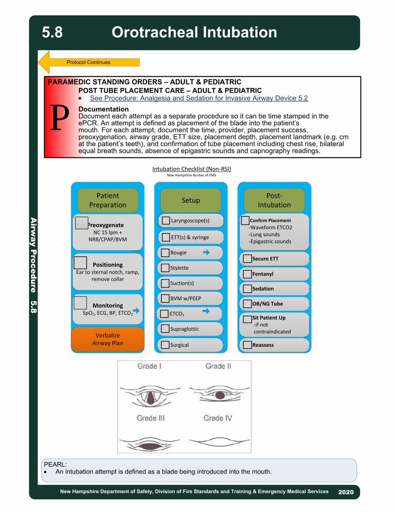
Airw
ay P
roc
ed
ure
5.8
Orotracheal Intubation
New Hampshire Department of Safety, Division of Fire Standards and Training & Emergency Medical Services 2020
5.8
POST TUBE PLACEMENT CARE – ADULT & PEDIATRIC· See Procedure: Analgesia and Sedation for Invasive Airway Device 5.2DocumentationDocument each attempt as a separate procedure so it can be time stamped in theePCR. An attempt is defined as placement of the blade into the patient’smouth. For each attempt, document the time, provider, placement success, preoxygenation, airway grade, ETT size, placement depth, placement landmark (e.g. cmat the patient’s teeth), and confirmation of tube placement including chest rise, bilateralequal breath sounds, absence of epigastric sounds and capnography readings.
PARAMEDIC STANDING ORDERS – ADULT & PEDIATRIC
P
PEARL:· An intubation attempt is defined as a blade being introduced into the mouth.
Protocol Continues
PatientPreparation
Post-Intubation
Setup
PreoxygenateNC 15 lpm +
NRB/CPAP/BVM
PositioningEar to sternal notch, ramp,
remove collar
MonitoringSpO2, ECG, BP, ETCO2
VerbalizeAirway Plan Surgical
Stylette
Bougie
ETT(s) & syringe
Laryngoscope(s)
Suction(s)
BVM w/PEEP
ETCO2
Supraglottic
Secure ETT
Fentanyl
Sedation
OB/NG Tube
Sit Patient Up -if not contraindicated
Reassess
Confirm Placement
-Waveform ETCO2 -Lung sounds -Epigastric sounds
Intubation Checklist (Non-RSI)New Hampshire Bureau of EMS

EMT/ ADVANCED EMT / PARAMEDIC STANDING ORDERS
2013
INDICATIONS· Obstruction of the airway (secondary to secretions, blood, and/or any other
substance) in a patient currently being assisted by an inserted airway such as an endotracheal tube or supraglottic airway. For tracheostomy tube see Tracheostomy Care 5.11.
CONTRAINDICATIONS· None.
PROCEDURE1. Ensure the suction device is operable.2. Pre-oxygenate the patient.3. While maintaining aseptic technique, attach the suction catheter to the suction
unit.4. If applicable, remove ventilation device from the airway.5. Insert the sterile end of the suction catheter into the tube without suction.
Insert until resistance is met; pull back approximately 1 – 2 cm.6. Once the desired depth is met, apply suction by occluding the port of the
suction catheter and slowly remove the catheter from the tube using a twisting motion.
7. Suctioning duration should not exceed 15 seconds, using lowest pressure that effectively removes secretions.
8. Saline flush may be used to help loosen secretions and facilitate suctioning.9. Re-attach the ventilation device to the patient.
EA
P
//
Suctioning of Inserted AirwayA
irwa
y P
roc
ed
ure
5.9
New Hampshire Department of Safety, Division of Fire Standards and Training & Emergency Medical Services 2020
5.9

P
EMT/ADVANCED EMT STANDING ORDERSINDICATIONS:· Cardiac Arrest.RELATIVE CONTRAINDICATIONS:· Severe maxillofacial or oral trauma.· For devices inserted into the esophagus:
o The patient has known esophageal disease.o The patient has ingested a caustic substance.o The patient has burns involving the airway.
PROCEDURE:· Insertion procedure should follow manufacturer guidelines as each device is
unique.· Confirm appropriate placement by symmetrical chest-wall rise, auscultation of
equal breath sounds over the chest and a lack of epigastric sounds with bag valve mask ventilation, and capnography, if available.
· Secure the device. · Document the time, provider, provider level and success for the procedure.
Complete all applicable airway confirmation fields including chest rise, bilateral, equal breath sounds, absence of epigastric sounds and end-tidal CO2 readings.
· Reassess placement frequently, especially after patient movement.
INDICATIONS:· Inability to adequately ventilate a patient with a bag-valve-mask or longer EMS
transports requiring a more definitive airway.· Back up device for failed endotracheal intubation attempt.POST TUBE PLACEMENT CARE – ADULT & PEDIATRIC· See Procedure: Analgesia and Sedationf for Invasive Airway Device 5.2
2013
EA/
Airw
ay P
roc
ed
ure
5.1
0
Supraglottic AirwayAdult & Pediatric
New Hampshire Department of Safety, Division of Fire Standards and Training & Emergency Medical Services 2020
5.10This protocol applies to commercially available supraglottic airway devices. These airways must be used as directed by the manufacturer’s guidelines. They may be used in all age groups for which the devices are designed. Providers must be trained on and competent with the airway device they will be using. Note: Double Lumen Device (e.g., Combitube) are not longer approved.
PARAMEDIC STANDING ORDERS

2013
INDICATIONS· An adult or pediatric patient with an established tracheostomy in respiratory
distress or failure.
PROCEDURE· Consult with the patient’s caregivers for assistance.· Assess tracheostomy tube: Look for possible causes of distress which may be
easily correctable, such as a detached oxygen source.· If the patient’s breathing is adequate but exhibits continued signs of respiratory
distress, administer high-flow oxygen via non-rebreather mask or blow-by, as tolerated, over the tracheostomy.
· If patient’s breathing is inadequate, assist ventilations using bag-valve-mask device with high-flow oxygen.
· If on a ventilator, remove the patient from the ventilator prior to using bag valve mask device as there may be a problem with the ventilator or oxygen source.
· Suction if unable to ventilate via tracheostomy or if respiratory distress continues.
· Use no more than 100 mmHg suction pressure. · If the tracheostomy tube has a cannula, remove it prior to suctioning. · Determine proper suction catheter length by measuring the obturator. · If the obturator is unavailable, insert the suction catheter approximately 2 – 3
inches into the tracheostomy tube. Do not use force! · 2 – 3 ml saline flush may be used to help loosen secretions.· If the patient remains in severe distress, continue ventilation attempts using bag
valve mask with high-flow oxygen via the tracheostomy. Consider underlying reasons for respiratory distress and refer to the appropriate protocol for intervention.
EMT/ADVANCED EMT STANDING ORDER – ADULT & PEDIATRIC
EA
P
/
PARAMEDIC STANDING ORDERS – ADULT & PEDIATRICINDICATIONS· An adult or pediatric patient with an established tracheostomy, in respiratory
distress or failure where EMT and Advanced EMT tracheostomy interventions have been unsuccessful.
· Dislodged tracheostomy tube. CONTRAINDICATIONS· None.PROCEDURE:· If the patient continues in severe respiratory distress, remove tracheostomy
tube and attempt bag valve mask ventilation. · If another tube is available from caregivers, insert into stoma and resume
ventilation (a standard endotracheal tube may be used or the used tracheostomy tube, after being cleaned).o Bougie may be used to assist with placement of endotracheal tube into
stoma.· If unable to replace tube with another tracheostomy tube or endotracheal tube,
assist ventilations with bag valve mask and high-flow oxygen.
Airw
ay P
roc
ed
ure
5.1
1Tracheostomy Care
New Hampshire Department of Safety, Division of Fire Standards and Training & Emergency Medical Services 2020
5.11

Airw
ay P
roc
ed
ure
5.1
2
New Hampshire Department of Safety, Division of Fire Standards and Training & Emergency Medical Services
2013
2020
P
PURPOSE· To define the methodology and practice for using a mechanical ventilator.· To optimize oxygenation and ventilation of endotracheally intubated patients as
well as patients with supraglottic airways.
INDICATIONS· Adult patients with advanced airways placed by EMS prehospitally. The use of
ventilators in the PIFT environment is not addressed by this protocol.· Adult and pediatric patients on their own ventilator:
o If the ventilator is operational, transport patient with their ventilator and caregiver on previously prescribed ventilator settings.
o If the ventilator is inoperable, assist caregiver with troubleshooting using the SCOPE mnemonic (see below). Use bag valve mask device and transition to EMS ventilator as necessary, if available.
CONTRAINDICATIONS· Pediatric patients with advanced airway placed by EMS.
SPECIAL CONSIDERATIONS· All patients receiving mechanical ventilation will have an appropriate size BVM
with mask, an appropriately sized OPA, and a 10cc luer lock syringe readily accessible.
SETTINGSThe following initial settings are recommended:Mode: Assist Control (AC) – Volume.Tidal Volume: 6-8 mL/kg of Ideal Body Mass (see charts below):
PARAMEDIC – ADULT & PEDIATRIC
Ventilator
Height in Ft/In
5.0
5.1
5.2
5.3
5.4
5.5
5.6
5.7
5.8
5.9
5.10
5.11
6.0
6 mL/kg
314
320
328
341
355
369
383
397
410
424
438
452
466
8 mL/kg
418
426
437
455
474
492
510
529
547
566
584
602
621
6.1 479 639
MALE
Height in Ft/In
5.0
5.1
5.2
5.3
5.4
5.5
5.6
5.7
5.8
5.9
5.10
5.11
6.0
6 mL/kg
286
293
300
314
328
342
356
370
383
397
411
425
439
8 mL/kg
382
390
400
406
438
456
474
493
511
530
548
566
585
6.1 452 603
FEMALE
Protocol Continues
5.12

VentilatorA
irwa
y P
roc
ed
ure
5.1
1
New Hampshire Department of Safety, Division of Fire Standards and Training & Emergency Medical Services 2020
P
PARAMEDIC – ADULT & PEDIATRICRate: Initially 8 - 12, titrate to appropriate EtCO2 based on patient’s condition(e.g. severe asthma, aspirin overdose, traumatic brain injury).
FiO2: Start at 100% FiO2, then titrate to maintain SpO2 >94% (90% for COPD patients).
PEEP: 2 to 5 cmH2O.
ALARM SETTINGS
· High pressure alarm: 30 cmH20. · Low pressure alarm, if available: 4 cmH20.
Further adjustments in ventilator settings may be done in conjunction with on or offline Medical Control.
SCOPE
S: SuctionC: ConnectionsO: ObstructionsP: PneumothoraxE: Equipment/Tube Dislodgement
5.11Protocol Continues

2013
Obtain 12 lead ECG with baseline vitals within 10 minutes if available and practical. Transmit per local guidelines. INDICATIONS· Congestive Heart Failure/Pulmonary Edema.· Dysrhythmias and/or Palpitations · Suspected Acute Coronary Syndrome.· Syncope.· Shortness of breath.· Stroke/CVA.· Cardiac Arrest with Return of Spontaneous Circulation (ROSC).· Upper Abdominal Pain· Dizziness/ lightheadedness
PROCEDURE1. Prepare ECG Monitor and connect cable with electrodes.2. Properly position the patient (supine or semi-reclined).3. Enter patient information (e.g., age, gender, name) into monitor, when able.4. Prep chest as necessary, (e.g., hair removal, skin prep pads). 5. Apply chest and extremity leads using recommended landmarks:
· RA – Right arm or shoulder.· LA – Left arm or shoulder.· RL – Right leg or hip.· LL – Left leg or hip.· V1 – 4TH intercostal space at the right sternal border.· V2 – 4TH intercostal space at the left sternal border.· V3 – Directly between V2 and V4.· V4 – 5th intercostal space midclavicular line.
· V5 – Level with V4 at left anterior axillary line.
· V6 – Level with V5 at left midaxillary line.6. Instruct patient to remain still.7. Acquire the 12 lead ECG.8. If 12 lead ECG indicates a STEMI (e.g., ECG identifies ***Acute MI Suspected***
and/or Paramedic interpretation) transport patient to the most appropriate facility in accordance with local STEMI guidelines/agreements. Notify receiving facility of a “STEMI Alert” and patient information as requested.
9. For patients with continued symptoms consistent with acute coronary syndrome, perform repeat ECGs, as indicated, during transport to evaluate for evolving STEMI. Leave 12 lead attached.
10. Copies of 12 lead ECG labeled with the patient’s name and date of birth should be left with the receiving hospital.
11. Document the procedure and time of the ECG acquisition in appropriate section of the Patient Care Record. Include the ECG printout/image in the PCR, if possible.
EMT/ADVANCED EMT/PARAMEDIC STANDING ORDER
EA
P
//P
roc
ed
ure
6.0
12-Lead ECG Acquisition
New Hampshire Department of Safety, Division of Fire Standards and Training & Emergency Medical Services 2020
6.0
Protocol Continues

Pro
ce
du
re 6
.012-Lead ECG Acquisition
New Hampshire Department of Safety, Division of Fire Standards and Training & Emergency Medical Services 2020
PEARLS:· Enter the patient’s age for proper interpretation.· When transmitting either include the patient’s name or notify the receiving facility of the
patient’s identity.· Be alert for causes of artifact: dry or sweaty skin, dried out electrodes, patient movement,
cable movement, vehicle movement, electromagnetic interference, static electricity· Dried out electrodes are a major source of artifact; keep in original sealed foil pouches;
plastic bags are not sufficient. Use all the same kind of electrodes. Press firmly around the edge of the electrode, not the center.
· Sweaty patients should be dried thoroughly. Consider tincture of benzoin. Dry skin is especially problematic. Clean the site (e.g., alcohol prep pad) and gently abrade skin using a towel or 4x4 gauze.
· Check for subtle movement: toe tapping, shivering, muscle tension (e.g., hand grasping rail or head raised to “watch”)
· For isolated ST depression in leads VI – V3 consider posterior ECG· For suspect inferior MI consider right sided ECG.
o Label these ECG printouts as applicable.
6.0Protocol Continues

2013
Indications:· Routine monitoring of ventilation status and indirectly circulatory and metabolic status
in adults and children with:o Respiratory distress (e.g., CHF, COPD, Asthma, Pulmonary embolus)o Altered mental statuso Traumatic brain injuryo Diabetic ketoacidosiso Circulatory shocko Sepsiso Cyanide and/or carbon monoxide poisoningo Administration of sedative medication
· Advanced Airway Devices:o Confirm and document placement of advanced airway devices, see Airway
Management 5.0 and 5.1 A&Po To confirm continued placement of advanced airway devices after every patient
move and at transfer of care.· Monitoring of CPR quality and for signs of return of spontaneous circulation (ROSC).
o High quality chest compressions are achieved when the ETCO2 is at least 20 mmHg. If ETCO2 abruptly increases it is reasonable to consider that this as an indicator of ROSC.
· To assist with termination of resuscitation efforts when ETCO2 is <20 mmHg despite adjusting the quality of chest compressions. o Low CO2 production after 20 minutes of effective CPR is a predictor of mortality.
See Resuscitation Initiation & Termination Policy 8.16. Procedure: 1. Attach the sensor to endotracheal tube, supraglottic airway, BVM or apply cannula with ETCO2 mouth scoop or bi-cannula. 2. Assess ETCO2 numeric levels and waveform:
o Normal ETCO2 range 35-45 mmHgo Elevated ETCO2 may indicate hypoventilation/CO2 retention.o Low ETCO2 may indicate hyperventilation, low perfusion, pulmonary embolus,
sepsis. 3.With abnormal ETCO2 levels consider adjusting rate and depth of ventilations.
EMT/ADVANCED EMT/PARAMEDIC STANDING ORDERS
EA
P
//
Pro
ce
du
re 6
.1
Capnography
New Hampshire Department of Safety, Division of Fire Standards and Training & Emergency Medical Services 2020
6.1
Any abrupt loss of ETCO2 detection or loss of continuous waveform may indicate a catastrophic failure of the airway, apnea, drug overdose, deep sedation and/or cardiac arrest warranting assessment of the airway, breathing, circulation, and/ or airway device.
PEARLS· Colorimetric CO2 detectors are not an approved alternative to quantitative
waveform capnography. Airway device placement confirmation and device monitoring should always be confirmed using quantitative waveform capnography.
· Numeric capnometry and capnography waveform morphology should be documented in the ePCR.

Do
ub
le S
eq
ue
ntia
l De
fibrilla
tion
6.2
2013
2013
P
INDICATION: Refractory Ventricular Fibrillation / Tachycardia after 5 unsuccessful shocks and a second manual defibrillator is available.
PROCEDURE:1. Prior to attempting Double Sequential Defibrillation, at least one shock should be given using a different vector. Change pad placement from anterior-apex to anterior-posterior.2. Ensure quality CPR and minimally interrupted chest compressions during pad application and procedure.3. Apply a new set of external defibrillation pads adjacent to, but not touching the pad set currently in use.4. Assure that controls for the second manual defibrillator are accessible to the team leader5. Verify that both cardiac manual defibrillators are attached to the patient, that all pads are well adhered, and simultaneously charge both manual defibrillators. 6. When both monitors are charged to maximum energy settings and all persons are clear, push both shock buttons as synchronously as possible.7. May repeat procedure every 2 minutes as indicated if refractory ventricular fibrillation/tachycardia persists
PARAMEDIC STANDING ORDERS – ADULT
Double Sequential Defibrillation – Adult
New Hampshire Department of Safety, Division of Fire Standards and Training & Emergency Medical Services 2020
6.2
PEARLS· Continue compressions when defibrillators are charging.· During interruptions compressor’s hands should hover over chest.· Pre-charge manual defibrillators prior to rhythm check to ensure rapid
defibrillation if a shockable rhythm is present. If no shock is indicated, disarm the device (dump the charge)
· Depending on your local hospital resources, some refractory ventricular fibrillation patients may benefit from emergent cardiac catheterization. For this small patient population, transportation (ideally with a mechanical CPR device) may be indicated. Transporting these patients directly to the cath lab should be done in collaboration with on-line medical control and interventional cardiology.
· Recurrent ventricular fibrillation/tachycardia is defined as SUCCESSFULLY CONVERTED by standard defibrillation techniques but subsequently returns. It should NOT be treated by double sequential external defibrillation. It is managed by treatment of correctable causes and use of anti-arrhythmic medications in addition to standard defibrillation
· Refractory ventricular fibrillation/tachycardia is defined as NOT CONVERTED by standard defibrillation. It is initially managed by treating correctable causes and with antiarrhythmic medications. If these methods fail to produce a response, double sequential external defibrillation may be beneficial.
Photo Courtesy of Emergency Medicine Reviews and Prespectives

2013
2013
P
INDICATIONS· Intubated patients (Orogastric preferred)
CONTRAINDICATIONS· If suspected basilar skull fracture, do not use nasogastric tube.· Severe facial trauma with distortion of airway anatomy
EQUIPMENT· Salem sump gastric tube of appropriate size; for pediatric size refer to the length
based tape. · 60 mL syringe with Toomey tip (catheter tip); use 5-10 mL syringe for pediatric· Lubricant· Stethoscope· Method of securing
OROGASTRIC TUBE PROCEDURE1. Size a Salem sump gastric tube by measuring from the epigastrium, around the ear, and to the mouth.2. Lubricate the distal portion of the tube with water based lubricant.3. If possible, flex the head forward to better align the esophagus for tube placement.4. Insert the tube into the mouth and advance until the measured depth is reached. If the tube coils or does not advance, pull it back, reposition, and try again. A maximum of three attempts are allowed.5. Once the tube is in place confirm placement by instilling air into the tube using 60 mL syringe and auscultating the epigastrium for gastric sounds.6. Secure the tube with tape or other device as necessary.7. Perform low intermittent suctioning.
NASOGASTRIC TUBE PROCEDURE1. Size a Salem sump gastric tube by measuring from the epigastrium, around the ear, and to the nose. The largest and least occluded nares should be utilized.2. Lubricate the distal portion of the tube with water based lubricant.3. If possible, flex the head forward to better align the esophagus for tube placement.4. Insert the tube into the nares and advance until the measured depth is reached. If the tube coils or does not advance, pull it back, reposition, and try again. A maximum of three attempts are allowed.5. Once the tube is in place confirm placement by instilling air into the tube using 60 mL syringe and auscultating the epigastrium for gastric sounds.6. Secure the tube with tape or other device as necessary.7. Perform low intermittent suctioning.
PARAMEDIC STANDING ORDERS – ADULT & PEDIATRIC
Pro
ce
du
re 6
.3
Gastric Tube Insertion
New Hampshire Department of Safety, Division of Fire Standards and Training & Emergency Medical Services 2020
6.3

2013
2013
A/P
ADVANCED EMT/PARAMEDIC STANDING ORDERS– ADULT & PEDIATRIC
Provider Level Approved · Advanced EMT, commercial intraosseous introduction device (e.g., EZ-IO).· Paramedic.
DefinitionIntraosseous insertion establishes access in a patient where venous access cannot be rapidly obtained. The bone marrow space serves as a “noncollapsible vein” and provides access to the general circulation for the administration of fluids and resuscitation drugs. This protocol applies to all appropriate IO insertion sites.
Indication· Drug or fluid resuscitation of a patient in need of immediate life-saving
intervention and unable to rapidly obtain peripheral IV access.· May be used as a primary vascular device in cardiac arrest.
Contraindications· Placement in or distal to a fractured bone.· Placement near prosthetic limb, joint or orthopedic procedure.· Placement at an infected site.· Inability to find landmarks.
Complications· Infusion rate may not be adequate for resuscitation of ongoing hemorrhage
or severe shock, extravasation of fluid, fat embolism, and osteomyelitis (rare).
Equipment:· 15 – 19 gauge bone marrow needle or FDA-approved commercial
intraosseous infusion device. · Povidone-iodine or chlorhexidine solution and gloves.· Primed IV tubing, IV stopcock, solution.· 10 ml syringe with 0.9% NaCl.· Pressure pump/bag or 60 ml syringe for volume infusion or slow push.· 1 vial of 2% lidocaine (preservative free) .· 5 mL syringe.
Procedure Continues
Pro
ce
du
re 6
.4Intraosseous Access
New Hampshire Department of Safety, Division of Fire Standards and Training & Emergency Medical Services 2020
6.4

A/P
Approved sites:· Per FDA-approved manufacturer’s recommendation.
Procedure:When using an FDA-approved commercial IO device, follow manufacturer’s instructions.
1. Place the patient in a supine position.2. Identify the bony landmarks as appropriate for device. 3. Prep the site.4. Needle is appropriately placed if the following are present:
· Aspiration with syringe yields blood with marrow particulate matter.· Infusion of saline does not result in infiltration at the site. · Needle stands without support.
5. Attach IV tubing, with or without stopcock.6. For alert patients prior to IO syringe bolus (flush) or continuous infusion:
· Ensure that the patient has no allergies or sensitivity to lidocaine.· If using an extension tubing without stopcock, prime with lidocaine
2% (preservative free).· SLOWLY administer lidocaine 2% (preservative free) through the IO
device catheter into the medullary space. · Allow 2 – 5 minutes for anesthetic effects, if feasible:
o Adult: 1 – 2.5 ml (20 – 50 mg) 2% lidocaine. o Pediatric: 0.5 mg/kg 2% lidocaine.
7. Flush with 10 ml of 0.9% NaCl rapid bolus prior to use:· Recommend use of a stop cock inline with syringe for bolus
infusions.· Use a pressure bag for continuous 0.9% NaCl infusions.· Infuse emergent pressors using an IV pump.
8. Stabilize needle: · Consider utilizing a commercially available stabilization device as
recommended by the manufacturer, OR· Stabilize needle on both sides with sterile gauze and secure with
tape (avoid tension on needle).
Pro
ce
du
re 6
.4
Intraosseous Access
New Hampshire Department of Safety, Division of Fire Standards and Training & Emergency Medical Services 2020
6.4Protocol Continues

PEARLS:· There is an increased risk of apnea with >2 doses of benzodiazepines. · Causes of combativeness may be due to comorbid medical conditions or due to hypoxia,
hypoglycemia, drug and/or alcohol intoxication, drug overdose, brain trauma.· Verbal de-escalation is the safest method and should be delivered in an honest, straightforward,
friendly tone avoiding direct eye contact and encroachment of personal space.
EMT/ ADVANCED EMT STANDING ORDERS
2013
Pro
ce
du
re 6
.5Restraints
New Hampshire Department of Safety, Division of Fire Standards and Training & Emergency Medical Services 2020
INDICATIONSPatients who are a potential harm to themselves or others, or interfere with their own care and lack the ability to refuse care under the Refusal of Care Protocol may be restrained to prevent injury to the patient or crew and facilitate necessary medical care. Restraining must be performed in a humane manner and used only as a last resort.
PROCEDURE1. Request law enforcement assistance, as necessary.2. When appropriate, attempt less restrictive means of managing the patient, including
verbal de-escalation.3. Ensure that there are sufficient personnel available to physically restrain the patient
safely.4. Restrain the patient in a lateral or supine position. No devices such as backboards,
splints, or other devices may be placed on top of the patient. Never hog-tie a patient. In order to gain control, the patient may need to be in a prone position, but must be moved to supine or lateral position as soon as possible.
5. The patient must be under constant observation by the EMS crew at all times. This includes direct visualization of the patient as well as cardiac, pulse oximetry, and quantitative waveform capnography monitoring, if available.6. The extremities that are restrained should have a circulation check at least every 15
minutes. The first of these checks should occur as soon possible after restraints are placed.
7. Documentation should include the reason for the use of restraints, the type of restraints used, the time restraints were placed, and circulation checks.
8. If a patient is restrained by law enforcement personnel with handcuffs or other devices that EMS personnel cannot remove, a law enforcement officer should accompany the patient to the hospital in the transporting ambulance. If this is not feasible, the officer MUST follow directly behind the transporting ambulance to the receiving hospital.
Resistant or Aggressive Management (Resisting necessary treatment/interventions)
Goal is alert and calm, consider:
· Midazolam 2.5 mg IV, may repeat once in 5 minutes, OR · *Midazolam 5 mg IM/IN, may repeat once in 10 minutes (*for IN use 5 mg/mL
concentration), OR· Lorazepam 1 mg IV, may repeat once in 5 minutes, OR· Diazepam 5 mg IV, may repeat once in 5 minutes.
6.5
EA/
Protocol Continues
Continued patient struggling against restraints may lead to hyperkalemia, rhabdomyolysis, and/or cardiac arrest. Chemical restraint may be necessary to prevent continued forceful struggling by the patient.
The Restraints Procedure was placed after Behavorial Emergencies for ease of finding. It is also listed under procedures.
The patient must be under constant observation by the EMS crew at all times. This includes direct visualization of the patient as well as cardiac, pulse oximetry, and quantitative waveform capnography monitoring, if available.
PARAMEDIC STANDING ORDERS - ADULT
P

Pro
ce
du
re 6
.5
Restraints
New Hampshire Department of Safety, Division of Fire Standards and Training & Emergency Medical Services
For patients with suspected Excited/Agitated Delirium (Immediate danger to self/others) OR extreme agitation OR ineffective control with benzodiazepines. Goal is safe and compliant:· **Ketamine: 4 mg/kg IM rounded to nearest 50 mg, maximum dose 500 mg, repeat 100
mg IM in 5 – 10 minutes. OR· Benzodiazepines:
o Midazolam 5 mg IV, repeat every 5 minutes as needed OR o *Midazolam 10 mg IM/IN, repeat every 5 minutes as needed ORo Lorazepam 2 - 4 mg IV, repeat every 5 minutes as needed ORo Diazepam 10 mg IV, repeat every 5 minutes as needed
· Consider in addition to benzodiazepines:o ***Haloperidol 10 mg IM; may repeat once in 10 minutes.
Contact Medical Control for additional doses.
After chemical restraint, re-evaluate whether the patient continues to meet criteria for physical restraint and remove if they are no longer necessary to ensure the safety of the patient, providers or both, taking into account transport times, the depth of sedation and the need to transfer the patient at destination.
· If cardiac arrest occurs with suspected excited delirium, consider early administration of: fluid bolus, sodium bicarbonate, calcium chloride/gluconate, see Cardiac Arrest Protocol 3.2A.
For acute dystonic reaction to haloperidol:· Diphenhydramine 25 – 50 mg IV/IM.
P
PARAMEDIC STANDING ORDERS - ADULT
· Excited/Agitated Delirium is characterized by extreme restlessness, irritability, and/or high fever. Patients exhibiting these signs are at high risk for sudden death.
· Medications should be administered cautiously in frail or debilitated patients; lower doses should be considered.
· Administer haloperidol with caution to patients who are already on psychotropic medications which may precipitate serotonin syndrome or malignant hyperthermia.
· Placing a patient in prone position creates a severe risk of airway and ventilation compromise and death.
Protocol Continued
2020
6.5
*For IN administration of midazolam use a 5 mg/mL concentration.
**For ketamine use 100 mg/mL concentration
***Administer haloperidol with caution to patients who are already on psychotropic medication
which may precipitate serotonin syndrome or malignant hyperthermia.
Resistant or Aggressive Management (Resisting necessary treatment/interventions)Contact Medical Control, to discuss treatment options.
Violent and/or Excited Delirium Management(Immediate danger to self/others)Target Goal is safe and compliant.Contact Medical Control and consider:· **Ketamine 4 mg/kg IM rounded to nearest 25 mg, maximum dose 250 mg, repeat x 1
in 5-10 minutes OR· Benzodiazepines:
o *Midazolam 0.2 mg/kg IM/IN (single maximum dose 10 mg) repeat every 5 minutes as needed, OR
o Midazolam 0.1 mg/kg IV (single maximum dose 5 mg) repeat every 5 minutes as needed, OR
o Lorazepam 0.1 mg/kg IV (single maximum dose 4 mg) repeat every 5 minutes as needed, OR
o Diazepam 0.2 mg/kg IV (single maximum dose 10 mg IV) repeat every 5 minutes as needed.
· If cardiac arrest occurs with suspected excited delirium, consider early administration of: fluid bolus, sodium bicarbonate, calcium chloride/gluconate, see Cardiac Arrest Protocol
PARAMEDIC STANDING ORDERS - PEDIATRIC

EMT/ ADVANCED EMT / PARAMEDIC STANDING ORDERS
2013
EMS should be activated following Taser application in the following circumstances:· The probe is embedded in the eye, genitals or bone.· Seizure is witnessed after Taser application.· There is excessive bleeding from probe site after probe removal.· Cardiac arrest, complaints of chest pain, palpitations.· Respiratory distress.· Change in mental status after application.· Pregnancy.
INDICATIONS FOR REMOVAL· Patient with uncomplicated conducted electrical weapon (Taser) probes
embedded subcutaneously in non-vulnerable areas of skin.
CONTRAINDICATIONS TO REMOVAL· Patients with probe penetration in vulnerable areas of the body as
mentioned below should be transported for further evaluation and probe removal.
· Genitalia, female breast, or skin above level of clavicles.· Suspicion that probe might be embedded in bone, blood vessel, or other
sensitive structure.
PROCEDURE1. Ensure wires are disconnected from weapon.2. Stabilize skin around probe using non-dominant hand.3. Grasp probe by metal body using dominant hand.4. Remove probe by pulling straight out in a single quick motion.5. Removed probes should be handled and disposed of like contaminated
sharps in a designated sharps container, unless requested as evidence by police.
6. Cleanse wound and apply dressing.7. If last tetanus immunization was greater than 5 years, advise the patient that
they may need one.8. Obtain a refusal of care for patients refusing transport.
EA
P
//
Me
dic
al P
roc
ed
ure
s 6
.6Tasers
(Conductive Electrical Weapon)
New Hampshire Department of Safety, Division of Fire Standards and Training & Emergency Medical Services 2020
State and local law enforcement may use a conductive energy weapon called a Taser. When used, the device discharges a wire that, at the distal end, contains an arrow-like barbed projectile that penetrates the suspect’s skin and embeds itself, allowing a 5-second incapacitating electric shock. Current medical literature does not support routine medical evaluation for an individual after Taser application. In most circumstances probes can be removed by law enforcement without further medical intervention.
6.6

2013
2013
P
PROVIDER LEVEL:· Medical Director approved program and/or the NH Bureau of EMS and Medical
Control Board approved learning objectives.INDICATIONS· In the presence of a life threatening condition, with clear indications for immediate
use of medication or fluid bolus. (Not for prophylactic IV access.)CONTRAINDICATIONS· Suspected infection at skin site.PROCEDUREDetermine the type of catheter present: PICC, Broviac, Hickman, Groshong, Mediport, etc.Procedure for peripherally inserted Central Catheter (Cook, Neo-PICC, etc.) and Tunneled Catheter (Broviac, Hickman, Groshong, etc.) 1. Utilize good hand-hygiene with either alcohol gel based cleanser or soap and water. 2. Utilize respiratory precautions if indication of respiratory infection in provider or patient:
· Mask the provider and/or the patient.3. Prepare equipment:
· 2 - 3 10 ml prefilled syringes of 0.9% NaCl. · Sterile gloves (if available).
4. If more than one lumen is available (PICCs, Hickmans and Broviacs can have one, two, or three lumens), select the largest lumen available.5. Vigorously cleanse the cap of the lumen with chlorhexidine or 70% alcohol prep pad.
· Allow to dry.6. Unclamp the selected catheter lumen and using a prefilled 10 ml syringe.
· Vigorously flush the catheter using a pulsating technique and maintaining pressure at the end of the flush to prevent reflux of fluid or blood.
· If catheter does not flush easily (note that a PICC line will generally flush more slowly and with greater resistance than a typical intravenous catheter), re-clamp the selected lumen and attempt to use another lumen (if present).
· If unable to flush any of the lumens, the catheter is unable to be used.7. Attach primed IV administration set and observe for free flow of IV fluid.
· Utilizing an IV pump, set the flow rate based on the patient condition and in accordance to NH Protocols.
PARAMEDIC – ADULT & PEDIATRIC
· Do not exceed recommended flow rates.· Avoid taking a blood pressure reading in the same arm as the PICC.
Pro
ce
du
re 6
.7
Vascular Access viaCentral Catheters
New Hampshire Department of Safety, Division of Fire Standards and Training & Emergency Medical Services 2020
6.7
PEARLS:· There are many peripherally inserted, tunneled and/or implanted ports options.
Providers should do their best to discern what option the patient has. Patient may be carrying a reference/wallet card about their device.
· PICC lines will not tolerate rapid infusions or infusions under pressure. Procedure Continues
CATHETERPICCPICCGroshong PICCGroshong PICC NXTGroshong PICC NXTHickman/BroviacHickman/Broviac – Power Port
SIZELess than 2.0 frGreater than 2.0 fr3 fr4 fr5 fr
8 – 9.5 fr
MAX FLOW RATE125 mL/hr250 mL/hr240 mL/hr540 mL/hr200 mL/hr
3000 mL/hr

Procedure for implanted catheter (Port-a-Cath, P.A.S. port, Medi-port)1. Utilize good hand-hygiene with either alcohol gel based cleanser or soap and water. 2. Utilize respiratory precautions if indication of respiratory infection in provider or patient.
· Mask the provider and/or the patient.3. Prepare all necessary equipment:
· Non-coring, right angle needle specific for implanted vascular access ports. · 2 - 3 10 mL prefilled syringes of 0.9% NaCl. · Sterile infusion port cap.· Sterile gloves (if available).· Sterile occlusive dressing large enough to completely cover the insertion site
4. Identify the access site; usually located in the chest.5. Vigorously cleanse the access site with chlorhexidine or 70% alcohol prep pad.
· Allow to dry.6. Attach the infusion port cap to the end of the non-coring, right angle needle tubing.7. Prime the non-coring needle with attached tubing with saline using one of the prefilled 10 ml syringes.
· Leave the syringe attached to the tubing.8. Palpate the port to determine the size and center of the device.
· If not utilizing sterile gloves, re-clean the skin and apply new gloves.9. Secure the access point port firmly between two fingers and firmly insert the non- coring needle into the port, entering at a direct 90o angle. 10. Aspirate 3 – 5 ml of blood with the syringe.
· If unable to aspirate blood, re-clamp the catheter and do not attempt further use.
· Dispose of aspirated blood in bio hazard container.· Asking the patient to cough may facilitate access of the port.
11. Flush the catheter with 3 – 5 ml 0.9% NaCl using a prefilled 10ml syringe. · If catheter does not flush easily, do not attempt further use.
12. Attach IV administration set and observe for free flow of IV fluid.· Utilizing an IV pump, set the flow rate based on the patient condition and in
accordance with NH Protocols. 13. Cover the needle and insertion site with the sterile occlusive dressing.
Procedure Continued
· Only non-coring, right angle needles specific for implanted ports are to be used for vascular access devices that are implanted in the patient. These are generally not carried by EMS units but may be provided by the patient.
· Priming the tubing of the non-coring needle is essential to prevent air embolism.
P Pro
ce
du
re 6
.7Vascular Access via
Central Catheter
New Hampshire Department of Safety, Division of Fire Standards and Training & Emergency Medical Services 2020
6.7
PEARLS:· Many of the newer implanted ports are double lumen ports. Providers should ask the patient or
family if they have a double lumen port or palpate carefully to discern this.· Newer non-coring, right angle insertion needles have a hard plastic top which later serves as a
safety device, housing the needle when the port is de-accessed.

2013
Sepsis - Advanced Care
P
PARAMEDIC - PREREQUISITES REQUIRED - ADULT ONLY
This procedure is only to be used by paramedics who are trained and credentialed to perform sepsis advanced care by the NH Bureau of EMS.
INDICATION:· Patient MUST meet Severe Sepsis or Septic Shock criteria.
CONTRAINDICATIONS:· Known allergies to available antibiotics
PROCEDURE:1. Draw labs:· Rainbow top draw (blue, purple, yellow and green)· Blood cultures X 2
2. Run POC lab testing and attach results· Lactate acid ______· PT/INR (optional) ______
3. Administer appropriate antibiotics per your Medical Resource Agreement. If the patient meets Severe Sepsis or Septic Shock definitions.4. For Septic Shock administer 0.9% NaCl for hypotension 30 mL/kg at a minimum rate of 125 mL/hr within the first 3 hours. If refractory to fluid challenge:· Norepinephrine infusion 1 – 30 microgram/minute (preferred) via pump, OR· Epinephrine infusion 2 – 10 micrograms/minute via pump.· Where vasopressor infusion is not immediately practical, epinephrine by push
dose (dilute boluses) prepare 10 mcg/mL by adding 1 mL 0.1 mg/mL Epinephrine to 9 mL normal saline, then administer 10 - 20 mcg boluses (1 – 2 mL) every 3 minutes (where feasible, switch to infusion as soon as practical).
Pre
req
uis
ite P
roto
co
l 7.0
New Hampshire Department of Safety, Division of Fire Standards and Training & Emergency Medical Services 2020
7.0
ACUTE ORGAN DYSFUNCTION:q Altered mental statusq Mean Arterial Pressure (MAP) < 65 mmHgq Lactate > 2 mmol/L
q Low UO or AKIq Coagulopathy (INR > 1.5)
Systemic Inflammatory Response Syndrome (SIRS)q Temperature < 96.8 °F or > 101°Fq Heart rate > 90 bpmq Respiratory rate > 20 bpm
SEPSIS2 or more SIRS criteria AND one or more of the following:q Infection known or
suspected
SEPTIC SHOCK q Meets SEPSIS criteria
with either a lactic acid > 4 mmol/L OR
q Persistent hypotension after 30 mL/kg fluid bolus OR
q Vasopressors needed to maintain MAP > 65 mmHg.
SEVERE SEPSISq Meets SEPSIS
criteria ANDq 1 of the acute organ
dysfunction ANDq Lactate > 2 mmol/L

Introduction
The purpose of this prerequisite protocol is to recognize the unique aspects of critical care resources and to facilitate their abilities within current protocol. Through this protocol providers, affiliated with licensed rotary wing units and their affiliated ground assets, will be granted an expanded scope of practice. This expanded scope of practice can only be used within protocols/standards approved by the State of New Hampshire EMS Medical Director and must be in line with current NH EMS law and licensure requirements. It is intended to provide flexibility, when possible, for individual agencies, providers, and communities to meet their unique needs.
Definition of Critical Care
Critical care is defined as the direct delivery of medical care for a critically ill or critically injured patient. Critical illness acutely impairs one or more vital organ systems such that there is a high probability of imminent or life threatening deterioration in the patient’s condition. Critical care requires high complexity medical decision-making to assess, manipulate and support vital organ system function in order to treat single or multiple vital organ system failure. For the purpose of
this protocol, critical care is only authorized to be utilized by licensed air medical units and
their associated ground resources.
Unit Protocols/Standards
Submission of unit protocols/standards, for approval by the State of New Hampshire EMS
Medical Director is required to operate within the expanded scope of practice. These unit protocols/standards must not exceed the established expansion of scope of practice outlined in this protocol. The most currently approved unit protocols/standards must be utilized for patient care activities. Any deviations may be subject to Compliance review as specified in Administrative Rule Saf-C 5922.
Medical Direction and Quality Management Program
Must establish a collaborative working relationship between the local EMS Physician Medical Director or designee, who will be responsible for operations and continuous quality improvement, and a practicing critical care physician providing medical direction for critical care services. The EMS Unit shall conduct a quality management (QM) program specifically for the critical care program. The QM program will incorporate all the components of an EMS QM program as specified in Administrative Rule Saf-C 5921.
Data Collection Plan
The EMS Unit will participate in electronic data collection as required by the NHBEMS and as specified in Administrative Rule Saf-C 5902.08.
Equipment and Staffing Plan
All equipment will be made available to affiliated providers to appropriately deliver care at the critical care level and what is outlined in the expanded scope of practice as well as in the approved protocols/standards. At minimum this equipment shall follow levels specified in Administrative Rule Saf-C 5906. Define who will be providing the critical care services. Provide a roster of licensed EMS providers as well as all other medical providers associated with the unit and involved in patient care activities in the State of New Hampshire.
2013
2013
Protocol Continues
Pre
req
uis
ite P
roto
col 7
.1
Critical Care
New Hampshire Department of Safety, Division of Fire Standards and Training & Emergency Medical Services 2020
7.1

Training PlanDescribe what training will be provided to enable the providers to deliver the services described above. List the objectives and outcomes of the training plan. Document who is responsible for training oversight and coordination and their qualifications. There must be a continuing education and credentialing process in place, with documentation of each EMS Provider’s participation in it. Such a process shall be approved by the EMS Unit’s Medical Director(s).
Expanded Scope of PracticeExpansion of the applicable EMS providers' scopes of practice include EMS skills in addition to those skills included in these EMS providers' general scopes of practice. Skills identified may be performed by these EMS providers only if the provider has successfully completed training (cognitive, affective and psychomotor) on the specified skill, which includes training to perform the skill on adults, children and infants, as appropriate. In addition to the skills, techniques and management procedures identified, latitude is extended to EMS providers with pharmacology to include expanded authorization in medication to be delivered as well as the dosing variations of medications. The following list outlines the expanded scope of practice for critical care units and affiliated EMS providers,
Airway/Respiratory Fiberoptic layngoscopy DSI/Awake Intubation Rapid Sequence Induction (RSI) Complex ventilator settings High flow nasal cannula (using blenders, etc.) Inhaled pulmonary vasodilators Needle/finger/tube thoroscotomy
Cardiovascular Management of Ventricular Assist Device (VAD) to include, but not limited to percutaneous or
central LVAD, RVAD, and BiVAD Management of Extracorporeal Membrane Oxygenation (ECMO) Management of Intra-Aortic Balloon Pump (IABP) Indwelling port access (e.g. Port-a-Cath) Transvenous and Epicardial wire pacemaker capabilities Pericardiocentesis Invasive monitoring devices to include, but not limited to Arterial Pressures, Central Venous
Pressures (CVP), Pulmonary Artery Pressures (e.g. Swan Ganz), Abdominal Pressures, Intracranial Pressures (ICP)
Blood/fluid warming devices Blood Product Administration (e.g. Packed Red Blood Cells (PRBCs), Platelets, Fresh Frozen
Plasma (FFP)) Operation of Single and Multi-Channel Infusion Pumps to include, but not limited to
Intravascular, Intraosseous, Intrathecal, Intra-arterial Cardiovascular Doppler/Ultrasound monitoring Arterial Cannulation (Radial and/or Femoral) Central Venous Cannulation (Femoral, Subclavian, and/or Internal Jugular)
Pre
req
uis
ite P
roto
col 7
.1Critical Care
New Hampshire Department of Safety, Division of Fire Standards and Training & Emergency Medical Services 2020
7.1
Protocol Continues
Protocol Continues
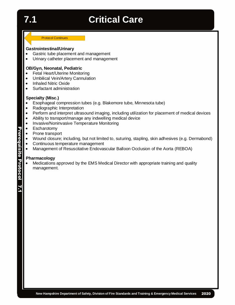
Pre
req
uis
ite P
roto
col 7
.1
Critical Care
New Hampshire Department of Safety, Division of Fire Standards and Training & Emergency Medical Services 2020
7.1
Gastrointestinal/Urinary Gastric tube placement and management Urinary catheter placement and management
OB/Gyn, Neonatal, Pediatric Fetal Heart/Uterine Monitoring Umbilical Vein/Artery Cannulation Inhaled Nitric Oxide Surfactant administration
Specialty (Misc.) Esophageal compression tubes (e.g. Blakemore tube, Minnesota tube) Radiographic Interpretation Perform and interpret ultrasound imaging, including utilization for placement of medical devices Ability to transport/manage any indwelling medical device Invasive/Noninvasive Temperature Monitoring Escharotomy Prone transport Wound closure; including, but not limited to, suturing, stapling, skin adhesives (e.g. Dermabond) Continuous temperature management Management of Resuscitative Endovascular Balloon Occlusion of the Aorta (REBOA)
Pharmacology Medications approved by the EMS Medical Director with appropriate training and quality
management.
Protocol Continues

2013
2013
Protocol Continues
Prerequisite Required
This procedure is only to be used by Paramedics or AEMTs who are trained and credentialed to perform immunization by the NH Bureau of EMS and the NH Medical Control Board.
INDICATIONS: Pre-hospital providers may be called upon to provide certain immunizations as necessary to assist state health officials in the event of a public health or public safety incident, or under the written order of a physician.
Non-Patient Specific Orders:
A non-patient specific order authorizes Paramedic or AEMT to administer specified immunizations for a specified period of time to an entire group of persons such as school children, employees, patients of a nursing home, etc. Some examples of non-patient specific orders are:
o Administer influenza vaccine 0.5 ml IM to all incoming freshmen students at X College who are eligible per protocol.
o Administer influenza vaccine 0.5 ml IM to all employees of X organization who request it and who are eligible by protocol.
o Administer influenza vaccine 0.5 ml IM to all X town residents who request it and who are eligible by protocol.
o Administer hepatitis B series to all employees of X organization eligible per protocol.
Immunizations
Many of the immunizations listed in the Centers for Disease Control and Prevention (CDC) guidelines fall under this protocol. The list of authorized immunizations differs for adults and children. For the purposes of immunizations, adults are persons who are 18 years of age or older; children are persons under 18 years of age. Immunizations for adults:
o Acellular pertussis o Diphtheria o Hepatitis A o Hepatitis B o Inactivated polio o Influenza o Measles o Meningococcus o Mumps o Pneumococcus o Rubella o Smallpox vaccine o Tetanus o Varicella
Immunizations for children:o Acellular pertussis o Diphtheria o Haemophilus influenza Type b (hiB) o Hepatitis A o Hepatitis B o Inactivated polio
Pre
req
uis
ite P
roto
col 7
.2Immunization
New Hampshire Department of Safety, Division of Fire Standards and Training & Emergency Medical Services 2020
7.2

o Influenza o Measles o Meningococcus o Mumps o Pneumococcal Conjugate o Rubella o Tetanus o Varicella
Note: NH EMS Medical Director may add immunizations in accordance with the recommendations of the Centers for Disease Control and Prevention and the New Hampshire Department of Health and Human Services.
Administration of Immunizations
The non-patient specific standing order and protocol must be authorized by a physician.
Public Health or Public Safety Incident
Any Paramedic or AEMT may administer immunizations that are authorized by a non-patient specific standing order and protocol as part of an immunization program when the immunization program is instituted as a result of a public health or public safety incident by public health officials.
Protocol requirements
Ensure that the potential immunization recipient is assessed for contraindications to immunizations.
Inform each potential immunization recipient of the potential side effects and adverse reactions, orally and in writing, prior to immunization, and inform each potential immunization recipient, in writing, of the appropriate course of action in the event of an untoward or adverse event. Vaccine Information Statements (VIS), developed by the Centers for Disease Control and prevention (CDC), United States Department of Health and Human Services are recommended for this use. http://www.cdc.gov/vaccines/pubs/vis/
Before the immunization is administered, obtain consent for the immunization from the potential recipient.
In cases of minors and persons incapable of personally consenting to immunization consent may be gained by informing the legally responsible person of the potential side effects and adverse reactions in writing and obtaining a written consent prior to administering the immunization.
Provide to each legally responsible immunization recipient a signed certificate of immunization noting the recipient’s name, date of immunization, address, administering Paramedic or AEMT, immunizing agent, manufacturer and lot number.
Have available on-site medications to treat anaphylaxis including, but not limited to, epinephrine and necessary needles and syringes.
Protocol Continues
Pre
req
uis
ite P
roto
col 7
.2
Immunization
New Hampshire Department of Safety, Division of Fire Standards and Training & Emergency Medical Services 2020
7.2
Protocol Continues

Report all adverse immunization outcomes to the Vaccine Adverse Event Reporting System (VAERS) using the appropriate form from the Centers for Disease Control and Prevention, United States Department of Health and Human Services. https://vaers.hhs.gov/esub/index
Coordinate with program site managers to ensure that the record of all persons immunized includes: the non-patient specific standing order and protocol utilized, recipient’s name, date, address of immunization site, immunization, manufacturer and lot number of administered vaccine(s), and recommendations for future immunizations.
For the administration of the influenza vaccine to adults only it is acceptable to maintain a log of the names, addresses, and phone numbers of all adult patients immunized with the influenza vaccine under non-patient specific orders, in a dated file.
Coordinate with program site managers to ensure that a record is kept of all potential recipients, noting those who declined immunization.
Pre
req
uis
ite P
roto
col 7
.2Immunization
New Hampshire Department of Safety, Division of Fire Standards and Training & Emergency Medical Services 2020
7.2
Protocol Continues
Introduction
The purpose of this section is to reconcile the unique aspects of interfacility transfers with current NH EMS law, licensure, and acute care protocols. It is intended to provide flexibility, when possible, for individual agencies, institutions, and communities to meet their unique needs.
Interfacility Transfer
An interfacility transfer is defined as any EMS ambulance transport from one healthcare facility to another. Examples include hospital-to-hospital, hospital-to-rehabilitation, and hospital-to-long-term care (Guide for interfacility patient Transfer, NHTSA, April 2006).
Transferring Facility Responsibilities
Certify benefits of transfer outweigh all expected risks. Ensure that patient has an accepting provider and bed assignment at destination facility. Transferring provider must ensure ongoing care will be sufficient and appropriate, and provide
resources as necessary. Transferring provider point of contact who will be immediately available to serve as medical
control for transporting agency during transfer. Provide complete set of patient care orders for the transporting agency. In any case where the number of patients requiring transport exceeds the number of available
EMS resources, the transferring institution shall decide the order in which patients are transported.
Transporting Agency Responsibilities
Assign personnel and resources that are most appropriate (consider training/experience, environmental factors, equipment needs)
Decline transports when proper resources cannot or will not be provided and/or their level of training/experience is not compatible with patients acuity
Consult medical control as necessary during transport Seek education or information about therapies or medications outside of normal formulary as
necessary
Shared Responsibilities
Assign the appropriate transport agency level for patient transport including sending hospital staff, if necessary (see following pages)
Receive and relay a complete patient care report Ensure every effort has been made to mitigate risk, including environmental factors.
2013
2013
Protocol Continues
Pre
req
uis
ite P
roto
col 7
.3
Interfacility Transfers
New Hampshire Department of Safety, Division of Fire Standards and Training & Emergency Medical Services 2020
7.3
Nothing in this protocol shall preclude EMS personnel from providing any medication or therapy that is already within their scope of practice unless it is explicitly forbidden by the transferring facility provider’s written orders for transport.
If at any time during transport a patient develops new signs/symptoms or has a change in status, EMS personnel shall refer to the appropriate NH EMS Protocol.
If there is a conflict between NH EMS Protocols and the transferring facility provider ’s written orders for transport, the transferring facility provider’s written orders shall prevail.

Transport Agency Levels
EMT AEMT Paramedic Interfacility Transport (PIFT)* Critical Care Teams (CCT)*
At a minimum, 2 licensed EMS providers in the vehicle, of which 1 may be the driver.
*Only to be used by paramedics and EMS units who have been trained and credentialed by the NH Bureau of EMS and the NH EMS Medical Control Board.
Interfacility transfers that are appropriate for EMT or AEMT level of care do not require additional levels of credentialing beyond training requirements defined in the NH EMS protocols and by Saf-C 5900.
CAPABILITIES Pre
req
uis
ite P
roto
col 7
.3Interfacility Transfers
New Hampshire Department of Safety, Division of Fire Standards and Training & Emergency Medical Services 2020
7.3
Protocol Continues
Protocol Continues
EMT
Care and treatment of stable patients. Therapies within the EMT scope of practice LINK Medications within EMT scope of practice LINK Non-invasive monitoring (BP, HR, RR, Spo2, EtCo2, temperature). Previously inserted Foley catheter, suprapubic tube, established feeding tube (NG,
PEG, J-tube not connected to infusion or suction). Saline lock permitted. Chest tube capped and without need for suction during transport. Maintenance of stable, long term ventilated patients with any mode of ventilation so
long as the patient is familiar and capable of operating the equipment OR patient is accompanied by a care provider who is capable of the same
Advanced EMT
Care and treatment of stable patients. Therapies within the AEMT scope of practice LINK Medications within AEMT scope of practice, with or without pump LINK Any crystalloid infusion Patient-controlled analgesic (PCA) pump. Cardiac monitoring with non-cardiac diagnoses (4 lead ECG as vital sign, non-
interpretive) with no anticipated need for ACLS intervention. CPAP
A
E
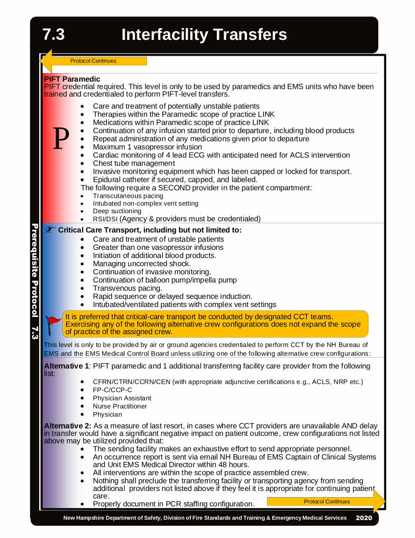
PIFT ParamedicPIFT credential required. This level is only to be used by paramedics and EMS units who have been trained and credentialed to perform PIFT-level transfers.
Care and treatment of potentially unstable patients Therapies within the Paramedic scope of practice LINK Medications within Paramedic scope of practice LINK Continuation of any infusion started prior to departure, including blood products Repeat administration of any medications given prior to departure Maximum 1 vasopressor infusion Cardiac monitoring of 4 lead ECG with anticipated need for ACLS intervention Chest tube management Invasive monitoring equipment which has been capped or locked for transport. Epidural catheter if secured, capped, and labeled.The following require a SECOND provider in the patient compartment: Transcutaneous pacing Intubated non-complex vent setting Deep suctioning RSI/DSI (Agency & providers must be credentialed)
🚁 Critical Care Transport, including but not limited to:
Care and treatment of unstable patients Greater than one vasopressor infusions Initiation of additional blood products. Managing uncorrected shock. Continuation of invasive monitoring. Continuation of balloon pump/impella pump Transvenous pacing. Rapid sequence or delayed sequence induction. Intubated/ventilated patients with complex vent settings
This level is only to be provided by air or ground agencies credentialed to perform CCT by the NH Bureau of EMS and the EMS Medical Control Board unless utilizing one of the following alternative crew configurations :
Alternative 1: PIFT paramedic and 1 additional transferring facility care provider from the following list:
CFRN/CTRN/CCRN/CEN (with appropriate adjunctive certifications e.g., ACLS, NRP etc.) FP-C/CCP-C Physician Assistant Nurse Practitioner Physician
Alternative 2: As a measure of last resort, in cases where CCT providers are unavailable AND delay in transfer would have a significant negative impact on patient outcome, crew configurations not listed above may be utilized provided that:
The sending facility makes an exhaustive effort to send appropriate personnel. An occurrence report is sent via email NH Bureau of EMS Captain of Clinical Systems
and Unit EMS Medical Director within 48 hours. All interventions are within the scope of practice assembled crew. Nothing shall preclude the transferring facility or transporting agency from sending
additional providers not listed above if they feel it is appropriate for continuing patient care.
Properly document in PCR staffing configuration.
Pre
req
uis
ite P
roto
col 7
.3
Interfacility Transfers
New Hampshire Department of Safety, Division of Fire Standards and Training & Emergency Medical Services 2020
7.3
It is preferred that critical-care transport be conducted by designated CCT teams. Exercising any of the following alternative crew configurations does not expand the scope of practice of the assigned crew.
Protocol Continues
P
Protocol Continues

Definitions
Unstable Patient: A critically ill or injured patient who cannot be stabilized at the transporting facility, who is deteriorating or likely to deteriorate during transport. (From “Guide for Interfacility Patient Transfer,” NHTSA.)
Potentially Unstable: A critically ill or injured patient who is currently stable (as defined below) but whose disease process will likely lead to instability or an acute change in condition enroute.
Stable Patient: Hemodynamically stable patient with a secure airway and who is NOT in acute distress or likely to deteriorate during transport
Resources: Could refer to personnel, equipment, medications or therapies. Sufficient & Appropriate: Transferring facilities are responsible for the coordination of
ongoing care during transfer until the patient arrives at the destination facility. Patient must continue receiving care that is commensurate with their condition and potential for deterioration throughout transfer within the limits of the system. This may mean providing additional transferring facility or transporting agency personnel, up to and including physicians if necessary .
Non-complex vent settings: Volume or pressure modes of ventilation provided that:o No inverse I:E ratioso No PEEP > 20 cmH20o No pediatric patients < 5 years of ageo No High frequency oscillationo No Mode of ventilation without apnea backupo Complex vent settings: Any mode of ventilation outside the above parameters.
Pre
req
uis
ite P
roto
col 7
.3Interfacility Transfers
New Hampshire Department of Safety, Division of Fire Standards and Training & Emergency Medical Services 2020
7.3
Protocol Continues
Protocol Continues

Pre
req
uis
ite P
roto
col 7
.3
Interfacility Transfers
New Hampshire Department of Safety, Division of Fire Standards and Training & Emergency Medical Services 2020
7.3
Protocol Continues
EMT
StableAEMT
StableCCT
UnstablePIFT
Potentially Unstable
q EMT therapiesq EMT medicationsq Vital signsq EtCO2q Temperature
monitoringq Foley catheterq Suprapubic
catheterq Feeding tube with
no need to access or adjust
q Saline lockq Capped chest
tubeq Maintenance of
stable, long term ventilated patients with any mode of ventilation so long as the patient is familiar and capable of operating the equipment OR patient is accompanied by a care provider who is capable of the same
q AEMT therapiesq AEMT
Medicationsq Any crystalloid
infusionq Patient-controlled
analgesic (PCA) pump that is locked
q Cardiac monitoring with non-cardiac diagnoses (4 lead ECG as vital sign, non-interpretive) with no anticipated need for ACLS intervention.
q CPAP
Including but not limited to:q Multiple vasoactive
medications/pressorsq Initiation of
additional blood products
q Managing uncorrected shock.
q Continuation of invasive monitoring.
q Continuation of balloon pump/impella pump
q Transvenous pacing.q Intubated/ventilated
patients with complex vent settings
q Paramedic therapiesq Paramedic medicationsq Any infusion started prior to
departureq Repeat administration of
any medications given prior to departure
q Max 1 vasopressorq Continuation of blood or
blood productsq Cardiac monitoring of 4
lead ECG with anticipated need for ACLS intervention
q Serial 12 leadsq Chest tube managementq Invasive monitoring
equipment which has been capped or locked for transport.
q Epidural catheter if secured, capped, and labeled.
The following require a SECOND provider in the patient compartment:q Transcutaneous pacing
cardioversionq Intubated/sedated patientsq Deep suctioningq RSI/DSI*q Non-complex vent settings
Transport Levels
Crew Options: CCT (Air/Ground) PIFT & hospital staff:o 1 CFRN/CTRN oro 1 CCRN/CEN oro 1 FPC-C/CCP-C oro 1 NP/PA oro 1 Physician
Last Resorto Any other
appropriate crew

2013
Indications:
EMS units may choose to stock their ambulances with Naloxone administration kits to be left at the scene where a suspected overdose patient was treated. Routine Patient Care Consider leaving behind a naloxone administration kit, and instructions, with a
patient or household member in the following circumstances:o When treating and transporting a patient who is suspected of an overdoseo When treating a patient who is suspected of an overdose, but transport is
refused
Pre
req
uis
ite P
roc
edu
re 7
.4
Leave – Behind Naloxone
New Hampshire Department of Safety, Division of Fire Standards and Training & Emergency Medical Services 2020
7.4
This prerequisite protocol is only to be used by EMS Units and providers who are authorized by the NH Bureau of EMS.
EA
P
//
EMR/EMT/AEMT/Paramedic Standing Orders
PEARLS:
Leave-behind naloxone administration kits must be separate from the medication used for patient care.
Naloxone administration kits should not be left at the scene where a patient has a known allergy to Naloxone or kit constituents.
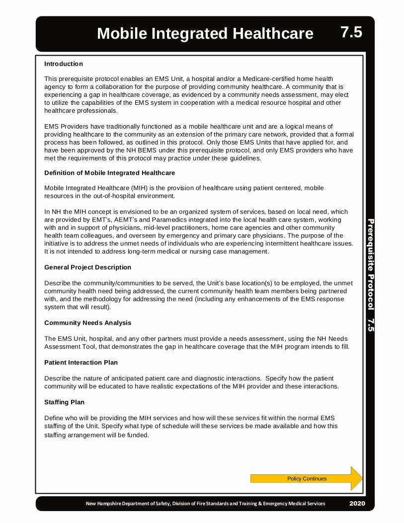
2013
2013
Policy Continues
Introduction
This prerequisite protocol enables an EMS Unit, a hospital and/or a Medicare-certified home health agency to form a collaboration for the purpose of providing community healthcare. A community that is experiencing a gap in healthcare coverage, as evidenced by a community needs assessment, may elect to utilize the capabilities of the EMS system in cooperation with a medical resource hospital and other healthcare professionals.
EMS Providers have traditionally functioned as a mobile healthcare unit and are a logical means of providing healthcare to the community as an extension of the primary care network, provided that a formal process has been followed, as outlined in this protocol. Only those EMS Units that have applied for, and have been approved by the NH BEMS under this prerequisite protocol, and only EMS providers who have met the requirements of this protocol may practice under these guidelines.
Definition of Mobile Integrated Healthcare
Mobile Integrated Healthcare (MIH) is the provision of healthcare using patient centered, mobile resources in the out-of-hospital environment.
In NH the MIH concept is envisioned to be an organized system of services, based on local need, which are provided by EMT’s, AEMT’s and Paramedics integrated into the local health care system, working with and in support of physicians, mid-level practitioners, home care agencies and other community health team colleagues, and overseen by emergency and primary care physicians. The purpose of the initiative is to address the unmet needs of individuals who are experiencing intermittent healthcare issues. It is not intended to address long-term medical or nursing case management.
General Project Description
Describe the community/communities to be served, the Unit’s base location(s) to be employed, the unmet community health need being addressed, the current community health team members being partnered with, and the methodology for addressing the need (including any enhancements of the EMS response system that will result).
Community Needs Analysis
The EMS Unit, hospital, and any other partners must provide a needs assessment , using the NH Needs Assessment Tool, that demonstrates the gap in healthcare coverage that the MIH program intends to fill.
Patient Interaction Plan
Describe the nature of anticipated patient care and diagnostic interactions. Specify how the patient community will be educated to have realistic expectations of the MIH provider and these interactions.
Staffing Plan
Define who will be providing the MIH services and how will these services fit within the normal EMS staffing of the Unit. Specify what type of schedule will these services be made available and how this staffing arrangement will be funded.
Pre
req
uis
ite P
roto
col 7
.5Mobile Integrated Healthcare
New Hampshire Department of Safety, Division of Fire Standards and Training & Emergency Medical Services 2020
7.5

Training Plan
Describe what training will be provided to enable the providers to deliver the services described above. List the objectives and outcomes of the training plan. Document who is responsible for training oversight and coordination and their qualifications.
There must be a continuing education and credentialing process in place, with documentation of each EMS Provider’s participation in it. Such a process shall be approved by the EMS Unit ’s Medical Director(s).
Quality Management Program and Data Collection
The EMS Unit shall conduct a quality management (QM) program specifically for the community healthcare program. The QM program will incorporate all the components of an EMS QM program as specified in Administrative Rule Saf-C 5923.
Describe what data demonstrates the need for this project, if any. Describe the data to be collected to demonstrate the impact of this project on the population served. Describe the data reporting plan and how the NH Bureau of EMS will be included in it.
Documentation
The EMS Provider may at any time, using their own discretion, decide to activate the 911 system for emergency treatment and transport to appropriate care.
Electronic patient care reports of all community healthcare patient encounters must be submitted to the requesting medical practice according to policies developed in coordination between the EMS Unit, MRH, collaborating home health agency and medical practice. Copies of these records shall be maintained by the EMS Unit, and be available for review by the NHBEMS.
The EMS Unit will participate in electronic data collection as required by the NHBEMS.
Medical Direction
Must establish a collaborative working relationship between the EMS Physician Medical Director or designee, who will be responsible for operations and continuous quality improvement, and a primary care provider providing medical direction for MIH services.
Pre
req
uis
ite P
roto
col 7
.5
Mobile Integrated Healthcare
New Hampshire Department of Safety, Division of Fire Standards and Training & Emergency Medical Services 2020
7.5
Protocol Continues

2013
Rapid Sequence Intubation (RSI)
P
This procedure is only to be used by paramedics who are trained and credentialed to perform RSI. This protocol provides a brief outline of the scope of the RSI paramedic but is not comprehensive of the entire RSI procedure. For full RSI guidelines refer to the New Hampshire Prehospital RSI Manual. The guidelines in this manual are part of the RSI protocol and are incorporated in this protocol by reference.
PARAMEDIC - PREREQUISITES REQUIRED - ADULT ONLY
SUCCINYLCHOLINE CONTRAINDICATIONS: Extensive recent burns or crush injuries > 24 hours old. Known or suspected hyperkalemia. History of malignant hyperthermia.
Pre
req
uis
ite P
roto
col 7
.6
New Hampshire Department of Safety, Division of Fire Standards and Training & Emergency Medical Services2020
7.6
Each RSI procedure must be performed in a controlled fashion and must involve careful planning and preparation. RSI requires at least one RSI credentialed paramedic and one credentialed RSI assistant or non-RSI paramedic. Intubation must be performed by the most appropriate provider as determined by the RSI paramedic leading the call. After intubation, the RSI paramedic must remain with the patient at all times unless there are extenuating circumstances (mass casualty, etc.) and ensure that adequate staff remain.RSI may only be performed on adults (i.e., patients who are taller than a length based resuscitation tape).MedicationsThe correct medication regimen should be chosen on a case-by-case basis by the RSI paramedic and care team. Weight-based dosages are listed below. Dosages for all medications are based on actual body weight. Use of a dosing chart with precalculated dosage ranges such as the one reproduced below is recommended; dosing charts reduce cognitive load and risk of error.
Premedication (if indicated) Fentanyl 2 mcg/kg IV at least three minutes prior to induction
Induction Ketamine 2 mg/kg IV or 4 mg/kg IM (max 500 mg) (only if performing
Delayed Sequence Intubation)o For elderly, shock, or risk of hypotension: 1 mg/kg IV or 2 mg/kg IM
OR Etomidate 0.3 mg/kg IV, maximum single dose 30 mg
o For elderly, shock, or risk of hypotension: 0.15 mg/kg IVParalysis
Rocuronium 1 mg/kg IVOR Succinylcholine 1.5 mg/kg IV, maximum 150
Protocol Continues
Sample Dosing Chart:IMPORTANT: Chart must be recalculated for the medication concentrations used by your service.
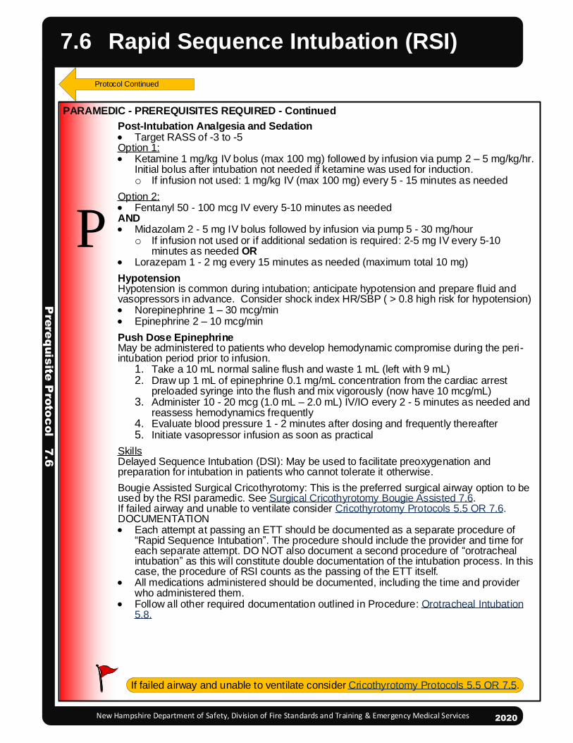
Pre
req
uis
ite P
roto
col 7
.6
Rapid Sequence Intubation (RSI)
New Hampshire Department of Safety, Division of Fire Standards and Training & Emergency Medical Services
Post-Intubation Analgesia and Sedation Target RASS of -3 to -5 Option 1: Ketamine 1 mg/kg IV bolus (max 100 mg) followed by infusion via pump 2 – 5 mg/kg/hr.
Initial bolus after intubation not needed if ketamine was used for induction.o If infusion not used: 1 mg/kg IV (max 100 mg) every 5 - 15 minutes as needed
Option 2: Fentanyl 50 - 100 mcg IV every 5-10 minutes as neededAND Midazolam 2 - 5 mg IV bolus followed by infusion via pump 5 - 30 mg/hour
o If infusion not used or if additional sedation is required: 2-5 mg IV every 5-10 minutes as needed OR
Lorazepam 1 - 2 mg every 15 minutes as needed (maximum total 10 mg)HypotensionHypotension is common during intubation; anticipate hypotension and prepare fluid and vasopressors in advance. Consider shock index HR/SBP ( > 0.8 high risk for hypotension) Norepinephrine 1 – 30 mcg/min Epinephrine 2 – 10 mcg/minPush Dose EpinephrineMay be administered to patients who develop hemodynamic compromise during the peri-intubation period prior to infusion.
1. Take a 10 mL normal saline flush and waste 1 mL (left with 9 mL)2. Draw up 1 mL of epinephrine 0.1 mg/mL concentration from the cardiac arrest
preloaded syringe into the flush and mix vigorously (now have 10 mcg/mL)3. Administer 10 - 20 mcg (1.0 mL – 2.0 mL) IV/IO every 2 - 5 minutes as needed and
reassess hemodynamics frequently4. Evaluate blood pressure 1 - 2 minutes after dosing and frequently thereafter5. Initiate vasopressor infusion as soon as practical
SkillsDelayed Sequence Intubation (DSI): May be used to facilitate preoxygenation and preparation for intubation in patients who cannot tolerate it otherwise.Bougie Assisted Surgical Cricothyrotomy: This is the preferred surgical airway option to be used by the RSI paramedic. See Surgical Cricothyrotomy Bougie Assisted 7.6.If failed airway and unable to ventilate consider Cricothyrotomy Protocols 5.5 OR 7.6.DOCUMENTATION Each attempt at passing an ETT should be documented as a separate procedure of
“Rapid Sequence Intubation”. The procedure should include the provider and time for each separate attempt. DO NOT also document a second procedure of “orotracheal intubation” as this will constitute double documentation of the intubation process. In this case, the procedure of RSI counts as the passing of the ETT itself.
All medications administered should be documented, including the time and provider who administered them.
Follow all other required documentation outlined in Procedure: Orotracheal Intubation 5.8.
P
Protocol Continued
PARAMEDIC - PREREQUISITES REQUIRED - Continued
2020
7.6
If failed airway and unable to ventilate consider Cricothyrotomy Protocols 5.5 OR 7.5.

Rapid Sequence Intubation (RSI)P
rere
quis
ite P
roto
col 7
.6
New Hampshire Department of Safety, Division of Fire Standards and Training & Emergency Medical Services2020
7.6

2013
INDICATIONS:Inability to adequately oxygenate and ventilate using less invasive methods
CONTRAINDICATIONS: Ability to oxygenate and ventilate using less invasive measures Age less than 12 years old
EQUIPMENT: Chlorhexidine #10 blade scalpel Bougie 6.0 mm endotracheal tube 10 ml Syringe BVM Quantitative ETCO2
PROCEDURE:1. Position the patient supine and extend the neck as needed to improve anatomic view.2. Prep neck with Chlorhexidine 3. The provider performing the procedure should be on the side of the patient corresponding to their dominant hand (i.e., right handed provider to the right of the patient).4. While resting dominant hand on patient's sternum, make an approximately 3 cm vertical incision, 0.5 cm deep, through the skin and fascia. Incision should start just above the thyroid cartilage and extend below the cricoid ring. With finger, dissect tissue and locate the cricothyroid membrane.5. Make approximately a 1.5 cm horizontal incision through the cricothyroid membrane.6. With your finger, bluntly dilate the opening through the cricothyroid membrane.7. Insert the bougie curved-tip first through the incision and angled towards the patient’s feet.8. Advance the bougie into the trachea feeling for “clicks” of tracheal rings and until “hold up” when it cannot be advanced any further. This confirms tracheal position.9. Advance a 6.0 mm endotracheal tube (ensure all air aspirated out of cuff) over the bougie and into the trachea.10.Remove bougie while stabilizing ETT ensuring it does not become dislodged11.Inflate the cuff with 5 – 10 ml of air.12.Confirm appropriate proper placement by symmetrical chest-wall rise, auscultation of equal breath sounds over the chest and a lack of epigastric sounds with ventilations using bag-valve-mask, condensation in the ETT, and quantitative waveform capnography.13.Secure the ETT.14.Reassess tube placement frequently, especially after movement of the patient.15.Ongoing monitoring of ETT placement and ventilation status using waveform capnography is required for all patients.
PARAMEDIC - PREREQUISITE REQUIRED– ADULT
Pre
req
uis
ite P
roc
edu
re 7
.7
Surgical Cricothyrotomy
Bougie Assisted — ADULT
New Hampshire Department of Safety, Division of Fire Standards and Training & Emergency Medical Services 2020
7.7
P cricothyroid membrane
Written notification will be provided to the Medical Resource Hospital’s EMS Medical Director, Hospital EMS Coordinator, and Bureau of EMS within 48 hours of an event. Use of this procedure documented under “Procedures Used” in the Patient Care Report constitutes notification of the Bureau of EMS.

Po
licy 8
.0
2013
EMS personnel may request Air Medical Transport (AMT) when operational and/or clinical conditions are present that would benefit from decrease in time to definitive care and/or advanced clinical capabilities offered by the AMT team.
The use of AMT is determined by the prehospital provider with the highest medical level providing patient care. It should not be determined by police or bystanders.
AMT does not require approval of on-line Medical Control. However, if in doubt of the appropriateness of a patient for AMT, please contact Medical Control as soon as possible.
Operational Conditions· When a patient meets the defined clinical criteria listed below and the ground transport time to
the closest hospital capable of providing definitive care (e.g., Level I or 2 trauma hospital, PCI center, stroke center) exceeds the ETA of air medical transport, OR
· Patient location, weather, or road conditions preclude the use of ambulance, OR· Multiple patients are present that will exceed the capabilities of local hospital and agencies.
Clinical Conditions· Severe respiratory compromise with respiratory arrest or abnormal respiratory rate.· Circulatory insufficiency: sustained systolic blood pressure < 90mmHg or MAP of < 65 mmHg in
adults, age appropriate hypotension in children or other signs of shock. · Neurologic compromise: Patient cannot follow commands (GCS motor component ≤ 5) or total
GCS ≤ 13. If the patient’s neurologic status improves above these limits, consider canceling the helicopter and transporting to the local hospital.
· Trauma: All penetrating injuries to head, neck, torso, and extremities proximal to elbow or knee; chest wall instability or deformity (e.g., flail chest); two or more proximal long-bone fractures; crushed, degloved, mangled, or pulseless extremity; amputation proximal to wrist or ankle; pelvic fracture; open or depressed skull fracture; paralysis.
· Major burns with greater than 20% BSA and/or inhalation injury with risk of airway compromise.· Electrocution injuries with loss of consciousness, arrhythmia, or any respiratory abnormality.· STEMI: If 12-lead ECG indicates a STEMI (e.g., machine reads ***Acute MI Suspected*** and/or
Paramedic interpretation), per your local STEMI plan.· Stroke: 1 or more abnormal signs of the stroke scale; per local stroke plans.· Critically ill children, including those with acute decompensation of chronic and/or special
healthcare needs.
Additional Notes· Patients with an uncontrolled airway or uncontrollable hemorrhage should be brought to the
nearest hospital unless advanced life support (ALS) service (by ground or air) can intercept in a more timely fashion.
· AMT is NOT indicated for patients in cardiac arrest. Should the patient go into cardiac arrest after AMT request the AMT crew may be utilized for resuscitation and stabilization.
· AMT is NOT indicated for a contaminated patient until AFTER decontamination.· AMT may be indicated in a wide range of conditions other than those listed above. In cases
where the patient’s status is uncertain, consult with Medical Control and proceed as directed.· Transfers from ground-ambulance to air-ambulance shall occur at the closest appropriate landing
site, including a hospital heliport, an airport, or an unimproved landing site deemed safe per pilot discretion. In cases where a hospital heliport is used strictly as the ground-to-air ambulance transfer point, no transfer of care to the hospital is implied or should be assumed by hospital personnel, unless specifically requested by the EMS providers.
2013
Air Medical Transport
New Hampshire Department of Safety, Division of Fire Standards and Training & Emergency Medical Services 2020
8.0
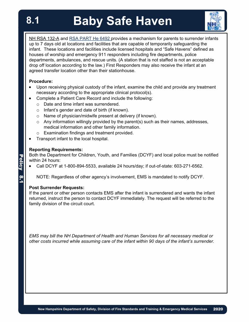
Baby Safe Haven
2013
2013
NH RSA 132-A and RSA PART He 6492 provides a mechanism for parents to surrender infants up to 7 days old at locations and facilities that are capable of temporarily safeguarding the infant. These locations and facilities include licensed hospitals and “Safe Havens” defined as houses of worship and emergency 911 responders including fire departments, police departments, ambulances, and rescue units. (A station that is not staffed is not an acceptable drop off location according to the law.) First Responders may also receive the infant at an agreed transfer location other than their stationhouse.
Procedure:· Upon receiving physical custody of the infant, examine the child and provide any treatment
necessary according to the appropriate clinical protocol(s).· Complete a Patient Care Record and include the following:
o Date and time infant was surrendered.o Infant’s gender and date of birth (if known).o Name of physician/midwife present at delivery (if known).o Any information willingly provided by the parent(s) such as their names, addresses,
medical information and other family information. o Examination findings and treatment provided.
· Transport infant to the local hospital.
Reporting Requirements:Both the Department for Children, Youth, and Families (DCYF) and local police must be notified within 24 hours: · Call DCYF at 1-800-894-5533, available 24 hours/day; if out-of-state: 603-271-6562.
NOTE: Regardless of other agency’s involvement, EMS is mandated to notify DCYF.
Post Surrender Requests:If the parent or other person contacts EMS after the infant is surrendered and wants the infant returned, instruct the person to contact DCYF immediately. The request will be referred to the family division of the circuit court.
EMS may bill the NH Department of Health and Human Services for all necessary medical or other costs incurred while assuming care of the infant within 90 days of the infant’s surrender.
Po
licy 8
.1
New Hampshire Department of Safety, Division of Fire Standards and Training & Emergency Medical Services 2020
8.1

2013
2013
Policy Continues
Purpose: This policy provides guidance for providers concerning the triage, extrication, care and transport for bariatric patients. At times, even a single patient can exceed the capacity of the immediately available resources. Like a multi-system trauma patient, a bariatric patient requires:· Appropriate EMS resources to respond· Appropriate protocols and equipment for the provision of care · Specialized equipment for transfer to the ambulance and transport · Careful selection of the appropriate destination hospital· Pre-alerting of the ED to ensure adequate resources to manage the patientOn scene times may be significantly extended for bariatric patients.For additional County Cache information and assistance in EMS bariatric planning, contact the NH Bureau of EMS at 603-223-4228.
Equipment· Deployment of equipment and procedures shall be done under local or regional operating
guidelines. DefinitionsA bariatric patient is a patient: · Weight exceeds 400 pounds OR· Weight, girth, body contours and/or co-morbidities challenge the ability of a two person
EMS crew to effectively manage.DispatchBariatric Ambulance: Based on dispatch information or previous planning, consider requesting a bariatric transport ambulance to respond to the scene. The arrival onscene of a bariatric ambulance may require between 30 and 90 minutes, and should be requested as soon as it becomes clear that bariatric capabilities may be required. The State of New Hampshire has 10 bariatric equipment caches (1 per county) While standard ambulance stretchers can potentially handle some patients up to 750 pounds or more, the use of a specialized bariatric stretcher increases the ability to provide effective care, is more comfortable for the patient and enhances provider safety.Additional Manpower: Consider requesting additional responders. In general, bariatric patients should be moved with a minimum of personnel. Larger bariatric patients may require additional personnel to participate in moving the patient. For significant extrications, consider designating a Safety Officer to oversee the safety of the operation in conjunction with Incident Command. It may be necessary to remove doors, walls or windows to carry out a safe extrication. The priorities are similar to extrication from a vehicle, although fixed property repair costs might be higher.Paramedic: Consider requesting a paramedic. Even BLS bariatric patients present unique treatment challenges which may benefit from a higher level of care.Medical CareMedical care must take into account the unique challenges presented by the bariatric patient as well as the likelihood of extended on-scene times. Providers should use appropriately sized equipment to the extent it is available or can be readily obtained. For example, an appropriately sized blood pressure cuff will need to be used and intramuscular injection will be given with a longer needle.If there are significant barriers to removing the patient from the structure in a timely manner (long narrow stairs, patient in the attic, etc.), there may be situations where EMS will provide extended care to the patient at the scene. In such cases, consult Medical Control and consider use of the extended care protocols.
Po
licy 8
.2Bariatric Triage, Care & Transport
New Hampshire Department of Safety, Division of Fire Standards and Training & Emergency Medical Services 2020
8.2

Transfer to AmbulanceSpecialized equipment will be needed to transfer the patient safely from the scene to the ambulance stretcher for transport. If a bariatric equipment cache is utilized, both the bariatric ambulance and cache equipment needs to be dispatched.Many services utilize large transfer flats for moving bariatric patients. Be sure before you use any patient transfer device that you understand the procedure for using it safely and that you know the weight limits of the device.
Hospital DestinationEnsure that you select a destination hospital that has the capabilities to care for your patient. Bariatric patients may require specialized hospital stretchers, CT scanners, catheterization laboratory equipment, operating room equipment, etc. It may be appropriate to bypass a local hospital to take the patient to a facility with the capabilities to properly care for the patient. This may even be appropriate in the case of life threatening emergencies if the closer emergency department does not have needed equipment.Pre-notification serves both to ensure that the hospital is capable of caring for the patient and allows hospital staff time for adequate preparation. Communication with the hospital shall be in a professional manner. Respect for the patient's privacy and feelings will match the respect for all EMS patients.
Transport to the Hospital A bariatric stretcher should be used to transport the patient to the hospital and equipment cache transfer devices may be utilized to facilitate transfer of the patient to the hospital stretcher. Be alert to ensure that the stretcher is adequately secured in the patient compartment. Transfer flats or other specialized transfer equipment may be left in place to facilitate transfer of the patient to the hospital stretcher.
Po
licy 8
.2
Bariatric Triage, Care & Transport
New Hampshire Department of Safety, Division of Fire Standards and Training & Emergency Medical Services 2020
8.2
PEARLS· It may be difficult to establish IV and IO access. Consider intramuscular or intranasal as
alternatives for some medications. For IM, ensure that the needle used is sufficiently long.· Weight-based calculations may yield inappropriately large doses in obese patients. Consult
with medical control when in doubt.· Bariatric patients often have decreased functional residual capacity, and are at risk of rapid
desaturation. Extremely obese individuals require more oxygen than non-obese individuals due to their diminished lung capacity. Pulse oximetry may not be reliable due to poor circulation. Even patients without respiratory distress may not tolerate the supine position.
· Bariatric patients may present with severe airway challenges. Carefully plan your approach to the airway, and be prepared with backup airway plans.
· If the patient has had recent bariatric surgery, possible complications may include anemia, dehydration, leakage, ulcers, localized infection, sepsis, etc.
Protocol Continues
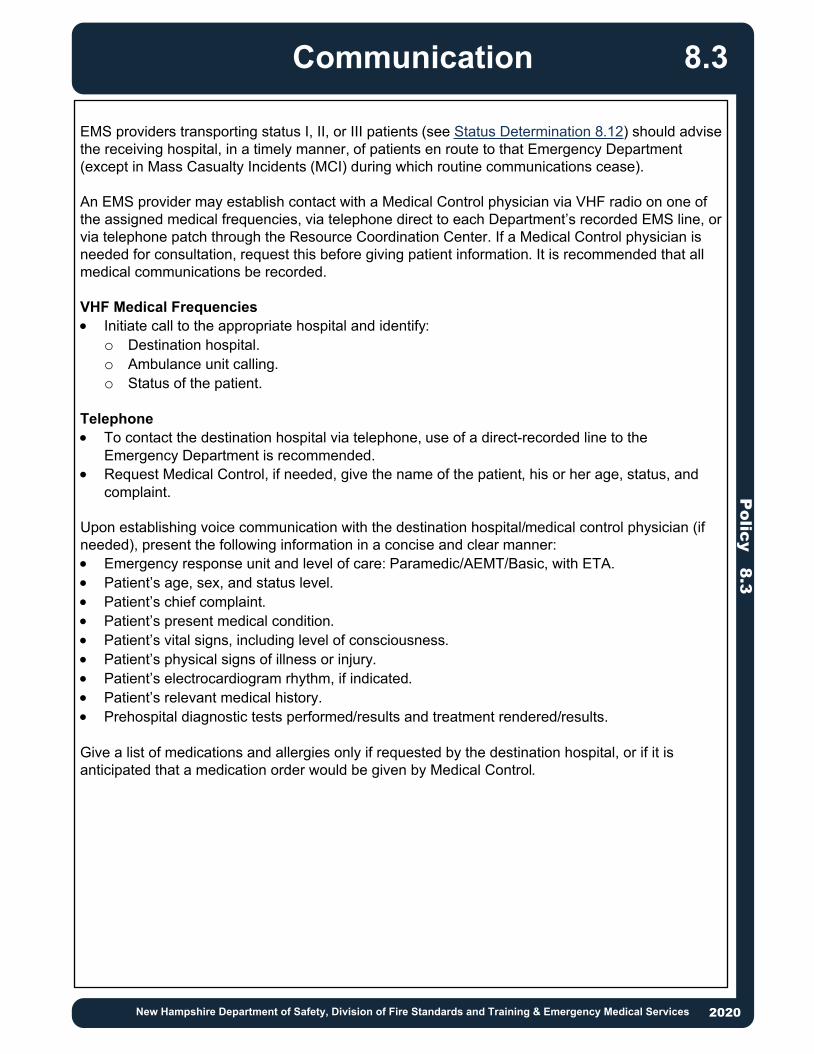
Po
licy 8
.3
2013
EMS providers transporting status I, II, or III patients (see Status Determination 8.12) should advise the receiving hospital, in a timely manner, of patients en route to that Emergency Department (except in Mass Casualty Incidents (MCI) during which routine communications cease).
An EMS provider may establish contact with a Medical Control physician via VHF radio on one of the assigned medical frequencies, via telephone direct to each Department’s recorded EMS line, or via telephone patch through the Resource Coordination Center. If a Medical Control physician is needed for consultation, request this before giving patient information. It is recommended that all medical communications be recorded.
VHF Medical Frequencies· Initiate call to the appropriate hospital and identify:
o Destination hospital.o Ambulance unit calling. o Status of the patient.
Telephone· To contact the destination hospital via telephone, use of a direct-recorded line to the
Emergency Department is recommended. · Request Medical Control, if needed, give the name of the patient, his or her age, status, and
complaint.
Upon establishing voice communication with the destination hospital/medical control physician (if needed), present the following information in a concise and clear manner:· Emergency response unit and level of care: Paramedic/AEMT/Basic, with ETA. · Patient’s age, sex, and status level.· Patient’s chief complaint.· Patient’s present medical condition. · Patient’s vital signs, including level of consciousness.· Patient’s physical signs of illness or injury.· Patient’s electrocardiogram rhythm, if indicated. · Patient’s relevant medical history.· Prehospital diagnostic tests performed/results and treatment rendered/results.
Give a list of medications and allergies only if requested by the destination hospital, or if it is anticipated that a medication order would be given by Medical Control.
2013
Communication
New Hampshire Department of Safety, Division of Fire Standards and Training & Emergency Medical Services 2020
8.3

Po
licy 8
.4
2013
In case of a communications failure with Medical Control due to equipment (cell phone, landline, radio, IHERN) malfunction or incident location, the following will apply:
· EMS personnel may, within the limits of their license, perform necessary ALS procedures that under normal circumstances would require a direct physician order.
· These procedures shall be the minimum necessary to prevent the loss of life or the critical deterioration of a patient’s condition.
· All procedures performed under this order, and the conditions that created the communications failure, need to be thoroughly documented.
· Attempts must be made to establish contact with Medical Control as soon as possible.
· The EMS provider shall provide a written notification pertaining to the communications failure describing the events, including the patient’s condition and treatment given, and referencing the EMS Incident Report. This report must be filed with the Medical Resource Hospital’s EMS Medical Director and/or Hospital EMS Coordinator within 48 hours of the event.
2013
Communications Failure
New Hampshire Department of Safety, Division of Fire Standards and Training & Emergency Medical Services 2020
8.4

Po
licy 8
.5
2013
The word “minor” is a legal term for a person who has not yet reached his/her eighteenth birthday and is under the control of parent(s) or legal guardian. Emancipated minors may make their own determinations regarding medical care and include those minors who are married or members of the armed forces. A minor patient bears the burden of establishing, by legal documentation or otherwise, that he/she is emancipated. New Hampshire recognizes emancipation decrees issued by other states.
Implied ConsentEMS personnel may treat minors under the doctrine of implied consent when the minor’s parent or other authorized representative is unavailable to provide expressed consent. (RSA 153-A:18)
Obtaining Consent:With the exception of life-threatening emergencies, personnel should attempt to contact the minor’s parent or legal guardian to obtain informed consent to treat and transport the child.
Refusal of CareA parent, or legal guardian or other authorized representative may refuse care for a minor and should understand the minor’s medical condition and potential consequences of refusing care. Carefully document all refusals. · When a parent or legal guardian is unavailable, another authorized representative (e.g.,
daycare/school/camp official), who has been expressly authorized by the minor’s parent, may consent to health care treatment. Another adult family member (e.g., grandparent) having custody of the minor may also give consent, see Refusal of Care Protocol 8.15.
· EMS personnel may accept a telephonic refusal of care, provided that they have explained the consequences of refusing care; telephonic refusal of care should be carefully documented.
A minor may not refuse care. When a minor attempts to refuse care and/or transport to the hospital, EMS personnel should enlist the assistance of the police, including requesting that the police place the minor in protective custody. Minors should be restrained only as a last resort.
Special CircumstancesA minor parent who has not yet reached his/her eighteenth birthday may consent to or refuse care on behalf of his or her minor children, provided that the minor parent has the capacity to understand the nature of the treatment and the possible consequences of consenting to or refusing care.
A minor may consent without parental permission for the following care:· An adolescent patient under the age of 18 must give his/her consent for a sexual assault
forensic exam ( "Sexual Assault: An Acute Care Protocol for Medical/Forensic Evaluation", Office of the NH Attorney General, Sixth Edition, 2011).
· Treatment for sexually transmitted diseases at age 14 and older (RSA 141-C:18).· Treatment for drug and alcohol abuse at age 12 and older (RSA 318-B:12-a).
2013
Consent for Treatment of a Minor
New Hampshire Department of Safety, Division of Fire Standards and Training & Emergency Medical Services 2020
8.5

2013
E
EMT/ADVANCED EMT STANDING ORDERS – ADULT & PEDIATRIC
2013
Po
licy 8
.6
Continuity of Care
New Hampshire Department of Safety, Division of Fire Standards and Training & Emergency Medical Services 2020
8.6· EMS providers responding to a 911 emergency may encounter patients with pre-existing
medical devices (e.g., ventilator) or pre-established medication infusions (e.g., antibiotics) that are outside of NH EMS Protocols and beyond the EMS provider’s scope of practice. The medical emergency may be unrelated to the pre-existing medical care (e.g,. chest pain in a patient receiving an infusion) or may relate to the pre-existing care (e.g., problems with a ventilator supporting a patient’s breathing).
· Pre-existing medical care may include ventilators, CPAP, BiPAP, ventricular assist devices (VADs), continuous or intermittent IV medication infusions (analgesics, antibiotics, chemotherapeutic agents, vasopressors, cardiac drugs), and nontraditional out-of-hospital drug infusion routes (subcutaneous infusaports, central venous access lines, direct subcutaneous infusions, self-contained implanted pumps). The type of pre-existing care potentially encountered by EMS providers is extensive.
· The device or medication administration may be supported or maintained by the patient or the patient’s caregiver.
· Routine Patient Care· Consider early consultation with on-line medical control· If the device or infusion is functioning properly and is maintained by an alert/oriented
patient (or caregiver), transport the patient with the device or infusion in place and operating normally.
· If the device or infusion is not functioning properly or may be the cause of the medical emergency, the provider should utilize all appropriate and available resources:o The patient/family/caregiverso Specialty resources available via telephone (e.g., LVAD Coordinator, hospice
nurse or physician), computer, smartphone or telemedicine device or application.o Product literature for the device or infusion (paper or digital)
· EMTs should not continue the administration of a newly initiated, i.e., not pre-existing medication that is outside their scope of practice. o Consider requesting that any healthcare providers or other trained personnel on
scene who are involved in the patient’s pre-existing care (e.g., nurse or physician) accompany the patient and the ambulance during transport to support the device or infusion.
o Request paramedic intercept for any medication outside the EMT or AEMT formulary.
EMS providers are not required to continue treatments which they believe are harmful to the patient or caregivers, (e.g. chemotherapy agents). If an EMS provider is not comfortable with a pre-existing treatment they should seek additional resources or discontinue treatment.
A/
PARAMEDIC STANDING ORDERS – ADULT & PEDIATRIC
P· Any treatment initiated recently or acutely by other healthcare providers
(e.g., urgent care) may be continued. Collaboration between sending providers, EMS, and medical control may be necessary.

2013
2013
If you have been dispatched to a possible crime scene, including motor vehicle incidents, or if you believe a crime has been committed, immediately contact law enforcement.
Protect yourself and other EMS personnel. You will not be held liable for failing to act if a scene is not safe to enter. Once a crime scene is deemed safe by law enforcement, initiate patient contact and medical care if necessary.
· Have all EMS providers use the same path of entry and exit, if feasible. · Do not walk through fluids.· Do not touch or move anything at a crime scene unless it is necessary to do so for patient
care (notify law enforcement prior to moving so if possible).· Avoid moving or touching firearms; notify law enforcement to secure. If necessary to move
a firearm for safety or resuscitation, document location and position.· Observe and document original location of items moved by crew.· When removing patient clothing, leave it intact as much as possible.
o Do not cut through clothing holes made by gunshot or stabbing.· If you remove any items from the scene, such as impaled objects or medication bottles,
document your actions and advise a law enforcement official.· Do not sacrifice patient care to preserve evidence.· Consider requesting a law enforcement officer to accompany the patient in the ambulance
to the hospital.· Document statements made by the patient or bystanders on the EMS patient care report.
o Comments made by a patient or bystanders should be denoted in quotation marks.· Document locations of needle punctures by EMS.· Inform staff at the receiving hospital that this is a “crime scene” patient.· If the patient is obviously dead consistent with Resuscitation Initiation and Termination
Policy, notify law enforcement of decision not to initiate resuscitation/patient care.· At motor vehicle incidents, preserve the scene by not driving over debris, not moving debris
and parking away from tire marks, if feasible.
Po
licy 8
.7Crime Scene
Preservation of Evidence
New Hampshire Department of Safety, Division of Fire Standards and Training & Emergency Medical Services 2020
8.7

2013
2013
Do No Resuscitate (DNR) Orders, Provider Orders for Life Sustaining Treatment (POLST)
and Advanced Directives
Po
licy 8
.8
New Hampshire Department of Safety, Division of Fire Standards and Training & Emergency Medical Services
Policy Continues
Recognized DNR Options in New Hampshire
The following are the only recognized DNR options in New Hampshire:1. “P-DNR” (portable DNR) order: statewide recognized document of any color and/or a “DNR” (Portable DNR) wallet card signed by a physician or Advanced Practiced Registered Nurse (APRN).2. Medical orders form documenting the patient’s name and signed by a physician or APRN and that clearly documents the DNR order.3. DNR bracelet or necklace worn by a patient, inscribed with the patient’s name, date of birth (in numerical form), and “NH DNR” or “NH Do not resuscitate.”4. “POLST” constitutes a DNR if it states ‘This will constitute a DNR Order, and no separate DNR Order will be required.’Note: Under state law, a DNR bracelet or necklace may only be issued to patients who have a valid DNR order.
When a written DNR order is not available but the patient has a DPOAH and the patient’s healthcare agent requests that resuscitation be withheld, contact online Medical Control for guidance.
For patients present or residing In a healthcare facility, the following is also acceptableA DNR order written by a physician or APRN at the nursing home, hospital, or other healthcare facility issued in accordance with the healthcare facility’s policies and procedures.
For Patients Being TransferredAll forms of DNR identified above remain valid during a transfer from one healthcare facility to another.
DNR Orders from Other States EMS providers should honor any DNR order that is substantially similar to the NH statutory form. (see NH form below) Medical orders from other states must be signed by a physician or APRN that clearly documents the DNR order.
Revocation of a DNR Order The following are the only recognized methods for revoking a DNR order:
Patients residing at home A patient residing at home may revoke a DNR order by destroying the DNR order and removing a DNR bracelet or necklace. If the patient lacks the capacity to make health care decisions, the patient’s healthcare agent (under a DPOAH—see below) may revoke the DNR order by destroying the DNR order and removing any DNR bracelet or necklace.
Patients residing in a healthcare facility A patient in a healthcare facility may revoke his or her previous consent to a DNR order by making a written, oral, or other act of communication to the attending physician or APRN or other professional staff of the healthcare facility. For a patient who lacks the capacity to make health care decisions, the patient’s healthcare agent (under a DPOAH—see below) may revoke a DNR order by notifying the attending physician or APRN in writing or, if a witness over the age of 18 is present, orally.
2020
8.8
Neither a Living will nor a Durable Power of Attorney for Healthcare (DPOAH) form is a valid DNR order. Neither a patient’s spouse nor a healthcare agent under a DPOAH may direct EMS providers to withhold resuscitation in the absence of a valid DNR Order.

New Hampshire Department of Safety, Division of Fire Standards and Training & Emergency Medical Services
Procedures not to be PerformedIf there is a valid DNR order and the patient is in cardiac or respiratory arrest, or cardiac or respiratory arrest is imminent, EMS providers should observe the following guidelines:
Do not perform chest compressions. Do not actively assist ventilations via BVM. Do not intubate or place advanced airway devices. Do not defibrillate. Do not administer resuscitation drugs to treat cardiac arrest or the rhythms identified below:
o Ventricular fibrillation. o Pulseless ventricular tachycardia. o Pulseless electrical activity.o Asystole.
Procedures that may be performedIf the patient is not in imminent cardiac or respiratory arrest, all appropriate medical treatment for all injuries, pain, difficult or insufficient breathing, hemorrhage, and/or other medical conditions should be provided despite the presence of a DNR order. Competent patients (and healthcare agents) retain the right to refuse any treatments indicated.
EMS providers MAY perform any other measures, including comfort measures, for these patients, within their scope of practice per the usual treatment guidelines, including but not limited to: Oxygen therapy via nasal cannula, non-rebreather mask, and/or CPAP. Medications for treatment of pain, respiratory distress, dysrhythmias (except for those
identified above). Intravenous fluid therapy for medication access and/or delivery. Mouth or airway suctioning.
NH Statutory DNR FormDo Not Resuscitate Order.As attending physician or APRN of [patient’s name here] and as a licensed physician or Advanced Practice Registered Nurse, I order that this person SHALL NOT BE Resuscitated in the event of cardiac or respiratory arrest.This order has been discussed with [patient’s name here] (or, if applicable, with his/her agent,) [name of DPOAH], who has given consent as evidenced by his/her signature below.Attending physician or APRN name:________________________________________________ Attending physician or APRN signature:_____________________________________________ Address:______________________________________________________________________ Patient signature:_______________________________________________________________ Address:______________________________________________________________________ Agent signature (if applicable):_____________________________________________________ Address: _____________________________________________________________________
Policy Continued
Policy Continues
Po
licy 8
.8
2020
8.8Do No Resuscitate (DNR) Orders,
Provider Orders for Life Sustaining Treatment (POLST) and Advanced Directives
8.8

Do No Resuscitate (DNR) Orders, Provider Orders for Life Sustaining Treatment (POLST)
and Advanced Directives
Po
licy 8
.8
New Hampshire Department of Safety, Division of Fire Standards and Training & Emergency Medical Services
Durable Power of Attorney for HealthcareUnder a Durable Power of Attorney for Healthcare, a patient may designate another person—a healthcare agent—to make health care decisions for themselves.
Before a healthcare agent may make decisions on behalf of the patient, the patient’s attending physician or APRN must certify in writing that the patient lacks capacity (this certification is filed within the patient’s medical record).
A patient who, in the clinical judgment of the EMS provider, retains the capacity to make health care decisions, shall direct his or her health care, even where a healthcare agent has been appointed. That is, EMS providers shall follow the wishes of the patient rather than the healthcare agent unless the patient lacks the capacity to make health care decisions.
The healthcare agent must make an informed decision. It is generally advisable for EMS providers to perform at least a preliminary assessment and inform the healthcare agent of the options for caring for the patient.
Note: in the absence of a valid DNR order, a healthcare agent does not have the authority to direct prehospital providers to withhold resuscitation in the event of a cardiac arrest. When a written DNR order is not available and a DPOAH is present and requests that resuscitation be withheld, contact online Medical Control for guidance.
Living WillA Living Will is intended to address patients who have been admitted to a healthcare facility. Living Wills rarely, if ever, have application in the prehospital environment.
POLST (Provider Orders for Life-Sustaining Treatment)Section AThe POLST constitutes a DNR if it states ‘This will constitute a DNR Order, and no separate DNR Order will be required.’ Otherwise, if the patient has indicated they do not want resuscitation but does not have a separate valid DNR order, contact Medical Control for guidance
Section BWhen confronted with a seriously ill patient who has a POLST form (yellow form),and is not in cardiac arrest: see POLST Appendix A5 If “Full Treatment” box is checked: Use all appropriate measures to stabilize/resuscitate patient. If “Selective Interventions” box is checked: The maximum respiratory interventions are non-
rebreather mask, CPAP, and suctioning. All appropriate IV medications may be utilized. No electrical therapies are to be provided.
If “Comfort-focused Care” box is checked: Limit respiratory interventions to non-rebreather mask, suctioning and treatment of airway obstruction, as needed. Medications to relieve pain or discomfort may be utilized.
Note: Section C refers to IV therapy for hydration and nutrition. Advanced EMTs and Paramedics may start an IV for the purpose of medication administration outlined in Section B.
Policy Continued
PEARLS: Your decision to withhold resuscitation is protected under the New Hampshire DNR law as
long as it is based on the good faith belief that you have been presented with a valid DNR order or DNR jewelry.
2020
8.8

Po
licy 8
.9
2013
2013
Hospice
New Hampshire Department of Safety, Division of Fire Standards and Training & Emergency Medical Services 2020
8.9
Protocol Continues
EMT/ADVANCED EMT STANDING ORDERS
EA/
· Routine Patient Care.· Contact the hospice team (preferred) or Medical Control to coordinate care and
determine administration of hospice kit medications.· Consider paramedic response for medication administration.· Breakthrough Pain: Suggest administration of breakthrough pain medication by
patients / families. For pain of sudden onset, seek to determine and ameliorate or treat the underlying cause (e.g., pathological fracture).
· Anxiety: Consider potential causes for patient’s anxiety, such as increased pain and shortness of breath.
· Dyspnea: Administer oxygen via nasal cannula to relieve shortness of breath and achieve a respiration rate of < 20. Use a fan to blow air directly at the patient’s face.
· Constipation: Suggest administration of constipation medication by patient/family. · Nausea/Vomiting: Suggest administration of nausea medication by patient/family. · Terminal Secretions: Reassure family that noisy breathing is generally not
distressing to the patient. Suggest administration of medication by patients/families.· Terminal Dehydration: Moisten lips with petroleum jelly; use artificial saliva/mouth
sponges and ice chips.· Confusion/Delirium: Speak slowly and calmly to the person. Remind the patient of
where they are, and who you are. Avoid contradicting the patient’s statements. Ensure a patient’s hearing aid and glasses are available. Limit activity/noise in the room.
This protocol is specific to those patients enrolled in Hospice. Treatment should be based on consultation with their Hospice team.
IntroductionThe treatment goals of hospice patients differ significantly from those of other patients. Maintaining patient dignity and quality of life, rather than treating medical conditions, is the objective. If a specific cause of discomfort is identified (e.g., bronchospasm), traditional EMS treatment may be appropriate depending on the invasiveness of the therapy and the patient’s preferences. Hospice patients generally wish to remain at home and transport to the hospital should be the exception.If the patient is unable to make medical decisions and the hospice team cannot be contacted, determine the patient’s wishes and contact Medical Control.
EMS providers should avoid the following interventions:· Sirens, lights or aggressive interventions with family or caregivers.· IV therapy (except where other forms of medication administration are not possible). · Cardiac resuscitation: CPR, resuscitation medications, BVM ventilations.· Cardiac pacing, cardioversion, and defibrillation.· Hospice patients should not be transported to the hospital except where transport is specifically
requested by the patient or his healthcare agent or surrogate, and preferably only after consultation with the hospice team and exhaustion of other treatment pathways that do not require transport to the hospital.
· Many hospice patients will have a hospice comfort kit that contains medications that patient’s caregivers are instructed to use to treat commonly encountered medical issues.

Po
licy 8
.9
Hospice
New Hampshire Department of Safety, Division of Fire Standards and Training & Emergency Medical Services 2020
Protocol Continues
P
PARAMEDIC STANDING ORDERSConsider following the written orders for medications in hospice kit.As an adjunct to the hospice kit medication consider:Breakthrough Pain: · See Pain Protocol 2.16 (All IV formulated opiates may be given PO for hospice
patients.)Anxiety: · Midazolam: 2.5 mg IN, repeat every 10 - 15 minutes as needed to a maximum of
6mg· Lorazepam: 0.25 - 2 mg PO or SL.Dyspnea:· Morphine or other opiate, dosing per Pain Protocol 2.16, maintaining respiratory
rate above 8 bpm.· Bronchospasm: See Asthma/COPD 2.3, subject to patient’s goals.· Heart Failure: See Congestive Heart Failure Protocol 3.3, subject to patient’s
goals.Nausea / Vomiting:· See Nausea/Vomiting Protocol 2.11
PEARLS· Breakthrough Pain assessment and management is important in patients with advanced
disease as they may have a high burden of pain, be opiate tolerant, and already be receiving high doses of opioids.
· Anxiety ranges from mild to severe, is common in patients nearing death, and should be treated promptly.
· Terminal Secretions are noisy, gurgling respirations caused by secretions accumulating in the lungs or oropharynx.
· Terminal Dyspnea is exhibited by patients that are expected to die within hours to days. Individuals experiencing dyspnea often experience heightened anxiety.
· Constipation is a frequent cause of nausea and vomiting. Opioid-related constipation is dose-related, and patients do not develop tolerance to this side effect. Surgical treatment is often not appropriate.
· Nausea / Vomiting can be extremely debilitating symptoms at the end of life. Effective control of nausea can be achieved in most patients.
· Fever and Infection treatment should be guided by an understanding of where the patient is in the dying trajectory and goals of care. Overwhelming sepsis may be a sign of active death not to be reversed.
· Delirium is common at end of life and is often caused by a combination of medications, dehydration, infections or hypoxia. It is distressing to families. It often heralds the end of life and may require active sedation.
8.9

2013
2013
Policy Continues
Blood Borne PathogensAssume that all bodily fluids and tissues are potentially infectious and personnel must protect themselves accordingly by use of appropriate Body Substance Isolation (BSI) and approved procedures.
Transmission of pathogens has been shown to occur when infected blood or Other Potentially Infectious Materials (OPIM) enter another individual’s body through skin, mucous membrane, or parenteral contact.
Screen symptomatic patients for out of country travel within the past month or close contact with another symptomatic individual who has recently traveled out of the country. If possible, determine where patient or contacts have recently traveled. Provide early notification to receiving hospital.
Body Substance Isolation (BSI) Procedures.
· BSI procedures include using protective barriers (such as gloves, masks, goggles, etc.), thorough hand washing, and proper use and disposal of needles and other sharp instruments.
· Centers for Disease Control and Prevention Guidelines for hand hygiene include:o When hands are visibly dirty, contaminated, or soiled, wash with soap and water. o If hands are not visibly soiled, use an alcohol-based hand sanitizer for routinely
decontaminating hands.· Personnel with any open wounds should refrain from all direct patient care and from
handling patient-care equipment, unless they can ensure complete isolation of these lesions and protection against seepage.
· Personnel who are potentially at risk of coming into contact with blood or OPIM are encouraged to obtain appropriate vaccines to decrease the likelihood of disease and transmission.
Body Substance Exposure - Procedures and Considerations
· Personnel with blood borne pathogen exposure should immediately flush exposed area or wash with an approved solution. At a minimum, use warm water and soap.
· If skin integrity is broken, cover area with a sterile dressing.
Po
licy 8
.10
Infection Control
New Hampshire Department of Safety, Division of Fire Standards and Training & Emergency Medical Services 2020
8.10

Airborne Pathogens
Assume that all patients who present with respiratory distress, cough, fever, or rash are potentially infectious with airborne pathogens and personnel must protect themselves by use of appropriate Airborne Personal Protective Equipment (APPE), Body Substance Isolation (BSI), and approved procedures.
Screen symptomatic patients for out of country travel within the past month, or close contact with another symptomatic individual who has recently traveled out of the country. If possible, determine where patient or contacts have recently traveled. Provide early notification to receiving hospital.
Airborne Personal Protective Equipment (APPE)
· Preferred APPE for EMS personnel is an N95 mask, to be worn whenever patient is suspected of having any communicable respiratory disease.
· N95 mask should be properly sized for each individual provider, having been previously determined through an annual fit-test procedure.
· A surgical mask should also be placed on suspect patients, if tolerated. If oxygen therapy is indicated, a surgical mask should be placed over the oxygen mask to block pathogen release. Close monitoring of the patient’s respiratory status and effort will be required.
Airborne Procedures and Considerations
· Provide early notification to receiving hospital so hospital may enact its respective airborne pathogen procedures.
· Limit number of personnel in contact with suspected patients to reduce potential exposure to others.
· Limit procedures that may result in the spread of suspected pathogen, (e.g., nebulizer treatments), if feasible.
· Utilize additional HEPA filtration on equipment, (e.g., BVM or suction), if available.· Exchange of fresh air into the patient compartment is recommended during transport.
Policy Continued
Po
licy 8
.10
Infection Control
New Hampshire Department of Safety, Division of Fire Standards and Training & Emergency Medical Services 2020
8.10
Policy Continues

Enteric Pathogens· Emergency medical services personnel should assume that patients who present with
gastrointestinal illnesses accompanied by nausea, vomiting and/or diarrhea are potentially infectious with enteric pathogens and must protect themselves by use of appropriate contact and droplet precautions and approved procedures.
· Screen symptomatic patients for recent antibiotic use or contact with others who have had Closteria Difficile or Noro Virus. Provide early notification to receiving hospital.
Decontamination and Follow-up
· In addition to accepted procedures for cleaning and disinfecting surfaces and equipment with approved solutions and for the proper disposal of contaminated items, the use of fresh air ventilation should be incorporated (e.g., open all doors and windows to allow fresh air after arrival at the hospital).
· In the case of suspected enteric pathogen contamination, personnel should clean all areas of patient contact with cleaners that are effective against E. coli, Noro Virus or C. Difficile. This should be clearly stated on the cleaner label, as most products do NOT effectively kill the pathogen. See The Centers for Disease Control and Preventions (Guideline for Disinfection and Sterilization in Health Care Facilities) If the patient was actively vomiting during transport to the hospital, surfaces in close proximity to the patient should also be cleaned.
· All personnel in contact with the patient should wash their hands thoroughly with warm water and soap. When soap and water are not immediately available, a hand sanitizer containing 60% isopropyl alcohol is recommended as an interim step until thorough hand washing is possible.
· Contaminated clothing should not be brought home by the employee for laundering, but laundered in a department provided washer or by other uniform cleaning arrangements.
· Ambulances equipped with airborne pathogen filtration systems should be cleaned and maintained in accordance with the manufacturer’s guidelines.
Po
licy 8
.10
Infection Control
New Hampshire Department of Safety, Division of Fire Standards and Training & Emergency Medical Services
8.10
2020
Policy Continued
· As soon as possible following any suspected exposures, EMS provider should complete all appropriate documentation as identified in service department’s specific policies, including Worker Compensation Notice of Accidental Injury or Occupational Disease 8aWCA form.

Po
licy 8
.11
2013
2013
On-Scene Medical Personnel
New Hampshire Department of Safety, Division of Fire Standards and Training & Emergency Medical Services
· The medical care provided at the scene is the responsibility of the highest level of EMS provider who has responded by usual dispatch systems to that scene. Passersby who stop to help, even though possibly more highly trained than the system providers, may NOT assume responsibility (except as outlined below) but may be allowed to help in care at the discretion of the lead EMS provider assuming they have proof of licensure.
· When an EMS provider, under Medical Control (on- or off-line), arrives at the scene of an emergency, the provider acts as the agent of Medical Control, (i.e., the on-line physician is ultimately responsible).
· Any health care provider (MD, PA, RN, nurse midwife, EMS provider, etc.) who is not an active member of the responding EMS unit or the unit’s medical director, and who is either at the scene at the time of the EMS unit’s arrival or arrives after an EMS unit has initiated care, and who desires to assume primary patient care, should be put in touch with the on-line Medical Control and: o Continue to provide care during transport of the patient; ORo Transfer patient care to another provider at the same licensing level for transport of the
patient to a medical hospital/facility; o Document all advanced care procedures performed while rendering care, which shall
include an emergency care provider’s current license number assigned by the Division; AND
o Submit all documentation to the unit in charge of the incident.
· Where a higher level provider offers to assist, but that assistance is declined by the lead responding agency, the higher level provider shall not have any responsibility or liability for the patient’s care.
See Saf-C 5922.01 (c)
2020
8.11
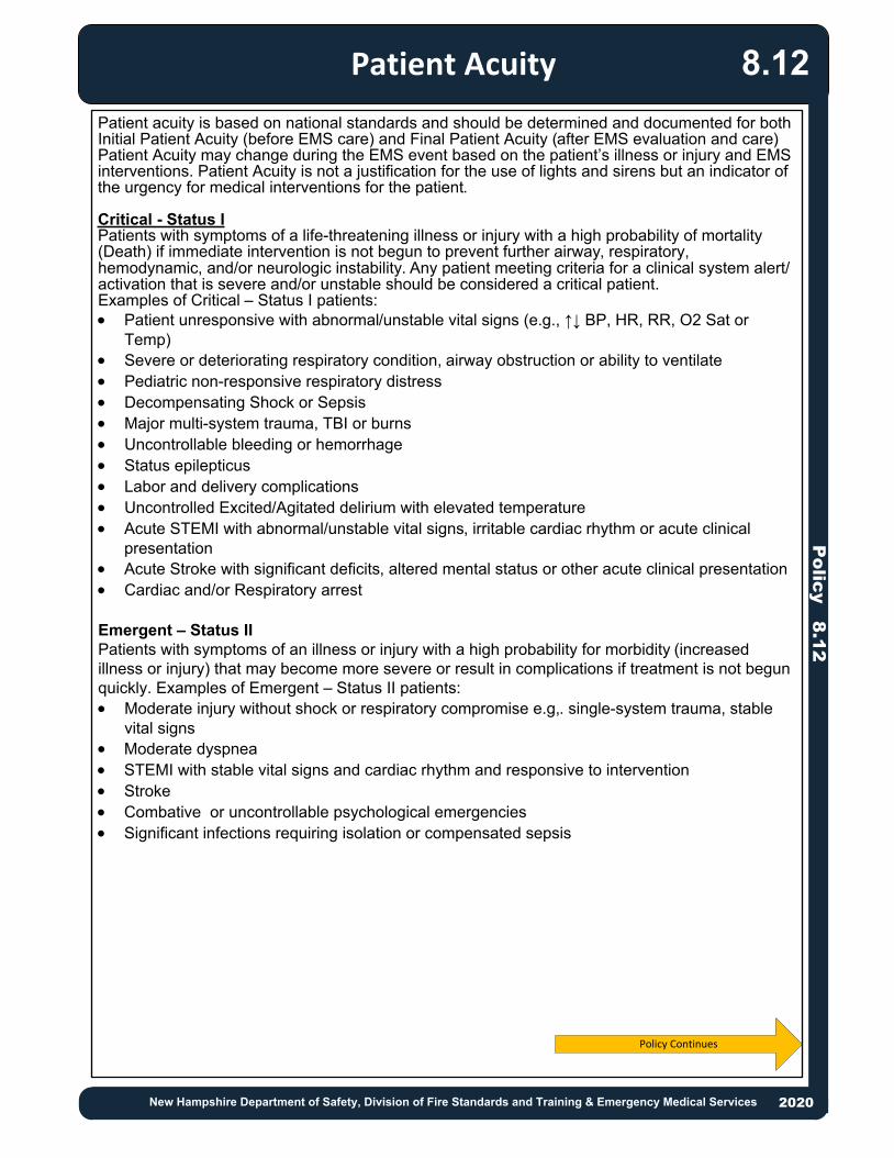
Patient Acuity
2013
Patient acuity is based on national standards and should be determined and documented for both Initial Patient Acuity (before EMS care) and Final Patient Acuity (after EMS evaluation and care) Patient Acuity may change during the EMS event based on the patient’s illness or injury and EMS interventions. Patient Acuity is not a justification for the use of lights and sirens but an indicator of the urgency for medical interventions for the patient.
Critical - Status I Patients with symptoms of a life-threatening illness or injury with a high probability of mortality (Death) if immediate intervention is not begun to prevent further airway, respiratory, hemodynamic, and/or neurologic instability. Any patient meeting criteria for a clinical system alert/activation that is severe and/or unstable should be considered a critical patient. Examples of Critical – Status I patients: · Patient unresponsive with abnormal/unstable vital signs (e.g., ↑↓ BP, HR, RR, O2 Sat or
Temp)· Severe or deteriorating respiratory condition, airway obstruction or ability to ventilate · Pediatric non-responsive respiratory distress· Decompensating Shock or Sepsis· Major multi-system trauma, TBI or burns· Uncontrollable bleeding or hemorrhage· Status epilepticus · Labor and delivery complications· Uncontrolled Excited/Agitated delirium with elevated temperature· Acute STEMI with abnormal/unstable vital signs, irritable cardiac rhythm or acute clinical
presentation· Acute Stroke with significant deficits, altered mental status or other acute clinical presentation· Cardiac and/or Respiratory arrest
Emergent – Status IIPatients with symptoms of an illness or injury with a high probability for morbidity (increased illness or injury) that may become more severe or result in complications if treatment is not begun quickly. Examples of Emergent – Status II patients:· Moderate injury without shock or respiratory compromise e.g,. single-system trauma, stable
vital signs· Moderate dyspnea· STEMI with stable vital signs and cardiac rhythm and responsive to intervention· Stroke · Combative or uncontrollable psychological emergencies · Significant infections requiring isolation or compensated sepsis
Po
licy 8
.12
8.12
New Hampshire Department of Safety, Division of Fire Standards and Training & Emergency Medical Services 2020
Policy Continues

Patient Acuity
Po
licy 8
.12
New Hampshire Department of Safety, Division of Fire Standards and Training & Emergency Medical Services 2020
Lower Acuity – Status IIIPatients with symptoms of an illness or injury that have a low probability of progression to more serious disease or development of complication. Examples of Lower Acuity – Status III patients:· Patient alert, vitals signs within normal limits, and with simple uncomplicated injuries or
medical complaints· Soft tissue injuries including minor burns· Isolated extremity fractures and dislocations· Maxillofacial injuries without airway compromise· Asthma attack that has responded to bronchodilators· Status: post seizure· Psychological emergencies
Non-Acute – Status IVEMS evaluation with no interventions provided Examples of Non-Acute – Status IV patients:· Scheduled medical transport, e.g., dialysis or return home· Public assists, Good intent calls, Medical alarm with false activation
Notes of Clarification· Patients should always be transported to the most appropriate available facility based on
their Patient status.· Patient status / acuity are based on definitions and conditions published in the National
EMS Core Content and the 2016 Model of Clinical Practice of Emergency Medicine. EMS providers should reference these documents for a more comprehensive list of conditions under each level of patient status/acuity.
· Determining and documenting initial and final patient status provides a picture of how acutely ill or injured a patient was when contacted and the effectiveness of EMS protocols for care or patient decline despite quality care at the end of EMS interaction.
Protocol Continues
8.12

Po
licy 8
.13
2013
2013
Pediatric Transportation
New Hampshire Department of Safety, Division of Fire Standards and Training & Emergency Medical Services
Policy Continues
PATIENT TRANSPORT
NH RSA 265:107-a requires all children be properly restrained when riding in a vehicle. Any child who fits on a length-based resuscitation tape must be properly restrained in a safety seat or harness.An ill or injured child must be restrained in a manner that minimizes injury in an ambulance crash. The best location for transporting a pediatric patient is secured directly to the ambulance cot. It is not acceptable, under any circumstance, to transport a pediatric patient in the arms of an adult.It is recommended that agencies develop standard operating procedure/policy for pediatric transport that reflects their ambulance configurations and specific pediatric transport equipment/devices.TYPES OF RESTRAINTS:1. Convertible car seat with two belt paths (front and back) with four points for belt attachment to
the cot is considered best practice for pediatric patients who can tolerate a semi-upright position.· Position safety seat on cot facing foot-end with
backrest elevated to meet back of child safety seat.
· Secure safety seat with 2 pairs of belts at both forward and rear points of seat.
· Place shoulder straps of the harness through slots just below child’s shoulders and fasten snugly to child.
· Follow manufacturer’s guidelines regarding child’s weight.
Note: Non-convertible safety seats cannot be secured safely to cot. If child’s personal
safety seat is not a convertible seat, it cannot be used on the cot.
2. Stretcher harness device with 5 point harnessRestraint device (marketed to EMS) with 5-point harness (examples: Ferno Pedi-Mate, SafeGuard Transport, ACR) · Attach securely to cot utilizing upper back strap behind cot and lower straps around cot’s
frame. · 5-point harness must rest snugly against child. Secure belt at child’s shoulder level so no
gaps exists above shoulders. · Adjust head portion of cot according to manufacturer’s recommendation.· Pedi-mate fits children weighing 10 – 40 lbs. SafeGuard Transport fits children weighing
22 – 100 lbs.Follow manufacturer's guidelines regarding weight.
2020
8.13

Po
licy 8
.13
PEDIATRIC TRANSPORTATION
New Hampshire Department of Safety, Division of Fire Standards and Training & Emergency Medical Services
3. Car bed with both a front and rear belt path (example: Cosco Dream Ride SE)· For infants who cannot tolerate a semi-upright position or who must lie flat.· Position car bed so infant lies perpendicular to cot, keeping infant’s head toward center of patient
compartment.· Fully raise backrest and anchor car bed to cot with 2 belts, utilizing the 4 attachment sites supplied
with car bed.· Only appropriate for infants from 5 – 20 lbs.
4. Isolette/Incubator must be secured to ambulance according to manufacturer’s guidelines.· Secure infant using manufacturer’s restraint. (Five point harness restraint is preferred.)· Blankets or towels may be used for additional stabilization
MOTHER AND NEWBORN TRANSPORT· It is not acceptable, under any circumstance to transport a pediatric patient in the arms of an adult.· Secure and transport mother on the cot.
o If mother and newborn are both stable and a commercial device is available to fasten newborn to mom (examples: Aegis, Kangoofix) follow manufacturer’s guidelines.
o If mother and/or newborn are not stable or commercial device is not available, best practice is to request two ambulances; transporting each in their own ambulance.
o If a second ambulance is not available, transport stable newborn secured to the rear-facing provider seat /captain’s chair using a size-appropriate child restraint system, infant should be facing the rear of the ambulance. Either a convertible safety seat with a forward-facing belt path or an integrated child restraint system certified by the manufacturer to meet FMVSS No. 213 may be used to secure infant.
o Do NOT use a rear-facing only safety seat in the rear-facing provider seat / captain’s chair as this is dangerous and may lead to significant injuries.
o Special attention should be paid to the high risk of hypothermia in newbornsNON-PATIENT TRANSPORTBest practice is to transport well children in a vehicle other than the ambulance, whenever possible, for safety. If no other vehicle is available and circumstances dictate that the ambulance must transport a well child, he/she may be transported in the following locations:· Passenger seat of the driver’s compartment if child is large enough (according to manufacturer’s
guidelines) to ride forward-facing in a child safety seat or booster seat. Airbag should be turned off. If the air bag can be deactivated, an infant, restrained in a rear-facing infant seat, may be placed in the passenger seat of the driver’s compartment.
· Captain’s chair in patient compartment using a size appropriate integrated seat or a convertible safety seat.
USE OF PATIENT’S CHILD SAFETY SEAT AFTER INVOLVEMENT IN MOTOR VEHICLE CRASHThe patient’s safety seat may be used to transport child to hospital after involvement in a minor crash if ALL of the following apply:· It is a convertible seat with both front and rear belt paths.· Visual inspection, including under movable seat padding, does not reveal cracks or deformation. · Vehicle in which safety seat was installed was capable of being driven from the scene of the crash. · Vehicle door nearest the child safety seat was undamaged.· The air bags (if any) did not deploy.
Policy Continued
2020
8.13

Po
licy 8
.14
New Hampshire Department of Safety, Division of Fire Standards and Training & Emergency Medical Services 2020
PurposeThe purpose of this policy is to give EMS guidance for patients who are in police custody, restrained, and/or protective custody is required.
Protective CustodyProtective custody is a civil status in which an incapacitated person is detained by a peace officer for the purposes of: (a) Assuring the safety of the individual or the public or both; and (b) Assisting the individual to return to a functional condition.
· Patients with evidence of suicidal ideation who refuse care may be placed into protective custody under RSA 135C:28 III.
· Patients who present with an altered level of consciousness, diminished mental capacity, or evidence of impaired judgment from alcohol or drug use may be placed into protective custody under RSA 172 and 172:B3.
· If law enforcement refuses to place a patient into protective custody at the request of EMS, on-line medical control must be contacted and a law enforcement supervisor should be requested for further guidance.
Police Custody· Police custody for this policy, shall mean a person under arrest.· Patients who EMS believe require medical care should be transported to a medical facility.
If police and EMS disagree about whether a patient in police custody requires transport to a medical facility for further assessment or treatment, on-line medical control must be contacted and a law enforcement supervisor should be requested for guidance.
EMS Initiated RestraintsFor any patient potentially requiring restraints by EMS, see the Restraints Procedure 6.5.
Police Restraint DevicesPatients transported by EMS who have been restrained by law enforcement devices (e.g., handcuffs) should be accompanied, in the patient compartment, by a law enforcement officer who is capable of removing the device. If this is not feasible, the officer MUST follow directly behind the transporting ambulance to the receiving hospital.
Tasers® (Conductive Electrical Weapon)Patients who have been subdued by a Taser device, see Tasers Procedure 6.6.
Pepper SprayPatients who have been subdued by pepper spray, see Eye and Dental Protocol 4.3.
Excited DeliriumExcited/Agitated Delirium is characterized by extreme restlessness, irritability, and/or high fever. Patients exhibiting these signs are at high risk for sudden death, see Restraints Procedure 6.5.
Police Custody 8.14

2013
Po
licy 8
.15
Refusal of Care
New Hampshire Department of Safety, Division of Fire Standards and Training & Emergency Medical Services
Policy Continues
PURPOSE: Establish guidelines for the management and documentation of situations where patients refuse treatment or transportation.
Refusal of careThere are three components to a valid refusal of care. Absence of any of these components will most likely result in an invalid refusal. The three components are as follows:
1. Competence: In general, a patient who is an adult or a legally emancipated minor is considered legally competent to refuse care. A parent or legal guardian who is on-scene or available by phone, may refuse care on his or her minor children’s behalf. 2. Capacity: In order to refuse medical assistance a patient must have the capacity to understand the nature of his or her medical condition, the risks and benefits associated with the proposed treatment, and the risks associated with refusal of care. 3. Informed Refusal: A patient must be fully informed about his or her medical condition, the risks and benefits associated with the proposed treatment and the risks associated with refusing care.
Patients who meet criteria to allow self-determination shall be allowed to make decisions regarding their medical care, including refusal of evaluation, treatment, or transport. These criteria include:
1. Adults (≥ 18 years of age or a legally emancipated minor).2. Orientation to person, place, time, and situation. 3. No evidence of altered level of consciousness resulting from head trauma, medical illness, intoxication, dementia, psychiatric illness or other causes. 4. No evidence of impaired judgment from alcohol or drug influence. 5. No language communication barriers. Reliable translation available (e.g., on scene interpreter, language line). 6. No evidence or admission of suicidal ideation resulting in any gesture or attempt at self harm. No verbal or written expression of suicidal ideation regardless of any apparent inability to complete a suicide.
EMS providers will make every reasonable effort to convince reluctant patients to access medical care at the emergency department via the EMS system before accepting a Refusal of Care.
Consider on-line medical control for all patients who present a threat to themselves, present with an altered level of consciousness or diminished mental capacity, or have history or examination findings consistent with a high-risk refusal.
The on-line medical control provider should be provided with all relevant information and may need to speak directly with the patient by radio or preferably a recorded landline. The physician should determine if protective custody is to be pursued in consultation with the Law Enforcement.
2020
8.15

Po
licy 8
.15
Refusal of Care
New Hampshire Department of Safety, Division of Fire Standards and Training & Emergency Medical Services
If the patient is intoxicated and in need of medical treatment or protective custody, and refuses care, police can take custody of the individual under NH RSA 172 and 172:B3.
Examples of high-risk refusals include but are not limited to:1. Treated / resolved hypoglycemia.2. Patient with obvious head trauma and taking anticoagulant medications.3. Intoxicated patients.4. Abnormal vital signs. 5. Treated / resolved narcotic overdose.6. High risk mechanism of injuries, see Spinal Injury Protocol 4.7.7. Patient / witness reports suicidal ideations.8. Possible Brief Resolved Unexplained Event see BRUE Protocol 2.5.
Procedure1. Clearly offer the patient both treatment and transportation to the hospital and document the
offer in your Patient Care Report.2. Perform an assessment of the patient’s mental capacity and, to the extent permitted by the
patient, a physical exam including vital signs. Your assessment, or the patient’s refusal of care, must be fully documented in your Patient Care Report
3. Explain to the patient the nature, severity or uncertainty of his/her illness or injury, the treatments being proposed, the risks and consequences of accepting or refusing treatment, and the potential alternatives. Fully document the explanation given to the patient in your patient care report.
4. A parent or legal guardian may refuse care for a minor or:· When a parent or legal guardian is not reasonably available, another adult family
member (e.g., grandparent), or other authorized representative having custody of the minor, may refuse care.
· EMS personnel may accept a telephonic refusal of care, provided that they have explained the consequences of refusing care; telephonic refusal of care should be carefully documented.
5. Prepare and explain the Refusal of Care form to the patient (or, in the case of a minor patient, the patient’s parent, legal guardian, or authorized representative).
6. The Refusal of Care form should be signed by the patient (or, in the case of a minor patient, by the minor patient’s parent, legal guardian, or authorized representative) at the time of the refusal. The form should also be dated and, where possible, signed by a witness, preferably a competent relative, friend, police officer, or impartial third person.
7. If on-line medical control was consulted for a refusal of care, obtain and document the provider’s name in the patient care report.
8. All patients in police custody retain the right to request transport. This should be coordinated with law enforcement.
9. If child or adult abuse and/or neglect is suspected and a refusal of care situation exists, the EMT provider must contact police immediately, see Victims of Violence 8.20.
Policy Continued
2020
8.15

Po
licy 8
.16
2013
2013
Resuscitation Initiation and Termination
New Hampshire Department of Safety, Division of Fire Standards and Training & Emergency Medical Services
WHEN NOT TO STARTResuscitation efforts should be withheld or discontinued under the following circumstances:· VALID DO NOT RESUSCITATE ORDER: Refer to DNR, POLST & Advanced Directives
Protocol 8.8.· SCENE SAFETY: The physical environment is not safe for providers.· DEAD ON ARRIVAL (DOA): A person is presumed dead on arrival when all five “Signs
of Death” are present AND at least one associated “Factor of Death” is present.Signs of Death (All five signs of death must be present)Ø Unresponsiveness.Ø Apnea.Ø Absence of palpable pulses at carotid, radial, and femoral sites.Ø Unresponsive pupils.Ø Absence of heart sounds.
Factors of Death (At least one associated factor of death must be present)Ø Damage or destruction of the body incompatible with life, such as:ü Decapitation.ü Decomposition.ü Deforming brain injury.ü Incineration or extensive full thickness burns.
Ø Lividity/Rigor mortis of any degree.Ø Major blunt or penetrating trauma incompatible with life.
SUDDEN UNEXPLAINED INFANT DEATH SYNDROME (SUIDS). · An infant <12 months who is apneic, asystolic (no heartbeat or umbilical cord pulse),
and exhibiting lividity and/or rigor mortis should be presumed dead. · For unexpected, unexplained infant death, record carbon monoxide level in room
where infant was found unresponsive, if possible.
NEONATE: · A neonate who is apneic, asystolic, and exhibits either neonatal maceration
(softening or degeneration of the tissues after death in utero) or anencephaly (absence of a major portion of the brain, skull, and scalp) may be presumed dead.
· Contact Medical Control if gestational age is less than 20 weeks and neonate shows signs of obvious immaturity (e.g., translucent and gelatinous skin, lack of fingernails, fused eyelids).
NOTE: Infant and/or neonatal resuscitation and transport may be initiated in cases where the family does not accept the idea of nonintervention.
Patients with ventricular assist devices (VAD) should almost never be pronounced dead at the scene, see VAD Policy 8.19.
2020
8.16
Policy Continues

Po
licy 8
.16
Resuscitation Initiation and Termination
New Hampshire Department of Safety, Division of Fire Standards and Training & Emergency Medical Services
Resuscitation may be stopped under the following circumstances:
Policy Continued
2020
8.16
· The physical environment becomes unsafe for providers.· The exhaustion of EMS providers.· The automatic external defibrillator has advised “no shock” for 20 minutes and
Paramedic/hospital care is not available within 20 minutes (hypothermia is an exception) and the ETCO2 is less than 20 mmHg (if available).
· Extrication is prolonged (>15 minutes) with no resuscitation possible during extrication (hypothermia is an exception).
· If directed to do so by Medical Control
EMT/ADVANCED EMT STANDING ORDERS – ADULT & PEDIATRIC
PARAMEDIC STANDING ORDER – ADULT & PEDIATRIC
P
EA/
· Asystole and slow wide complex PEA o If there is no return of spontaneous circulation after 20 minutes in the
absence of hypothermia and the ETCO2 is less than 20 mmHg· · Narrow complex PEA with a rate above 40 or refractory and recurrent ventricular
fibrillation / ventricular tachycardiao Consider early expert consultation with Medical Controlo Consider resuscitation for up to 60 minutes from the time of dispatch,
including transport for potential reversible causes if no ROSC after initial efforts.
o Resuscitation efforts may be ceased before 60 minutes based on factors including but not limited to ETCO2 less than 20 mmHg, age, co-morbidities, distance from, and resources available at the closest hospital.
Policy Continues
· EMS providers are not required to transport every victim of cardiac arrest to a hospital. Unless special circumstances are present, it is expected that most resuscitations will be performed on-scene until the return of spontaneous circulation or a decision to cease resuscitation efforts is made based on the criteria listed under “when to stop” (above). Transportation with continuing CPR may be justified if hypothermia is present or suspected. Current AHA guidelines state: “cessation of efforts in the out-of-hospital setting…should be standard practice.”
PEARLS:· For patients that do not achieve return of spontaneous circulation on scene, termination of
resuscitation should be considered before the patient is loaded into the ambulance for transport.

Resuscitation Initiation and Termination
New Hampshire Department of Safety, Division of Fire Standards and Training & Emergency Medical Services
DETERMINING DEATH IN THE FIELDWhen efforts to resuscitate are not initiated or are terminated under the above provisions, EMS providers shall:· Document time of death.· Notify law enforcement. · Consider possibility of a crime scene and restrict access.· Any decision to move the body must be made in collaboration with law enforcement
and the medical examiner.· Leave any resuscitation adjuncts such as advanced airway devices, IV/IO access
devices, electrode pads, etc., in place.· Inform family on scene of patient’s death and offer to contact family, friends, clergy, or
other support systems.
The above requirements apply to situations in which law enforcement or the medical examiner may take jurisdiction. Law enforcement and the medical examiner are not required to take jurisdiction of hospice or other patients who are known to have been terminally ill from natural causes or congenital anomaly, and death was imminent and expected. Where law enforcement is not involved, EMS providers may provide appropriate assistance to families or other caregivers.
Mass Casualty Incident (MCI): See MCI Protocol 9.1.
Documentation· Complete a Patient Care Record (PCR) in all cases. If available, include ECG rhythm
strips with the patient care report.· Document special orders including DNR, on-line Medical Control, etc.· MCI conditions may require a triage tag in addition to an abbreviated PCR.· Record any special circumstances or events that might impact patient care or forensic
issues.
Policy Continued
2020
Po
licy 8
.16
8.16

Although often described as 'choking' by patients, it should be distinguished as strangulation when being documented by providers (as opposed to choking, i.e., foreign body obstruction). Include all information and observations regarding attempted strangulation in documentation provided to receiving hospital.Assessment:How was the patient strangled:· Left, right, or both hands; forearm; knee or foot; ligature or smothered; other, describeWas patient shaken, beaten or held against wall, ground:· Quantify grip strength and level of pain using 1-10 scale; duration in min/sec.· Prior incidents of strangulation, domestic violence, or threats?Signs and symptoms:· Petechiae on face, eyes/eyelids, nose, ears, head· Deformity of or bleeding from nose, ears; bruising, swelling of mouth/lips· Redness, scratches, abrasions, bruising under chin, on neck, shoulders, chest· Ligature marks, swelling, fingernail impressions (offensive or defensive) on neck· Missing hair, fracture, or swelling/bruising on head, signs of concussion· Difficulty breathing or speaking; coughing, hoarse or raspy voice; drooling, difficulty or pain
swallowing· Vision disturbances or changes (spots, light flashes, tunnel vision, etc.)· Hearing disturbances or changes (buzzing or ringing in the ears, etc.)· Headache· Subcutaneous emphysema· IncontinenceBehavioral signs:· Agitation, amnesia, hallucinations, dizziness, fainting, or combativeness due to hypoxiaDocumentation and Reporting ResponsibilitiesStrangulation is a felony-level crime*. Per NH RSA 631:6, it must be reported to the police unless the patient age 18 or older refuses to have the information released.Strangulation is also an indicator of increasing lethality in a violent relationship. Every effort should be made to connect patient with support services.
· 24-Hour Domestic Violence Crisis Line: 1-866-644-3574.· 24-Hour Sexual Assault Crisis Line: 1-800-277-5570.· Emergency shelter and transportation.· Hospital and court accompaniment; legal advocacy· Information about public assistance.
*(http://www.gencourt.state.nh.us/rsa/html/LXII/631/631-2.htm)
Po
licy 8
.17
2013
2013
Strangulation
New Hampshire Department of Safety, Division of Fire Standards and Training & Emergency Medical Services 2020
8.17
PEARLS:· Patient's spouse/partner, caregiver or parent may be the perpetrator; their presence may
hinder patient's disclosure of information. · Providers' reactions can impact patient recovery and strengthen or hinder prosecution of the
perpetrator. Non-judgmental and compassionate care and thorough documentation and preservation of evidence are essential.
Strangulation is defined as asphyxiation caused by closure of blood vessels and or air passageways of the neck due to external pressure. External pressure can be manual via a body part such as hands, arms, knees, etc., or can be by an object such as a belt, rope, etc. Patients are at risk of delayed death due to internal swelling, anoxia, hematoma or structural damage that cannot be identified externally. Patients should be encouraged to seek medical care; if transported, communicate reported strangulation attempt to hospital staff.

Po
licy 8
.18
Trauma Triage and Transport Decision
New Hampshire Department of Safety, Division of Fire Standards and Training & Emergency Medical Services 2020
Divert to the nearest acute care hospital if a patient with major trauma is in cardiac arrest, peri-arrest or immediately needs a life-saving intervention that cannot be delivered by available prehospital resources.
Measure Vital Signs and Level of ConsciousnessGlasgow Coma Scale ≤13Systolic Blood Pressure <90 mmHg or signs of shockRespiratory Rate <10 or >29 breaths per minute
or need for ventilatory support (<20 in infants aged <1 year)
Glasgow Coma Scale ≤13Systolic Blood Pressure <90 mmHg or signs of shockRespiratory Rate <10 or >29 breaths per minute
or need for ventilatory support (<20 in infants aged <1 year)
Assess Anatomy of Injury· All penetrating injuries to head, neck, torso, and extremities
proximal to elbow or knee· Chest wall instability or deformity (e.g., flail chest)· Two or more proximal long-bone fractures· Crushed, degloved, mangled, or pulseless extremity· Amputation proximal to wrist or ankle· Pelvic fractures· Open or depressed skull fracture· Paralysis
· Fallso Adult: >20 feet (1 story is equal to 10 feet)o Pediatric: >10 feet or 2 to 3 times the height of the child.
· High-risk auto crasho Intrusion, including roof: >12 inches occupant site; >18 inches
any siteo Ejection (partial or complete) from automobileo Death in same passenger compartment
· Auto vs. pedestrian/bicyclist: thrown, run over, or with significant (>20 mph) impact
· Motorcycle crash >20 mph
Assess Mechanism of Injury andEvidence of High-Energy impact
Assess Special Patient orSystem Considerations
· Older Adultso Risk of injury/death increases after age 55 yearso SBP <110 mmHg may represent shock after age 65o Low impact mechanisms (e.g., ground level falls) may result in
severe injury· Pediatric
o Should be triaged preferentially to pediatric capable trauma centers
· Anticoagulants and bleeding disorderso Patients with head injury are at high risk for rapid deterioration
· Burns (See Burn Protocol)o Without other trauma mechanism: triage to burn facilityo With trauma mechanism: triage to trauma center
· Pregnancy >20 weeks (See Obstetric Protocol)· EMS Provider judgment
Transport to closest appropriate acute care hospital
· If feasible, transport directly to a Level I or II Trauma Center by ground or air.
· If above is not feasible and air transport is unavailable, transport to nearest Trauma Center (preferred) or acute care hospital with emergency department and consider requesting ALS intercept.
· For a child <15 years of age, direct transport to a Level 1 or 2 Pediatric Trauma Center is desired.
· Contact destination hospital and activate the trauma system in accordance with local guidelines.
· Transport to the closest Trauma Center (preferred) or acute care hospital.
· Contact destination hospital and activate the trauma system in accordance with local guidelines.
· Consider transport to nearest Trauma Center
Reference: CDC 2011 Guidelines for Field Triage of Injured Patients and NH Trauma Plan
NO
NO
NO
NO
YES
YES
YES
YES
8.18
For more information on Trauma Center assignments and hospital services click on this LINK

EMT/ ADVANCED EMT / PARAMEDIC STANDING ORDERS
2013
EA
P
// P
olic
y 8
.19
New Hampshire Department of Safety, Division of Fire Standards and Training & Emergency Medical Services 2020
Implantable Ventricular Assist Devices (VAD)
Protocol Continues
Patient Care Goals· Rapid identification of, and interventions for, cardiovascular compromise in
patients with VADs· Rapid identification of, and interventions for VAD-related malfunctions or
complications
Indications· Patients with an implanted ventricular assist device (VAD) and symptoms of
cardiovascular compromise or cardiac arrest.· Patients with VADs experiencing a medical or injury-related event not involving the
cardiovascular system or VAD malfunction
Assessment:· Assess for possible pump malfunction
o Assess for alarmso Auscultate for pump sound “hum” o Signs of hypoperfusion including pallor, diaphoresis, altered mental status
· Utilize available resources to troubleshoot potential VAD malfunctions and to determine appropriate corrective actions to restore normal VAD function:o Contact the patient’s VAD-trained companion, if available, o Contact the patient’s VAD coordinator (phone number may be on device)o Check all the connections to system controllero Change VAD batteries one at a time and/or system controller if indicated
Treatment and Interventions:· Routine Patient Care· Cardiac monitoring and acquire 12-lead EKG· For VAD-related complications or cardiovascular problems, expedite transport to
the medical facility where VAD was placed if clinical condition and time allows.· If patient has a functioning VAD and is experiencing a non-cardiovascular-related
problem, transport to a facility that is appropriate for the patient’s main presenting problem without manipulating the device.
· Signs of poor perfusion, (e.g., MAP < 50 mmHg and/or ETCO2 <20) follow ACLS algorithm
· CPR should only may be initiated if:o You have confirmed the pump has stopped AND troubleshooting efforts to
restart it have failed, AND o The patient is unresponsive and has no detectable signs of life
· See Cardiac Arrest Protocol 3.2.
8.19

Po
licy 8
.19
Implantable Ventricular Assist Device (VAD)
New Hampshire Department of Safety, Division of Fire Standards and Training & Emergency Medical Services 2020
Protocol Continued
8.19
PEARLS· The most common cause for VAD alarms are low batteries or battery failures· You do not need to disconnect the controller or batteries in order to defibrillate or
cardiovert or acquire a 12-lead EKG· Flow through many VAD devices is not pulsatile (except older models) and
patients may not have a palpable pulse or accurate pulse oximetry · Ventricular fibrillation, ventricular tachycardia, or asystole/PEA may be the
patient’s “normal” underlying rhythm. Evaluate clinical condition and provide care in consultation with VAD coordinator
· The patient’s travel bag must accompany him/her at all times with back-up controller and spare batteries and accessory equipment
· If feasible, bring the patient’s power module, cable and display module with patient to the hospital
· Although automatic non-invasive blood pressure cuffs are often ineffective in measuring systolic and diastolic pressure, if they do obtain a measurement, the MAP is usually accurate
· Other VAD complications: o Infectiono Stroke / TIAo Bleeding
o Arrhythmiaso Cardiac
Tamponade
o CHFo Aortic Insufficiency
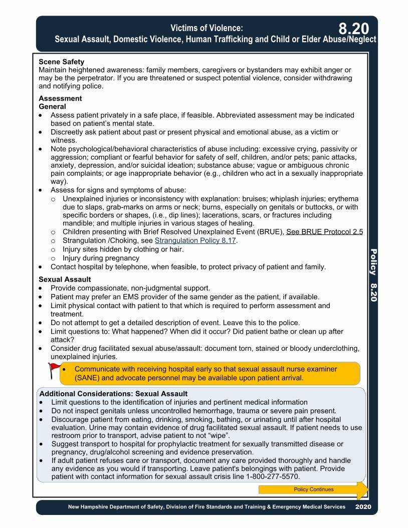
Victims of Violence:
Sexual Assault, Domestic Violence, Human Trafficking and Child or Elder Abuse/Neglect
2013
Scene SafetyMaintain heightened awareness: family members, caregivers or bystanders may exhibit anger or may be the perpetrator. If you are threatened or suspect potential violence, consider withdrawing and notifying police. Assessment General· Assess patient privately in a safe place, if feasible. Abbreviated assessment may be indicated
based on patient’s mental state.· Discreetly ask patient about past or present physical and emotional abuse, as a victim or
witness. · Note psychological/behavioral characteristics of abuse including: excessive crying, passivity or
aggression; compliant or fearful behavior for safety of self, children, and/or pets; panic attacks, anxiety, depression, and/or suicidal ideation; substance abuse; vague or ambiguous chronic pain complaints; or age inappropriate behavior (e.g., children who act in a sexually inappropriate way).
· Assess for signs and symptoms of abuse:o Unexplained injuries or inconsistency with explanation: bruises; whiplash injuries; erythema
due to slaps, grab-marks on arms or neck; burns, especially on genitals or buttocks, or with specific borders or shapes, (i.e., dip lines); lacerations, scars, or fractures including mandible; and multiple injuries in various stages of healing.
o Children presenting with Brief Resolved Unexplained Event (BRUE), See BRUE Protocol 2.5o Strangulation /Choking, see Strangulation Policy 8.17.o Injury sites hidden by clothing or hair. o Injury during pregnancy
· Contact hospital by telephone, when feasible, to protect privacy of patient and family.
Sexual Assault· Provide compassionate, non-judgmental support. · Patient may prefer an EMS provider of the same gender as the patient, if available.· Limit physical contact with patient to that which is required to perform assessment and
treatment.· Do not attempt to get a detailed description of event. Leave this to the police.· Limit questions to: What happened? When did it occur? Did patient bathe or clean up after
attack?· Consider drug facilitated sexual abuse/assault: document torn, stained or bloody underclothing,
unexplained injuries.
2013
Policy Continues
Po
licy 8
.20
New Hampshire Department of Safety, Division of Fire Standards and Training & Emergency Medical Services 2020
8.20
· Communicate with receiving hospital early so that sexual assault nurse examiner (SANE) and advocate personnel may be available upon patient arrival.
Additional Considerations: Sexual Assault· Limit questions to the identification of injuries and pertinent medical information · Do not inspect genitals unless uncontrolled hemorrhage, trauma or severe pain present.· Discourage patient from eating, drinking, smoking, bathing, or urinating until after hospital
evaluation. Urine may contain evidence of drug facilitated sexual assault. If patient needs to use restroom prior to transport, advise patient to not “wipe”.
· Suggest transport to hospital for prophylactic treatment for sexually transmitted disease or pregnancy, drug/alcohol screening and evidence preservation.
· If adult patient refuses care or transport, document any care provided thoroughly and handle any evidence as you would if transporting. Leave patient's belongings with patient. Provide patient with contact information for sexual assault crisis line 1-800-277-5570.
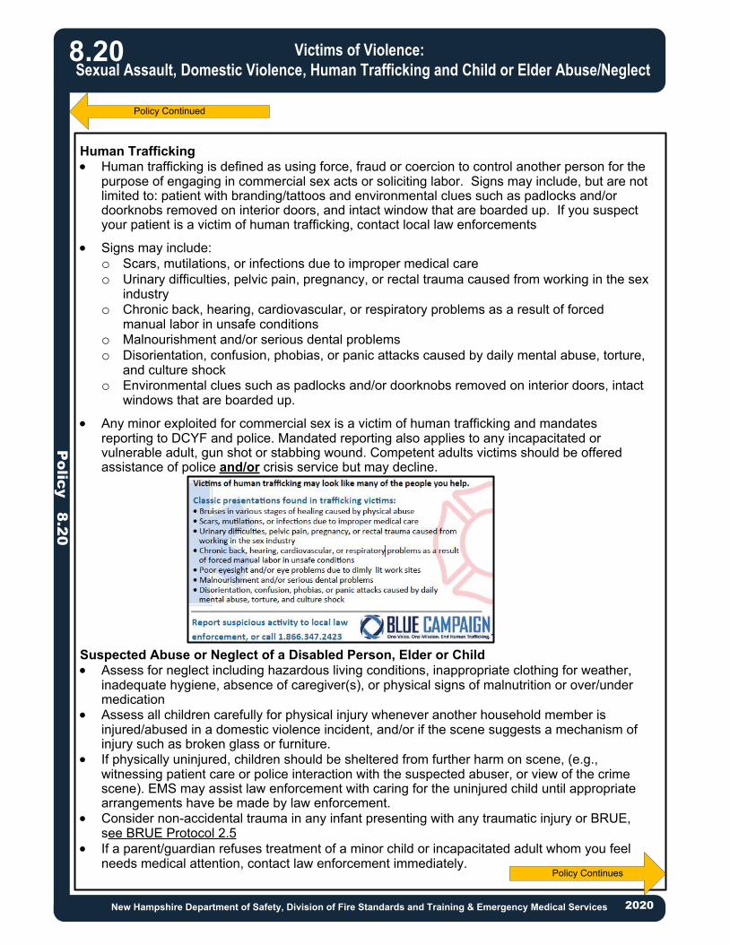
Human Trafficking · Human trafficking is defined as using force, fraud or coercion to control another person for the
purpose of engaging in commercial sex acts or soliciting labor. Signs may include, but are not limited to: patient with branding/tattoos and environmental clues such as padlocks and/or doorknobs removed on interior doors, and intact window that are boarded up. If you suspect your patient is a victim of human trafficking, contact local law enforcements
· Signs may include:o Scars, mutilations, or infections due to improper medical careo Urinary difficulties, pelvic pain, pregnancy, or rectal trauma caused from working in the sex
industryo Chronic back, hearing, cardiovascular, or respiratory problems as a result of forced
manual labor in unsafe conditionso Malnourishment and/or serious dental problemso Disorientation, confusion, phobias, or panic attacks caused by daily mental abuse, torture,
and culture shocko Environmental clues such as padlocks and/or doorknobs removed on interior doors, intact
windows that are boarded up.
· Any minor exploited for commercial sex is a victim of human trafficking and mandates reporting to DCYF and police. Mandated reporting also applies to any incapacitated or vulnerable adult, gun shot or stabbing wound. Competent adults victims should be offered assistance of police and/or crisis service but may decline.
Suspected Abuse or Neglect of a Disabled Person, Elder or Child· Assess for neglect including hazardous living conditions, inappropriate clothing for weather,
inadequate hygiene, absence of caregiver(s), or physical signs of malnutrition or over/under medication
· Assess all children carefully for physical injury whenever another household member is injured/abused in a domestic violence incident, and/or if the scene suggests a mechanism of injury such as broken glass or furniture.
· If physically uninjured, children should be sheltered from further harm on scene, (e.g., witnessing patient care or police interaction with the suspected abuser, or view of the crime scene). EMS may assist law enforcement with caring for the uninjured child until appropriate arrangements have be made by law enforcement.
· Consider non-accidental trauma in any infant presenting with any traumatic injury or BRUE, see BRUE Protocol 2.5
· If a parent/guardian refuses treatment of a minor child or incapacitated adult whom you feel needs medical attention, contact law enforcement immediately.
Policy Continued
Policy Continues
Po
licy 8
.20
Victims of Violence:
Sexual Assault, Domestic Violence, Human Trafficking and Child or Elder Abuse/Neglect
New Hampshire Department of Safety, Division of Fire Standards and Training & Emergency Medical Services 2020
8.20

Policy Continued
Po
licy 8
.20
Victims of Violence:
Sexual Assault, Domestic Violence, Human Trafficking and Child or Elder Abuse/Neglect
New Hampshire Department of Safety, Division of Fire Standards and Training & Emergency Medical Services 2020
8.20
REPORTING REQUIREMENTS: According to New Hampshire State law, most domestic violence injuries are not required to be reported to the police. If the patient is 18 years of age or older and has received a gunshot wound or other serious bodily injury, the injuries must be reported to the police. As defined in RSA 625:11 "Serious bodily injury" means any harm to the body which causes or could cause severe, permanent, or protracted loss of or impairment to the health or of the function of any part of the body.
Documentation and Evidence Preservation · Document verbatim everything the patient or caregiver says that may be relevant. Do not
paraphrase. Capture inconsistencies. · If necessary to remove patient's clothing, do not damage evidence (rips, stains) if possible.
Cut along seam lines.Preserve all evidence, see Crime Scene/Preservation of Evidence Policy 8.7
Reporting Procedures/Requirements· Suspected abuse, neglect, or exploitation of children or adults must be reported
immediately, whether or not the patient is transported. Informing hospital personnel or involving law enforcement does not fulfill legal reporting requirements.
Child Abuse Both the Department for Children, Youth, and Families (DCYF) and local police must be notified within 24 hours: · Call DCYF at 1-800-894-5533, available 24 hours/day; if out-of-state: 603-271-6562.· NOTE: Regardless of other agency’s involvement, EMS is mandated to notify DCYF.· Do not send reports of suspected child abuse by email.
NOTE: If an uninjured child witnesses violence, this qualifies as child abuse and neglect and mandates a report.Abuse of Elders and Incapacitated Adults · Call Bureau of Elderly & Adult Services at 800-949-0470 or 603-271-7014 and leave a
message. For adults residing in:o Independent living situation (own home/apartment, home/apartment of friends or
relatives, boarding home, or no fixed address).o Homes or programs affiliated with Bureau of Behavioral Health or Bureau of
Developmental Services.o Hospital or rehabilitation center.
· Call Office of Long-Term Care Ombudsman at 800–442–5640 or 603–271–4375 between 8:00 am to 4:30 pm, Monday through Friday, for adult residents of nursing or assisted living facilities.
Call local police department during non-work hours and holidays and follow up with a telephone call to Bureau of Elderly & Adult Services or Office of Long-Term Care Ombudsman during work hours.Referral Information for Domestic Violence and Sexual AssaultThe NH Coalition Against Domestic and Sexual Violence (NHCADSV) supports survivors of domestic and sexual violence and offers free, confidential services (emergency shelter and transportation, legal advocacy, hospital and court accompaniment, information about public assistance). Provide patient with referral information.24-Hour Domestic Violence Crisis Line: 1-866-644-3574.24-Hour Sexual Assault Crisis Line: 1-800-277-5570.

RESPONSE
2013
2013
Hazardous Material Exposure
Ha
zm
at &
MC
I 9.0
New Hampshire Department of Safety, Division of Fire Standards and Training & Emergency Medical Services 2020
9.0Hazardous Material: A hazardous material is any item or agent (biological, chemical, radiological, and/or physical), which has the potential to cause harm to humans, animals, or the environment, either by itself or through interaction with other factors.
Hazardous Material Exposure: Any patient with an illness, injury, or complaint which has been caused by or is suspected of being caused by a hazardous material.
When to use this protocol: During any response to a hazardous material exposure where the public, responders, environment, or valuable property are at risk of continued harm or exposure AND the hazard has not been previously mitigated or contained.
· SAFETY: Your safety is priority #1. DO NOT PROCEED beyond staging or cold zone unless directed by HAZMAT team and Incident Command.
· LOCAL PROTOCOL: Follow your dispatch area’s HAZMAT response notification and response plan! This protocol is not a substitute for a comprehensive notification, response, decontamination, treatment and transport plan
· Activate ICS and HAZMAT response plan· Request specific staging instructions· Position ambulance uphill and upwind >300ft· Be alert for patients self extricating from
scene· Declare MCI see MCI Protocol
Resources:NH Fire Marshall: 603-223-4289 NH Bureau of Emergency Mgmt: 603-271-2231NH National Guard 12th CST: 603-227-1555
HAZARDOUS MATERIAL IDENTIFICATION· Name and proper spelling of material if known· SDS sheet, bill of lading, waybill, other
documentation· Emergency Response Guide ID# (4 digits)· DOT classification on placard· Bystanders, technicians or employees at
location· Physical description of material (color, odor,
etc.)
Resources:North American Emergency Response Guide (NAERG) Poison Control: 1-800-222-1222Chemtrec: 1-800-424-9300Military Shipments: 1-800-851-8061Wireless Information System for Emergency Responders (WISER)
Note: Many household chemicals may not require activation of a HAZMAT team. Utilizemanufacturer’s recommendation for decontamination and treatment, or contact Poison Control
HOSPITAL NOTIFICATION· Estimate number of patients if possible· Estimate triage/acuity level of patients· Determine time frame for transportation· Determine capacity of receiving hospitals
ResourcesTriage Tags with “contaminated” identifierSee Mass Casualty and Triage Protocol
Receiving hospitals should be notified as soon as it is determined you have contaminatedpatient(s) to ensure the facility is capable and prepared to receive a potentially contaminated
patient, include level of hazardous materials suit, if known. Protocol Continues

Hazardous Materials Exposure H
azm
at &
MC
I 9.0
New Hampshire Department of Safety, Division of Fire Standards and Training & Emergency Medical Services
Protocol Continued
2020
9.0
TREATMENT DURING DECONTAMINATION
· Limit medication administration route to IM/IN or nebulizer· Intravenous therapy and advanced airway interventions should be delayed until after gross
decontamination. · · Specific individual treatment should be referenced from Poison Control or SDS sheets. · Encourage the use of warmed water 100o to prevent hypothermia
RECORD EXPOSURE AND TREAMENT INFORMATION
· Name of chemical(s).· Amount, time, and route of exposure. · Decontamination information.· Treatment/antidotes administered.
TRANSPORT· EMS personnel transporting potentially contaminated patients (e.g., patients who have received
gross decontamination) should wear personal protective equipment as recommended by Incident Command.
· If an ambulance has transported a contaminated patient, it can only be used to transport similarly contaminated patients until proper decontamination of the vehicle is complete.
· Contaminated patients should not be transported by helicopter.
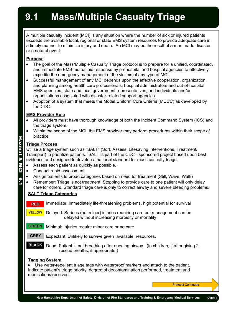
2013
2013
Mass/Multiple Casualty Triage
Ha
zm
at &
MC
I 9.1
New Hampshire Department of Safety, Division of Fire Standards and Training & Emergency Medical Services
Protocol Continues
A multiple casualty incident (MCI) is any situation where the number of sick or injured patients exceeds the available local, regional or state EMS system resources to provide adequate care in a timely manner to minimize injury and death. An MCI may be the result of a man made disaster or a natural event.
Purpose· The goal of the Mass/Multiple Casualty Triage protocol is to prepare for a unified, coordinated,
and immediate EMS mutual aid response by prehospital and hospital agencies to effectively expedite the emergency management of the victims of any type of MCI.
· Successful management of any MCI depends upon the effective cooperation, organization, and planning among health care professionals, hospital administrators and out-of-hospital EMS agencies, state and local government representatives, and individuals and/or organizations associated with disaster-related support agencies.
· Adoption of a system that meets the Model Uniform Core Criteria (MUCC) as developed by the CDC.
EMS Provider Role· All providers must have thorough knowledge of both the Incident Command System (ICS) and
the triage system.· Within the scope of the MCI, the EMS provider may perform procedures within their scope of
practice.
Triage ProcessUtilize a triage system such as “SALT” (Sort, Assess, Lifesaving Interventions, Treatment/Transport) to prioritize patients. SALT is part of the CDC - sponsored project based upon best evidence and designed to develop a national standard for mass casualty triage.· Assess each patient as quickly as possible.· Conduct rapid assessment.· Assign patients to broad categories based on need for treatment (Still, Wave, Walk)· Remember: Triage is not treatment! Stopping to provide care to one patient will only delay
care for others. Standard triage care is only to correct airway and severe bleeding problems.
2020
9.1
SALT Triage Categories
Immediate: Immediately life-threatening problems, high potential for survival
Delayed: Serious (not minor) injuries requiring care but management can be delayed without increasing morbidity or mortality
Minimal: Injuries require minor care or no care
Expectant: Unlikely to survive given available resources.
Dead: Patient is not breathing after opening airway. (In children, if after giving 2 rescue breaths, if appropriate.)
Tagging System· Use water-repellent triage tags with waterproof markers and attach to the patient.Indicate patient's triage priority, degree of decontamination performed, treatment and medications received.
RED
YELLOW
GREEN
BLACK
GREY

Mass/Multiple Casualty TriageH
azm
at &
MC
I 9.1
New Hampshire Department of Safety, Division of Fire Standards and Training & Emergency Medical Services
Triage in Hazardous Material IncidentsDecontaminationThe need for decontamination is the "first triage decision." Since decontamination can be a lengthy process, the "second decision" is which patient(s) are the first to be decontaminated. The "third decision" is based on need for treatment during the decontamination process, since only simple procedures such as antidote administration can be accomplished while wearing PPE.
Identification and Treatment· Signs and symptoms of exposure will usually dictate the treatment required, however, at the
earliest possible time, identification of the specific chemical should be made.· Reference additional hazardous materials protocols as necessary.· Request additional resources. Initial antidote and medical supplies may be limited to priority
patients.· Respiratory compromise is a leading factor of fatalities due to hazardous material exposure.· Symptoms of chemical exposure may be delayed and occur suddenly. Constant reevaluation
of respiratory status is necessary.
Protocol Continued
2020
9.1
SALT Mass Casualty Triage Algorithm (Sort, Assess, Lifesaving Interventions, Treatment/Transport)

· Remove the patient from scene and decontaminate by appropriately trained personnel.
· Triage tools for mass casualty incidento If vomiting starts:§ Within 1 hour of exposure, survival is unlikely and patient should be
tagged “Expectant.”§ Less than 4 hours after exposure, patient needs immediate
decontamination and evaluation and should be tagged “immediate.”§ 4 hours after exposure, reevaluation can be delayed 24 – 72 hours if no
other injury is present and patient should be tagged “Delayed”.· Routine Patient Care.· Treat traumatic injuries and underlying medical conditions.· Patients with residual contamination risk from wounds, shrapnel, or internal
contamination should be wrapped in water repellent dressings to reduce cross contamination.
· Consider Air Medical Transport after proven definitive decontamination of patient.
2013
· Consider pain management, see Pain Management Protocol 2.16. P
EMT/ADVANCED EMT STANDING ORDERS
PARAMEDIC STANDING ORDERS
2013
Ra
dia
tion
Inju
ries 9
.2
Radiation InjuriesAdult & Pediatric
New Hampshire Department of Safety, Division of Fire Standards and Training & Emergency Medical Services 2020
Exposure to radioactive source or radioactive material/debris
PEARLS:· In general, trauma patients who have been exposed to or contaminated by radiation should be
triaged and treated on the basis of the severity of their conventional injuries· A patient who is contaminated with radioactive material (e.g. flecks of radioactive material
embedded in their clothing and skin) generally poses a minimal exposure risk to medical personnel.
9.2
A· Consider anti-emetic, see Nausea/Vomiting Protocol 2.11.
ADVANCED EMT STANDING ORDERS
E

Appendix 1New Hampshire Department of Safety, Division of Fire Standards and Training & Emergency Medical Services 2020
NH Approved EMS Medications

Appendix 1 New Hampshire Department of Safety, Division of Fire Standards and Training & Emergency Medical Services 2020
NH Approved EMS Medications
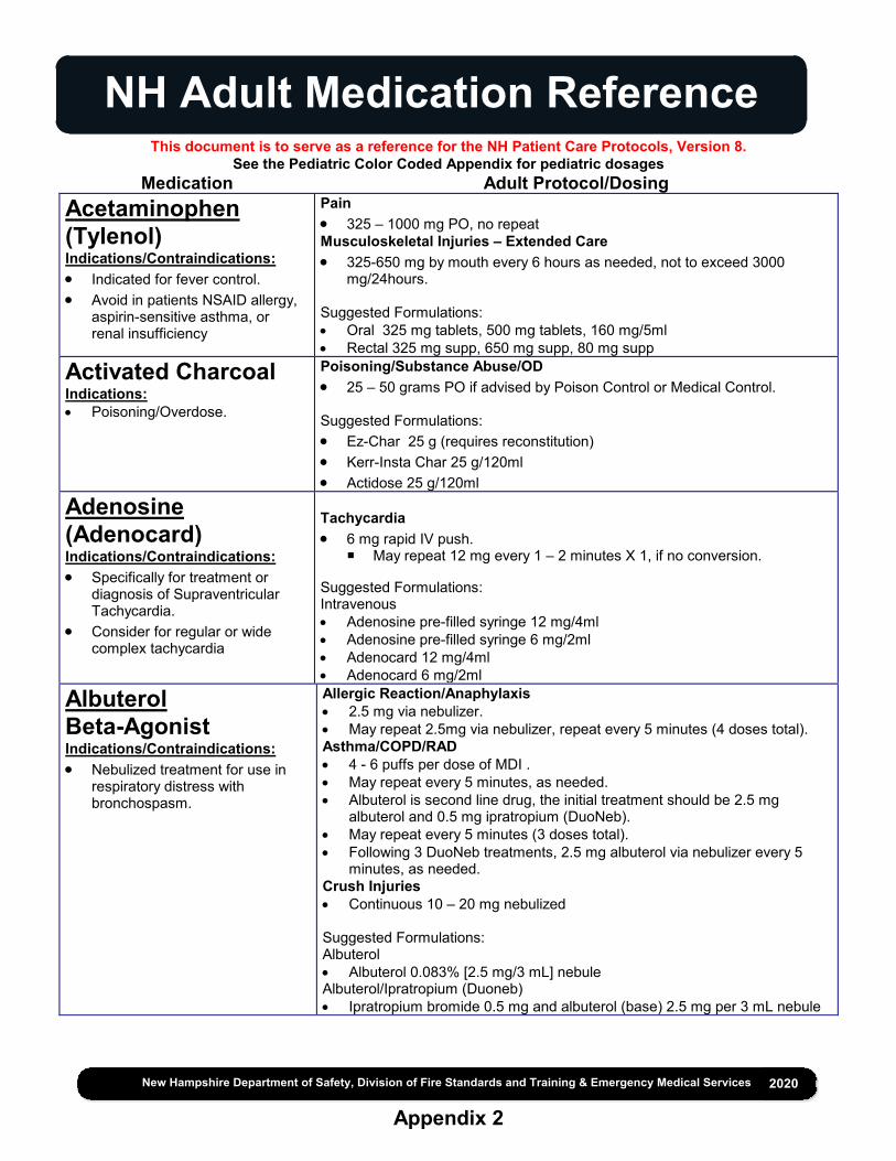
NH Adult Medication Reference
This document is to serve as a reference for the NH Patient Care Protocols, Version 8. See the Pediatric Color Coded Appendix for pediatric dosages
New Hampshire Department of Safety, Division of Fire Standards and Training & Emergency Medical Services 2020
Appendix 2
Medication Adult Protocol/Dosing Acetaminophen (Tylenol) Indications/Contraindications: Indicated for fever control. Avoid in patients NSAID allergy,
aspirin-sensitive asthma, or renal insufficiency
Pain 325 – 1000 mg PO, no repeat Musculoskeletal Injuries – Extended Care 325-650 mg by mouth every 6 hours as needed, not to exceed 3000
mg/24hours.
Suggested Formulations: Oral 325 mg tablets, 500 mg tablets, 160 mg/5ml Rectal 325 mg supp, 650 mg supp, 80 mg supp
Activated Charcoal Indications: Poisoning/Overdose.
Poisoning/Substance Abuse/OD 25 – 50 grams PO if advised by Poison Control or Medical Control. Suggested Formulations: Ez-Char 25 g (requires reconstitution) Kerr-Insta Char 25 g/120ml Actidose 25 g/120ml
Adenosine (Adenocard) Indications/Contraindications: Specifically for treatment or
diagnosis of Supraventricular Tachycardia.
Consider for regular or wide complex tachycardia
Tachycardia 6 mg rapid IV push.
May repeat 12 mg every 1 – 2 minutes X 1, if no conversion. Suggested Formulations: Intravenous Adenosine pre-filled syringe 12 mg/4ml Adenosine pre-filled syringe 6 mg/2ml Adenocard 12 mg/4ml Adenocard 6 mg/2ml
Albuterol Beta-Agonist Indications/Contraindications: Nebulized treatment for use in
respiratory distress with bronchospasm.
Allergic Reaction/Anaphylaxis 2.5 mg via nebulizer. May repeat 2.5mg via nebulizer, repeat every 5 minutes (4 doses total). Asthma/COPD/RAD 4 - 6 puffs per dose of MDI . May repeat every 5 minutes, as needed. Albuterol is second line drug, the initial treatment should be 2.5 mg
albuterol and 0.5 mg ipratropium (DuoNeb). May repeat every 5 minutes (3 doses total). Following 3 DuoNeb treatments, 2.5 mg albuterol via nebulizer every 5
minutes, as needed. Crush Injuries Continuous 10 – 20 mg nebulized Suggested Formulations: Albuterol Albuterol 0.083% [2.5 mg/3 mL] nebule Albuterol/Ipratropium (Duoneb) Ipratropium bromide 0.5 mg and albuterol (base) 2.5 mg per 3 mL nebule

NH Adult Medication Reference
This document is to serve as a reference for the NH Patient Care Protocols, Version 8. See the Pediatric Color Coded Appendix for pediatric dosages
New Hampshire Department of Safety, Division of Fire Standards and Training & Emergency Medical Services 2020
Appendix 2
Medication Adult Protocol/Dosing Amiodarone (Cordarone) Indications/Contraindications: Antiarrhythmic used mainly in
wide complex tachycardia and ventricular fibrillation.
Avoid in patients with heart block or profound bradycardia.
Contraindicated in patients with iodine hypersensitivity.
Cardiac Arrest V-Fib/Pulseless V-Tach 300 mg IV push. Repeat dose of 150 mg IV push for recurrent episodes. Tachycardia - Wide complex tachycardia 150 mg in 50 – 100 mL normal saline or D5W over 10 min. May repeat once in 10 minutes. If successful, consider maintenance infusion of 1 mg/minute. Suggested Formulations: Amiodarone HCL 150 mg/3 mL (3 mL); 450 mg/9ml (9ml)
Antibiotics Severe Sepsis Prerequisite Protocol Per Medical Resource Hospital Agreement
Aspirin Indications/Contraindications: An antiplatelet drug for use in
cardiac chest pain. History of anaphylaxis to aspirin
or NSAIDs Active GI bleeding
Acute Coronary Syndrome 324 mg by mouth (chewable). Suggested Formulations: Oral 81 mg chewable tablets
Atropine Indications/Contraindications: Anticholinergic drug used in
bradycardias and organophosphate poisonings.
Bradycardia 0.5 mg IV every 3 – 5 minutes up to maximum of 3 mg. Organophosphate Poisoning and Nerve Agent 2 mg IM or IV every 5 minutes until secretions clear. Suggested Formulations: Solution, Injection, as sulfate [preservative free]:
0.4 mg/mL (1 mL) 0.8 mg/mL (0.5 mL) 1 mg/mL (1 mL)
Device, Intramuscular, as sulfate:
AtroPen: 0.25 mg/0.3 mL (0.3 mL) [pyrogen free] AtroPen: 0.5 mg/0.7 mL (0.7 mL); 1 mg/0.7 mL (0.7 mL); 2 mg/0.7 mL (0.7 mL) [pyrogen free; contains phenol]

NH Adult Medication Reference
This document is to serve as a reference for the NH Patient Care Protocols, Version 8. See the Pediatric Color Coded Appendix for pediatric dosages
New Hampshire Department of Safety, Division of Fire Standards and Training & Emergency Medical Services 2020
Appendix 2
Medication Adult Protocol/Dosing Atropine and Pralidoxime Auto-Injector Nerve Agent Kit Indications/Contraindications: Antidote for Nerve Agents or
Organophosphate Overdose.
Nerve Agents Patients experiencing: apnea, convulsions, unconsciousness, flaccid
paralysis administer 3 atropine/pralidoxime auto-injectors and 1 diazepam (10 mg) auto-injectors.
Patients experiencing: dyspnea, twitching, nausea, vomiting, sweating, anxiety, confusion, constricted pupils, restlessness, weakness administer 1 atropine/pralidoxime auto-injectors.
Maintenance Dose: 1 atropine/pralidoxime auto-injectors every 3 hours.
Calcium Chloride 10% solution Indications/Contraindications: Indicated for hyperkalemia or
calcium channel blocker overdose.
Caution in patient takes digoxin.
Bradycardia For suspected hyperkalemia with ECG changes or symptomatic calcium channel blocker overdose consider: 1 gram IV / IO over 10 minutes, with continuous cardiac monitoring. May
repeat in 10 minutes if clinical indications persists. Cardiac Arrest - Wide Complex PEA 1 gram IV Crush Injuries 1 gram IV/IO over 10 minutes, may repeat in 10 minutes Suggested Formulations: Solution, Intravenous: Generic: 10% (10 mL
Calcium Gluconate Indications/Contraindications: Indicated for hyperkalemia or
calcium channel blocker overdose.
Bradycardia For suspected hyperkalemia with ECG changes or symptomatic calcium channel blocker overdose consider: 2 grams IV / IO over 10 minutes, with constant cardiac monitoring, may
repeat in 10 minutes if clinical indication persists Cardiac Arrest - Wide Complex PEA 2 grams IV Crush Injuries 2 grams IV/IO over 10 minutes, may repeat in 10 minutes
Dexamethasone Indications/Contraindications: COPD/Asthma
Asthma – Adult 10 mg PO/IM/IV
Dextrose Indications/Contraindications: Symptomatic hypoglycemia. Use in medication infusion
medium.
Hypoglycemia Administer dextrose 10% IV via premixed infusion bag (preferred) or
prefilled syringe until mental status returns to baseline and glucose level is greater than 60mg/dL. IV pump not required.
Suggested Formulations: Solution, Intravenous:
10% (250 mL, 500 mL, 1000 mL);

NH Adult Medication Reference
This document is to serve as a reference for the NH Patient Care Protocols, Version 8. See the Pediatric Color Coded Appendix for pediatric dosages
New Hampshire Department of Safety, Division of Fire Standards and Training & Emergency Medical Services 2020
Appendix 2
Medication Adult Protocol/Dosing Diazepam (Valium) Benzodiazepine Indications/Contraindications: Seizure control. Sedation. Anxiolytic.
Behavioral Emergencies 2.5 mg IV, may repeat every 10 minutes to a total of 10 mg. Bradycardia 5 mg IV, may repeat 2.5 mg once in 5 minutes. BiPAP Consider administering anxiolytic: 5 mg IV (then 2.5 mg every 5 minutes to total of 20 mg) CPAP Consider administering anxiolytic: 5 mg IV (then 2.5 mg every 5 minutes to a total of 20 mg) Hyperthermia 2 mg IV, may repeat once in 5 minutes. Nerve Agent 5 mg IV every 5 minutes; or 10 mg IM OR Diazepam auto-injector (10mg).
Repeat 10 minutes as needed Poisoning/Substance Abuse/OD 2 mg IV, may repeat once in 5 minutes, OR 5 mg IM, may repeat once in 20 minutes Seizure 5 – 10 mg IV (then 2.5 mg every 5 minutes to a total of 20 mg). Restraints – Resistant or Aggressive Management (goal alert and calm) 5 mg IV/IM (IV preferred route), may repeat once in 10 minutes Restraints – Excited/Agitated Delirium (goal is safe and compliant) 10 mg IV (IV preferred route), may repeat once in 5 minutes; or 5 mg IM,
may repeat once in 20 minutes Tachycardia 5 mg IV, may repeat 2.5 mg once in 5 minutes. Traumatic Brain Injury 2 mg IV, may repeat once in 5 minutes. Suggested Formulations: Solution, Injection:
Generic: 5 mg/mL (2 mL, 10 mL)
Diltiazem (Cardizem) Indications/Contraindications: Calcium channel blocker used to
treat narrow complex SVT. Contraindicated in patients with
heart block, ventricular tachycardia, WPW, and/or acute MI.
Tachycardia - Narrow Complex Tachycardia 0.25 mg/kg IV (maximum dose 20 mg) over 2 minutes.
May repeat dose in 15 minutes at 0.35 mg/kg (maximum dose 20 mg) if necessary.
Consider maintenance infusion 5 – 15 mg/hour. Suggested Formulations:
Solution, Intravenous, as hydrochloride: 25 mg/5 mL (5 mL, 25 mL); 50 mg/10 mL (10 mL); 125 mg/25 mL (25 mL)

NH Adult Medication Reference
This document is to serve as a reference for the NH Patient Care Protocols, Version 8. See the Pediatric Color Coded Appendix for pediatric dosages
New Hampshire Department of Safety, Division of Fire Standards and Training & Emergency Medical Services 2020
Appendix 2
Medication Adult Protocol/Dosing Diphenhydramine (Benadryl) Indications/Contraindications: Antihistamine used as an
adjunctive treatment in allergic reactions.
Antidote for dystonic reaction.
Allergic Reaction/Anaphylaxis 25 - 50 mg IM/IV or by mouth Extended Care: 25 - 50 mg IM/IV or by mouth. May repeat every 4-6 hours as needed;
maximum dose of 300mg in 24 hours. Nausea/Vomiting 25 – 50 mg IV/IM. Extended Care: For motion sickness: administer diphenhydramine:
Adult: 25 mg by mouth Ages 2 – 5 years: 6.25 mg by mouth Ages 6 – 11 years: 12.5 - 25 mg by mouth
Poisoning/Substance Abuse/OD 25 – 50 mg IV/IM. Restraints 25 – 50 mg IV/IM. Suggested Formulations: Oral
25 mg Capsule Solution, Injection, as hydrochloride [preservative free]: 50 mg/mL (1 mL)
Epinephrine 1 mg/mL Indications/Contraindications: Bronchodilation in Asthma and
COPD exacerbation. Primary treatment for anaphylaxis
Vasopressor in cardiac arrest. Hemodynamic instability
Allergic Reaction/Anaphylaxis Adult epinephrine autoinjector 0.3 mg IM (0.3 mL) IM OR Epinephrine 1mg/1mL: Administer 0.3 mg (0.3 mL) IM If signs and symptoms do not resolve may repeat in 5 minutes Anaphylaxis refractory to 3 or more doses of IM epinephrine, (e.g. persistent hemodynamic compromise, bronchospasm) consider: Infusion 2 – 10 micrograms/minute until symptoms resolve, pump require,
see Drip Rate Reference Appendix 5 Asthma/COPD/RAD 0.3 mg IM, lateral thigh preferred Suggested Formulations: Device, Injection: EpiPen 2-Pak: 0.3 mg/0.3 mL (2 ea) [latex free; contains sodium
metabisulfite] EpiPen Jr 2-Pak: 0.15 mg/0.3 mL (2 ea) [contains sodium metabisulfite] Auvi-Q: 0.15 mg/0.15 mL (2 ea); 0.3 mg/0.3 mL (2 ea) [contains sodium
bisulfite] Nebulization Solution, Inhalation [preservative free]: S2: 2.25% (1 ea) [sulfite free; contains edetate disodium] Solution, Intravenous [preservative free]: Generic: 1 mg/mL (1 mL)

NH Adult Medication Reference
This document is to serve as a reference for the NH Patient Care Protocols, Version 8. See the Pediatric Color Coded Appendix for pediatric dosages
New Hampshire Department of Safety, Division of Fire Standards and Training & Emergency Medical Services 2020
Appendix 2
Medication Adult Protocol/Dosing Epinephrine 0.1 mg/mL Indications/Contraindications: Vasopressor used in cardiac
arrest.
Cardiac Arrest 1 mg IV.
Repeat every 3 – 5 minutes. Bradycardia, Non – Traumatic Shock, Post Resuscitative Care, Rapid Sequence Intubation & Sepsis/Advanced Sepsis: Infusion 2 -10 micrograms/minute via pump Epinephrine by push dose (dilute boluses) prepare 10 mcg/mL by adding 1
mL 0.1 mg/mL Epinephrine to 9 mL normal saline, then administer 10 - 20 mcg boluses (1 – 2 mL) every 2 minutes (where feasible, switch to infusion as soon as practical)
Suggested Formulations: Solution, Injection:
Generic: 0.1 mg/mL (10 mL); 1 mg/mL (1 mL)
Etomidate (Amidate) Indications/Contraindications: Sedative used in Rapid
Sequence Intubation.
Rapid Sequence Intubation 0.3 mg/kg IBW IV, maximum single dose 30 mg
o For elderly, shock, or risk of hypotension: 0.15 mg/kg IV Suggested Formulations: Solution, Intravenous:
Amidate: 2 mg/mL (10 mL, 20 mL) [contains propylene glycol] Generic: 2 mg/mL (10 mL, 20 mL)
Fentanyl (Sublimaze) Indications/Contraindications: Narcotic analgesic Avoid use if BP < 100 mmHg.
Acute Coronary Syndrome 25 – 100 micrograms slow IV push, every 2 minutes to a total of 300
micrograms and systolic BP remains > 100 mmHg Pain 25 – 100 micrograms slow IV push, every 2 – 5 minutes to a total of 300
micrograms titrated to pain relief. 50 – 100 micrograms IM/IN, every 5 minutes to a total of 300 micrograms
titrated to pain relief Analgesia and Sedation for Invasive Airway Device 50 – 100 micrograms, slow IV push every 5 – 10 minutes as needed RSI Premedication (if indicated) 2 micrograms/kg IBW IV at least three minutes prior to induction Suggested Formulations:
Injection, solution, as citrate [strength expressed as base, preservative free]: 0.05 mg/mL (2 ml)

NH Adult Medication Reference
This document is to serve as a reference for the NH Patient Care Protocols, Version 8. See the Pediatric Color Coded Appendix for pediatric dosages
New Hampshire Department of Safety, Division of Fire Standards and Training & Emergency Medical Services 2020
Appendix 2
Medication Adult Protocol/Dosing Glucagon Indications/Contraindications: Converts glycogen to glucose in
the liver to increase blood sugar Use in patients with no IV access Indicated for beta blocker or
calcium channel blocker overdose
Hypoglycemia 1 mg IM.
Recheck glucose 15 minutes after administration of glucagon. May repeat glucagon 1mg IM if glucose level is < 60mg/dl with
continued altered mental status. Bradycardia 5 mg IV over 3 – 5 minutes. Suggested Formulations: Kit, Injection:
Glucagon Emergency: 1 mg Solution Reconstituted, Injection, as hydrochloride:
GlucaGen: 1 mg (1 ea) GlucaGen HypoKit: 1 mg (1 ea)
Glucose Oral Glucose Solutions Indications/Contraindications: Use in conscious hypoglycemic
states.
Diabetic Emergencies Administer 15 – 30 mg commercially prepared glucose gel or equivalent. Suggested Formulations: Gel, Oral:
Glutose 15: 40% (37.5 g) Glutose 15: 40% (37.5 g) [lemon flavor] Glutose 45: 40% (112.5 g) [lemon flavor] Insta-Glucose: 77.4% (31 g)
Liquid, Oral: Glutol: 55 % (180 mL) [lemon flavor]
Haloperidol (Haldol) Phenothiazine Preparation Indications/Contraindications: Medication to assist with sedation
of agitated patients. Chemical restraint.
Restraints For patients with suspected Excited/Agitated Delirium, extreme agitation or ineffective control with benzodiazepines: Consider in addition to benzodiazepines: Haloperidol 10 mg IM, may repeat once in 10 minutes.
Suggested Formulations: Solution, Injection, as lactate [strength expressed as base]: Haldol: 5 mg/mL (1 mL)
Generic: 5 mg/mL (1 mL, 10 mL)
Heparin Indications/Contraindications STEMI and no affirmative finding
from fibrinolytic questionnaire. Contraindication - history of
Heparin Induced Thrombocytopenia
Acute Coronary Syndrome 60 unit/kg to a maximum of 4000 unit IV bolus. Suggested Formulations: Solution, Injection, as sodium:
Generic: 1000 units (500 mL); 2000 units (1000 mL); 12,500 units (250 mL); 25,000 units (250 mL, 500 mL); 1000 units/mL (1 mL, 10 mL, 30 mL); 2500 units/mL (10 mL); 5000 units/mL (1 mL, 10 mL); 10,000 units/mL (1 mL, 4 mL, 5 mL); 20,000 units/mL (1 mL)
Solution, Intravenous, as sodium: Hep Flush-10: 10 units/mL (10 mL)

NH Adult Medication Reference
This document is to serve as a reference for the NH Patient Care Protocols, Version 8. See the Pediatric Color Coded Appendix for pediatric dosages
New Hampshire Department of Safety, Division of Fire Standards and Training & Emergency Medical Services 2020
Appendix 2
Medication Adult Protocol/Dosing Hydrocortisone (Solu-Cortef) Indications/Contraindications: Adrenal Insufficiency
Adrenal Insufficiency 100 mg IV/IM. Adrenal Insufficiency - Extended Care After the initial hydrocortisone (100 mg IV/IM), give hydrocortisone 50 mg
IV bolus administered every 6 hours until stabilization of vital signs and capacity to eat and take medication orally.
Suggested Formulations: Solution Reconstituted, Injection, as sodium succinate [strength expressed as base]:
A-Hydrocort: 100 mg (1 ea) Solu-CORTEF: 100 mg (1 ea)
Hydromorphone Indications/Contraindications: Pain control
Pain – Adult 0.5 - 1mg IV, every 10 minutes to a total 4mg titrated to pain relief and
if systolic BP is > 100 mmHg.
Hydroxocobalamin (Cyanokit) Indications/Contraindications: Smoke Inhalation
Smoke Inhalation Via use of Cyanokit.
Ibuprofen (Motrin) Indications/Contraindications: A non-steroidal anti-inflammatory
drug (NSAID) fever control. Avoid in women who are
pregnant or could be pregnant. Use with caution in patients with
dehydration, cardiovascular disease, or preexisting renal disease.
Musculoskeletal – Extended Care 400 – 600 mg by mouth; repeat every 6 hours as needed. Pain 400 mg PO, no repeat Suggested Formulations: Capsule, Oral: Generic: 200 mg Advil: 200 mg
Ipratropium Bromide (Atrovent) Indications/Contraindications: Anticholinergic bronchodilator.
Blocks the muscarinic receptors of acetylcholine.
Relief of bronchospasm in patients with reversible obstructive airway disease and bronchospasm.
Asthma/COPD/RAD 0.5 mg ipratropium and 2.5 mg albuterol (DouNeb).
o May repeat every 5 minutes (3 doses total). Suggested Formulations: Solution, Inhalation, as bromide:
Generic: 0.02% (2.5 mL) Aerosol Solution, Inhalation, as bromide:
Atrovent HFA: 17 mcg/actuation (12.9 g) [contains alcohol, usp]
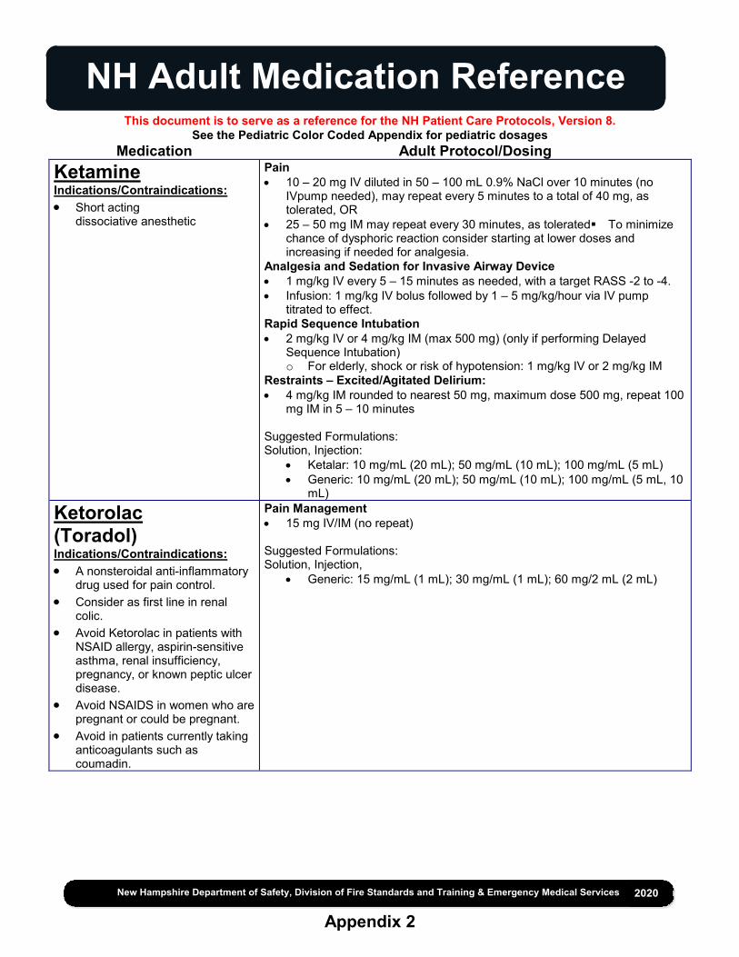
NH Adult Medication Reference
This document is to serve as a reference for the NH Patient Care Protocols, Version 8. See the Pediatric Color Coded Appendix for pediatric dosages
New Hampshire Department of Safety, Division of Fire Standards and Training & Emergency Medical Services 2020
Appendix 2
Medication Adult Protocol/Dosing Ketamine Indications/Contraindications: Short acting
dissociative anesthetic
Pain 10 – 20 mg IV diluted in 50 – 100 mL 0.9% NaCl over 10 minutes (no
IVpump needed), may repeat every 5 minutes to a total of 40 mg, as tolerated, OR
25 – 50 mg IM may repeat every 30 minutes, as toleratedTo minimize chance of dysphoric reaction consider starting at lower doses and increasing if needed for analgesia.
Analgesia and Sedation for Invasive Airway Device 1 mg/kg IV every 5 – 15 minutes as needed, with a target RASS -2 to -4. Infusion: 1 mg/kg IV bolus followed by 1 – 5 mg/kg/hour via IV pump
titrated to effect. Rapid Sequence Intubation 2 mg/kg IV or 4 mg/kg IM (max 500 mg) (only if performing Delayed
Sequence Intubation) o For elderly, shock or risk of hypotension: 1 mg/kg IV or 2 mg/kg IM
Restraints – Excited/Agitated Delirium: 4 mg/kg IM rounded to nearest 50 mg, maximum dose 500 mg, repeat 100
mg IM in 5 – 10 minutes Suggested Formulations: Solution, Injection:
Ketalar: 10 mg/mL (20 mL); 50 mg/mL (10 mL); 100 mg/mL (5 mL) Generic: 10 mg/mL (20 mL); 50 mg/mL (10 mL); 100 mg/mL (5 mL, 10
mL)
Ketorolac (Toradol) Indications/Contraindications: A nonsteroidal anti-inflammatory
drug used for pain control. Consider as first line in renal
colic. Avoid Ketorolac in patients with
NSAID allergy, aspirin-sensitive asthma, renal insufficiency, pregnancy, or known peptic ulcer disease.
Avoid NSAIDS in women who are pregnant or could be pregnant.
Avoid in patients currently taking anticoagulants such as coumadin.
Pain Management 15 mg IV/IM (no repeat) Suggested Formulations: Solution, Injection,
Generic: 15 mg/mL (1 mL); 30 mg/mL (1 mL); 60 mg/2 mL (2 mL)

NH Adult Medication Reference
This document is to serve as a reference for the NH Patient Care Protocols, Version 8. See the Pediatric Color Coded Appendix for pediatric dosages
New Hampshire Department of Safety, Division of Fire Standards and Training & Emergency Medical Services 2020
Appendix 2
Medication Adult Protocol/Dosing Lidocaine Indications/Contraindications: Antiarrhythmic used for control of
ventricular dysrhythmias. Anesthetic for nasotracheal
intubation and intraosseous infusion.
Cardiac Arrest 1 – 1.5 mg/kg IV.
o Repeat dose 0.75 mg/kg up to a maximum dose of 3 mg/kg. Tachycardia 1 – 1.5 mg/kg IV. (considered second-line therapy to Amiodarone).
o May repeat once in 5 minutes to maximum of 3mg/kg. o If successful, consider a maintenance infusion of 1 – 4mg/minute.
Nasotracheal Intubation 2% lidocaine jelly. Intraosseous 1 – 2.5 ml (20 – 50 mg) 2% lidocaine.
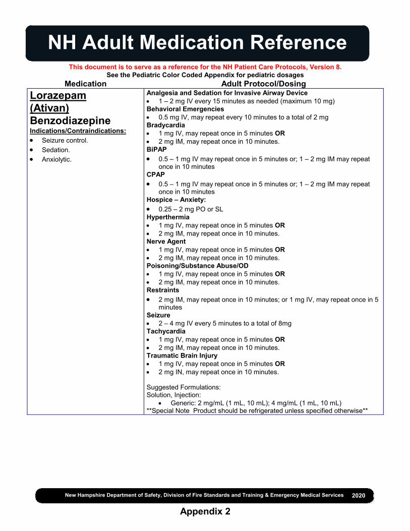
NH Adult Medication Reference
This document is to serve as a reference for the NH Patient Care Protocols, Version 8. See the Pediatric Color Coded Appendix for pediatric dosages
New Hampshire Department of Safety, Division of Fire Standards and Training & Emergency Medical Services 2020
Appendix 2
Medication Adult Protocol/Dosing Lorazepam (Ativan) Benzodiazepine Indications/Contraindications: Seizure control. Sedation. Anxiolytic.
Analgesia and Sedation for Invasive Airway Device 1 – 2 mg IV every 15 minutes as needed (maximum 10 mg) Behavioral Emergencies 0.5 mg IV, may repeat every 10 minutes to a total of 2 mg Bradycardia 1 mg IV, may repeat once in 5 minutes OR 2 mg IM, may repeat once in 10 minutes. BiPAP 0.5 – 1 mg IV may repeat once in 5 minutes or; 1 – 2 mg IM may repeat
once in 10 minutes CPAP 0.5 – 1 mg IV may repeat once in 5 minutes or; 1 – 2 mg IM may repeat
once in 10 minutes Hospice – Anxiety: 0.25 – 2 mg PO or SL Hyperthermia 1 mg IV, may repeat once in 5 minutes OR 2 mg IM, may repeat once in 10 minutes. Nerve Agent 1 mg IV, may repeat once in 5 minutes OR 2 mg IM, may repeat once in 10 minutes. Poisoning/Substance Abuse/OD 1 mg IV, may repeat once in 5 minutes OR 2 mg IM, may repeat once in 10 minutes. Restraints 2 mg IM, may repeat once in 10 minutes; or 1 mg IV, may repeat once in 5
minutes Seizure 2 – 4 mg IV every 5 minutes to a total of 8mg Tachycardia 1 mg IV, may repeat once in 5 minutes OR 2 mg IM, may repeat once in 10 minutes. Traumatic Brain Injury 1 mg IV, may repeat once in 5 minutes OR 2 mg IN, may repeat once in 10 minutes.
Suggested Formulations: Solution, Injection:
Generic: 2 mg/mL (1 mL, 10 mL); 4 mg/mL (1 mL, 10 mL) **Special Note Product should be refrigerated unless specified otherwise**

NH Adult Medication Reference
This document is to serve as a reference for the NH Patient Care Protocols, Version 8. See the Pediatric Color Coded Appendix for pediatric dosages
New Hampshire Department of Safety, Division of Fire Standards and Training & Emergency Medical Services 2020
Appendix 2
Medication Adult Protocol/Dosing Magnesium Sulfate Indications/Contraindications: Elemental electrolyte used to
treat eclampsia during the third trimester of pregnancy.
A smooth muscle relaxer used in refractory respiratory distress resistant to beta-agonists.
Torsades de Pointes.
Asthma/COPD/RAD 2 grams in 100 ml NS given IV over 10 minutes. Obstetrical Emergencies Magnesium sulfate, 4 grams IV (mix in 100 mL 0.9% NaCl) bolus over 10
minutes, then consider 1 gram/hr continuous infusion Seizure Magnesium sulfate, 4 grams IV (mix in 100 mL 0.9% NaCl) bolus over 10
minutes, then consider 1 gram/hr continuous infusion .Cardiac Arrest/Tachycardia – Torsades de Pointes. 1 – 2 grams IV over 5 minutes. Suggested Formulations: Solution, Injection: Generic: 40 mg/mL (50 mL, 100 mL, 500 mL, 1000 mL); 80 mg/mL (50
mL); 50% (2 mL, 10 mL, 20 mL, 50 mL) Solution, Intravenous: Generic: 10 mg/mL (100 mL); 20 mg/mL (500 mL **Special Note 1 g of magnesium sulfate = elemental magnesium 98.6 mg = magnesium 8.12 mEq = magnesium 4.06 mmol**
Methylprednisolone (Solu-medrol) Indications/Contraindications: Steroid used in respiratory
distress to reverse inflammatory and allergic reactions.
Allergic Reaction Extended Care 125 mg IV. Asthma/COPD/RAD 125 mg IV. Suggested Formulations: Solution Reconstituted, Injection, as sodium succinate: Solu-MEDROL: 40 mg (1 ea); 125 mg (1 ea); 500 mg (1 ea); 1000 mg (1
ea)
Metoclopramide (Reglan) Indications/Contraindications: Anti-Emetic used to control
Nausea and/or Vomiting.
Nausea/Vomiting 5mg IV. May repeat once after 10 minutes if nausea/vomiting persists Extended care, may repeat every 4 – 6 hours as needed. Suggested Formulations: Solution, Injection: Generic: 5 mg/mL (2 mL)

NH Adult Medication Reference
This document is to serve as a reference for the NH Patient Care Protocols, Version 8. See the Pediatric Color Coded Appendix for pediatric dosages
New Hampshire Department of Safety, Division of Fire Standards and Training & Emergency Medical Services 2020
Appendix 2
Medication Adult Protocol/Dosing Metoprolol (Lopressor)
Tachycardia 5mg IV over 2 – 5 minutes.
May repeat every five minutes to a maximum of 15mg as needed to achieve a ventricular rate of 90 – 100.
Suggested Formulations: Solution, Intravenous, as tartrate: Generic: 1 mg/mL (5 mL); 5 mg/5 mL (5 mL) Lopressor: 1 mg/mL (5 mL)

NH Adult Medication Reference
This document is to serve as a reference for the NH Patient Care Protocols, Version 8. See the Pediatric Color Coded Appendix for pediatric dosages
New Hampshire Department of Safety, Division of Fire Standards and Training & Emergency Medical Services 2020
Appendix 2
Medication Adult Protocol/Dosing Midazolam (Versed) Benzodiazepine Indications/Contraindications: Seizure control Sedation Anxiolytic Antidote for ketamine
Analgesia and Sedation for Invasive Airway Device 1 – 5 mg IV every 5 – 10 minutes, as needed (in conjunction with fentanyl)
o Infusion 2.5 – 5 mg IV bolus followed by 5 – 30 mg/hour via IV pump titrated to effect
Behavioral Emergencies 2.5 mg IV/IM/IN, may repeat once in 10 minutes to a total of 5 mg Bradycardia 2.5 mg IV/IN may repeat once in 5 minutes OR 5 mg IM may repeat once in 10 minutes. BiPAP 2.5 mg IV/IN may repeat once in 5 minutes OR 5 mg IM may repeat once in 10 minutes. CPAP 2.5 mg IV/IN may repeat once in 5 minutes OR 5 mg IM may repeat once in 10 minutes.
Hospice – Anxiety: 2.5 mg IN, repeat every 10 – 15 minutes as needed to a maximum of 6 mg Hyperthermia 2.5 mg IV/IN may repeat once in 5 minutes OR 5 mg IM may repeat once in 10 minutes. Nerve Agent 2.5 mg IV/IN may repeat once in 5 minutes OR 5 mg IM may repeat once in 10 minutes. Pain – Antidote for ketamine 1 – 2 mg IV/IM every 5 minutes as needed Poisoning/Substance Abuse/OD 2.5 mg IV/IN may repeat once in 5 minutes OR 5 mg IM may repeat once in 20 minutes. Rapid Sequence Intubation 2 – 5 mg IV bolus followed by infusion via pump 5 – 30 mg/hour
o If infusion not used or if additional sedation is required: 2 -5 mg IV every 5 – 10 minutes as needed
Seizure Midazolam 10 mg IM (preferred route) every 10 minutes or 5 – 10 mg IV/IN
every 5 minutes Restraints – Resistant or Aggressive Management (goal is alert & calm)
2 – 5 mg IV/IM/IN, may repeat once in 10 minutes Restraints - Excited/Agitated Delirium (goal is safe and compliant) 10 mg IV/IM/IN; (IM preferred route) may repeat once in 10 minutes Tachycardia 2.5 mg IV/IN may repeat once in 5 minutes OR 5 mg IM may repeat once in 10 minutes. Traumatic Brain Injury 2.5 mg IV/IN may repeat once in 5 minutes OR 5 mg IM may repeat once in 10 minutes. Suggested Formulations: Solution, Injection
Generic: 2 mg/2 mL (2 mL); 5 mg/5 mL (5 mL); 5 mg/mL (1 mL); 10 mg/2 mL (2 mL)

NH Adult Medication Reference
This document is to serve as a reference for the NH Patient Care Protocols, Version 8. See the Pediatric Color Coded Appendix for pediatric dosages
New Hampshire Department of Safety, Division of Fire Standards and Training & Emergency Medical Services 2020
Appendix 2
Medication Adult Protocol/Dosing Morphine Sulfate Indications/Contraindications: Narcotic analgesic Avoid use if BP < 100 mmHg.
Acute Coronary Syndrome 2 – 5 mg IV/IM every 5 minutes to a maximum of 15 mg titrated to pain and
systolic BP remains >100 mmHg. Hospice Per Pain Protocol Pain 2 – 10mg IV/IM every 10 minutes to a total of 20 mg titrated to pain relief
and if systolic BP is >100 mmHg. Suggested Formulations: Solution, Injection, as sulfate: Generic: 2 mg/mL (1 mL); 10 mg/mL (1 mL)
Naloxone (Narcan) Narcotic Antagonist Indications/Contraindications: Narcotic overdose.
Pain Antidote: For hypoventilation from opiate administration by EMS personnel, assist ventilations and administer naloxone as directed in the Opioid Overdose Protocol Opioid Overdose 1mg (1mL) per nostril (IN) via prefilled syringe and atomizer for a total of
2mg OR 4 mg (0.5 mL) commercially prepared nasal spray Repeat every 5 – 10 minutes (maximum) 10 mg) until respiratory
depression resolves and not necessarily until return of consciousness 0.4 – 2.0 mg IV, repeat every 3 – 5 minutes (maximum 10 mg) until
respiratory depression resolves and not necessarily until return of consciousness
0.4 – 2.0 IM, repeat every 5 – 10 minutes (maximum 10 mg) until respiratory depression resolves and not necessarily until return of consciousness
Suggested Formulations: Solution, Injection, as hydrochloride Generic: 1 mg/mL (2 mL) Solution Auto-injector, Injection, as hydrochloride: Evzio: 0.4 mg/0.4 mL (0.4 mL)

NH Adult Medication Reference
This document is to serve as a reference for the NH Patient Care Protocols, Version 8. See the Pediatric Color Coded Appendix for pediatric dosages
New Hampshire Department of Safety, Division of Fire Standards and Training & Emergency Medical Services 2020
Appendix 2
Medication Adult Protocol/Dosing Nitroglycerin Indications/Contraindications: Vasodilator used in the treatment
of CHF and chest pain secondary to acute coronary syndrome
Infusion pump required for infusion.
Acute Coronary Syndrome Facilitate administration of the patient’s own nitroglycerin every 3 – 5
minutes while symptoms persist and systolic BP remains >100 mmHg, to a total of 3 doses
0.4 mg SL every 3 – 5 minutes while symptoms persist and if systolic BP remains >100 mmHg.
10 micrograms/minute if symptoms persist after 3rd SL nitroglycerin (must be on a pump). Increase IV nitroglycerin by 10 micrograms/minute every 5 minutes
while symptoms persist and systolic remains >100 mmHg. Congestive Heart Failure For patients with known history of congestive heart failure, consider: For systolic BP of 140 - 160 mmHg: nitroglycerin 0.4 mg SL. For systolic BP of 160 - 200 mmHg: nitroglycerin 0.8 mg SL (2
tabs/sprays). For systolic BP > 200 mmHg: nitroglycerin 1.2 mg SL (3 tabs/sprays). The above doses may be repeated every 5 minutes until symptomatic
improvement or systolic BP of 140 mmHg. Assess blood pressure every 3 – 5 minutes during nitroglycerin
administration Titrate until symptomatic improvement or systolic BP of 140 mmHg. For systolic BP of 140 - 160 mmHg: IV nitroglycerin start at 50
micrograms/minute. For systolic BP of 160 - 200 mmHg: IV nitroglycerin start at 100
micrograms/minute. For systolic BP > 200 mmHg: nitroglycerin start at 200 micrograms/minute. Note: It is recommended two (2) IV lines in place and the IV nitroglycerin must be on an infusion pump. Generally, accepted maximum dose: 400 micrograms/minute
Nitrous Oxide Indications/Contraindications: "non-narcotic analgesic gas Contraindicated in abdominal
pain, pneumothorax, head injury, or diving emergency patients.
Pain Patient self administers gas for pain control as needed

NH Adult Medication Reference
This document is to serve as a reference for the NH Patient Care Protocols, Version 8. See the Pediatric Color Coded Appendix for pediatric dosages
New Hampshire Department of Safety, Division of Fire Standards and Training & Emergency Medical Services 2020
Appendix 2
Medication Adult Protocol/Dosing Norepinephrine (Levophed) Indications/Contraindications: Alpha and Beta 1 receptor
adronergic receptor agonist vasopressor
Infusion pump required.
Bradycardia Infusion 1 – 30 microgram/minute via pump Non – Traumatic Shock If there is not adequate hemodynamic response after 2,000 mL IV fluid infused, consider: Infusion 1 - 30 micrograms/minute via pump Cardiogenic Shock Infusion 1 – 30 micrograms/minute via pump Post Resuscitation Care Infusion 1 – 30 microgram/min via pump Sepsis & Advanced Sepsis Infusion 1 – 30 microgram/minute via pump Suggested Formulations: Solution, Intravenous [strength expressed as base]: Levophed: 1 mg/mL (4 mL) [contains sodium metabisulfite] Generic: 1 mg/mL (4 mL)
Ondansetron (Zofran) Anti-emetic Indications/Contraindications: Anti-Emetic used to control
Nausea and/or Vomiting.
Nausea/Vomiting 4 mg by mouth or IV/PO/IM Tablet Dispersible, Oral: Zofran ODT: 4 mg, Generic: 4 mg, Solution, Injection [preservative free]:
Generic: 4 mg/2 mL (2 mL)
Oxygen Indications/Contraindications: Indicated in any condition with
increased cardiac work load, respiratory distress, or illness or injury resulting in altered ventilation and/or perfusion. Goal oxygen saturation 94 - 98%.
Indicated for pre-oxygenation whenever possible prior to endotracheal intubation. Goal oxygen saturation 100%.
1-4 liters/min via nasal cannula. 6-15 liters/min via NRB mask. 15 liters via BVM / ETT / supraglottic airway.
Oxytocin (Pitocin) Indications/Contraindications: Routine administration after placental delivery
Obstetrical & Childbirth Oxytocin 10 units IM.

NH Adult Medication Reference
This document is to serve as a reference for the NH Patient Care Protocols, Version 8. See the Pediatric Color Coded Appendix for pediatric dosages
New Hampshire Department of Safety, Division of Fire Standards and Training & Emergency Medical Services 2020
Appendix 2
Medication Adult Protocol/Dosing Pralidoxime (2-PAM) Indications/Contraindications: Antidote for Nerve Agents or
Organophosphate Overdose. Administered with Atropine.
Nerve Agent 1 – 2 grams in 50 – 250 mL of 0.9% NaCl, over 15 – 30 minutes (pump not
required), may repeat within 1 hour if muscle weakness and fasciculations are not relieved. Additional doses may be needed every 3 – 8 hours, if signs of poisoning recur.
Medical Control: Maintenance infusion: up to 500mg per hour (maximum of 12 grams/day).
Prochlorperazine (Compazine) Indications/Contraindications: Anti-Emetic used to control
Nausea and/or Vomiting.
Nausea/Vomiting 5 – 10 mg IV, or 5mg IM. Nausea/Vomiting Extended Care Repeat IM every 4 – 6 hours, as needed Suggested Formulations: Solution, Injection, as edisylate [strength expressed as base]: Generic: 5 mg/mL (2 mL, 10 mL)
Proparacaine (Alcaine) Indications/Contraindications: Topical anesthetic
Eye & Dental 2 drops to affected eye; repeat every 5 minutes as needed. Suggested Formulations: Solution, Ophthalmic, as hydrochloride: Alcaine: 0.5% (15 mL) Parcaine: 0.5% (15 mL) Generic: 0.5% (15 mL)
Rocuronium Indications/Contraindications: Non-depolarizing paralytic agent
used as a component of rapid sequence intubation, when succinylcholine is contraindicated and for post intubation paralysis..
Rapid Sequence Intubation 1mg/kg IBW IV. Suggested Formulations: Solution, Intravenous, as bromide: Zemuron: 50 mg/5 mL (5 mL); 100 mg/10 mL (10 mL) Generic: 50 mg/5 mL (5 mL); 100 mg/10 mL (10 mL)

NH Adult Medication Reference
This document is to serve as a reference for the NH Patient Care Protocols, Version 8. See the Pediatric Color Coded Appendix for pediatric dosages
New Hampshire Department of Safety, Division of Fire Standards and Training & Emergency Medical Services 2020
Appendix 2
Medication Adult Protocol/Dosing Sodium Bicarbonate Indications/Contraindications: A buffer used in acidosis to
increase the pH in Cardiac Arrest, Hyperkalemia or Tricyclic Overdose.
Cardiac Arrest 2 mEq/kg IV Crush Injuries 1 mEq/kg (maximum dose of 50 mEq) IV/IO bolus over 5 minutes Extended Care Secondary to initial bolus, consider sodium bicarbonate infusion: 150 mEq in1000 mL 0.9 % NaCl or D5W at a rate of 250 mL/hr or 4 mL/min Restraints If cardiac arrest occurs with suspected excited delirium, consider early
administration of: fluid bolus, sodium bicarbonate, calcium chloride/gluconate, see Cardiac Arrest Protocol 3.2A.
Poisoning/Substance Abuse/OD Tricyclic with symptomatic dysrhythmias, (eg. tachycardia and wide QRS): 2 mEq/kg IV.
Succinylcholine Paralytic Agent Indications/Contraindications: Paralytic Agent used as a
component of rapid sequence intubation.
Avoid in patients with burns >24 hours old, chronic neuromuscular disease (e.g., muscular dystrophy), ESRD, or other situation in which hyperkalemia is likely.
Rapid Sequence Intubation 1.5mg/kg IV, maximum 150mg Suggested Formulations: Solution, Injection, as chloride: Generic: 20 mg/mL (10 mL) [contains methylparaben] Quelicin: 20 mg/mL (10 mL)
Tetracaine Indications/Contraindications: Topical anesthetic
Eye & Dental 2 drops to affected eye; repeat every 5 minutes as needed. Suggested Formulations: Generic: 0.5% (1 mL, 2 mL, 15 mL)

NH Adult Medication Reference
This document is to serve as a reference for the NH Patient Care Protocols, Version 8. See the Pediatric Color Coded Appendix for pediatric dosages
New Hampshire Department of Safety, Division of Fire Standards and Training & Emergency Medical Services 2020
Appendix 2
Medication Adult Protocol/Dosing Tranexamic Acid (TXA) Indications/Contraindications: Evidence of significant trauma Evidence of severe bleeding The presence of hemodynamic instability AND The injury occurred within the past 3 hours in patients greater than 15 years old without a known allergy to TXA, without an isolated head injury, who has not or will not be receiving factors and is not pregnant with a viable fetus.
Hemorrhage Control Mix 1 gram TXA in 50 - 100mL 0.9% NaCl or LR. Infuse over
approximately 10 minutes IV or IO. Notify receiving facility of TXA administration prior to arrival.
Obstetrical Emergencies Mix 1 gram TXA in 50 - 100mL 0.9% NaCl or LR. Infuse over
approximately 10 minutes IV or IO. Notify receiving facility of TXA administration prior to arrival.
Shock – Trauma Consider tranexamic acid see, Hemorrhage Control Protocol 4.4

New Hampshire Department of Safety, Division of Fire Standards and Training & Emergency Medical Services 2020
Epinephrine Preparation: 1 mg in 250 mL NaCl 0.9%
Concentration: 4 mcg/mL
Dose wanted (mcg/min)
Rate for Infusion (mL/hr)
2 mcg/min 30 mL/hr
3 mcg/min 45 mL/hr
4 mcg/min 60 mL/hr
5 mcg/min 75 mL/hr
6 mcg/min 90 mL/hr
7 mcg/min 105 mL/hr
8 mcg/min 120 mL/hr
9 mcg/min 135 mL/hr
10 mcg/min 150 mL/hr
Epinephrine Preparation: 1 mg in 500 mL NaCl 0.9%
Concentration: 2 mcg/mL
Dose wanted (mcg/min)
Rate for Infusion (mL/hr)
2 mcg/min 60 mL/hr
3 mcg/min 90 mL/hr
4 mcg/min 120 mL/hr
5 mcg/min 150 mL/hr
6 mcg/min 180 mL/hr
7 mcg/min 210 mL/hr
8 mcg/min 240 mL/hr
9 mcg/min 270 mL/hr
10 mcg/min 300 mL/hr
Epinephrine Preparation: 1 mg in 1000 mL NaCl 0.9%
Concentration: 1 mcg/mL
Dose wanted (mcg/min)
Rate for Infusion (mL/hr)
2 mcg/min 120 mL/hr
3 mcg/min 180 mL/hr
4 mcg/min 240 mL/hr
5 mcg/min 300 mL/hr
6 mcg/min 360 mL/hr
7 mcg/min 420 mL/hr
8 mcg/min 480 mL/hr
9 mcg/min 540 mL/hr
10 mcg/min 600 mL/hr
Norepinephrine (Levophed) Preparation: 4 mg in 500 mL NaCl 0.9%
Concentration: 8 mcg/mL
Dose wanted (mcg/min)
Rate for Infusion(mL/hr)
2 mcg/min 15
4 mcg/min 30
6 mcg/min 45
8 mcg/min 60
10 mcg/min 75
12 mcg/min 90
14 mcg/min 105
16 mcg/min 120
18 mcg/min 135
20 mcg/min 150
22 mcg/min 165
24 mcg/min 180
26 mcg/min 195
28 mcg/min 210
30 mcg/min 225
Norepinephrine (Levophed) Preparation: 4 mg in 1000 mL NaCl 0.9%
Concentration: 4 mcg/mL
Dose wanted (mcg/min)
Rate for Infusion(mL/hr)
2 mcg/min 30
4 mcg/min 60
6 mcg/min 90
8 mcg/min 120
10 mcg/min 150
12 mcg/min 180
14 mcg/min 210
16 mcg/min 240
18 mcg/min 270
20 mcg/min 300
22 mcg/min 330
24 mcg/min 360
26 mcg/min 390
28 mcg/min 420
30 mcg/min 450

New Hampshire Department of Safety, Division of Fire Standards and Training & Emergency Medical Services 2020
Nitroglycerin Preparation: 50 mg in 250 mL Concentration: 200 mcg/mL
Dose wanted (mcg/min)
Rate for Infusion (mL/hr)
10 mcg/min 3 mL/hr
20 mcg/min 6 mL/hr
30 mcg/min 9 mL/hr
40 mcg/min 12 mL/hr
50 mcg/min 15 mL/hr
60 mcg/min 18 mL/hr
70 mcg/min 21 mL/hr
80 mcg/min 24 mL/hr
90 mcg/min 27 mL/hr
100 mcg/min 30 mL/hr
120 mcg/min 36 mL/hr
140 mcg/min 42 mL/hr
160 mcg/min 48 mL/hr
180 mcg/min 54 mL/hr
200 mcg/min 60 mL/hr
250 mcg/min 75 mL/hr
300 mcg/min 90 mL/hr
350 mcg/min 105 mL/hr
400 mcg/min 120 mL/hr ACS – start at 10; increase by 10 q 5 min CHF – 140-160 50mcg/min, 160-200 100 mcg/min, >200 – 200 mcg/min
Nitroglycerin Preparation: 100 mg in 250 mL
Concentration: 400 mcg/mL
Dose wanted (mcg/min)
Rate for Infusion (mL/hr)
10 mcg/min 1.5 mL/hr
20 mcg/min 3 mL/hr
30 mcg/min 4.5 mL/hr
40 mcg/min 6 mL/hr
50 mcg/min 7.5 mL/hr
60 mcg/min 9 mL/hr
70 mcg/min 10.5 mL/hr
80 mcg/min 12 mL/hr
90 mcg/min 13.5 mL/hr
100 mcg/min 15 mL/hr
120 mcg/min 18 mL/hr
140 mcg/min 21 mL/hr
160 mcg/min 24 mL/hr
180 mcg/min 27 mL/hr
200 mcg/min 30 mL/hr
250 mcg/min 37.5 mL/hr
300 mcg/min 45 mL/hr
350 mcg/min 52.5 mL/hr
400 mcg/min 60 mL/hr ACS – start at 10; increase by 10 q 5 min CHF – 140-160 50mcg/min, 160-200 100 mcg/min, >200 200 mcg/min

New Hampshire Department of Safety, Division of Fire Standards and Training & Emergency Medical Services 2020
Cardizem (Diltiazem) Preparation: 25 mg in 100 mL NaCl 0.9%
Concentration: 0.25 mg/mL
Dose wanted (mg/hr) Rate for Infusion (mL/hr)
5 mg/hr 20 mL/hr
10 mg/hr 40 mL/hr
15 mg/hr 60 mL/hr
Lidocaine Preparation: 2 grams in 500 mL
Concentration: 4 mg/mL
1 mg/min 15 mL/hr
2 mg/min 30 mL/hr
3 mg/min 45 mL/hr
4 mg/min 60 mL/hr
Midazolam Preparation: 5 mg in 250 mL Concentration: 0.02 mg/mL
2 mg/hr 100 mL/hr
3 mg/hr 150 mL/hr
4 mg/hr 200 mL/hr
5 mg/hr 250 mL/hr

Weight 8-9 Kg (Avg 8.5 Kg)
Pediatric Color Coded AppendixLe
ngth
66.
5-74
cm
Red
(7-1
0 M
onth
s)
Weight 3-5 Kg (Avg 4.0 Kg)
Leng
th <
59.
5 cm
Gra
y (0
-3 m
onth
s)Vital SignsHeart Rate: 120-150Respirations: 24-48BP Systolic: 70 (+/-25)
EquipmentET Tube: 2.5 - 3.5Blade Size: 0 - 1
DefibrillationDefibrillation: 8 J, 15 JCardioversion: 2 J, 4 J
Albuterol 2.5 mgAmiodarone 20 mgAtropine- Bradycardia 0.08 mg - Organophosphate Poison 0.2 mgCalcium Chloride 80 mgCalcium Gluconate 400mgDexamethasone 2 mgDextrose 10% 20 mLDiazepam (IV) 0.5 mg
(Rectal) 2.0 mg Diphenhydramine HOLDEpinephrine 1:10,000 0.04 mgEpinephrine 1:1000 Nebulized 3 mgEpinephrine 1:1000 IM 0.15 mgEpinephrine (Racemic 2.25%) 0.5 mLFentanyl HOLDGlucagon 0.5 mgGlucose Oral 15 gm tubeHydrocortisone 10 mgHydromorphone 0.04 mgHydroxocobalamin 280 mg (11.2 mL)Ibuprofen HOLD
Normal Saline 40-80 ml
Weight 6-7 Kg (Avg 6.5 Kg)
Ipratropium w/ albuterol 500 mcgLidocaine: - Cardiac Arrest 4 mg - Intraosseous 2 mgLorazepam 0.4 mgMagnesium Sulfate - Asthma 150 mg - Torsades 200 mgMethylprednisolone
1mg/kg 4 mg2mg/kg 8 mg
Midazolam (0.1mg/kg) 0.4 mg Morphine Sulfate 0.4 mgNaloxone – IN 1 mg Ondansetron - IV 0.4 mg - ODT 2 mgPralidoxime IV 60 mgProparacaine 2 dropsSodium Bicarbonate 8 mEqTetracaine 2 drops
Leng
th 5
9.5-
66.5
cm
Pink
(3-6
Mon
ths)
Appendix 3 (Page 1 of 3) 2020
Acetaminophen 60 mgAdenosine: 1st Dose- 0.4 mg Repeat Dose- 0.8 mg Activated Charcoal 4.2grams
Vital SignsHeart Rate: 120-125Respirations: 24-48BP Systolic: 85 (+/-25)
EquipmentET Tube: 3.5Blade Size: 1
DefibrillationDefibrillation: 10 J, 20 JCardioversion: 2 J, 5 J
Albuterol 2.5 mgAmiodarone 30 mgAtropine- Bradycardia 0.12 mg - Organophosphate Poison 0.320 mgCalcium Chloride 120 mgCalcium Gluconate 600mgDexamethasone 4 mgDextrose 10% 35 mLDiazepam (IV) 0.5 mg
(Rectal) 3 mg Diphenhydramine HOLDEpinephrine 1:10,000 0.06 mgEpinephrine 1:1000 Nebulized 3 mgEpinephrine 1:1000 IM 0.15 mgEpinephrine (Racemic 2.25%) 0.5 mLFentanyl HOLDGlucagon 0.5 mgGlucose Oral 15 gm tubeHydrocortisone 15 mgHydromorphone 0.065 mgHydroxocobalamin 450 mg (18 mL)Ibuprofen HOLD
Normal Saline 65-130 ml
Ipratropium w/ albuterol 500 mcgLidocaine: - Cardiac Arrest 6 mg - Intraosseous 3 mgLorazepam 0.65 mgMagnesium Sulfate - Asthma 250mg - Torsades 300 mgMethylprednisolone
1mg/kg 8 mg2mg/kg 12 mg
Midazolam (0.1mg/kg) 0.6 mg Morphine Sulfate 0.6 mgNaloxone – IN 1 mg Ondansetron - IV 0.6mg - ODT 2 mgPralidoxime IV 100 mgProparacaine 2 dropsSodium Bicarbonate 13 mEqTetracaine 2 drops
Acetaminophen 97.5 mgAdenosine: 1st Dose- 0.65 mg Repeat Dose- 1.3 mg Activated Charcoal 6.3 grams
Vital SignsHeart Rate: 120Respirations: 24-32BP Systolic: 92 (+/-25)
EquipmentET Tube: 3.5 -4.0Blade Size: 1
DefibrillationDefibrillation: 20 J, 40 JCardioversion: 5 J, 9 J
Albuterol 2.5 mgAmiodarone 40 mgAtropine- Bradycardia 0.18 mg - Organophosphate Poison 0.420 mgCalcium Chloride 180 mgCalcium Gluconate 800mgDexamethasone 5 mgDextrose 10% 45 mLDiazepam (IV) 1 mg
(Rectal) 4 mg Diphenhydramine HOLDEpinephrine 1:10,000 0.08 mgEpinephrine 1:1000 Nebulized 3 mgEpinephrine 1:1000 IM 0.15 mgEpinephrine (Racemic 2.25%) 0.5 mLFentanyl 10 mcgGlucagon 0.5 mgGlucose Oral 15 gm tubeHydrocortisone 18 mgHydromorphone 0.085 mgHydroxocobalamin 595 mg (23.8 mL)Ibuprofen HOLD
Normal Saline 85-170 ml
Ipratropium w/ albuterol 500 mcgLidocaine: - Cardiac Arrest 8 mg - Intraosseous 4 mgLorazepam 0.8 mgMagnesium Sulfate - Asthma 350 mg - Torsades 400 mgMethylprednisolone
1mg/kg 8 mg2mg/kg 16 mg
Midazolam (0.1mg/kg) 0.8 mg Morphine Sulfate 0.8 mgNaloxone – IN 1 mg Ondansetron - IV 0.8 mg - ODT 2 mgPralidoxime IV 128 mgProparacaine 2 dropsSodium Bicarbonate 21 mEqTetracaine 2 drops
Acetaminophen 120 mgAdenosine: 1st Dose- 0.85 mg Repeat Dose- 1.7 mg Activated Charcoal 8.3grams

Weight 12-14 Kg (Avg 13 Kg)
Leng
th 8
4.5-
97.5
cm
Yello
w (1
9-35
Mon
ths)
Weight 15-18 Kg (Avg 16.5 Kg)
Leng
th 9
7.5-
110
cm
Whi
te (3
-4 y
rs)
Weight 10-11 Kg (Avg 10.5 Kg)
Leng
th 7
4-84
.5 c
m
Purp
le (1
1-18
Mon
ths)
Appendix 3 (Page 2 of 3) 2020
Pediatric Color Coded Appendix
Vital SignsHeart Rate: 115-120Respirations: 22-30BP Systolic: 96 (+/-30)
EquipmentET Tube: 4.0Blade Size: 1
DefibrillationDefibrillation: 20 J, 40 JCardioversion: 5 J, 10 J
Albuterol 2.5 mgAmiodarone 50 mgAtropine- Bradycardia 0.2 mg - Organophosphate Poison 0.5 mgCalcium Chloride 200 mgCalcium Gluconate 1000 mgDexamethasone 6 mgDextrose 10% 55 mLDiazepam (IV) 1 mg
(Rectal) 5 mg Diphenhydramine 10 mgEpinephrine 1:10,000 0.1 mgEpinephrine 1:1000 Nebulized 3 mgEpinephrine 1:1000 IM 0.15 mgEpinephrine (Racemic 2.25%) 0.5 mLFentanyl 10 mcgGlucagon 0.5 mgGlucose Oral 15 gm tubeHydrocortisone 20 mgHydromorphone 0.1 mgHydroxocobalamin 740 mg (29.5 mL)Ibuprofen 100 mg
Normal Saline 105-210 ml
Ipratropium w/ albuterol 500 mcgLidocaine: - Cardiac Arrest 12 mg - Intraosseous 6 mgLorazepam 1 mgMagnesium Sulfate - Asthma 400 mg - Torsades 550 mgMethylprednisolone
1mg/kg 12 mg2mg/kg 20 mg
Midazolam (0.1mg/kg) 1 mg Morphine Sulfate 1 mgNaloxone - IN 1 mg Ondansetron - IV 1 mg - ODT 2 mgPralidoxime IV 160 mgProparacaine 2 dropsSodium Bicarbonate 21 mEqTetracaine 2 drops
Acetaminophen 160 mgAdenosine: 1st Dose- 1.05 mg Repeat Dose- 2.1 mg Activated Charcoal 10.4 grams
Vital SignsHeart Rate: 110-115Respirations: 20-28BP Systolic: 100 (+/-30)
EquipmentET Tube: 4.5Blade Size: 2
DefibrillationDefibrillation: 30 J, 50 JCardioversion: 6 J, 15 J
Albuterol 2.5 mgAmiodarone 65 mgAtropine- Bradycardia 0.26 mg - Organophosphate Poison 0.65 mgCalcium Chloride 260 mgCalcium Gluconate 1300 mgDexamethasone 8 mgDextrose 10% 65 mLDiazepam (IV) 1.5 mg
(Rectal) 6 mg Diphenhydramine 20 mgEpinephrine 1:10,000 0.12 mgEpinephrine 1:1000 Nebulized 3 mgEpinephrine 1:1000 IM 0.15 mgEpinephrine (Racemic 2.25%) 0.5 mLFentanyl 15 mcgGlucagon 0.5 mgGlucose Oral 15 gm tubeHydrocortisone 25 mgHydromorphone 0.13 mgHydroxocobalamin 900 mg (36 mL)Ibuprofen 130 mg
Normal Saline 130-260 ml
Ipratropium w/ albuterol 500 mcgLidocaine: - Cardiac Arrest 12 mg - Intraosseous 6 mgLorazepam 1.4 mgMagnesium Sulfate - Asthma 550 mg - Torsades 650 mgMethylprednisolone
1mg/kg 12 mg2mg/kg 28 mg
Midazolam (0.1mg/kg) 1.3 mg Morphine Sulfate 1.2 mgNaloxone – IN 1 mg Ondansetron - IV 1.4 mg - ODT 2 mgPralidoxime IV 200 mgProparacaine 2 dropsSodium Bicarbonate 26 mEqTetracaine 2 drops
Acetaminophen 195 mgAdenosine: 1st Dose- 1.3mg Repeat Dose- 2.6 mg Activated Charcoal 12.5 grams
Vital SignsHeart Rate: 100 - 115Respirations: 20-26BP Systolic: 100 (+/-20)
EquipmentET Tube: 5.0Blade Size: 2
DefibrillationDefibrillation: 30 J, 70 JCardioversion: 8 J, 15 J
Albuterol 2.5 mgAmiodarone 80 mgAtropine- Bradycardia 0.3 mg - Organophosphate Poison 0.8 mgCalcium Chloride 320 mgCalcium Gluconate 1600mgDexamethasone 10 mgDextrose 10% 85 mLDiazepam (IV) 2 mg
(Rectal) 8 mg Diphenhydramine 20 mgEpinephrine 1:10,000 0.16 mgEpinephrine 1:1000 Nebulized 3 mgEpinephrine 1:1000 IM 0.15 mgEpinephrine (Racemic 2.25%) 0.5 mLFentanyl 16 mcgGlucagon 0.5 mgGlucose Oral 15 gm tubeHydrocortisone 30 mgHydromorphone 0.165 mgHydroxocobalamin 1150 mg (46 mL)Ibuprofen 130 mg
Normal Saline 165-330 ml
Ipratropium w/ albuterol 500 mcgLidocaine: - Cardiac Arrest 16 mg - Intraosseous 8 mgLorazepam 1.6 mgMagnesium Sulfate -Asthma 650 mg - Torsades 850 mgMethylprednisolone
1mg/kg 16 mg2mg/kg 32 mg
Midazolam (0.1mg/kg) 1.6 mg Morphine Sulfate 1.6 mgNaloxone – IN 1 mg Ondansetron - IV 1.6 mg - ODT 4 mgPralidoxime IV 250 mgProparacaine 2 dropsSodium Bicarbonate 33 mEqTetracaine 2 drops
Acetaminophen 247.5 mgAdenosine: 1st Dose- 1.65 mg Repeat Dose- 3.3 mg Activated Charcoal 16.7 grams

Leng
th 1
37-1
50 c
m
Gre
en (1
0-12
yrs
)
Weight 24-30 Kg (Avg 27 Kg)
Leng
th 1
22-1
37 c
m
Ora
nge
(7-9
yrs
)
Weight 19-22 Kg (Avg 20.75 Kg)
Leng
th 1
10-1
22 c
m
Blu
e (5
-6 y
rs)
Pediatric Color Coded Appendix
Appendix 3 (Page 3 of 3) 2020
Weight 32-40 Kg (Avg 36 Kg)Vital SignsHeart Rate: 85-90Respirations: 16-22BP Systolic: 115 (+/-20)
EquipmentET Tube: 6.5Blade Size: 3
DefibrillationDefibrillation: 60 J, 150 JCardioversion: 15 J, 30 J
Albuterol 2.5 mgAmiodarone 180 mgAtropine- Bradycardia 0.5 mg - Organophosphate Poison 1.8 mgCalcium Chloride 720 mgCalcium Gluconate 2 gramsDexamethasone 10 mgDextrose 10% 180 mLDiazepam (IV) 3.5 mg
(Rectal) 18 mg Diphenhydramine 50 mgEpinephrine 1:10,000 0.36 mgEpinephrine 1:1000 Nebulized 3 mgEpinephrine 1:1000 IM 0.3 mgEpinephrine (Racemic 2.25%) 0.5 mLFentanyl 40 mcgGlucagon 1 mgGlucose Oral 15 gm tubeHydrocortisone 70 mgHydromorphone 0.36 mgHydroxocobalamin 2520 mg (100.8 mL)Ibuprofen 360 mg
Normal Saline 250-500 ml
Ipratropium w/ albuterol 500 mcgLidocaine: - Cardiac Arrest 36 mg - Intraosseous 18 mgLorazepam 3.6 mgMagnesium Sulfate - Asthma 1450 mg - Torsades 1800 mgMethylprednisolone
1mg/kg 36 mg2mg/kg 72 mg
Midazolam (0.1mg/kg) 3.6 mg Morphine Sulfate 3.6 mgNaloxone – IN 2 mg Ondansetron - IV 3.6 mg - ODT 4 mgPralidoxime IV 540 mgProparacaine 2 dropsSodium Bicarbonate 72 mEqTetracaine 2 drops
Acetaminophen 540 mgAdenosine: 1st Dose- 3.6 mg Repeat Dose- 7.2 mg Activated Charcoal 33.3
Vital SignsHeart Rate: 90Respirations: 18-22BP Systolic: 105 (+/-15)
EquipmentET Tube: 6.0Blade Size: 2-3
DefibrillationDefibrillation: 50 J, 100 JCardioversion: 15 J, 30 J
Albuterol 2.5 mgAmiodarone 140 mgAtropine- Bradycardia 0.5 mg - Organophosphate Poison 1.2 mgCalcium Chloride 540 mgCalcium Gluconate 2 gramsDexamethasone 10 mgDextrose 10% 135 mLDiazepam (IV) 3 mg
(Rectal) 13 mg Diphenhydramine 40 mgEpinephrine 1:10,000 0.22 mgEpinephrine 1:1000 Nebulized 3 mgEpinephrine 1:1000 IM 0.3 mgEpinephrine (Racemic 2.25%) 0.5 mLFentanyl 25 mcgGlucagon 0.27 mgGlucose Oral 15 gm tubeHydrocortisone 50 mgHydromorphone 1 mgHydroxocobalamin 1890 mg (75.6 mL)Ibuprofen 270 mg
Normal Saline 250-500 ml
Ipratropium w/ albuterol 500 mcgLidocaine: - Cardiac Arrest 28 mg - Intraosseous 14 mgLorazepam 2.8 mgMagnesium Sulfate - Asthma 1100 mg - Torsades 1350 mgMethylprednisolone
1mg/kg 28 mg2mg/kg 56 mg
Midazolam (0.1mg/kg) 2.7 mg Morphine Sulfate 2.8 mgNaloxone – IN 2 mg Ondansetron - IV 2.8 mg - ODT 4 mgPralidoxime IV 400 mgProparacaine 2 dropsSodium Bicarbonate 54 mEqTetracaine 2 drops
Acetaminophen 405 mgAdenosine: 1st Dose- 2.7 mg Repeat Dose- 5.4 mg Activated Charcoal 27 grams
Vital SignsHeart Rate: 100Respirations: 20-24BP Systolic: 100 (+/-15)
EquipmentET Tube: 5.5Blade Size: 2
DefibrillationDefibrillation: 40 J, 85 JCardioversion: 10 J, 20 J
Albuterol 2.5 mgAmiodarone 105 mgAtropine- Bradycardia 0.4 mg - Organophosphate Poison 1 mgCalcium Chloride 400 mgCalcium Gluconate 2 gramsDexamethasone 10 mgDextrose 10% 105 mLDiazepam (IV) 2 mg
(Rectal) 10 mg Diphenhydramine 30 mgEpinephrine 1:10,000 0.2 mgEpinephrine 1:1000 Nebulized 3 mgEpinephrine 1:1000 IM 0.15 mgEpinephrine (Racemic 2.25%) 0.5 mLFentanyl 20 mcgGlucagon 0.2 mgGlucose Oral 15 gm tubeHydrocortisone 40 mgHydroxocobalamin 1450 mg (58 mL)Ibuprofen 205 mg
Normal Saline 205-410 ml
Ipratropium w/ albuterol 500 mcgLidocaine: - Cardiac Arrest 20 mg - Intraosseous 10 mgLorazepam 2 mgMagnesium Sulfate - Asthma 850 mg - Torsades 1050 mgMethylprednisolone
1mg/kg 20 mg2mg/kg 40 mg
Midazolam (0.1mg/kg) 2 mg Morphine Sulfate 2 mgNaloxone – IN 2 mg Ondansetron - IV 2 mg - ODT 4 mgPralidoxime IV 320 mgProparacaine 2 dropsSodium Bicarbonate 41mEqTetracaine 2 drops
Acetaminophen 311.25 mgAdenosine: 1st Dose- 2.075 mg Repeat Dose- 4.15 mg Activated Charcoal 20.8 grams
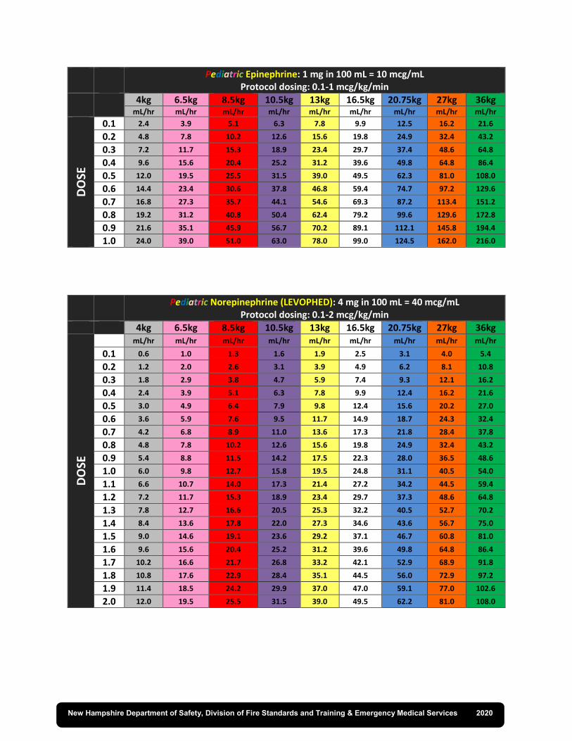
Pediatric Epinephrine: 1 mg in 100 mL = 10 mcg/mL Protocol dosing: 0.1-1 mcg/kg/min
4kg 6.5kg 8.5kg 10.5kg 13kg 16.5kg 20.75kg 27kg 36kg
mL/hr mL/hr mL/hr mL/hr mL/hr mL/hr mL/hr mL/hr mL/hr
DO
SE
0.1 2.4 3.9 5.1 6.3 7.8 9.9 12.5 16.2 21.6
0.2 4.8 7.8 10.2 12.6 15.6 19.8 24.9 32.4 43.2
0.3 7.2 11.7 15.3 18.9 23.4 29.7 37.4 48.6 64.8
0.4 9.6 15.6 20.4 25.2 31.2 39.6 49.8 64.8 86.4
0.5 12.0 19.5 25.5 31.5 39.0 49.5 62.3 81.0 108.0
0.6 14.4 23.4 30.6 37.8 46.8 59.4 74.7 97.2 129.6
0.7 16.8 27.3 35.7 44.1 54.6 69.3 87.2 113.4 151.2
0.8 19.2 31.2 40.8 50.4 62.4 79.2 99.6 129.6 172.8
0.9 21.6 35.1 45.9 56.7 70.2 89.1 112.1 145.8 194.4
1.0 24.0 39.0 51.0 63.0 78.0 99.0 124.5 162.0 216.0
Pediatric Norepinephrine (LEVOPHED): 4 mg in 100 mL = 40 mcg/mL Protocol dosing: 0.1-2 mcg/kg/min
4kg 6.5kg 8.5kg 10.5kg 13kg 16.5kg 20.75kg 27kg 36kg
DO
SE
mL/hr mL/hr mL/hr mL/hr mL/hr mL/hr mL/hr mL/hr mL/hr
0.1 0.6 1.0 1.3 1.6 1.9 2.5 3.1 4.0 5.4
0.2 1.2 2.0 2.6 3.1 3.9 4.9 6.2 8.1 10.8
0.3 1.8 2.9 3.8 4.7 5.9 7.4 9.3 12.1 16.2
0.4 2.4 3.9 5.1 6.3 7.8 9.9 12.4 16.2 21.6
0.5 3.0 4.9 6.4 7.9 9.8 12.4 15.6 20.2 27.0
0.6 3.6 5.9 7.6 9.5 11.7 14.9 18.7 24.3 32.4
0.7 4.2 6.8 8.9 11.0 13.6 17.3 21.8 28.4 37.8
0.8 4.8 7.8 10.2 12.6 15.6 19.8 24.9 32.4 43.2
0.9 5.4 8.8 11.5 14.2 17.5 22.3 28.0 36.5 48.6
1.0 6.0 9.8 12.7 15.8 19.5 24.8 31.1 40.5 54.0
1.1 6.6 10.7 14.0 17.3 21.4 27.2 34.2 44.5 59.4
1.2 7.2 11.7 15.3 18.9 23.4 29.7 37.3 48.6 64.8
1.3 7.8 12.7 16.6 20.5 25.3 32.2 40.5 52.7 70.2
1.4 8.4 13.6 17.8 22.0 27.3 34.6 43.6 56.7 75.0
1.5 9.0 14.6 19.1 23.6 29.2 37.1 46.7 60.8 81.0
1.6 9.6 15.6 20.4 25.2 31.2 39.6 49.8 64.8 86.4
1.7 10.2 16.6 21.7 26.8 33.2 42.1 52.9 68.9 91.8
1.8 10.8 17.6 22.9 28.4 35.1 44.5 56.0 72.9 97.2
1.9 11.4 18.5 24.2 29.9 37.0 47.0 59.1 77.0 102.6
2.0 12.0 19.5 25.5 31.5 39.0 49.5 62.2 81.0 108.0
New Hampshire Department of Safety, Division of Fire Standards and Training & Emergency Medical Services 2020

Pediatric Vital Signs
Normal Heart Rates by age Normal Respiratory Rates by age
Age Awake
Rate Mean
Sleeping Rate
Age Breaths per
Minute
Newborn to 3 months
85-205 140 80-160 Infant (<1 year) 30-60
3 months to 2 years 100-190 130 75-160 Toddler (1-3 years) 24-40 2 years to 10 years 60-140 80 60-90 Preschooler (4-5 years) 22-34
>10 years 60-100 75 50-90 School-age child (6-12
years) 18-30
Adolescent > 12 12-16
Normal Blood Pressures by age
Age Systolic Blood Pressure (mmHg) Diastolic Blood Pressure (mmHg)
Female Male Female Male
Neonate (1 day) 60-76 60-74 31-45 30-44
Neonate (4 days) 67-83 68-84 37-53 35-53
Infant (1 month) 73-91 74-94 36-56 37-55
Infant (3 months) 78-100 81-103 44-64 45-65
Infant (6 months) 82-102 87-105 46-66 48-68
Child (2 years) 88-105 88-106 45-63 42-61
Child (7 years) 96-113 97-115 57-75 57-76
Adolescent (15 years) 110-127 113-131 65-83 64-83
Threshold by age of Systolic Blood Pressure Indicating Hypotension
Age Systolic Blood Pressure
Term neonates (0-28) Less than 60 mmHg
Infant (1-12 months) Less than 70 mmHg
Children 1-10 years Less than 70 + (age in years x 2) mmHg
Children > 10 Less than 90 mmHg
Equipment
GRAY 3-5 Kg
PINK 6-7
RED 8-9 Kg
PURPLE 10-11 Kg
YELLOW 12-14 Kg
WHITE 15-18 Kg
BLUE 19-23 Kg
ORANGE 24-29 Kg
GREEN 30-36 Kg
BVM Anesthesia Bag/Infant
Infant Infant
Pedi Pedi Pedi Pedi Pedi Adult
NRB Infant Infant Infant Pedi Pedi Pedi Pedi Pedi Adult
Oral airway 40-50 50 50 60 60 60 70 80 80
NPA 14 14 - 15 14 - 15 18 20 22 24 26 26
Laryng blade (size)
1 Straight
1 Straight
1 Straight
1-1.5 Straight
2 Straight or
curved
2 Straight
2 Straight or curved
2 Straight or curved
3 Straight or curved
ET tube (mm)
3-3.5 Uncuff 3.5
Uncuff 3 Cuff
3.5 Uncuff 3 Cuff
4.0 Uncuff 3.5 Cuff
4.5 Uncuff 4.0 Cuff
5.0 Uncuff 4.5 Cuff
5.5 Uncuff 5.0 Cuff
6.0 Cuffed 6.5 Cuffed
ETT insertion Length
(cm)
3k: 9-9.5 4k: 9.5 - 10
5K: 10 - 10.5 10.5 - 11 10.5 - 11
11 - 12
12.5 - 13.5
14 - 15
15.5 - 16.5
17 - 18
18.5 - 19.5
Suction Catheter
8 8 8 8 10 10 10 10 10-12
BP cuff Neo # 2- 4 Infant/
Pedi Infant/
Pedi Pedi Pedi Pedi Pedi Pedi
Small Adult
IV catheter 24G (14mm) 22 - 24 22 - 24 20 - 24 18 - 22 18 - 22 18 - 20 18 - 20 16 - 20
NG tube (F) 5-8 5 - 8 5 - 8 8 - 10 10 10 12 - 14 14 - 18 16 - 18
Urinary Catheter
5 8 8 8 - 10 10 10 - 12 10 -12 12 12
Chest Tube 10-12 10-12 10-12 16-20 20-24 20-24 24-28 28-32 32-38
New Hampshire Department of Safety, Division of Fire Standards and Training & Emergency Medical Services 2020

X Skill allowed under protocol and taught in the National Education Standards.
* Skill allowed under protocol after completion of a scope of practice module.
Revised 08/20/21 Appendix 6
ADULT Scope of Practice
Airway Management EMR EMT AEMT PARAMEDIC
BVM X X X X
Chest Tube Maintenance X
Cleared, Opened, Heimlich X X X X
BiPAP X
CPAP * * X
Cricothyrotomy – Percutaneous X
Cricothyrotomy – Surgical (Bougie Assisted) Prerequisite
Endotracheal Intubation X
Endotracheal Suctioning X X
Nasogastric Tube X
Nasopharyngeal Airway X X X
Nasotracheal Intubation X
Nebulizer Treatment X X
Needle Decompression X
Oral Suctioning X X X X
Oropharyngeal Airway X X X X
Oxygen Administration X X X X
Pulse Oximetry X X X
Rapid Sequence Intubation Prerequisite
Supraglottic Airways * X X
Tracheostomy Maintenance X X X
Ventilator Operation X

X Skill allowed under protocol and taught in the National Education Standards.
* Skill allowed under protocol after completion of a scope of practice module.
Revised 08/20/21 Appendix 6
ADULT Scope of Practice
Medication Administration Route EMR EMT AEMT PARAMEDIC
Auto Injector Provider
Protection X X X
Blood Products X
Endotracheal X
Inhalation MDI X X
Intramuscular X X
Intraosseous * X
Intravenous X X
Intravenous Pump Interfacility
Transfers X
Oral X X X
Intranasal * * X X
Rectal X
Subcutaneous X X
Sublingual Assist X X
Vascular Access EMR EMT AEMT PARAMEDIC
Blood Draw * X
Blood Glucose Analysis * X X
Central Line Maintenance X
External Jugular X
Peripheral Venous Access X X
Intraosseous - Adult
X
Commercial Devices X

X Skill allowed under protocol and taught in the National Education Standards.
* Skill allowed under protocol after completion of a scope of practice module.
Revised 08/20/21 Appendix 6
ADULT Scope of Practice
Cardiac Management EMR EMT AEMT PARAMEDIC
Application of 12 Lead ECG * * X
Application of 3 or 4 lead
ECG * * X
CPR - Cardiopulmonary Resuscitation X X X X
Defibrillation - AED X X X X
Defibrillation - Manual X
Interpretation of 12 Lead
ECG X
Interpretation of 3 or 4 lead X
Synchronized Cardioversion X
Transcutaneous Pacing X

X Skill allowed under protocol and taught in the National Education Standards.
* Skill allowed under protocol after completion of a scope of practice module.
Revised 08/20/21 Appendix 6
PEDIATRIC Scope of Practice
Airway Management EMR EMT AEMT PARAMEDIC
BVM X X X X
Cleared, Opened, Heimlich X X X X
CPAP * * X
Endotracheal Intubation X
Endotracheal Suctioning X X
Nasogastric Tube X
Nasopharyngeal Airway X X X
Nebulizer Treatment X X
Needle Decompression X
Oral Suctioning X X X X
Oropharyngeal Airway X X X X
Oxygen Administration X X X X
Pulse Oximetry X X X
Supraglottic Airways * X X
Tracheostomy Maintenance X X X
Ventilator Operation X

X Skill allowed under protocol and taught in the National Education Standards.
* Skill allowed under protocol after completion of a scope of practice module.
Revised 08/20/21 Appendix 6
PEDIATRIC Scope of Practice
Medication Administration Route EMR EMT AEMT PARAMEDIC
Auto Injector X X X
Endotracheal X
Inhalation MDI X X
Intramuscular X X
Intraosseous X X
Intravenous X X
Intravenous Pump Interfacility
transfers X
Oral X X X
Rectal Assist
Diastat
Assist
Diastat X
Subcutaneous X X
Vascular Access EMR EMT AEMT PARAMEDIC
Blood Draw X
Blood Glucose Analysis * X X
Central Line Access X
Intraosseous X X
Peripheral Venous Access X X

X Skill allowed under protocol and taught in the National Education Standards.
* Skill allowed under protocol after completion of a scope of practice module.
Revised 08/20/21 Appendix 6
PEDIATRIC Scope of Practice
Cardiac Management EMR EMT AEMT PARAMEDIC
Application of 12 Lead ECG * * X
Application of 3 or 4 Lead ECG * * X
CPR - Cardiopulmonary Resuscitation X X X X
Defibrillation - AED X X X X
Defibrillation - Manual X
Interpretation of 12 Lead ECG X
Interpretation of 3 or 4 lead X
Synchronized Cardioversion X
Transcutaneous Pacing X

X Skill allowed under protocol and taught in the National Education Standards.
* Skill allowed under protocol after completion of a scope of practice module.
Revised 08/20/21 Appendix 6
ADULT & PEDIATRIC Scope of Practice
OTHER SKILLS EMR EMT AEMT PARAMEDIC
Advanced Spinal Assessment * * *
Burn Care X X X X
Cervical Spinal Immobilization Manual
Stabilization X X X
Childbirth X X X X
Cold Pack X X X X
Extrication X X X
Eye Irrigation (Morgan Lens) X
Hot Pack X X X X
Immunization X Prerequisite
Restraints - Pharmacological X
Restraints - Physical X X X
Spinal Motion Restriction Manual
Stabilization X X X
Splinting Manual
Stabilization X X X
Splinting - Traction Manual
Stabilization X X X
Stroke Scale X X X
Temperature X X X
Wound Care - Occlusive Dressing X X X X
Wound Care Pressure Bandage X X X X

NEW HAMPSHIRE HIPAA PERMITS DISCLOSURE TO HEALTH PROFESSIONALS INVOLVED IN THE PATIENT’S CARE
Provider Orders for Life-Sustaining Treatment (POLST)
This is a Physician/APRN Order Sheet. First follow these orders, then contact physician or APRN. These medical orders are based
on the patient’s current medical condition and preferences. Any section not completed does not invalidate the form and
implies full treatment for that section.
Patient Last Name
Patient First Name/Middle Initial
Date of Birth (mm/dd/yyyy)
, ,
Gender
Section A
Check One
Cardiopulmonary Resuscitation (CPR): Patient has no pulse and/or is not breathing. YES, Attempt CPR
NO, Do Not Attempt Resuscitation/DNR Follow orders in B, C and D when not in cardiopulmonary arrest.
This will constitute a DNR order, and no separate DNR Order will be required.
Section
B
Check One
Interventions: Full Treatment – Includes treatment described below, use intubation, advanced airway interventions, mechanical
ventilation, and cardioversion as indicated. Transfer to hospital if indicated. Includes intensive care. Treatment Plan: Full treatment including life support measures in the intensive care unit.
Selective Interventions – Includes treatments described below. Use medical treatment, IV fluids and cardiac monitor as indicated. Do not use intubation, advanced airway interventions, or mechanical ventilation. Transfer to hospital level of treatment to meet need, if indicated. Avoid intensive care. Treatment Plan: Provide basic medical treatments.
Comfort-focused Care – Use medication by any route, positioning, wound care, oxygen, and other measures to relieve pain and discomfort. Patient prefers no transfer to hospital for life-sustaining treatment. Transfer only if comfort needs cannot be met in current location. Treatment Plan: Maximize comfort through symptom management.
Other Orders (e.g. time limited treatment, hospice evaluation, etc.):
Section C
Check Only One in Each Column
Medically Administered Fluids and Nutrition. Oral fluids and nutrition must be offered if medically feasible and consistent with patient’s goals of care. IV fluids long-term for hydration and nutrition Feeding tube long-term IV fluids for a defined trial period Feeding tube for a defined trial period No IV Fluids for hydration and nutrition No feeding tube Section
D Check One
Antibiotics if life prolonging No antibiotics Antibiotics only if likely to contribute to comfort
Section E
Check All That Apply
The basis for these orders is: Patient Parent(s) of minor DPOAH agent Surrogate Court-appointed guardian Other (specify):______________________
This order has been discussed with the patient named above (or agent, guardian, or parent named below), who has given consent as evidenced by signature below.
Documentation of discussion is located in medical chart at: Date of Discussion:
Mandatory Signature of Patient or Activated DPOAH, Guardian, Surrogate or Parent of Minor, and Physician/ARPN Name (Print) Signature (Mandatory) Date Relationship (write “self” if patient)
Physician/APRN Name: (Print) Physician/APRN Phone Number: Physician/APRN State License Number:
Physician/APRN Signature: (Mandatory) Date: (Mandatory)
05/2017

ORIGINAL TO ACCOMPANY PATIENT IF TRANSFERRED / DISCHARGED. FAX OR PHOTOCOPY SHALL BE REGARDED AS VALID IF CONSISTENT WITH FACILITY OR AGENCY POLICY.
HIPAA PERMITS DISCLOSURE TO HEALTH PROFESSIONALS INVOLVED IN THE PATIENT’S CARE Information for Patient Named on this form – Patient’s Name (print):
This voluntary form records your preferences for life-sustaining treatment in your current state of health. It can be reviewed and updated by you and your health care professional at any time if your preferences change. If you are unable to make your own health care decisions, the orders should reflect your preferences as best understood by your DPOAH, Guardian or by your written Advance Care Plan.
(Optional) Contact Information for DPOAH, Guardian or Parent of Minor
Name: Relationship: Phone Number: Address:
(Optional) Health Care Professional Preparing Form Name: Preparer Title: Phone Number:
Date Prepared: Directions for Health Care Professionals
Completing POLST • Encourage completion of an Advance Directive. • Should reflect current preferences of patient with serious illness or frailty whose death within the next year would not
surprise you. • Verbal/phone orders are acceptable with follow-up signature by physician/APRN in accordance with facility policy. • Use original form if patient is transferred/discharged.
Reviewing POLST This POLST should be reviewed periodically and if:
• The patient is transferred from one care setting or care level to another, or • There is a substantial change in the patient’s health status, or • The patient’s treatment preferences change.
Voiding POLST
• A patient with capacity, or the activated DPOAH or Court appointed Guardian of a patient without capacity, can void the form and request alternative treatment.
• Draw line through sections A through E and write “VOID” in large letters if POLST is replaced or becomes invalid if in a Health Care facility.
• At any time a patient at home or agent or guardian may revoke this POLST by destroying it. Review of this POLST Form
Review Date Reviewer Location of Review Signature
Review Outcome: No Change Form Voided New form completed Review Date Reviewer Location of Review Signature
Review Outcome: No Change Form Voided New form completed Review Date Reviewer Location of Review Signature
Review Outcome: No Change Form Voided New form completed
