st3gal.i sialyltransferase relevance in bladder cancer tissues and cell lines
TRANSCRIPT

BioMed CentralBMC Cancer
ss
Open AcceResearch articleST3Gal.I sialyltransferase relevance in bladder cancer tissues and cell linesPaula A Videira*1, Manuela Correia1, Nadia Malagolini2, Hélio J Crespo1, Dário Ligeiro3, Fernando M Calais4, Helder Trindade1,3 and Fabio Dall'Olio2Address: 1CEDOC, Departamento de Imunologia, Faculdade de Ciências Médicas, FCM, Universidade Nova de Lisboa, Lisboa, Portugal, 2Department of Experimental Pathology, University of Bologna, Bologna, Italy, 3Centro de Histocompatibilidade do Sul, Lisboa, Portugal and 4Grupo Português Génito-Urinário, Hospital São José, Lisboa, Portugal
Email: Paula A Videira* - [email protected]; Manuela Correia - [email protected]; Nadia Malagolini - [email protected]; Hélio J Crespo - [email protected]; Dário Ligeiro - [email protected]; Fernando M Calais - [email protected]; Helder Trindade - [email protected]; Fabio Dall'Olio - [email protected]
* Corresponding author
AbstractBackground: The T antigen is a tumor-associated structure whose sialylated form (the sialyl-Tantigen) involves the altered expression of sialyltransferases and has been related with worseprognosis. Since little or no information is available on this subject, we investigated the regulationof the sialyltransferases, able to sialylate the T antigen, in bladder cancer progression.
Methods: Matched samples of urothelium and tumor tissue, and four bladder cancer cell lineswere screened for: ST3Gal.I, ST3Gal.II and ST3Gal.IV mRNA level by real-time PCR. Sialyl-T antigenwas detected by dot blot and flow cytometry using peanut lectin. Sialyltransferase activity wasmeasured against the T antigen in the cell lines.
Results: In nonmuscle-invasive bladder cancers, ST3Gal.I mRNA levels were significantly higherthan corresponding urothelium (p < 0.001) and this increase was twice more pronounced incancers with tendency for recurrence. In muscle-invasive cancers and matching urothelium,ST3Gal.I mRNA levels were as elevated as nonmuscle-invasive cancers. Both non-malignant bladdertumors and corresponding urothelium showed ST3Gal.I mRNA levels lower than all the otherspecimen groups. A good correlation was observed in bladder cancer cell lines between theST3Gal.I mRNA level, the ST activity (r = 0.99; p = 0.001) and sialyl-T antigen expression,demonstrating that sialylation of T antigen is attributable to ST3Gal.I. The expression of sialyl-Tantigens was found in patients' bladder tumors and urothelium, although without a markedrelationship with mRNA level. The two ST3Gal.I transcript variants were also equally expressed,independently of cell phenotype or malignancy.
Conclusion: ST3Gal.I plays the major role in the sialylation of the T antigen in bladder cancer. Theoverexpression of ST3Gal.I seems to be part of the initial oncogenic transformation of bladder andcan be considered when predicting cancer progression and recurrence.
Published: 7 October 2009
BMC Cancer 2009, 9:357 doi:10.1186/1471-2407-9-357
Received: 16 June 2009Accepted: 7 October 2009
This article is available from: http://www.biomedcentral.com/1471-2407/9/357
© 2009 Videira et al; licensee BioMed Central Ltd. This is an Open Access article distributed under the terms of the Creative Commons Attribution License (http://creativecommons.org/licenses/by/2.0), which permits unrestricted use, distribution, and reproduction in any medium, provided the original work is properly cited.
Page 1 of 15(page number not for citation purposes)

BMC Cancer 2009, 9:357 http://www.biomedcentral.com/1471-2407/9/357
BackgroundBladder cancer is one of the most common cancers inhumans and its incidence has been increasing during thepast years [1]. 70-80% of all bladder cancers are nonmus-cle-invasive and have a low mortality rate. However,despite the complete resection of the primary lesion, andthe success of treatments such as the intravesical instilla-tion with bacillus Calmette-Guérin (BCG) [2], 30-50% ofpatients with nonmuscle invasive bladder cancer experi-ence recurrence within the first year after resection and15% of these patients manifest worsening of tumor gradeand stage. For those reasons, bladder cancers are becom-ing a serious public health problem and a social and eco-nomic burden.
The oligosaccharide chains of glycoproteins and glycolip-ids are often decorated by sialic acids, a family of nine car-bon sugars derived from neuraminic acid. In humans,sialylation of glycoconjugates is mediated by different sia-lyltransferase enzymes which, depending on their nature,may establish different types of linkages [linkage throughan α2-3- or an α2-6-bond to galactose (Gal); through anα2-6-bond to N-acetylgalactosamine (GalNAc) or N-acetylglucosamine (GlcNAc); or through an α2-8-bond toanother sialic acid, forming polysialic acid] (reviewed in[3]). During neoplastic transformation, the activity of sia-lyltransferases may be altered and, as a consequence, can-cer cells express more heavily sialylated glycans at thesurface [4]. This aberrant sialylation may mediate keypathophysiological events during the various steps oftumor progression, including invasion and metastasis for-mation. This is due to the fact that sialylated structures canprevent cell-cell interactions through non-specific chargerepulsion, but they can be specifically bound by cell adhe-sion molecules, such as selectins [5]. On the other hand,the addition of sialic acids may mask the underlying sugarstructure, thus avoiding recognition by other specific gly-can binding molecules, such as galectins [6]. Moreover,specific sialylated structures, aberrantly expressed in carci-nomas, have been used as targets for cancer immuno-therapy in preclinical and clinical studies [7-9].
The T antigen, or Thomsen-Friedenreich antigen, is a sim-ple glycan, formed by the dissacharide, galactose (Gal) β1-3-linked to N-acetylgalactosamine (GalNAc), O-glycosidi-caly-linked to serine or threonine [10]. The addition ofsialic acid to this core 1 structure, forming the sialyl-T anti-gen, inhibits any further chain extension (with the excep-tion of the possible addition of another sialic acid toGalNAc). These antigens are largely increased in sometypes of cancers (colon, prostate, cervix, ovary, breast)and, according to the above mentioned role of sialylation,the sialylated form (sialyl-T) has been associated withworse prognosis (reviewed in [11]). According to theknown specificities, three sialyltransferases, namely
ST3Gal.I, ST3Gal.II and ST3Gal.IV can mediate the sialyla-tion of the T antigen and the increase in the expression ofthese sialyltransferases has been shown to be one of themajor mechanisms responsible for the sialylation of Tantigen [10]. There is growing evidence that these mole-cules can act as good markers in cancer. In fact, in breastcancer, ST3Gal.I has been found increased compared withnormal tissue and its expression is related to the grade ofthe tumor [12]. Altered mRNA expressions of these sialyl-transferases were also shown to be of importance inmalignant epithelial ovarian cancers [13] and in coloncarcinoma [14,15].
The relevance of sugar structures related with the T antigenin the control of bladder cell proliferation is indicated bythe fact that the antiproliferative factor (AFP), a sialoglyc-opeptide comprised of the sialyl-T trisaccharide linked toa peptide derived from a membrane receptor [16], whichis secreted by bladder cells of interstial cystitis patients,exerts a strong inhibitory effect on bladder cell prolifera-tion, both in vivo and in vitro. Nevertheless, in spite of thefew studies dealing with the expression of T and sialyl-Tantigens [17-19] or other types of sialylated structures[20,21] in bladder cancer and there are no published dataabout the expression of related sialyltransferases.
In this study, we attempted to investigate the relevance ofthe sialyltransferases ST3Gal.I, ST3Gal.II or ST3Gal.IVexpression in bladder cancer progression, by comparingtheir expression in neoplastic tissue, from different gradebladder tumors, with those of normal urothelium. Wefound that the mRNA levels of ST3Gal.I were significantlyhigher in malignant bladder tumors. The analysis of fourbladder cancer cell lines showed that this sialyltransferasegives the most relevant contribution for the sialylation ofT antigens in bladder cancer cells.
MethodsPatient and tissue specimensThis study involves 54 patients [mean age of 65.3 years(range 45 to 80)], who underwent bladder surgery,between July 2005 and July 2007, due to various indica-tions. Matched pairs of histologically verified bladdertumors and normal appearing mucosa remote from thetumor were collected from patients with diagnosed non-muscle-invasive bladder cancer (Group I, n = 43) or inva-sive bladder cancer (Group II, n = 6). Patients withcarcinoma in situ (CIS) and cancers with low risk for pro-gression and recurrence (primary TaG1 bladder cancers)were excluded from this study. To serve as control, 4patients with benign tumors (papilloma) were included(Group III). Within the group of patients with nonmus-cle-invasive bladder cancer, further subgroup was studied:Group IA-patients with no recurrence or progression in aminimum period of 12 months (n = 29) and Group IB-
Page 2 of 15(page number not for citation purposes)

BMC Cancer 2009, 9:357 http://www.biomedcentral.com/1471-2407/9/357
patients with subsequent recurrence or progression in lessthan 12 months (n = 14). Both subgroups received thesame treatment: transurethral resection of the bladdertumors (TURBT) followed by the same dose and scheduleof BCG instillations (TICE® BCG). A summary of the clin-ical data is available in Table 1. Patients with at least oneof the following criteria were excluded from this study,i.e., presence of upper tract malignancy, other malignan-cies, chronic infections, women expectant or lactating and
congenital or acquired immunodeficiency. For the use ofthese clinical materials, prior patient consent andapproval from the institute research ethics committeewere obtained.
After collection, samples were immediately divided: afraction was immersed in RNAlater® RNA StabilizationReagent (Sigma Aldrich Inc., St. Louis, MO, USA) and
Table 1: Stage and grade distribution of bladder cancer in our study patients
Patients groups and subgroupsa Ib(n = 43) II (n = 6)
IA (n = 29)/IB (n = 14)
Number of cases per group
Stage
Ta 18 (41.9%) -
14 (48.3%)/4 (28.5%)
T1 25 (58.1%) -
15 (51.7%)/10 (71.4%)
T2 - 6 (100%)
Stage/grade
TaG1c 12 (27.9%) -
9 (31.0%)/4 (28.5%)
TaG2 6 (13.9%) -
5 (17.2%)/-(0%)
T1G1 6 (13.9%)
5 (17.2%)/1 (7.1%)
T1G2 14 (32.6%)
7 (24.1%)/7 (50.0%)
T1G3 5 (11.6%)
3 (10.3%)/2 (14.3%)
T2G2 - 3 (50%)
T2G3 - 3 (50%)
aThe control group III used in this study was omitted from the table since it does not includes malignant tumors.bThe group II of patients were all treated by TURBT followed by the same dose and schedule of BCG instillations (TICE® BCG).cPatients with low risk, primary TaG1 cancers were not included in the study.
Page 3 of 15(page number not for citation purposes)

BMC Cancer 2009, 9:357 http://www.biomedcentral.com/1471-2407/9/357
then preserved at -20°C and another fraction was storedat -80°C, until further processing.
RNA isolation and characterizationSamples preserved in RNAlater (20 to 50 mg) werehomogenized and the total RNA was isolated using theGenElute Mammalian Total RNA Purification kit andDNAase treatment (Sigma), according to the manufac-turer's instructions. RNA concentrations were measuredand only samples with A260/A280ratios between 1.9 and2.1 were considered further. 250 to 500 ng of total RNA (1μg for cell lines) was reverse transcribed into cDNA, byusing the random-primers-based High Capacity cDNAArchive Kit (Applied Biosystems, Foster City, CA, USA).
Gene expression measurementsReverse transcriptase-polymerase chain reaction (RT-PCR)in real time was performed using Taqman probes method-ology, mainly as we described [22,23]. For each primer/probe set, the Assay ID (Applied Biosystems), the detectedreference sequences and the location on the gene were thefollowing: ST3Gal.I (Hs00161688_m1;NM_173344.2NM_003033.3; exons 5, 6), ST3Gal.II(Hs00199480_m1; NM_006927.2; exons 1, 2) andST3Gal.IV (Hs00272170_m1; NM_006278.1; exons 1, 2).The mRNA expression was normalized using the geomet-ric mean of the expression of the endogenous controls, β-actin and GAPDH genes [23]. The relative mRNA levelswere calculated by using the adapted formula 2-ΔCt
*1000[24], which infers the number of mRNA moleculesof a certain gene per 1000 molecules of the endogenouscontrols. ΔCt stands for the difference between the cyclethreshold of the target gene and that of the endogenouscontrol genes. The efficiency of the amplification reactionfor each primer-probe was above 95% (determined by themanufacturer).
Cell linesThe human bladder cancer cell lines, HT1374, MCR, T24and 5637, were grown in Dulbecco's modified Eaglemedium (DMEM) (Sigma), supplemented with 10% foe-tal calf serum (Sigma), 2 mM L-glutamine and 100 μg/mlpenicillin/streptomycin.
Sialyltransferase assaySialyltransferase activity was assayed in whole cellhomogenates as we previously described [25,26], usingGalβ1,3GalNAcα1-O-benzyl (Sigma), as acceptor. Theenzyme reaction was performed at 37°C for 2 hours andthen the products were isolated by hydrophobic chroma-tography, eluted and counted by liquid scintillation. Con-trols without exogenous acceptors were run in paralleland their incorporation was subtracted. The activity wasmeasured as the amount of radioactive sialic acid trans-
ferred from the donor to the acceptor substrate, per hourand per amount of protein (nmol/h* μg protein).
Neuraminidase treatmentCells were resuspended in DMEM medium (5 × 10⁶/ml)and treated with 200 mU/ml neuraminidase fromClostridium perfringens (Roche Diagnostics GmbH, Man-nheim, Germany) in serum-free medium at 37°C for 90min. In the case of dot blots, 25 μg of protein homogenatewere treated with 0.8 mU/μl neuraminidase at 37°C for 6hours. In parallel, identical samples were mock-treated inthe same conditions with heat-inactivated neuraminidase(neuraminidase previously submitted 15 min at 100°C).
Analysis of the phenotypic expression of T antigen by flow cytometryStaining with Arachis hypogaea lectin, a peanut agglutinin(PNA) was used to examine the expression of T antigensin the cell lines. 105 cells, treated or not with neuramini-dase, were resuspended in serum free RPMI medium andincubated with 50 μg/ml of Fluorescein isothiocyanate(FITC)-labelled PNA (EYLaboratories, San Mateo, CA), for15 min, at 18°C in the dark. Cells were then washed andresuspended in FACS Flow supplemented with 0.1% BSA.Cells were then analyzed in a FacsCalibur Flow cytometerusing CellQuest software (Becton Dickinson), followingstandard procedures.
Lectin blot analysisThe expression of T antigens in tissue samples wasassessed by PNA lectin dot blot. 12 μg of protein fromneuraminidase treated and mock-treated samples weretransferred to a Hybond nitrocellulose membrane (GEHealthcare). After blocking, the membranes were stainedwith PNA-digoxigenin conjugate (1 μg/ml) for 1 h andsubsequently incubated with 7.5 U/μl anti-digoxigeninantibody conjugated with horse-radish peroxidase(Roche). After washing, the membranes were incubatedwith Immobilon™ Western Chemiluminescent Substrate(Millipore) according to manufacturer's instructions anddetected with autoradiography film.
Analysis of ST3Gal.I transcript variantsComparison between ST3Gal.I mRNA (ReferenceSequences: NM_003033.2 and NM_173344.2) andgenomic sequences [NCBI http://www.ncbi.nlm.nih.govand Ensembl http://www.ensembl.org databases] wereperformed using Basic Local Alignment Search Tool(BLAST) and refined manually. In order to estimate thesize of ST3Gal.I transcript and distinguish its two variantsin bladder cancer cells, 2 μl of cDNA (obtained as referredabove) were amplified by conventional RT-PCR, usingforward primers located in exon Y (F1: 5'-CTATGCCA-GACAGTTTCGACA-3'), exon ×1 (F2: 5'-CAAGAGCT-GCAGTGAGCAAA-3') and exon 1 (F3: 5'-
Page 4 of 15(page number not for citation purposes)

BMC Cancer 2009, 9:357 http://www.biomedcentral.com/1471-2407/9/357
CGACAGGATGGGAAAGAAAA-3'); and reverse primerslocated in exon 1 (R1: 5'-TGTGGGAGTAGTTCAGGAA-GAA-3'), exon 2 (R2: 5'-CTCCAGCATAGGGTCCACAT-3')and exon 6 (R3: 5'-AGACATGCTCTGCCACGC-3'). PCRamplification was carried out with a 5 min of preincuba-tion at 95°C, followed by 35 cycles of the following pro-file: denaturation at 95°C for 60 s, annealing at 55°C for60 s, and extension at 72°C for 60 s. PCR products wereanalyzed on a 2% agarose gel stained with ethidium bro-mide. In some cases their identity was confirmed by DNAsequencing.
Statistical analysisData from healthy urothelium were paired with data fromtumor tissue and statistically analyzed using Kruskal-Wal-lis (one-way ANOVA) test. The correlation between thesialyltransferase gene expression and activity, in the celllines, was analyzed using Spearman and Pearson meth-ods. All statistical tests were considered statistically signif-icant when p < 0.05. All data are expressed as the mean ±SEM. Analyses were conducted using GraphPad Prismsoftware, version 5.0 (GraphPad Software, La Jolla, CA).
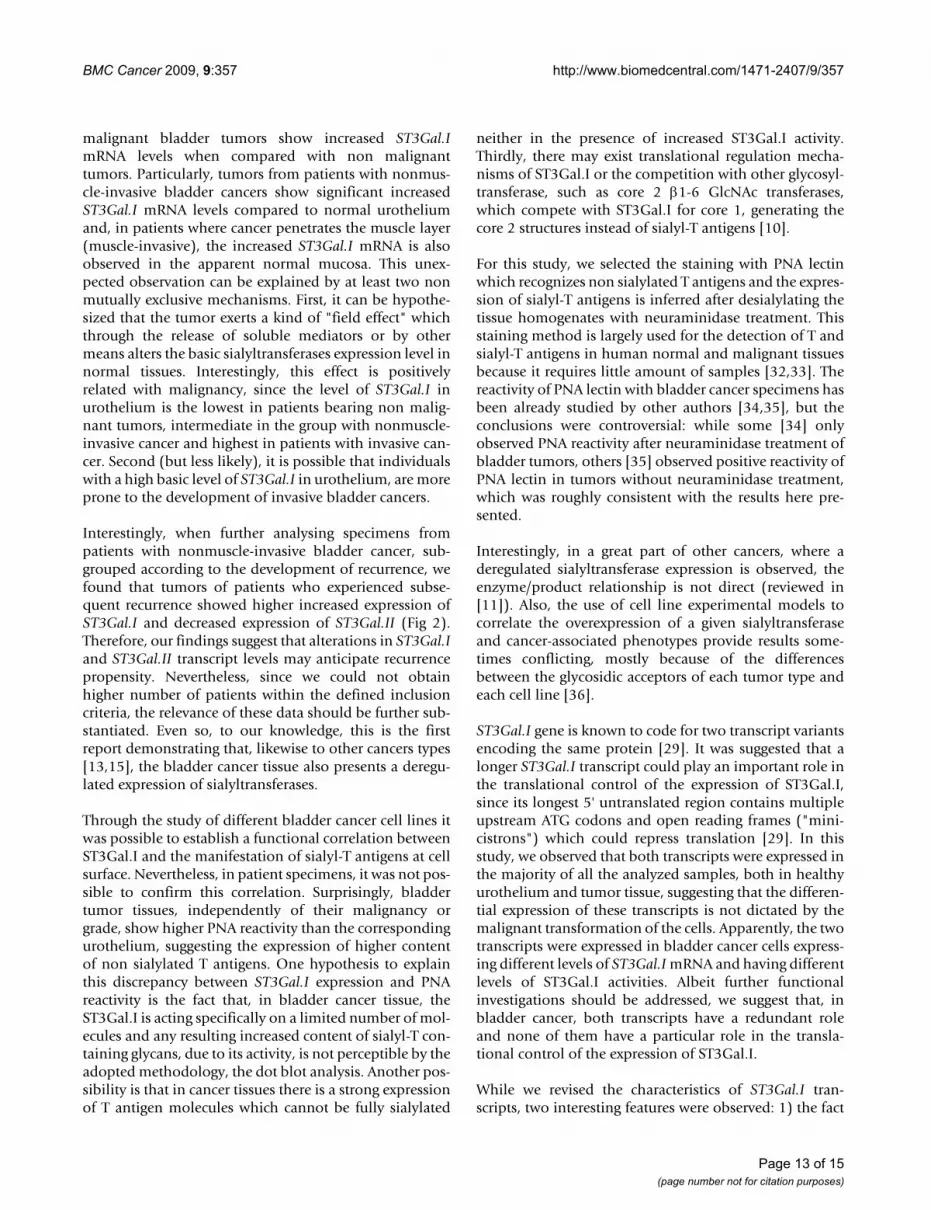
ResultsST3Gal.I expression is increased in bladder cancerTo understand whether there is an altered expression ofST3Gal.I, ST3Gal.II or ST3Gal.IV in bladder tumors, weanalyzed their mRNA levels. In the three analyzed groupsof patients, we found that ST3Gal.I expression was signif-icantly increased in tissue of nonmuscle-invasive bladdercancer (group I), compared with corresponding urothe-lium [105.3 ± 20.1 (tumors) and 43.0 ± 6.1 (urothelium)relative mRNA levels; p < 0.001] (Fig 1). In tissues frominvasive bladder cancer (group II) and from non malig-nant bladder tumors (group III), ST3Gal.I expression wasnot significantly different from the corresponding urothe-lium. Interestingly, in group II of patients, ST3Gal.I tran-scripts ranged from 84.1 ± 32.2 (urothelium) to 88.0 ±37.2 (tumors) mean relative mRNA levels, and wereapproximately eight times higher than group III, whichranged from 11.7 ± 6.6 (urothelium) to 7.9 ± 3.6 (tumors)mean relative mRNA levels (Fig 1A).
The expression of ST3Gal.II was not statistically differentbetween tumor tissue and normal urothelium in all theanalyzed groups of patients. However, in the group II ofpatients, ST3Gal.II mean mRNA levels were twice higher[ranging from 47.9 ± 20.9 (urothelium) to 38.8 ± 11.2(tumors)] than in group I [ranging from 27.8 ± 4.1(urothelium) to 23.4 ± 4.0 (tumors)] and five timeshigher than group III [ranging from 9.4 ± 3.6 (urothe-lium) to 5.1 ± 2.0 (tumors)] (Fig 1B).
Similarly, while ST3Gal.IV expression was also not statis-tically different between tumor tissue and normal urothe-
lium in all the analyzed groups of patients, in samplesfrom patients with non malignant tumors (group III) itsmRNA expression was 3 to 4 times lower [ranging from1.9 ± 1.1 (urothelium) to 3.5 ± 0.9 (tumors)] than sam-ples from other patient's groups. Taken as a whole, theST3Gal.IV mRNA levels were quite inferior to the mRNAlevels of the ST3Gal.I and ST3Gal.II (Fig 1C).
Although, not statistically significant, we observed unex-pected differences mRNA sialyltransferase expression lev-els among the normal urothelium of patients from thethree groups. This is most evident for ST3Gal.I, which isexpressed at the lowest level by patients who developednon malignant tumors (group III), at an intermediatelevel by those who developed malignant but nonmuscle-invasive cancers (group I) and at the highest level by thosewho developed invasive cancers (group II). These data canbe interpreted as the result of a "field effect" of the tumor,whose presence and degree of malignancy would affectsialyltransferase expression also in apparently uninvolvedtissues [27].
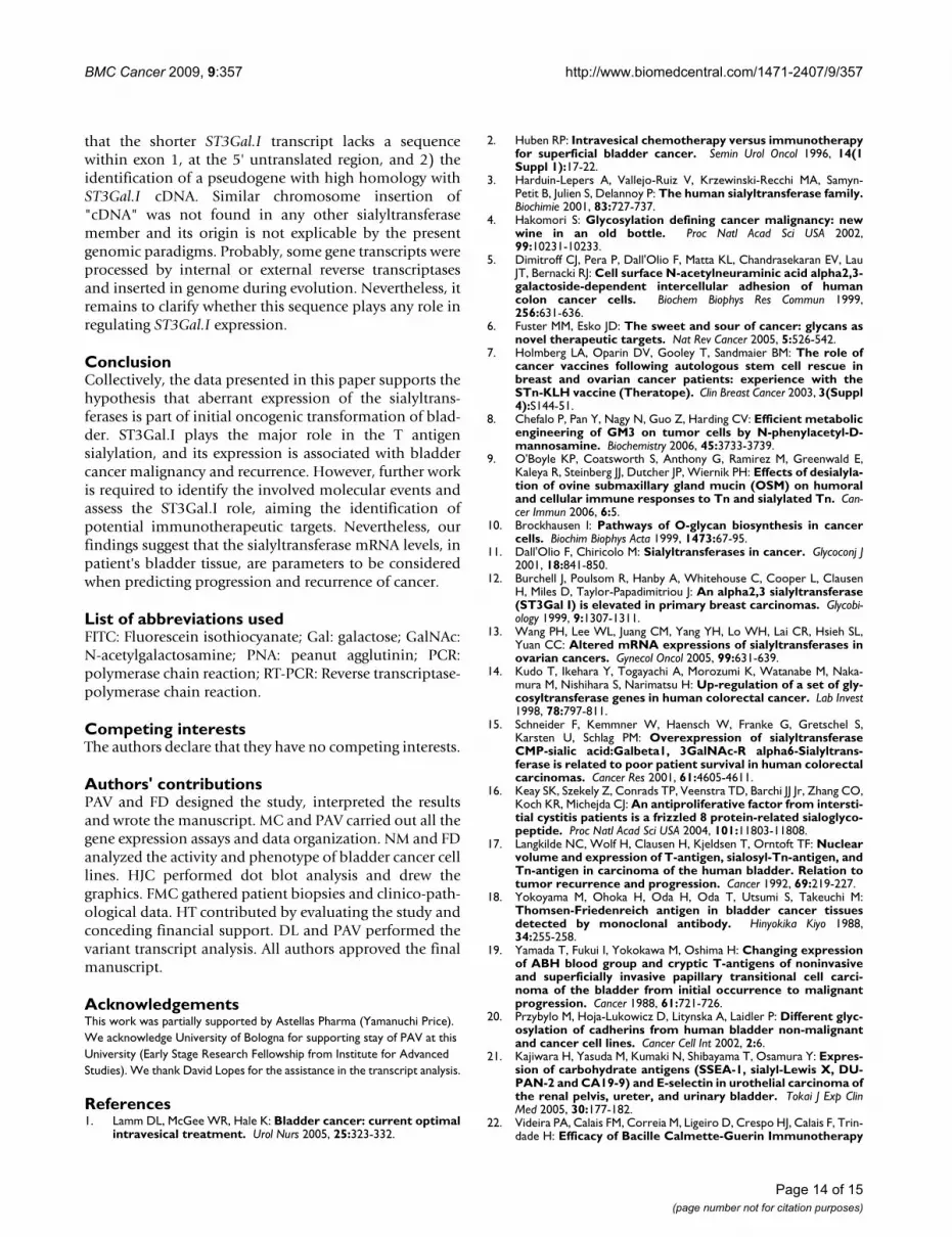
Nonmuscle-invasive bladder cancer with tendency for recurrence show pronounced ST3Gal.I mRNA increase and decreased ST3Gal.II mRNA levels33% of the patients with nonmuscle-invasive bladder can-cer, included in this study, have experienced recurrence inless than 12 months after TURBT plus intravesical BCG.To elucidate whether the expression of ST3Gal.I, ST3Gal.IIor ST3Gal.IV genes was dissimilar among patients withoutrecurrence (Group IA) and those with subsequent recur-rence (Group IB), we examined the transcripts levels ofthese sialyltransferases in these subgroups of patients.
Compared to urothelium, ST3Gal.I mRNA levels were sig-nificantly higher in both tumors from group IA and groupIB (p < 0.05) (Fig 2A). However, ST3Gal.I expression wasremarkably higher in tumors from group IB of patients(285.3 ± 93.6) than tumors from group IA (89.8 ± 18.3)(p = 0.058) (Fig 2A), suggesting that a high level ofST3Gal.I expression is associated with propensity to recur-rence. Conversely, ST3Gal.II mRNA expression, in groupIB of patients, was significantly decreased in tumors (15.3± 5.6), compared to urothelium (30.7 ± 7.6) (p = 0.0046)(Fig 2B). In group IA, no significant differences werefound for the expression of this gene. ST3Gal.IV mRNAlevels were not significantly different among group IA andgroup IB of patients (not shown). All together, these datashow that patients with nonmuscle-invasive bladder can-cer, with tendency for recurrence present a significant aug-ment in ST3Gal.I and reduction in ST3Gal.II expression intheir tumors.
Page 5 of 15(page number not for citation purposes)

BMC Cancer 2009, 9:357 http://www.biomedcentral.com/1471-2407/9/357
Page 6 of 15(page number not for citation purposes)
Bladder cancer tissue has increased expression of ST3Gal.IFigure 1Bladder cancer tissue has increased expression of ST3Gal.I. The relative mRNA levels of ST3Gal.I (A), ST3Gal.II (B) and ST3Gal.IV (C) genes were analyzed by Real Time RT-PCR as described in the Methods section, in bladder tumor tissue (T) and corresponding normal urothelium (U) from patients with nonmuscle-invasive (I), with invasive (II) and with non malignant blad-der tumors (III). Values infer the number of mRNA molecules of a certain gene per 1000 molecules of the average of the endogenous controls (β-actin and GAPDH). ST3Gal.I expression is significantly increased in tissue of nonmuscle-invasive blad-der cancer (I), compared with the corresponding urothelium (p < 0.001).
ST3Gal.IV
I - U I - T II - U II - T III - U III - T0.1
1
10
100
Rel
ativ
e m
RN
A l
evel
(0 / 00
of-a
ctin
/GA
DP
H)
ST3Gal.II
I - U I - T II - U II - T III - U III - T0.1
1
10
100
Rel
ativ
e m
RN
A le
vel
(0 / 00
of-a
ctin
/GA
DP
H)
ST3Gal.I
I - U I - T II - U II - T III - U III - T0.1
1
10
100
1000
Rel
ativ
e m
RN
A l
evel
(0 / 00
of-a
ctin
/GA
DP
H)
ST3Gal.I A
***
ST3Gal.II B
ST3Gal.IV C

BMC Cancer 2009, 9:357 http://www.biomedcentral.com/1471-2407/9/357
Figure 2 (see legend on next page)
ST3Gal.I
ST3Gal.I A
I A - U I A - T I B - U I B - T1
10
100
1000
Rel
ativ
e m
RN
A l
evel
(0 / 00
of-a
ctin
/GA
DP
H)
*
*
**
ST3Gal.II
ST3Gal.II B
10
1
100
Rel
ativ
e m
RN
A l
evel
(0 / 00
of-a
ctin
/GA
DP
H)
0.1I A - U I A - T I B - U I B - T
Page 7 of 15(page number not for citation purposes)

BMC Cancer 2009, 9:357 http://www.biomedcentral.com/1471-2407/9/357
ST3Gal.I is responsible for the sialylation of T antigens in bladder cancer cell linesTo investigate whether, in bladder cancer, there was a cor-relation between the activity of at least one of theST3Gal.I, ST3Gal.II or ST3Gal.IV sialyltransferases and theexpression of sialyl-T antigens, we examined in detail fourdistinct bladder cancer cell lines. According to the tran-scriptional analysis, the ST3Gal.I gene gave the most scat-tering expression among the four cell lines used in thisstudy (Fig 3A). The 5637 cell line showed the highestexpression for ST3Gal.I (16.0 ± 1.8 relative mRNA levels);contrasting with the HT1376 cell line, which showed thesmallest expression and the MCR and T24 cell lines whichexpressed approximately forty times less ST3Gal.I mRNAlevels than the 5637 cell line (Fig 3A). ST3Gal.II transcriptlevels were higher in the MCR and T24 cell lines (6.1 ± 0.4and 7.5 ± 0.4, respectively), followed by the 5637 cell line,which showed around half expression, and the HT1376cell line which showed one tenth of this expression (Fig3A). The ST3Gal.IV gene was poorly expressed in all thefour cell lines, ranging from 0.03 ± 0.003 to 0.46 ± 0.022relative mRNA molecules (Fig 3A).
Regarding the analysis of sialyltransferase activity, weobserved that only 5637 cells show a high level of activitytowards T antigens while, in the other three cell lines, theactivity was very low or undetectable (Fig 3B). Among theanalyzed genes, the expression of ST3Gal.I gave the bestcorrelation with the observed sialyltransferase activitytowards the T antigen (r = 0.99; p = 0.001). According tothe cell staining with PNA, a lectin which recognizes the Tantigen [28], it was evident that the T antigen wasexpressed at high levels by the HT1376 and MCR celllines, but only weakly by 5637 cells and at an intermedi-ate level by T24 (Fig 4). After removing the cell surfacesialic acid, by neuraminidase treatment, part of the 5637and T24 become PNA positive, indicating that these celllines express the T antigen masked by sialic acid (Fig 4).These data propose the ST3Gal.I sialyltransferase is themajor responsible for the sialylation of T antigens in blad-der cancer cells.
Sialyl-T antigens are expressed in bladder tumor tissue and urotheliumTo ascertain whether sialyl-T antigens were also expressedin the normal urothelium and bladder tumor specimens,we performed a PNA dot blot analysis. The majority of theanalyzed samples stained with PNA (Fig 5), demonstrat-ing the expression of T antigen. It appears that tumor sam-ples had enhanced reactivity with PNA, but we couldn'tfind any peculiarity among the tumor malignancies andgrades. After neuraminidase treatment, the PNA stainingincreased in nearly all the samples (Fig 5), demonstratingthat part of the T antigens is masked by sialic acid, in theurothelium and bladder tumors. Altogether, these resultssuggest that tumors may have slightly increased expres-sion of T antigens compared with urothelium, but wefailed to find a significant different expression of T andsialyl-T antigens, between the different grades of tumors.
Bladder cancer cells express two ST3Gal.I transcript variantsIn the first paper describing the molecular cloning ofhuman ST3Gal.I [29], it was reported the presence of twotypes of ST3Gal.I cDNAs which differ in the 5'untrans-lated region; and the shorter of the two transcript variantsmisses one or more exons. While the sequences of thesetwo ST3Gal.I transcript variants were deposited at Gen-Bank (Reference Sequences: NM_003033.2 andNM_173344.2 for the longer and shorter form, respec-tively), there was no further reference in the literature con-cerning this issue. In addition, it was not consensualwhether there were six or seven exons in the codingregion.
In a first investigation, we compared the two transcriptvariants and the genomic ST3Gal.I sequences. Accordingto our in silico analysis, the shorter ST3Gal.I cDNA(NM_173344.2) does not lack any specific exon at the5'untranslated region, but a 203 nucleotide sequence,within exon 1, 51 nucleotides upstream the translationinitiation codon (Fig 6A). Both transcript variants arecomposed of nine exons with six of them containing thecoding region (Fig 6A). The PCR amplification of cDNAsfrom the bladder cancer cell lines, using primers located in
Nonmuscle-invasive bladder cancers with tendency for recurrence show slight increased expression of ST3Gal I and ST3Gal.II, compared with normal urotheliumFigure 2 (see previous page)Nonmuscle-invasive bladder cancers with tendency for recurrence show slight increased expression of ST3Gal I and ST3Gal.II, compared with normal urothelium. Only patients with nonmuscle-invasive bladder cancer were consid-ered: without recurrence within a minimal period of 12 months following TURBT (IA) and experiencing recurrence in that period (IB). The relative mRNA levels of ST3Gal.I (A) and ST3Gal.II (B) genes were analyzed in bladder tumor (T) tissue and corresponding normal urothelium (U), by Real Time RT-PCR, as described in the Methods section. Values infer the number of mRNA molecules of a certain gene per 1000 molecules of the average of the endogenous controls (β-actin and GAPDH). ST3Gal.I expression is significantly increased in tissue of both subgroups of nonmuscle-invasive bladder cancer, compared with the corresponding urothelium (p < 0.05), while ST3Gal.II expression is significantly decreased in tumors from group IB patients, compared with the corresponding urothelium (p < 0.01).
Page 8 of 15(page number not for citation purposes)

BMC Cancer 2009, 9:357 http://www.biomedcentral.com/1471-2407/9/357
Page 9 of 15(page number not for citation purposes)
The ST3Gal.I expression correlates with the sialyltransferase activity towards T antigen in bladder cancer cell linesFigure 3The ST3Gal.I expression correlates with the sialyltransferase activity towards T antigen in bladder cancer cell lines. A: The relative mRNA levels of ST3Gal.I, ST3Gal.II and ST3Gal.IV genes were analyzed in 5637, HT1376, MCR and T24 bladder cancer cell lines, by Real Time PCR. Values infer the number of mRNA molecules of a certain gene per 1000 molecules of the average of the endogenous controls (β-actin and GAPDH). Results are the mean of 3 independent assays. B: The sialyl-transferase activity towards Galβ1,3GalNAcα1-O-Benzyl glycoside was assessed in sample homogenates of 5637, HT1376, MCR and T24 bladder cancer cell lines, as described in the Methods section. In the four analyzed bladder cancer cell lines, the ST3Gal.1 mRNA levels are correlated with the observed sialyltransferase activity towards the T antigen (r = 0.99; p = 0.001).
B
0
20
40
60
80
5637 HT1376 MCR T24
Sial
yltra
nsfe
rase
act
ivity
(nm
ol/h
*g)
.
A
0.001
0.01
0.1
1
10
100
5637 HT1376 MCR T24
rela
tive
mRN
A (‰
of
actin
/GA
PDH
)
ST3Gal.I
ST3Gal.II
ST3Gal.IV

BMC Cancer 2009, 9:357 http://www.biomedcentral.com/1471-2407/9/357
different ST3Gal.I exons, originated products whose sizeswere in agreement with the in silico analysis and thereforepresuppose the existence of two ST3Gal.I transcripts, eachwith nine exons (data not shown). The 203 nucleotidesshortage which distinguish NM_173344.2 fromNM_003033.2 transcript was confirmed by cDNA ampli-fication, using primers located at the extremities of exon 1(Fig 6A), where two PCR products with the expected size(609 bp and 406 bp) were obtained (Fig 6B), and bysequencing in both directions.
Although the intensities of the PCR products differedamong the analyzed cell lines, all samples expressed bothtranscripts (Fig 6B). The two ST3Gal.I transcripts were alsoobserved in the patient's specimens, with no significant
differences regarding their nature (urothelium or tumortissue) and the tumor grade (Fig 6B).
Considering the augmented ST3Gal.I mRNA expressionwhich we observed in bladder cancer, it was not possibleto correlate it with one of the transcripts. Although furtherstudies should be conducted, these results suggest that, inthese bladder cancer tissue and urothelium, both ST3Gal.Itranscripts are randomly expressed, with no particularassociation with the overall expression of the gene and thetype of tissue.
It is relevant to note that during the in silico analysis wehave observed that the shorter cDNA had the highestidentity (89%) with a pseudogene located at chromosome
Bladder cancer cell lines express both T and sialyl-T antigensFigure 4Bladder cancer cell lines express both T and sialyl-T antigens. The bladder cancer cell lines, 5637, HT1376, MCR and T24, treated with heat-inactivated neuraminidase (A) or with active neuraminidase (B) were analyzed by Flow cytometry using fluorescent PNA lectin. The gray line shows the controls without PNA. The T antigen is expressed at high levels by the HT1376 and MCR cell lines, but only weakly by 5637 cells and at an intermediate level by T24. According to the results obtained after removing the cell surface sialic acid by neuraminidase treatment, the 5637 and T24 cell lines express the T anti-gen masked by sialic acid.
A
B
5637 HT1376 MCR T24
256
256
256
256
256
256
256
256
5637 HT1376 MCR T24
Page 10 of 15(page number not for citation purposes)

BMC Cancer 2009, 9:357 http://www.biomedcentral.com/1471-2407/9/357
Page 11 of 15(page number not for citation purposes)
T and sialyl-T antigens are expressed in bladder tumor tissue and urotheliumFigure 5T and sialyl-T antigens are expressed in bladder tumor tissue and urothelium. Tumor (T) and Urothelium (U) sam-ples from patients were analyzed by dot blot staining with PNA, which binds to T-antigen. Samples were arranged by tumor type, namely, nonmuscle-invasive without (IA) and with recurrence (IB) and muscle-invasive (II) bladder cancer and non-malig-nant tumors (III) and by treatment with neuraminidase (N) or mock treated (M). PNA binds to non neuraminidase treated samples, of all patients' groups, indicating the presence of T-antigens. In the majority of the samples, PNA binding is strongly increased after neuraminidase treatment.
IA
IB
II III
U T U T U T U T
M
N
M
N
M
N

BMC Cancer 2009, 9:357 http://www.biomedcentral.com/1471-2407/9/357
4, followed by homology with a chimpanzee pseudogene,also chromosome 4, and then by genomic ST3Gal.Isequence, at chromosome 8. This sequence has no homol-ogy with any viral or bacterial gene and it is not flanked byany insertion element, which excludes the hypothesis of apathogenic chromosome insertion.
DiscussionA great part of the aberrant glycosylation found in cancercells is due to the altered expression of sialyltransferases.These enzymes are considered as prognostic factors insome cancers [11] and recent data has revealed that theyare important targets for therapeutic approaches [30,31].
Investigating the relevance of sialyltransferases in cancer ishowever a complex task, due the diversity expressed byhuman cells and for presenting some overlapping sub-strate specificities, tissue-restricted patterns of expressionand individual variations.
Our aim was to investigate, in bladder cancers, the expres-sion of the sialyltransferases, ST3Gal.I, ST3Gal.II andST3Gal.IV, which are proposed to have a decisive role inthe T antigen sialylation [10]. Our study with differentgroups of patients with bladder tumors, demonstratemarkedly that the ST3Gal.I mRNA levels increases duringthe malignant transformation of urothelium. In fact,
The two ST3Gal.I transcripts are expressed in the majority of the bladder cancer and urothelium cellsFigure 6The two ST3Gal.I transcripts are expressed in the majority of the bladder cancer and urothelium cells. A: Sche-matic diagram of human ST3Gal.I cDNA (on the top) and genomic (on the centre) sequences. The black rectangle in ST3Gal.I cDNA represents the sequence which is present in ST3Gal.I transcript variant NM_003033.2 and absent on the transcript vari-ant NM_173344.2. Exons containing the coding region (represented in grey) are denoted as 1-6 with the sizes shown above. The total of exon 1 nucleotide sequence is shown below: DNA sequence absent in transcript variant NM_173344.2 is repre-sented on black background and the location of the primers F3 and R1 used to discriminate the transcript variants is under-lined. B: RT-PCR analysis of NM_003033.2 and NM_173344.2ST3Gal.I transcripts in bladder cancer cell lines and patient's specimens. The ST3Gal.I exon 1 was amplified in cDNAs obtained from HT1374, MCR, T24 and 5637 bladder cancer cell lines and bladder tumor (T) tissue and corresponding normal urothelium (U) from 6 randomly chosen patients (subgrouped as described above), using F3 and R1 primers. The predicted sizes of the products are 609 bp (NM_003033.2) and 406 bp (NM_173344.2).
A -1035 bp +1
232 169 55 885 197 180 46 120 >1036 bp
6 1 X1 Y X2 2 4 5 3
cDNA variants NM_003033.2 NM_173344.2
Genomic DNA
F3 CAGATGACAGACAACCTTCTGTGCCTTCATCAAGCTGGTTGTGTACCCACCATGTCCCTGGCGACAGGATGGGAAAGAAAAAGCCCTAATTAAGGATCGTCAGAAACCACAGTTGGAGGAGGACGGCAGAGACAGTTTCCCTCCCCGCTATACCAACACCCTTCCTTCGAGGTCCTCGCTCCTGAGGGACCCTGGACTGTCACAGAGATTAATGACCCCTTATCTTCTTTGGATGTGAAAGGAAATCACTGGTTAAAGCTTGATCGAGAGACATTATCAGCTCTTTAAGGATTGCAGAAGAATAGGCTACTTTATTTTCTGAAAAGGTAAATATATGCAAGCAAAGCCAACATGCCACGAATGGCGTTGGTCTACCACACAGCCGTGTCTGGGACACAGTTGGGGGTCATCCCCCAGCAGGAGTGAAGTCGAGCTTAGCGGCCCTTGTGTCCTCCCTTGGAATTCCTGCCATCCCTTTTGATTGAGCCTCCACCTCTGGGATTTTTCTTCCATTTTTCTCCTCTCTTAGGAGGGAGTTCCTGCTACCCATCGTGGGAGGCCACCATCAGGACTGCGAAGATGGTGACCCTGCGGAAGAGGACCCTGAAAGTGCTCACCTTCCTCGTGCTCTTCATCTTCCTCACCTCCTTCTTCCTGAACTACTCCCACACCATGGTGGCCACCACCTGGTTCCCCAAGCAGATGGTCCTGGAGCTCTCCGAGAACCTGAAGAGACTG
R1 ATCAAGCACAGGCCTTGCACCTGCACCCACTGCATCGGGCAGCGCAAGCTCTCGGCCTGGTTCGATGAGAGGTTCAACCAGACCATGCAGCCGCTGCTGACCGCCCAGAACGCGCTCTTGGAGGACGACACCTACCGATGGTGGCTG
B
HT
1376
MC
R
5637
T24
IA IB II III
U T U T U T U T
Page 12 of 15(page number not for citation purposes)
BMC Cancer 2009, 9:357 http://www.biomedcentral.com/1471-2407/9/357
malignant bladder tumors show increased ST3Gal.ImRNA levels when compared with non malignanttumors. Particularly, tumors from patients with nonmus-cle-invasive bladder cancers show significant increasedST3Gal.I mRNA levels compared to normal urotheliumand, in patients where cancer penetrates the muscle layer(muscle-invasive), the increased ST3Gal.I mRNA is alsoobserved in the apparent normal mucosa. This unex-pected observation can be explained by at least two nonmutually exclusive mechanisms. First, it can be hypothe-sized that the tumor exerts a kind of "field effect" whichthrough the release of soluble mediators or by othermeans alters the basic sialyltransferases expression level innormal tissues. Interestingly, this effect is positivelyrelated with malignancy, since the level of ST3Gal.I inurothelium is the lowest in patients bearing non malig-nant tumors, intermediate in the group with nonmuscle-invasive cancer and highest in patients with invasive can-cer. Second (but less likely), it is possible that individualswith a high basic level of ST3Gal.I in urothelium, are moreprone to the development of invasive bladder cancers.
Interestingly, when further analysing specimens frompatients with nonmuscle-invasive bladder cancer, sub-grouped according to the development of recurrence, wefound that tumors of patients who experienced subse-quent recurrence showed higher increased expression ofST3Gal.I and decreased expression of ST3Gal.II (Fig 2).Therefore, our findings suggest that alterations in ST3Gal.Iand ST3Gal.II transcript levels may anticipate recurrencepropensity. Nevertheless, since we could not obtainhigher number of patients within the defined inclusioncriteria, the relevance of these data should be further sub-stantiated. Even so, to our knowledge, this is the firstreport demonstrating that, likewise to other cancers types[13,15], the bladder cancer tissue also presents a deregu-lated expression of sialyltransferases.
Through the study of different bladder cancer cell lines itwas possible to establish a functional correlation betweenST3Gal.I and the manifestation of sialyl-T antigens at cellsurface. Nevertheless, in patient specimens, it was not pos-sible to confirm this correlation. Surprisingly, bladdertumor tissues, independently of their malignancy orgrade, show higher PNA reactivity than the correspondingurothelium, suggesting the expression of higher contentof non sialylated T antigens. One hypothesis to explainthis discrepancy between ST3Gal.I expression and PNAreactivity is the fact that, in bladder cancer tissue, theST3Gal.I is acting specifically on a limited number of mol-ecules and any resulting increased content of sialyl-T con-taining glycans, due to its activity, is not perceptible by theadopted methodology, the dot blot analysis. Another pos-sibility is that in cancer tissues there is a strong expressionof T antigen molecules which cannot be fully sialylated
neither in the presence of increased ST3Gal.I activity.Thirdly, there may exist translational regulation mecha-nisms of ST3Gal.I or the competition with other glycosyl-transferase, such as core 2 β1-6 GlcNAc transferases,which compete with ST3Gal.I for core 1, generating thecore 2 structures instead of sialyl-T antigens [10].
For this study, we selected the staining with PNA lectinwhich recognizes non sialylated T antigens and the expres-sion of sialyl-T antigens is inferred after desialylating thetissue homogenates with neuraminidase treatment. Thisstaining method is largely used for the detection of T andsialyl-T antigens in human normal and malignant tissuesbecause it requires little amount of samples [32,33]. Thereactivity of PNA lectin with bladder cancer specimens hasbeen already studied by other authors [34,35], but theconclusions were controversial: while some [34] onlyobserved PNA reactivity after neuraminidase treatment ofbladder tumors, others [35] observed positive reactivity ofPNA lectin in tumors without neuraminidase treatment,which was roughly consistent with the results here pre-sented.
Interestingly, in a great part of other cancers, where aderegulated sialyltransferase expression is observed, theenzyme/product relationship is not direct (reviewed in[11]). Also, the use of cell line experimental models tocorrelate the overexpression of a given sialyltransferaseand cancer-associated phenotypes provide results some-times conflicting, mostly because of the differencesbetween the glycosidic acceptors of each tumor type andeach cell line [36].
ST3Gal.I gene is known to code for two transcript variantsencoding the same protein [29]. It was suggested that alonger ST3Gal.I transcript could play an important role inthe translational control of the expression of ST3Gal.I,since its longest 5' untranslated region contains multipleupstream ATG codons and open reading frames ("mini-cistrons") which could repress translation [29]. In thisstudy, we observed that both transcripts were expressed inthe majority of all the analyzed samples, both in healthyurothelium and tumor tissue, suggesting that the differen-tial expression of these transcripts is not dictated by themalignant transformation of the cells. Apparently, the twotranscripts were expressed in bladder cancer cells express-ing different levels of ST3Gal.I mRNA and having differentlevels of ST3Gal.I activities. Albeit further functionalinvestigations should be addressed, we suggest that, inbladder cancer, both transcripts have a redundant roleand none of them have a particular role in the transla-tional control of the expression of ST3Gal.I.
While we revised the characteristics of ST3Gal.I tran-scripts, two interesting features were observed: 1) the fact
Page 13 of 15(page number not for citation purposes)
BMC Cancer 2009, 9:357 http://www.biomedcentral.com/1471-2407/9/357
that the shorter ST3Gal.I transcript lacks a sequencewithin exon 1, at the 5' untranslated region, and 2) theidentification of a pseudogene with high homology withST3Gal.I cDNA. Similar chromosome insertion of"cDNA" was not found in any other sialyltransferasemember and its origin is not explicable by the presentgenomic paradigms. Probably, some gene transcripts wereprocessed by internal or external reverse transcriptasesand inserted in genome during evolution. Nevertheless, itremains to clarify whether this sequence plays any role inregulating ST3Gal.I expression.
ConclusionCollectively, the data presented in this paper supports thehypothesis that aberrant expression of the sialyltrans-ferases is part of initial oncogenic transformation of blad-der. ST3Gal.I plays the major role in the T antigensialylation, and its expression is associated with bladdercancer malignancy and recurrence. However, further workis required to identify the involved molecular events andassess the ST3Gal.I role, aiming the identification ofpotential immunotherapeutic targets. Nevertheless, ourfindings suggest that the sialyltransferase mRNA levels, inpatient's bladder tissue, are parameters to be consideredwhen predicting progression and recurrence of cancer.
List of abbreviations usedFITC: Fluorescein isothiocyanate; Gal: galactose; GalNAc:N-acetylgalactosamine; PNA: peanut agglutinin; PCR:polymerase chain reaction; RT-PCR: Reverse transcriptase-polymerase chain reaction.
Competing interestsThe authors declare that they have no competing interests.
Authors' contributionsPAV and FD designed the study, interpreted the resultsand wrote the manuscript. MC and PAV carried out all thegene expression assays and data organization. NM and FDanalyzed the activity and phenotype of bladder cancer celllines. HJC performed dot blot analysis and drew thegraphics. FMC gathered patient biopsies and clinico-path-ological data. HT contributed by evaluating the study andconceding financial support. DL and PAV performed thevariant transcript analysis. All authors approved the finalmanuscript.
AcknowledgementsThis work was partially supported by Astellas Pharma (Yamanuchi Price). We acknowledge University of Bologna for supporting stay of PAV at this University (Early Stage Research Fellowship from Institute for Advanced Studies). We thank David Lopes for the assistance in the transcript analysis.
References1. Lamm DL, McGee WR, Hale K: Bladder cancer: current optimal
intravesical treatment. Urol Nurs 2005, 25:323-332.
2. Huben RP: Intravesical chemotherapy versus immunotherapyfor superficial bladder cancer. Semin Urol Oncol 1996, 14(1Suppl 1):17-22.
3. Harduin-Lepers A, Vallejo-Ruiz V, Krzewinski-Recchi MA, Samyn-Petit B, Julien S, Delannoy P: The human sialyltransferase family.Biochimie 2001, 83:727-737.
4. Hakomori S: Glycosylation defining cancer malignancy: newwine in an old bottle. Proc Natl Acad Sci USA 2002,99:10231-10233.
5. Dimitroff CJ, Pera P, Dall'Olio F, Matta KL, Chandrasekaran EV, LauJT, Bernacki RJ: Cell surface N-acetylneuraminic acid alpha2,3-galactoside-dependent intercellular adhesion of humancolon cancer cells. Biochem Biophys Res Commun 1999,256:631-636.
6. Fuster MM, Esko JD: The sweet and sour of cancer: glycans asnovel therapeutic targets. Nat Rev Cancer 2005, 5:526-542.
7. Holmberg LA, Oparin DV, Gooley T, Sandmaier BM: The role ofcancer vaccines following autologous stem cell rescue inbreast and ovarian cancer patients: experience with theSTn-KLH vaccine (Theratope). Clin Breast Cancer 2003, 3(Suppl4):S144-51.
8. Chefalo P, Pan Y, Nagy N, Guo Z, Harding CV: Efficient metabolicengineering of GM3 on tumor cells by N-phenylacetyl-D-mannosamine. Biochemistry 2006, 45:3733-3739.
9. O'Boyle KP, Coatsworth S, Anthony G, Ramirez M, Greenwald E,Kaleya R, Steinberg JJ, Dutcher JP, Wiernik PH: Effects of desialyla-tion of ovine submaxillary gland mucin (OSM) on humoraland cellular immune responses to Tn and sialylated Tn. Can-cer Immun 2006, 6:5.
10. Brockhausen I: Pathways of O-glycan biosynthesis in cancercells. Biochim Biophys Acta 1999, 1473:67-95.
11. Dall'Olio F, Chiricolo M: Sialyltransferases in cancer. Glycoconj J2001, 18:841-850.
12. Burchell J, Poulsom R, Hanby A, Whitehouse C, Cooper L, ClausenH, Miles D, Taylor-Papadimitriou J: An alpha2,3 sialyltransferase(ST3Gal I) is elevated in primary breast carcinomas. Glycobi-ology 1999, 9:1307-1311.
13. Wang PH, Lee WL, Juang CM, Yang YH, Lo WH, Lai CR, Hsieh SL,Yuan CC: Altered mRNA expressions of sialyltransferases inovarian cancers. Gynecol Oncol 2005, 99:631-639.
14. Kudo T, Ikehara Y, Togayachi A, Morozumi K, Watanabe M, Naka-mura M, Nishihara S, Narimatsu H: Up-regulation of a set of gly-cosyltransferase genes in human colorectal cancer. Lab Invest1998, 78:797-811.
15. Schneider F, Kemmner W, Haensch W, Franke G, Gretschel S,Karsten U, Schlag PM: Overexpression of sialyltransferaseCMP-sialic acid:Galbeta1, 3GalNAc-R alpha6-Sialyltrans-ferase is related to poor patient survival in human colorectalcarcinomas. Cancer Res 2001, 61:4605-4611.
16. Keay SK, Szekely Z, Conrads TP, Veenstra TD, Barchi JJ Jr, Zhang CO,Koch KR, Michejda CJ: An antiproliferative factor from intersti-tial cystitis patients is a frizzled 8 protein-related sialoglyco-peptide. Proc Natl Acad Sci USA 2004, 101:11803-11808.
17. Langkilde NC, Wolf H, Clausen H, Kjeldsen T, Orntoft TF: Nuclearvolume and expression of T-antigen, sialosyl-Tn-antigen, andTn-antigen in carcinoma of the human bladder. Relation totumor recurrence and progression. Cancer 1992, 69:219-227.
18. Yokoyama M, Ohoka H, Oda H, Oda T, Utsumi S, Takeuchi M:Thomsen-Friedenreich antigen in bladder cancer tissuesdetected by monoclonal antibody. Hinyokika Kiyo 1988,34:255-258.
19. Yamada T, Fukui I, Yokokawa M, Oshima H: Changing expressionof ABH blood group and cryptic T-antigens of noninvasiveand superficially invasive papillary transitional cell carci-noma of the bladder from initial occurrence to malignantprogression. Cancer 1988, 61:721-726.
20. Przybylo M, Hoja-Lukowicz D, Litynska A, Laidler P: Different glyc-osylation of cadherins from human bladder non-malignantand cancer cell lines. Cancer Cell Int 2002, 2:6.
21. Kajiwara H, Yasuda M, Kumaki N, Shibayama T, Osamura Y: Expres-sion of carbohydrate antigens (SSEA-1, sialyl-Lewis X, DU-PAN-2 and CA19-9) and E-selectin in urothelial carcinoma ofthe renal pelvis, ureter, and urinary bladder. Tokai J Exp ClinMed 2005, 30:177-182.
22. Videira PA, Calais FM, Correia M, Ligeiro D, Crespo HJ, Calais F, Trin-dade H: Efficacy of Bacille Calmette-Guerin Immunotherapy
Page 14 of 15(page number not for citation purposes)

BMC Cancer 2009, 9:357 http://www.biomedcentral.com/1471-2407/9/357
Publish with BioMed Central and every scientist can read your work free of charge
"BioMed Central will be the most significant development for disseminating the results of biomedical research in our lifetime."
Sir Paul Nurse, Cancer Research UK
Your research papers will be:
available free of charge to the entire biomedical community
peer reviewed and published immediately upon acceptance
cited in PubMed and archived on PubMed Central
yours — you keep the copyright
Submit your manuscript here:http://www.biomedcentral.com/info/publishing_adv.asp
BioMedcentral
Predicted by Expression of Antigen-presenting Moleculesand Chemokines. Urology 2009, 74(4):944-50.
23. Videira PA, Ligeiro D, Correia M, Trindade H: Gene expressionanalysis in superficial bladder cancer: comparison of twosuitable endogenous reference genes. Curr Urol 2007,1:145-150.
24. Livak KJ, Schmittgen TD: Analysis of relative gene expressiondata using real-time quantitative PCR and the 2(-Delta DeltaC(T)) Method. Methods 2001, 25:402-408.
25. Dall'Olio F, Malagolini N, Guerrini S, Lau JT, Serafini-Cessi F: Differ-entiation-dependent expression of human beta-galactosidealpha 2,6-sialyltransferase mRNA in colon carcinoma CaCo-2 cells. Glycoconj J 1996, 13:115-121.
26. Videira PA, Amado IF, Crespo HJ, Alguero MC, Dall'Olio F, CabralMG, Trindade H: Surface alpha 2-3- and alpha 2-6-sialylation ofhuman monocytes and derived dendritic cells and its influ-ence on endocytosis. Glycoconj J 2008, 25:259-268.
27. Heney NM, Daly J, Prout GR Jr, Nieh PT, Heaney JA, Trebeck NE:Biopsy of apparently normal urothelium in patients withbladder carcinoma. J Urol 1978, 120:559-560.
28. Pereira ME, Kabat EA, Lotan R, Sharon N: Immunochemical stud-ies on the specificity of the peanut (Arachis hypogaea) agglu-tinin. Carbohydr Res 1976, 51:107-118.
29. Kitagawa H, Paulson JC: Differential expression of five sialyl-transferase genes in human tissues. J Biol Chem 1994,269:17872-17878.
30. Hsu CC, Lin TW, Chang WW, Wu CY, Lo WH, Wang PH, Tsai YC:Soyasaponin-I-modified invasive behavior of cancer bychanging cell surface sialic acids. Gynecol Oncol 2005,96:415-422.
31. Zhu Y, Srivatana U, Ullah A, Gagneja H, Berenson CS, Lance P: Sup-pression of a sialyltransferase by antisense DNA reducesinvasiveness of human colon cancer cells in vitro. Biochim Bio-phys Acta 2001, 1536:148-160.
32. Amado M, Yan Q, Comelli EM, Collins BE, Paulson JC: Peanutagglutinin high phenotype of activated CD8+ T cells resultsfrom de novo synthesis of CD45 glycans. J Biol Chem 2004,279:36689-36697.
33. Shio Y, Suzuki H, Kawaguchi T, Ohsugi J, Higuchi M, Fujiu K, Kanno R,Ohishi A, Gotoh M: Carbohydrate status detecting by PNA ischangeable through cancer prognosis from primary to met-astatic nodal site: A possible prognostic factor in patient withnode-positive lung adenocarcinoma. Lung Cancer 2007,57:187-192.
34. Langkilde NC, Orntoft TF: A comparative study of peanutagglutinin and amaranthin binding to human urinary bladdertumor glycoproteins. Scand J Urol Nephrol Suppl 1995, 172:57-64.
35. Nakanishi K, Kawai T, Suzuki M: Lectin binding and expression ofblood group-related antigens in carcinoma-in-situ and inva-sive carcinoma of urinary bladder. Histopathology 1993,23:153-158.
36. Malagolini N, Santini D, Chiricolo M, Dall'Olio F: Biosynthesis andexpression of the Sda and sialyl Lewis × antigens in normaland cancer colon. Glycobiology 2007, 17:688-697.
Pre-publication historyThe pre-publication history for this paper can be accessedhere:
http://www.biomedcentral.com/1471-2407/9/357/prepub
Page 15 of 15(page number not for citation purposes)