spinal muscular atrophy pathogenic mutations impair the axonogenic properties of axonal-survival of...
TRANSCRIPT

Autosomal recessive spinal muscular atrophy (SMA) is theleading genetic cause of infant mortality, with an incidenceof 1 : 10 000 live births and an estimated carrier frequencyof 1 : 31 (Prior et al. 2010). It is due to motor neuron lossleading to progressive amyotrophic paralysis, respiratoryfailure and eventually death in the more severe cases. SMA isclassified into three main clinical types (I–III), in relation tothe age of onset and disease severity. All clinical types ofSMA are mainly caused by homozygous deletion of theSurvival of Motor Neuron gene SMN1 (Hahnen et al. 1995;Lefebvre et al. 1995; Rodrigues et al. 1995). However, aminority of SMA patients (about 4%) are compoundheterozygous with one deleted SMN1 allele and the otherSMN1 allele carrying a subtle mutation (Lefebvre et al. 1998;Parsons et al. 1998).
The major product of the SMN1 gene is the ‘full-length’FL-SMN protein, a well-established assembly factor forsmall nucleolar ribonucleoprotein particles (RNPs) and smallnuclear RNPs (snRNPs) that plays a key role in spliceosomalbiogenesis and mRNA splicing (Pellizzoni et al. 1998;
Received October 10, 2011; revised manuscript received January 10,2012; accepted February 6, 2012.Address correspondence and reprint requests to Giorgio Battaglia,
MD, Molecular Neuroanatomy and Pathogenesis Unit, NeurologicalInstitute ‘C. Besta’, via Temolo 4, 20126 Milano, Italy.E-mail: [email protected] used: a-SMN, axonal SMN; NSC34, Neuroblastoma-
spinal cord hybrid cell line; PB, phosphate buffer; RNP, ribonucleo-protein particle; snRNP, small nuclear RNP; SMA, spinal muscularatrophy; SMN, survival of motor neuron; TBS, Tris-buffered saline.
*Molecular Neuroanatomy and Pathogenesis Unit, Neurological Institute ‘C. Besta’, Milano, Italy
�Brain Mind Institute, School of Life Sciences, Ecole Polytechnique Federale de Lausanne, Lausanne,
Switzerland
�Laboratory of Molecular Biology, Istituto di Ricerche Farmacologiche ‘Mario Negri’, Milano, Italy
Abstract
The axonal survival of motor neuron (a-SMN) protein is a
truncated isoform of SMN1, the spinal muscular atrophy
(SMA) disease gene. a-SMN is selectively localized in axons
and endowed with remarkable axonogenic properties. At
present, the role of a-SMN in SMA is unknown. As a first step
to verify a link between a-SMN and SMA, we investigated by
means of over-expression experiments in neuroblastoma-
spinal cord hybrid cell line (NSC34) whether SMA pathogenic
mutations located in the N-terminal part of the protein affected
a-SMN function. We demonstrated here that either SMN1
missense mutations or small intragenic re-arrangements lo-
cated in the Tudor domain consistently altered the a-SMN
capability of inducing axonal elongation in vitro. Mutated hu-
man a-SMN proteins determined in almost all NSC34 motor
neurons the growth of short axons with prominent morphologic
abnormalities. Our data indicate that the Tudor domain is
critical in dictating a-SMN function possibly because it is an
association domain for proteins involved in axon growth. They
also indicate that Tudor domain mutations are functionally
relevant not only for FL-SMN but also for a-SMN, raising the
possibility that also a-SMN loss of function may contribute to
the pathogenic steps leading to SMA.
Keywords: axon growth, axon swellings, cytoskeletal
abnormalities, motor neuron, spinal muscular atrophy, Tudor
domain.
J. Neurochem. (2012) 121, 465–474.
JOURNAL OF NEUROCHEMISTRY | 2012 | 121 | 465–474 doi: 10.1111/j.1471-4159.2012.07689.x
� 2012 The AuthorsJournal of Neurochemistry � 2012 International Society for Neurochemistry, J. Neurochem. (2012) 121, 465–474 465
Zhang et al. 2008). However, SMN is also specificallylocalized in motor neuron axons in vivo (Pagliardini et al.2000), and recent papers have indicated that SMN plays arole in motor axon development, which may be independentfrom that played in snRNPs biogenesis (Rossoll et al. 2002;Zhang et al. 2006; Shafey et al. 2008). In line with thishypothesis, a novel product of the SMN gene, the axonalSMN or a-SMN protein, has been recently identified (Setolaet al. 2007; Gunadi et al. 2008). a-SMN is a truncated,alternatively spliced isoform of the SMN1 gene, originatingfrom the retention of intron3. Like FL-SMN, a-SMN ispreferentially encoded by the SMN1 gene (Setola et al.2007). At variance with FL-SMN, a-SMN does not localizein nuclear gems, but it is more selectively localized in axonsand endowed with remarkable axonogenic properties (Setolaet al. 2007).
SMN1 mutations have been already used in in vitro and invivo assays to verify what functional alteration of the FL-SMN protein could be linked to SMA pathogenesis. Previousdata have shown that SMA derived mutations impairfunctional properties of the FL-SMN protein both related tosplicing, such as the formation of gems and the binding to theSm class of snRNPs or to gemin proteins, and to axongrowth, like the ability in rescuing motor axon defects inSmn-deficient zebrafish motor neurons or in stimulating axongrowth in cultured cells (Mohaghegh et al. 1999; Jones et al.2001; Lefebvre et al. 2002; Sun et al. 2005; Carrel et al.2006; Kotani et al. 2007; Nolle et al. 2011).
Some SMA pathogenic mutations are localized in the 5¢portion of the SMN1 gene (Sun et al. 2005) encoding theN-terminal part of the protein, particularly the Tudor domain,and they therefore alter the structure of the a-SMN transcriptand protein. We have selected five mutations (W92S,A111G, E134K, 133–134 4 bp del, and 147–148 5 bp del)described in type I patients (Brahe et al. 1996; Clermontet al. 1997; Mohaghegh et al. 1999; Cusco et al. 2003, Sunet al. 2005; Kotani et al. 2007). In the present paper, bymeans of over-expression experiments in immortalized motorneurons, we have verified whether the selected pathogenicmutations of the SMN1 gene could alter the functionalproperties of a-SMN in stimulating axon growth. Our datademonstrated that all mutated a-SMN proteins displayedaltered axonogenic function, thus representing the firstindication possibly linking a-SMN loss of function to SMApathogenesis.
Materials and methods
Cultured NSC34 motor neuronsNSC34 are hybrid cell lines obtained by fusing a mouse neuroblas-toma cell line with embryonic, motoneuron-enriched spinal cordcells (Cashman et al. 1992). NSC34 motor neurons were routinelymaintained in Dulbecco’s modified Eagle’s medium (Gibco,Rockville, MD, USA) supplemented with 5% fetal bovine serum,
1 mM glutamine and antibiotics (penicillin G K-salt, 100 UI/mLand streptomycin sulphate, 100 lg/mL) and grown at 37�C in ahumidified atmosphere (5% CO2 -95% air) in 25 cm2 flasks(Corning, Cambridge, MA, USA). The culture medium wasreplaced every 2–3 days. Every week cells were detached fromthe plates by mechanical dissociation in culture medium, and thenreplated at a density of 5 · 104 cells/flask.
Plasmid generation and cell transfectionIn all experiments, we used expression vectors containing the wthuman a-SMN (ha-SMN) sequence encompassing exon1 to theentire length of intron 3 (Setola et al. 2007). To produce mutated ha-SMN constructs, mutations were inserted by using the QuickChange Site directed mutagenesis kit (Stratagene, La Jolla, CA,USA) following manufacture’s instruction. PCR amplified and gelpurified fragments for wt and mutated ha-SMN were in frame clonedin the pcDNA4/HisMaxTOPO expression vector (Invitrogen,Carlsbad, CA, USA). All clones were fully sequenced. The pEGFPvector (Clontech, Mountain View, CA, USA) was used as a control.Transfection was performed with Lipofectamine 2000 (Invitrogen)by standard procedures, the efficiency of transfection was around15% of total cells. For western blot analysis, NSC34 cells werewashed twice in ice-cold phosphate-buffered saline without Ca++
and Mg++ ions, detached with a cell scraper in 2 mL of the samebuffer, and centrifuged at 1000 g for 5 min. For immunofluores-cence analysis, cells were fixed after 24, or 48, or 72 h post-transfection with 4% paraformaldehyde in 4% sucrose in phosphatebuffer (PB) pH 7.2.
AntibodiesThe anti-peptide polyclonal antibody directed against the C-terminalregion of the human a-SMN (no. 910) was prepared in rabbits byNeoMPS (Strasbourg, France) and used at 1 : 1000 dilution in allexperiments. Mouse anti-SMN (clone 8) was purchased from BDBiosciences (Franklin Lakes, NJ, USA; diluted 1 : 25 000); mouseanti-tag (anti-Xpress) from Invitrogen (diluted 1 : 500); mouse anti-neurofilament 200 (NF-200) and mouse anti-Tubulin from Sigma-Aldrich (St Louis, MO, USA; diluited 1 : 500); anti-peptidepolyclonal paraplegin, diluted 1 : 500 (residues 89–304, kindlyprovided by Dr Franco Taroni; Di Bella et al. 2010).
Western blot analysisTransfected NSC34 cells were lysed in buffer containing 0.1 M Na-phosphate (pH 7.4), 0.2% Triton X-100, 0.1 mM EDTA, 0.2 mMphenylmethanesulfonylfluoride or phenylmethylsulfonyl fluorite(PMSF), 1 mg/mL aprotinin and 1 mg/mL leupeptin by three-repeated freezing and thawing cycles. The resulting cell lysates werecentrifuged at 13 000 g. Proteins were separated by sodium dodecylsulfate–polyacrylamide gel electrophoresis (12% acrylamide) andelectro-blotted on nitrocellulose paper for 1 h at 180 mA. Thenitrocellulose was blocked overnight with 10% no-fat milk in Tris-buffered saline (TBS). The primary antibodies were diluted in 3%no-fat milk in TBS and incubated with the nitrocellulose for 1.5 h.The membranes were rinsed in TBS–Tween 20, and incubated withthe peroxidase-coniugated secondary antibody (anti-mouse IgG1 : 10 000; Kierkeegard and Perry Laboratory, Gaithersburg, MD,USA) in 3% no-fat milk in TBS for 45 min. After washes in TBS/Tween 20, the immunoreactive signal was revealed by enhanced
Journal of Neurochemistry � 2012 International Society for Neurochemistry, J. Neurochem. (2012) 121, 465–474� 2012 The Authors
466 | D. Locatelli et al.
chemiluminescence (ECL; Amersham International, Little Chalfont,UK). The mouse anti-actin (from Chemicon, Temecula, CA, USA;diluted 1 : 5000) was used as loading control.
Immunofluorescence on NSC34 motor neuronsAfter fixation, to avoid crosslink with non-specific epitopes, NSC34cells were rinsed three times in low concentration salt buffer(150 mM NaCl and 10 mM PB at pH 7.4) and three times in highconcentration salt buffer (500 mM NaCl and 20 mM PB at pH 7.4),and then incubated in goat serum dilution buffer (3% normal goatserum, 0.1% Triton X-100, 500 mM NaCl and 20 mM PB at pH7.4). The primary antibodies were incubated over-night at 4�C. Thesecondary Alexa Fluor 546 and 488 antibodies were incubated for1 h at 25�C. In selected experiments, transfected NSC34 cells wereprocessed for sequential double-labeling immunofluorescence. Cellswere incubated with monoclonal antibodies first (anti-Xpress oranti-tubulin) and polyclonal antibodies thereafter (anti-parapleginand anti-NF200, or no. 910 anti-ha-SMN). To avoid interferencebetween fluorophore signals, Alexa 488 and 647 far-red secondaryantibodies were used. After all experiments, cells were repeatedlyrinsed, coverslipped with Fluorsave (Calbiochem, Darmstadt,Germany) and examined on a confocal microscope (Radiance2100; Bio-Rad, Hercules, CA; Leica SP5, Wetzlar, Germany; NikonA1, Tokio, Japan).
Cell measurements and statistical analysisConfocal microscope images of cells of interest were selected. Axonlength, axon abnormalities, and the density of transfected cells werequantified at different time-points after transfection (24, 48, and72 h) by means of Image-ProPlus software. For statistical evalua-tion, at least fifty neurons transfected with a control empty vectorexpressing GFP, wt or mutated ha-SMN were analyzed at everytime-points considered (n = 3 experiments). To quantify the densityof anti-tag labeled neurons, three adjacent non-overlapping0.36 mm2 subfields were chosen for each construct. Density values(number of cells/area) from triplicate experiments were averaged perwild-type and mutated ha-SMN. Data were statistically analyzed byone-way or two-way ANOVA followed by Bonferroni as post hoccomparison test.
Results
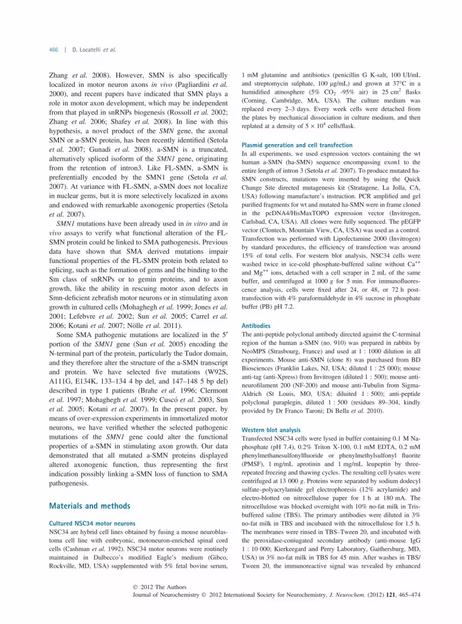
Preparation of a-SMN mutated constructsSMN1 pathogenic mutations from compound heterozygousSMA patients have been already used in in vitro and in vivoassays to verify FL-SMN functional alterations. The selectedconstructs (W92S, A111G, E134K, 133–134 4 bp del, and147–148 5 bp del: Fig. 1a were obtained by inserting pointmutations or small intragenic re-arrangements in the alreadyavailable human a-SMN vector (pcDNA4/wt ha-SMN:Setola et al. 2007; : Fig. 1a). As summarized in Fig. 1a,the selected constructs produced either (i) a-SMN proteinswith single aminoacid substitutions (W92S, A111G, andE134K mutated ha-SMN proteins), or (ii) SMN proteinsidentical to a-SMN and FL-SMN in the N-terminal part butwith a different C-terminal tail, encoded in part by the normal
sequence of exon 3, in part by the frameshifted sequence ofexon 3 (133–134 4 bp del and 147–148 5 bp del: seeFig. 1a). The sequence of all mutated constructs was verifiedby sequence analysis.
Expression levels of the different a-SMN polypeptidesWe next transiently transfected the selected constructs inNSC34 motor neurons. As illustrated in Fig. 1, western blotanalysis of cell lysates demonstrated that all constructsdetermined the production of ha-SMN polypeptides of theexpected molecular size (Fig. 1b and c). The optical densityquantification of WB immunoreactive bands (ha-SMN vs.actin) revealed that wt and mutated ha-SMN proteins withsingle aminoacid substitutions produced at both 24 and 48 hafter transfection comparable protein amounts (Fig. 1b andc). In contrast, 147–148 5 bp del ha-SMN expression levelswere clearly reduced at 48 h if compared with 24 h post-transfection (Fig. 1d). The expression levels of the 133–1344 bp del ha-SMN were already low at 24 h and almost absentat 48 h after transfection (Fig. 1b–d). These findings couldsignify that the latter SMN proteins exert a negative effectnot only on axon growth (see below) but also on cell survivaland homeostasis. We checked this possibility by analyzingthe density of cells transfected with the different ha-SMNconstructs at 24, 48, and 72 h (Fig. 1e). As indicated inFig. 1e, the number of transfected NSC34 motor neuronsdeclined slowly over time for all constructs considered. Thisindicates that the frameshift mutated ha-SMN proteins arenot toxic for motor neurons but rather less produced or lessstable/more rapidly degraded.
Effect on axon growthWe have already shown that a-SMN is capable of inducingtime-dependent axon growth upon forced expression inNSC34 motor neurons (Setola et al. 2007). We used thesame experimental approach to compare the axonogenicproperties of wt and mutated ha-SMN proteins. To this end,we analyzed a sizeable number of NSC34 motor neuronstransfected with a control vector expressing GFP and thedifferent ha-SMN constructs at three different time-points(24, 48, and 72 h after transfection) taken from triplicateexperiments. As illustrated in Fig. 2a, the forced expressionof the ha-SMN protein determined time-dependent axongrowth in NSC34 cells, in keeping with what reportedpreviously by our group (Setola et al. 2007). No increase inaxon length was observed after transfection with the GFP-expressing vector. The ha-SMN polypeptides carrying singleaminoacid substitutions were capable of progressivelyinducing axon growth in a way similar to that of wt ha-SMN (Fig. 2a and b). However, quantification of axonallength revealed that the axonogenic function of all missensemutated ha-SMN was impaired in comparison to that of wtha-SMN and that reduction of axon growth was statisticallysignificant (at 24 h for W92S/ha-SMN and E134K/ha-SMN,
� 2012 The AuthorsJournal of Neurochemistry � 2012 International Society for Neurochemistry, J. Neurochem. (2012) 121, 465–474
Mutated ha-SMN proteins impair axonogenesis | 467

at 72 h for A111G/ha-SMN: Fig. 2a and b). The axonogenicfunction was completely abrogated for the two ha-SMNpolypeptides with primary structure of the C-terminus moredivergent from that of the ha-SMN protein. In NSC34 motorneurons transfected with 147–148 5 bp del/ha-SMN, axongrowth was not different from that of cells transfected withthe GFP control vector. The axonogenic function of 133–1344 bp del/ha-SMN was completely abolished: the transfected
motor neurons did not show any tendency to grow at allconsidered time-points (Fig. 2a and b).
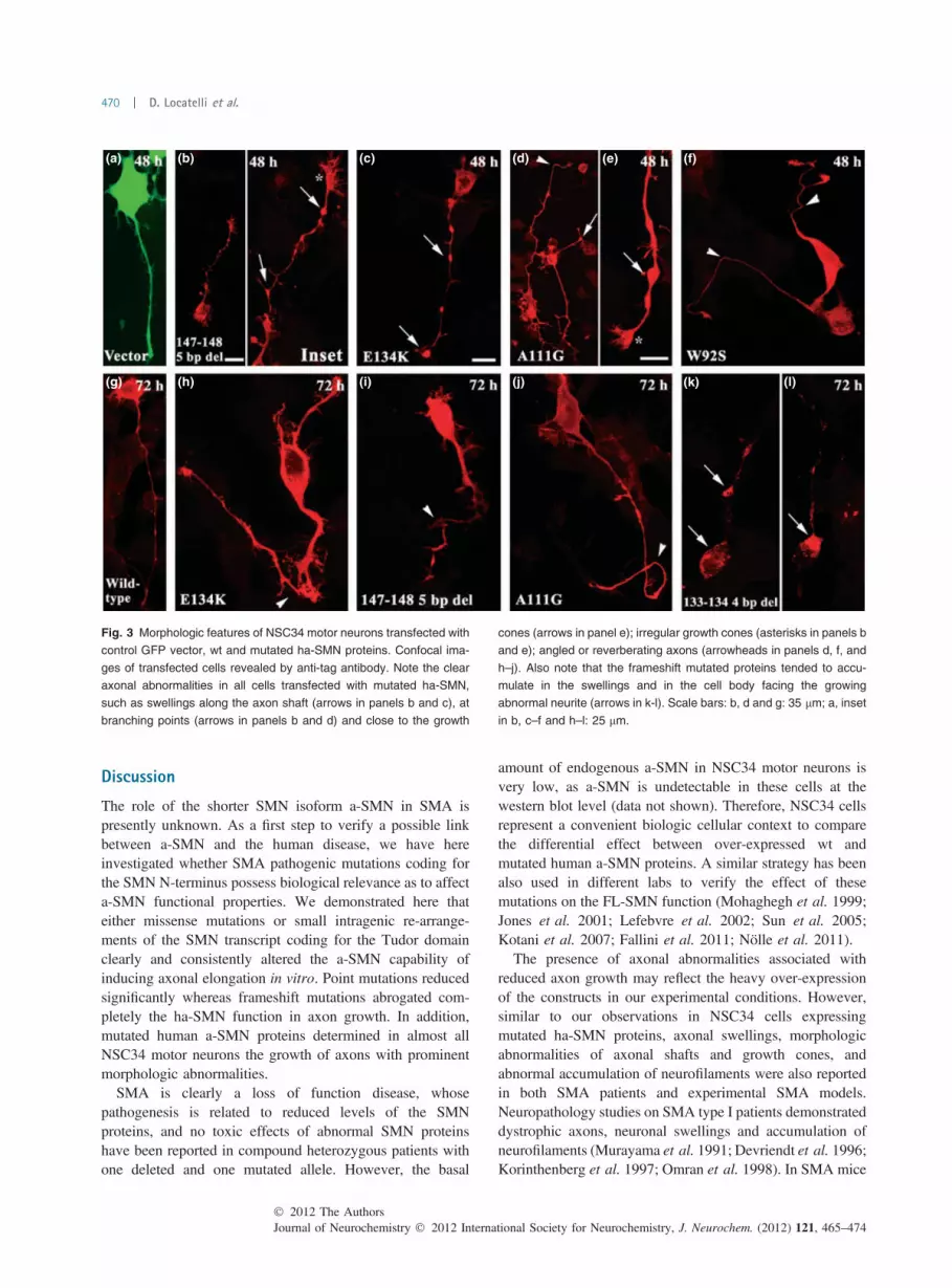
Axonal abnormalitiesWe next verified the morphology of neurites whose growthwas stimulated by the different human a-SMN constructs andthe GFP vector as control. The NSC34 cells transfected withthe GFP vector (Fig. 3a) and wt a-SMN (Fig. 3g) showed
(a)
(b) (c)
(d) (e)
Fig. 1 (a) Schematic representation of the full-length SMN protein,
and the wt and mutated ha-SMN constructs employed in the present
study. Note that the W92S, A111G, and E134K mutated ha-SMN all
retain the wt ha-SMN C-terminus with the Tudor and the intron 3 en-
coded domains. By contrast, this domain is disrupted in the ha-SMN
proteins with frameshift mutations, particularly in the 133–134 4 bp del
polypeptide. (b, c) WB analysis showing the production of the different
a-SMN polypeptides of the expected molecular size at 24 (b) and 48 h
after transfection (c). Blots were probed with an antibody directed
against the SMN N-terminus (clone 8, BD) recognizing both endoge-
nous FL-SMN (upper bands) and transfected ha-SMN (lower bands).
(d) Bargraphs showing the expression levels of wt and mutated ha-
SMN proteins (OD ratio vs. actin) after 24–48 h transfection. (e) Bar-
graphs showing the cell density of NSC34 motor neurons at different
time-points after transfection (transfected cells/mm2). The density of
NSC34 cells similarly decreased over time for all constructs consid-
ered, even if was lower for the frameshift mutated ha-SMN at all time-
points considered.
Journal of Neurochemistry � 2012 International Society for Neurochemistry, J. Neurochem. (2012) 121, 465–474� 2012 The Authors
468 | D. Locatelli et al.

normal neurites at all time-points considered. By contrast,cells transfected with mutated ha-SMN polypeptides werecharacterized by neurites not only shorter than those of cellsover-expressing wt ha-SMN but also displaying clearmorphologic abnormalities. In particular, we noticed thepresence of evident swellings along the course of neurites.The swellings were present in cells transfected with allmutated ha-SMN proteins, at all time-points considered,along the neuritic shaft close to the cell body (Fig. 3b and c),at branching sites (Fig. 3b and d) and up to the growth cone(Fig. 3e). Irregularly shaped, enlarged growth cones were notinfrequently found at the end of neurites with multipleswellings (Fig. 3b and e). In addition to the presence of theswellings, neurites also displayed an irregular course, as theywere frequently angled (Fig. 3f and i) and reverberatingtoward the cell body (Fig. 3h and j). These abnormal featureswere present at all time-points considered, but they tended toincrease progressively at later time-points (see below). In
cells transfected with the frameshift mutated ha-SMNproteins, the over-expressed proteins tended to accumulatein granules within the cell body frequently in the regionfacing the growing abnormal neurite (Fig. 3k and l).
For a better estimate of the morphologic abnormalitiesinduced by the different mutated ha-SMN proteins, wequantified the percentage of cells showing axonal defects atthe different time-points. As indicated in Figure 4, noabnormalities were observed in NSC34 cells transfectedwith wt ha-SMN at 24 h after transfection. At 48 and 72 hafter transfection, only a minority of wt ha-SMN transfectedcells (about 10%) were characterized by a few swellingsalong the neuritic shaft. By contrast, at all time-pointsconsidered, the majority of cells transfected with the mutatedha-SMN displayed axonal abnormalities (Fig. 4). In partic-ular, the negative effect of the missense mutations wasprogressive over time, whereas the frameshift mutationsinduced even after 24 h axonal abnormalities (and a highernumber of axonal abnormalities per axon: data not shown) inalmost all transfected motor neurons (Fig. 4a). If comparedwith wt ha-SMN, all mutations eventually prove to bedetrimental to the axonal morphology and growth in a highlysignificant fashion (Fig. 4b).
Cytoskeletal abnormalities and altered localization ofsubcellular organellesTo characterize further the morphologic abnormalitiesreported above, we performed double-labeling confocalexperiments with cytoskeletal and mitochondrial markers incells transfected with wt and mutated ha-SMN. Double-labeling staining revealed that cells transfected with wtha-SMN showed neurites and growth cones characterized byregular organization of microtubules and no evidence ofstructural abnormalities or mitochondria accumulation(Fig. 5a). By contrast, in cells transfected with mutated ha-SMN swellings were consistently associated with irregular-ities of the axonal cytoskeleton. As illustrated in Fig. 5,microtubules were consistently splayed apart in the axonalswellings (Fig. 5b–d). In addition, in the neurites character-ized by swellings, irregular disposition of microtubules wasobserved also at branching points (Fig. 5b, inset) and in thegrowth cone (Fig. 5c, inset). Swellings were also consistentlycharacterized by accumulation of mitochondria (Fig. 5e).Interestingly, mitochondria tended to be concentrated mostlyin the proximal part of the swellings (Fig. 5e). In addition, incells transfected with the frameshift mutated ha-SMN, theprotein accumulation in granules within the cell body wasalways associated with axonal swellings characterized byneurofilament irregularities (Fig. 5f). The quantification ofneurofilament/tubulin accumulation in axon swellings(Fig. 5g) and growth cones (Fig. 5h) confirmed the signif-icantly higher frequency of cytoskeletal abnormalities in cellstransfected with mutated versus wt ha-SMN at all time-pointsconsidered.
(a)
(b)
Fig. 2 (a) Diagram illustrating the quantification of mean axonal
elongation in NSC34 motor neurons transfected with the ‘empty’ GFP
vector, and the wt and mutated ha-SMN proteins at different time-
points. Data are indicated as mean ± SE of triplicate experiments.
Note that cells transfected with 133–134 4 bp del ha-SMN did not
show any axonal growth at the different time-points considered. (b)
Bargraphs illustrating the statistical evaluation at the three time-points
considered. Axon length (for control vector vs. wt vs. mutated ha-
SMN) was analyzed with two-way ANOVA followed by Bonferroni as
post hoc comparison test.
� 2012 The AuthorsJournal of Neurochemistry � 2012 International Society for Neurochemistry, J. Neurochem. (2012) 121, 465–474
Mutated ha-SMN proteins impair axonogenesis | 469
Discussion
The role of the shorter SMN isoform a-SMN in SMA ispresently unknown. As a first step to verify a possible linkbetween a-SMN and the human disease, we have hereinvestigated whether SMA pathogenic mutations coding forthe SMN N-terminus possess biological relevance as to affecta-SMN functional properties. We demonstrated here thateither missense mutations or small intragenic re-arrange-ments of the SMN transcript coding for the Tudor domainclearly and consistently altered the a-SMN capability ofinducing axonal elongation in vitro. Point mutations reducedsignificantly whereas frameshift mutations abrogated com-pletely the ha-SMN function in axon growth. In addition,mutated human a-SMN proteins determined in almost allNSC34 motor neurons the growth of axons with prominentmorphologic abnormalities.
SMA is clearly a loss of function disease, whosepathogenesis is related to reduced levels of the SMNproteins, and no toxic effects of abnormal SMN proteinshave been reported in compound heterozygous patients withone deleted and one mutated allele. However, the basal
amount of endogenous a-SMN in NSC34 motor neurons isvery low, as a-SMN is undetectable in these cells at thewestern blot level (data not shown). Therefore, NSC34 cellsrepresent a convenient biologic cellular context to comparethe differential effect between over-expressed wt andmutated human a-SMN proteins. A similar strategy has beenalso used in different labs to verify the effect of thesemutations on the FL-SMN function (Mohaghegh et al. 1999;Jones et al. 2001; Lefebvre et al. 2002; Sun et al. 2005;Kotani et al. 2007; Fallini et al. 2011; Nolle et al. 2011).
The presence of axonal abnormalities associated withreduced axon growth may reflect the heavy over-expressionof the constructs in our experimental conditions. However,similar to our observations in NSC34 cells expressingmutated ha-SMN proteins, axonal swellings, morphologicabnormalities of axonal shafts and growth cones, andabnormal accumulation of neurofilaments were also reportedin both SMA patients and experimental SMA models.Neuropathology studies on SMA type I patients demonstrateddystrophic axons, neuronal swellings and accumulation ofneurofilaments (Murayama et al. 1991; Devriendt et al. 1996;Korinthenberg et al. 1997; Omran et al. 1998). In SMA mice
(a) (b) (c) (d) (e) (f)
(g) (h) (i) (j) (k) (l)
Fig. 3 Morphologic features of NSC34 motor neurons transfected with
control GFP vector, wt and mutated ha-SMN proteins. Confocal ima-
ges of transfected cells revealed by anti-tag antibody. Note the clear
axonal abnormalities in all cells transfected with mutated ha-SMN,
such as swellings along the axon shaft (arrows in panels b and c), at
branching points (arrows in panels b and d) and close to the growth
cones (arrows in panel e); irregular growth cones (asterisks in panels b
and e); angled or reverberating axons (arrowheads in panels d, f, and
h–j). Also note that the frameshift mutated proteins tended to accu-
mulate in the swellings and in the cell body facing the growing
abnormal neurite (arrows in k-l). Scale bars: b, d and g: 35 lm; a, inset
in b, c–f and h–l: 25 lm.
Journal of Neurochemistry � 2012 International Society for Neurochemistry, J. Neurochem. (2012) 121, 465–474� 2012 The Authors
470 | D. Locatelli et al.

prominent axonal swellings (McGovern et al. 2008) orneurofilament accumulation in neuromuscular junctions (Cifu-entes-Diaz et al. 2002) were reported. Cultured motor neuronsand sensory neurons from severe (SMN2+/+; Smn)/)) SMAmice showed decreased axonal length and reduced growthcones size (Rossoll et al. 2003; Jablonka et al. 2006; Tinget al. 2007). Smn knockdown in zebrafish to levels similar tothose occurring in SMA patients determined the developmentof altered motor neurons with truncated or excessivelybranched axons that reached the target muscles withinappropriate trajectories (McWhorter et al. 2003).
Regarding the potential severity of the considered muta-tions in SMA pathogenesis, it should be noted that theW92S, A111G, E134K and 147–148 5bp del mutations weredescribed in type I patients (Brahe et al. 1996; Clermontet al. 1997; Mohaghegh et al. 1999; Sun et al. 2005; Kotaniet al. 2007), thus indicating that even subtle abnormalities ofthe Tudor domain are critical for SMN function. Thesemutations were previously reported to disrupt the SMNinteraction with fundamental components of nuclear RNA-protein complexes, such as fibrillarin and the Sm proteins(Buhler et al. 1999; Jones et al. 2001; Sun et al. 2005), thusimpairing snoRNP and snRNP biogenesis. However, it has
been more recently demonstrated that FL-SMN also interactsthrough the Tudor domain with HuD, a protein of theembryonic lethal abnormal vision (ELAV) family, in both thecytoplasm and axons of cultured motor neurons to formmultiprotein complexes that are actively transported alongthe axon (Akten et al. 2011; Fallini et al. 2011; Hubers et al.2011). The E134K mutation significantly impaired theinteraction SMN-HuD in the axonal compartment (Falliniet al. 2011; Hubers et al. 2011). Given that the ha-SMNsequence contains the entire Tudor domain, it is possible thatthis specific region might subserve the anchoring of ha-SMNto other proteins or mRNA involved in neuronal develop-ment, growth and transport (such as HuD, neuritin, GAP43),and that abnormalities in this region may explain the a-SMNloss of function reported here. The relevance of the Tudorand the shorter protein domain encoded by intron3 is alsosuggested by the relationship between the severity ofmutation and axonogenic function, because the more theha-SMN C-terminal sequence is altered, the more the a-SMNaxonogenic properties are affected.
The 133–134 4 bp del mutation has been mostly reportedin Spanish SMA families, associated with a wide variety ofclinical manifestations, ranging from severe type I toasymptomatic patients (Cusco et al. 2003). This mutationwas the most severe in our in vitro experiments in affectingthe a-SMN axonogenic functions. However, as clearlyindicated by our data on the survival of transfected motorneurons, the mutated 133–134 4 bp del ha-SMN did notpossess toxic effects (as shown in Fig. 1e) but it was likelymore rapidly degraded. Therefore, other factors such as thenumber of copies of SMN2 and the ratio between FL and D7proteins may underscore the important difference in severityreported in the Spanish families (Cusco et al. 2003; Also-Rallo et al. 2011).
The double-labeling experiments demonstrated that theaxonal swellings induced by mutated human a-SMN proteinswere structural abnormalities of the axonal shafts with alteredcytoskeletal elements and altered distribution of subcellularorganelles such as mitochondria. These findings may indicatethat the a-SMN protein plays a relevant role in theestablishment of the proper cytoskeletal structure of theaxon, and that axonal transport may be impaired as aconsequence of such cytoskeletal impairment. The relevanceof a-SMN in the proper building up of the axon is alsosuggested by the accumulation of the mutated a-SMNpolypeptides (particularly the 133–134 4 bp del ha-SMN,the mutated a-SMN with the C-terminus most divergent fromthe wt ha-SMN) in the region of the cell body facing thenewly growing neurite (see Fig. 3l).
In conclusion, we have demonstrated here that SMApathogenic mutations strategically placed in the Tudordomain deeply alter the functional capabilities of a-SMN.The clear relationship between a-SMN structural alterationand functional impairment indicates that the Tudor and
(a)
(b)
Fig. 4 (a) Histogram showing the percentage of transfected cells with
axonal abnormalities at the different time-points considered. Note that
the negative effect of the missense point mutations was significantly
progressive over time, whereas the frameshift mutations induced ax-
onal defects even after 24 h. (b) By contrast, only 10% of cells
transfected with wt ha-SMN displayed axonal abnormalities at 72 h.
The difference was significant for all considered mutated ha-SMN
proteins.
� 2012 The AuthorsJournal of Neurochemistry � 2012 International Society for Neurochemistry, J. Neurochem. (2012) 121, 465–474
Mutated ha-SMN proteins impair axonogenesis | 471

intron3-encoded domains are critical in dictating a-SMNfunction possibly because they work as an associationdomain for proteins involved in axonal growth or mainte-nance. Two limitations of our experimental approach shouldbe underlined. First, these data do not provide a direct proofthat a-SMN is pathogenetically linked with SMA. Rather,they more simply indicate that Tudor domain mutations are
functionally relevant not only for FL-SMN function, raisingthe possibility of diverse, multiple pathogenic steps leadingto the disease in humans. Second, for the limitations intrinsicto immortalized NSC34 cells, the potential pathogenic role ofa-SMN for SMA would ultimately need to be studied in morephysiologic biological systems, such as primary motoneuro-nal cultures or motor neurons in vivo.
(a)
(g) (h)
(a1)
(b) (b1)
(c) (c1)
(d) (d1)
(e) (e1)
(f) (f1)
Fig. 5 Confocal images of NSC34 cells transfected with wt or mutated
a-SMN constructs, double labeled for ha-SMN (red, pseudo-color from
Alexa 647: no. 910 in panels a–d and anti-tag in panels e and f), and
anti-tubulin (green, from Alexa 488: a1–d1), anti-paraplegin (green: e1)
and anti- neurofilaments NF-200 (green: f1) antibodies. Note the
irregular disposition of microtubules in axonal swellings, branch points
and growth cones (arrows in panels b–d, insets) and the neurofilament
irregularities in axonal swellings (arrows in panel f, inset). Also note
the accumulation of mitochondria in axonal swellings (arrowhead in e
and inset). Scale bars: a, b, d–f: 25 lm; c: 20 lm; inset in a–f: 10 lm.
(g–h) Bargraphs showing the presence of cytoskeletal abnormalities in
swellings and growth cones in cells transfected with wt and mutated
ha-SMN constructs at the different time-points considered. The
significant difference in cytoskeletal abnormalities between wt and
mutants was obtained with one-way ANOVA followed by Bonferroni as
post hoc comparison test.
Journal of Neurochemistry � 2012 International Society for Neurochemistry, J. Neurochem. (2012) 121, 465–474� 2012 The Authors
472 | D. Locatelli et al.

Acknowledgements
The authors wish to thank Dr Maddalena Fratelli for her help in thestatistical analysis, and Dr Franco Taroni for providing the anti-paraplegin antibody. This work was supported by grant no.GGP07223 from Telethon-Italy Foundation; grant no. A0000842from the ‘Sovvenzione Globale Ingenio Regione Lombardia/SocialEuropean Fund; grant no. R0868 from the Pierfranco e LuisaMariani Foundation; grant no. RA48 from Cariplo Foundation.Financial supports from Girotondo/ONLUS and Smarathon-ONLUSassociations is also gratefully acknowledged. The authors declarethat they have no competing financial interests.
References
Akten B., Kye M. J., Hao le T. et al. (2011) Interaction of survival ofmotor neuron (SMN) and HuD proteins with mRNA cpg15 rescuesmotor neuron axonal deficits. Proc. Natl Acad. Sci. USA 108,10337–10342.
Also-Rallo E., Alıas L., Martınez-Hernandez R., Caselles L., Barcelo M.J., Baiget M., Bernal S. and Tizzano E. F. (2011) Treatment ofspinal muscular atrophy cells with drugs that upregulate SMNexpression reveals inter- and intra-patient variability. Eur. J. Hum.Genet. 19, 1059–1065.
Brahe C., Clermont O., Zappata S., Tiziano F., Melki J. and Neri G.(1996) Frameshift mutation in the survival motor neuron gene in asevere case of SMA type I. Hum. Mol. Genet. 5, 1971–1976.
Buhler D., Raker V., Luhrmann R. and Fischer U. (1999) Essential rolefor the tudor domain of SMN in spliceosomal U snRNP assembly:implications for spinal muscular atrophy. Hum. Mol. Genet. 8,2351–2357.
Carrel T. L., McWhorter M. L., Workman E., Zhang H., WolstencroftE. C., Lorson C., Bassell G. J., Burghes A. H. and Beattie C. E.(2006) Survival motor neuron function in motor axons is inde-pendent of functions required for small nuclear ribonucleoproteinbiogenesis. J. Neurosci. 26, 11014–11022.
Cashman N. R., Durham H. D., Blusztajn J. K., Oda K., Tabira T., ShawI. T., Dahrouge S. and Antel J. P. (1992) Neuroblastoma x spinalcord (NSC) hybrid cell lines resemble developing motor neurons.Dev. Dyn. 194, 209–221.
Cifuentes-Diaz C., Nicole S., Velasco M. E., Borra-Cebrian C., PanozzoC., Frugier T., Millet G., Roblot N., Joshi V. and Melki J. (2002)Neurofilament accumulation at the motor endplate and lack ofaxonal sprouting in a spinal muscular atrophy mouse model. Hum.Mol. Genet. 11, 1439–1447.
Clermont O., Burlet P., Cruaud C., Bertrandy S., Melki J., Munnich A.and Lefebvre S. (1997) Mutation analysis of the SMN gene inundeleted SMA patients. Am. J. Hum. Genet. 61, A329.
Cusco I., Lopez E., Soler-Botija C., Jesus Barcelo M., Baiget M. andTizzano E. F. (2003) A genetic and phenotypic analysis in Spanishspinal muscular atrophy patients with c.399_402del AGAG, themost frequently found subtle mutation in the SMN1 gene. Hum.Mutat. 2, 136–143.
Devriendt K., Lammens M., Schollen E., Van Hole C., Dom R., Dev-lieger H., Cassiman J. J., Fryns J. P. and Matthijs G. (1996)Clinical and molecular genetic features of congenital spinal mus-cular atrophy. Ann. Neurol. 40, 731–738.
Di Bella D., Lazzaro F., Brusco A. et al. (2010) Mutations in themitochondrial protease gene AFG3L2 cause dominant hereditaryataxia SCA28. Nat. Genet. 4, 313–321.
Fallini C., Zhang H., Su Y., Silani V., Singer R. H., Rossoll W. andBassell G. J. (2011) The survival of motor neuron (SMN) protein
interacts with the mRNA-binding protein HuD and regulateslocalization of poly(A) mRNA in primary motor neuron axons.J. Neurosci. 31, 3914–3925.
Gunadi, Sasongko T. H., Yusoff S., Lee M. J., Nishioka E., Matsuo M.and Nishio H. (2008) Hypomutability at the polyadenine tract inSMN intron 3 shows the invariability of the a-SMN proteinstructure. Ann. Hum. Genet. 72, 288–291.
Hahnen E., Forkert R., Marke C., Rudnik-Schoneborn S., Schonling J.,Zerres K. and Wirth B. (1995) Molecular analysis of candidategenes on chromosome 5q13 in autosomal recessive spinal mus-cular atrophy: evidence of homozygous deletions of the SMN genein unaffected individuals. Hum. Mol. Genet. 4, 1927–1933.
Hubers L., Valderrama-Carvajal H., Laframboise J., Timbers J., SanchezG. and Cote J. (2011) HuD interacts with survival motor neuronprotein and can rescue spinal muscular atrophy-like neuronal de-fects Hum. Mol. Genet. 20, 553–579.
Jablonka S., Karle K., Sandner B., Andreassi C., von Au K. andSendtner M. (2006) Distinct and overlapping alterations in motorand sensory neurons in a mouse model of spinal muscular atrophy.Hum. Mol. Genet., 15, 511–518.
Jones K. W., Gorzynski K., Hales C. M., Fischer U., Badbanchi F.,Terns R. M. and Terns M. P. (2001) Direct interaction of the spinalmucular atrophy disease protein SMN with the small nucleolarRNA-associated protein fibrillarin. J. Biol. Chem. 276, 38645–38651.
Korinthenberg R., Sauer M., Ketelsen U. P. et al. (1997) Congenitalaxonal neuropathy caused by deletions in the spinal muscularatrophy region. Ann. Neurol. 42, 364–368.
Kotani T., Sutomo R., Sasongko T. H. et al. (2007) A novel mutation atthe N-terminal of SMN Tudor domain inhibits its interaction withtarget proteins. J. Neurol. 254, 624–630.
Lefebvre S., Burglen L., Reboullet S. et al. (1995) Identification andcharacterization of a spinal muscular atrophy-determining gene.Cell 80, 155–165.
Lefebvre S., Burglen L., Reboullet S., Munnich A. and Melki J. (1998)The role of the SMN gene in proximal spinal muscular atrophy.Hum. Mol. Genet. 7, 1531–1536.
Lefebvre S., Burlet P., Viollet L., Bertrandy S., Huber C., Belser C. andMunnich A. (2002) A novel association of the SMN protein withtwo major non-ribosomal nucleolar proteins and its implication inspinal muscular atrophy. Hum. Mol. Genet. 11, 1017–1027.
McGovern V. L., Gavrilina T. O., Beattie C. E. and Burghes A. H.(2008) Embryonic motor axon development in the severe SMAmouse. Hum. Mol. Genet. 17, 2900–2909.
McWhorter M. L., Monani U. R., Burghes A. H., and Beattie C. E.(2003) Knockdown of the survival motor neuron (Smn) protein inzebrafish causes defects in motor axon outgrowth and pathfinding.J. Cell Biol. 162, 919–931.
Mohaghegh P., Rodrigues N. R., Owen N., Ponting C. P., Le T. T.,Burghes A. H. and Davies K. E. (1999) Analysis of mutations inthe tudor domain of the survival motor neuron protein SMN. Eur.J. Hum. Gen. 7, 519–525.
Murayama S., Bouldin T. W. and Suzuki K. (1991) Immunocytochem-ical and ultrastructural studies of Werdnig-Hoffmann disease. ActaNeuropathol. 81, 408–417.
Nolle A., Zeug A., van Bergeijk J. et al. (2011) The spinal muscularatrophy disease protein SMN is linked to the rho-kinase pathwayvia profilin. Hum. Mol. Genet. 20, 4865–4878.
Omran H., Ketelsen U. P., Heinen F., Sauer M., Rudnik-Schoneborn S.,Wirth B., Zerres K., Kratzer W. and Korinthenberg R. (1998)Axonal neuropathy and predominance of type II myofibers ininfantile spinal muscular atrophy. J. Child Neurol. 13, 327–331.
Pagliardini S., Giavazzi A., Setola V., Lizier C., Di Luca M., DeBiasi S.and Battaglia G. (2000) Subcellular localization and axonal
� 2012 The AuthorsJournal of Neurochemistry � 2012 International Society for Neurochemistry, J. Neurochem. (2012) 121, 465–474
Mutated ha-SMN proteins impair axonogenesis | 473
transport of the survival motor neuron (SMN) protein in thedeveloping rat spinal cord. Hum. Mol. Genet. 9, 47–56.
Parsons D. W., McAndrew P. E., Iannaccone S. T., Mendell J. R.,Burghes A. H. and Prior T. W. (1998) Intragenic telSMN muta-tions: frequency, distribution, evidence of a founder effect, andmodification of the spinal muscular atrophy phenotype by cenSMNcopy number. Am. J. Hum. Genet. 63, 1712–1723.
Pellizzoni L., Kataoka N., Charroux B. and Dreyfuss G. (1998) A novelfunction for SMN, the spinal muscolar atrophy disease geneproduct, in pre-mRNA splicing. Cell 95, 615–624.
Prior T. W., Snyder P. J., Rink B. D. et al. (2010) Newborn and carrierscreening for spinal muscular atrophy. Am. J. Med. Genet. A. 152,1608–1616.
Rodrigues N. R., Owen N., Talbot K., Ignatius J., Dubowitz V. andDavies K. E. (1995) Deletions in the survival motor neuron geneon 5q13 in autosomal recessive spinal muscular atrophy. Hum.Mol. Genet. 4, 631–634.
Rossoll W., Kroning A. K., Ohndorf U. M., Steegborn C., Jablonka S.and Sendtner M. (2002) Specific interaction of Smn, the spinalmuscular atrophy determining gene product, with hnRNP-R andgry-rbp/hnRNP-Q: a role for Smn in RNA processing in motoraxons? Hum. Mol. Genet. 11, 93–105.
Rossoll W., Jablonka S., Andreassi C., Kroning A. K., Karle K., MonaniU. R. and Sendtner M. (2003) Smn, the spinal muscular atrophy-determining gene product, modulates axon growth and localization
of b-actin mRNA in growth cones of motoneurons. J. Cell Biol.163, 801–812.
Setola V., Terao M., Locatelli D., Bassanini S., Garattini E. and BattagliaG. (2007) Axonal-SMN (a-SMN), a protein isoform of the SurvivalMotor Neuron gene, is specifically involved in axonogenesis. Proc.Natl Acad. Sci. USA 104, 1959–1964.
Shafey D., MacKenzie A. E. and Kothary R. (2008) Neurodevelop-mental abnormalities in neurosphere-derived neural stem cells fromSMN-depleted mice. J. Neurosci. Res. 86, 2839–2847.
Sun Y., Grimmler M., Schwarzer V., Schoenen F., Fischer U. and WirthB. (2005) Molecular and functional analysis of intragenic SMN1mutations in patients with spinal muscular atrophy. Hum. Mutat.25, 64–71.
Ting C. H., Lin C. W., Wen S. L., Hsieh-Li H. M. and Li H. (2007) Stat5constitutive activation rescues defects in spinal muscular atrophy.Hum. Mol. Genet. 5, 499–514.
Zhang H., Xing L., Rossoll W., Wichterle H., Singer R. H. and BassellG. J. (2006) Multiprotein complexes of the survival of motorneuron protein SMN with Gemins traffic to neuronal processes andgrowth cones of motor neurons. J. Neurosci. 26, 8622–8632.
Zhang Z., Lotti F., Dittmar K., Younis I., Wan L., Kasim M. andDreyfuss G. (2008) SMN deficiency causes tissue-specific pertur-bations in the repertoire of snRNAs and widespread defects insplicing. Cell 4, 585–600.
Journal of Neurochemistry � 2012 International Society for Neurochemistry, J. Neurochem. (2012) 121, 465–474� 2012 The Authors
474 | D. Locatelli et al.