spatially fractionated radiation induces cytotoxicity and changes in gene expression in bystander...
TRANSCRIPT

Spatially Fractionated Radiation Induces Cytotoxicity andChanges in Gene Expression in Bystander and RadiationAdjacent Murine Carcinoma Cells
Rajalakshmi S. Asura, Sunil Sharmaa, Ching-Wei Changb, Jose Penagaricanoa, Indira M.Kommurua, Eduardo G. Morosc, Peter M. Corrya, and Robert J. Griffina,1
aDepartment of Radiation Oncology, University of Arkansas for Medical Sciences, Little Rock,Arkansas 72205bDivision of Personalized Nutrition and Medicine, National Center for Toxicological Research,U.S. Food and Drug Administration, Jefferson, Arkansas 72079cH. Lee Moffitt Cancer Center and Research Institute, MCC RADONC, Tampa, Florida 33612
AbstractRadiation-induced bystander effects have been extensively studied at low doses, since evidence ofbystander induced cell killing and other effects on unirradiated cells were found to be predominantat doses up to 0.5 Gy. Therefore, few studies have examined bystander effects induced byexposure to higher doses of radiation, such as spatially fractionated radiation (GRID) treatment. Inthe present study, we evaluate the ability of GRID treatment to induce changes in GRID adjacent(bystander) regions, in two different murine carcinoma cell lines following exposure to a singleirradiation dose of 10 Gy. Murine SCK mammary carcinoma cells and SCCVII squamouscarcinoma cells were irradiated using a brass collimator to create a GRID pattern of nine circularfields 12 mm in diameter with a center-to-center distance of 18 mm. Similar to the typical clinicalimplementation of GRID, this is approximately a 50:50 ratio of direct and bystander exposure. Wealso performed experiments by irradiating separate cultures and transferring the medium tounirradiated bystander cultures. Clonogenic survival was evaluated in both cell lines to determinethe occurrence of radiation-induced bystander effects. For the purpose of our study, we havedefined bystander cells as GRID adjacent cells that received approximately 1 Gy scatter dose orunirradiated cells receiving conditioned medium from irradiated cells. We observed significantbystander killing of cells adjacent to the GRID irradiated regions compared to sham treatedcontrols. We also observed bystander killing of SCK and SCCVII cells cultured in conditionedmedium obtained from cells irradiated with 10 Gy. Therefore, our results confirm the occurrenceof bystander effects following exposure to a high-dose of radiation and suggest that cell-to-cellcontact is not required for these effects. In addition, the gene expression profile for DNA damageand cellular stress response signaling in SCCVII cells after GRID exposure was studied. Theoccurrence of GRID-induced bystander gene expression changes in significant numbers of DNAdamage and cellular stress response signaling genes, providing molecular evidence for possiblemechanisms of bystander cell killing.
© 2012 by Radiation Research Society1Address for correspondence: Department of Radiation Oncology, University of Arkansas for Medical Sciences, 4301 West MarkhamStreet, Little Rock, Arkansas 72205; [email protected].
NIH Public AccessAuthor ManuscriptRadiat Res. Author manuscript; available in PMC 2013 June 01.
Published in final edited form as:Radiat Res. 2012 June ; 177(6): 751–765.
NIH
-PA Author Manuscript
NIH
-PA Author Manuscript
NIH
-PA Author Manuscript

INTRODUCTIONA long-standing concept in radiobiology has been that cells must be directly exposed toionizing radiation for DNA damage, and possible cell death, to occur. However,considerable evidence now exists that challenges this belief. In fact, as early as the 1940s,published literature provided evidence for DNA damage brought about by targeting not onlythe cells or tissues, but also the surrounding medium (1, 2). The term “radiation-inducedbystander effects” refers to effects seen in cells that have not directly been exposed toionizing radiation. Kotval and Gray (3) demonstrated that α particles passing close to, butnot through, chromatin threads produced chromosomal breaks and exchanges. An increasein sister chromatid exchanges in cells that were not directly exposed to ionizing radiation,but were in the vicinity of directly irradiated cells (4), provided further proof of bystandereffects. Among the numerous studies, each with its own nuances and protocols nowaccumulated in the literature, radiation-induced bystander effects can be broadly classifiedinto two types: those mediated by gap-junctions and requiring cell-to-cell communicationand those brought about by the presence of medium secreted factors, which do not requirecell-to-cell contact (5–10). There are also reports in the literature where evidence ofbystander effects from irradiation was not found, especially when low-LET radiationsources were used. Therefore, the phenomenon appears to have specific requirements tooccur.
Spatially fractionated radiation therapy (sometimes referred to as “GRID” therapy) refers tothe delivery of a single radiation fraction (10–20 Gy peak doses) by dividing a radiationfield into smaller segments interspersed with segments receiving no (or very low doses)direct irradiation. This approach has been shown to have potent palliative benefits withoutincreasing toxicity (11, 12). Although originally designed mostly to avoid normal tissuetoxicity, over the last 100 years, this approach has been sporadically, and in generalsuccessfully, used to improve treatment of bulky and deep-seated tumors. Our recent clinicalexperience along with that of others suggests that GRID may be combined with traditionaldose/time fractionated radiation therapy or used along with other treatment modalities,including chemotherapy, to achieve better control of bulky tumors while minimallyextending the treatment course (11, 13). Although GRID dose distribution is nonuniform,regression of the tumor mass receiving GRID has exhibited uniform regression clinically(11, 13). One plausible explanation might be the enhanced reoxygenation of the tumorfollowing GRID, which would be expected to improve the effect of subsequently appliedradiation or chemotherapy. We have recently observed evidence of reoxygenation of tumorsafter spatially fractionated micro-beam radiation (61). Induction of tumor necrosis factor αand ceramide, as well as down regulation of transforming growth factor β1, have also beenobserved following GRID (14, 15). Increased cytokine production has resulted in broadsystemic effects as well (11). It also is possible that bystander effects might play a role inkilling adjacent nonirradiated or partially irradiated cells.
In the present study, we sought to evaluate the possible contribution of bystander effects tothe overall therapeutic effect of spatially fractionated (GRID) radiation therapy. In ourexperiments, we considered the regions that were exposed to 10 Gy of radiation as “directlyirradiated”. The cells adjacent to the direct irradiation fields that did not receive directirradiation, but were exposed to indirect radiation (i.e., scattering and some degree of a verysteep dose gradient), which amounted to a valley dose of approximately 1 Gy, wereconsidered as “bystander cells”. Clonogenic survival was used to determine bystander cellkilling in two different murine carcinoma cell lines following exposure to a single dose of 10Gy using either confluent cultures and selectively harvesting bystander cells or byperforming medium exchange experiments. In addition, we evaluated the expression profilesof genes involved in DNA damage signaling and stress response signaling in murine cells.
Asur et al. Page 2
Radiat Res. Author manuscript; available in PMC 2013 June 01.
NIH
-PA Author Manuscript
NIH
-PA Author Manuscript
NIH
-PA Author Manuscript

MATERIALS AND METHODSCell Culture
Two murine tumor cell lines (passage 3–5) were used in these experiments. SCK is a mousemammary carcinoma cell line derived from A/J mice (16, 17). SCK cells are considered tobe radiation resistant (18). We chose these cells to determine the occurrence of bystandereffects in response to spatially fractionated radiation therapy at clinically relevant doses. Thecells were grown in RPMI 1640 culture medium, with 2 mM L-glutamine (Cellgro,Manassas, VA) supplemented with 10% bovine serum albumin (Hyclone, Logan, UT) and1% penicillin-streptomycin (10,000 units/ml penicillin G sodium, 10,000 μg/mlstreptomycin in 0.85% saline) (Hyclone). SCCVII is a mouse squamous cell carcinoma cellline. We used these cells in our experiments, since GRID therapy has been used at ourinstitution and others to treat mainly head and neck cancers. SCCVII cells were cultured inD-MEM culture medium with 4.5 g/L glucose, sodium pyruvate and 2 mM (concentration)L-glutamine (Cellgro, Manassas, VA), supplemented with 10% fetal bovine serum (AtlasBiologicals, Fort Collins, CO) and 1% penicillin-streptomycin (10,000 units/ml penicillin Gsodium, 10,000 μg/ml streptomycin in 0.85% saline) (Hyclone). SCK and SCCVII cellswere sub-cultured by seeding at a concentration of 2.0 × 10⁵ cells in 25-cm2 culture flasks(Corning, NY), and were grown in a fully humidified incubator with 5% CO2 at 37°C. Thedoubling time of these cell lines is approximately 24 h.
Radiation Survival StudiesSCK and SCCVII cells were sparsely plated (200–250 cells) in 25-cm2 culture flaskscontaining 5 ml culture medium. The irradiation was performed approximately 16–20 h laterusing a CP-160 cabinet X-radiator system (Faxitron, Lincolnshire, IL) at room temperature.The cells were exposed to 0, 1 and 2 Gy X rays at a dose rate of 1 Gy per min at 150 kV and6.6 mA. The medium was changed immediately after irradiation and the flasks werereturned to the incubator for 8 days, after which the colonies (>50 cells) were stained usingcrystal violet and counted to determine colony forming efficiency. Colony formingefficiency was defined as the ratio of the number of colonies to the number of plated cells.In all cases, the clonogenic survival was normalized to the cloning efficiency ofappropriately sham treated and time-matched incubations.
Small Animal Conformal Radiation Research System (SACRRS) Used for GRID IrradiationThe irradiation system has a 225kVp X-ray tube with focal spots of 0.4 mm (used forimaging) and 3 mm (used for therapy). The precise positioning of the target/beam for boththerapy and imaging is achieved by a programmable robotic arm (Adept Viper S650, AdeptTechnology Inc., Pleasanton, CA). The system is equipped with a flat-panel amorphoussilicon digital X-ray imager (XRD 0820 CM3, Perking Elmer Inc., San Jose, CA), whichcaptures 1024 × 1024 pixel image (200 μm pixel size) at a frame rate of 7.5 Hz. The systemcan deliver an accurate and quantifiable conformal radiation dose to selected targets insingle or multiple fractions. In addition, the system is also equipped with a variable aperturecollimation system mounted at 20 cm from the X-ray source that can produce field size up to20 cm2 at the isocenter. These specially designed brass collimators are mounted on twosliders that move in opposite direction from the center using a uni-axial motor (Velmex Inc.)to make square fields. During the GRID irradiation, cells are placed on the “palm” of therobot and aligned with the X-ray beam. The GRID pattern of irradiation is then created byprogramming the robot platform to move normal to X-ray beam direction (Fig. 1A). Thecells were then irradiated to create a pattern of nearly 50:50 direct and bystander exposurepattern of 9 circular fields, 12 mm in diameter with a center-to-center distance of 18 mm.The cells were irradiated with 10 Gy using GRID, at a dose rate of 1.9 Gy per min at 255 kVand 13 mA.
Asur et al. Page 3
Radiat Res. Author manuscript; available in PMC 2013 June 01.
NIH
-PA Author Manuscript
NIH
-PA Author Manuscript
NIH
-PA Author Manuscript

A Gafchromic EBT-2 film (International Specialty Products, Wayne, NJ) was used fordosimetry. A calibration curve was plotted using the pixel values against the doses byirradiating the EBT-2 film under a Cobalt-60 beam. Subsequently, the EBT-2 films wereirradiated at 1 cm below the isocenter, at 225 kVp energy, with 12 mm brass collimatorduplicating the conditions under which the murine cells were irradiated (Fig. 1B). The filmswere exposed at different times and pixel values were recorded. The dose rate at 1 cm depthwas then calculated.
Bystander Survival StudiesMedium transfer based studies—SCK and SCCVII cells were plated at confluentdensity in 25-cm2 culture flasks containing 5 ml culture medium and were irradiated 16–20h later with 10 Gy. Flasks were returned to the incubator and cells were harvested byscraping at 0, 24, 48 and 168 h after irradiation. The 168 h (1 week) time point was includedto determine the long-term effect of direct or bystander radiation exposure. Any clumps ofcells were manually disrupted using a 500-μl pipette tip and the cells were then re-platedsparsely in 6-well cell culture dishes (Greiner Bio-One, Germany) and stained for colony-forming efficiency following 8 days to determine the effect of direct irradiation on thesecells. Donor cell cultures for providing the conditioned medium were plated with 2 millionconfluent cells in 5 ml culture medium in 25-cm2 flasks and were exposed to 10 Gy ofradiation after 16–20 h. Medium from sham-exposed cells was used for the controls.Medium transfer was performed 4 h after irradiation by collecting all medium from donorflasks and passing it through a 0.22 μm filter (Corning, NY) to ensure that no cells werepresent in the medium, but that soluble proteins or other factors could be retained. Thisfiltrate was considered as the conditioned medium. The original medium was aspirated frombystander recipient cells and was replaced with filtered conditioned medium. Cells wereharvested by scraping at 0, 24, 48 and 168 h after medium transfer and were re-plated (200–250 cells) in 6-well cell culture dishes and stained for clonogenic survival after 8 days.
GRID based bystander studies—SCK and SCCVII cells were plated at confluentdensity in 100 mm cell culture dishes (Greiner Bio-One, Germany) a day prior to irradiation.In our experiments, we considered the regions that were exposed to 10 Gy of radiation as“directly irradiated” and the adjacent cells which did not receive direct irradiation, but wereexposed to indirect radiation (i.e., scattering) which amounted to a valley dose ofapproximately 1 Gy, were considered as “bystander cells”. The irradiated and adjacentbystander cells were demarcated using a waterproof histological marker, Pap pen (ResearchProducts International Corp, Mt. Prospect, IL) to separate the irradiated and bystander fieldsusing a small square of Gafchromic film that was attached to the bottom of the flask duringirradiation as a guide and separately isolated. Cells were harvested at 0, 4, 24 and 48 h afterGRID irradiation. Clumps were manually disrupted using a 500-μl pipette tip and cells werethen sparsely re-plated (200–250 cells) in fresh medium, in 6-well culture plates todetermine clonogenic survival.
RNA Isolation and Reverse TranscriptionSCCVII cells were plated at confluent density in 100 mm culture dishes a day prior toirradiation. Twenty-four hours later, cells were exposed to 10 Gy of radiation in a GRIDpattern using SACRRS. The directly irradiated cells, as well as bystander cells, wereharvested at different times to determine the effects of GRID exposure (direct or indirect) asa function of time. Cells were harvested at 0, 4 and 24 h after GRID exposure by separatelyscraping the irradiated and bystander cells into 5 ml of culture medium. Cultures were thencentrifuged at 300g for 5 min. Cell pellets were flash-frozen using dry ice and stored at−80°C for subsequent use. Total RNA was isolated from the cell pellet using the RNeasyMini RNA isolation kit (Qiagen, Valencia, CA). Total RNA purity and yield was determined
Asur et al. Page 4
Radiat Res. Author manuscript; available in PMC 2013 June 01.
NIH
-PA Author Manuscript
NIH
-PA Author Manuscript
NIH
-PA Author Manuscript

by measuring the absorbance at 260 nm and 280 nm using a NanoDrop (ThermoFisher,Rockford, IL), and the RNA integrity number (RIN) was determined using a Bioanalyzer(Agilent, Santa Clara, CA). Five micrograms of total RNA was then reverse transcribed withrandom hexamer primers using the High-Capacity cDNA Reverse Transcription Kit(Applied Biosystems, Foster City, CA) as per the manufacturer’s protocol.
Gene Expression AnalysisGene expression was quantified using the RT2 Profiler PCR Array Mouse DNA DamageSignaling Pathway and Mouse Stress Response to Cellular Damage Pathway(SABiosciences, Frederick, MD). Reactions were performed as per the manufacturer’sprotocol, using a 7900HT Fast Real-Time PCR System (Applied Biosystems, Foster City,CA) and results were analyzed using the 7900 HT Fast System SDS Software. Thresholdcycle (Ct) values obtained from real-time PCR were used in our calculations. The 2−ΔΔCt
method (19) was used to calculate the normalized Ct values. The formula used can berepresented as:
where GOI represents the Ct value for the gene of interest, RG represents the reference(housekeeping) gene Ct value, ‘exp’ represents the exposed (direct or bystander) cells and‘cntr’ represents the sham-exposed controls. The 84 genes within each pathway werenormalized against the average of 2 reference genes: Gapdh and Actb. Ct values greater than32.0 were considered to be beyond the limit of detection
Statistical AnalysesMeasurements of clonogenic survival are presented as mean values with ±SE of 5independent experiments, each performed in duplicate. Student’s paired t test was used tocalculate the significance of the difference irradiated (direct or bystander) and sham-exposedsurvival values. The O’Brien’s OLS statistic was used for gene expression testing due tosample-size constraints. The t test is also used for gene-specific testing. The level ofsignificance was set at a P value of 0.05.
The patterns of gene expression were then compared between the different times andtreatment groups. If a gene exhibited a fold-change greater than 2, it was considered asshowing an “effect” and was selected for further analysis. If no gene in a particularfunctional group exhibited an effect, the group was considered as showing “no effect”. Thenumber of effect genes were then counted and compared to determine whether more genesshowed an increase or a decrease in expression. In the case that equal number of genesshowed both up and down regulation, the effect was considered “unknown”.
RESULTSGRID Film Dosimetry
In the case of GRID based cell irradiation experiments, a Gafchromic EBT-2 film wasattached to the bottom of a 100 mm Petri dish containing cultured cells, and the irradiationwas performed with a 12 mm diameter brass collimator and the 225 kVp SACRRS beam.Cells were placed on the top of the robot platform while on top of a 3-inch thick Styrofoamboard. The robot was programmed to create a GRID pattern of 9 circular areas and center-center distances of each irradiated beam center was 18 mm in horizontal and verticaldirections. For each irradiation point, the cell layer was positioned 1 cm below the isocenterand was irradiated with 10 Gy. The same batch of EBT-2 films was used in all
Asur et al. Page 5
Radiat Res. Author manuscript; available in PMC 2013 June 01.
NIH
-PA Author Manuscript
NIH
-PA Author Manuscript
NIH
-PA Author Manuscript

measurements and irradiated films were marked for their orientation and scanned at thesame area of the scanner. The films were scanned 24 h after irradiation using the Epsonperfection V700 scanner and were analyzed using ImageJ. In vivo film measurementsshowed that cells growing on the plate surface were irradiated with a dose of 9.81 ± 0.26 Gywithin the peak region and cells in the valley region (20) received 0.91 ± 0.26 Gy on average(Fig. 2).
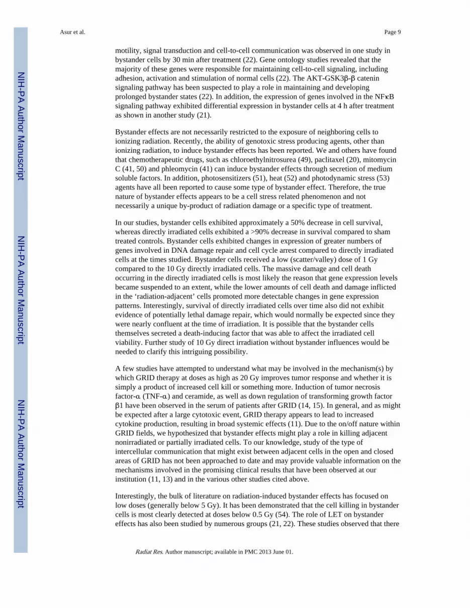
Cell SurvivalTraditional radiation survival studies were used to determine cell survival after exposure toabout 1 Gy, since that was the observed background dose in the bystander (valley) region(Fig. 3). We observed a negligible decrease in survival in confluent SCK or SCCVII cellsexposed to 1 Gy irradiation (data not shown). We observed on average a 3–10% decrease insurvival in sparsely plated SCK or SCVII cells exposed to 1 Gy irradiation (Fig. 3). Survivalof confluent SCK and SCCVII cells after direct exposure to 10 Gy of radiation is illustratedin Fig. 4. SCK and SCCVII cells exhibited an average of 10% survival after exposure to 10Gy of radiation using either the Faxitron (Fig. 4A) or the GRID system (Fig. 4B). Thedecrease in survival of directly irradiated cells was found to be statistically significant (P <0.05) at all times evaluated compared to the sham treated controls.
When medium from irradiated cells was transferred to unirradiated cultures bystander killingwas observed (Fig. 5A). We also observed significant (P < 0.05) bystander killing above andbeyond what could be expected by background/scatter irradiation (3 to 10% cell killingnoted above for 1 Gy in our cells) after GRID irradiation. Therefore, clonogenic cellsurvival was reduced in bystander cells after a medium exchange that was nearly identical tothat obtained within the bystander cell contact (GRID) experiments (Fig. 5B). Both SCK andSCCVII bystander clonogenic cell survival was reduced to nearly 50% compared to shamtreated cells. Bystander SCK and SCCVII cell survival was significantly different (P < 0.05)from the respective controls when assessed 4 and 24 h after irradiation. However, when cellswere harvested immediately after medium transfer or GRID exposure (0 h), there was littleto no bystander killing observed. Survival values were corrected for multiplicity bynormalizing to sham treated controls, which were handled with the exact same time courseas the treated cells and would account for any degree of multiplicity in the plating efficiencyobtained.
Gene Expression Changes in Bystander CellsWe proceeded to evaluate the effects of direct and bystander exposure to a GRID treatmentof 10 Gy on SCCVII cells using the SYBR green based real-time PCR technology at 0, 4and 24 h after exposure. Real-time PCR arrays specific for mouse DNA damage and cellularstress response pathways were studied in our experiments. GRID-induced bystander geneexpression changes were observed in several groups of genes involved in the repair/responseto DNA damage and stress response signaling genes, thus providing molecular evidence forpossible mechanisms of cell killing in nonirradiated cells.
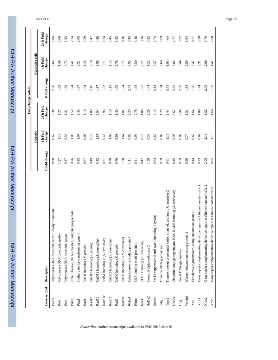
DNA damage response genes—Figure 6A and B illustrate the expression of genesinvolved in DNA damage repair that exhibited a greater than 2- and 4-fold change,respectively, in directly irradiated and bystander SCCVII cells. DNA damage responsegenes exhibiting 2-fold or more change in expression in directly irradiated and bystandercells are presented in Table 1. Table 2 illustrates the statistical analysis of genes involved inthe DNA damage signaling pathway. The fold-change values were compared betweenbystander and directly irradiated cells at each time, to determine the effect of exposure type(direct or bystander) on gene expression. Most DNA repair genes demonstrated an increasein fold-change expression in bystander cells compared to directly irradiated cells (Table 2).
Asur et al. Page 6
Radiat Res. Author manuscript; available in PMC 2013 June 01.
NIH
-PA Author Manuscript
NIH
-PA Author Manuscript
NIH
-PA Author Manuscript

Expression of DNA repair genes was found to be significantly (P < 0.03) higher in bystandercells compared to GRID exposed cells at the times evaluated (0–24 h). Genes involved indamaged DNA binding, including Rad51c, Xpc and Xrcc1 (P = 0.002, 0.04 and 0.05,respectively), showed higher expression in the bystander cells. Base excision repair genes,such as Mpg, Nthl1 and Parp1 (P = 0.02, 0.01 and 0.009, respectively), were to besignificantly higher. Dclre1a (nucleotide excision repair) and Xrcc6 and H2afx1 (double-strand break repair) all exhibited significant changes in bystander expression (P < 0.01). Inthe “mismatch repair” and “other genes related to DNA repair” subgroups, Mlh1, Mlh3,Apex1, Chaf1a, Fen1, Gtf2h1, Gtf2h2, Lig1, Pold1, Rbm4, Rev1, Sumo1, Tdg and Ube2aall exhibited higher (P < 0.05) expression in bystander cells than in directly irradiated cells.Genes involved in apoptosis (Fig. 7A) exhibited a change in expression in bystander cellscompared to directly irradiated cells. Expression of genes involved in apoptosis was foundto be significantly different in bystander cells compared to directly irradiated cells at 4 hafter exposure (P = 0.014). Mgmt (P = 0.03) and Rad21 (P = 0.009) were expressedsignificantly higher in bystander cells compared to directly irradiated cells. Cell cycle genesalso exhibited a significant difference in expression between the two exposure groups (Fig.7B). Directly irradiated cells showed a decrease in expression at 0 and 4 h after GRIDtreatment, whereas we observed an increase in expression in bystander cells. Cell cyclearrest genes, such as Hus1 and Msh2 (P = 0.03), cell cycle checkpoint genes like Rad9 andRad17 and Rad21 (P = 0.02, 0.02 and 0.09, respectively), all exhibited significantly (P <0.03) higher expression in cells harvested from the bystander region compared to GRID-irradiated cells.
Cellular stress response—The values of cellular stress response genes exhibiting 2-foldor more change in directly irradiated and bystander cells are presented in Table 3.Antioxidant and pro-oxidant genes demonstrated an increase in fold-change expression inbystander cells, while directly irradiated cells showed a decrease (Fig. 8A). Several genesinvolved in xenobiotic metabolism (Fig. 8B) exhibited an increase in expression inbystander cells at all times evaluated. Figure 9A and B illustrate the expression of molecularchaperone genes that exhibited a greater than 2- and 4-fold change, respectively, in directlyirradiated and bystander SCCVII cells. We observed an increase in expression in bystandercells at all times evaluated compared to directly irradiated cells. Table 4 illustrates thestatistical analysis of the genes involved in the cellular stress signaling pathway. The fold-change values were compared between bystander and directly irradiated cells at each time,to determine the effect of exposure type (direct or bystander) on gene expression.Expression of genes coding for antioxidant and pro-oxidant enzymes, as well as molecularchaperones, including heat shock proteins, were found to be significantly different inbystander cells compared to directly irradiated cells at 0–24 h after exposure (P = 0.05).Genes coding for antioxidant and pro-oxidant enzymes, such as Gpx1, Sod1 and Xdh wereexpressed at significantly higher levels (P < 0.01) in bystander cells compared to directlyirradiated cells at 0–24 h after exposure. Heat shock proteins, such as Dnaja1, Dnajb1,Hspa4, Hspd1 and Hspe1 all exhibited significantly (P < 0.05) different expression inbystander cells at all times evaluated. Cct7 (P < 0.02), a molecular chaperone and Gsta1 (P <0.05), a gene involved in response to xenobiotic metabolism (detoxification) bothdemonstrated changes in bystander expression compared to expression in directly irradiatedcells.
In summary, our results demonstrated higher expression of genes involved in DNA repair,cell cycle arrest and checkpoint, antioxidant and molecular chaperone pathways in the GRIDirradiated and bystander cells compared to sham treated controls.
Asur et al. Page 7
Radiat Res. Author manuscript; available in PMC 2013 June 01.
NIH
-PA Author Manuscript
NIH
-PA Author Manuscript
NIH
-PA Author Manuscript

DISCUSSIONIn the current study, we evaluated the ability of spatially fractionated radiation (GRID) toinduce bystander effects in murine carcinoma cells after exposure to a single dose of 10 Gy,analogous to the high single doses (usually 10–20 Gy) used in spatial fractionation. Asignificant decrease in clonogenic survival was observed in bystander cells after exposure tomedium obtained from cells exposed to 10 Gy. We also observed a significant bystanderkilling in cells adjacent to irradiated regions of the same monolayer compared to the shamtreated controls. The decrease in survival of cells in the adjacent regions was found to bemore than that expected from exposure to only background “valley” or scatter doses,suggesting the existence of true cytotoxic bystander effects after GRID irradiation.
We observed greater than a 2-fold increase in expression of genes involved in DNA damagerepair, cell cycle arrest and apoptosis in GRID irradiated cells 24 h after exposure.Bystander (GRID-adjacent) cells exhibited increased expression of genes involved in DNArepair, cell cycle arrest, and apoptosis immediately after exposure or 4 h after exposure. Insome instances, the increase persisted up to 24 h after GRID irradiation. We observedincreased expression of antioxidant, heat shock and chaperone genes immediately afterirradiation or 4 h after GRID exposure in the bystander cells. In a few instances, weobserved a persistent increase in expression up to 24 h after GRID exposure. We did notobserve significant increase in expression of these genes in the GRID irradiated cells. Incontrast, it has been reported that p53-related genes exhibited minimal activation inbystander cells, while genes involved in NFκB were activated to equal degrees in direct andbystander cells (21, 22) after α-particle irradiation of fibroblasts.
Of the many genes studied using the arrays we selected, we observed significantly higherlevels of genes encoding Glutathione peroxidase, Gpx1 and superoxide dismutase, Sod1 inbystander cells compared to sham treated controls. These antioxidant enzymes have beenknown to be important in cellular defense to oxidative stress (23, 24). Factors responsiblefor transmitting the bystander signal or agents that cause cytotoxicity or cell stress fromirradiated to naive bystander cells have been previously hypothesized to be proteins that canwithstand freezing and thawing (25–29). In addition, certain studies have indicated theinvolvement of reactive oxygen species (26, 30, 31), growth factors and cytokines in themaintenance of the bystander signal (32). Our results suggest that secreted factors that leadto reactive oxygen species are very likely candidates for the effects observed. We observedthe highest increase in expression of antioxidant genes in the bystander cells immediatelyafter GRID treatment suggesting that oxidative stress was occurring in these cells.
An increase in expression of DNA damage response genes, such as those involved inapoptosis, cell cycle arrest and repair was also observed in bystander cells compared to shamtreated controls in our experiments. In past studies, exposure of cells to low doses of αparticles have resulted in increased expression of DNA damage response genes, such asTP53, CDKN1A, CDC2, CCN1 and RAD51 (33). Therefore, as might be expected, ourresults agree with those of others to suggest that a part of the killing mechanism involved inbystander effects is through DNA damage, which is something that can be induced by avariety of reactive oxygen species.
Overall, radiation-induced bystander effects have been demonstrated using a wide variety ofend points, including decreased cell survival (34, 35), apoptosis (36–40), increasedchromosomal damage (4, 41–44) and DNA double-strand breaks (6, 45, 46). Changes inexpression of mitogen (commonly referred to as stress) activated protein kinases (31, 47, 48)have been observed after exposure to medium obtained from cells exposed to ionizingradiation. Differential expression of genes involved in maintaining cell structure and
Asur et al. Page 8
Radiat Res. Author manuscript; available in PMC 2013 June 01.
NIH
-PA Author Manuscript
NIH
-PA Author Manuscript
NIH
-PA Author Manuscript
motility, signal transduction and cell-to-cell communication was observed in one study inbystander cells by 30 min after treatment (22). Gene ontology studies revealed that themajority of these genes were responsible for maintaining cell-to-cell signaling, includingadhesion, activation and stimulation of normal cells (22). The AKT-GSK3β-β cateninsignaling pathway has been suspected to play a role in maintaining and developingprolonged bystander states (22). In addition, the expression of genes involved in the NFκBsignaling pathway exhibited differential expression in bystander cells at 4 h after treatmentas shown in another study (21).
Bystander effects are not necessarily restricted to the exposure of neighboring cells toionizing radiation. Recently, the ability of genotoxic stress producing agents, other thanionizing radiation, to induce bystander effects has been reported. We and others have foundthat chemotherapeutic drugs, such as chloroethylnitrosurea (49), paclitaxel (20), mitomycinC (41, 50) and phleomycin (41) can induce bystander effects through secretion of mediumsoluble factors. In addition, photosensitizers (51), heat (52) and photodynamic stress (53)agents have all been reported to cause some type of bystander effect. Therefore, the truenature of bystander effects appears to be a cell stress related phenomenon and notnecessarily a unique by-product of radiation damage or a specific type of treatment.
In our studies, bystander cells exhibited approximately a 50% decrease in cell survival,whereas directly irradiated cells exhibited a >90% decrease in survival compared to shamtreated controls. Bystander cells exhibited changes in expression of greater numbers ofgenes involved in DNA damage repair and cell cycle arrest compared to directly irradiatedcells at the times studied. Bystander cells received a low (scatter/valley) dose of 1 Gycompared to the 10 Gy directly irradiated cells. The massive damage and cell deathoccurring in the directly irradiated cells is most likely the reason that gene expression levelsbecame suspended to an extent, while the lower amounts of cell death and damage inflictedin the ‘radiation-adjacent’ cells promoted more detectable changes in gene expressionpatterns. Interestingly, survival of directly irradiated cells over time also did not exhibitevidence of potentially lethal damage repair, which would normally be expected since theywere nearly confluent at the time of irradiation. It is possible that the bystander cellsthemselves secreted a death-inducing factor that was able to affect the irradiated cellviability. Further study of 10 Gy direct irradiation without bystander influences would beneeded to clarify this intriguing possibility.
A few studies have attempted to understand what may be involved in the mechanism(s) bywhich GRID therapy at doses as high as 20 Gy improves tumor response and whether it issimply a product of increased cell kill or something more. Induction of tumor necrosisfactor-α (TNF-α) and ceramide, as well as down regulation of transforming growth factorβ1 have been observed in the serum of patients after GRID (14, 15). In general, and as mightbe expected after a large cytotoxic event, GRID therapy appears to lead to increasedcytokine production, resulting in broad systemic effects (11). Due to the on/off nature withinGRID fields, we hypothesized that bystander effects might play a role in killing adjacentnonirradiated or partially irradiated cells. To our knowledge, study of the type ofintercellular communication that might exist between adjacent cells in the open and closedareas of GRID has not been approached to date and may provide valuable information on themechanisms involved in the promising clinical results that have been observed at ourinstitution (11, 13) and in the various other studies cited above.
Interestingly, the bulk of literature on radiation-induced bystander effects has focused onlow doses (generally below 5 Gy). It has been demonstrated that the cell killing in bystandercells is most clearly detected at doses below 0.5 Gy (54). The role of LET on bystandereffects has also been studied by numerous groups (21, 22). These studies observed that there
Asur et al. Page 9
Radiat Res. Author manuscript; available in PMC 2013 June 01.
NIH
-PA Author Manuscript
NIH
-PA Author Manuscript
NIH
-PA Author Manuscript

appears to be a greater incidence of bystander effect with high-LET irradiation when equaldoses are compared to low-LET.
It has also been suggested that bystander effects plateau at low doses and further increases inthe radiation dose has no effect on bystander response (5). Gow et al. observed a decrease insurviving fraction of bystander HPV-G cells receiving medium from 0.5 Gy or 5 Gyirradiated cells, as well as recipients of medium from 10 Gy of radiation when plateddensely. However, the surviving fractions returned to near control levels when sparselyplated recipient cells received medium from cells that had been exposed to 10 Gy (55). Inour study, using a dose of 10 Gy, both medium transfer and GRID irradiation assays reducedbystander cell viability and, at least in the case of the GRID exposures, the bystander cellswere densely plated which would agree with the previous study results. In another study,cells receiving medium from cells irradiated with fractionated doses (i.e., 2 Gy per fraction),rather surprisingly exhibited decreased survival fractions compared to cells that directlyreceived the fractionated irradiation (i.e., bystander cells receiving medium fromfractionated radiation exhibited more cell killing than cells that were directly exposed tofractionated doses of radiation) (56, 57). In terms of radiation treatment, this suggests thatalthough the direct effect of fractionated radiation doses is hoped to be normal tissuesparing, the occurrence of bystander effects coming from the irradiated tumor or normalcells after each fraction of radiation may increase the overall tumor and normal tissueresponses.
Recently, intensity modulated radiation therapy (IMRT), with doses ranging from 3 to 20Gy, was used to study cellular response to radiation (58). The authors observed three distincttypes of bystander effects. Type 1 was a decrease in cell survival observed in unirradiatedcells adjacent to cells irradiated with a moderate dose (3 Gy). Type 2 was an increase insurvival in cells adjacent to cells receiving a lethal dose (20 Gy) of radiation. Type 3 was anincreased survival of cells receiving a high dose of radiation, when they were adjacent tocells receiving a low dose, possibly due to the generation of unspecified mechanisms ofsurvival response in the low-dose cells, which were conducive to colony formation. In ourstudy, we observed decrease in survival of cells adjacent to cells exposed to 10 Gyirradiation. Other studies involving IMRT determined the effect of modulated fields ofirradiation on cell survival. Radiation-resistant and radiation-sensitive cells were exposed tomodulated and nonmodulated irradiation up to 8 Gy using a multi-leaf collimator. Nosignificant difference was found in survival of cells exposed to modulated radiation ornonmodulated fields of irradiation, suggesting the occurrence of bystander effects inmodulated fields (59). In addition, survival of cells that were in the same flask but notexposed to radiation (out-of-field) was found to be less than that expected solely due toradiation scattering (60). Inhibition of intercellular communication using nitric oxidesynthase inhibitor increased cell survival, suggesting the occurrence of bystander effects inthe out-of-field cellular responses.
In our radiation therapy clinic, treatment of head and neck cancer usually involves GRIDtherapy followed by a conventional course of 2 Gy fractionated radiation therapy andchemotherapy. In addition to bystander killing by the GRID dose, the occurrence of sub-lethal bystander effects after GRID therapy might make these cells more susceptible to thesubsequent chemotherapy and radiation treatment, further contributing to the overall tumorresponse. A detailed understanding of possible bystander effects involved in low or highdose or spatially fractionated radiation therapy, and the factors that mediate such effects mayuncover molecular mechanisms involved and suggest new therapeutic targets to improvetreatment outcomes, either by increasing tumor cell killing or enhancing normal cellprotection. Studies designed in vivo will need to take into consideration possiblecontributions of tissue and tumor effects, including hypoxia, on bystander effects.
Asur et al. Page 10
Radiat Res. Author manuscript; available in PMC 2013 June 01.
NIH
-PA Author Manuscript
NIH
-PA Author Manuscript
NIH
-PA Author Manuscript

Understanding the involvement of bystander effects within various dose gradients present instate-of-the art radiation techniques may enable us to optimize therapies and design potentcombination treatment approaches.
AcknowledgmentsThis work was supported by Central Arkansas Radiation Therapy Institute (CARTI) and NIH grant CA44114. Theauthors thank N. Koonce and A. Jamshidi-Parsian, for their input and guidance.
References1. Dale WM. The effect of X-rays on enzymes. Biochem J. 1940; 34:1367–73. [PubMed: 16747266]
2. Dale WM. The effect of X-rays on the conjugated protein d-aminoacid oxidase. Biochem J. 1942;36:80–5. [PubMed: 16747494]
3. Kotval JP, Gray LH. Structural changes produced in microspores of Tradescantia by alpha-radiation.J Genet. 1947; 48:135–54. 1947. [PubMed: 20266728]
4. Nagasawa H, Little JB. Induction of sister chromatid exchanges by extremely low doses of alpha-particles. Cancer Res. 1992; 52:6394–6. [PubMed: 1423287]
5. Mothersill C, Seymour CB. Radiation-induced bystander effects–implications for cancer. Nat RevCancer. 2004; 4:158–64. [PubMed: 14964312]
6. Hu B, Wu L, Han W, Zhang L, Chen S, Xu A, Hei TK, Yu Z. The time and spatial effects ofbystander response in mammalian cells induced by low dose radiation. Carcinogenesis. 2006;27:245–51. [PubMed: 16150894]
7. Azzam EI, de Toledo SM, Little JB. Oxidative metabolism, gap junctions and the ionizing radiation-induced bystander effect. Oncogene. 2003; 22:7050–7. [PubMed: 14557810]
8. Azzam EI, de Toledo SM, Little JB. Direct evidence for the participation of gap junction-mediatedintercellular communication in the transmission of damage signals from alpha-particle irradiated tononirradiated cells. Proc Natl Acad Sci USA. 2001; 98:473–8. [PubMed: 11149936]
9. Azzam EI, Little JB. The radiation-induced bystander effect: evidence and significance. Hum ExpToxicol. 2004; 23:61–5. [PubMed: 15070061]
10. Mothersill C, Seymour C. Radiation-induced bystander effects: past history and future directions.Radiat Res. 2001; 155:759–67. [PubMed: 11352757]
11. Penagaricano JA, Moros EG, Ratanatharathorn V, Yan Y, Corry P. Evaluation of spatiallyfractionated radiotherapy (GRID) and definitive chemoradiotherapy with curative intent for locallyadvanced squamous cell carcinoma of the head and neck: initial response rates and toxicity. Int JRadiat Oncol Biol Phys. 2010; 76:1369–75. [PubMed: 19625138]
12. Penagaricano JA, Griffin R, Corry P, Moros E, Yan Y, Ratanatharathorn V. Spatially fractionated(GRID) therapy for large and bulky tumors. J Ark Med Soc. 2009; 105:263–5. [PubMed:19475814]
13. Huhn JL, Regine WF, Valentino JP, Meigooni AS, Kudrimoti M, Mohiuddin M. Spatiallyfractionated GRID radiation treatment of advanced neck disease associated with head and neckcancer. Technol Cancer Res Treat. 2006; 5:607–12. [PubMed: 17121437]
14. Sathishkumar S, Boyanovsky B, Karakashian AA, Rozenova K, Giltiay NV, Kudrimoti M,Mohiuddin M, Ahmed MM, Nikolova-Karakashian M. Elevated sphingomyelinase activity andceramide concentration in serum of patients undergoing high dose spatially fractionated radiationtreatment: implications for endothelial apoptosis. Cancer Biol Ther. 2005; 4:979–86. [PubMed:16096366]
15. Sathishkumar S, Dey S, Meigooni AS, Regine WF, Kudrimoti MS, Ahmed MM, Mohiuddin M.The impact of TNF-alpha induction on therapeutic efficacy following high dose spatiallyfractionated (GRID) radiation. Technol Cancer Res Treat. 2002; 1:141–7. [PubMed: 12622521]
16. Burgher AH, Swanlund DJ, Griffin RJ, Song CW, Bischof JC, Roberts KP. Sensitization ofthermotolerant SCK cells to hyperthermia and freezing with reduction of intracellular pH:implications for cryosurgery. J Surg Oncol. 2003; 82:160–9. [PubMed: 12619059]
Asur et al. Page 11
Radiat Res. Author manuscript; available in PMC 2013 June 01.
NIH
-PA Author Manuscript
NIH
-PA Author Manuscript
NIH
-PA Author Manuscript

17. Griffin RJ, Williams BW, Wild R, Cherrington JM, Park H, Song CW. Simultaneous inhibition ofthe receptor kinase activity of vascular endothelial, fibroblast, and platelet-derived growth factorssuppresses tumor growth and enhances tumor radiation response. Cancer Res. 2002; 62:1702–6.[PubMed: 11912143]
18. Dings RP, Williams BW, Song CW, Griffioen AW, Mayo KH, Griffin RJ. Anginex synergizeswith radiation therapy to inhibit tumor growth by radiosensitizing endothelial cells. Int J Cancer.2005; 115:312–9. [PubMed: 15688384]
19. Livak KJ, Schmittgen TD. Analysis of relative gene expression data using real-time quantitativePCR and the 2(−Delta Delta C(T)) Method. Methods. 2001; 25:402–8. [PubMed: 11846609]
20. Alexandre J, Hu Y, Lu W, Pelicano H, Huang P. Novel action of paclitaxel against cancer cells:bystander effect mediated by reactive oxygen species. Cancer Res. 2007; 67:3512–7. [PubMed:17440056]
21. Ghandhi SA, Yaghoubian B, Amundson SA. Global gene expression analyses of bystander andalpha particle irradiated normal human lung fibroblasts: synchronous and differential responses.BMC Med Genomics. 2008; 1:63. [PubMed: 19108712]
22. Ghandhi SA, Ming L, Ivanov VN, Hei TK, Amundson SA. Regulation of early signaling and geneexpression in the alpha-particle and bystander response of IMR-90 human fibroblasts. BMC MedGenomics. 2010; 3:31. [PubMed: 20670442]
23. Mitchell JB, Russo A. The role of glutathione in radiation and drug induced cytotoxicity. Br JCancer Suppl. 1987; 8:96–104. [PubMed: 3307879]
24. Gao Z, Sarsour EH, Kalen AL, Li L, Kumar MG, Goswami PC. Late ROS accumulation andradiosensitivity in SOD1-overexpressing human glioma cells. Free Radic Biol Med. 2008;45:1501–9. [PubMed: 18790046]
25. Narayanan PK, Goodwin EH, Lehnert BE. Alpha particles initiate biological production ofsuperoxide anions and hydrogen peroxide in human cells. Cancer Res. 1997; 57:3963–71.[PubMed: 9307280]
26. Lehnert BE, Goodwin EH. A new mechanism for DNA alterations induced by alpha particles suchas those emitted by radon and radon progeny. Environ Health Perspect. 1997; 5(105 Suppl):1095–101. [PubMed: 9400706]
27. Mothersill C, Seymour CB. Cell-cell contact during gamma irradiation is not required to induce abystander effect in normal human keratinocytes: evidence for release during irradiation of a signalcontrolling survival into the medium. Radiat Res. 1998; 149:256–62. [PubMed: 9496888]
28. Iyer R, Lehnert BE, Svensson R. Factors underlying the cell growth-related bystander responses toalpha particles. Cancer Res. 2000; 60:1290–8. [PubMed: 10728689]
29. Shao C, Prise KM, Folkard M. Signaling factors for irradiated glioma cells induced bystanderresponses in fibroblasts. Mutat Res. 2008; 638:139–45. [PubMed: 17977565]
30. Konopacka M, Rzeszowska-Wolny J. The bystander effect-induced formation of micronucleatedcells is inhibited by antioxidants, but the parallel induction of apoptosis and loss of viability arenot affected. Mutat Res. 2006; 593:32–8. [PubMed: 16040062]
31. Lyng FM, Maguire P, McClean B, Seymour C, Mothersill C. The involvement of calcium andMAP kinase signaling pathways in the production of radiation-induced bystander effects. RadiatRes. 2006; 165:400–9. [PubMed: 16579652]
32. Barcellos-Hoff MH, Brooks AL. Extracellular signaling through the microenvironment: ahypothesis relating carcinogenesis, bystander effects, and genomic instability. Radiat Res. 2001;156:618–27. [PubMed: 11604083]
33. Azzam EI, de Toledo SM, Gooding T, Little JB. Intercellular communication is involved in thebystander regulation of gene expression in human cells exposed to very low fluences of alphaparticles. Radiat Res. 1998; 150:497–504. [PubMed: 9806590]
34. Mothersill C, Seymour C. Medium from irradiated human epithelial cells but not human fibroblastsreduces the clonogenic survival of unirradiated cells. Int J Radiat Biol. 1997; 71:421–7. [PubMed:9154145]
35. Sawant SG, Zheng W, Hopkins KM, Randers-Pehrson G, Lieberman HB, Hall EJ. The radiation-induced bystander effect for clonogenic survival. Radiat Res. 2002; 157:361–4. [PubMed:11893236]
Asur et al. Page 12
Radiat Res. Author manuscript; available in PMC 2013 June 01.
NIH
-PA Author Manuscript
NIH
-PA Author Manuscript
NIH
-PA Author Manuscript

36. Mothersill C, Seymour RJ, Seymour CB. Increased radiosensitivity in cells of two human cell linestreated with bystander medium from irradiated repair-deficient cells. Radiat Res. 2006; 165:26–34.[PubMed: 16392959]
37. Belyakov OV, Folkard M, Mothersill C, Prise KM, Michael BD. Bystander-induced apoptosis andpremature differentiation in primary urothelial explants after charged particle microbeamirradiation. Radiat Prot Dosimetry. 2002; 99:249–51. [PubMed: 12194297]
38. Belyakov OV, Folkard M, Mothersill C, Prise KM, Michael BD. Bystander-induceddifferentiation: a major response to targeted irradiation of a urothelial explant model. Mutat Res.2006; 597:43–9. [PubMed: 16423374]
39. Belyakov OV, Malcolmson AM, Folkard M, Prise KM, Michael BD. Direct evidence for abystander effect of ionizing radiation in primary human fibroblasts. Br J Cancer. 2001; 84:674–9.[PubMed: 11237389]
40. Lyng FM, Seymour CB, Mothersill C. Production of a signal by irradiated cells which leads to aresponse in unirradiated cells characteristic of initiation of apoptosis. Br J Cancer. 2000; 83:1223–30. [PubMed: 11027437]
41. Asur RS, Thomas RA, Tucker JD. Chemical induction of the bystander effect in normal humanlymphoblastoid cells. Mutat Res. 2009; 676:11–6. [PubMed: 19486859]
42. Prise KM, Belyakov OV, Folkard M, Michael BD. Studies of bystander effects in humanfibroblasts using a charged particle microbeam. Int J Radiat Biol. 1998; 74:793–8. [PubMed:9881726]
43. Deshpande A, Goodwin EA, Bailey SM, Marrone BL, Lehnert BE. Alpha-particle-induced sisterchromatid exchange in normal human lung fibroblasts: evidence for an extranuclear target. RadiatRes. 1996; 145:260–7. [PubMed: 8927692]
44. Lorimore SA, Kadhim MA, Pocock DA, Papworth D, Stevens DL, Goodhead DT, Wright EG.Chromosomal instability in the descendants of unirradiated surviving cells after alpha-particleirradiation. Proc Natl Acad Sci USA. 1998; 95:5730–3. [PubMed: 9576952]
45. Sedelnikova OA, Nakamura A, Kovalchuk O, Koturbash I, Mitchell SA, Marino SA, et al. DNAdouble-strand breaks form in bystander cells after microbeam irradiation of three-dimensionalhuman tissue models. Cancer Res. 2007; 67:4295–302. [PubMed: 17483342]
46. Sokolov MV, Smilenov LB, Hall EJ, Panyutin IG, Bonner WM, Sedelnikova OA. Ionizingradiation induces DNA double-strand breaks in bystander primary human fibroblasts. Oncogene.2005; 24:7257–65. [PubMed: 16170376]
47. Asur R, Balasubramaniam M, Marples B, Thomas RA, Tucker JD. Bystander effects induced bychemicals and ionizing radiation: evaluation of changes in gene expression of downstream MAPKtargets. Mutagenesis. 2010; 25:271–9. [PubMed: 20130020]
48. Azzam EI, De Toledo SM, Spitz DR, Little JB. Oxidative metabolism modulates signaltransduction and micronucleus formation in bystander cells from alpha-particle-irradiated normalhuman fibroblast cultures. Cancer Res. 2002; 62:5436–42. [PubMed: 12359750]
49. Demidem A, Morvan D, Madelmont JC. Bystander effects are induced by CENU treatment andassociated with altered protein secretory activity of treated tumor cells: a relay for chemotherapy?Int J Cancer. 2006; 119:992–1004. [PubMed: 16557598]
50. Rugo RE, Almeida KH, Hendricks CA, Jonnalagadda VS, Engelward BP. A single acute exposureto a chemotherapeutic agent induces hyper-recombination in distantly descendant cells and in theirneighbors. Oncogene. 2005; 24:5016–25. [PubMed: 15856014]
51. Dahle J, Angell-Petersen E, Steen HB, Moan J. Bystander effects in cell death induced byphotodynamic treatment UVA radiation and inhibitors of ATP synthesis. Photochem Photobiol.2001; 73:378–87. [PubMed: 11332033]
52. Dabrowska A, Gos M, Janik P. “Bystander effect” induced by photodynamically or heat-injuredovarian carcinoma cells (OVP10) in vitro. Med Sci Monit. 2005; 11:BR316–24. [PubMed:16127353]
53. Chakraborty A, Held KD, Prise KM, Liber HL, Redmond RW. Bystander effects induced bydiffusing mediators after photodynamic stress. Radiat Res. 2009; 172:74–81. [PubMed: 19580509]
Asur et al. Page 13
Radiat Res. Author manuscript; available in PMC 2013 June 01.
NIH
-PA Author Manuscript
NIH
-PA Author Manuscript
NIH
-PA Author Manuscript

54. Seymour CB, Mothersill C. Relative contribution of bystander and targeted cell killing to the low-dose region of the radiation dose-response curve. Radiat Res. 2000; 153:508–1. [PubMed:10790270]
55. Gow MD, Seymour CB, Byun SH, Mothersill CE. Effect of dose rate on the radiation-inducedbystander response. Phys Med Biol. 2008; 53:119–32. [PubMed: 18182691]
56. Mothersill C, Seymour CB. Bystander and delayed effects after fractionated radiation exposure.Radiat Res. 2002; 158:626–33. [PubMed: 12385640]
57. Mothersill CE, Moriarty MJ, Seymour CB. Radiotherapy and the potential exploitation ofbystander effects. Int J Radiat Oncol Biol Phys. 2004; 58:575–9. [PubMed: 14751530]
58. Mackonis EC, Suchowerska N, Zhang M, Ebert M, McKenzie DR, Jackson M. Cellular responseto modulated radiation fields. Phys Med Biol. 2007; 52:5469–82. [PubMed: 17804876]
59. Butterworth KT, McGarry CK, O’Sullivan JM, Hounsell AR, Prise KM. A study of the biologicaleffects of modulated 6 MV radiation fields. Phys Med Biol. 2010; 55:1607–18. [PubMed:20164535]
60. Butterworth KT, McGarry CK, Trainor C, O’Sullivan JM, Hounsell AR, Prise KM. Out-of-fieldcell survival following exposure to intensity-modulated radiation fields. Int J Radiat Oncol BiolPhys. 2011; 9:1516–22. [PubMed: 21277116]
61. Griffin RJ, Koonce NA, Dings RPM, Siegel E, Moros EG, et al. Microbeam radiation therapyalters vascular architecture and tumor oxygenation and is enhanced by a galectin-1 targeted anti-angiogenic peptide. Radiat Res. 2012; 177:804–12. [PubMed: 22607585]
Asur et al. Page 14
Radiat Res. Author manuscript; available in PMC 2013 June 01.
NIH
-PA Author Manuscript
NIH
-PA Author Manuscript
NIH
-PA Author Manuscript

FIG. 1.Schematic diagram of set-up used in GRID irradiation experiments. Panel A: The SmallAnimal Conformal Radiation Research System (SACCRS) used in GRID irradiationexperiments. Panel B: A Gafchromic EBT-2 film was attached to the bottom of a 100 mmPetri dish containing cells. The bottom of the dish was placed on top a 3-inch thickstyrofoam board, and the entire set-up was placed on a robot platform and irradiated in aGRID pattern.
Asur et al. Page 15
Radiat Res. Author manuscript; available in PMC 2013 June 01.
NIH
-PA Author Manuscript
NIH
-PA Author Manuscript
NIH
-PA Author Manuscript

FIG. 2.Cells were irradiated using spatially fractionated radiation to evaluate bystander effects.Cells were irradiated at a peak dose of 10 Gy using a brass collimator to create a GRIDpattern of 9 open circular areas, 12 mm in diameter with a center-to-center distance of 18mm. The bystander cells were harvested from the valley dose region along the diagonal linesillustrated, which represents about 10% of the total radiation.
Asur et al. Page 16
Radiat Res. Author manuscript; available in PMC 2013 June 01.
NIH
-PA Author Manuscript
NIH
-PA Author Manuscript
NIH
-PA Author Manuscript
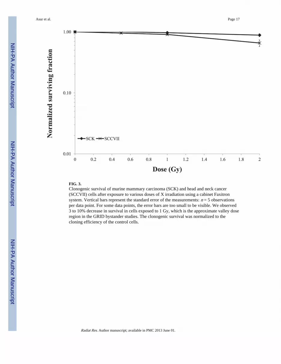
FIG. 3.Clonogenic survival of murine mammary carcinoma (SCK) and head and neck cancer(SCCVII) cells after exposure to various doses of X irradiation using a cabinet Faxitronsystem. Vertical bars represent the standard error of the measurements: n = 5 observationsper data point. For some data points, the error bars are too small to be visible. We observed3 to 10% decrease in survival in cells exposed to 1 Gy, which is the approximate valley doseregion in the GRID bystander studies. The clonogenic survival was normalized to thecloning efficiency of the control cells.
Asur et al. Page 17
Radiat Res. Author manuscript; available in PMC 2013 June 01.
NIH
-PA Author Manuscript
NIH
-PA Author Manuscript
NIH
-PA Author Manuscript
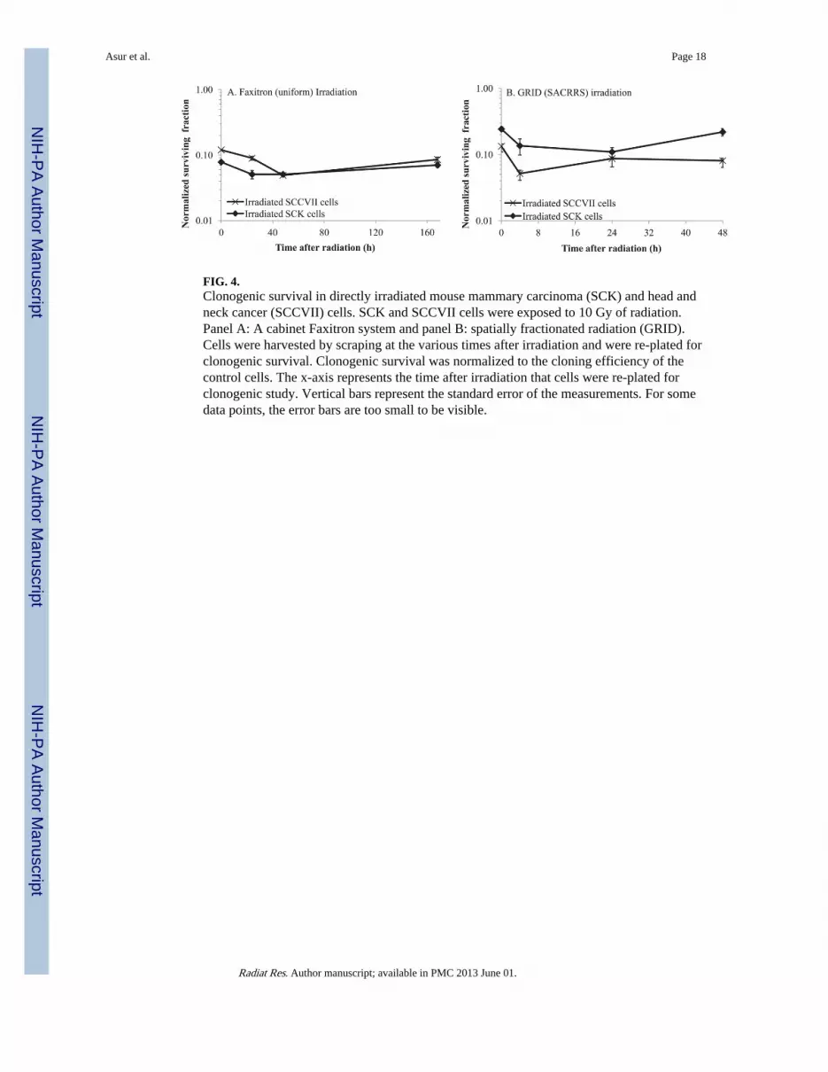
FIG. 4.Clonogenic survival in directly irradiated mouse mammary carcinoma (SCK) and head andneck cancer (SCCVII) cells. SCK and SCCVII cells were exposed to 10 Gy of radiation.Panel A: A cabinet Faxitron system and panel B: spatially fractionated radiation (GRID).Cells were harvested by scraping at the various times after irradiation and were re-plated forclonogenic survival. Clonogenic survival was normalized to the cloning efficiency of thecontrol cells. The x-axis represents the time after irradiation that cells were re-plated forclonogenic study. Vertical bars represent the standard error of the measurements. For somedata points, the error bars are too small to be visible.
Asur et al. Page 18
Radiat Res. Author manuscript; available in PMC 2013 June 01.
NIH
-PA Author Manuscript
NIH
-PA Author Manuscript
NIH
-PA Author Manuscript

FIG. 5.Clonogenic survival in bystander mouse mammary carcinoma (SCK) and head and neckcancer (SCCVII) cells. SCK and SCCVII cells were irradiated in two different systems.Panel A: Cells were irradiated in a cabinet Faxitron system and subsequently medium fromirradiated cells was transferred 4 h later to bystander cells. Panel B: Cells were irradiatedusing a conformal X-ray system (SACRRS) and bystander cells in the diagonal region(received a background dose of 1 Gy, which resulted in 3 to 10% decrease in survival) of aGRID irradiated pattern were selectively isolated at various times and the clonogenicsurvival was determined. The clonogenic survival was normalized to the cloning efficiencyof the control cells. The x-axis represents the time following medium transfer (panel A) orGRID irradiation (panel B) when cells were re-plated for clonogenic study. N = 3–5experiments for all data points. Vertical bars represent the standard error of themeasurements. For some data points, the error bars are too small to be visible.
Asur et al. Page 19
Radiat Res. Author manuscript; available in PMC 2013 June 01.
NIH
-PA Author Manuscript
NIH
-PA Author Manuscript
NIH
-PA Author Manuscript

FIG. 6.Expression patterns of genes involved in DNA damage repair. Mouse head and neckcarcinoma (SCCVII) cells were exposed to a single fraction of 10 Gy using GRID. Cellsfrom the directly irradiated and bystander regions were selectively and gene expressionchanges were evaluated using quantitative real-time PCR. Panel A: DNA damage repairgenes exhibiting 2- to 4-fold change in expression. Panel B: DNA damage repair genesexhibiting greater than a 4-fold change in expression.
Asur et al. Page 20
Radiat Res. Author manuscript; available in PMC 2013 June 01.
NIH
-PA Author Manuscript
NIH
-PA Author Manuscript
NIH
-PA Author Manuscript
FIG. 7.Expression patterns of genes involved in apoptosis (panel A) and cell cycle exhibiting (panelB) greater than 2-fold change in expression analyzed by quantitative real-time PCR.
Asur et al. Page 21
Radiat Res. Author manuscript; available in PMC 2013 June 01.
NIH
-PA Author Manuscript
NIH
-PA Author Manuscript
NIH
-PA Author Manuscript

FIG. 8.Expression patterns of antioxidant (panel A) and xenobiotic metabolism genes (panel B)exhibiting greater than 2-fold change in expression, harvested from directly irradiated andbystander cells after exposure to 10 Gy using GRID in SCCVII cells.
Asur et al. Page 22
Radiat Res. Author manuscript; available in PMC 2013 June 01.
NIH
-PA Author Manuscript
NIH
-PA Author Manuscript
NIH
-PA Author Manuscript

FIG. 9.Expression patterns of molecular chaperone genes exhibiting (panel A) 2- to 4-fold changein expression and (panel B) greater than 4-fold change in expression after direct or bystanderexposure to 10 Gy GRID in SCCVII cells.
Asur et al. Page 23
Radiat Res. Author manuscript; available in PMC 2013 June 01.
NIH
-PA Author Manuscript
NIH
-PA Author Manuscript
NIH
-PA Author Manuscript

NIH
-PA Author Manuscript
NIH
-PA Author Manuscript
NIH
-PA Author Manuscript
Asur et al. Page 24
TAB
LE 1
Fold
-Cha
nge
Val
ues
of D
NA
Dam
age
Res
pons
e G
enes
Exh
ibiti
ng 2
-Fol
d or
Mor
e C
hang
e at
one
or
Mor
e T
imep
oint
s St
udie
d in
Eith
er D
irec
tlyIr
radi
ated
or
Bys
tand
er C
ells
, Com
pare
d to
Sha
m T
reat
ed C
ontr
ols
Gen
e sy
mbo
lD
escr
ipti
on
Fol
d ch
ange
val
ues
Dir
ectl
yB
ysta
nder
cel
ls
0 F
old
chan
ge4
h F
old
chan
ge24
h F
old
chan
ge0
Fol
d ch
ange
4 h
Fol
dch
ange
24 h
Fol
dch
ange
Ape
x1A
puri
nic/
apyr
imid
inic
end
onuc
leas
e 1
0.30
1.25
1.54
2.03
2.12
1.16
Atr
xA
lpha
thal
asse
mia
/men
tal r
etar
datio
n sy
ndro
me
X-l
inke
d ho
mol
og (
hum
an)
1.00
1.99
1.28
1.52
1.55
2.01
Dcl
re1a
DN
A c
ross
-lin
k re
pair
1A
, PSO
2 ho
mol
og (
S. c
erev
isia
e)0.
640.
831.
992.
032.
531.
13
Erc
c1E
xcis
ion
repa
ir c
ross
-com
plem
entin
g ro
dent
rep
air
defi
cien
cy,
com
plem
enta
tion
grou
p 1
1.06
0.85
1.42
1.82
2.24
1.12
Exo
1E
xonu
clea
se 1
1.12
1.68
2.45
2.61
3.22
1.43
Fanc
cFa
ncon
i ane
mia
, com
plem
enta
tion
grou
p C
0.61
0.99
1.97
2.04
2.92
2.16
Fen1
Flap
str
uctu
re s
peci
fic
endo
nucl
ease
11.
311.
382.
043.
284.
422.
83
Gad
d45a
Gro
wth
arr
est a
nd D
NA
-dam
age-
indu
cibl
e 45
alp
ha0.
120.
270.
650.
490.
452.
65
Gtf
2h1
Gen
eral
tran
scri
ptio
n fa
ctor
II
H, p
olyp
eptid
e 1
0.21
0.43
3.42
3.02
4.72
2.20
Gtf
2h2
Gen
eral
tran
scri
ptio
n fa
ctor
II
H, p
olyp
eptid
e 2
0.63
1.04
2.10
2.01
2.94
1.89
H2a
fxH
2A h
isto
ne f
amily
, mem
ber
X0.
520.
641.
983.
314.
644.
32
Hus
1H
us1
hom
olog
(S.
pom
be)
0.65
1.25
2.83
2.88
4.52
2.73
Lig
1L
igas
e I,
DN
A, A
TP-
depe
nden
t0.
730.
783.
994.
175.
251.
85
Mar
eA
lpha
glo
bin
regu
lato
ry e
lem
ent c
onta
inin
g ge
ne0.
850.
913.
751.
191.
361.
33
Mbd
4M
ethy
l-C
pG b
indi
ng d
omai
n pr
otei
n 4
0.99
0.97
1.56
0.49
0.72
1.16
Mgm
tO
-6-m
ethy
lgua
nine
-DN
A m
ethy
ltran
sfer
ase
0.38
0.41
1.55
1.03
1.08
0.89
Mif
Mac
roph
age
mig
ratio
n in
hibi
tory
fac
tor
0.31
0.38
2.63
1.94
2.09
0.86
Mlh
1M
utL
hom
olog
1 (
E. c
oli)
0.73
0.98
5.71
3.28
3.19
3.72
Mlh
3M
utL
hom
olog
3 (
E c
oli)
0.63
0.51
1.17
1.10
1.01
2.09
Mpg
N-m
ethy
lpur
ine-
DN
A g
lyco
syla
se0.
740.
771.
682.
302.
582.
22
Msh
2M
utS
hom
olog
2 (
E. c
oli)
0.31
0.44
4.76
2.04
3.09
2.05
Nth
l1N
th (
endo
nucl
ease
III
)-lik
e 1
(E. c
oli)
0.45
0.49
1.63
1.71
1.77
1.33
Parp
1Po
ly (
AD
P-ri
bose
) po
lym
eras
e fa
mily
, mem
ber
10.
760.
833.
532.
753.
632.
74
Pms1
Post
mei
otic
seg
rega
tion
incr
ease
d 1
(S. c
erev
isia
e)0.
900.
811.
801.
961.
653.
63
Radiat Res. Author manuscript; available in PMC 2013 June 01.
NIH
-PA Author Manuscript
NIH
-PA Author Manuscript
NIH
-PA Author Manuscript
Asur et al. Page 25
Gen
e sy
mbo
lD
escr
ipti
on
Fol
d ch
ange
val
ues
Dir
ectl
yB
ysta
nder
cel
ls
0 F
old
chan
ge4
h F
old
chan
ge24
h F
old
chan
ge0
Fol
d ch
ange
4 h
Fol
dch
ange
24 h
Fol
dch
ange
Pold
1Po
lym
eras
e (D
NA
dir
ecte
d), d
elta
1, c
atal
ytic
sub
unit
0.90
0.69
2.38
2.68
2.64
1.86
Pole
Poly
mer
ase
(DN
A d
irec
ted)
, eps
ilon
1.27
1.19
1.27
1.62
1.40
2.06
Polk
Poly
mer
ase
(DN
A d
irec
ted)
, kap
pa0.
470.
541.
311.
090.
731.
33
Prkd
cPr
otei
n ki
nase
, DN
A a
ctiv
ated
, cat
alyt
ic p
olyp
eptid
e0.
761.
031.
591.
741.
412.
24
Pttg
1Pi
tuita
ry tu
mor
-tra
nsfo
rmin
g ge
ne 1
0.33
1.07
2.43
1.37
2.21
2.05
Rad
1R
AD
1 ho
mol
og (
S. p
ombe
)0.
570.
071.
101.
341.
351.
29
Rad
17R
AD
17 h
omol
og (
S. p
ombe
)0.
490.
791.
832.
102.
181.
47
Rad
51R
AD
51 h
omol
og (
S. c
erev
isia
e)0.
821.
872.
941.
872.
584.
88
Rad
51c
Rad
51 h
omol
og c
(S.
cer
evis
iae)
0.71
0.45
0.93
1.03
0.71
1.24
Rad
52R
AD
52 h
omol
og (
S. c
erev
isia
e)0.
781.
061.
181.
121.
152.
45
Rad
9R
AD
9 ho
mol
og (
S. p
ombe
)0.
790.
981.
401.
792.
781.
83
Rad
9bR
AD
9 ho
mol
og B
(S.
cer
evis
iae)
1.38
1.61
1.82
1.74
2.71
8.16
Rbb
p4R
etin
obla
stom
a bi
ndin
g pr
otei
n 4
1.11
0.88
2.20
2.42
2.95
3.45
Rbm
4R
NA
bin
ding
mot
if p
rote
in 4
0.41
0.99
2.78
1.98
3.50
3.48
Rev
1R
EV
1 ho
mol
og (
S. c
erev
isia
e)0.
420.
721.
801.
632.
273.
36
Srd5
a2St
eroi
d 5
alph
a-re
duct
ase
21.
580.
572.
291.
441.
223.
22
Sum
o1SM
T3
supp
ress
or o
f m
if tw
o 3
hom
olog
1 (
yeas
t)0.
640.
802.
142.
143.
371.
73
Tdg
Thy
min
e D
NA
gly
cosy
lase
0.59
0.82
1.20
1.64
2.60
2.69
Trp
c2T
rans
ient
rec
epto
r po
tent
ial c
atio
n ch
anne
l, su
bfam
ily C
, mem
ber
20.
611.
182.
443.
751.
934.
64
Ube
2aU
biqu
itin-
conj
ugat
ing
enzy
me
E2A
, RA
D6
hom
olog
(S.
cer
evis
iae)
0.45
0.97
3.67
2.62
3.82
1.77
Ung
Ura
cil D
NA
gly
cosy
lase
0.59
0.82
3.44
2.89
3.46
3.22
Wrn
ip1
Wer
ner
helic
ase
inte
ract
ing
prot
ein
10.
560.
741.
511.
842.
501.
84
Xpc
Xer
oder
ma
pigm
ento
sum
, com
plem
enta
tion
grou
p C
0.44
0.62
1.64
1.70
2.47
8.77
Xrc
c1X
-ray
rep
air
com
plem
entin
g de
fect
ive
repa
ir in
Chi
nese
ham
ster
cel
ls 1
0.73
0.69
1.89
1.94
2.31
2.50
Xrc
c2X
-ray
rep
air
com
plem
entin
g de
fect
ive
repa
ir in
Chi
nese
ham
ster
cel
ls 2
2.05
2.16
1.52
2.05
2.86
1.75
Xrc
c3X
-ray
rep
air
com
plem
entin
g de
fect
ive
repa
ir in
Chi
nese
ham
ster
cel
ls 3
0.95
1.18
1.68
1.39
4.41
2.50
Radiat Res. Author manuscript; available in PMC 2013 June 01.

NIH
-PA Author Manuscript
NIH
-PA Author Manuscript
NIH
-PA Author Manuscript
Asur et al. Page 26
TAB
LE 2
P V
alue
s an
d Pa
ttern
s fo
r D
NA
Dam
age
Res
pons
e G
enes
0 h
4 h
24 h
PP
atte
rnP
Pat
tern
PP
atte
rn
DN
A r
epai
r
D
amag
ed D
NA
bin
ding
0.01
4In
crea
se0.
023
Incr
ease
0.27
1In
crea
se
B
ase-
exci
sion
rep
air
0.07
3In
crea
se0.
004
Incr
ease
0.28
7D
ecre
ase
N
ucle
otid
e-ex
cisi
on r
epai
r0.
017
Incr
ease
0.00
4In
crea
se0.
35In
crea
se
D
oubl
e-st
rand
bre
ak r
epai
r0.
051
Incr
ease
0.00
7In
crea
se0.
2In
crea
se
M
ism
atch
rep
air
0.03
4In
crea
se0.
004
Incr
ease
0.43
5U
nkno
wn
O
ther
gen
es r
elat
ed to
DN
A R
epai
r0.
006
Incr
ease
0.00
2In
crea
se0.
433
Dec
reas
e
Apo
ptos
is0.
106
Incr
ease
0.01
4In
crea
se0.
443
No
effe
ct
Cel
l cyc
le
C
ell c
ycle
arr
est
0.02
4In
crea
se0.
007
Incr
ease
0.46
Unk
now
n
C
ell c
ycle
che
ckpo
int
0.06
5In
crea
se0.
119
Incr
ease
0.23
5N
o ef
fect
O
ther
gen
es r
elat
ed to
the
cell
cycl
e0.
035
Incr
ease
0.01
Incr
ease
0.42
3N
o ef
fect
Not
es. T
he f
old-
chan
ge e
xpre
ssio
n va
lues
wer
e co
mpa
red
betw
een
dire
ct a
nd b
ysta
nder
gro
ups
usin
g th
e O
’Bri
en’s
OL
S st
atis
tic. P
< 0
.05
wer
e co
nsid
ered
sig
nifi
cant
. The
exp
ress
ion
patte
rns
for
the
diff
eren
t tre
atm
ent g
roup
s in
dica
te e
xpre
ssio
n in
bys
tand
er c
ells
com
pare
d to
dir
ectly
irra
diat
ed c
ells
at t
he v
ario
us ti
mes
.
Radiat Res. Author manuscript; available in PMC 2013 June 01.

NIH
-PA Author Manuscript
NIH
-PA Author Manuscript
NIH
-PA Author Manuscript
Asur et al. Page 27
TAB
LE 3
Fold
-Cha
nge
Val
ues
of C
ellu
lar
Stre
ss R
espo
nse
Gen
es E
xhib
iting
2-F
old
or M
ore
Cha
nge
in D
irec
tly I
rrad
iate
d an
d B
ysta
nder
Cel
ls, C
ompa
red
to S
ham
Tre
ated
Con
trol
s
Gen
e sy
mbo
lD
escr
ipti
on
Fol
d-ch
ange
val
ues
Dir
ectl
y ir
radi
ated
cel
lsB
ysta
nder
cel
ls
0 h
Fol
d ch
ange
4 h
Fol
d ch
ange
24 h
Fol
d ch
ange
0 h
Fol
d ch
ange
4 h
Fol
d ch
ange
24 h
Fol
d ch
ange
Bag
1B
cl2-
asso
ciat
ed a
than
ogen
e 1
1.10
1.35
1.02
3.64
2.90
1.37
Cal
rC
alre
ticul
in0.
850.
600.
981.
072.
971.
24
Cct
3C
hape
roni
n co
ntai
ning
Tcp
1, s
ubun
it 3
(gam
ma)
0.81
0.95
1.00
4.52
3.86
1.71
Cct
4C
hape
roni
n co
ntai
ning
Tcp
1, s
ubun
it 4
(del
ta)
0.70
0.64
0.99
2.17
2.35
1.32
Cct
7C
hape
roni
n co
ntai
ning
Tcp
1, s
ubun
it 7
(eta
)0.
981.
061.
144.
935.
022.
36
Cct
8C
hape
roni
n co
ntai
ning
Tcp
1, s
ubun
it 8
(the
ta)
0.83
0.84
0.89
2.05
2.43
1.36
Ces
1C
arbo
xyle
ster
ase
10.
230.
280.
350.
480.
350.
29
Cry
aaC
ryst
allin
, alp
ha A
1.74
0.69
0.50
6.33
3.74
1.31
Cyp
17a1
Cyt
ochr
ome
P450
, fam
ily 1
7, s
ubfa
mily
a, p
olyp
eptid
e 1
1.24
1.02
1.01
5.17
1.13
2.20
Cyp
1a1
Cyt
ochr
ome
P450
, fam
ily 1
, sub
fam
ily a
, pol
ypep
tide
11.
111.
011.
552.
741.
510.
74
Cyp
2c40
Cyt
ochr
ome
P450
, fam
ily 2
, sub
fam
ily c
, pol
ypep
tide
400.
540.
690.
750.
330.
430.
51
Cyp
2d22
Cyt
ochr
ome
P450
, fam
ily 2
, sub
fam
ily d
, pol
ypep
tide
220.
240.
200.
470.
450.
360.
76
Dna
ja1
Dna
J (H
sp40
) ho
mol
og, s
ubfa
mily
A, m
embe
r 1
0.98
1.21
1.09
4.24
4.67
1.64
Dna
ja2
Dna
J (H
sp40
) ho
mol
og, s
ubfa
mily
A, m
embe
r 2
0.70
0.77
0.86
2.38
2.44
1.29
Dna
ja2
Dna
J (H
sp40
) ho
mol
og, s
ubfa
mily
A, m
embe
r 2
0.70
0.77
0.86
2.38
2.44
1.29
Dna
jb1
Dna
J (H
sp40
) ho
mol
og, s
ubfa
mily
B, m
embe
r 1
1.08
0.96
1.02
3.22
3.11
1.40
Dna
jb5
Dna
J (H
sp40
) ho
mol
og, s
ubfa
mily
B, m
embe
r 5
0.70
0.85
1.37
0.39
0.58
0.89
Dna
jc8
Dna
J (H
sp40
) ho
mol
og, s
ubfa
mily
C, m
embe
r 8
1.17
0.78
1.72
4.00
3.03
2.77
Fmo5
Flav
in c
onta
inin
g m
onoo
xyge
nase
50.
580.
480.
931.
081.
351.
75
Gpx
1G
luta
thio
ne p
erox
idas
e 1
0.66
0.73
0.95
7.73
5.70
1.96
Gpx
2G
luta
thio
ne p
erox
idas
e 2
0.74
1.52
0.88
1.55
2.35
2.67
Gsr
Glu
tath
ione
red
ucta
se1.
261.
351.
242.
122.
321.
47
Gst
a1G
luta
thio
ne S
-tra
nsfe
rase
, alp
ha 1
(Y
a)0.
360.
400.
330.
971.
040.
23
Gst
m1
Glu
tath
ione
S-t
rans
fera
se, m
u 1
0.80
0.16
1.06
1.08
1.08
1.15
Hop
xH
OP
hom
eobo
x0.
470.
351.
271.
170.
620.
37
Hsp
a12a
Hea
t sho
ck p
rote
in 1
2A0.
520.
300.
790.
700.
661.
20
Radiat Res. Author manuscript; available in PMC 2013 June 01.

NIH
-PA Author Manuscript
NIH
-PA Author Manuscript
NIH
-PA Author Manuscript
Asur et al. Page 28
Gen
e sy
mbo
lD
escr
ipti
on
Fol
d-ch
ange
val
ues
Dir
ectl
y ir
radi
ated
cel
lsB
ysta
nder
cel
ls
0 h
Fol
d ch
ange
4 h
Fol
d ch
ange
24 h
Fol
d ch
ange
0 h
Fol
d ch
ange
4 h
Fol
d ch
ange
24 h
Fol
d ch
ange
Hsp
a1a
Hea
t sho
ck p
rote
in 1
A0.
370.
701.
240.
910.
971.
10
Hsp
a2H
eat s
hock
pro
tein
20.
260.
141.
041.
711.
911.
21
Hsp
a4H
eat s
hock
pro
tein
40.
490.
921.
265.
097.
383.
12
Hsp
a9H
eat s
hock
pro
tein
90.
530.
491.
161.
021.
931.
92
Hsp
b1H
eat s
hock
pro
tein
10.
740.
270.
993.
041.
733.
90
Hsp
b2H
eat s
hock
pro
tein
20.
160.
621.
182.
232.
381.
95
Hsp
d1H
eat s
hock
pro
tein
1 (
chap
eron
in)
0.63
0.87
0.96
6.23
5.82
2.21
Hsp
e1H
eat s
hock
pro
tein
1 (
chap
eron
in 1
0)0.
770.
800.
872.
262.
031.
42
Ppid
Pept
idyl
prol
yl is
omer
ase
D (
cycl
ophi
lin D
)0.
650.
680.
772.
893.
941.
73
Serp
inh1
Seri
ne (
or c
yste
ine)
pep
tidas
e in
hibi
tor,
cla
de H
, mem
ber
10.
760.
761.
132.
051.
831.
63
Radiat Res. Author manuscript; available in PMC 2013 June 01.
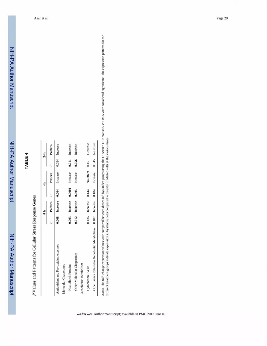
NIH
-PA Author Manuscript
NIH
-PA Author Manuscript
NIH
-PA Author Manuscript
Asur et al. Page 29
TAB
LE 4
P V
alue
s an
d Pa
ttern
s fo
r C
ellu
lar
Stre
ss R
espo
nse
Gen
es
0 h
4 h
24 h
PP
atte
rnP
Pat
tern
PP
atte
rn
Ant
ioxi
dant
and
Pro
-oxi
dant
enz
ymes
0.00
8In
crea
se0.
004
Incr
ease
0.08
4In
crea
se
Mol
ecul
ar C
hape
rone
s
H
eat S
hock
Pro
tein
s0.
003
Incr
ease
0.00
01In
crea
se0.
011
Incr
ease
O
ther
Mol
ecul
ar C
hape
rone
s0.
012
Incr
ease
0.00
5In
crea
se0.
056
Incr
ease
Xen
obio
tic M
etab
olis
m
C
ytoc
hrom
e P4
50s
0.13
6In
crea
se0.
144
No
effe
ct0.
15D
ecre
ase
O
ther
Gen
es R
elat
ed to
Xen
obio
tic M
etab
olis
m0.
187
Incr
ease
0.18
4In
crea
se0.
345
No
effe
ct
Not
es. T
he f
old-
chan
ge e
xpre
ssio
n va
lues
wer
e co
mpa
red
betw
een
dire
ct a
nd b
ysta
nder
gro
ups
usin
g th
e O
’Bri
en’s
OL
S st
atis
tic. P
< 0
.05
wer
e co
nsid
ered
sig
nifi
cant
. The
exp
ress
ion
patte
rns
for
the
diff
eren
t tre
atm
ent g
roup
s in
dica
te e
xpre
ssio
n in
bys
tand
er c
ells
com
pare
d to
dir
ectly
irra
diat
ed c
ells
at t
he v
ario
us ti
mes
.
Radiat Res. Author manuscript; available in PMC 2013 June 01.