mathematical modelling of the radiation-induced bystander effect and transmissible genomic...
TRANSCRIPT

Non-targeted effects of ionising radiation
Proceedings of the RISC-RAD specialised training course “Non-targeted effects of ionising radiation”
STUK – Radiation and Nuclear Safety Authority, Helsinki, Finland 14 – 16 February 2005
O.V. Belyakov (Ed.)
STUK-A234 / DECEMBER 2008
SäteilyturvakeskusStrålsäkerhetscentralen
Radiation and Nuclear Safety Authority
A


STUK • SÄTEILYTURVAKESKUSSTRÅLSÄKERHETSCENTRALEN
RADIATION AND NUCLEAR SAFETY AUTHORITY
Osoite / Address • Laippatie 4 , 00880 HelsinkiPostiosoite / Postal address • PL / P.O. Box 14, FI-00881 Helsinki, FINLANDPuh. / Tel. +358 9 759 881 • Fax +358 9 759 88 500 • www.stuk.fi
Non-targeted effects of ionising radiation
Proceedings of the RISC-RAD specialised training course “Non-targeted effects of ionising radiation”
STUK – Radiation and Nuclear Safety Authority, Helsinki, Finland 14 – 16 February 2005
O.V. Belyakov (Ed.)
STUK-A234 / DECEMBER 2008

The conclusions in the STUK report series are those of the of the authors and do not necessarily represent the official position of STUK.
ISBN 978-952-478-432-0 (print)ISBN 978-952-478-433-7 (pdf)ISSN 0781-1705
Edita Prima Oy, Helsinki 2008
Sold by:STUK – Radiation and Nuclear Safety AuthorityP.O.Box 14, FI-00881 Helsinki, FinlandTel. +358 9 759 881Fax +358 9 759 88500

3
STUK-A234
BELYAKOV Oleg V. Non-targeted effects of ionising radiation. Proceedings of the RISC-RAD specialised training course “Non-targeted effects of ionising radiation”. STUK – Radiation and Nuclear Safety Authority, Helsinki, Finland 14 – 16 February 2005. STUK-A234. Helsinki 2008, 175 pp.
Key words: non-targeted effects of ionising radiation, health effects, mathematical modelling
Preface
Scope and ideas of the workshopThe training course “Non-targeted effects of ionising radiation” took place at the STUK – Radiation and Nuclear Safety Authority, Helsinki, Finland 14 – 16 February 2005. Proceeding of this course is collected in this volume. The idea of the course was to convene a number of scientists leading in the area of non-targeted effects of ionising radiation with the aim to outline their visions for the role of these effects and outline the future directions of radiation research on the basis of their expertise.
The course was generously supported by the RISC-RAD IP FI6R-CT-2003-508842, Euratom specific programme for research and training on nuclear energy, 6th FP of the EC.
It was clear for almost a decade that the universality of the target theory of radiation-induced effects is challenged by observations on non-targeted effects such as bystander effects, genomic instability and adaptive response. Essential features of non-targeted effects are that they do not require direct nuclear exposure by radiation and they are particularly significant at low doses. This new evidence suggests a need for a new paradigm in radiation biology. A better understanding of non-targeted effects may have important consequences for health risk assessment and, consequently, on radiation protection. It is important to explore the mechanisms involved in the non-targeted effects of ionising radiation, to determine the dose-effect relationships of non-targeted effects in space and time, to address the role of individual susceptibility in response and to determine whether the non-targeted effects relate to protective or harmful responses to radiation. The linkage between the bystander response, adaptive response and genomic instability needs to be studied. A longer term objective is to establish a conceptual framework for the generation of a new radiobiological paradigm that covers both targeted (direct) and non-targeted (indirect) effects of ionising radiation. This, in turn would help in

4
STUK-A234
setting the scientific basis for development of a new, more realistic, radiation protection system.
Some questions can be addressed by employing a range of low-dose broad field and microbeam irradiation approaches to investigate both high- and low-LET responses, and by employing well-defined biological systems, such as human cell cultures, 3D artificial tissue systems and ex vivo tissue explants. At the cell and molecular levels, new research should focus particularly on identifying the signals and signal receptors for the non-targeted effects. It will be important to understand whether such signals are produced by all cell types and whether reception and response is general or limited by cell type or organ. Identifying and understanding the action of the signalling process could lead to a means of predicting the outcome of an exposure in an individual.
While research at the cellular, molecular and ex vivo tissue levels will be critical for understanding the mechanisms of these processes, their influence on risk must also be determined more directly. To properly assess the net impact of targeted and non-targeted radiation effects, new research should specifically employ whole animal models, using both strains that are genetically normal and strains that are suspected to be radiation sensitive or cancer prone. Overall measures of risk need to be used together with tissue specific measures, and these tissues need to be assessed for cellular and molecular changes. These results will also be important in understanding the relationship between dose and tissue weighting factors as dose decreases. The animal models could additionally provide clarification on interactions of non-targeted effects with exogenous (e.g. dietary) and endogenous (heritable) variables as a possible part of an inflammatory-type response to radiation-induced stress under in vivo conditions. Long-term clonal variability of non-targeted responses and cell type differences needs to be studied. More information is required on the influence of LET, and on simultaneous exposures to radiations of different LET. More information is also required on the relationship of dose rate and total dose for induction of these responses. Mathematical and statistical modelling is likely to improve the understanding of the potential role of non-targeted effects in the development of different pathologies.
To summarise, the main objectives of the training course were: (1) to clarify the mechanisms of non-targeted effects, in particular, bystander effects, genomic instability and adaptive response; (2) to look if and how non-targeted effects modulate the cancer risk in the low dose region, and whether they relate to protective or harmful functions; (3) to clarify if ionising radiation can cause non-cancer diseases or beneficial effects at low and intermediate doses; (4) address the issue of individual susceptibility and other factors modifying non-targeted responses; (5) attempt to assess the relevance of non-targeted effects
5
STUK-A234
for radiation protection and to set the scientific basis for a modern, more realistic, radiation safety system; (6) and finally to contribute to the conceptualisation of a new paradigm in radiation biology that would cover both the classical direct (DNA-targeted) and non-targeted (indirect) effects.
Oleg Belyakov

6
STUK-A234
BELYAKOV Oleg V. RISC-RAD – epäsuorat isonisoivan säteilyn soluvaikutuk-set.Kurssijulkaisu. Säteilyturvakeskus, Helsinki, 14. – 16.2.2005. STUK-A234. Helsinki 2008, 175 s.
Avainsanat: ionisoivan säteilyn epäsuorat vaikutukset, terveysvaikutukset, matemaattinen mallintaminen
Esipuhe
Kurssin aihe ja tavoitteetKurssi ”Ionisoivan säteilyn epäsuorat soluvaikutukset” järjestettiin Säteily-turvakeskuksessa (STUK), Helsingissä 14 – 16.2.2005. Kurssin ohjelma on koottu tähän kirjaan. Kurssin ajatuksena oli kutsua koolle alan johtavia asiantunti-joita tavoitteena luonnostella heidän asiantuntemuksensa pohjalta näkemyk-siä säteilyn epäsuorien vaikutusten roolista ja suunnitella säteilytutkimuksen tulevaisuuden suuntalinjoja.
Kurssin järjestämistä tuki Euroopan komission (6th FP) Euratomin ydin-energian tutkimus ja koulutusohjelma RISC-RAD IP FI6R-CT-2003-508842.
Jo vuosikymmenen ajan on ollut ilmeistä, että havainnot säteilyn epä-suorista vaikutuksista, kuten naapurisoluvaikutuksesta, perimän epävakai-suudesta ja adaptiivisesta vasteesta haastavat yleispätevän teorian säteilyn suorista vaikutuksista. Epäsuorien säteilyvaikutusten oleellinen piirre on, että niiden syntyminen ei vaadi suoraa säteilyaltistumista ja ne ovat erityisen mer-kittäviä alhaisilla säteilyannoksilla. Epäsuorien säteilyvaikutusten ymmärtä-minen voi johtaa terveysriskien uudelleen arvioimiseen ja tämän myötä vai-kuttaa myös säteilysuojeluun. Ionisoivan säteilyn aikaansaamien epäsuorien vaikutusten mekanismien tutkiminen on tärkeää määritettäessä annosvaste-suhdetta ajallisesti ja paikallisesti, tutkittaessa yksilöllistä alttiutta ja määri-tettäessä liittyvätkö epäsuorat vaikutukset säteilyn suojaavaan vai haitalliseen vasteeseen. Pitkän aikavälin tavoite on muodostaa käsitteellinen kehys uudelle säteily biologian paradigmalle, joka pitää sisällään sekä ionisoivan säteilyn suo-rat että epäsuorat vaikutukset. Tämä vuorostaan auttaa asettamaan tieteelliset perusteet uudelle realistisemmalle säteilysuojelujärjestelmälle.
Altistamalla soluja matalille säteilyannoksille leveä- (broad field)- ja kapeakenttä (microbeam) olosuhteissa voidaan selvittää sekä tiheään että har-vaan ionisoivan säteilyn vaikutusta tarkoin määritetyissä biologisissa malleissa, kuten ihmisen soluviljelmissä, kolmiulotteisissa keinokudosviljelmissä ja ex vivo kudossiirrännäisissä. Solu- ja molekyylitasolla tutkimuksen tulee keskittyä eri-

7
STUK-A234
tyisesti tunnistamaan epäsuorien vaikutusten aikaansaamia signaaleja ja niitä vastaanottavia reseptorimolekyylejä. On tärkeää tietää, tuottavatko kaikki solut näitä signaaleja ja onko signaalien vastaanottaminen ja niihin vastaaminen yleistä, vai rajoittuvatko ne tiettyihin solutyyppeihin tai elimiin. Signaalinvä-litystapahtumien tunnistaminen ja ymmärtäminen mahdollistaa säteilyaltis-tumisen vaikutusten ennustamisen yksilötasolla.
Säteilyn epäsuorien vaikutusten mekanismien ymmärtämisen kannalta on oleellista tutkimus solu-, molekyyli- ja ex vivo kudostasolla. On kuitenkin tärkeää myös määrittää tarkemmin epäsuorien säteilyvaikutusten mahdollinen vaikutus riskiin. Suorien ja epäsuorien säteilyn nettovaikutusten huolellinen arvioiminen vaatii, että tutkimuksen tulee keskittyä eläinmalleihin ja kantoihin, jotka ovat geneettisesti normaaleja ja kantoihin, jotka ovat säteilylle ja syövälle alttiita. Riskin kokonaismääritys tulee arvioida yhdessä kudosspesifisten mää-ritysten kanssa ja edelleen solu- ja molekulaaristen muutosten suhteen. Tämä on myös tärkeää määritettäessä suhdetta annoksen ja kudoskorjauskertoimen välillä kun annos laskee. Eläinmallit saattavat lisäksi tarjota selvennystä epä-suorien vaikutusten interaktiosta ulkoisten (esim. ravitsemuksellisten) tai sisä-syntyisten (perinnöllisten) tekijöiden välillä, kun arvioidaan säteilyn aikaan-saamaa tulehduksellista vastetta in vivo olosuhteissa. Epäsuorien vaikutusten aikaansaama pitkän ajan klonaalinen vaihtelevuus ja erot solutyyppien välillä tulee myös tutkia. Tietoa tarvitaan myös LET:n (LET Linear Energy Transfer, säteilyn luonteenomainen energiansiirtokyky) vaikutuksesta ja samanaikaisesta altistumisesta erilaiselle säteilylle. On myös tärkeää selvittää annosnopeuden ja kokonaisannoksen suhteesta muutosten syntymisessä. Matemaattiset ja tilas-tolliset mallit tulevat lisäksi valaisemaan epäsuorien säteilyvaikutusten mah-dollista roolia erilaisten sairauksien synnyssä.
Yhteenvetona, kurssin päätavoitteet olivat 1) selventää epäsuorien sätei-lyvaikutusten mekanismeja, erityisesti naapurisoluvaikutusten, perimän epä-vakaisuuden ja adaptiivisen vasteen mekanismeja; 2) tarkastella muuttavatko epäsuorat vaikutukset syöpäriskiä alhaisilla annoksilla ja liittyvätkö ne suojaa-viin vai haitallisiin vaikutuksiin; 3) selventää voiko ionisoiva säteily aiheuttaa muita kuin syöpätauteja ja osoittaa säteilyn mahdolliset hyödylliset vaikutuk-set alhaisilla ja keskisuurilla annoksilla; 4) painottaa yksilöllistä herkkyyttä ja muita tekijöitä, jotka vaikuttavat epäsuoriin vaikutuksiin; 5) yrittää arvioida epäsuorien vaikutusten merkityksellisyyttä säteilysuojelulle ja asettaa tieteel-linen pohja uudenaikaiselle säteilysuojelujärjestelmälle; 6) ja lopulta osallistua uuden paradigman luomiseen säteilybiologiassa, joka kattaa sekä klassiset suo-rat (DNA:han kohdistuvan) että epäsuorat vaikutukset.
Oleg Belyakov


9
STUK-A234
Contents
PrefAce 3
esiPuhe 6
NoN-tArgeted effects of ioNisiNg rAdiAtioN 13Oleg V. Belyakov
Abstract 13Non-targeted effects of ionising radiation 13Significance of the bystander effects for radiotherapy 34Applicability to radiation protection and contribution to LNT discussion 34Acknowledgements 37References 37
euroPeAN NucleAr MicroProbes with rAdiobiologicAl ProgrAMs 47T. Butz, R. Cherubini, G. Dollinger, B. Fischer, M. Folkard, U. Giesen, G. W. Grime, K. Hjort, Ph. Moretto, J. Pallon and Z. Stachura
Abstract 471 Introduction 482 The components of nuclear microprobes with “external beams” 493 Cell types and endpoints 534 Outlook 54
MAtheMAticAl ModelliNg of the rAdiAtioN-iNduced bystANder effect ANd trANsMissible geNoMic iNstAbility APPlied to cANcer 59Mark P. Little, Kevin Prise, Melvyn Folkard and Oleg Belyakov
Abstract 591 Mechanistic carcinogenesis models 602 Mechanistic bystander effect models 83References 89
10
STUK-A234
AbscoPAl iNductioN of leuKAeMiA ANd osteosArcoMA followiNg AdMiNistrAtioN of AlPhA-eMittiNg rAdioNuclides 98Brian I. Lord
Abstract 981 Introduction 992 Haemopoiesis 993 Practical implications 1014 Transgenerational induction of leukaemia 1025 Leukaemia and osteosarcoma following injection
of 239Pu, 241Am and 233U in adult mice 1066 Conclusion 108References 108
sigNAlliNg PAthwAys iNduced iN cells exPosed to MediuM froM irrAdiAted cells 113Fiona M. Lyng, Paula Maguire, Brendan McClean, Colin Seymour and Carmel Mothersill
Abstract 1131 Radiation induced bystander effects 1142 Medium transfer experiments 1153 Apoptotic bystander responses 1154 Conclusions 119References 119
the bystANder effect of cANcer geNe therAPy 125Katalin Lumniczky and Géza Sáfrány
Abstract 1251 Introduction 1262 Basic gene therapy protocols,
combination with radiation therapy 1263 Bystander effects 1304 Conclusion 137References 137

11
STUK-A234
rAdiAtioN-iNduced geNoMic iNstAbility ANd bystANder effects: iNter-relAted iNflAMMAtory-tyPe NoN-tArgeted effects of exPosure to ioNiziNg rAdiAtioN 143Eric G. Wright
Abstract 1431 DNA damage and repair 1442 Signalling from DNA strand breaks 1463 Activation of the p53 pathway 1464 Indirect untargeted effects of radiation 1495 Clastogenic factors:
long-range acting non-targeted mechanisms 1526 Radiation-induced bystander effects and
genomic instability are interrelated 1527 Microenvironmental factors and indirect DNA damage 1538 Inflammation and pathological consequences
of genome damage 1559 Conclusions 156References 157


13
STUK-A234
Non-targeted effects of ionising radiation
Oleg V. BelyakovNon-Targeted Ionising Radiation Effects Research Group, Radiation Biology Laboratory, Research and Environmental Surveillance, STUK – Radiation and Nuclear Safety Authority, Helsinki, Finland, P.O. Box 14, FI-00881 Helsinki, Finland
AbstractThe universality of the target theory of radiation-induced effects is challenged by observations on non-targeted effects such as bystander effects and genomic instability. Essential features of non-targeted effects are that they do not require direct nuclear exposure by radiation and they are particularly significant at low doses. This new evidence suggests a need for a new paradigm in radiation biology. The new paradigm would cover both the classical (targeted) and the non-targeted effects. New aspects include the role of cellular communication and tissue-level responses. A better understanding of non-targeted effects may have important consequences for health risk assessment and, consequently, on radiation protection. Non-targeted effects may contribute to the estimation of cancer risk from occupational, medical and environmental exposures. In particular, they may have implications for the applicability of the Linear-No-Threshold (LNT) model in extrapolating radiation risk data into the low-dose region. This also means that the adequacy of the concept of dose to estimate risk is challenged by these findings. Moreover, these effects may provide new mechanistic explanations for the development of non-cancer diseases. Further research is required to determine if these effects, typically measured in cell cultures, are applicable in tissue level, whole animals, and ultimately in humans.
Non-targeted effects of ionising radiation
Cellular targets for radiation damageThe target theory of radiation induced effects [1, 2] postulates that cells contain at least one critical site or target that must be hit by radiation in order to kill a cell. Radiation damage outside of the target does not cause cell death. It is widely accepted that nuclear DNA is the critical target for radiation induced cell death. Early experiments demonstrated that damage to the DNA is more than 3.000 times more effective than membrane damage in the killing of cells in vitro [3].
14
STUK-A234
However, there is evidence suggesting that the cell membrane might also be a target of death in some instances [4, 5].
When a tissue absorbs ionising radiation, its energy results in the production of a fast recoil electron. This electron may then cause damage, either by direct interaction with the DNA, or indirectly through production of free radicals, particularly the hydroxyl radical (OH•), which can cause a break to the DNA helix. Charged particles with high linear energy transfer (LET) radiation such as 3He2+ or α-particle would induce predominantly “direct” damage, whereas low LET radiation (γ and x-rays) predominantly cause “indirect” damage through the action of free radicals [6].
There are a few major types of DNA damage that can be produced by ionising radiation. Single-strand breaks (SSBs) occur due to the deposition of radiation energy on one strand of DNA. Double-strand breaks (DSBs) can be formed by a single ionising event or by the coincidence of random single-strand breaks on the complementary strands, DNA base damage occurs when radiation damages the purine and pyrimidine bases of DNA and finally DNA-DNA and DNA-protein crosslinks [7].
Radiation induced DNA damage can be repaired. There are three types of repair: error-free repair includes excision repair and generally does not result in mutations or lethality, error-prone repair may result in non-lethal or lethal mutations and incomplete repair does not result in the re-establishment of continuity in the DNA sequence and thus may be considered lethal [6].
Non-repaired DNA breaks may lead to chromosomal aberrations. Many types of chromosomal aberrations are produced, some of them lethal (unstable aberrations like dicentrics, rings, fragments), and some non-lethal (stable aberrations i.e. reciprocal translocations). Non-lethal aberrations may lead to oncogenesis. Unstable aberrations may result in the formation of micronuclei, which are the consequences of separation of acentric fragments (or whole chromosome) from the mitotic spindle, and are clearly visible in cellular cytoplasm at the first post-irradiation mitosis [8]. These ultimately lead to loss of clonogenic survival.
In addition to repair, cells may respond rapidly to irradiation, through a number of biological pathways by the initiation of signal transduction pathways, the activation of gene transcription, and cell cycle-specific growth arrest. These early events precondition and predetermine the later consequences of irradiation. Depending on the efficacy of the repair processes, damaged cells may undergo necrosis, apoptosis, proliferative death, senescence (premature differentiation) or ultimately survive and proliferate [6].
There is a range of delayed effects, which may occur in remote descendants of irradiated cells several generations after irradiation. If a cell survives and

15
STUK-A234
Figure 1. New paradigms for Low-Dose Radiation Response.
Targeted effects Non-targeted effects
• Bystander effect
• Radiation-induced genomic instability
• Low dose hypersensitivity
• Adaptive response
• Abscopal (out-of field) effects
• Clastogenic factors
• Delayed reproductive death
• Induction of genes by radiation
New evidenceClassical paradigm of radiation biology
• DNA damage occursduring or very shortly after irradiationof the nuclei in targeted cells.
• The potential for biological consequences can be expressedwithin one or two cell generations
produces progeny then the initial biological response to the irradiation may influence cell differentiation, shorten life-span, induce genomic instability [9], or carcinogenesis [10].
Non-targeted effects, a new paradigm of radiation biologyAccording to the target theory of radiation induced effects, which forms a central core of radiation biology, DNA damage occurs during or very shortly after irradiation of the nuclei in targeted cells and the potential for biological consequences can be expressed within one or two cell generations [11, 12].
A range of evidence has now emerged that challenges the classical effects resulting from targeted damage to DNA (Fig. 1). These effects have also been termed “non-(DNA)-targeted” [11] and include radiation-induced bystander effects [13], genomic instability [14, 15], adaptive response [16], low dose hyper-radiosensitivity (HRS) [17], delayed reproductive death [18] and induction of genes by radiation [19]. An essential feature of “non-targeted” effects is that they do not require a direct nuclear exposure by irradiation to be expressed and they are particularly significant at low doses.
This new evidence suggests a new paradigm [20] for radiation biology that challenges the universality of target theory.
16
STUK-A234
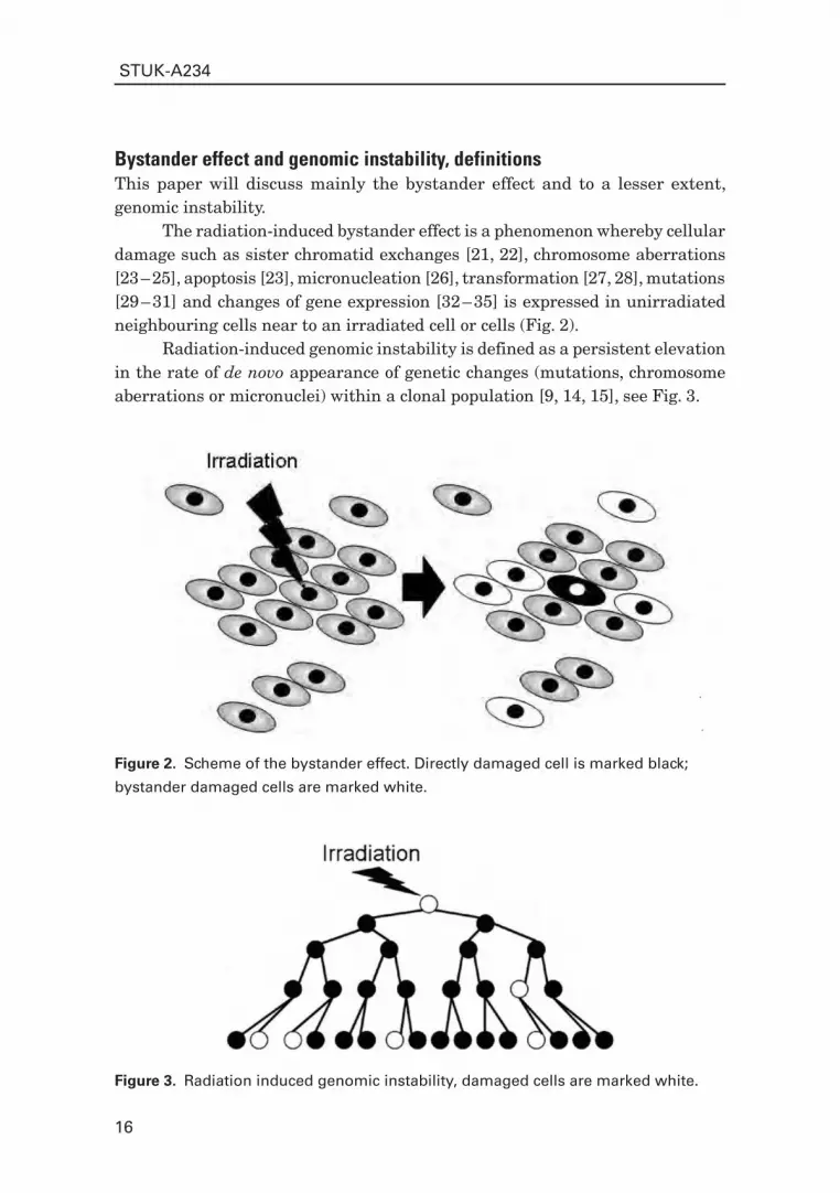
Figure 2. Scheme of the bystander effect. Directly damaged cell is marked black;
bystander damaged cells are marked white.
Figure 3. Radiation induced genomic instability, damaged cells are marked white.
Bystander effect and genomic instability, definitionsThis paper will discuss mainly the bystander effect and to a lesser extent, genomic instability.
The radiation-induced bystander effect is a phenomenon whereby cellular damage such as sister chromatid exchanges [21, 22], chromosome aberrations [23 – 25], apoptosis [23], micronucleation [26], transformation [27, 28], mutations [29 – 31] and changes of gene expression [32 – 35] is expressed in unirradiated neighbouring cells near to an irradiated cell or cells (Fig. 2).
Radiation-induced genomic instability is defined as a persistent elevation in the rate of de novo appearance of genetic changes (mutations, chromosome aberrations or micronuclei) within a clonal population [9, 14, 15], see Fig. 3.

17
STUK-A234
Genomic instability and the bystander effect are both non-targeted effects of irradiation. They have a cross-section much larger than the nucleus. The bystander effect and genomic instability might be related phenomena. There is as yet no evidence that the bystander effect persists for many generations. On the other hand, it was reported that persistent genomic instability can be induced via a bystander mechanism under in vitro [24] and in vivo [25] conditions. This evidence suggests that the initial cross-section for radiation damage is increased by the bystander effect, and cells that are affected by the bystander mechanism may remain at an increased risk of genetic change for many generations.
Evidence for bystander effectsInteractions between hit and non-hit cells after exposure to ionising radiation have been known for many years in radiation biology. Much of the early data was obtained from studies of chromosome damage induced by plasma from radiotherapy patients [36, 37] and accidental exposures [38] in test cell cultures. These indirect effects were explained by the production of “clastogenic factors” [39]. These clastogenic factors were extensively studied in victims of the Chernobyl Accident [40 – 42]. It was hypothesised that they may be related to lipid peroxide products [43] ionisine nucleotides [44], cytokines [45] and reactive oxygen species (ROS) such as superoxide radicals [39].
Other evidence has come from abscopal or “out-of-field” effects, which are well known in radiotherapy [46 – 49]. These phenomena are defined as the effects of radiation on tissues of the same person or organism at some distance from the actual radiation site or target. A recent paper by [50] related radiation-induced out-of-field effects in lung of rodents with DNA damage. A strong correlation between lethality and DNA damage was found.
In the last few years, a large number of papers were published demonstrating evidence for the radiation induced bystander effect [13, 51]. Nagasawa and Little first published a paper, describing the bystander effect [22], measured as an increase of sister chromatid exchanges (SCE). They irradiated Chinese hamster ovary cells with low doses of α-particles from a conventional broad field source in a way that only a few cells within a population were actually traversed by a particle. A much higher level of SCEs were produced in cells than would be predicted on the basis of the number of cell nuclei targeted. The authors proposed a hypothesis that cell irradiation induces some indirect effects within neighboring cells via free radical cascades or signal transduction pathways.
Significant numbers of the recent publications with evidence for bystander effects have come from the studies with α-particle irradiation delivered with

18
STUK-A234
specially constructed conventional low doses broad-field sources [52]. In this case irradiation have been delivered to a population of cells in such a way that only a few cells within a population were actually traversed by α-particles. Hickman measured changes in the TP53 expression after rat lung epithelial cells were exposed to low doses of α-particles [35]. They found that a higher fraction of cells demonstrated an increased TP53 expression than were hit by α-particles.
A series of papers from the Los Alamos National Laboratory demonstrated that extracellular factors are involved in SCE formation following low dose α-particle exposure. Deshpande and co-workers [21] irradiated cell cultures of primary human fibroblasts with α-particles and observed a high level of sister chromatid exchanges. The percentage of cells showing SCEs was 9-fold higher than expected on the basis of the number of nuclei traversed. The authors provided convincing evidence for the production of extracellular factors, released into the cell culture medium [53]. Later, the same group [54] attributed the observed bystander effects to the action of TGF-β1 and reactive oxygen species (ROS).
In a series of studies, Mothersill and Seymour demonstrated that medium from γ-ray irradiated cell cultures reduces the survival of unirradiated cells [23, 55 – 58]. Under this protocol supernatant from irradiated cells was transferred to test “reporter” cell cultures, which were analysed using the Puck and Marcus clonogenic assay [59] and for presence of micronucleated, apoptotic and cells with chromosome aberrations.
Another approach was utilized by Bishayee and co-workers [60, 61]. They detected a pronounced bystander effect in a V79 three-dimensional tissue culture model labelled with 3H-thymidine when the isotope is localised in the cell nucleus and distributed non-uniformly among the cells. A related class of effects was demonstrated in thymocytes [62]. They demonstrated that interactions between different types of γ-irradiated cells lead to different degrees of radiation-induced apoptosis via the production of soluble autotoxic mediators. When irradiated cells were mixed with non-irradiated ones, less interphase-induced cell killing was observed than would be predicted on the basis of ratios of the cells mixed together. This protection effect is not observed when the medium from non-irradiated cells is added to the irradiated thymocytes.
Previous studies at the Gray Cancer Institute demonstrated that the target for chromosomal damage is larger than the nucleus on basis of calculations of the fraction of micronucleated Chinese hamster V79 cells after α-particle irradiation [63]. It has been demonstrated a direct evidence of bystander effects in normal human AG01522B fibroblasts using the Gray Cancer Institute charged particle microbeam [64, 65]. Irradiation of a single fibroblast with a single 3He2+ particle delivered by the microbeam through the nucleus would give a significant rise of bystander damaged cells measured as micronucleated and apoptotic cells.

19
STUK-A234
In general a 2 – 3 fold increase in the level of damaged cells was measured in comparison to controls.
Other groups have also utilised microbeam approaches to study bystander effects. Evidence for the existence of extra-nuclear target(s) for radiation-induced effects [66] was observed when the cytoplasm of human-hamster hybrid A(L) cells was irradiated avoiding traversal of the nucleus. Cytoplasmic irradiation led to considerable mutagenesis at the CD59 (S1) locus with minimal cytotoxicity. The mutations found were similar to those of spontaneous origin and are entirely different from those of nuclear irradiation. On other hand, it was demonstrated that cytoplasmic irradiation initiates the generation of reactive oxygen species. The final conclusion from the paper was that cytoplasmic irradiation might be more dangerous than nuclear irradiation, as mutagenicity is accomplished by little killing of the target cells.
Zhou and co-authors [30] demonstrated a bystander mutagenic effect after α-particle microbeam irradiation. They showed that cells, irradiated with a microbeam, could induce a bystander mutagenic response in neighbouring cells, which were not directly traversed by an α-particle. Intercellular communication plays a critical role in mediating the bystander phenomenon under these conditions. It was shown that irradiation of 20% of randomly selected human-hamster hybrid A(L) cells with 20 α-particles each, resulted in a mutant fraction that is 3-fold higher than expected, assuming no bystander effect. Analysis by multiplex PCR demonstrated that the types of mutations induced are significantly different from those of spontaneous origin.
Another study from the same group [31] showed that irradiation of even 10% of confluent human-hamster hybrid A(L) cells with a single α-particle per cell through the nucleus results in a mutant yield similar to that observed when all cells in the population are irradiated. This effect was significantly eliminated by an inhibitor of gap junction-mediated intercellular communication, or in cells carrying a dominant negative connexin 43 vector.
An important question is whether the bystander effect contributes to carcinogenesis. Lewis and co-authors [27] tested the response of non-irradiated cell cultures when these were exposed to medium from X-irradiated human CGL1 hybrid cells. They reported an increased radiation-induced bystander neoplastic transformation after treatment with medium from irradiated cells. Medium, exposed with 5 or 7 Gy of X-ray increased the frequency of neoplastic transformation significantly from 6.3 × 10⁻⁶ in control to 2.3 × 10⁻⁵ (~ 4-fold).
Sawant and co-authors [28] used the Columbia University microbeam system to delivered 0, 1, 2, 4 or 8 α-particles through the nuclei of all or 10% of C3H 10T1/2 cells. They demonstrated that when 10% of the cells are exposed to
20
STUK-A234
α-particles, the frequency of induced transformation is the same as that observed when every cell was exposed to the same number of α-particles.
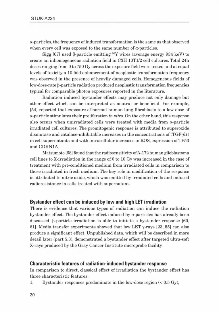
Sigg [67] used β-particle emitting 90Y wires (average energy 934 keV) to create an inhomogeneous radiation field in C3H 10T1/2 cell cultures. Total 24h doses ranging from 0 to 750 Gy across the exposure field were tested and at equal levels of toxicity a 10 fold enhancement of neoplastic transformation frequency was observed in the presence of heavily damaged cells. Homogeneous fields of low-dose-rate β-particle radiation produced neoplastic transformation frequencies typical for comparable photon exposures reported in the literature.
Radiation induced bystander effects may produce not only damage but other effect which can be interpreted as neutral or beneficial. For example, [54] reported that exposure of normal human lung fibroblasts to a low dose of α-particle stimulates their proliferation in vitro. On the other hand, this response also occurs when unirradiated cells were treated with media from α-particle irradiated cell cultures. The promitogenic response is attributed to superoxide dismutase and catalase-inhibitable increases in the concentrations of (TGF-β1) in cell supernatants and with intracellular increases in ROS, expression of TP53 and CDKN1A.
Matsumoto [68] found that the radiosensitivity of A-172 human glioblastoma cell lines to X-irradiation in the range of 0 to 10 Gy was increased in the case of treatment with pre-conditioned medium from irradiated cells in comparison to those irradiated in fresh medium. The key role in modification of the response is attributed to nitric oxide, which was emitted by irradiated cells and induced radioresistance in cells treated with supernatant.
Bystander effect can be induced by low and high LET irradiationThere is evidence that various types of radiation can induce the radiation bystander effect. The bystander effect induced by α-particles has already been discussed. β-particle irradiation is able to initiate a bystander response [60, 61]. Media transfer experiments showed that low LET γ-rays [23, 55] can also produce a significant effect. Unpublished data, which will be described in more detail later (part 5.3), demonstrated a bystander effect after targeted ultra-soft X-rays produced by the Gray Cancer Institute microprobe facility.
Characteristic features of radiation-induced bystander responseIn comparison to direct, classical effect of irradiation the bystander effect has three characteristic features:
Bystander responses predominate in the low-dose region (< 0.5 Gy);1.

21
STUK-A234
The bystander effect has a non-linear dose dependence, suggesting a 2. switch-on (“all or nothing”) mechanism for its activation;The bystander effect is maximally induced by very low doses.3.
Nagasawa and Little first demonstrated evidence of the bystander effect induced by a very low dose of 0.16 mGy and saturating at 0.31 mGy without further statistically significant increases up to 4.9 mGy [22]. Hickman in his experiments with irradiation of rat lung epithelial cells, showed that the dose-effect for TP53 expression was different for α-particles in comparison to X-rays [35]. α-particles gave a no-threshold response whereas there was a low dose threshold observed with X-rays at around 0.1 Gy. Overall, the shape of the dose-effect curve for both types of irradiation had a tendency to flatten after exposure with 0.2 – 0.5 Gy and did not demonstrate a statistically significant increase with increasing dose. Deshpande and co-workers [21] did not observe a dose-dependence of the bystander effect above 0.02 Gy with saturation up to highest does tested, 13 Gy of α-particles. Zhou [30, 31] noted that a level of bystander mutagenesis effect after α-particle microbeam irradiation did not depend on the number of particles delivered. Lewis [27] also showed that the amount of cell death induced by bystander effects is not dependent on dose.
The bystander effect contributes to a significant proportion of the overall damage yield in the low-dose region by an apparently distinct mechanism from the “classical” radiation response. Recently obtained data [26, 64] demonstrated that the fraction of damaged (micronucleated and apoptotic) human fibroblasts was independent of the number of charged particles delivered to the targeted cell. One 3He2+ ion, delivered to the nucleus of one cell among a few hundred non-irradiated neighbours induced the bystander effect to the maximum extent. Further increase of dose to the targeted cell does not change the dose response. Similarly, the effect was independent of the number of cells irradiated. The same level of damage was observed whether 1 or 4 cells were targeted within the dish. These data are considered in detail in [26, 64].
The general shape of the bystander effect dose response in comparison to direct radiation consequences is illustrated at Fig. 4. Most observations of bystander effects have shown a saturation of the response above the threshold dose (0.2 Gy is an estimation) and do not demonstrate a linear relationship to the dose, see review [69].
The model proposed here (Fig. 4) is supported by data, obtained with normal human fibroblast cell cultures published in [26, 64]. Experiments with primary urothelial explants similarly demonstrated the absence of a dose response.
The model proposed here is in marked contrast to that proposed by Brenner [70] as a quantitative model for the application of the bystander effect

22
STUK-A234
Figure 4. Comparison of ”classical” and ”bystander” types of response to ionising irradiation.
Classicaleffects
Bystander effects
Dose~0.2 Gy
Classicaleffects
Bystander effects
Dose~0.2 Gy
to carcinogenic risk. The BaD (Bystander and Direct) model of radiation response is supported by the data from the same group on in vitro oncogenic transformation after broad-field or microbeam α-particle irradiation [28]. BaD postulates that the bystander effect is a binary “all or nothing” phenomenon and might be expressed in a small sensitive sub-population of “interested neighbors” [71],
which do not cover the entire cell population. The authors believe that there may be purely geometrical reasons for the existence of a subpopulation, susceptible to bystander effects. They assume that a hypothetical bystander factor has a limited penetration distance. However, in this case some clustering of damage should be observed around the irradiated cell. To date, no evidence of clustering has been reported and in contrast, data published in [64] suggest that cellular damage is uniformly distributed throughout the cell culture dish.
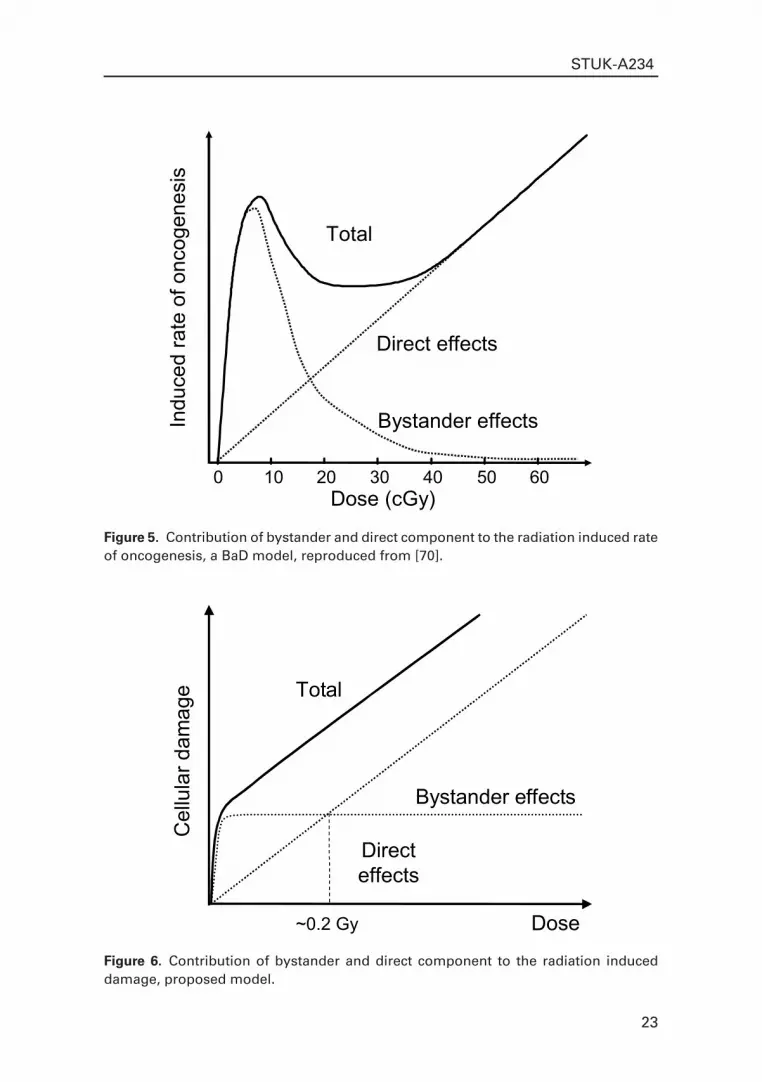
The BaD model also suggests that the bystander effect can only be observed at low doses (Fig. 5). At low doses, the bystander effect dominates the direct response. The authors point out that this may lead to an underestimation of low-dose risks extrapolated from high doses, where direct effects dominate. Similar to the model proposed here, BaD assume that the total response of a cellular system to ionising radiation has two components: direct and bystander damage. Direct damage has a linear dose-relationship, whereas bystander damage is induced to the maximum extent by very low doses (less than 1 cGy).
23
STUK-A234
Figure 5. Contribution of bystander and direct component to the radiation induced rate of oncogenesis, a BaD model, reproduced from [70].
Indu
ced
rate
of o
ncog
enes
is
Direct effects
Bystander effects
Dose (cGy)0 10 20 30 40 50 60
Total
Figure 6. Contribution of bystander and direct component to the radiation induced damage, proposed model.
Dose
Bystander effects
Directeffects
~0.2 Gy
Total
Cel
lula
r dam
age

24
STUK-A234
In contrast to the proposed model, the authors believe that the bystander effect would decline with increasing dose because bystander signal-sensitive cells, whose nuclei are hit directly, cannot produce a bystander response Therefore, the total effect would be (as presented at Fig. 5) a result of summing bystander and direct effects at low dose region (up to 30 cGy). At the higher dose (from about 30 cGy) it would be predominantly influenced by direct effects. To date, however, there is not enough experimental data to assume that a direct hit would prevent a cell from releasing a bystander factor.
Therefore, a different model can be suggested (see Fig. 6), which has a more pronounced plateau in the low dose region and fits better to both the results, obtained during this project and other published experimental data. We assume that bystander signal-sensitive cells, whose nuclei are hit directly, can produce a bystander response. Finally, the model proposed here can be utilised to describe any dose-effect relationship for cellular damage whereas the BaD model is designed for the estimation of carcinogenic risk.
Recently another novel stochastic model was proposed [72] A model of the radiation-induced bystander effect is developed that takes account of spatial location, cell killing and repopulation. The ionizing radiation dose- and time-responses of this model are explored, and it is shown to exhibit pronounced downward curvature in the high dose-rate region, similar to that observed discussed above. One significant advantage of this model is that this model is suitable for 3D modelling of bystander effect can be applied to the tissue data.
Bystander versus direct effectsFor studies of cell killing, it is important to determine numerically the relative contribution of “classical” and “bystander” effects. Recently, Seymour and Mothersill [58] have presented a method of correcting the overall survival curve to enable analysis of the relative contributions of the bystander effect and the effects attributable to direct interaction of the radiation with the target cell. They used a standard Puck and Marcus assay [59] to obtain a clonogenic survival curve for HPV-G human keratinocytes. Two separate sets of cell culture flask were used. One set was irradiated with broad field of γ-rays with various doses, medium was harvested, filtered and added to a second set of flasks, which had not seen a direct radiation exposure. The survival results were converted to clonogenic death for both bystander and total effect and by subtraction, the percentage of cell death due to non-bystander induced death was determined. The data show that for this human epithelial cell line, doses within the range 0.01 – 0.5 Gy of γ-rays would induce clonogenic death only by the bystander effect (see Fig. 7).

25
STUK-A234
Figure 7. Clonogenic cell death measured in human keratinocytes. The total bar represents the total death detected after exposure of cells to the radiation dose. The death measured after exposure to medium from irradiated cell cultures (Bystander) is represented by the blue portion of the bar, and the remaining death determined by subtraction is represented by the red portion of the bar, giving a value (Direct) for death not attributable to bystander effects of radiation. Adapted from [58].
Dose (Gy),-rays0
102030405060708090100
0 0.01 0.03 0.05 0.1 0.3 0.5 2.5 5
Percentageclonogenic
celldeath
DirectBystander
Dose (Gy),-rays0
102030405060708090100
0 0.01 0.03 0.05 0.1 0.3 0.5 2.5 5
Percentageclonogenic
celldeath
DirectBystander
It can be seen that there is a large bystander component at low doses but at doses of 0.5 Gy and above the direct effects of radiation begin to appear. The magnitude of the bystander effect is relatively constant and it appears to saturate at doses in the range of 0.03 – 0.5 Gy. After doses greater than 0.5 Gy, the clonogenic death curves are the result of a dose dependent non-bystander effect and a dose independent bystander effect.
Mechanisms of the bystander effectsIt is known that the bystander effect is cell type dependent [23], depends on cell proliferative state (discussed in [73]) and that energy / REDOX metabolism may be involved in the expression of a radiation induced bystander response [74]. The exact mechanisms of the bystander effect are not yet known. However, it is clear that bystander signal production and cellular response may involve different pathways [51]. Bystander signalling is a complex and well-tuned system, which most likely involves more than one messenger and is connected with tissue microenvironment signalling [75, 76].
There is experimental evidence that the bystander effect may have at least two separate pathways for the transfer of damage from irradiated cells to unirradiated neighbours: by gap junction intercellular communication (GJIC) or cell culture mediated factors. A junction between cells, which consists of many

26
STUK-A234
pores, mediates GJIC. Each pore is formed by a hexagonal array (connexon) of six transmembrane proteins (connexins) in each plasma membrane: when joined together the pores open, allowing communication and the interchange of metabolites between cells [77].
Azzam and co-workers [32] have demonstrated that the bystander effect is dependent on gap junction intercellular communication in confluent cultures of 5 different primary human diploid fibroblast lines exposed to low fluences of α-particles. They showed that TP53 and CDKN1A expression are activated in bystander cells after low dose α-particle irradiation. Importantly, they also observed clustering of expression in neighbouring cells. Treatment of the culture with lindane, which inhibits GJIC, led to a marked reduction in the increase in the levels of TP53 and CDKN1A. A recent paper from the same authors suggested direct evidence for the participation of GJIC in the transmission of damage signals from irradiated to non-irradiated cells [33]. Other workers have also shown that lindane treatment leads to inhibition of bystander-induced cell killing in hamster V79 cells [61]. The bystander effect was also significantly reduced in cells pretreated with 1 mM of octanol, which inhibits gap junction-mediated intercellular communication [31]. The same paper also reports that the bystander effect was suppressed in cells carrying a dominant negative connexin 43 vector, which is a part of the connexon complex.
Little is known concerning the signals, which may be transferred via GJIC. The connexin proteins, which form the gap junctions, allow ions, secondary messengers and small molecules to pass between cells and modification of these proteins, by phosphorylation, can open or close the pores. Whether specific signal molecules are transmitted between cells or the junctions are specifically opened, as part of a bystander response needs to be addressed.
The second proposed mechanism of the bystander effect is mediation by secretion of factors into the culture medium. Medium transfer experiments [23, 58] suggest the existence of a relatively long-lived bystander effect mediator, which cannot be eliminated by media filtering. A series of studies suggested another possible mechanism in which the irradiated cells secrete cytokines or other factors that act to increase intracellular levels of reactive oxygen species in unirradiated cells. Lehnert and co-workers [53] demonstrated that the culture medium harvested from cells irradiated with low fluences of α-particles could induce an increase in sister chromatid exchanges when incubated with unirradiated test cells. According to results [53, 78], α-particle irradiated cells secrete into the serum containing medium some short-lived factor(s). It was found that the activity of this factor(s) could be inhibited by superoxide dismutase, can survive freeze and thawing but not heating. A recent paper by Lewis and co-authors [27] used a medium transfer protocol and observed delayed death and
27
STUK-A234
neoplastic transformation. And finally, Mothersill and Seymour [55] reported data which suggest that the bystander effect does not depend on communication through gap junctions formed between cells in contact but is due solely to media release factors, in contrast to that predicted from other studies.
Hypothetical messenger(s)The exact nature of bystander signalling is not known. Two mechanisms of transmission from an irradiated cell to an unirradiated neighbour have been proposed as described above. A bystander messenger can be either a soluble factor excreted into the cell culture medium from the irradiated cells or be directly transmitted by GJIC – gap junction intercellular communication between hit and non-hit cells [51].
Based on this distinction it can be speculated that at least two types of the bystander messenger might exist. Primary messenger is emitted by targeted cell. It is short lived, not very stable, travels through gap junctions, should be water soluble and most likely not a protein. One suitable candidate here could be long-lived organic radicals capable of transferring through gap junctions. Such radicals could have lifetimes of up to 20 hours [79, 80]. Among other candidates for GJIC mediated primary bystander messenger are antioxidants (thiols) [81], Ca2+ [82] Ip3 (storage form of intercellular Ca2+) and cAMP [83], which is an important secondary messenger involved in Ca2+ metabolism.
Secondary bystander messenger should be long-lived, more stable, most likely emitted by activated, not directly traversed, cells. It might be a media borne factor and most likely a protein. Suitable candidates here would be lipid hydroperoxidases [84], ceramide [5], death ligand (TNFSF6) produced from exfoliation [85]. Other evidence supports a role for cytokines as key signalling molecules in the transfer of bystander damage cytokines such as TNF-α [43, 86], TGF-β [54, 76] or IL-1 [43].
There is a range of possible candidates for bystander effect mediation, which are medium borne and could be either primary or secondary messengers. Reactive oxygen species (H
2O
2/O-2) have been proposed as possible signals involved
in bystander responses [54, 87]. Another group proposed that nitric oxide (NO) might play a central role in mediation of bystander effect [68, 88] potentially having a protective value.
In conclusion, it is most likely that there is no single mechanism underlying the bystander effect and both media borne and GJIC factors are involved in its induction and perpetuation. The mechanisms involved are probably cell type specific which may reflect a lot of the current uncertainty in the literature as to the processes involved.

28
STUK-A234
The relationship between radiation induced bystander effect and genomic instabilityThe relationship between the bystander effect and genomic instability is not clear. It was reported that persistent genomic instability could be induced in vitro via a bystander mechanism. Chromosomal instability was demonstrated in the clonal descendants of haemopoietic stem cells after irradiating murine bone marrow with α-particles [24]. The authors studied the effects of interposing a grid between the cells and the α-particle source so that the surviving population consisted predominantly of non-traversed stem cells. It was shown that the number of clonogenic cells transmitting chromosomal instability was greater than the number expected to be hit and survive. Later, the same group utilised a bone marrow transplantation protocol in which a mixture of irradiated and non-irradiated murine bone marrow cells was transplanted into mice. It was demonstrated that genomic instability could be observed in the progeny of non-irradiated haemopoietic stem cells under in vivo conditions [25].
The data published in [89] suggest that the same AG01522B normal human fibroblast cell line is susceptible to radiation induced genomic instability (after both α-particle and X-ray irradiations), and bystander response after microbeam 3He2+ irradiation according to the results, published in [26, 64]. Also, the urothelial model, which demonstrates a pronounced bystander response [73, 90, 91] may express genomic instability as a part of the response.
Other studies have suggested a common relationship between genomic instability and the bystander response. Some evidence of protective function of bystander effect is available [76]. This issue is discussed in greater detail in part 6.1 of this thesis. There is some indication that genomic instability may play a protective role as well. It was recently demonstrated [92] that chromosome instability in GM10115 cells can lead to the development of cell variants that are more resistant to radiation. Bystander effect and genomic instability might be parts of a comprehensive system of oxidative damage control, which aims to reduce the risk of carcinogenesis [93, 94] and both have been observed in vivo [25, 95]. Finally there are suggestions that both the bystander effect [96] and genomic instability [97] are controlled through epigenetic mechanisms [98] such as DNA methylation [99].
Bystander effect in multicellular systemsThe bystander effect cannot be comprehensively explained on the basis of a single cell reaction. It is well known that an organism is composed of different cell types that interact as functional units in a way to maintain normal tissue [100] function. Radiation effects at the tissue level under normal conditions prove
29
STUK-A234
that individual cells cannot be considered as an isolated functional unit within most tissues of a multicellular organism. Therefore the radiation response is not simply the sum of cellular responses as assumed in classical radiobiology, predominantly from studies using cell cultures. Experimental models, which maintain tissue-like intercellular cell signalling and 3-D structure, are essential for proper understanding of the bystander effect. The tissue microenvironment is also important for proper manifestation of the bystander effect [75]. Barcellos-Hoff and Brooks hypothesise that the radiation bystander effect and genomic instability are positive and negative manifestations of a tissue homeostatic process [76]. Extracellular signalling in normal tissues plays a crucial role in initiation and perpetuation of bystander effect.
Only a few papers have been published on bystander effects in multicellular systems. The radiosensitivity of HPV-G and HaCaT epithelial cells lines irradiated within microcolonies (> 50 cells) was found to be lower than those irradiated as single cells [23, 101]. A series of papers by Bishayee and co-workers [60, 61] detected a pronounced bystander effect in a V79 three-dimensional tissue culture model labelled with 3H-thymidine when the isotope is localised in the cell nucleus and distributed non-uniformly among the cells. Jen and co-workers [102] found that the radiosensitivity of mouse kidney cells that are irradiated under in vivo conditions in situ or in vitro as fragments was higher than those irradiated in vitro as single cells.
Our recent work [103] clarifies mechanisms of bystander responses in a 3D normal human-tissue system. Endpoints were induction of micronucleated and apoptotic cells. A charged-particle microbeam was used, allowing irradiation of cells in defined locations in the tissue yet guaranteeing that no cells located more than a few micrometers away receive any radiation exposure. Unirradiated cells up to 1 mm distant from irradiated cells showed a significant enhancement in effect over background, with an average increase in effect of 1.7-fold for micronuclei and 2.8-fold for apoptosis. The surprisingly long range of bystander signals in human tissue suggests that bystander responses may be important in extrapolating radiation risk estimates from epidemiologically accessible doses down to very low doses where non-hit bystander cells will predominate.
With the exception of abscopal effects and clastogenic factors in blood plasma of patient undergo radiation therapy, which were discussed above, little evidence of bystander effect under in vivo conditions is available. The one experimental paper, which deals with bystander effect under in vivo conditions is work by Watson and co-authors [25]. They utilised a bone marrow transplantation protocol to demonstrate that genomic instability could be induced in bystander cells. Mixture of irradiated and non-irradiated cells distinguished by a cytogenetic marker, was transplanted into CBA/H mice. Genomic instability was demonstrated

30
STUK-A234
in the progeny of non-irradiated cells. Another recent paper [104] demonstrated oncogenic bystander radiation effects in mouse cerebellum. Authors reported bystander (in fact “abscopal”) tumour induction in cerebellum of radiosensitive Patched-1 (Ptch1) heterozygous mice after x-ray exposure of the other parts of the body. They also provided evidence supporting the role of gap-junction intercellular communication (GJIC) in transmission of bystander signals in the central nervous system.
Rationale for the current interest in non-targeted responsesThe current interest in non-targeted effects such as bystander responses is particularly timely. Firstly there is currently a tremendous shift of emphasis from high-dose effects towards low and ultra-low doses, of relevance to environmental and occupational exposures both in terms of research needs and public interest. This has coincided with tremendous advances in the technical possibilities for precise low dose irradiation such as development of microbeams [105, 106], imaging and computerised automation. Apart from technical developments, low dose studies would not be possible without development of more specific and sensitive methods of cellular and molecular biology. Apoptosis assays, techniques to measure changes in cell cycle regulation, protein expression, advanced methods of cytogentic analysis has enabled radiation biology to start to probe low frequency changes in individual cells. This allows the systematic studies of processes (i.e. apoptosis, genomic instability or bystander effect) now considered to be important and are ultimately challenging the existing fundamentals of the understanding of the action of radiation on biological systems.
Hypothesis: bystander effect is a protective mechanism of tissue damage controlThe discovery of a bystander effect is important for understanding the dose-response mechanisms relevant to low-dose irradiation in vivo. One important question is whether the bystander effect is a protective mechanism or whether, conversely, it amplifies the number of cells damaged by the isolated radiation tracks of low-dose exposures leading to an increased risk of carcinogenesis.
One theory, supported by the experimental data obtained during this project is that the main function of the bystander effect is to decrease the risk of transformation in a multicellular organism exposed to radiation. It can be speculated that individual cells within a tissue may not have the ability to detect irradiation such that an individual cell response is not expressed. An integrated multicellular system may be able to detect damage from irradiation and respond

31
STUK-A234
to it by removing a functional group of cells, which could be potentially damaged. The existence of a potentially sensitive group of cells, susceptible to the bystander response has also been proposed by [70]. However, not every cell will respond to the hypothetical bystander factor, which is released by targeted cells. Only 1 – 3% of the total number of cells in the system would express damage [26, 73] and approximately 10 – 15% would go on to bystander induced differentiation [91, 107]. Lehnert and co-workers believe that differences in the gene expression profiles and temporal and spatial patterns of key proteins expressed in directly irradiated and bystander cells may determine how the cells ultimately respond to low doses of radiation [108]. The data obtained during this project are consistent with every cell being able to initiate the bystander effect. Such a mechanism of co-operative response would make the tissue system much more robust. It would work only for low doses of charged particle irradiation (below ~ 0.1 – 0.2 Gy, depending on system and type of radiation) because only in this case is the damage localised within a small fraction of the cell population.
In some systems, the most convenient way to remove potentially damaged cells is via apoptosis. In particular, apoptosis allows the removal of affected cells without a negative impact on other cells via inflammatory responses. However many apoptotic pathways are controlled by cellular signals, which would also enable the selective removal of certain functional groups of cells. Apoptosis is not playing a significant role in the urothelial explant system [26, 73]. Another way to isolate damage is to prompt affected cells into irreversible differentiation. Results [91, 107], which support this mechanism, have been obtained. Underlying this theory is that a normal 3-dimensional tissue microarchitecture is essential for the manifestation of the bystander effect [103, 109].Therefore, the bystander effect might be a tissue-specific epigenetic phenomenon, which can be observed in full scale when there is presence of natural cellular stratification with differentiated and dividing cells present and an intact tissue microenvironment. However, the data suggest that initial nuclear damage seems to be essential for initiation of this system. Perpetuation of the bystander effect might involve cascade-like epigenetic mechanisms.
Tissues remove all potentially damaged cells from the system to avoid the risk of carcinogensis following sparse low dose irradiation or any other local oxidative damage [75]. Bystander induced differentiation seems to play a central role in this process. It is known that cellular senescence is a powerful tumour suppressor mechanism [110].
A general scheme explaining the proposed theory is illustrated in Fig. 8. Tissue, exposed to sparse natural irradiation, would respond as a single unit (1). The damaged cells would produce some bystander signal or signals. Some sensitive sub-population of potentially damaged cells would respond to the

32
STUK-A234
Figure 8. A general scheme of radiation induced bystander effect in tissue systems.
1. Sparse irradiation 2. Bystander signal 3.Tissue response
TrackIntercellular communication
Premature differentiated cellApoptotic cell
Targeted cell
Potentially damaged cell
bystander messenger (2). The tissue response to sparse irradiation would affect just a fraction of cells within the tissue (estimated at 10 – 15%). A minor fraction of the cells will be eliminated (probably by apoptosis – estimated as < 1%). The majority of the cells would be removed from proliferating pool by being prompted into differentiation (3). Such a significant response of tissue might be explained by the great danger of even one transformation event induced by natural background radiation. Removing from the proliferating pool all the potentially damaged cells would significantly reduce the risk of transformation for any one cell.
Recently, two theories were proposed concerning the possible meaning of the bystander effect. One of them hypothesises that the radiation-induced bystander effect is a manifestation of a tissue homeostatic process [76]. Cell growth, differentiation and death are directed significantly by extracellular signaling through the interactions of cells with other cells and with the extracellular matrix and the tissue microenvironment. According to the authors’ theory the bystander effect eliminates abnormal cells in order to inhibit neoplastic behavior and preserve tissue integrity. Genomic instability is interpreted by the authors as results of absence the bystander effect. They write: “radiation-induced bystander effects and genomic instability, are, respectively, positive and negative cellular manifestation of multicellular programs of damage response” [76]. Therefore, the bystander effect is hypothesised to be an important mechanism of tissue integrity maintenance.
33
STUK-A234
Another theory concerning a possible role of the bystander effect for the genome as a whole was recently proposed by Baverstock [111]. The author proposed that the radiation induced bystander effect (as well as genomic instability) can be understood in the terms of the dynamic genome concept proposed in this paper. These phenomena are interpreted not just as the result of loss of stability from specific modifications of the genome sequence, but, as a response of the genome in order to preserve the integrity of the genomic sequence.
The relationship between the bystander effect and genomic instabilityRadiation induced bystander effect and genomic instability are both non-targeted effects of irradiation. However, the relationship between the bystander effect and genomic instability is not clear. Genomic instability and bystander effect can both be induced in vitro and in vivo [25, 95]. The data published in [64, 65, 89] suggest that the same cell line (primary human fibroblasts) can express radiation induced genomic instability and bystander response, although a direct relationship between the two endpoints has not been tested implicitly.
On the other hand, the experiments with irradiation of ureter tissue fragments [107, 112] demonstrate that genomic instability (i.e. de novo appearance of cellular damage) and the bystander effect could be closely linked. With the damaged or differentiated cells that are expressed 7 days later in the explant outgrowth, many must be several generations removed from the initially targeted cells and those which initially express the bystander phenotype. It is likely that a cascade mechanism of bystander cell damage probably dominates the initial phase of the targeted exposures. However a significant contribution of genomic instability (probably, bystander-induced) on the later stages cannot be ruled out.
Where the bystander effects might be important?Bystander effect could be important in a few areas related to radiation. The bystander effect might contribute to the estimation of cancer risk from domestic radon exposure [113]; the effects of HZE particles during space mission to the Mars, see discussion on cosmic radiation at [114]; health effects of air crew personnel, exposed to radiation during i.e. inter-continental flight [115]; high energy radiotherapy outcome.
I would like to concentrate on two issues, where the bystander effects might contribute significantly: cancer radiotherapy and radiation protection.
34
STUK-A234
Significance of the bystander effects for radiotherapyThe bystander effect is a low dose (up to 200 mGy) phenomenon. Therefore, at the first look, it cannot play any considerable role in radiotherapy, which operates with doses of tenth of Grays and more. However, the spectrum of secondary malignancies in radiotherapy patients may suggest some contribution of the bystander effect [116]. On the other hand, Trott [117] points out that the future experiments are needed to prove the potential therapeutic value of the bystander effect in radiotherapy and nuclear medicine.
The theory concerning a protective role of the bystander effect may be supported by the recent data of microbeam radiation therapy [118]. It was demonstrated that arrays of parallel X-ray microbeams could be efficiently used for treatment of central nervous system tumours because of minimal damage to normal tissues. Another group of publications [119 – 121] deals with microbeam radiation therapy of brain tumours. They have demonstrated an unusually high resistance of normal tissues irradiated with array microbeams of energetic synchrotron-generated X-rays and that this method can be successfully used for either curative or palliative treatment of brain tumours. It may point to that fact that the bystander effect, induced by microbeams would remove all potential targets in normal tissues, making them more radioresistant. On the other hand, the bystander effect as a phenomenon, which requires normal tissue microarchitecture and microenvironment would not act in the same way in tumours being either switched-off or damaging.
Finally, the finding of a significant bystander induced differentiation after microbeam irradiation would suggest a potential value of the bystander effect for differentiation therapy of cancer treatment; see review of [122].
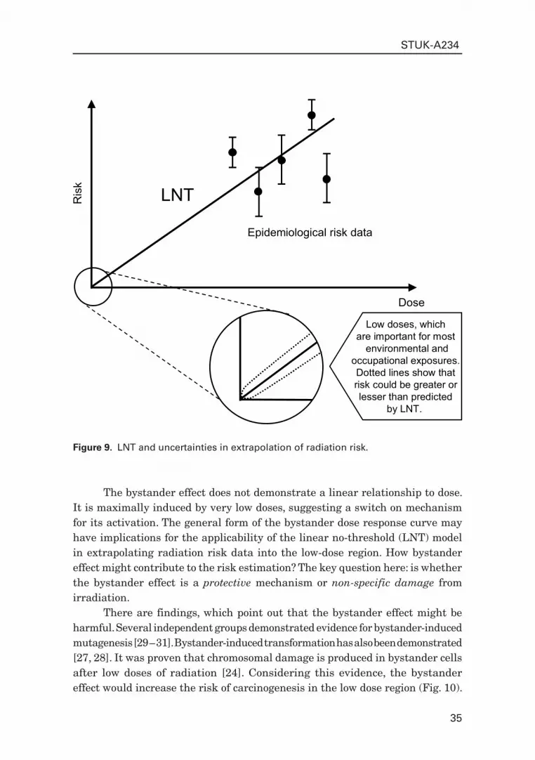
Applicability to radiation protection and contribution to LNT discussionAccording to the Linear-Non-Threshold (LNT) model, which currently dominates in radiation protection, cancer risk for low dose low LET exposures is derived from high-dose epidemiological data, mainly obtained from A-bomb survivors cohort [123]. The average dose of the A-bomb survivors was about 0.3 Gy, which corresponds to about 300 electron tracks at the cellular level (ignoring the very small neutron component) and which were delivered in a short time. Low-dose environmental exposures correspond to around 1 mGy per year of low LET radiation, which is roughly equivalent to 1 electron track per cell per year. The risk at low doses might be different than predicted by a linear extrapolation of the high dose epidemiological data. There is not any reliable epidemiological information in this dose region (Fig. 9).
35
STUK-A234
Figure 9. LNT and uncertainties in extrapolation of radiation risk.
Ris
k
Dose
LNT
Epidemiological risk data
Low doses, whichare important for most
environmental andoccupational exposures.Dotted lines show thatrisk could be greater orlesser than predicted
by LNT.
The bystander effect does not demonstrate a linear relationship to dose. It is maximally induced by very low doses, suggesting a switch on mechanism for its activation. The general form of the bystander dose response curve may have implications for the applicability of the linear no-threshold (LNT) model in extrapolating radiation risk data into the low-dose region. How bystander effect might contribute to the risk estimation? The key question here: is whether the bystander effect is a protective mechanism or non-specific damage from irradiation.
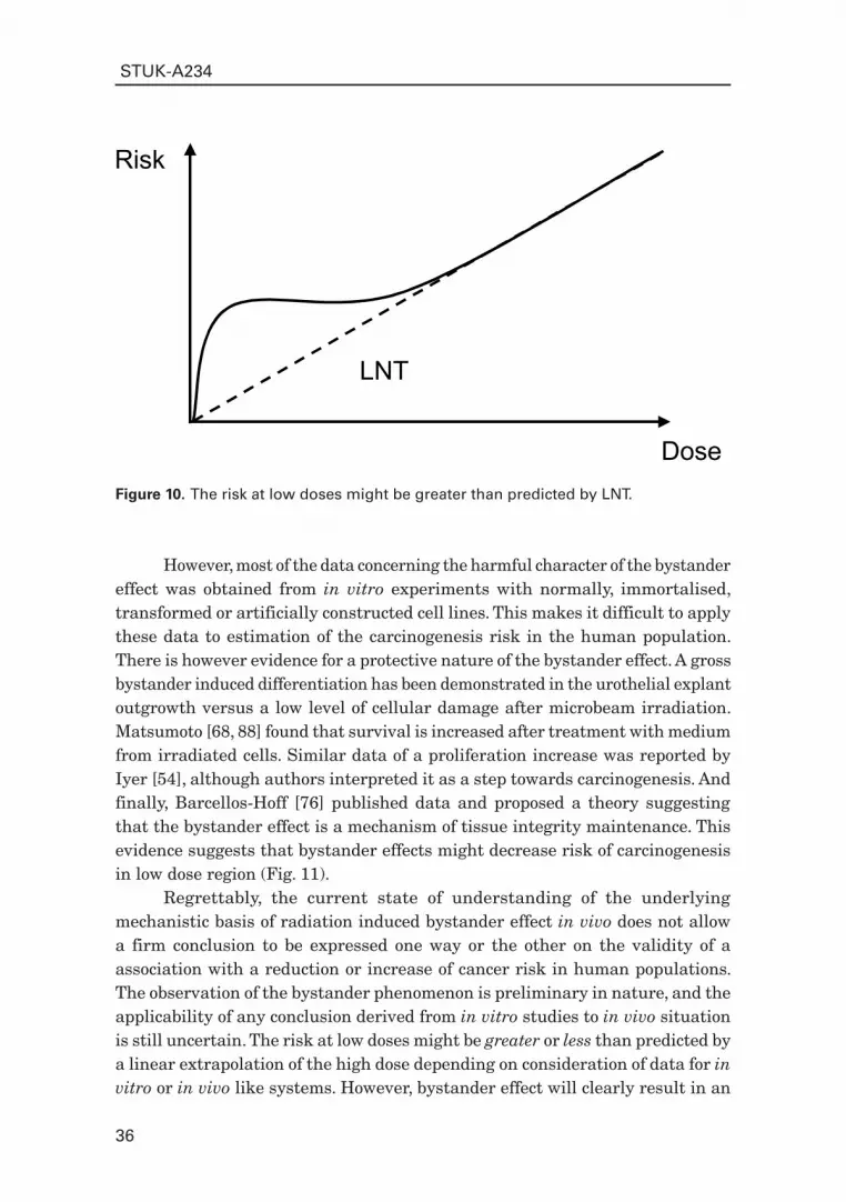
There are findings, which point out that the bystander effect might be harmful. Several independent groups demonstrated evidence for bystander-induced mutagenesis [29 – 31]. Bystander-induced transformation has also been demonstrated [27, 28]. It was proven that chromosomal damage is produced in bystander cells after low doses of radiation [24]. Considering this evidence, the bystander effect would increase the risk of carcinogenesis in the low dose region (Fig. 10).
36
STUK-A234
Figure 10. The risk at low doses might be greater than predicted by LNT.
Risk
Dose
LNT
However, most of the data concerning the harmful character of the bystander effect was obtained from in vitro experiments with normally, immortalised, transformed or artificially constructed cell lines. This makes it difficult to apply these data to estimation of the carcinogenesis risk in the human population. There is however evidence for a protective nature of the bystander effect. A gross bystander induced differentiation has been demonstrated in the urothelial explant outgrowth versus a low level of cellular damage after microbeam irradiation. Matsumoto [68, 88] found that survival is increased after treatment with medium from irradiated cells. Similar data of a proliferation increase was reported by Iyer [54], although authors interpreted it as a step towards carcinogenesis. And finally, Barcellos-Hoff [76] published data and proposed a theory suggesting that the bystander effect is a mechanism of tissue integrity maintenance. This evidence suggests that bystander effects might decrease risk of carcinogenesis in low dose region (Fig. 11).
Regrettably, the current state of understanding of the underlying mechanistic basis of radiation induced bystander effect in vivo does not allow a firm conclusion to be expressed one way or the other on the validity of a association with a reduction or increase of cancer risk in human populations. The observation of the bystander phenomenon is preliminary in nature, and the applicability of any conclusion derived from in vitro studies to in vivo situation is still uncertain. The risk at low doses might be greater or less than predicted by a linear extrapolation of the high dose depending on consideration of data for in vitro or in vivo like systems. However, bystander effect will clearly result in an

37
STUK-A234
Figure 11. The risk at low doses might be less than predicted by LNT.
Risk
Dose
LNT
overall risk, which is a non-linear function of dose. It would be highly premature to consider revising current risk calculations on the basis of current in vitro and in vivo like studies of bystander phenomena. On other hand, the LNT model is important for radiation protection as a simple method to optimise procedures and regulations. However, it should not be mistaken as a scientific model directly derived from the present state of knowledge of the processes involved in radiation carcino genesis [124].
Acknowledgements This work was partially supported by the NOTE IP 036465 (FI6R) and RISC-RAD IP FI6R-CT-2003-508842, Euratom specific programme for research and training on nuclear energy, 6th FP of the EC.
References1. Lea DE. Actions of Radiation on Living Cells. Cambridge: University Press; 1946.2. Marshall M, Gibson JA, Holt PD. An analysis of the target theory of Lea
with modern data. Int J Radiat Biol Relat Stud Phys Chem Med 1970; 18 (2): 127 – 138.
3. Warters RL, Hofer KG. Radionuclide toxicity in cultured mammalian cells. Elucidation of the primary site for radiation-induced division delay. Radiat Res 1977; 69 (2): 348 – 358.

38
STUK-A234
4. Gillies NE. Radiation damage to cell membranes: insights from the oxygen effect. Int J Radiat Biol 1997; 71 (6): 643 – 648.
5. Haimovitz-Friedman A et al. Ionizing radiation acts on cellular membranes to generate ceramide and initiate apoptosis. J Exp Med 1994; 180 (2): 525 – 535.
6. Hall EJ. Radiobiology for the radiologist. 5th ed. Philadelphia: Lippincott Williams & Wilkins; 2000.
7. Ward JF. DNA damage produced by ionizing radiation in mammalian cells: identities, mechanisms of formation, and reparability. Prog Nucleic Acid Res Mol Biol 1988; 35: 95 – 125.
8. Muller WU et al. Micronuclei: a biological indicator of radiation damage. Mutat Res 1996; 366 (2): 163 – 169.
9. Morgan WF et al. Genomic instability induced by ionizing radiation. Radiat Res 1996; 146 (3): 247 – 258.
10. Little JB. Radiation carcinogenesis. Carcinogenesis 2000; 21 (3): 397 – 404.
11. Ward J. New paradigms for Low-Dose Radiation Response in Proceedings of the American Statistical Association Conference on Radiation and Health. San Diego, California, USA. June 14 – 17, 1998. Radiat Res 1999; 151 (1): 92 – 117.
12. Ward J. The radiation-induced lesions which trigger the bystander effect. Mutation Research, Fundamental and Molecular Mechanisms of Mutagenesis 2002; 499 (2): 151 – 154.
13. Iyer R, Lehnert BE. Effects of ionizing radiation in targeted and nontargeted cells. Arch Biochem Biophys 2000; 376 (1): 14 – 25.
14. Wright EG. Radiation-induced genomic instability in haemopoietic cells. Int J Radiat Biol 1998; 74 (6): 681 – 687.
15. Wright EG. Inducible genomic instability: new insights into the biological effects of ionizing radiation. Med Confl Surviv 2000; 16 (1): 117 – 130; discussion 131 – 133.
16. Wolff S. The adaptive response in radiobiology: evolving insights and implications. Environ Health Perspect 1998; 106 Suppl 1: 277 – 283.
17. Joiner MC. et al. Low-dose hypersensitivity: current status and possible mechanisms. Int J Radiat Oncol Biol Phys 2001; 49 (2): 379 – 389.
18. Seymour CB, Mothersill C, Alper T. High yields of lethal mutations in somatic mammalian cells that survive ionizing radiation. Int J Radiat Biol Relat Stud Phys Chem Med 1986; 50 (1): 167 – 179.
19. Amundson SA et al. Induction of gene expression as a monitor of exposure to ionizing radiation. Radiat Res 2001; 156 (5 Pt 2): 657 – 661.
20. Baverstock K,Belyakov OV. Classical radiation biology, the bystander effect and paradigms: a reply. Hum Exp Toxicol 2005; 537 – 542.

39
STUK-A234
21. Deshpande A et al. Alpha-particle-induced sister chromatid exchange in normal human lung fibroblasts: evidence for an extranuclear target. Radiat Res 1996; 145 (3): 260 – 267.
22. Nagasawa H, Little JB. Induction of sister chromatid exchanges by extremely low doses of alpha-particles. Cancer Res 1992; 52 (22): 6394 – 6396.
23. Mothersill C, Seymour C. Medium from irradiated human epithelial cells but not human fibroblasts reduces the clonogenic survival of unirradiated cells. Int J Radiat Biol 1997; 71 (4): 421 – 427.
24. Lorimore SA et al. Chromosomal instability in the descendants of unirradiated surviving cells after alpha-particle irradiation. Proc Natl Acad Sci USA 1998; 95 (10): 5730 – 5733.
25. Watson GE et al. Chromosomal instability in unirradiated cells induced in vivo by a bystander effect of ionizing radiation. Cancer Res 2000; 60 (20): 5608 – 5611.
26. Belyakov OV et al. Direct evidence for a bystander effect of ionizing radiation in primary human fibroblasts. Br J Cancer 2001; 84 (5): 674 – 679.
27. Lewis DA et al. Production of delayed death and neoplastic transformation in CGL1 cells by radiation-induced bystander effects. Radiat Res 2001; 156 (3): 251 – 258.
28. Sawant SG et al. The bystander effect in radiation oncogenesis: I. Transformation in C3H 10T1/2 cells in vitro can be initiated in the unirradiated neighbors of irradiated cells. Radiat Res 2001; 155 (3): 397 – 401.
29. Nagasawa H, Little JB. Unexpected Sensitivity to the Induction of Mutations by Very Low Doses of Alpha-Particle Radiation: Evidence for a Bystander Effect. Radiat Res 1999; 152 (5): 552 – 557.
30. Zhou H et al. Induction of a bystander mutagenic effect of alpha particles in mammalian cells. Proc Natl Acad Sci USA 2000; 97 (5): 2099 – 2104.
31. Zhou H et al. Radiation risk to low fluences of alpha particles may be greater than we thought. Proc Natl Acad Sci USA 2001; 98 (25): 14410 – 14415.
32. Azzam EI et al. Intercellular communication is involved in the bystander regulation of gene expression in human cells exposed to very low fluences of alpha particles. Radiat Res 1998; 150 (5): 497 – 504.
33. Azzam EI, de Toledo SM, Little JB. Direct evidence for the participation of gap junction-mediated intercellular communication in the transmission of damage signals from alpha-particle irradiated to nonirradiated cells. Proc Natl Acad Sci USA 2001; 98 (2): 473 – 478.
34. Azzam EI et al. High and low fluences of alpha-particles induce a G1 checkpoint in human diploid fibroblasts. Cancer Res 2000; 60 (10): 2623 – 2631.
40
STUK-A234
35. Hickman A et al. Alpha-particle-induced p53 protein expression in a rat lung epithelial cell strain. Cancer Res 1994; 54 (22): 5797 – 5800.
36. Hollowell JG Jr, Littlefield LG. Chromosome damage induced by plasma of x-rayed patients: an indirect effect of x-ray. Proc Soc Exp Biol Med 1968; 129 (1): 240 – 244.
37. Littlefield LG, Hollowell JG Jr, Pool WH Jr. Chromosomal aberrations induced by plasma from irradiated patients: an indirect effect of X radiation. Further observations and studies of a control population. Radiology 1969; 93 (4): 879 – 886.
38. Goh K, Sumner H. Breaks in normal human chromosomes: are they induced by a transferable substance in the plasma of persons exposed to total-body irradiation? Radiat Res 1968; 35 (1): 171 – 181.
39. Emerit I. Reactive oxygen species, chromosome mutation, and cancer: possible role of clastogenic factors in carcinogenesis. Free Radic Biol Med 1994; 16 (1): 99 – 109.
40. Emerit I et al. Clastogenic factors in the plasma of Chernobyl accident recovery workers: anticlastogenic effect of Ginkgo biloba extract. Radiat Res 1995; 144 (2): 198 – 205.
41. Emerit I et al. Oxidative stress-related clastogenic factors in plasma from Chernobyl liquidators: protective effects of antioxidant plant phenols, vitamins and oligoelements. Mutat Res 1997; 377 (2): 239 – 246.
42. Emerit I et al. Transferable clastogenic activity in plasma from persons exposed as salvage personnel of the Chernobyl reactor. J Cancer Res Clin Oncol 1994; 120 (9): 558 – 561.
43. Khan MA, Hill RP, Van Dyk J. Partial volume rat lung irradiation: an evaluation of early DNA damage. Int J Radiat Oncol Biol Phys 1998; 40 (2): 467 – 476.
44. Auclair C et al. Clastogenic inosine nucleotide as components of the chromosome breakage factor in scleroderma patients. Arch Biochem Biophys 1990; 278 (1): 238 – 244.
45. Morgan GW, Breit SN. Radiation and the lung: a reevaluation of the mechanisms mediating pulmonary injury. Int J Radiat Oncol Biol Phys 1995; 31 (2): 361 – 369.
46. Ehlers G, Fridman M. Abscopal effect of radiation in papillary adenocarcinoma. Br J Radiol 1973; 46 (543): 220 – 222.
47. Nobler MP. The abscopal effect in malignant lymphoma and its relationship to lymphocyte circulation. Radiology 1969; 93 (2): 410 – 412.
48. Robin HI et al. The abscopal effect: demonstration in lymphomatous involvement of kidneys. Med Pediatr Oncol 1981; 9 (5): 473 – 476.
49. Sham RL. The abscopal effect and chronic lymphocytic leukemia. Am J Med 1995; 98 (3): 307 – 308.

41
STUK-A234
50. Moiseenko VV et al. In-field and out-of-field effects in partial volume lung irradiation in rodents: possible correlation between early dna damage and functional endpoints. Int J Radiat Oncol Biol Phys 2000; 48 (5): 1539 – 1548.
51. Grosovsky AJ. Radiation-induced mutations in unirradiated DNA. Proc Natl Acad Sci USA 1999; 96 (10): 5346 – 5347.
52. Metting NF et al. Design of a benchtop alpha particle irradiator. Health Phys 1995; 68 (5): 710 – 715.
53. Lehnert BE, Goodwin EH. Extracellular factor(s) following exposure to alpha particles can cause sister chromatid exchanges in normal human cells. Cancer Res 1997; 57 (11): 2164 – 2171.
54. Iyer R, Lehnert BE. Factors underlying the cell growth-related bystander responses to alpha particles. Cancer Res 2000; 60 (5): 1290 – 1298.
55. Mothersill C, Seymour CB. Cell-cell contact during gamma irradiation is not required to induce a bystander effect in normal human keratinocytes: evidence for release during irradiation of a signal controlling survival into the medium. Radiat Res 1998; 149 (3): 256 – 262.
56. Mothersill C, Seymour C. Radiation-induced bystander effects: past history and future directions. Radiat Res 2001; 155 (6): 759 – 767.
57. Mothersill C et al. Individual variation in the production of a ‘bystander signal’ following irradiation of primary cultures of normal human urothelium. Carcinogenesis 2001; 22 (9): 1465 – 1471.
58. Seymour CB, Mothersill C. Relative contribution of bystander and targeted cell killing to the low-dose region of the radiation dose-response curve. Radiat Res 2000; 153 (5 Pt 1): 508 – 511.
59. Puck TT, Marcus P. Action of X-ray on mammalian cells. Journal of Experimental Medicine 1956; 103: 653 – 666.
60. Bishayee A et al. Protection by DMSO against cell death caused by intracellularly localized iodine-125, iodine-131 and polonium-210. Radiat Res 2000; 153 (4): 416 – 427.
61. Bishayee A, Rao DV, Howell RW. Evidence for pronounced bystander effects caused by nonuniform distributions of radioactivity using a novel three-dimensional tissue culture model. Radiat Res 1999; 152 (1): 88 – 97.
62. Korystov Yu N et al. Intercellular interactions in the interphase death of irradiated thymocytes. Radiat Res 1993; 134 (3): 301 – 306.
63. Manti L et al. Genomic instability in Chinese hamster cells after exposure to X rays or alpha particles of different mean linear energy transfer. Radiat Res 1997; 147 (1): 22 – 28.
64. Prise KM et al. Studies of bystander effects in human fibroblasts using a charged particle microbeam. Int J Radiat Biol 1998; 74 (6): 793 – 798.

42
STUK-A234
65. Belyakov OV et al. Direct evidence for a bystander effect of ionizing radiation in primary human fibroblasts. Br J Cancer 2001; 84 (5): 674 – 679.
66. Wu LJ et al. Targeted cytoplasmic irradiation with alpha particles induces mutations in mammalian cells. Proc Natl Acad Sci USA 1999; 96 (9): 4959 – 4964.
67. Sigg M, Crompton NE, Burkart W. Enhanced neoplastic transformation in an inhomogeneous radiation field: an effect of the presence of heavily damaged cells. Radiat Res 1997; 148 (6): 543 – 547.
68. Matsumoto H et al. Induction of radioresistance by a nitric oxide-mediated bystander effect. Radiat Res 2001; 155 (3): 387 – 396.
69. Michael B et al. Implications of bystander effects for radiobiology. In: Proceedings of 11th ICRR. Dublin; 2000.
70. Brenner DJ, Little JB, Sachs RK. The bystander effect in radiation oncogenesis: II. A quantitative model. Radiat Res 2001; 155 (3): 402 – 408.
71. Hall EJ. Personal communication. 2001.72. Little MP et al. A model for radiation-induced bystander effects, with
allowance for spatial position and the effects of cell turnover. J Theor Biol 2005; 232 (3): 329 – 338.
73. Belyakov OV et al. A proliferation-dependent bystander effect in primary porcine and human urothelial explants in response to targeted irradiation. Br J Cancer 2003; 88 (5): 767 – 774.
74. Mothersill C et al. Involvement of energy metabolism in the production of ‘bystander effects’ by radiation. Br J Cancer 2000; 82 (10): 1740 – 1746.
75. Barcellos-Hoff MH. How do tissues respond to damage at the cellular level? The role of cytokines in irradiated tissues. Radiat Res 1998; 150 (5 Suppl): S109 – S120.
76. Barcellos-Hoff MH, Brooks AL. Extracellular signaling through the microenvironment: a hypothesis relating carcinogenesis, bystander effects, and genomic instability. Radiat Res 2001; 156 (5 Pt 2): 618 – 627.
77. Trosko JE, Chang CC. Mechanism of up-regulated gap junctional intercellular communication during chemoprevention and chemotherapy of cancer. Mutat Res 2001; 480 – 481: 219 – 229.
78. Lehnert BE, Goodwin EH. A new mechanism for DNA alterations induced by alpha particles such as those emitted by radon and radon progeny. Environ Health Perspect 1997; 105 (Suppl 5): 1095 – 1101.
79. Koyama S et al. Radiation-induced long-lived radicals which cause mutation and transformation. Mutat Res 1998; 421 (1): 45 – 54.
80. Watanabe M et al. Radioprotective effects of dimethyl sulfoxide in golden hamster embryo cells exposed to gamma rays at 77 K. II. Protection

43
STUK-A234
from lethal, chromosomal, and DNA damage. Radiat Res 1990; 124 (1): 73 – 78.
81. Prise KM. Personal communication. 2002.82. Lyng FM, Seymour CB, Mothersill C. Oxidative stress in cells exposed to low
levels of ionizing radiation. Biochem Soc Trans 2001; 29 (Pt 2): 350 – 353.83. Lehnert S. Modification of postirradiation survival of mammalian cells by
intracellular cyclic AMP. Radiat Res 1975; 62 (1): 107 – 116.84. Lehnert BE et al. Temporal sequence of early alterations in rat lung
following thoracic X-irradiation. Int J Radiat Biol 1991; 60 (4): 657 – 675.85. Albanese J, Dainiak N. Ionizing radiation alters Fas antigen ligand at
the cell surface and on exfoliated plasma membrane-derived vesicles: implications for apoptosis and intercellular signaling. Radiat Res 2000; 153 (1): 49 – 61.
86. Ramesh R et al. In vivo analysis of the ‘bystander effect’: a cytokine cascade. Exp Hematol 1996; 24 (7): 829 – 838.
87. Narayanan PK, Goodwin EH, Lehnert BE. Alpha particles initiate biological production of superoxide anions and hydrogen peroxide in human cells. Cancer Res 1997; 57 (18): 3963 – 3971.
88. Matsumoto H et al. Induction of radioresistance to accelerated carbon-ion beams in recipient cells by nitric oxide excreted from irradiated donor cells of human glioblastoma. Int J Radiat Biol 2000; 76 (12): 1649 – 1657.
89. Belyakov OV et al. Delayed lethality, apoptosis and micronucleus formation in human fibroblasts irradiated with X-rays or alpha-particles. Int J Radiat Biol 1999; 75 (8): 985 – 993.
90. Belyakov OV et al. Studies of bystander effects in primary urothelial cells using a charged particle microbeam. In Proceedings of the 4th International Workshop: Microbeam Probes of Cellular Radiation Response. Radiation Research 2000; 153 (2): 220.
91. Belyakov OV et al. Bystander-induced apoptosis and premature differentiation in primary urothelial explants after charged particle microbeam irradiation. Radiation Protection Dosimetry 2002; 99 (1 – 4): 249 – 251.
92. Limoli CL et al. A role for chromosomal instability in the development of and selection for radioresistant cell variants. Br J Cancer 2001; 84 (4): 489 – 492.
93. Pollycove M. Nonlinearity of radiation health effects. Environ Health Perspect 1998; 106 Suppl 1: 363 – 368.
94. Pollycove M, Feinendegen LE. Molecular biology, epidemiology, and the demise of the linear no-threshold (LNT) hypothesis. C R Acad Sci III 1999; 322 (2 – 3): 197 – 204.

44
STUK-A234
95. Watson GE et al. In vivo chromosomal instability and transmissible aberrations in the progeny of haemopoietic stem cells induced by high- and low-LET radiations. Int J Radiat Biol 2001; 77 (4): 409 – 417.
96. Neriishi K, Nakashima E, Delongchamp RR. Persistent subclinical inflammation among A-bomb survivors. Int J Radiat Biol 2001; 77 (4): 475 – 482.
97. Limoli CL et al. Genomic instability induced by high and low LET ionizing radiation. Adv Space Res 2000; 25 (10): 2107 – 2117.
98. Trosko JE. Hierarchical and cybernetic nature of biologic systems and their relevance to homeostatic adaptation to low-level exposures to oxidative stress-inducing agents. Environ Health Perspect 1998; 106 Suppl 1: 331 – 339.
99. Bird AP. The relationship of DNA methylation to cancer. Cancer Surv 1996; 28: 87 – 101.
100. Prise KM et al. Non-targeted effects of radiation: bystander responses in cell and tissue models. Radiat Prot Dosimetry 2002; 99 (1 – 4): 223 – 226.
101. Cummins RJ et al. The effect of microcolony size, at time of irradiation, on colony forming ability. Int J Radiat Biol 1999; 75 (2): 225 – 232.
102. Jen Y, West C, Hendry J. The lower radiosensitivity of mouse kidney cells irradiated in vivo than in vitro: a cell contact effect phenomenon. Int J Radiat Oncol Biol Phys 1991; 20 (6): 1243 – 1248.
103. Belyakov OV et al. Biological effects in unirradiated human tissue induced by radiation damage up to 1 mm away. Proc Natl Acad Sci USA 2005; 102 (40): 14203 – 14208.
104. Mancuso M et al. Oncogenic bystander radiation effects in Patched heterozygous mouse cerebellum. Proc Natl Acad Sci USA 2008; 105 (34): 12445 – 124450.
105. Prise KM et al. Investigating the cellular effects of isolated radiation tracks using microbeam techniques. Adv Space Res 2002; 30 (4): 871 – 876.
106. Folkard M et al. The impact of microbeams in radiation biology. Nuclear Instruments and Methods in Physics Research, Section B, Beam Interactions with Materials and Atoms (NIMB) 2001; 181: 426 – 430.
107. Belyakov OV et al. Bystander-induced differentiation: A major response to targeted irradiation of a urothelial explant model. Mutation Research/Fundamental and Molecular Mechanisms of Mutagenesis 2006; 597 (1 – 2): 43 – 49.
108. Lehnert BE et al. Low Dose Ionizing Radiation-Induced Effects in Irradiated and Unirradiated Cells: Pathways Analysis in Support of Risk Assessment in the research Abstracts from the DOE Human Genome Program Contractor-Grantee Workshop IX, January 27 – 31, 2002 Oakland, CA.
45
STUK-A234
2002 [cited; Available from: http://www.ornl.gov/hgmis/publicat/02santa/lowdose_infra.html#195.]
109. Belyakov OV et al. Bystander effect and genomic instability. Challenging the classic paradigm of radiobiology. In: Modern problems of Radiobiology, Radioecology and Evolution. Proceedings of the International conference dedicated to the centenary of the birth of N.W. Timofeeff-Ressovsky. Dubna, Russia 6 – 9 September, 2000. 2001; Dubna: JINR.
110. Yeager TR et al. Overcoming cellular senescence in human cancer pathogenesis. Genes Dev 1998; 12 (2): 163 – 174.
111. Baverstock K. Radiation-induced genomic instability: a paradigm-breaking phenomenon and its relevance to environmentally induced cancer. Mutation Research / Fundamental and Molecular Mechanisms of Mutagenesis 2000; 454 (1 – 2): 89 – 109.
112. Belyakov OV et al. A proliferation-dependent bystander effect in primary porcine and human urothelial explants in response to targeted irradiation. Br J Cancer 2003; 88 (5): 767 – 774.
113. Brenner D, Sachs RK. Do low dose-rate bystander effects influence domestic radon risks? Int J Radiat Biol; 2002. Accepted.
114. Blakely EA. Biological effects of cosmic radiation: deterministic and stochastic. Health Phys 2000; 79 (5): 495 – 506.
115. De Angelis G et al. Health risks from radiation exposure for civilian aviation flight personnel: a study of Italian airline crew members. Radiat Res 2001; 156 (5 Pt 2): 689 – 694.
116. Hall EJ. Radiation, the two-edged sword: cancer risks at high and low doses. Cancer J 2000; 6 (6): 343 – 350.
117. Trott KR, Kamprad F. Estimation of cancer risks from radiotherapy of benign diseases. Strahlenther Onkol 2006; 182 (8): 431 – 436.
118. Thomlinson W et al. Research at the European Synchrotron Radiation Facility medical beamline. Cell Mol Biol (Noisy-le-grand) 2000; 46 (6): 1053 – 1063.
119. Stepanek J et al. Physics study of microbeam radiation therapy with PSI-version of Monte Carlo code GEANT as a new computational tool. Med Phys 2000; 27 (7): 1664 – 1675.
120. Laissue JA et al. Neuropathology of ablation of rat gliosarcomas and contiguous brain tissues using a microplanar beam of synchrotron-wiggler-generated X rays. Int J Cancer 1998; 78 (5): 654 – 660.
121. Slatkin DN et al. Microbeam radiation therapy. Med Phys 1992; 19 (6): 1395 – 1400.
122. Beere HM, Hickman JA. Differentiation: a suitable strategy for cancer chemotherapy? Anticancer Drug Des 1993; 8 (4): 299 – 322.

46
STUK-A234
123. Kellerer AM. Risk estimates for radiation-induced cancer--the epidemiological evidence. Radiat Environ Biophys 2000; 39 (1): 17 – 24.
124. Trott KR, Rosemann M. Molecular mechanisms of radiation carcinogenesis and the linear, non-threshold dose response model of radiation risk estimation. Radiat Environ Biophys 2000; 39 (2): 79 – 87.
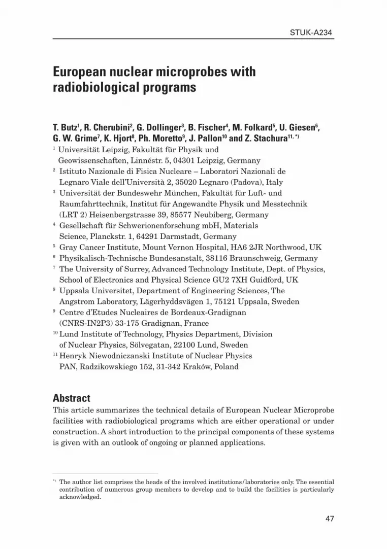
47
STUK-A234
European nuclear microprobes with radiobiological programs
T. Butz1, R. Cherubini2, G. Dollinger3, B. Fischer4, M. Folkard5, U. Giesen6, G. W. Grime7, K. Hjort8, Ph. Moretto9, J. Pallon10 and Z. Stachura11, *)
1 Universität Leipzig, Fakultät für Physik und Geowissenschaften, Linnéstr. 5, 04301 Leipzig, Germany
2 Istituto Nazionale di Fisica Nucleare – Laboratori Nazionali de Legnaro Viale dell’Università 2, 35020 Legnaro (Padova), Italy
3 Universität der Bundeswehr München, Fakultät für Luft- und Raumfahrttechnik, Institut für Angewandte Physik und Messtechnik (LRT 2) Heisenbergstrasse 39, 85577 Neubiberg, Germany
4 Gesellschaft für Schwerionenforschung mbH, Materials Science, Planckstr. 1, 64291 Darmstadt, Germany
5 Gray Cancer Institute, Mount Vernon Hospital, HA6 2JR Northwood, UK6 Physikalisch-Technische Bundesanstalt, 38116 Braunschweig, Germany7 The University of Surrey, Advanced Technology Institute, Dept. of Physics,
School of Electronics and Physical Science GU2 7XH Guidford, UK8 Uppsala Universitet, Department of Engineering Sciences, The
Angstrom Laboratory, Lägerhyddsvägen 1, 75121 Uppsala, Sweden9 Centre d’Etudes Nucleaires de Bordeaux-Gradignan
(CNRS-IN2P3) 33-175 Gradignan, France10 Lund Institute of Technology, Physics Department, Division
of Nuclear Physics, Sölvegatan, 22100 Lund, Sweden11 Henryk Niewodniczanski Institute of Nuclear Physics
PAN, Radzikowskiego 152, 31-342 Kraków, Poland
AbstractThis article summarizes the technical details of European Nuclear Microprobe facilities with radiobiological programs which are either operational or under construction. A short introduction to the principal components of these systems is given with an outlook of ongoing or planned applications.
*) The author list comprises the heads of the involved institutions / laboratories only. The essential contribution of numerous group members to develop and to build the facilities is particularly acknowledged.

48
STUK-A234
1 IntroductionThe investigation of biological effects due to ionising radiation is usually carried out with broadbeam X-ray or γ-ray or alpha-sources or ion accelerators. In these experiments the interaction point between the photon or ion and tissue or a cell, respectively, is not known (except for in-situ ion track detection behind the cells) nor controllable. Since cells communicate with each other, a superposition of various interaction points renders the interpretation of data difficult. Most of the targeted irradiations were performed thus far on isolated cells, sometimes also on tissue. In the following, only isolated cells will be considered. Furthermore, the discussion is restricted to ions. In order to distinguish between an interaction of the incident particle with the cell nucleus, the cytoplasm, or the media, collimated or focused beams are required with beam diameters in the m-range or below. Such systems are usually called “Nuclear Microprobes”. For work with living cells the beam has to be extracted from vacuum to the ambient (“external beams”). In addition, very efficient cell-recognition procedures are required in order to position the target exactly in front of the beam or to direct the beam exactly to the target. If such irradiation facilities are equipped with a fast beam-switch, an exactly counted number of particles – from one to an arbitrary number – can be placed at the desired target.
This new approach in radiobiological research allows to address questions such as the investigation of the spatially resolved radiation sensitivity of cells, cell-to-cell communication and bystander-effects, DNA-damage without hitting the cell nucleus and many more. The pioneering institutions in this field of research are the Texas A&M University, USA, the Columbia University, USA and the Gray Cancer Institute, UK. In recent years, many more Nuclear Microprobe institutions started radiobiological programs in Japan, in the USA and – above all – in Europe. Most of the European institutions collaborate within the Marie Curie Research and Training Network CELLION (6th framework program of the European Community) and thus guarantee the education and training of young researchers in this multidisciplinary field of research. Admittedly, most institutions / laboratories are not yet fully operational. Whereas large research institutions in general have resources to develop their infrastructure, Universities depend more heavily on funding via research projects which is particularly difficult in the start phase.
This article summarizes technical details of European Nuclear Microprobe facilities with radiobiological programs which are either operational or under construction. A short introduction to the principal components of these systems is given with an outlook of ongoing or planned applications. In some cases, reference is made to the installation at the Columbia University, USA. Instead of giving an extensive reference list with scattered pieces of information here and there
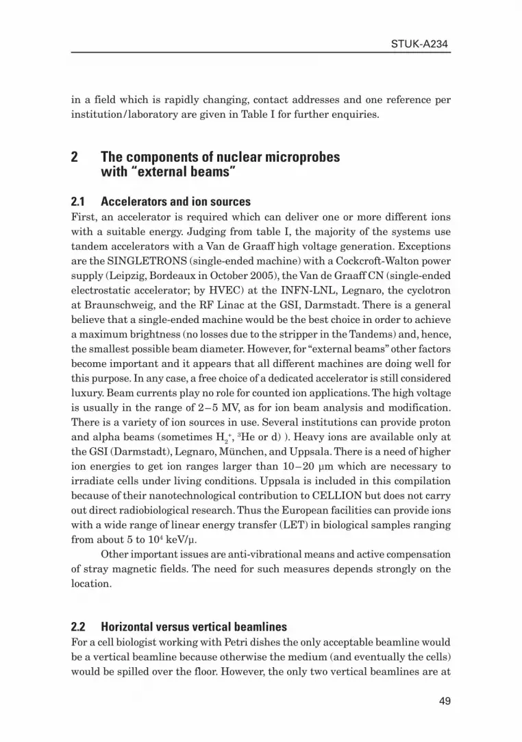
49
STUK-A234
in a field which is rapidly changing, contact addresses and one reference per institution / laboratory are given in Table I for further enquiries.
2 The components of nuclear microprobes with “external beams”
2.1 Accelerators and ion sourcesFirst, an accelerator is required which can deliver one or more different ions with a suitable energy. Judging from table I, the majority of the systems use tandem accelerators with a Van de Graaff high voltage generation. Exceptions are the SINGLETRONS (single-ended machine) with a Cockcroft-Walton power supply (Leipzig, Bordeaux in October 2005), the Van de Graaff CN (single-ended electrostatic accelerator; by HVEC) at the INFN-LNL, Legnaro, the cyclotron at Braunschweig, and the RF Linac at the GSI, Darmstadt. There is a general believe that a single-ended machine would be the best choice in order to achieve a maximum brightness (no losses due to the stripper in the Tandems) and, hence, the smallest possible beam diameter. However, for “external beams” other factors become important and it appears that all different machines are doing well for this purpose. In any case, a free choice of a dedicated accelerator is still considered luxury. Beam currents play no role for counted ion applications. The high voltage is usually in the range of 2 – 5 MV, as for ion beam analysis and modification. There is a variety of ion sources in use. Several institutions can provide proton and alpha beams (sometimes H2
+, 3He or d) ). Heavy ions are available only at the GSI (Darmstadt), Legnaro, München, and Uppsala. There is a need of higher ion energies to get ion ranges larger than 10 – 20 µm which are necessary to irradiate cells under living conditions. Uppsala is included in this compilation because of their nanotechnological contribution to CELLION but does not carry out direct radiobiological research. Thus the European facilities can provide ions with a wide range of linear energy transfer (LET) in biological samples ranging from about 5 to 104 keV/ µ.
Other important issues are anti-vibrational means and active compensation of stray magnetic fields. The need for such measures depends strongly on the location.
2.2 Horizontal versus vertical beamlinesFor a cell biologist working with Petri dishes the only acceptable beamline would be a vertical beamline because otherwise the medium (and eventually the cells) would be spilled over the floor. However, the only two vertical beamlines are at
50
STUK-A234
the GCI, London, and at the PTB, Braunschweig. The first one comes upwards from the cellar whereas the latter one is downwards (!). The majority of the institutions uses horizontal beamlines for a number of reasons: frequently one uses cells which adhere to the bottom of the Petri dish and a lack of adhesion is used as indicator for cell death; furthermore, often the medium is removed anyway in order to avoid irradiation and radiolysis in the medium and a minimum film covering the cells – eventually by exposing them to humidified air – is usually sufficient to keep them alive for a period of say 10 minutes or so. So, e.g. Legnaro, Leipzig and München do not use transmission detectors before the Petri dish but rather behind it and cannot tolerate thicker media layers. Interestingly enough, at the Columbia University a vertical beamline is used and the medium is removed nevertheless.
2.3 Beam formationThere are two possibilities for the production of beams with µm diameters: collimation and focussing. In the first case, the beam passes though a pinhole or a capillary which actually defines the beamsize (and eventually divergence). The target has to be scanned over the beam. In the latter case, the beam passes through object and aperture slits (defining beam diameter and divergence) and is subsequently demagnified by quadrupole lenses. In this way, minimum beam diameters of below 50 nm were achieved in vacuum. Usually, with focussed beams a scanning unit (magnetic or electrostatic) is used to allow for scanning the beam over the target. An advantage of beam scanning compared to mechanical sample scanning is speed. Scanning devices can be placed before or after the last lens. In the first case, the working distance can be rather small, the disadvantage being a greater influence of lens aberrations for non-paraxial rays.
Since the beam has to be extracted to the ambient, further factors like the ion exit window, (often used as transmission detector, see below), air gap between exit window and bottom of Petri dish, as well as Petri dish entrance window deteriorate the beam diameter and, hence, the hit accuracy. Reported accuracies in air are between 1 – 3 µm (collimated systems) and below 500 nm (Leipzig and München, focussed systems, relative accuracies). These are usually obtained by single (or few) ion(s) bombardment of a radiation sensitive polymer like CR39 with which single ion tracks can be visualized by etching.
Several groups are using various codes for ion beam optics. Bordeaux uses a “fully integrated” ray tracing approach which includes beamline, windows and cells / media.
Another important component is a fast beam switch or beam blanker, usually electrostatic. After the detection of a single particle, the beam blanker
51
STUK-A234
must be able to “shut” the beamgate fast enough that no further ion hits the sample at the very same position. Since usually 1000 – 3000 ions / s are used for single hit applications, this does not represent a real challenge with hardware pulses and high-voltage supplies with fast risetimes.
2.4 Exit windowsIn principle pinholes and differential pumping is possible to extract the beam to the ambient. However, an air-jet enters the beamline with ultrasonic velocities and, hence, renders this method impractical for applications with Petri dishes as close as possible to the hole. Therefore, all institutions use vacuum-tight windows consisting of suitable polymers (e.g. mylar, kapton) or Si3N4. It is interesting to note that a 1mm × 1mm window of Si3N4 with a thickness of 100 nm only can withstand a pressure difference of 1 atmosphere. Unfortunately, with such an exit window the area of the Petri dish which can be irradiated without sample translation stage will be 1 mm x 1mm at best only. With polymer foils, larger exit windows are feasible. One should keep in mind that any window material bends towards the vacuum chamber leaving an air gap between ion exit window and entrance window of the Petri dish. An air gap of about 75 – 100 µm does deteriorate the beam quality. Therefore the München group works with an air gap < 25 µm.
A nearby fast automatic valve should be provided to prevent ventilation of the beamline (and the accelerator tube) in case of rupture of the ion exit window.
2.5 Ion detectors and on-line microscopyWith the exceptions of Legnaro, Leipzig, München and the Columbia University all institutions install transmission detectors in front of the Petri dish, despite the deterioration of the beam quality. A common technique is the use of a plastic scintillator (in combination with a photomultiplier behind the cells. The München group uses a plastic scintillator after the Petri dish while the Legnaro group uses a silicon diode, an arrangement which requires higher ion energies in order not to suffer from the macroscopic amount of material (air, Petri dish exit window) between cells and detector. A disadvantage is that the on-line microscope (München) has to be retracted for particle detection. At the Columbia University, a gas proportional counter was built into the objective of the on-line microscope. A miniaturized low pressure gas proportional counters has been developed as transmission detector in Bordeaux and is used in routine conditions. The signal to background ratio is sufficient to allow 100% efficiency for MeV

52
STUK-A234
protons and alphas with a very low level of “false positives”. The only drawback is a tail in the lateral distribution of the beam due to multiple scattering on gas molecules. Studies of semiconductor devices as transmission detectors are going on at Lund, but are not yet satisfactory. Several groups are using Si3N4 exit windows which are coated with a thin layer of CsI and Au (to prevent charging up) and use secondary electrons and an upstream channeltron as transmission detectors. Doped diamond windows were tried as well. Such devices work very well for ions heavier than protons. A satisfactory transmission detector for 2 – 3 MeV protons does not exist yet. By satisfactory we mean that every proton is detected with close to 100% efficiency and that no “false-positive” signals are produced in the absence of transmitted protons. A level below 1% false-positives is desirable. To place a downstream particle detector behind the cells with about 100% efficiency requires the removal of media but poses no serious constraints to the distance between cells and detector because the lateral straggling in air is of minor importance after cell traversal. Here, a retractable on-line microscope is required, too; alternatively, cells could be observed on-line with a long-distance or retractable microscope through the ion exit window towards downstream, as done at Leipzig. Some institutions are equipped with on-line UV illumination for better recognition of stained cells (stains for the cell nucleus or the cytoplasm or other).
2.6 Petri dishesThere is a whole variety of Petri dish designs. Common to all is the requirement of a thin entrance window, usually a polymer foil. Leipzig uses a Si3N4 window. Cells are plated on the other side of the window. Thus the material must be biocompatible and cells must adhere, at least for the horizontal beamlines. Typical dimensions of the entrance window range from 2 mm × 2 mm to 30 mm in diameter. In general, cells are plated all over the Petri dish bottom, not only over the entrance window. This raises questions about possible differences between cells residing on different materials. Some vertical Petri dishes are equipped with exit windows as well. Within the CELLION project “intelligent” Petri dishes using nanotechnology will be developed which could eventually result in cell arrays or even cells on electrodes.
2.7 Target translation stagesFor collimated beams a target translation stage is indispensable. However, due to the small size of common exit windows, a target translation stage is also advisable for focussed beams. Computer controlled x-, y- (and -z) translation

53
STUK-A234
stages with µm precision or better are commercially available or can be custom-made. Precision is one issue, reproducibility another. Speed is also important for rapid scanning over thousands of cells. For this purpose voice-coil devices proved useful. Thus far, no sub-micrometer intracellular targets other than the nucleus were aimed at due to the limited hit accuracy. Hence, the existing translation stages are good enough at present.
2.8 Automatic cell recognitionThe standard procedure is to use an off-line microscope to automatically register the cell nuclei positions of stained cells. The x- and y-coordinates are then used for automatic target translation movements or automatic beam scanning. Some institutions have such systems already installed. The need of stains always raises the question to what extent the stress which the cells suffer from the stain and the UV illumination might spoil the low dose results. The Legnaro group already developed a semi-automatic cell visualization, recognition and re-visiting system based on an inverted phase contrast optical microscope equipped with a CCD camera and on a dedicated software, without using any fluorescent staining or UV light. The Krakow group is working on an automatic cell recognition system which does not require stains and UV illumination. It uses three microscopy images, one in focus and two above / below focus and exploits the phase information.
An obvious difficulty results from the requirement of absolute x- and y-coordinates. A reference or fiducial marker would be advantageous. However, tests with predetermined target positions in CR39 which are subsequently revisited with ions turned out useful for hit verification tests, too. Image overlay procedures for off- and on-line images would be useful and are under construction in several laboratories. For studies where a very high throughput of cells is required, an automatic cell recognition system is indispensable, contrary to the case where one or a few cells are targeted only.
3 Cell types and endpointsSince this is a compilation of technical aspects, information on radiobiological programs will be kept rather short. A few comments, however, are appropriate to see where we are going. The GCI, London investigated by far the largest variety of cells. Thus far it is also the only institution which started studies of tissue. In most other laboratories which have started with single ion irradiation of living cells the choice of cells was dictated more by availability, ease of cell culturing, sometimes size of cells, adhesion properties and alike. As soon as routine work begins, a careful selection of cells is advisable. As far as possible endpoints are
54
STUK-A234
concerned, cell survival experiments and bystander effect studies in order to catch up with existing literature (time consuming) are required. Apoptosis, DNA damage and repair mechanisms and dynamics, protein expression, stress markers are further possibilities, just to mention a few. An interesting “technical” aspect is the possibility to visualize tracks of individual ions using double-strand break markers (“foci”). Since the cells stay alive upon traversal of a single ion (or even more ions), any deviation from a straight trajectory in 3D-images taken with laser scanning confocal microscopes hours later is due to intranuclear dynamics, as shown by the München group.
In order to discriminate true foci from spurious ones, several groups started to irradiate patterns, e.g. a cross consisting of 5 particles, or equidistant lines of points, which can be easily identified. The GSI, Darmstadt group did so with targeted irradiation whereas Leipzig and München “wrote” regular patterns all over a certain area of the Petri dish. It is astonishing how much new information can be extracted from such patterned irradiation. Thus far, 2.25 MeV protons did not lead to a clearly recognizable pattern of foci whereas alpha particles seem to work.
4 OutlookAmong radiation biologists a frequently encountered objection against the use of nuclear microprobes is the statement that such studies are carried out using molecular biology nowadays. This statement is misleading because molecular biology techniques are most useful and welcome, but do not eliminate the urgent need for targeted irradiation at the single particle level. In other words, very refined tools to introduce controlled damage with subcellular accuracy must be combined with state of the art detection schemes for the cellular response. The rapid grows of the European nuclear microprobe community with radiobiological programs is encouraging and parallels worldwide efforts. It is of utmost importance to understand the cellular response to ionising radiation in the low-dose regime, both for radiation protection issues as well as for the development of better radiation therapies.
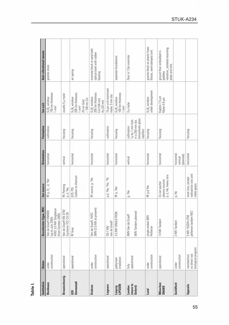
55
STUK-A234
Tab
le I.
Inst
itutio
nSt
atus
Acc
eler
ator
(typ
e, M
V)Io
n so
urce
Ori
enta
tion
Form
atio
nIo
n ex
it A
nti-
vibr
atio
nal m
eans
Bor
deau
xun
der
Van
de G
raaf
f KN
4000
RF
p, H
2+ , d, 4 He
+ ho
rizon
tal
colli
mat
ion
Si3N
4 win
dow
gr
anite
sto
neco
nstru
ctio
n(u
p to
Jul
y 20
05)
150
nm th
ickn
ess
HVEE
3.5
MV
Sing
letro
n1
mm
2
(from
Oct
ober
200
5)
Bra
unsc
hwei
gop
erat
iona
lVa
n de
Gra
aff K
N 3
750
RF, P
enni
ngve
rtica
lfo
cusi
ngus
ually
5 µ
myl
ar
Cycl
otro
n TC
C CV
-28
p, d
, 4 He
GSI
op
erat
iona
lRF
lina
c EC
R, P
IGho
rizon
tal
focu
sing
Si
3N4 w
indo
wai
r spr
ing
Dar
mst
adt
Carb
on to
Ura
nium
200
nm th
ickn
ess
1 m
m2
20 n
m G
old
~ 10
0 nm
CsI
Krak
owun
der
Van
de G
raaf
f, HV
ECRF
sou
rce,
p, 4 He
ho
rizon
tal
focu
sing
Si3N
4 win
dow
conc
rete
blo
ck in
san
d ba
thco
nstru
ctio
n3M
V (2
.5 M
V at
pre
sent
)20
0 nm
thic
knes
sop
tical
ban
k w
ith ru
bber
Cs
I (10
0 nm
)ba
ckin
g Au
(20
nm)
Legn
aro
oper
atio
nal
CN 7
MV
p,d,
3 He, 4 He
, 15N
horiz
onta
lco
llim
atio
n10
µm
a-b
i-lum
inize
dva
n de
Gra
aff
myl
ar, 5
mm
dia
.
Leip
zig
patte
rned
3.5
MV
SIN
GLET
RON
RF p
, 4 He+
horiz
onta
lfo
cusi
ngSi
3N4 w
indo
wse
para
te fo
unda
tions
LIPS
ION
irrad
iatio
n10
0 nm
thic
knes
s1
mm
2
Lond
onfu
lly4M
V Va
n de
Gra
aff
p, 3 He
verti
cal
colli
mat
ion
3 µ
myl
arflo
or is
1.5
m c
oncr
ete
(mov
e to
oper
atio
nal
1µ b
y 1
mm
hol
e O
xfor
d)5M
V Ta
ndem
pla
nned
in a
250
mm
dia
.bo
rosi
licat
e gl
ass
capi
llary
Lund
unde
rsi
ngle
-end
ed 3
MV
RF p
,d,4 He
horiz
onta
lfo
cusi
ngSi
3N4 w
indo
wgr
anite
blo
ck o
n pl
astic
foam
cons
truct
ion
Pelle
tron
unde
r dev
elop
men
tbl
ocks
, san
d-da
mpe
d co
nt.
Mün
chen
oper
atio
nal
14 M
V Ta
ndem
Cs-io
n sp
utte
rho
rizon
tal
focu
sing
Kapt
on 7
.5 µ
m
grou
nd fl
oor e
mbe
dded
in
SNA
KE
plas
ma
sour
ces
Myl
ar 0
.9 µ
m
pebb
les
p, d
, He,
hea
vy io
nsvi
brat
ion_
isol
ated
mou
ntin
g
plat
e an
d sl
its
Gui
ldfo
rdun
der
2 M
V Ta
ndem
p, H
eho
rizon
tal
cons
truct
ion
verti
cal
(pla
nned
)
Upp
sala
nano
tech
nol.
5 M
V 15
SDH-
2TM
m
ost i
ons,
exc
ept
horiz
onta
lfo
cusi
ngno
dire
ct ra
d.-
pelle
tron
tand
em N
ECra
dioa
ctiv
e io
ns a
ndbi
olog
ical
pro
gram
nobl
e ga
ses

56
STUK-A234
Tab
le I
(co
nti
nu
ed).
Inst
itutio
nA
nti-
mag
netic
str
ay fi
elds
mea
nsO
ptic
s co
deRe
mar
ks a
ccel
lera
tor
Scan
ning
Ion
tran
smis
sion
det
ecto
r
Bor
deau
xIro
n sh
ield
ing
for s
ome
parts
of
TRAN
SPOR
T, TR
AXDe
tect
or u
pstre
am th
at a
llow
s irr
adia
tion
with
cel
ls im
mer
sed
in m
ediu
m
targ
et
prop
ortio
nal c
ount
erth
e be
amlin
eGE
ANT
IV, Z
GOUB
IEl
ectro
stat
ic b
eam
bla
nkin
g (p
erfo
rman
ce w
ithin
500
ns)
isob
uten
e at
low
pre
ssur
e Lo
w m
icro
beam
cur
rent
(unc
ontro
lled)
(100
0 – 30
00 io
ns p
er s
ec)
Bra
unsc
hwei
gTR
ANSP
ORT
fast
ele
ctro
stat
ic b
eam
def
lect
ion
for s
ingl
e- o
r cou
nted
-ion
irrad
iatio
nta
rget
scin
tilla
tor
fast
ele
ctro
stat
ic b
eam
sca
nnin
g up
to 5
0000
cel
ls o
r pos
itons
per
hou
r
GSI
µ-
met
al s
hiel
d al
ong
beam
tube
200
ns b
eam
sw
itch
usin
g el
ectro
stat
ic d
efle
ctor
at p
ositi
on o
f obj
ect s
litbe
amse
cond
ary
elec
tron
Dar
mst
adt
em
issi
on fr
om v
acuu
mw
indo
w
Krak
owSI
MIO
N, P
RAM
, Be
am b
lank
ing
prov
ided
by
defle
ctio
n el
ectro
des
targ
etCh
anne
ltron
as
a se
con-
TRAN
SPOR
T(+
/ -22
0V, 1
000V
/ µs
slew
rate
); be
am b
lank
ing
time
13 µ
s fro
m io
n
dary
ele
ctro
n de
tect
or
pass
age
regi
stra
tion
to fu
ll vo
ltage
on
elec
trode
s.
(will
be
rede
sign
ed)
Legn
aro
150
ns b
eam
ta
rget
none
, sili
con
behi
nd c
ells
Leip
zig
activ
e co
mpe
nsat
ion
with
fa
st e
lect
rost
atic
bea
m d
efle
ctio
n fo
r sin
gle-
or c
ount
ed-io
n irr
adia
tion
beam
none
, det
ecto
r beh
ind
LIPS
ION
Helm
holtz
-coi
lsirr
adia
tion
tunn
el w
ith P
etri
dish
slid
ePe
tri d
ish
350
nm F
WHM
hit
accu
racy
in a
ir
Lond
onno
neno
nebe
tter t
han
99%
par
ticle
det
ectio
n ef
ficie
ncy
targ
et18
mic
ron
plas
tic B
C400
(mov
e to
Erro
rs a
re u
sual
ly ‘f
alse
-pos
itive
’ cou
nts
Ham
mam
atsu
pho
tom
ultip
lier
Oxf
ord)
tube
abo
ve d
ish
Lund
none
PRAM
, Oxt
race
beam
bla
nker
: see
V. A
uzel
yte
et a
l. N
IMB
219 –
220
(200
4) 4
85 –
489
beam
parti
cle
dete
ctor
beh
ind
cells
(und
er d
evel
opm
ent)
Mün
chen
soft
iron
beam
tube
18
m lo
ngTR
ANSP
ORT
supe
rcon
duct
ing
mul
tipol
e le
ns c
onsi
stin
g of
a th
ree
sect
ion
quad
rupo
le
beam
sc
intil
lato
r beh
ind
Petri
SNA
KEin
fron
t of t
he s
uper
cond
uctin
gho
me
mad
e ra
y le
ns s
pher
ical
ly c
orre
cted
, di
shm
ultip
ole
lens
tra
cig
code
hi
t acc
urac
y < 5
00 n
m in
x- a
nd y
(35
MeV
Li,
55 M
eV C
, 100
MeV
O)
at 1
000
ions
/ s. H
igh
beam
cur
rent
s (1
– 10
0 pA
) als
o av
aila
ble
Gui
ldfo
rd1
µm h
orizo
ntal
bea
mlin
e (u
nder
con
stru
ctio
n)ve
rtica
l nan
o-be
amlin
e (p
lann
ed)
Upp
sala
beam

57
STUK-A234
Tab
le I
(co
nti
nu
ed).
Inst
itutio
nTa
rget
tran
slat
ion
stag
ePe
tri d
ish
Mic
rosc
opy
Imag
e ov
erla
yA
utom
atic
cel
l rec
ogni
tion
Bor
deau
xM
icro
cont
rol X
-Y-Z
MFN
25-C
Cpo
lyet
hyle
neon
-line
, epi
fluor
esce
nce
UV/V
ISun
der d
evel
opm
ent
Imag
e pr
o pl
us (M
edia
Cyb
erne
tics)
repr
oduc
ibili
ty <
1 µ,
55
nm re
solu
tion
4 µ
poly
prop
ylen
e as
diffe
rent
pos
sibl
e fil
ters
as p
roce
ssin
g pa
ckag
eCo
ntro
ler N
ewpo
rt ES
P300
entra
nce
win
dow
/ cul
ture
sup
port
Obje
ctiv
es x
10 a
nd x
2010
mm
in d
iam
eter
, 3 m
m th
ickn
ess
CCD
Cam
era
Koda
k KA
I-209
2 ch
ipse
t
Bra
unsc
hwei
gco
mpu
ter-c
ontro
lled
x-y-
stag
ecu
stom
-mad
e st
ainl
ess
stee
l,on
-line
, off-
line
(Ipp+
, med
ia-c
yber
netic
s) w
ith
abou
t 1.5
µm
(FW
HM)
8 an
d 11
mm
ID, f
oil:
5 – 25
µm
re
visi
ting
cells
in-s
itu a
nd e
x-si
tufle
xibl
e cu
stom
-mad
e ro
utin
es
GSI
x-
y-st
age
(intre
gral
par
t of v
acuu
m
20 x
20
x 3
mm
sta
inle
ss s
teel
with
cen
tral
on li
ne 2
0 x
mic
rosc
ope
stai
ned
cell
nucl
eiD
arm
stad
tch
ambe
r’s e
nd fl
ange
)ro
und
plat
ing
area
of 8
mm
dia
met
erle
ns p
roje
ctin
g on
to C
CD c
amer
a(5
0 nM
of H
oech
st s
tain
and
less
8.5
mm
trav
el, 2
00 n
m a
ccur
acy
4 µm
pol
ypro
pyle
ne o
r myl
ar fo
il 50
W H
g la
mp;
Filt
er w
heel
for g
reen
is p
ossi
ble)
lig
ht; U
V fil
ter f
or fl
uore
scen
ceRe
visi
ting
cells
off-
line
Krak
owPh
ysik
Inst
rum
ente
, Voi
ce C
oil t
ype
unde
r con
stru
ctio
nOf
f-lin
e: Q
Pm s
yste
m
unde
r dev
elop
men
t Im
age
Pro
Plus
(Med
ia C
yber
netic
s)
V-10
6 2S
, sub
-mic
rom
eter
reso
lutio
n On
-line
: lon
g w
orki
ng d
ista
nce
Mitu
toyo
obj
ectiv
e +
CCD
cam
era
Legn
aro
Phys
ik In
stru
men
te, a
ccur
acy
0.1
µmen
tranc
e: 6
µm
myl
ar e
xit:
6 µm
myl
arOl
ympu
s in
verte
d ph
ase
cont
rast
cell
cham
ber t
hick
ness
20
µmof
f-lin
e; re
visi
ting
cells
Leip
zig
plan
ned
35 m
m O
D w
ithon
-line
VIS
un
der d
evel
opm
ent
plan
ned
LIPS
ION
2 m
m2 S
i 3N4 2
00 n
m th
ickn
ess
off-l
ine
AXIO
VERT
x40
with
UV
lam
pas
bot
tom
for p
latin
g
Lond
onM
arzh
auze
r, ac
cura
cy (r
epro
duci
bilit
y)3
µ m
ylar
med
ical
-gra
de s
uppo
rted
on-li
ne O
lym
pus
BX2
Visi
log
imag
ing
proc
essi
ng p
acka
ge(m
ove
toab
out 1
mic
ron
in s
tain
less
ste
el d
ish
Oxf
ord)
Lund
x-st
age
M41
0 re
s. 0
.008
5 µm
/ cou
ntun
der d
evel
opm
ent
on-li
ne: O
lym
pus
5x, 2
0x +
TV-
cam
era
unde
r dev
elop
men
tno
ney-
and
z-st
age
M11
1 re
s. 0
.006
8 µm
/ c.
Mün
chen
man
ual m
icro
scop
e tra
nsla
tion
stag
e,
plat
e-lik
e, s
tain
less
ste
el c
onta
iner
, on
-line
: pha
se c
ontra
st a
nd fl
uore
scen
ceno
ne
none
SN
AKE
accu
racy
1 µ
m
oute
r dim
ensi
ons
90 m
m *
90
mm
mic
rosc
ope:
Zei
ss A
xiov
ert 2
5 CF
L ce
ll fre
e sp
ace
66 m
m d
iam
eter
, of
f-lin
e: 3
D flu
ores
cenc
e m
icro
scop
e 3
mm
in h
eigh
tco
nfoc
al la
ser f
luor
esce
nce
mic
rosc
ope
Gui
ldfo
rd
Upp
sala
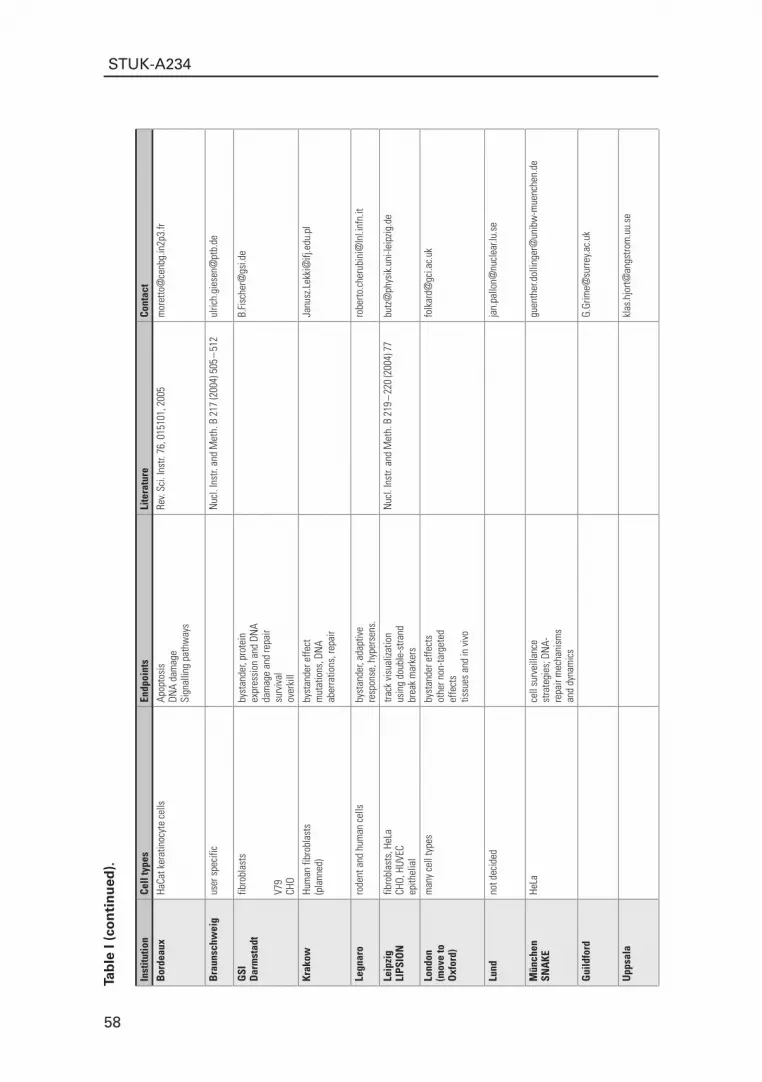
58
STUK-A234
Tab
le I
(co
nti
nu
ed).
Inst
itutio
nCe
ll ty
pes
Endp
oint
sLi
tera
ture
Cont
act
Bor
deau
xHa
Cat k
erat
inoc
yte
cells
Ap
opto
sis
Rev.
Sci.
Inst
r. 76
, 015
101,
200
5m
oret
to@
cenb
g.in
2p3.
fr DN
A da
mag
eSi
gnal
ling
path
way
s
Bra
unsc
hwei
gus
er s
peci
ficN
ucl.
Inst
r. an
d M
eth.
B 2
17 (2
004)
505
– 51
2ul
rich.
gies
en@
ptb.
de
GSI
fib
robl
asts
by
stan
der,
prot
ein
B.Fi
sche
r@gs
i.de
Dar
mst
adt
expr
essi
on a
nd D
NA
da
mag
e an
d re
pair
V79
surv
ival
CHO
over
kill
Krak
owHu
man
fibr
obla
sts
byst
ande
r effe
ctJa
nusz
.Lek
ki@
ifj.e
du.p
l (p
lann
ed)
mut
atio
ns, D
NA
aber
ratio
ns, r
epai
r
Legn
aro
rode
nt a
nd h
uman
cel
lsby
stan
der,
adap
tive
robe
rto.c
heru
bini
@ln
l.inf
n.it
resp
onse
, hyp
erse
ns.
Leip
zig
fibro
blas
ts, H
eLa
track
vis
ualiz
atio
nN
ucl.
Inst
r. an
d M
eth.
B 2
19 –
220
(200
4) 7
7bu
tz@
phys
ik.u
ni-le
ipzig
.de
LIPS
ION
CHO,
HUV
ECus
ing
doub
le-s
trand
epith
elia
lbr
eak
mar
kers
Lond
onm
any
cell
type
sby
stan
der e
ffect
sfo
lkar
d@gc
i.ac.
uk
(mov
e to
othe
r non
-targ
eted
O
xfor
d)ef
fect
stis
sues
and
in v
ivo
Lund
not d
ecid
edja
n.pa
llon@
nucl
ear.l
u.se
Mün
chen
HeLa
ce
ll su
rvei
llanc
e
guen
ther
.dol
linge
r@un
ibw
-mue
nche
n.de
SN
AKE
stra
tegi
es; D
NA-
repa
ir m
echa
nism
s
and
dyna
mic
s
Gui
ldfo
rdG.
Grim
e@su
rrey.a
c.uk
Upp
sala
klas
.hjo
rt@an
gstro
m.u
u.se

59
STUK-A234
Mathematical modelling of the radiation-induced bystander effect and transmissible genomic instability applied to cancer
Mark P. Littlea,*, Kevin Priseb, Melvyn Folkardb and Oleg Belyakovc
a Department of Epidemiology and Public Health, Imperial College Faculty of Medicine, London W2 1PG, UK
b Gray Cancer Institute, P.O. Box 100, Mount Vernon Hospital, Northwood HA6 2JR, UK
c Radiation and Nuclear Safety Authority, Research and Environmental Surveillance, Radiation Biology Laboratory, P.O. Box 14, Laippatie 4, FI-00881 Helsinki, Finland
* To whom all correspondence should be addressed via: Tel. +44 (0)20 7594 3312 Fax +44 (0)20 7402 2150 Email: [email protected]
Keywords: cancer, bystander effect, mechanistic carcinogenesis models, Armitage-Doll model, two-mutation model, dose response
AbstractA variety of quasi-mechanistic models of carcinogenesis are reviewed, and in particular, the multi-stage model of Armitage and Doll and the two-mutation model of Moolgavkar, Venzon, and Knudson. Both the latter models, and various generalizations of them also, are capable of describing at least qualitatively many of the observed patterns of excess cancer risk following ionizing radiation exposure. However, there are certain inconsistencies with the biological and epidemiological data both for the multi-stage model and the two-mutation model. In particular, there are indications that the two-mutation model is not totally suitable for describing the pattern of excess risk for solid cancers that is often seen after exposure to radiation, although leukaemia may be better fitted by this type of model. Generalizations of the model of Moolgavkar, Venzon, and Knudson which require three or more mutations, and models allowing for genomic instability, are easier to reconcile with the epidemiological and biological data relating to solid cancers.
60
STUK-A234
Bystander effects, whereby cells that are not directly exposed to ionizing radiation exhibit adverse biological effects, have been observed in a number of experimental systems. In contrast to the large amount of work on developing carcinogenesis models over the last 50 years, there has been comparatively little work on developing quasi-mechanistic models of the bystander effect, reflecting the comparatively recently available experimental data elucidating this phenomenon. The few quasi-mechanistic models of the bystander effect that have been developed are surveyed. In particular, a novel stochastic model of the radiation-induced bystander effect is considered that takes account of spatial location, cell killing and repopulation, features not explicitly taken into account in many previous models. The ionizing radiation dose- and time-responses of this model are explored, and it is shown to exhibit pronounced downward curvature in the high dose-rate region, similar to that observed in many experimental systems, reviewed in the paper. It is also shown to predict the augmentation of effect after fractionated delivery of dose that has been observed in certain experimental systems.
1 Mechanistic carcinogenesis models
1.1 IntroductionOne of the principal uncertainties that surround the calculation of population cancer risks from epidemiological data results from the fact that few radiation-exposed cohorts have been followed up to extinction. For example, 50 years after the atomic bombings of Hiroshima and Nagasaki, about half of the survivors were still alive (Preston et al. 2003). In attempting to calculate lifetime population cancer risks it is therefore important to predict how risks might vary as a function of time after radiation exposure, in particular for that group for whom the uncertainties in projection of risk to the end of life are most uncertain, namely those who were exposed in childhood.
One way to model the variation in risk is to use empirical models incorporating adjustments for a number of variables (e.g. age at exposure, time since exposure, sex) and indeed this approach has been used in the Fifth Report of the Biological Effects of Ionizing Radiations (BEIR V) Committee (National Research Council 1990) in its analyses of data on the Japanese atomic bomb survivors and various other irradiated groups. Recent analyses of solid cancers for these groups have found that the radiation-induced excess risk can be described fairly well by a relative risk model (ICRP 1991). The time-constant relative risk model assumes that if a dose of radiation is administered to a population, then, after some latent period, there is an increase in the cancer rate, the excess rate
61
STUK-A234
being proportional to the underlying cancer rate in an unirradiated population. For leukaemia, this model provides an unsatisfactory fit, consequently a number of other models have been used for this group of malignancies, including one in which the excess cancer rate resulting from exposure is assumed to be constant i.e. the time-constant additive risk model (UNSCEAR 1988).
It is well known that for all cancer subtypes (including leukaemia) the excess relative risk (ERR) diminishes with increasing age at exposure (UNSCEAR, 2000). For those irradiated in childhood there is evidence of a reduction in the ERR of solid cancer 25 or more years after exposure (Little et al. 1991, Little 1993, Thompson et al. 1994, Pierce et al. 1996, UNSCEAR 2000). For solid cancers in adulthood the ERR is more nearly constant, or perhaps even increasing over time (Little and Charles 1991, Little 1993, UNSCEAR 2000), although there are some indications to the contrary (Weiss et al. 1994). Clearly then, even in the case of solid cancers various factors have to be employed to modify the ERR.
Associated with the issue of projection of cancer risk over time is that of projection of cancer risk between two populations with differing underlying susceptibilities to cancer. Analogous to the relative risk time projection model one can employ a multiplicative transfer of risks, in which the ratio of the radiation-induced excess cancer rates to the underlying cancer rates in the two populations might be assumed to be identical. Similarly, akin to the additive risk time projection model one can use an additive transfer of risks, in which the radiation-induced excess cancer rates in the two populations might be assumed to be identical. The data that are available suggests that there is no simple solution to the problem (UNSCEAR 1994). For example, there are weak indications that the relative risks of stomach cancer following radiation exposure may be more comparable than the absolute excess risks in populations with different background stomach cancer rates (UNSCEAR 1994). Comparison of breast cancer risks observed in the Japanese atomic bomb survivor incidence data and those in various medically exposed populations, many from North America and Europe, where underlying breast cancer rates are higher than in Japan, suggests that ERRs are rather higher in the LSS than those in the medically irradiated groups, but (time- and age-adjusted) EARs are more similar (Little and Boice 1999, Preston et al. 2002). The observation that gender differences in solid tumour ERR are generally offset by differences in gender-specific background cancer rates (UNSCEAR 1994) might suggest that EARs are more alike than ERRs. Taken together, these considerations suggest that in various circumstances relative or absolute transfers of risk between populations may be advocated or, indeed, the use of some sort of hybrid approach such as that employed by Muirhead and Darby (1987) and Little et al. (1999).
62
STUK-A234
The exposed populations that are often used for deriving cancer risks e.g. the Japanese atomic bomb survivors, were exposed to ionizing radiation at high doses and high dose rates. However, it is the possible risks arising from low dose and low dose-rate exposure to ionizing radiation which are central to the setting of standards for radiological protection. The International Commission on Radiological Protection (ICRP) (1991) recommended application of a dose and dose-rate effectiveness factor of 2 to scale cancer risks from high dose and high dose-rate exposure to low dose and low dose-rate exposure on the basis of animal data, the shape of the cancer dose-response in the bomb survivor data and other epidemiological data. Although the linear-quadratic dose-response model (with upward curvature) found for leukaemia is perhaps the most often employed departure from linearity in analyses of cancer in radiation-exposed groups (Pierce and Vaeth 1991, Pierce et al. 1996), other shapes are possible for the dose-response curve (UNSCEAR 1993). While for most tumour types in the Japanese data linear-quadratic curvature adequately describes the shape of the dose-response curve, for non-melanoma skin cancer (NMSC) there is evidence for departures from linear-quadratic curvature. The NMSC dose-response in the Japanese cohort is consistent with a dose threshold of ≈ 1 Sv (Little and Muirhead 1996, Little and Charles 1997) or with an induction term proportional to the fourth power of dose, with in each case an exponential cell sterilization term to reduce NMSC risk at high doses (> 3 Sv).
Arguably, models which take account of the biological processes leading to the development of cancer can provide insight into these related issues of projection of cancer risk over time, transfer of risk across population and extrapolation of risks from high doses and dose-rates to low doses and dose-rates. For example, Little and Charles (1991) have demonstrated that a variety of mechanistic models of carcinogenesis predict an ERR which reduces with increasing time after exposure for those exposed in childhood, while for those exposed in adulthood the ERR might be approximately constant over time. Mechanistic considerations also imply that the interactions between radiation and the various other factors that modulate the process of carcinogenesis may be complex (Leenhouts and Chadwick, 1994), so that in general one would not expect either relative or absolute risks to be invariant across populations.
1.2 Armitage-Doll multi-stage modelMechanistic models of carcinogenesis were originally developed to explain phenomena other than the effects of ionizing radiation. One of the more commonly observed patterns in the age-incidence curves for epithelial cancers is that the cancer incidence rate varies approximately as ]age[C for some constants C
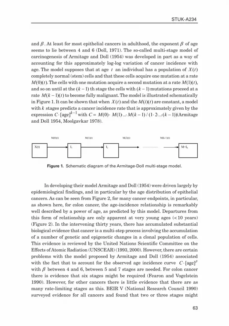
63
STUK-A234
X(t) I1
M(0)(t) M(1)(t)
I2
M(2)(t)
M=Ik
M(k-1)(t)
Figure 1. Schematic diagram of the Armitage-Doll multi-stage model.
and . At least for most epithelial cancers in adulthood, the exponent of age seems to lie between 4 and 6 (Doll, 1971). The so-called multi-stage model of carcinogenesis of Armitage and Doll (1954) was developed in part as a way of accounting for this approximately log-log variation of cancer incidence with age. The model supposes that at age t an individual has a population of X t( ) completely normal (stem) cells and that these cells acquire one mutation at a rate M t( )( )0 . The cells with one mutation acquire a second mutation at a rate M t( )( )1 , and so on until at the ( )k 1 th stage the cells with ( )k 1 mutations proceed at a rate M k t( )( )1 to become fully malignant. The model is illustrated schematically in Figure 1. It can be shown that when X t( ) and the M i t( )( ) are constant, a model with k stages predicts a cancer incidence rate that is approximately given by the expression C k [ ]age 1 with C M M M k k ( ) ( ) ... ( ) / ( ... ( ))0 1 1 1 2 1 (Armitage and Doll 1954, Moolgavkar 1978).
In developing their model Armitage and Doll (1954) were driven largely by epidemiological findings, and in particular by the age distribution of epithelial cancers. As can be seen from Figure 2, for many cancer endpoints, in particular, as shown here, for colon cancer, the age-incidence relationship is remarkably well described by a power of age, as predicted by this model. Departures from this form of relationship are only apparent at very young ages (< 10 years) (Figure 2). In the intervening thirty years, there has accumulated substantial biological evidence that cancer is a multi-step process involving the accumulation of a number of genetic and epigenetic changes in a clonal population of cells. This evidence is reviewed by the United Nations Scientific Committee on the Effects of Atomic Radiation (UNSCEAR) (1993, 2000). However, there are certain problems with the model proposed by Armitage and Doll (1954) associated with the fact that to account for the observed age incidence curve ]age[C
with between 4 and 6, between 5 and 7 stages are needed. For colon cancer there is evidence that six stages might be required (Fearon and Vogelstein 1990). However, for other cancers there is little evidence that there are as many rate-limiting stages as this. BEIR V (National Research Council 1990) surveyed evidence for all cancers and found that two or three stages might

64
STUK-A234
Figure 2. SEER (2002) colon cancer data, and observed data (with 95% confidence intervals (CI), adjusted for overdispersion (McCullagh and Nelder 1989)) (taken from Little (2005)). The use of double logarithmic (log-log) axes shows that except for the youngest age group (< 10 years) the age-incidence relationship is well described by C·[age]k-1.
1 10 100Age (years)
0.01
0.1
1
10
100
1000
Ann
ual i
ncid
ence
rate
(/10
0000
/yea
r)
Males
1 10 100
Females
Observed + 95% CI
be justifiable, but not a much larger number. To this extent the large number of stages predicted by the Armitage-Doll model appears to be verging on the biologically unlikely. Related to the large number of stages required by the Armitage-Doll multi-stage model is the high mutation rates predicted by the model. Moolgavkar and Luebeck (1992) fitted the Armitage-Doll multi-stage model to datasets describing the incidence of colon cancer in a general population and in patients with familial adenomatous polyposis. Moolgavkar and Luebeck (1992) found that Armitage-Doll models with five or six stages gave good fits to these datasets, but that both of these models implied mutation rates that were too high by at least two orders of magnitude. The discrepancy between the predicted and experimentally measured mutation rates might be eliminated, or at least significantly reduced, if account were to be taken of the fact that the experimental mutation rates are locus-specific. A “mutation” in the sense in which it is defined in this model might result from the “failure” of any one of a number of independent loci, so that the “mutation” rate would be the sum of the failure rates at each individual locus.
Notwithstanding these problems, much use has been made of the Armitage-Doll multi-stage model as a framework for understanding the time
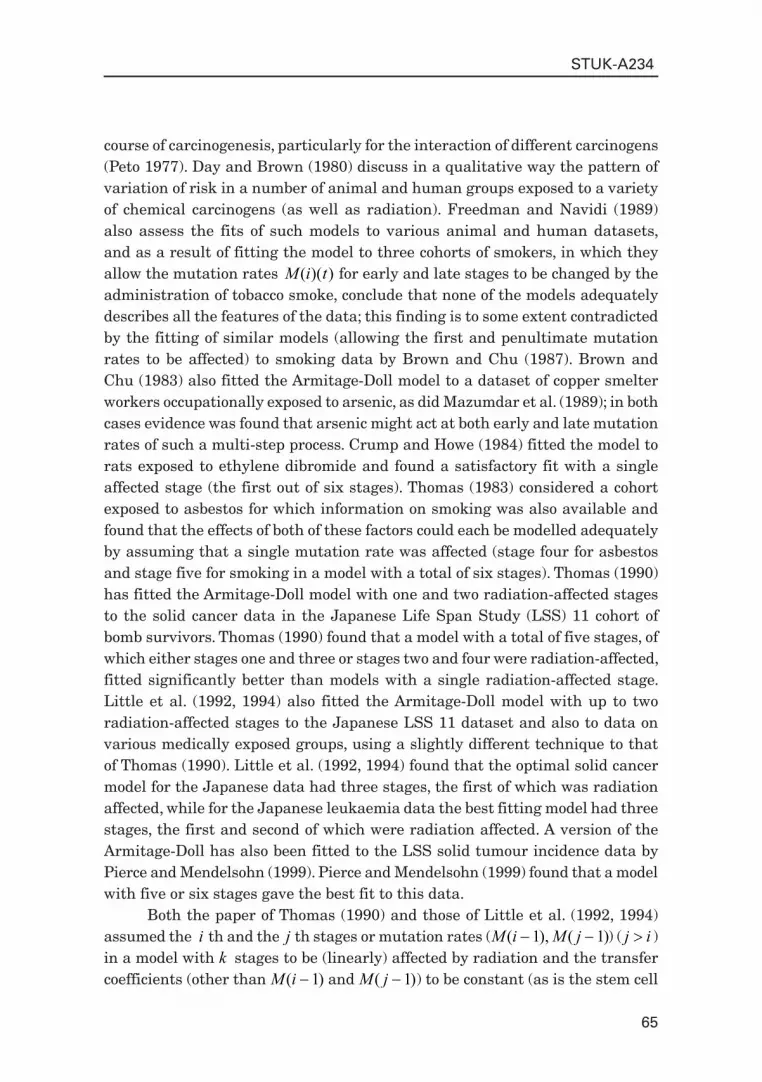
65
STUK-A234
course of carcinogenesis, particularly for the interaction of different carcinogens (Peto 1977). Day and Brown (1980) discuss in a qualitative way the pattern of variation of risk in a number of animal and human groups exposed to a variety of chemical carcinogens (as well as radiation). Freedman and Navidi (1989) also assess the fits of such models to various animal and human datasets, and as a result of fitting the model to three cohorts of smokers, in which they allow the mutation rates M i t( )( ) for early and late stages to be changed by the administration of tobacco smoke, conclude that none of the models adequately describes all the features of the data; this finding is to some extent contradicted by the fitting of similar models (allowing the first and penultimate mutation rates to be affected) to smoking data by Brown and Chu (1987). Brown and Chu (1983) also fitted the Armitage-Doll model to a dataset of copper smelter workers occupationally exposed to arsenic, as did Mazumdar et al. (1989); in both cases evidence was found that arsenic might act at both early and late mutation rates of such a multi-step process. Crump and Howe (1984) fitted the model to rats exposed to ethylene dibromide and found a satisfactory fit with a single affected stage (the first out of six stages). Thomas (1983) considered a cohort exposed to asbestos for which information on smoking was also available and found that the effects of both of these factors could each be modelled adequately by assuming that a single mutation rate was affected (stage four for asbestos and stage five for smoking in a model with a total of six stages). Thomas (1990) has fitted the Armitage-Doll model with one and two radiation-affected stages to the solid cancer data in the Japanese Life Span Study (LSS) 11 cohort of bomb survivors. Thomas (1990) found that a model with a total of five stages, of which either stages one and three or stages two and four were radiation-affected, fitted significantly better than models with a single radiation-affected stage. Little et al. (1992, 1994) also fitted the Armitage-Doll model with up to two radiation-affected stages to the Japanese LSS 11 dataset and also to data on various medically exposed groups, using a slightly different technique to that of Thomas (1990). Little et al. (1992, 1994) found that the optimal solid cancer model for the Japanese data had three stages, the first of which was radiation affected, while for the Japanese leukaemia data the best fitting model had three stages, the first and second of which were radiation affected. A version of the Armitage-Doll has also been fitted to the LSS solid tumour incidence data by Pierce and Mendelsohn (1999). Pierce and Mendelsohn (1999) found that a model with five or six stages gave the best fit to this data.
Both the paper of Thomas (1990) and those of Little et al. (1992, 1994) assumed the i th and the j th stages or mutation rates (M i M j( ), ( ) 1 1 ) ( j i ) in a model with k stages to be (linearly) affected by radiation and the transfer coefficients (other than M i( )1 and M j( )1 ) to be constant (as is the stem cell

66
STUK-A234
population X t( ) ). In these circumstances it can be shown (Little et al. 1992) that if an instantaneously administered dose of radiation d is given at age a , then at age t a( ) the cancer rate is approximately:
t d a t a d a t a d a t ak i k i j k j i k j1 1 1 1 1 2 1 1[ ] [ ] [ ]
for some positive constants , and , and where is given by:
( ) ( )( )
k i jk
j i
j i2
1
0 1
if
if
and (.) is the gamma function (Abramowitz and Stegun 1964).The first term ( t k 1 ) in expression corresponds to the cancer rate
that would be observed in the absence of radiation, while the second term ( d a t ai k i1 1[ ] ) and the third term ( d a t aj k j1 1[ ] ) represent the separate effects of radiation on the i th and j th stages respectively. The fourth term ( d a t ai k j2 1 1[ ] ), which is quadratic in dose d , represents the consequences of interaction between the effects of radiation on the i th and the j th stages and is only non-zero when the two radiation-affected stages are adjacent ( j i 1 ). Thus if the two affected stages are adjacent, a quadratic (dose plus dose-squared) relationship will occur, whereas the relationship will be approximately linear if the two affected stages have at least one intervening stage. Another way of considering the joint effects of radiation on two stages is that for a brief exposure, unless the two radiation-affected stages are adjacent, there will be insignificant interaction between the cells affected by radiation in the earlier and later of the two radiation-affected cell compartments. This is simply because very few cells will move between the two compartments in the course of the radiation exposure. If the i th and the j th stages are radiation-affected the result of a brief dose of radiation will be to cause some of the cells which have already accumulated ( i 1 ) mutations to acquire an extra mutation and move from the ( i 1 ) th to the i th compartment. Similarly, it will cause some of the cells which have already acquired ( j 1 ) mutations to acquire an extra mutation and so move from the ( j 1 ) th to the j th compartment. It should be noted that the model does not require that the same cells be hit by the radiation at the i th and j th stages, and in practice for low total doses, or whenever the two radiation-affected stages are separated by an additional unaffected stage or stages, an insignificant proportion of the same cells will be hit (and mutated) by the radiation at both the i th and the j th stages. The result is that, unless the radiation-affected stages are adjacent, for a brief exposure the total effect on cancer rate is approximately the sum of the effects, assuming radiation were to act on each of the radiation-affected stages alone. One interesting implication of

67
STUK-A234
models with two or more radiation-affected stages is that as a result of interaction between the effects of radiation at the various stages, protraction of dose in general results in an increase in cancer rate i.e. an inverse dose-rate effect (Little et al. 1992). However, it can be shown that in practice the resulting increase in cancer risk is likely to be small (Little et al. 1992).
The variant of the Armitage-Doll model fitted by Pierce and Mendelsohn (1999) is unusual in that it assumes that radiation equally affects all k mutation rates in the model except the last. (In the last stage radiation is not assumed to have any effect.) This assumption distinguishes their use of this model from the approaches of Little et al. (1992) or Thomas (1990), both of whom assumed that radiation affected at most two of the mutation rates (and did not constrain the effects of radiation to be equal in these stages). There are some technical problems with the paper of Pierce and Mendelsohn (1999) arising from the authors failure to take account of interactions between the effects of radiation on the ( 2−k ) pairs of adjacent stages, and which contribute significantly, by adding a quadratic term in the dose-response. These cannot be ignored, even to a first order approximation. The fact that in general there is little evidence for upward curvature in the solid cancer dose-response in the LSS (Pierce and Vaeth 1991, Little and Muirhead 1996, 1998, 2000) argues that if proper account were taken of these interaction terms the model of Pierce and Mendelsohn (1999) would not fit the data well. Moreover, one implication of the model of Pierce and Mendelsohn (1999) is that the ERR will be proportional to a/1 i.e. the inverse of attained age. However, this is known to provide a poor description of the ERR of solid cancer, even within the LSS cohort (Little et al. 1997a, Little et al. 1999). For these reasons, there are some grounds for regarding the model of Pierce and Mendelsohn (1999) as providing a poor description of the pattern of excess risk of solid tumours within the LSS cohort. Other problems with the model of Pierce and Mendelsohn (1999) are discussed by Heidenreich et al. (2002).
The optimal leukaemia model found by Little et al. (1992, 1994), having adjacent radiation-affected stages, predicts a linear-quadratic dose-response, in accordance with the significant upward curvature which has been observed in the Japanese dataset (Pierce and Vaeth 1991, Preston et al. 1994, Pierce et al. 1996). This leukaemia model, and also that for solid cancer, predicts the pronounced reduction of ERR with increasing age at exposure (see Figure 3) which has been seen in the Japanese atomic bomb survivors and other datasets (UNSCEAR 2000). The optimal Armitage-Doll leukaemia model predicts a reduction of ERR with increasing time after exposure for leukaemia. At least for those exposed in childhood, the optimal Armitage-Doll solid cancer model also predicts a reduction in ERR with time for solid cancers. These observations are consistent with the observed pattern of risk in the Japanese and other datasets (Little 1993,

68
STUK-A234
UNSCEAR 2000). Nevertheless, there are indications that the Armitage-Doll model may not provide an adequate fit to the Japanese data (Little et al. 1995). For this reason, and because of the other problems with the Armitage-Doll model discussed above, one needs to consider a slightly different class of models.
1.3 Two-mutation modelIn order to reduce the biologically implausible number of stages required by their first model, Armitage and Doll (1957) developed a further model of carcinogenesis, which postulated a two-stage probabilistic process whereby a cell following an initial transformation into a pre-neoplastic state (initiation) was subject to a period of accelerated (exponential) growth. At some point in this exponential growth a cell from this expanding population might undergo a second transformation (promotion) leading quickly and directly to the development of a neoplasm. Like their previous model, it satisfactorily explained the incidence of cancer in adults, but was less successful in describing the pattern of certain childhood cancers.
The two-mutation model developed by Knudson (1971) to explain the incidence of retinoblastoma in children took account of the process of growth and differentiation in normal tissues. Subsequently, the stochastic two-mutation model of Moolgavkar and Venzon (1979) generalized Knudson’s model, by taking account of cell mortality at all stages as well as allowing for differential growth of intermediate cells. The two-stage model developed by Tucker (1967) is very similar to the model of Moolgavkar and Venzon but does not take account of the differential growth of intermediate cells. The two-mutation model of Moolgavkar, Venzon and Knudson (MVK) supposes that at age t there are X t( ) susceptible stem cells, each subject to mutation to an intermediate type of cell at a rate M t( )( )0 . The intermediate cells divide at a rate G t( )( )1 ; at a rate D t( )( )1 they die or differentiate; at a rate M t( )( )1 they are transformed into malignant cells. The model is illustrated schematically in Figure 4. In contrast with the case of the (first) Armitage-Doll model, there is a considerable body of experimental biological data supporting this initiation-promotion type of model (see e.g. Moolgavkar and Knudson 1981, Tan 1991). The model has recently been developed to allow for time-varying parameters at the first stage of mutation (Moolgavkar et al. 1988). A further slight generalization of this model (to account for time varying parameters at the second stage of mutation) was presented by Little and Charles (1991), who also demonstrated that the ERR predicted by the model, when the first mutation rate was subject to instantaneous perturbation, decayed at least exponentially for a sufficiently long time after the perturbation. Moolgavkar et al. (1990), Luebeck et al. (1996), Heidenreich et al. (1999) and

69
STUK-A234
0 10 20 30 40 50
Years after exposure
0
5
10
15
20
Exce
ss re
lativ
e ris
k
Solid cancers
0 10 20 30 40 500
20
40
60
80
100Leukaemia
0 10 20 30 40 500.0
0.2
0.4
0.6
0.8
1.0
0 10 20 30 40 500
5
10
15
20
Age at exposure < 15 years
Age at exposure > 15 years
2-mutation model3-mutation model
Figure 3. Observed excess relative risk Sv-1 (+ 90% CI) for solid cancers and leukaemia and fitted excess relative risk (evaluated at 1 Sv) using optimal 2-mutation and 3-mutation generalized MVK models (Little, 1996).
Heidenreich et al. (2000) have used the two-mutation model to describe the incidence of lung cancer in rats exposed to radon, and in particular to model the inverse dose-rate effect that has been observed in this data. Moolgavkar et al. (1993), Luebeck et al. (1999), Leenhouts (1999), Hazelton et al. (2001) and Heidenreich et al. (2004) have applied the model to describe the interaction of radon, smoking and other agents causing lung cancer in various miner cohorts. The two-mutation model has also been utilised to model lung, stomach, and colon cancer in the Japanese atomic bomb survivor incidence data (Kai et al. 1997), and to model liver cancer in a cohort of Swedish Thorotrast-exposed patients (Heidenreich et al. 2003).

70
STUK-A234
Figure 4. Schematic diagram of the two-mutation (MVK) model.
I1
M(1)(t)
M=I2 I1 X(t)
M(0)(t)
D
D(1)(t)
I1 I1
G(1)(t)
A curious finding in many analyses of lung cancer in relation to radon-daughter exposure using the two-mutation model is that there is significant radon action on intermediate cell proliferation. This has been observed both in radon-exposed rats (Heidenreich et al. 1999, 2000), in the Colorado Plateau uranium miners (Luebeck et al. 1999, Little et al. 2002) and in the Chinese tin miners (Hazelton et al. 2001). This is very much an artifact of the two-mutation model. Analyses of rat data using a three-mutation generalized MVK model (see below) did not find any indications of an effect of radon daughter exposure on intermediate cell proliferation (Heidenreich et al. 2000). Likewise, analysis of the Colorado Plateau miners (the same dataset analysed by Luebeck et al. (1999)) using a three-mutation MVK generalized MVK model did not find any effect of radon daughter exposure on intermediate cell proliferation rates (Little et al. 2002).
Moolgavkar and Luebeck (1992) have used models with two or three mutations to describe the incidence of colon cancer in a general population and in patients with familial adenomatous polyposis. They found that both models gave good fits to both datasets, but that the model with two mutations implied biologically implausibly low mutation rates. The three-mutation model, which predicted mutation rates more in line with biological data, was therefore somewhat preferable. The problem of implausibly low mutation rates implied by the two-mutation model is not specific to the case of colon cancer, and is discussed at greater length by Den Otter et al. (1990) and Derkinderen et al. (1990), who argue that for most cancer sites a model with more than two stages is required.

71
STUK-A234
X(t) M(0)(t)
I1
I1
D
D(1)(t)
I1
G(1)(t)
I1
I2
I2
D
D(2)(t)
I2
G(2)(t)
M(2)(t)
I2
M(1)(t) I3
Ik-1
Ik-1
D
D(k-1)(t)
Ik-1
G(k-1)(t)
M(k-1)(t) M=Ik Ik-1
Figure 5. Schematic diagram of the generalized MVK model.
1.4 Generalized MVK and multi-stage modelsA number of generalizations of the Armitage-Doll and two- and three-mutation models have been developed (Tan 1991, Little 1995, Little and Wright 2003). In particular two closely related models have been developed, whose properties have been described in the paper of Little (1995). The first model is a generalization of the two-mutation model of Moolgavkar, Venzon, and Knudson and so will be termed the generalized MVK model. The second model generalizes the multi-stage model of Armitage and Doll and will be referred to as the generalized multi-stage model. For the generalized MVK model it may be supposed that at age t there are X t( ) susceptible stem cells, each subject to mutation to a type of cell carrying an irreversible mutation at a rate of M t( )( )0 . The cells with one mutation divide at a rate G t( )( )1 ; at a rate D t( )( )1 they die or differentiate. Each cell with one mutation can also divide into an equivalent daughter cell and another cell with a second irreversible mutation at a rate M t( )( )1 . For the cells with two mutations there are also assumed to be competing processes of cell growth, differentiation, and mutation taking place at rates G t( )2)( , D t( )2)( , and M t( )2)( respectively, and so on until at the ( )k 1 th stage the cells which have accumulated ( )k 1 mutations proceed at a rate M k t( )( )1 to acquire another mutation and become malignant. The model is illustrated schematically in Figure 5. The two-mutation model of Moolgavkar, Venzon, and Knudson corresponds to the case k 2 . The generalized multi-stage model differs from the generalized MVK model only in that the process whereby a cell is assumed to split into an identical daughter cell and a cell carrying an additional mutation is replaced by the process in which only the cell with an additional mutation results, i.e. an identical daughter cell is not produced. The classical Armitage-Doll multi-stage model corresponds to the case in which the intermediate cell proliferation rates G i t( )( ) and the cell differentiation rates D i t( )( ) are all zero.

72
STUK-A234
It can be shown (Little 1995) that the ERR for either model following a perturbation of the parameters will tend to zero as the attained age tends to infinity. One can also demonstrate that perturbation of the parameters M k( 2) , M k( )1 , G k( )1 , and D k( )1 will result in an almost instantaneous change in the cancer rate (Little 1995).
Generalized MVK models have been fitted to the Japanese atomic bomb survivor LSS 11 mortality data (Little 1996, 1997). Both for leukaemia and solid cancers the only models with a single radiation-affected parameter which give at all satisfactory fit are those in which radiation is assumed to affect M ( )0 (Little 1996). Both for leukaemia and for solid cancer generalized two- and three-mutation MVK models fit equally well. For leukaemia, the three-mutation model provides at all satisfactory a fit only when M ( )0 and M ( )1 are assumed affected by radiation. For solid cancer and leukaemia there are indications of lack of fit to the youngest age at exposure group for the three-mutation model; there is also some lack of fit of the optimal solid cancer three-mutation model to this age at exposure group (Figure 3). Little et al. (1996) also showed that the age-incidence relationship for lymphocytic leukemia incidence in the UK population could be adequately described by models with either two or three stages. Little et al. (2002) modelled lung cancer mortality in the Colorado Plateau uranium miner cohort using generalizations of the MVK model, and demonstrated that models with three mutations provided a superior fit to models with two mutations.
For solid cancer only M ( )0 is (linearly) affected by radiation for two- or three-mutation generalized MVK models. In contrast to the solid cancer models, both leukaemia models assume a linear-quadratic dose-dependence of the M i( ) . The non-linearity found in the leukaemia M i( ) dose-response reflects known curvature in the leukaemia dose-response in the Japanese (National Research Council 1990, Pierce and Vaeth 1991). There is some evidence e.g. for chromosome aberrations that the mutation induction curve is linear-quadratic at least for low LET radiation, although linearity is generally observed for high LET radiation (Lloyd and Edwards 1983).
Despite the indications of lack of fit discussed above, the variation of ERR with time since exposure and age at exposure predicted by the optimal two- and three-mutation models for solid cancer (Figure 3) is in qualitative agreement with the variation seen in the Japanese bomb survivors and in other irradiated groups (UNSCEAR 2000). In particular the optimal models demonstrate the progressive reduction in ERR with increasing age at exposure seen in many datasets (UNSCEAR 2000), together with the marked reduction in ERR with increasing time since exposure observed in various groups exposed in childhood (Little et al. 1991, Pierce et al. 1996, UNSCEAR 2000).

73
STUK-A234
Figure 3 reinforces the theoretical predictions of a previous paper (Little 1995), and shows that immediately after perturbing M ( )0 in the two-mutation model the ERR for solid cancers and leukaemia quickly increases. However, there are no data in the first 5 years of follow-up in the Japanese cohort (Pierce et al. 1996), so that it is difficult to test the predictions made in a previous paper (Little 1995) concerning the variation in risk shortly after exposure using that dataset.
There is a suggestive increase in the ERR of cancers other than leukaemia and colon cancer in the UK ankylosing spondylitis patients < 5 years after first treatment (the first two datapoints in the top-left panel of Figure 6), but the authors caution against interpreting this as the effect of the X-irradiation (Darby et al. 1987). There are no strong indications of an elevation in risk in the first five years after radiotherapy for cancers other than leukaemia and of the reproductive organs in a study of women followed up for second cancer after radiotherapy for cervical cancer (Boice et al. 1985). This corresponds to the first two datapoints in the bottom panel of Figure 6. (Lung cancers are also excluded from the International Radiation Study of Cervical Cancer (IRSCC) data shown in the lower left panel of Figure 6 because of indications of above-average smoking rates in this cohort (Boice et al. 1985).) In general there are no strong indications of an elevation in solid cancer risk soon after irradiation in other exposed groups (UNSCEAR 2000). To this extent there are indications of inconsistency for solid cancers between the predictions of the two-mutation model and the observed variation in risks shortly after exposure.
Moolgavkar et al. (1993) partially overcome the problem posed by this instantaneous rise in the hazard after perturbation of the two-mutation model parameters in their analysis of the Colorado uranium miners data by assuming a fixed period (3.5 years) between the appearance of the first malignant cell and the clinical detection of malignancy. However, the use of such a fixed latent period only translates a few years into the future the sudden step-change in the hazard. To achieve the observed gradual increase in ERR shortly after exposure, a stochastic process must be used to model the transition from the first malignant cell to detectable cancer, such as is provided by the final stage(s) in the three- or four-mutation generalized MVK models used in the analysis of Little (1996). In particular, an exponentially growing population of malignant cells could be modelled by a penultimate stage with G k( ) 1 0 and D k( ) 1 0 , the probability of detection of the clone being determined by M k( )1 . In their analysis of lung, stomach and colon cancer in the Japanese atomic bomb survivor incidence data Kai et al. (1997) did not assume any such period of latency, perhaps because of the long period after the bombings (12.4 years) before solid cancer incidence follow-up began in the LSS.
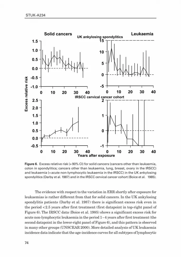
74
STUK-A234
0 10 20 30 40
Years after exposure
-1.0
-0.5
0.0
0.5
1.0
1.5
Exce
ss re
lativ
e ris
k
Solid cancers
0 10 20 30 40-5
0
5
10
15Leukaemia
0 10 20 30 40-0.5
0.0
0.5
1.0
1.5
2.0
2.5
0 10 20 30 40-1
0
1
2
UK ankylosing spondylitics
IRSCC cervical cancer cohort
Figure 6. Excess relative risk (+ 90% CI) for solid cancers (cancers other than leukaemia, colon in spondylitics; cancers other than leukaemia, lung, breast, ovary in the IRSCC) and leukaemia (= acute non-lymphocytic leukaemia in the IRSCC) in the UK ankylosing spondylitics (Darby et al. 1987) and in the IRSCC cervical cancer cohort (Boice et al. 1985).
The evidence with respect to the variation in ERR shortly after exposure for leukaemias is rather different from that for solid cancers. In the UK ankylosing spondylitis patients (Darby et al. 1987) there is significant excess risk even in the period < 2.5 years after first treatment (first datapoint in top-right panel of Figure 6). The IRSCC data (Boice et al. 1985) shows a significant excess risk for acute non-lymphocytic leukaemia in the period 1 – 4 years after first treatment (the second datapoint in the lower-right panel of Figure 6), and this pattern is observed in many other groups (UNSCEAR 2000). More detailed analysis of UK leukaemia incidence data indicate that the age-incidence curves for all subtypes of lymphocytic

75
STUK-A234
leukaemia can be adequately modelled by two- and three-mutation generalized MVK models (Little et al. 1996, 1997b), although the two-mutation models for acute lymphocytic leukaemia (ALL) imply a very small number of stem cells (< 104 cells) if the model is not to yield implausibly low mutation rates (Little et al. 1997b).
1.5 Multiple pathway modelsLittle et al. (1995) fitted a generalization of the Armitage-Doll model to the Japanese atomic bomb survivor and IRSCC leukaemia data which allowed for two cell populations at birth, one consisting of normal stem cells carrying no mutations, the second a population of cells each of which has been subject to a single mutation. The leukaemia risk predicted by such a model is equivalent to that resulting from a model with two pathways between the normal stem cell compartment and the final compartment of malignant cells, the second pathway having one fewer stage than the first. This model fitted the Japanese and IRSCC leukaemia datasets significantly better, albeit with biologically implausible parameters, than a model which assumed just a single pathway (Little et al. 1995). A number of other such models are described by Tan (1991), who also discusses at some length the biological and epidemiological evidence for such models of carcinogenesis.
We now discuss what may appear to be a special case of these multiple pathway models, but which are of sufficient flexibility to embrace most categories of multiple pathway models.
1.5.1 Multiple pathway models incorporating genomic instabilityThere is much biological data suggesting that the initiating lesion in the multistage process leading to cancer might be one involving a destabilization of the genome resulting in elevation of mutation rates. In particular, the findings of Kadhim et al. (1992, 1994), that exposure of mammalian haemopoietic stem cells to alpha particles could result in a general elevation of mutation rates to very much higher than normal levels, implies, if these findings are at all relevant to carcinogenesis, that there might be multiple pathways in the progression from normal stem cells to malignant cells. A carcinogenesis model based on genomic instability (GI) and clonal selection was proposed by Nowell (1976). More recently Loeb (1991, 2001) has presented evidence that an early step in carcinogenesis is mutation in a gene controlling genome stability. Stoler et al. (1999) showed that there are 11,000 mutations per carcinoma cell for a number of different cancer types, again implying that genomic destabilization is an early event in carcinogenesis. In particular, there is strong evidence of such an early genomic destabilization event for colon cancer (Loeb 1991, Stoler et al. 1999, Loeb 2001).

76
STUK-A234
There have been a few attempts to incorporate GI in mechanistic carcinogenesis models (Mao et al. 1998, Ohtaki and Niwa 2001), although in general these models have not been fitted to data in a statistically rigorous manner. Little and Wright (2003) developed a stochastic carcinogenesis model which allowed for genome destabilization, very close in spirit to the model of Mao et al. (1998), and generalizing the class of generalized MVK models developed by Little (1995, 1996, 1997), which in turn therefore generalize the two-mutation model of Moolgavkar, Venzon and Knudson (Knudson 1971, Moolgavkar and Venzon, 1979). Little and Wright (2003) fitted the model to Surveillance, Epidemiology and End Results (SEER) population-based Caucasian colon cancer incidence data (SEER 2000).
The model assumes that cells can acquire two sorts of mutation, those associated with progression to a malignant phenotype (‘cancer-stage’ mutations), and those associated with successive destabilization of the genome (‘destabilizing’ mutations). With acquisition of successively more destabilizing mutations the cancer-stage mutation rates are generally higher, corresponding to the genome destabilization that is characteristic of GI.
Specifically, the model supposes that at age t there are X t( ) susceptible stem cells, each subject to mutation to a type of cell carrying an irreversible cancer-stage mutation at a rate of ))(0,0( tM . The cells in the stem cell compartment can also acquire a destabilizing mutation at a rate ))(0,0( tA . Thereafter the cells in compartment ),( jiI with i cancer-stage mutation and j destabilizing mutations divide into two such cells at a rate ))(,( tjiG ; at a rate ))(,( tjiD they die or differentiate. Each such cell can also divide into an equivalent daughter cell and another cell with an additional cancer-stage mutation at a rate ))(,( tjiM . In addition, each such cell can also divide into an equivalent daughter cell and another cell with an additional destabilizing mutation, at a rate ))(,( tjiA . There are assumed to be a total of k cancer-stage mutations required for a cell to become malignant. Likewise, there are assumed to be m destabilizing mutations. Once a cell has acquired all m such destabilizing mutations it is assumed to remain at the m th destabilizing mutation level. This model is illustrated schematically in Figure 7. The acquisition of carcinogenic (cancer-stage) mutations amounts to moving horizontally (left to right) in Figure 7, whereas acquisition of destabilizing mutations amounts to moving vertically (top to bottom) in this figure. The asymmetric cell divisions associated with most of the cancer-stage and destabilizing mutations (all except )0,0(),( =ji ), in which each cell produces a daughter cell identical to the parent and another carrying an additional mutation, should be contrasted with the symmetric cell divisions associated with the cell proliferation processes (with rates ),( jiG ), in which each cell produces two identical daughter cells. The two-mutation MVK model corresponds to the

77
STUK-A234
24
G(2,0)(t)
X(t) M(0,0)(t) I(1,0)
D
D(1,0)(t)
G(1,0)(t)
A(0,0)(t)
I(2,0)
D
D(2,0)(t)
M(2,0)(t) M(1,0)(t) I(k-1,0)
D
D(k-1,0)(t)
G(k-1,0)(t)
M(k-1,0)(t) M=I(k,0)
I(1,1)
D D(1,1)(t)
G(1,1)(t) I(2,1)
D D(2,1)(t)
G(2,1)(t)
M(2,1)(t) M(1,1)(t) I(k-1,1)
D
D(k-1,1)(t)
G(k-1,1)(t)
M(k-1,1)(t) M=I(k,1) I(0,1)
D
G(0,1)(t)
M(0,1)(t)
D(0,1)(t)
A(0,m-1)(t) A(1,m-1)(t) A(2,m-1)(t) A(k-1,m-1)(t)
I(1,m)
D D(1,m)(t)
G(1,m)(t) I(2,m)
D D(2,m)(t)
G(2,m)(t)
M(2,m)(t) M(1,m)(t) I(k-1,m)
D
D(k-1,m)(t)
G(k-1,m)(t)
M(k-1,m)(t) M=I(k,m)
I(0,m)
D
G(0,m)(t)
M(0,m)(t)
D(0,m)(t)
A(1,0)(t) A(2,0)(t) A(k-1,0)(t)
A(0,1)(t) A(1,1)(t) A(2,1)(t) A(k-1,1)(t)
Asymmetric cell division: one mutant daughter cell, one non-mutant daughter cell
Mutation: one mutant daughter cell only Symmetric cell division: two non-mutant daughter cells
Figure 7. Schematic diagram of the generalized MVK model with k cancer-stage mutations and m destabilizing mutations.
case k 2 , 0=m , while the generalized MVK model with K stages developed by Little (1995, 1996, 1997) amounts to the case Kk = , 0=m . In fits to the SEER colon cancer data models with two cancer-stage mutations and one destabilizing mutation, with three cancer-stage mutations and one destabilizing mutation, and with five cancer-stage mutations and two destabilizing mutations all gave good fit (Little and Wright 2003, Little 2005).
Two other recent papers have appeared proposing formulations of stochastic carcinogenesis model that incorporate genomic instability (Luebeck and Moolgavkar 2002, Nowak et al. 2002), again both applied to colon cancer. These models are illustrated schematically in Figures 8 and 9. The model of Little and Wright (2003) includes as special cases the models proposed by Luebeck et al. (2002) and Nowak et al. (2002).
Little (2005) has compared the fits of all five of these models (Little and Wright two cancer-stage mutations + one destabilizing mutation, Little and Wright three cancer-stage mutations + one destabilizing mutation, Little and Wright five cancer-stage mutations + two destabilizing mutations three-mutation, Luebeck and Moolgavkar, Nowak et al.) to the colon cancer data

78
STUK-A234
31
I(1,0) I(2,0) M(2,0)(t)
G(3,0)(t)
M(1,0)(t) I(3,0)
D
D(3,0)(t) M(3,0)(t)
M=I(4,0) X(t) M(0,0)(t)
Asymmetric cell division: one mutant daughter cell, one non-mutant daughter cell
Mutation: one mutant daughter cell only Symmetric cell division: two non-mutant daughter cells
Figure 9. Schematic diagram of the model of Luebeck and Moolgavkar (2002), similar to a generalized MVK model with four cancer-stage mutations and no destabilizing mutations.
26
Asymmetric cell division: one mutant daughter cell, one non-mutant daughter cell
Mutation: one mutant daughter cell only Symmetric cell division: two non-mutant daughter cells
X(t) M(0,0)(t) I(1,0)
D
D(1,0)(t)
G(1,0)(t)
A(0,0)(t)
I(1,1)
D D(1,1)(t)
G(1,1)(t) M=I(2,1)
M(1,1)(t) I(0,1)
G(0,1)(t)
M(0,1)(t)
D(0,1)(t)
A(1,0)(t)
D
Figure 8. Schematic diagram of the model of Nowak et al. (2002), similar to a generalized MVK model with two cancer-stage mutations and one destabilizing mutation.
used by Little and Wright (2003). If the number of stem cells is fixed at a biologically plausible value, the best fitting model is that of Nowak et al. (2002), with the two-stage model of Little and Wright not markedly inferior, as shown in Figures 10 and 11. The fit of the three-stage model of Little and Wright (2003) is somewhat worse than these two, even more so that of Luebeck and Moolgavkar (2002) and the five stage model of Little and Wright (2003), as shown in Figures 10 and 11. Comparison of the predictions of the two-stage models of Little and Wright (2003) and Nowak et al. (2002), given in Figures

79
STUK-A234
Figure 10. Cancer hazards predicted by models of Nowak et al. (2002) (with two cancer-stage mutations and one destabilizing mutation), of Luebeck and Moolgavkar (2002) (with four cancer-stage mutations and no destabilizing mutations), and of Little and Wright (2003) (with two cancer-stage mutations and one destabilizing mutation, three cancer-stage mutations and one destabilizing mutation, five cancer-stage mutations and two destabilizing mutations), with stem cell population fixed to 108 cells, refitted to SEER (2002) colon cancer data, and observed data (with 95% confidence intervals (CI), adjusted for overdispersion (McCullagh and Nelder 1989)) (taken from Little (2005)).
0 20 40 60 80 90Age (years)
0.001
0.01
0.1
1
10
100
1000A
nnua
l inc
iden
ce ra
te (/
1000
00/y
ear)
Males
0 20 40 60 80 90
Females
X
X
X
X
X
X
X
X
X
XX
XX
XX
X
X
X
X
X
X
X
XX
XX
XX
XX
x x Observed + 95% CI2-stage 1-gate model
Nowak et al (2002) modelLuebeck and Moolgavkar (2002) model
3-stage 1-gate model5-stage 2-gate model
Figure 11. As for Figure 10, but showing cancer rates and model fits up to age 40 (taken from Little, 2005).
0 10 20 30 40Age (years)
0.001
0.01
0.1
1
10
Ann
ual i
ncid
ence
rate
(/10
0000
/yea
r)
Males
0 10 20 30 40
Females
X
X
X
X
X
X
XXXXXXXXX
X
X
X
X
X
X
XXXXXXXXX
x x Observed + 95% CI2-stage 1-gate model
Nowak et al (2002) modelLuebeck and Moolgavkar (2002) model
3-stage 1-gate model5-stage 2-gate model

80
STUK-A234
Years since exposure
0.00.20.40.60.81.0
Nor
mal
ized
exc
ess
rela
tive
risk
0.00.20.40.60.81.0
0 10 20 30 40 50 600.00.20.40.60.81.0
G(1,0) +1G(0,1) +1
M(0,0) x10M(1,0) x10M(1,1) x10
A(0,0) +0.001A(1,0) x10
Figure 12. Normalized excess relative hazard following perturbations of ),( jiG , ),( jiM and ),( jiA in generalized MVK model with two cancer-stage mutations and one destabilizing mutation of Little and Wright (2003), with stem cell population fixed to 108 cells, refitted to male SEER colon cancer incidence data (taken from Little (2005)). In the absence of perturbation the model is as described in Figures 7 and 10. The parameters
),( jiG are increased by 11 −y at the age of 25, for 1 year, the parameters ),( jiM and ),( jiA are generally multiplied by 10 at the age of 25 ( )0,0(A is augmented by
310 y 1), for 1 year.
12 and 13, with patterns of excess risk in the Japanese atomic bomb survivor colon cancer incidence data, shown in Figure 14, indicate that radiation might act on cell proliferation rates in the model, and at least for the model of Little and Wright also on one of the parameters governing progression to genomic destabilization.
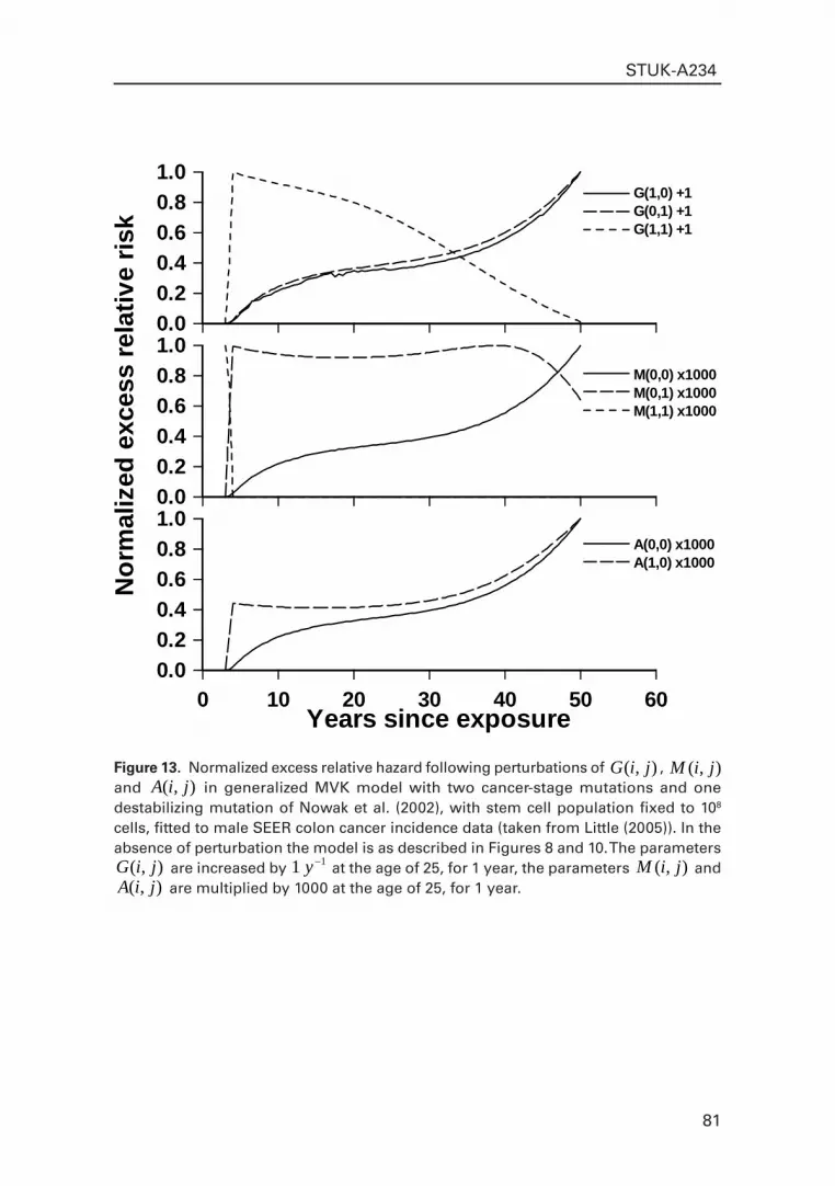
81
STUK-A234
Years since exposure
0.00.20.40.60.81.0
Nor
mal
ized
exc
ess
rela
tive
risk
0.00.20.40.60.81.0
0 10 20 30 40 50 600.00.20.40.60.81.0
G(1,0) +1G(0,1) +1
M(0,0) x1000M(0,1) x1000
A(0,0) x1000A(1,0) x1000
G(1,1) +1
M(1,1) x1000
Figure 13. Normalized excess relative hazard following perturbations of ),( jiG , ),( jiM and ),( jiA in generalized MVK model with two cancer-stage mutations and one destabilizing mutation of Nowak et al. (2002), with stem cell population fixed to 108 cells, fitted to male SEER colon cancer incidence data (taken from Little (2005)). In the absence of perturbation the model is as described in Figures 8 and 10. The parameters
),( jiG are increased by 11 −y at the age of 25, for 1 year, the parameters ),( jiM and ),( jiA are multiplied by 1000 at the age of 25, for 1 year.

82
STUK-A234
10 20 30 40 50Years since exposure
0
2
4
6
8
10
Exce
ss re
lativ
e ris
k/Sv
Figure 14. Excess relative risk (per Sv) and 95% confidence intervals for male colon cancer incidence as a function of years since exposure in the Japanese atomic bomb survivor data of Thompson et al. (1994) (taken from Little and Wright (2003))
1.6 Other carcinogenesis models: breaking the assumption of conditional independence
A common assumption of most carcinogenesis models is that cells are statistically conditionally independent, so that the cell populations may be described by a branching process. This is assumed for analytic tractability, but it is difficult to test. To the extent that it is known that cells communicate with each other via cell surface markers and otherwise, it is unlikely to be precisely true. One tissue in which, because of its spatial structure, this assumption may break down is the colon. The colon and small intestine are structured into crypts, each crypt containing some thousands of cells, and organized so that the stem cells are at the bottom of the crypt (Potten and Loeffler 1987, Nowak et al. 2003). There is evidence that there may be more than one stem cell at the bottom of each crypt (Bach et al. 2000). The progeny of stem cells migrate up the crypt and continue to divide, becoming progressively more differentiated. The differentiated cells

83
STUK-A234
eventually reach the top of the crypt where they are shed into the intestinal lumen. Potten and Loeffler (1987) and Nowak et al. (2003) have postulated similar models for cancers of the small intestine and colon taking account of the linear structure of the crypts, and in which necessarily the assumption of conditional independence breaks down.
1.7 Mechanistic carcinogenesis modelling: conclusionsThe classical multi-stage model of Armitage and Doll and the two-mutation model of Moolgavkar, Venzon, and Knudson, and various generalizations of them also, are capable of describing, at least qualitatively, many of the observed patterns of excess cancer risk following ionizing radiation exposure. However, there are certain inconsistencies with the biological and epidemiological data for both the multi-stage and two-mutation models. In particular, there are indications that the two-mutation model is not totally suitable for describing the pattern of excess risk for solid cancers that is often seen after exposure to ionizing radiation, although leukaemia may be better fitted by this type of model. Generalized MVK models which require three or more mutations, in particular ones with multiple pathways associated with genomic destabilization, are easier to reconcile with biological and epidemiological data relating to solid cancers.
2 Mechanistic bystander effect modelsIt has been generally accepted that most biological damage produced by ionizing radiation occurs when radiation interacts directly with DNA in the cell nucleus or indirectly through the action of free radicals (UNSCEAR 2000). However, in the last 10 or so years there have been a number of reports of cells exposed experimentally to α-particle radiation in which more cells showed damage than were traversed by α particles (Nagasawa and Little 1992, 1999, Azzam et al. 1998, 2000, Belyakov et al. 2001, Huo et al. 2001, Sawant et al. 2001, Zhou et al. 2001, Little et al. 2003) i.e. a bystander effect. This is observed for a number of end points, including cell killing, micronucleus induction, and mutation induction, as recently reviewed by Iyer and Lehnert (2000) and Morgan (2003a, 2003b). It is also clear, from the use of microbeam approaches, that direct DNA damage from energy deposition is not required to trigger the effect (Shao et al. 2004). The bystander effect implies that the dose response after broad-beam irradiation could be highly concave at low doses (i.e. with slope of the dose response generally decreasing with increasing dose), as shown in Figures 15 – 17. This sort of dose response occurs because of saturation of the bystander effect at high doses, and implies that predictions of low-dose effects obtained by linear extrapolation from

84
STUK-A234
0 2 4 6 8 10Exact number of alpha particles per irradiated cell
0
5
10
15
20
C3H
10T
1/2
tran
sfor
med
col
onie
s (p
er 1
04 cel
ls)
100% cells irradiated10% cells irradiated
Figure 16. Transformed C3H 10T½ colonies (and 95% CI) as a function of the exact number of α-particles received by each cell, and whether 10% or 100% of cells were irradiated, in the study of Sawant et al. (2001).
0 2 4 6 8 10 12 14 16Exact number of helium-3 ions per irradiated cell
0
1
2
3
4
Perc
enta
ge m
icro
nucl
ei
4 cells per dish irradiated1 cell per dish irradiated
Figure 15. Percentage micronuclei (and 95% CI) as a function of the exact number of helium-3 ions received by each irradiated cell, and whether 1 or 4 cells were irradiated per dish, in the study of Belyakov et al. (2001).

85
STUK-A234
Figure 17. Chromosome aberrations per cell (and 95% CI) as a function of the α-particle dose received by each irradiated cell, and by knockout status, in the study of Little et al (2003).
0.0 0.2 0.4 0.6 0.8 1.0 1.2Dose (Gy)
0
1
2
3
4
5
Chr
omos
ome
aber
ratio
ns p
er c
ell
Wild type (SV)Xrcc5-/-
G22p1-/-
Figure 18. Percentage cell killing (and 95% CI) as a function of the dose received by each cell from which the conditioned medium was taken, and whether the dose was delivered in a single fraction or two equal fractions separated by 3 hours, in the study of Mothersill and Seymour (2002).
0.001 0.01 0.1 1 10Dose (Gy)
0
10
20
30
40
50
60
Perc
enta
ge c
ell k
illin
g
Single dose fractionFractionated dose (2 equal doses separated by 3 hours)

86
STUK-A234
data for high-dose exposures would be substantial underestimates. However, other forms of dose response are also possible, including ones exhibiting low-dose convexity, particularly if protective bystander processes occur. An interesting feature is that the bystander effect is augmented following fractionated delivery of dose, in a manner dependent on the total dose delivered (Mothersill and Seymour 2002), and as illustrated in Figure 18.
Although the majority of work on the bystander effect has been with in vitro systems, bystander effects have also been observed in vivo, as reviewed by Mothersill and Seymour (2001) and Morgan (2003b). Two recent papers are particularly noteworthy in this respect. Camphausen et al. (2003) found that mice with wild-type p53 exhibit a reduction in growth of tumours associated with cells implanted distant from an irradiated leg, and that this occurs in a dose-dependent manner. However, p53 null mice or animals treated with a p53 blocker did not exhibit this effect, implicating p53 as a key mediator of the abscopal effect of radiation in these animals. Xue et al. (2002) injected nude mice with 125IUdR-labelled adenocarcinoma cells that accumulated lethal doses of radiation from the 125IUdR-label; they found that the mice exhibit a pronounced reduction in tumour growth associated with injected unlabelled adenocarcinoma cells, indicative of a bystander effect.
The form of dose response caused by the bystander effect is highlighted by a recent article by Brenner et al. (2001), who proposed a model for this phenomenon based on data for in vitro exposure of C3H 10T½ cells to α particles. The model proposed by Brenner et al. (2001), as extended by Little and Wakeford (2001), assumes that a certain fraction, , of cells respond to a bystander signal and become oncogenically transformed. The model assumes also that a cell has a probability q of surviving a single α-particle traversal of its nucleus, and the number of oncogenic transformations per surviving cell following N α-particle traversals for the micro-beam data is NTF . Following broad-beam irradiation with an average number, >< N , of α-particle traversals per cell nucleus, by elementary extensions of derivations of Brenner et al. (2001), Little and Wakeford (2001) showed that:
01]exp[ NNqNqTF
Here 01 N takes the value 0 for 0 N and is 1 otherwise. Brenner et al. (2001) discussed evidence from experimental systems that would be consistent with the linear extrapolation of high-dose effects to low doses underestimating oncogenic transformation rates by a factor of between 60 and 3000. However, Little and Wakeford (2001) analysed lung cancer risk in various human datasets and found little evidence of elevation of risk at low doses compared with linear extrapolations from the high-dose data. Fitting an extension of the model developed
87
STUK-A234
by Brenner et al. (2001) discussed above, Little and Wakeford concluded that the bystander effect observed in the experimental C3H 10T½ system probably does not play a large part in the process of radon-induced lung carcinogenesis in humans. Recently, Brenner and Sachs (2002, 2003) proposed a slight extension of their earlier model for the bystander effect, which they fitted to a dataset of lung cancers in 11 cohorts of underground miners (Lubin et al. 1995). Like their earlier model the model assumes two cell populations, normal and hypersensitive; the hypersensitive cells are assumed to be respond to a bystander signal and to be sensitive also to killing by direct hits from α-particles. The model suggested that bystander effects might account for a fourfold elevation in risks at low doses (Brenner and Sachs 2002, 2003). However, the inverse dose-rate effect underlying the model of Brenner and Sachs (2002, 2003) is capable of alternative descriptions (Little 2004).
A defect of all the above recently proposed models (Brenner et al. 2001, Little and Wakeford 2001, Brenner and Sachs 2002, 2003) of the bystander effect, and various other models also (Dahle et al. 1997, Nikjoo and Khvostunov 2003), is that they take no account of the spatial location of cells, which may well be important if the bystander signal is localized. Although all these models take account of cell killing, they take no account of cell repopulation. Little et al. (2005) have recently constructed a novel model of the bystander effect that takes account of spatial location, and also of cell killing and repopulation. The model assumes that each cell can either be: (i) alive (or undifferentiated) but unaffected; (ii) affected and signalling; (iii) affected and non-signalling; or (iv) dead (or differentiated). The affected (whether signalling or non-signalling) cells are assumed to have sustained the initiating transformation that may dispose them to further aberrant behaviour, possibly expressed as chromosome aberration, mutation or cancer; the affected signalling cells are the only one of the four cell populations capable of releasing the bystander signal. In these signalling cells the transformation is not complete and fixed, so that they are capable of re-conversion to unaffected cells, as well as to fully-affected cells (that do not produce a bystander signal).
As shown in Figure 19, for the illustrative parameters used by Little et al. (2005) the probability of being affected versus dose is an approximately linear function of the dose rate at low dose rates, although the dose response flattens out quite quickly for dose rates much above 1 Gy per hour. A particular feature of the model is the predicted augmentation of effect following fractionated delivery of dose, in a manner dependent on the total dose delivered, as shown in Figure 20.
There is evidence that the phenomena of the bystander effect and genomic destabilization may be linked. In particular, the work of Lorimore et al. (1998)

88
STUK-A234
Figure 19. Average (cumulative) probability (average over all 25 cells) of a cell in 5 × 5 rectangular lattice being affected and non-signalling, as a function of the radiation dose rate and time of follow-up, derived by 104 Monte Carlo simulations using the model of Little et al. (2005).
0 1 2 3 4 5Dose rate (Gy/hour)
0.00
0.05
0.10
0.15
0.20
0.25
Prob
abili
ty o
f cel
l bei
ng a
ffect
ed, n
on-s
igna
lling
Time=2 hoursTime=4 hoursTime=6 hoursTime=8 hoursTime=10 hours
Figure 20. Average (cumulative) probability (average over all 25 cells) of a cell in 5 × 5 rectangular lattice being affected and non-signalling, as a function of the radiation dose rate and time of follow-up, if the dose is administered over a single time period (0 – 2 hours) or split over 2 periods (0 – 2 hours, 5 – 7 hours), derived by 104 Monte Carlo simulations using the model of Little et al. (2005).
0 2 4 6 8 10
Hours after start of irradiation
0.00
0.05
0.10
0.15
0.20
0.25
0.30
Prob
abili
ty o
f cel
l bei
ng a
ffect
ed, n
on-s
igna
lling Split dose = 1 Gy
Single dose = 1 GySplit dose = 4 GySingle dose = 4 GySplit dose = 10 GySingle dose = 10 Gy

89
STUK-A234
suggests that chromosomal instability may develop in unirradiated haemopoietic stem cells in proximity to similar cells that are irradiated with α particles. If this is confirmed in other systems, and in particular in vivo, it has important implications for future models of the bystander effect and carcinogenesis.
2.1 Mechanistic bystander effect modelling: conclusionsIn contrast to the large amount of work on developing carcinogenesis models over the last 50 years, there has been comparatively little work on developing models of the bystander effect, reflecting the comparatively recently available experimental data elucidating this phenomenon. In particular, models accounting for the spatial location of cells, as well as cell killing and cell repopulation, all likely to be important in determining the magnitude of any bystander effect whether in vitro or in vivo, have only recently been developed.
ReferencesAbramowitz M, Stegun IA. Handbook of mathematical functions. National Bureau
of Standards, Washington, D.C.; 1964.Armitage P, Doll R. The age distribution of cancer and a multi-stage theory of
carcinogenesis. Br J Cancer 1954; 8: 1 – 12.Armitage P, Doll R. A two-stage theory of carcinogenesis in relation to the age
distribution of human cancer. Br J Cancer 1957; 9: 161 – 169.Azzam EI, de Toledo SM, Gooding T, Little JB. Intercellular communication is
involved in the bystander regulation of gene expression in human cells exposed to very low fluences of alpha particles. Radiat Res 1998; 150: 497 – 504.
Azzam EI, de Toledo SM, Waker AJ, Little JB. High and low fluences of α-particles induce a G1 checkpoint in human diploid fibroblasts. Cancer Res 2000; 60: 2623 – 2631.
Bach SP, Renehan AG, Potten CS. Stem cells: the intestinal stem cell as a paradigm. Carcinogenesis 2000; 21: 469 – 476.
Belyakov OV, Malcolmson AM, Folkard M, Prise KM, Michael BD. Direct evidence for a bystander effect of ionizing radiation in primary human fibroblasts. Br J Cancer 2001; 84: 674 – 679.
Boice JD Jr, Day NE, Andersen A, Brinton LA, Brown R, Choi NW, Clarke EA, Coleman MP, Curtis RE, Flannery JT, Hakama M, Hakulinen T, Howe GR, Jensen OM, Kleinerman RA, Magnin D, Magnus K, Makela K, Malker B, Miller AB, Nelson N, Patterson CC, Pettersson F, Pompe-Kirn V, Primic-Zakelj M, Prior P, Ravnihar B, Skeet RG, Skjerven JE, Smith

90
STUK-A234
PG, Sok M, Spengler RF, Storm HH, Stovall M, Tomkins GWO, Wall C. Second cancers following radiation treatment for cervical cancer. An international collaboration among cancer registries. J Natl Cancer Inst 1985; 74: 955 – 975.
Brenner DJ, Sachs RK. Do low dose-rate bystander effects influence domestic radon risks? Int J Radiat Biology 2002; 78: 593 – 604.
Brenner DJ, Sachs RK. Domestic radon risks may be dominated by bystander effects – but the risks are unlikely to be greater than we thought. Health Phys 2003; 85: 103 – 108.
Brenner DJ, Little JB, Sachs RK. The bystander effect in radiation oncogenesis: II. A quantitative model. Radiat Res 2001; 155: 402 – 408.
Brown CC, Chu CC. A new method for the analysis of cohort studies: implications of the multistage theory of carcinogenesis applied to occupational arsenic exposure. Environ Health Perspectives 1983; 50: 293 – 308.
Brown CC, Chu CC. Use of multistage models to infer stage affected by carcinogenic exposure: example of lung cancer and cigarette smoking. J Chron Dis 1987; 40: 171S – 179S.
Camphausen K, Moses MA, Ménard C, Sproull M, Beecken W-D, Folkman J, O’Reilly MS. Radiation abscopal antitumor effect is mediated through p53. Cancer Res 2003; 63: 1990 – 1993.
Crump KS, Howe RB. The multistage model with a time-dependent dose pattern: applications to carcinogenic risk assessment. Risk Anal 1984; 4: 163 – 176.
Dahle J, Kaalhus O, Moan J, Steen HB. Cooperative effects of photodynamic treatment of cells in microcolonies. Proc Nat Acad Sci USA 1997; 94: 1773 – 1778.
Darby SC, Doll R, Gill SK, Smith PG. Long term mortality after a single treatment course with X-rays in patients treated for ankylosing spondylitis. Br J Cancer 1987; 55: 179 – 190.
Day NE, Brown CC. Multistage models and primary prevention of cancer. J Natl Cancer Inst 1980; 64: 977 – 989.
Den Otter W, Koten JW, van der Vegt BJH, Beemer FA, Boxma OJ, Derkinderen DJ, de Graaf PW, Huber J, Lips CJM, Roholl PJM, Sluijter FJH, Tan KEWP, van der Heyden K, van der Ven L, van Unnik JAM. Oncogenesis by mutations in anti-oncogenes: a view. Anticancer Res 1990; 10: 475 – 488.
Derkinderen DJ, Boxma OJ, Koten JW, Den Otter W. Stochastic theory of oncogenesis. Anticancer Res 1990; 10: 497 – 504.
Doll R. The age distribution of cancer: implications for models of carcinogenesis. J Royal Statist Soc Series A 1971; 132: 133 – 166.
Fearon ER, Vogelstein B. A genetic model for colorectal tumorigenesis. Cell 1990; 61: 759 – 767.

91
STUK-A234
Freedman DA, Navidi WC. Multistage models for carcinogenesis. Environ Health Perspectives 1989; 81: 169 – 188.
Hazelton WD, Luebeck EG, Heidenreich WF, Moolgavkar SH. Analysis of a historical cohort of Chinese tin miners with arsenic, radon, cigarette smoke, and pipe smoke exposures using the biologically based two-stage clonal expansion model. Radiat Res 2001; 156: 78 – 94.
Heidenreich WF, Jacob P, Paretzke HG, Cross FT, Dagle GE. Two-step model for the risk of fatal and incidental lung tumors in rats exposed to radon. Radiat Res 1999; 151:209 – 217.
Heidenreich WF, Brugmans MJP, Little MP, Leenhouts HP, Paretzke HG, Morin M, Lafuma J. Analysis of lung tumour risk in radon-exposed rats: an intercomparison of multi-step modeling. Radiat Environ Biophys 2000; 39: 253 – 264.
Heidenreich WF, Luebeck EG, Hazelton WD, Paretzke HG, Moolgavkar SH. Multistage models and the incidence of cancer in the cohort of atomic bomb survivors. Radiat Res 2002; 158: 607 – 614.
Heidenreich WF, Nyberg U, Hall P. A biologically based model for liver cancer risk in the Swedish Thorotrast patients. Radiat Res 2003; 159: 656 – 662.
Heidenreich WF, Luebeck EG, Moolgavkar SH. Effects of exposure uncertainties in the TSCE model and application to the Colorado miners data. Radiat Res 2004; 161: 72 – 81.
Huo L, Nagasawa H, Little JB. HPRT mutants induced in bystander cell by very low fluences of alpha particles result primarily from point mutations. Radiat Res 2001; 156: 521 – 525.
International Commission on Radiological Protection (ICRP). 1990 recommendations of the International Commission on Radiological Protection. Annals of the ICRP 21 (1 – 3). Oxford: Pergamon Press; 1991.
Iyer R, Lehnert BE. Effects of ionizing radiation in targeted and nontargeted cells. Arch Biochem Biophys 2000; 376: 14 – 25.
Kadhim MA, Lorimore SA, Hepburn MD, Goodhead DT, Buckle VJ, Wright EG. α-particle-induced chromosomal instability in human bone marrow cells. Lancet 1994; 344: 987 – 988.
Kadhim MA, Macdonald DA, Goodhead DT, Lorimore SA, Marsden SJ, Wright EG. Transmission of chromosomal instability after plutonium α-particle irradiation. Nature 1992; 355: 738 – 740.
Kai M, Luebeck EG, Moolgavkar SH. Analysis of the incidence of solid cancer among atomic bomb survivors using a two-stage model of carcinogenesis. Radiat Res 1997; 148: 348 – 358.
Knudson AG Jr. Mutation and cancer: statistical study of retinoblastoma. Proc Nat Acad Sci USA 1971; 68: 820 – 823.

92
STUK-A234
Leenhouts HP. Radon-induced lung cancer in smokers and non-smokers: risk implications using a two-mutation carcinogenesis model. Radiat Environ Biophys 1999; 38: 57 – 71.
Leenhouts HP, Chadwick KH. A two-mutation model of radiation carcinogenesis: application to lung tumours in rodents and implications for risk evaluation. J Radiol Prot 1994; 14: 115 – 130.
Little JB, Nagasawa H, Li GC, Chen DJ. Involvement of the nonhomologous end joining DNA repair pathway in the bystander effect for chromosomal aberrations. Radiat Res 2003; 159: 262 – 267.
Little MP. Risks of radiation-induced cancer at high doses and dose rates. J Radiol Prot 1993; 13: 3 – 25.
Little MP. Are two mutations sufficient to cause cancer? Some generalizations of the two-mutation model of carcinogenesis of Moolgavkar, Venzon, and Knudson, and of the multistage model of Armitage and Doll. Biometrics 1995; 51: 1278 – 1291.
Little MP. Generalisations of the two-mutation and classical multi-stage models of carcinogenesis fitted to the Japanese atomic bomb survivor data. J Radiol Prot 1996; 16: 7 – 24.
Little MP. Are two mutations sufficient to cause cancer? Modelling radiation-induced cancer in the Japanese atomic bomb survivors using generalizations of the two-mutation model of Moolgavkar, Venzon and Knudson and of the multi-stage model of Armitage and Doll. In: Health Effects of Low Dose Radiation. Challenges of the 21st Century. Proceedings of the Conference Organized by the British Nuclear Energy Society and held in Stratford-upon-Avon, UK, on 11 – 14 May 1997. London: British Nuclear Energy Society; 1997. p. 169 – 174.
Little MP. The bystander effect model of Brenner and Sachs fitted to lung cancer data in 11 cohorts of underground miners, and equivalence of fit of a linear relative risk model with adjustment for attained age and age at exposure. J Radiol Prot 2004; 24: 243 – 255.
Little MP, Wright EG. A stochastic carcinogenesis model incorporating genomic instability fitted to colon cancer data. Math Biosci 2003; 183 (2): 111 – 134.
Little MP, Boice JD Jr. Comparison of breast cancer incidence in the Massachusetts tuberculosis fluoroscopy cohort and in the Japanese atomic bomb survivors. Radiat Res 1999; 151: 218 – 224.
Little MP, Charles MW. Time variations in radiation-induced relative risk and implications for population cancer risks. J Radiol Prot 1991; 11: 91 – 110.
Little MP, Charles MW. The risk of non-melanoma skin cancer incidence in the Japanese atomic bomb survivors. Int J Radiat Biol 1997; 71: 589 – 602.
93
STUK-A234
Little MP, Muirhead CR. Evidence for curvilinearity in the cancer incidence dose-response in the Japanese atomic bomb survivors. Int J Radiat Biol 1996; 70: 83 – 94.
Little MP, Muirhead CR. Curvature in the cancer mortality dose response in Japanese atomic bomb survivors: absence of evidence of threshold. Int. J. Radiat. Biol. 74: 471 – 480 (1998).
Little MP, Muirhead CR. Derivation of low-dose extrapolation factors from analysis of curvature in the cancer incidence dose response in Japanese atomic bomb survivors. Int J Radiat Biol 2000; 76: 939 – 953.
Little MP, Wakeford R. The bystander effect in C3H 10T½ cells and radon-induced lung cancer. Radiat Res 2001;156: 695 – 699.
Little MP, Wright EG. A stochastic carcinogenesis model incorporating genomic instability fitted to colon cancer data. Math Biosci 2003; 183: 111 – 134.
Little MP, Hawkins MM, Shore RE, Charles MW, Hildreth NG. Time variations in the risk of cancer following irradiation in childhood. Radiat Res 1991; 126: 304 – 316.
Little MP, Hawkins MM, Charles MW, Hildreth NG. Fitting the Armitage-Doll model to radiation-exposed cohorts and implications for population cancer risks. Radiat Res 1992; 132: 207 – 221.
Little MP, Hawkins MM, Charles MW, Hildreth NG. Corrections to the paper “Fitting the Armitage-Doll model to radiation-exposed cohorts and implications for population cancer risks” [letter]. Radiat Res 1994; 137: 124 – 128.
Little MP, Muirhead CR, Boice JD Jr, Kleinerman RA. Using multistage models to describe radiation-induced leukaemia. J Radiol Prot 1995; 15: 315 – 334.
Little MP, Muirhead CR, Stiller CA. Modelling lymphocytic leukaemia incidence in England and Wales using generalisations of the two-mutation model of carcinogenesis of Moolgavkar, Venzon and Knudson. Statist Med 1996; 15: 1003 – 1022.
Little MP, de Vathaire F, Charles MW, Hawkins MM, Muirhead CR. Variations with time and age in the relative risks of solid cancer incidence after radiation exposure. J Radiol Prot 1997a; 17: 159 – 177.
Little MP, Muirhead CR, Stiller CA. Modelling acute lymphocytic leukaemia using generalizations of the MVK two-mutation model of carcinogenesis: implied mutation rates and the likely role of ionising radiation. In: Goodhead DT, O’Neill P, H.G. Menzel HG (Eds.).Microdosimetry. An interdisciplinary approach. Cambridge: Royal Society of Chemistry; 1997b. p. 244 – 247
Little MP, Muirhead CR, Charles MW. Describing time and age variations in the risk of radiation-induced solid tumour incidence in the Japanese atomic bomb survivors using generalized relative and absolute risk models. Statist Med 1999; 18: 17 – 33.

94
STUK-A234
Little MP, Haylock RGE, Muirhead CR. Modelling lung tumour risk in radon-exposed uranium miners using generalizations of the two-mutation model of Moolgavkar, Venzon and Knudson. Int J Radiat Biol 2002; 78: 49 – 68.
Little MP, Filipe JAN, Prise KM, Folkard M, Belyakov OV. A model for radiation-induced bystander effects, with allowance for spatial position and the effects of cell turnover. J Theor Biol 2005; 232: 329 – 338.
Lloyd DC, Edwards AA. Chromosome aberrations in human lymphocytes: effect of radiation quality, dose, and dose rate. In: Ishihara T, Sasaki MS (Eds.). Radiation-induced chromosome damage in man. New York: Alan Liss; 1983. p. 23 – 49.
Loeb LA. Mutator phenotype may be required for multistage carcinogenesis. Cancer Res 1991; 51: 3075 – 3079.
Loeb LA. A mutator phenotype in cancer. Cancer Res 2001; 61: 3230 – 3239.Lorimore SA, Kadhim MA, Pocock DA, Papworth D, Stevens DL, Goodhead DT,
Wright EG. Chromosomal instability in the descendants of unirradiated surviving cells after α-particle irradiation. Proc Nat Acad Sci USA 1998; 95: 5730 – 5733.
Lubin JH, Boice JD Jr, Edling C, Hornung RW, Howe GR, Kunz E, Kusiak RA, Morrison HI, Radford EP, Samet JM, Tirmarche M, Woodward A, Yao SX. Radon-exposed underground miners and inverse dose-rate (protraction enhancement) effects. Health Phys 1995; 69: 494 – 500.
Luebeck EG, Curtis SB, Cross FT, Moolgavkar SH. Two-stage model of radon-induced malignant lung tumors in rats: effects of cell killing. Radiat Res 1996; 145: 163 – 173.
Luebeck EG, Heidenreich WF, Hazelton WD, Paretzke HG, Moolgavkar SH. Biologically based analysis of the data for the Colorado uranium miners cohort: age, dose and dose-rate effects. Radiat Res 1999; 152: 339 – 351.
Luebeck EG, Moolgavkar SH. Multistage carcinogenesis and the incidence of colon cancer. Proc Nat Acad Sci USA 2002; 99: 15095 – 15100.
Mao JH, Lindsay KA, Balmain A, Wheldon TE. Stochastic modelling of tumori-genesis in p53 deficient mice. Br J Cancer 1998; 77: 243 – 252.
Mazumdar S, Redmond CK, Enterline PE, Marsh GM, Constantino JP, Zhou SJY, Patwardhan RN. Multistage modeling of lung cancer mortality among arsenic-exposed copper-smelter workers. Risk Anal 1989; 9: 551 – 563.
McCullagh P, Nelder JA. Generalized linear models, 2nd edition. London: Chapman and Hall; 1989.
Moolgavkar SH. The multistage theory of carcinogenesis and the age distribution of cancer in man. J Natl Cancer Inst 1978; 61: 49 – 52.
Moolgavkar SH, Knudson AG. Mutation and cancer: a model for human carcinogenesis. J Natl Cancer Inst 1981; 66: 1037 – 1052.

95
STUK-A234
Moolgavkar SH, Luebeck EG. Multistage carcinogenesis: population-based model for colon cancer. J Natl Cancer Inst 1992; 84: 610 – 618.
Moolgavkar SH, Venzon DJ. Two-event models for carcinogenesis: incidence curves for childhood and adult tumors. Math Biosci 1979; 47: 55 – 77.
Moolgavkar SH, Cross FT, Luebeck G, Dagle GE. A two-mutation model for radon-induced lung tumors in rats. Radiat Res 1990; 121: 28 – 37.
Moolgavkar SH, Dewanji A, Venzon DJ. A stochastic two-stage model for cancer risk assessment. I. The hazard function and the probability of tumor. Risk Anal 1988; 8: 383 – 392.
Moolgavkar SH, Luebeck EG, Krewski D, Zielinski JM. Radon, cigarette smoke, and lung cancer: a re-analysis of the Colorado plateau uranium miners’ data. Epidemiology 1993; 4: 204 – 217.
Morgan WF. Non-targeted and delayed effects of exposure to ionizing radiation: I. Radiation-induced genomic instability and bystander effects in vitro. Radiat Res 2003a; 159: 567 – 580.
Morgan WF. Non-targeted and delayed effects of exposure to ionizing radiation: II. Radiation-induced genomic instability and bystander effects in vivo, clastogenic factors and transgenerational effects. Radiat Res 2003b; 159: 581 – 596.
Mothersill C, Seymour C. Radiation-induced bystander effects: past history and future directions. Radiat Res 2001; 155: 759 – 767.
Mothersill C, Seymour CB. Bystander and delayed effects after fractionated radiation exposure. Radiat Res 2002; 158: 626 – 633.
Muirhead CR, Darby SC. Modelling the relative and absolute risks of radiation- induced cancers. J Royal Statist Soc Series A 1987; 150: 83 – 118.
Nagasawa H, Little JB. Induction of sister chromatid exchanges by extremely low doses of α-particles. Cancer Res 1992; 52: 6394 – 6396.
Nagasawa H, Little JB. Unexpected sensitivity to the induction of mutations by very low doses of alpha-particle radiation: evidence for a bystander effect. Radiat Res 1999; 152: 552 – 557.
National Research Council. Health effects of exposure to low levels of ionizing radiation (BEIR V). Washington D.C.: National Academy Press; 1990.
Nikjoo H, Khvostunov IK. Biophysical model of the radiation-induced bystander effect. Int J Radiat Biol 2003; 79: 43 – 52.
Nowak MA, Komarova NL, Sengupta A, Jallepalli PV, Shih I-M, Vogelstein B, Lengauer C. The role of chromosomal instability in tumor initiation. Proc Nat Acad Sci USA 2002; 99: 16226 – 16231.
Nowak MA, Michor F, Isawa Y. The linear process of somatic evolution. Proc Nat Acad Sci USA 2003; 100: 14966 – 14969.
Nowell PC. The clonal evolution of tumor cell populations. Science 1976; 194: 23 – 28.
96
STUK-A234
Ohtaki M, Niwa O. A mathematical model of radiation carcinogenesis with induction of genomic instability and cell death. Radiat Res 2001; 156: 672 – 677.
Peto R. Epidemiology, multistage models, and short-term mutagenicity tests. In: Hiatt HH, Winsten JA (Eds.). Origins of human cancer. Cold Spring Harbor: Cold Spring Harbor Laboratory; 1977. p. 1403 – 1428.
Pierce DA, Mendelsohn ML. A model for radiation-related cancer suggested by atomic bomb survivor data. Radiat Res 1999; 152: 642 – 654.
Pierce DA, Vaeth M. The shape of the cancer mortality dose-response curve for the A-bomb survivors. Radiat Res 1991; 126: 36 – 42.
Pierce DA, Shimizu Y, Preston DL, Vaeth M, Mabuchi K. Studies of the mortality of atomic bomb survivors. Report 12, part I. Cancer: 1950 – 1990. Radiat Res 1996; 146: 1 – 27.
Potten CS, Loeffler M. A comprehensive model of the crypts of the small intestine of the mouse provides insight into the mechanisms of cell migration and the proliferation hierarchy. J Theor Biol 1987; 127: 381 – 391.
Preston DL, Kusumi S, Tomonaga M, Izumi S, Ron E, Kuramoto A, Kamada N, Dohy H, Matsui T, Nonaka H, Thompson DE, Soda M, Mabuchi K. Cancer incidence in atomic bomb survivors. Part III: leukemia, lymphoma and multiple myeloma, 1950 – 1987. Radiat Res 1994; 137: S68 – S97.
Preston DL, Mattsson A, Holmberg E, Shore R, Hildreth NG, Boice JD Jr. Radiation effects on breast cancer risk: a pooled analysis of eight cohorts. Radiat Res 2002; 158: 220 – 235; 666.
Preston DL, Shimizu Y, Pierce DA, Suyama A, Mabuchi K. Studies of mortality of atomic bomb survivors. Report 13: solid cancer and noncancer disease mortality: 1950 – 1997. Radiat Res 2003; 160: 381 – 407.
Sawant SG, Randers-Pehrson G, Geard CR, Brenner DJ, Hall EJ. The bystander effect in radiation oncogenesis: I. Transformation in C3H 10T½ cells in vitro can be initiated in the unirradiated neighbors of irradiated cells. Radiat Res 2001; 155: 397 – 401.
Shao C, Folkard M, Michael BD, Prise KM. Targeted cytoplasmic irradiation induces bystander responses. Proc Natl Acad Sci USA 2004; 101: 13495 – 13500.
Stoler DL, Chen N, Basik M, Kahlenberg MS, Rodriguez-Bigas MA, Petrelli NJ, Anderson GR. The onset and extent of genomic instability in sporadic colorectal tumor progression. Proc Nat Acad Sci USA 1999; 96: 15121 – 15126.
Surveillance, Epidemiology and End Results (SEER) Registry data, 1973 – 1999. Bethesda, MD, USA: Cancer Statistics Branch, Surveillance Research Program, Division of Cancer Control and Population Sciences, National Cancer Institute; 2000. [http://seer.cancer.gov/]
97
STUK-A234
Tan W-Y. Stochastic models of carcinogenesis. New York: Marcel Dekker; 1991.Tucker HG. A stochastic model for a two-stage theory of carcinogenesis. In: Fifth
Berkeley Symposium on mathematical statistics and probability. Berkeley: University of California Press; 1967. p. 387 – 403.
Thomas DC. Statistical methods for analyzing effects of temporal patterns of exposure on cancer risks. Scand J Work Environ Health 1983; 9: 353 – 366.
Thomas DC. A model for dose rate and duration of exposure effects in radiation carcinogenesis. Environ Health Perspectives 1990; 87: 163 – 171.
Thompson DE, Mabuchi K, Ron E, Soda M, Tokunaga M, Ochikubo S, Sugimoto S, Ikeda T, Terasaki M, Izumi S, Preston DL. Cancer incidence in atomic bomb survivors. Part II: solid tumors, 1958 – 1987. Radiat Res 1994; 137: S17 – S67.
United Nations Scientific Committee on the Effects of Atomic Radiation (UNSCEAR). Sources, effects and risks of ionizing radiation. New York: United Nations; 1988.
United Nations Scientific Committee on the Effects of Atomic Radiation (UNSCEAR). Sources and effects of ionizing radiation. New York: United Nations; 1993.
United Nations Scientific Committee on the Effects of Atomic Radiation (UNSCEAR). Sources and effects of ionizing radiation. New York: United Nations; 1994.
United Nations Scientific Committee on the Effects of Atomic Radiation (UNSCEAR). Sources and effects of ionizing radiation. Volume II: Effects. New York: United Nations; 2000.
Weiss HA, Darby SC, Doll R. Cancer mortality following X-ray treatment for ankylosing spondylitis. Int J Cancer 1994; 59: 327 – 338.
Xue LY, Butler NJ, Makrigiorgios GM, Adelstein SJ, Kassis AI. Bystander effect produced by radiolabelled tumor cells in vivo. Proc Nat Acad Sci USA 2002; 99: 13765 – 13770.
Zhou H, Suzuki M, Randers-Pehrson G, Vannais D, Chen G, Trosko JE, Waldren CA, Hei TK. Radiation risk to low fluences of α particles may be greater than we thought. Proc Nat Acad Sci USA 2001; 98:14410 – 14415.
98
STUK-A234
Abscopal induction of leukaemia and osteosarcoma following administration of alpha-emitting radionuclides
Brian I. LordPaterson Institute for Cancer Research, Christie Hospital Manchester, United Kingdom
Address for correspondence: Dr B. I. Lord, 152 Finney Lane, Heald Green, Cheadle, Cheshire SK8 3PU, EnglandTel. +161 437 6297Email: [email protected]
Key words: haemopoiesis, leukaemogenesis, osteosarcomagenesis, radiation-microdosimetry, genomic instability, bone-seeking radionuclides.
AbstractAlpha-particle-emitting, bone-seeking radionuclides can induce leukaemia and/or osteosarcoma in mice. Furthermore, plutonium-239, given to male mice before mating with normal females, while not directly leading to leukaemia in the progeny does lead to enhanced susceptibility to leukaemogenic agents. In the first case, the amounts of radionuclide are very small in experimental terms; and zero in the case of transgenerational activity. In both cases, the development of the disorders is remote in time and location relative to that of the contaminating radionuclide, making interpretation of the mechanisms and estimation of radiation risk problematic. It is necessary, then, to address questions involving the basis of haemopoiesis itself. Cellular kinetics of the development of blood from the pluripotent stem cells to the mature functional cells are outlined, describing compensatory proliferation mechanisms and extensive movement of cells throughout the marrow space. The locations of potential oncogenic target cells are identified and the nature of the stromal microenvironment that regulates haemopoiesis is defined. Plutonium-239, given to male mice, targets spermatogenesis at the stem cell level leaving unidentified damage that is inherited by his offspring. This leaves the offspring susceptible to a leukaemogenic agent encountered later in life. The characteristics of this, corroborated by

99
STUK-A234
consideration of the cellular kinetics, are of an inherited genomic instability. Cells of the microenvronment, inheriting the same genetic damage, probably act in the role of an enhancing ‘bystander’. In adult mice, the mechanisms are different. Bone turnover results in radioactivity being gradually transported through the marrow by long-lived macrophages. A model based on temporal microdistributions of activity, defining specific target cell regions, is able to illustrate that considering bone marrow as a uniform mass of cells is inadequate to describe the observed patterns of development of leukaemia and osteosarcoma.
1 IntroductionIt is recognized that bone-seeking, α-particle-emitting radionuclides can result in the later development of osteosarcoma and myeloid leukaemia in mice [1 – 3]. These disorders arise late (months) and over an extended time scale. It has generally been assumed that the cause is the direct effect of α-particles on the appropriate target cells. Over a considerable period of time we have studied the effects of the radionuclides on the haemopoietic tissues with the aim of defining the cell populations involved. We have considered administration, mainly of plutonium 239, at several stages of murine development, namely, adult [5, 6] fetal and neonatal development [6, 7] and ultimately during preconception via contamination of the potential father [8].
This paper will consider the modes of action that may lead to the development of leukaemia and / or osteosarcoma. It will be seen that induction, in the case of adult contamination, is the direct result of indirect delivery of radiation to the appropriate target cells while, in the case of transgenerational induction, the probable mechanism is via transmitted genomic instability, enhanced by bystander support. In both cases it is necessary, first, to consider the basis of haemopoiesis because a few generalized concepts are pertinent to both situations.
2 Haemopoiesis
2.1 The cells of haemopoietic tissueThe structure of haemopoietic tissue has been described fully on many occasions [see 9, 10]. Briefly, blood cells develop through a three-tiered production system comprising a self-maintaining, pluripotential stem cell compartment, progenitor cells which are committed to specific lines of blood production and finally the maturation compartments which lead to fully functional blood cells. Most important is that there is fine structure in the stem cell compartment – the location of potential leukaemogenic target cells – in which the most primitive

100
STUK-A234
stem cells have full or high self-renewal capacity, a property that diminishes as cells progress though mitotic divisions. Simultaneously, as self-renewal capacity falls, the probability of differentiation, induced by one or more of a large family of haemopoietic growth factors, into one of the recognized cell lineages, increases.
The phases of development are closely integrated and interdependent, the results of which mean compensatory mechanisms would come into play if part of the process were to be damaged. For example, chronic irradiation results in a long-term depleted stem cell population [11] that is countered by extra cell division in the maturing cell compartments [12, 13], thus maintaining a normal output of cells.
2.2 The haemopoietic inductive microenvironmentHaemopoiesis is driven by the stromal microenvironment in which it exists. Cells of the microenvironment provide the appropriate growth factors that regulate the proliferative growth and development of the stem cell population, and, the differentiation of those stem cells into committed progenitor cells. Maturation rates are more probably regulated by feedback from the populations of mature, functional cells.
2.3 Microarchitecture of the bone marrowBone marrow has commonly and traditionally been thought of as a loose mass of cells without any particular structure [14]. It seems, however, unreasonable to believe that such a complex, diverse tissue could function as efficiently as it does without showing as much formal structure as any other tissue in the body. Careful division of femoral marrow in mice allowed assays of all the different cell populations in different locations of the femur to be carried out. In this way it proved possible to define the distributions of those cells throughout the femur [15 – 19].
Multipotent stem cells were found in highest concentration close to bone surfaces – important, one might imagine, when dealing with bone-seeking radionuclides. The primitive stem cells, however, were found more specifically in regions distant from the bone. These stem cells are very dormant [20] while those close to the bone proliferate rapidly. Differentiation appears to occur close to the bone and the subsequent developing cell populations then progress to the central venous sinus from which the functional cells enter the circulation. Furthermore, it appeared that these defined spatial distributions hold fast also for human marrow [18, 21].

101
STUK-A234
3 Practical implicationsThus, bone marrow cells are in a constant, but variable, state of flux, remaining static in neither development nor location. Potential target cells for oncogenic transformation may, in the case of contamination with α-emitters be remote from the primary source of radiation. These factors make it unlikely that bone marrow can be considered simply as a cylinder of tissue in which radiation doses can be directly computed. Neither can the ultimate result of radiation ever be derived from studying the effects on specific cell types in isolation. It is worthwhile, therefore, to consider a few experimental examples of radiation in bone marrow where interpretation might be compromised.
3.1 Growth of the stem cell populationDuring the course of studies on the effects of plutonium-239 administered to the developing embryo / fetus via its mother, the growth of the multipotent stem cell population (measured as spleen colony forming units or CFU-S, [22]) was monitored post-natally. 239Pu had been injected to the pregnant mother at either 4 days or at 13 days of gestation. Following 4-day contamination, the infant was born with about half the normal number of CFU-S but these grew at a normal rate and stabilized, still at half population size by about 22 weeks [24]. By contrast, following 13-day contamination, the infant was born with a normal complement of CFU-S but these grew at a slow rate, ultimately achieving the same level as in the former group, and at about the same time. Measurements of the microenvironment 8 weeks after birth indicated damage in the case of 13-day plutonium, but not following 4-day contamination. From injection at 13 days, the CFU-S population was undamaged, but its microenvironment was and prevented its proper development. At 4 days, the microenvironment had not started to develop so the growth of the surviving, more sensitive early stem cells was not affected. There was the same long-term effect; but brought about by different mechanisms, thus illustrating the potential dangers of looking at effects on cell populations in isolation.
3.2 Dose weighting factors (effective RBE)In an attempt to put radiation of differing qualities on a common footing, dose-weighting factors are employed. For α-particles relative to γ-rays this is usually taken as 20 as recommended by ICRP [24]. Is this realistic for haemopoietic stem cells? Direct α-radiation to, and assay of, CFU-S gave a value of 1 – 2 rather than 20 [25]. Jiang et al [26] took a different approach.
102
STUK-A234
It had been estimated that 30Bq 239Pu given to the pregnant female at 13 days gestation delivered a dose of 10 – 14 mGy to the fetal liver, the major source of haemopoiesis, in the 6 days to birth [6]. As reported above, this was sufficient to result in a reduced CFU-S population in the adult offspring. Mason and his colleagues [7] also observed that the spatial distribution of CFU-S in the adult femur under these circumstances was disturbed as a long-term feature. However, they were unable to simulate the effect of plutonium by giving a total dose of 50 mGy per day γ-rays over the same period. Even 3 times this dose had relatively little effect. Jiang et al [26] did reproduce the alpha effect, however, by increasing the daily γ-ray dose to 600 mGy, a factor of ~ 250 over the α-dose. 1, 20 or 250? The effective relative biological efficiencies of differing qualities of radiation clearly depend on the end-point chosen.
Examples like these indicate a few generalizations that should always be borne in mind, both when interpreting experimentally observed phenomena, and in making theoretical interpretations as in model building. So,(i) The distribution of potential target cells is not uniform as is often
assumed.(ii) One should not look at segments or cell compartments of the bone marrow
in isolation.(iii) Effects on, and of, the haemopoietic stromal microenvironment must always
be taken into account.(iv) The definition of end-points is important when making relative dose assess-
ments.
4 Transgenerational induction of leukaemia
4.1 The experimentsAs part of our programme of studying the effects of α-emitters at various stages of development, and as a natural follow up to the Gardner Report [27], suggesting that childhood leukaemia clusters in Seascale (UK) could be related to a paternal history of contamination prior to conception, we looked for effects in the offspring of male mice that had been contaminated with plutonium-239 before mating with normal females.
The results of these experiments have been reported in full [28 – 30]. Briefly, two questions were addressed. One, were there defects arising in the development of haemopoiesis in the offspring? Secondly, did the earlier paternal exposure to plutonium leave the offspring more likely to develop leukaemia or, in any way sensitize them to the subsequent induction of leukaemia by other means? The experiments were conducted, in parallel, in two strains of mouse, neither of which

103
STUK-A234
is susceptible to any significant level of spontaneous development of leukaemia. The potential fathers were injected with a single dose of plutonium-239, 12 weeks before mating with normal females. The amount of plutonium used was up to the maximum that could be injected without measurable effect on spermatogenesis [31]. This was confirmed on observing normal litter sizes and male/female distributions at birth [28]. Given at 12 weeks before mating meant any potential, transmitted damage should originate from the spermatogenic stem cells, the normal spermatogenic cycle being ~ 56 days. 128 and 256 Bq.g-1 239Pu were calculated to give ~ 65 and 130 mGy radiation doses to the testes over the 12 week period to mating (0.77 and 1.55 mGy.d-1) and no plutonium was carried over to the female partner or transmitted to their offspring.
After birth, the male offspring were assessed for development of haemopoiesis over 18 weeks by measuring CFU-S (stem cells) and CFC-F (fibroblastoid colony-forming cells, a stromal cell measure). BDF1 (C57Bl x DBA2) female offspring were treated at 12 weeks of age with 50 mg.kg-1 of methyl nitroso urea (MNU): CBA-H (CBA-H × CBA-H) females with 3 Gy γ-rays. Both treatments are recognized as leukaemia inducing agents in mice [32, 33]. Accordingly, the offspring were monitored daily for up to 8 months. No cases of leukaemia developed in offspring not given MNU or radiation.
4.1.1 haemopoiesisAssayed in groups in the normal way [34], there were no apparent changes in either CFU-S or CFC-F numbers. However, when assayed as individual mice it became clear that although the average numbers remained the same the distribution of numbers was, unusually, widely spread – more mice had high numbers, more mice had low numbers [28]. These changes did not directly affect the welfare of the mice; they grew as normal, with a normal blood picture, probably due the compensatory, adaptive proliferation discussed above, but they did indicate that preconception, paternal irradiation (PPI) might not be so innocent as originally assumed. As mentioned above, however, this did not lead directly to any overt signs of developing leukaemia.
4.1.2 leukaemia induction50 mg.kg-1 MNU is known to induce initially, thymic lymphomas or, at a later stage myeloid leukaemia in a significant proportion of BDF1 mice. In the control mice the first disorders arose 3 months after injection of MNU and increased over the next 5 months to affect ~ 50% of the mice. Following paternal plutonium contamination, the first cases were seen after 2 months, and by 8 months these had risen to ~ 90% – a significant reduction in the latent period and increase in the rate of induction that was particularly evident in the myeloid leukaemias.

104
STUK-A234
In CBA mice, similarly, 3 Gy irradiation is a classic method for inducing myeloid leukaemia that develops in about 30% of the treated mice. Again, there was a significant increase in the level of leukaemia following paternal irradiation. It should be noted that a similar result was obtained following a single preconception, paternal γ-ray treatment [35].
4.1.3 Mechanisms of leukaemia inductionMNU and radiation are well recognized leukaemogenic agents, and since preconception paternal irradiation alone resulted neither in direct induction of leukaemia, nor in transmission of radioactive elements that might subsequently induce leukaemia, but yet led to an increased level of the disease, it is clear that PPI had effected some change that increased the sensitivity of the offspring to the effects of a secondary exposure to a leukaemogen. This was probably manifest in the disturbance to the patterns of haemopoietic development. At the same time, there is no direct experimental evidence to indicate the nature of the basis to this disturbance. It is clear however, that the normal approach of radiation-kill / radiation-survival measurements and radiation dosimetry will not help. Nevertheless, some pointers do exist.
The timing of the plutonium injection, and particularly of the PPI γ-rays – 12 weeks before mating; longer than the duration of a single spermatogenic cycle – implicated damage at the stem cell level of spermatogenesis and, therefore, transmissible genetic damage. This was corroborated by the development of a significant increase in the overall level of chromosome aberrations in the offspring [28, 36]. Classical radiation-induced, point mutation genetics would indicate implausibly high mutation rates, specific to development of leukaemia, from the radiation doses (up to 130 mGy over 12 weeks PPI) to contemplate direct transmission, but Cox [37] suggested that under such circumstances the normal rules of genetics may be suspended, “…that non-Mendelian (epigenetic) processes operating during gamete formation can influence tumour susceptibility…”. Genomic instability as a result of PPI that is transmitted to the offspring therefore becomes a potentially major factor. However, there are two complicating factors. First, PPI did not lead directly to leukaemia: any instability induced was not expressed spontaneously, either during development or over the longer term. It is expressed in the individual only during, or subsequent to, the recovery after a second leukaemogenic insult. Secondly, any inherited genetic defect should be expected to appear in all cells, but significantly increased chromosomal aberrations were seen only in bone marrow and not in splenic lymphocytes. Paradoxically, since the aberrations were clearly the result of inherited phenomena, intertissue variation in this expression may itself be interpreted as a manifestation of genomic instability. To appreciate this, knowledge of the kinetics of haemopoiesis is necessary.

105
STUK-A234
Since, by virtue of its definition, genomic instability is increasingly expressed over an extended phase of cell proliferation. Lymphoid and myeloid cells are both derived, separately, from the haemopoietic stem cell compartment. Lymphoid cells, however, arise from a more primitive cell than do myeloid cells. They have a shorter development phase, but the ‘end’ lymphocytes do retain the capacity for several additional divisions; when challenged by a mitogenic or antigenic stimulus. Taking account of the number of cells generated daily by the bone marrow and the life-spans of the mature cells, it can be shown that each primitive stem cell will generate about 220 (106) myeloid cells while resulting in only 210 (103) lymphoid cells [36]. Any inherited genomic instability expression rate might therefore be expected to result in ~ 1000 times as many unstable aberrations in myeloid cells as in lymphoid cells. This is, therefore, compatible with experimental observations where significant aberrations were seen in marrow myeloid cells but not in splenic lymphocytes.
Thus, mice born following PPI appear to be born primed for a higher potential instability rate than normal. Subsequent treatment with a carcinogen, MNU or irradiation, then exposes this potential. These secondary cytotoxic agents kill a significant proportion of the stem cells inducing further regenerative proliferation, exposing any latent instability in the surviving stem cells and enhancing the mutagenic properties of the insult.
A further problem remains. Unstable aberrations in the inherited primitive stem cells should eventually build up to 100% aberration levels. This did not occur, in corroboration of an earlier observation of long-term instability in transplanted stem cells [38]. This means that the integrity of the genome is protected at the stem cell level, probably by its regulatory microenvironment, to ensure that any inherited genomic instability remains latent. This protection is probably the reason there was no detectable level of leukaemia in those PPI offspring that were not secondarily challenged. It will be recalled that large perturbations in the microenvironental CFC-F developed following PPI [28]. These cells may be considered as bystanders that, in this case, protect against development of instability. Other parts of the microenvironment, however, probably act as more conventional ‘bystander’ in enhancing the expression of instabilities. A function of the microenvironment is to generate the haemopoietic growth regulatory factors that promote myelopoiesis. A depleted stem cell population, associated with a possibly dysfunctional microenvironment – in the case of PPI also inherited since it itself is derived from the transgenerational stem cell – requires compensensatory hyperproliferation of the maturing cells to maintain anything like a normal cell output.

106
STUK-A234
5 Leukaemia and osteosarcoma following injection of 239Pu, 241Am and 233U in adult mice
The post-injection kinetics of bone-seeking radionuclides such as 239Pu, 241Am or 233U, radionuclides that are known to induce leukaemia or osteosarcoma in mice [1 – 3] present a different set of complications in assessing radiation dosimetry and risk. The movement both of cells in the marrow and α-particle emissions in the bone and marrow make it essential to consider the locations of potential target cells in the marrow and to relate this to the changing microdistributions of α-particle activity over a period of time.
5.1 Experimental analysesFrom 1 to 448 days after injection of 40 Bq.g-1 of 239Pu, 241Am or 233U in 12-week-old mice α-activity in the femora was recorded [39] and parallel monitoring, primarily for onset of osteosarcoma and myeloid leukaemia, was conducted [40]. Using neutron-induced- and α-track autoradiography of those femora, we have systematically carried out computer-based image analyses of the distributions of α-activity and applied dosimetric methods to obtain radiation dose-rates to different regions of the marrow cavity. The methods and results have all been fully catalogued elsewhere [41 – 44]. Ultimately, a model was developed enabling dose-rates and accumulated dosages to be calculated and related to specific cell populations at any point in the bone marrow [45].
It is worth noting that measurements were made also on contaminated bones from baboons and from photographs of human bone autoradiographs, both kindly made available by Professor Nick Priest. It became clear that similar radionuclide kinetics held for other species and, with the inclusion of appropriate parameters defining these bones, the model was not limited to the murine studies.
5.2 Kinetics of radionuclides in bone and marrowBriefly, plutonium, a bone-surface seeking radionuclide is initially deposited primarily on the endosteal bone surfaces. Over the next 7 days, activity moves below the surface but thereafter, due to bone surface turnover, the plutonium spills out into the marrow space. On breakdown of the surface bone tissue, plutonium is picked up first by the osteoclasts that cause the breakdown, and ultimately by macrophages that consequently accumulate large amounts of α-activity. These macrophages are long-lived and progress throughout the marrow in something of a random manner, resulting in high levels of activity in the centre of the marrow spaces by about 224 days. By 448 days, the cycle is complete with activity in the marrow dissipated, recycled and redeposited on the bones.
107
STUK-A234
Americium presents a broadly similar distribution. Initially more evenly distributed throughout the endosteal, periosteal and vascular canal surfaces, it moves more quickly than plutonium from the bone into the marrow, peaking there by 122 days and ending with a more general distribution by 244 days.
Uranium, a bone-volume seeker, is initially deposited on the endosteal bone surface but then is gradually absorbed deeper into the bone volume as it equilibrates with the bone tissue. Only traces of activity appear in the marrow throughout the observation period.
A model, incorporating parameters of bone morphometry, the dynamic status of bone formation and turnover, surface affinities for the various radionuclides, recirculation parameters of the radionuclides and dynamic properties of osteoclasts and macrophages transporting the activity throughout the marrow, was constructed and validated using the data from the autoradiographic analyses of the mouse femur [45]. From the model it became possible it became possible to calculate radiation dose-rates and accumulated doses at any point in the marrow, at any time after incorporation of the radionuclide.
To understand the applications of this model and its implications for leukaemogenesis and osteosarcomagenesis it is necessary now to go, once again, back to the structure and micro-architecture of the bone marrow.
5.3 Application of the model to leukaemogenesis and osteosarcomagenesis
Myeloid leukaemia results from damage to the haemopoietic stem cell. It will be recalled that while the multipotent stem cells, the CFC-S, are more concentrated in the vicinity of bone surfaces, the more primitive, pluripotent stem cells, most likely to be the leukaemogenic target cells, are found more distant from those same surfaces. By contrast, osteogenic cells lie close to the bone surface and have generally been implicated in the onset of osteosarcoma. The target area for this has classically been taken as a ring of marrow up to 10 µm from the bone surface, well within the range of α-particle irradiation from radiaonuclides deposited in the bone. More recently there have been suggestions from histological observations that the target cells are somewhat more distant from the bone surface [46].
Use of the model to calculate doses to specific regions of the bone marrow volume has allowed us to compare dose with incidence of leukaemia and osteosarcoma in the original experiments [3, 44]. It was found that the ratios of leukaemia development due to 239Pu, 241Am and 233U were most closely matched by the cumulative doses, over 448 days, to the 5 – 10% of marrow closest to the centre of the marrow and corresponding to the highest incidence of primitive stem cells [44].

108
STUK-A234
Similarly, the incidence of osteosarcoma was most closely matched when calculating doses to a band of cells 20 – 40 µm from the bone surface [44], corroborating the histological suggestion that the appropriate target cells were not located within 10 µm of the surface [46].
Based on the results obtained from this model, the onset of leukaemia and osteosarcoma, although distant in time (and with much intervening cell proliferation history) and location with respect to the bone seeking nature of these radionuclides, is probably a direct effect of the α-radiation. In this case there appears to be little or no evidence of a genetic instability factor.
6 ConclusionLeukaemia and osteosarcoma are distant developments of contamination with α-particle emitting, bone seeking radionuclides, distant both in time and location with respect to the origins of the activities. However, the manifestation of these abscopal effects is dependent on the conditions under which contamination occurs. Use of an appropriate model, describing the movement of radionuclides and calculating the resultant cumulative dose-patterns in the marrow suggest that in the case of injection to adult animals, the cause is probably a direct result of radiation, delivered by an indirect or intermediary means that is related to the biology of the tissue, to the appropriate target cells. By contrast, transgenerational leukaemogenesis, where no α-activity is transferred to the offspring, is indirect. Genetic radiation damage to spermatogenesis (possibly epigenetic damage) is transmitted at a survival level but is probably amplified as genetic instability, and further so by the haemopoietic microenvironment acting as a bystander effector. This damage is not, of itself, leukaemogenic but is complementary to subsequent exposure to a secondary leukaemogenic agent. Since inherited, potential genetic instabilities are necessarily present throughout the body, it is likely that exposure to other carcinogenic factors could enhance the tumour incidence in the relevant tissues, in a similar manner; for example lung tumours with urethane [47, 48].
In assessing the nature of radiation effects, it is necessary, always to take into account structure of the tissue and the kinetics of the cellular processes.
ReferencesHumphreys ER, Loutit JF, Major IR, Stones V A. The induction by1. 224Ra
of myeloid leukaemia and osteosarcoma in male CBA mice. International Journal of Radiation Biology 1985; 47: 239 – 247.Humphreys ER, Loutit JF, Stones VA. The induction by 2. 239Pu of myeloid

109
STUK-A234
leukaemia and osteosarcoma in female CBA mice. International Journal of Radiation Biology 1987; 51: 331 – 339.Ellender M, Harrison JD, Pottinger H, Thomas JM. Induction of osteosarcoma 3. and acute myeloid leukaemia in CBA / H mice by the alpha-emitting nuclides, uranium-233, plutonium-239 and americium-241. International Journal of Radiation Biology 2001; 77: 41 – 52.Schofield R, Lord BI, Humphreys ER, Stones VA. Effects of plutonium-239 4. on haemopoiesis. 1. Quantitative and qualitative changes in CFU-S in different regions of the mouse femur and vertebrae. International Journal of Radiation Biology 1986; 49: 1021 – 1029.Lord BI, Molineux G, Humphreys ER, Stones VA. Long-term effects of 5. plutonium-239 and radium-224 on the distribution and performance of pluripotent haemopoietic progenitor cells and their regulatory microenvironment. International Journal of Radiation biology 1991; 59: 211 – 217.Mason TM, Humphreys ER, Lord BI. Alpha-particle irradiation of 6. haemopoietic tissue in pre- and postnatal mice. 1. Distribution of plutonium-239 after mid-term contamination. International Journal of Radiation Biology 1991; 59: 467 – 478.Mason TM, Lord BI, Molineux G, Humphreys ER. Alpha-irradiation 7. of haemopoiesis in pre- and postnatal mice. 2. Effects of mid-term contamination with 239Pu in utero. International Journal of Radiation Biology 1992; 61: 393 – 403.Lord BI, Humphreys ER, Stones VA. Preconcepual paternal plutonium-239 8. contamination: development of haemopoiesis in the offspring. In: van Kaick G, Karaglou A, Kellerer AM (Eds.). Health Effects of Internally Deposited Radionuclides; 1995. p. 341 – 345.Lord BI. Haemopoiesis. In: Hendry JH, Lord BI (Eds.). Radiation 9. Toxicology: Bone Marrow and Leukaemia. London: Taylor & Francis; 1995. p. 23 – 41.Lord BI, Heyworth C , Testa NG. An introduction to primitive hematopoietic 10. cells. In: Testa NG, Lord BI, Dexter TM (Eds.). Hematopoietic Lineages in Health and Disease. New York: Marcel Dekker Inc.; 1997. p. 1 – 27.Wu Chu-tse, Lajtha LG. Haemopoietic stem cell kinetics during continuous 11. irradiation. International Journal of Radiation Biology 1975; 27: 41 – 50. Lord BI. The effects of continuous irradiation on cell proliferation in rat 12. bone marrow. British Journal Of Haematology 1964; 10: 496 – 507.Lord BI. Cellular proliferation in normal and continuously irradiated 13. rat bone marrow studied by repeated labelling with tritiated thymidine. British Journal of Haematology 1965; 11: 130 – 143.

110
STUK-A234
McCulloch EA. Stem cell functions and the clonal haemopathies in man. 14. In: Lord BI, Potten CS, Cole RJ (Eds.). Stem Cells and Tissue Homeostasis. Cambridge, London, New York, Melbourne: Cambridge University Press: 1978. p. 139 – 156.Lord BI, Hendry JH. The distribution of haemopoietic colony-forming 15. units in the mouse femur and its modification by X-rays. British Journal of Radiology 1972; 45: 110 – 115.Xu CX, Hendry JH. The radial distribution of fibroblastic colony-forming 16. cells in mouse femoral marrow. Biomedical Express 1981; 35: 119 – 122.Lord BI, Testa NG, Hendry JH. The relative spatial distributions of CFU-S 17. and CFU-C in the normal mouse femur. Blood 1975; 46: 65 – 72.Lord BI. The architecture of bone marrow cell populations. In: Murphy 18. MJ Jr (Ed.). Concise Reviews in Clinical and Experimental Hematology Dayton: Alpha Med Press; 1992. p. 225 – 234.Cui Y-F, Lord BI, Woolford LB, Testa NG. The relative spatial distributions 19. of in vitro CFC-S in the bone marrow responding to specific growth factors. Cell Proliferation 1996; 29: 1243 – 1257.Lord BI, Woolford LB. Proliferation of spleen colony forming units (CFU-S20. 8, CFU-S11) and cells with marrow repopulating ability. Stem Cells 1993; 11: 212 – 217.Testa NG, Hendry JH, Molineux G. Long-term bone marrow damage to 21. experimental systems and in patients after radiation or chemotherapy. Anticancer Research 1985; 5: 101 – 110.Till JE, McCulloch EA. A direct measurement of the radiosensitivity 22. of normal mouse bone marrow cells. Radiation Research 1961; 14: 213 – 222.Lord BI, Mason TM, Humphreys ER. Age-dependent uptake and retention 23. of 239Pu: Its relationship to haemopoietic damage. Radiation Protection Dosimetry 1992; 41: 163 – 167.International Commission on Radiological Protection. 1990 recommendations 24. of the International Commission on Radiological Protection. ICRP Publication 60. Oxford: Pergamon Press; 1991.Lorimore SA, Goodhead DT, Wright EA. Inactivation of haemopoietic stem 25. cells by slow α-particles. International journal of Radiation Biology 1993; 63: 655 – 660.Jiang TN, Lord BI, Hendry JH. Alpha particles are more extensively 26. damaging to developing haemopoiesis compared to gamma irradiation. Radiation Research 1994; 137: 380 – 384.Gardner MJ, Snee MP, Hall AJ, Powell CA, Downes S, Terrell JD. Results of 27. a case controlled study of leukaemia and lymphoma among young people

111
STUK-A234
near Sellafield nuclear plant in West Cumbria. British Medical Journal 1990; 300: 423 – 429.Lord BI, Woolford LB, Wang L, Stones VA, McDonald D, Lorimore SA, 28. Papworth D, Wright EG, Scott D. Tumour induction by methyl-nitroso-urea following preconceptional paternal contamination with plutonium-239. British Journal of Cancer 1998; 78: 301 – 311.Lord BI, Woolford LB, Wang L, McDonald D, Lorimore SA, Stones VA, 29. Wright EG, Scott D. Induction of lympho-haemopoietic malignancy: impact of preconception paternal irradiation. International Journal of Radiation Biology 1998; 74: 721 – 728.Lord BI. Transgenerational susceptibility to leukaemia induction resulting 30. from preconception paternal irradiation. International Journal of Radiation Biology 1999; 75: 801 – 810.Searle AG, Beechey CV, Green D, Humphreys ER. Cytogenetic effects of 31. protracted exposure to alpha-particles from plutonium-239 and γ-rays from cobalt-60 compared in male mice. Mutation Research 1976; 41: 297 – 310.Dexter TM, Schofield R, Lajtha LG, Moore M. Studies on the mechanisms of 32. chemical leukaemogenesis. British Journal of Cancer 1974; 30: 325 – 331.Major IR. Induction of myeloid leukaemia by whole body single exposure of 33. CBA male mice to X-rays. British Journal of Cancer 1979; 40: 903 – 913.Lord BI. 34. In vivo assays for multipotential and marrow repopulating cells. In: Testa NG, Molineux G (Eds.). Haemopoiesis: A practical approach. Oxford: IRL Press; 1993. p. 1 – 20.Hoyes KB, Hendry, JH, Lord BI. Modification of murine adult haemopoiesis 35. and response to methyl nitrosourea following exposure to radiation at different developmental stages. International Journal of Radiation Biology 2000; 76: 77 – 85.Lord BI. Transgenerational genomic instability due to preconception 36. paternal irradiation. Radiation Research (Proceedings 11th International Congress of Radiation Research) 2000; 2: 520 – 523.Cox R. Transgenerational carcinogenesis: are there genes that break the 37. rules? Radiological Protection Bulletin 1992; 129: 15 – 23.Watson GE, Lorimore SA, Wright EA. Long-term 38. in vivo transmission of alpha-particle-induced chromosomal instability in murine haemopoietic cells. International Journal of Radiation Biology 1996; 69: 175 – 182.Ellender M, Haines JW, Harrison JD. The distribution and retention of 39. plutonium-239, americium-241 and uranium-233 in CBA / H mice. Human and Experimental Toxicology 1995; 14: 38 – 48.Ellender M, Harrison JD, Pottinger HE, Thomas JM. Osteosarcoma induction 40. in mice by the alpha-emitting nuclides, plutonium-239, americium-241 and

112
STUK-A234
uranium-233. Proceedings 9th International Congress of the International Radiological Protection Agency, Vienna 1996; 4: 67 – 69.Austin AL, Lord BI, Ellender M, Haines JW, Harrison JD. Microdosimetry 41. for leukaemogenic target cells for bone incorporated alpha-emitting radionuclides. Radiation Protection Dosimetry 1998; 79: 391 – 394.Austin AL, Ellender M, Haines JW, Harrison JD, Lord BI. Temporal 42. changes in microdosimetry to bone marrow and stromal progenitor cells from alpha-particle-emitting radionuclides incorporated in bone. Radiation Research 1999; 152: 538 – 542.Austin AL, Ellender M, Haines JW, Harrison JD, Lord BI. Microdistribution 43. and localized dosimetry of the α-emitting radionuclides 239Pu, 241Am and 233U in mouse femoral shaft. International Journal of Radiation Biology 2000; 76: 101 – 111.Lord BI, Austin AL, Ellender M, Haines JW, Harrison JD. International 44. Journal of Radiation Biology 2001; 77: 665 – 678.Austin AL, Lord BI. A biokinetic model for alpha-emitting bone surface-45. seeking radionuclides in the mouse skeleton. Radiation Protection Dosimetry 2000; 92: 233 – 238.Gössner W, Masse R, Stather JW. Cells at risk for dosimetric modelling 46. relevant to bone tumour induction. Radiation Protection Dosimetry 2000; 92: 209 – 213.Nomura T. X-ray induced germ-line mutation leading to tumours: its 47. manifestation in mice given urethane post-natally. Mutation Research 1983; 121: 59 – 66.Vorobtsova IE, Kitaev EM. Urethane-induced lung adenomas in the first 48. generation progeny of irradiated male mice. Carcinogenesis 1988; 9: 1931 – 1934.

113
STUK-A234
Signalling pathways induced in cells exposed to medium from irradiated cells
Fiona M. Lyng1,*, Paula Maguire1, Brendan McClean2, Colin Seymour2 and Carmel Mothersill2
1 Radiation and Environmental Science Centre, Focas Institute, Dublin Institute of Technology, Kevin St, Dublin 8, IRELAND
2 St Luke’s Hospital, Highfield Road, Rathgar, Dublin 8, IRELAND
* Person to whom all correspondence should be sent: Dr Fiona M Lyng, Radiation and Environmental Science Centre, FOCAS, Dublin Institute of Technology, Kevin St, Dublin 8, IRELAND Tel. ++ 353 1 4027972 Fax ++ 353 1 4027904 Email: [email protected]
Keywords: bystander effects, apoptosis, mitochondria, calcium, reactive oxygen species
AbstractIn recent years, radiation induced bystander effects have been reported in cells which were not themselves irradiated but were either in the vicinity of irradiated cells or exposed to medium from irradiated cells. The effects have been clearly shown to occur both in vivo and in vitro. This work has led to a paradigm shift in radiobiology over the last 5 – 10 years. The target theory of radiation induced effects is now being challenged because of an increasing number of studies which demonstrate non(DNA)-targeted effects. These effects appear to be particularly important at low doses.
Considerable evidence now exists relating to radiation-induced bystander effects but the mechanisms involved in the transduction of the signal are still unclear. Cell – cell communication through gap junctions and / or secretion of a cytotoxic factor into the medium are thought to be involved in the transduction of the bystander signal. Oxidative metabolism has been shown to be important in both mechanisms.
Signalling pathways leading to apoptosis, such as calcium, MAP kinase, mitochondrial and reactive oxygen species (ROS) signalling are discussed.
114
STUK-A234
The importance of oxidative metabolism and calcium signalling in bystander responses are demonstrated.
Further investigations of these signalling pathways may aid in the identification of novel therapeutic targets.
1 Radiation induced bystander effectsRadiation induced bystander effects occur when an irradiated cell communicates with non-irradiated cells via secreted factors and / or gap junctional intercellular communication and the non-irradiated cells exhibit responses that are normally characteristic of irradiated cells [see reviews; 1 – 4]. It has been suggested that bystander effects may be the predominant responses to low doses of low LET X or γ radiation [5] and to low doses of high LET α particle irradiation [6]. Bystander responses include sister chromatid exchanges [7 – 9], micronucleus formation [10 – 12], apoptosis [11, 13, 14], damage inducible stress responses [15 – 18], gene mutation [19 – 21], chromosomal instability [22] and transformation [23, 24]. While many of the studies have focussed on damage endpoints, other effects such as increased proliferation have been reported in bystander cells [25, 26]. A protective adaptive response has also been observed [27, 28] where bystander cells that are irradiated subsequently are more radioresistant than cells not exposed to bystander signals.
1.1 Nature of the bystander factorData are suggestive of a small peptide molecule but it is also possible that long lived radicals are produced [29]. These molecules could all be involved and interact to provide a progression from short lived radicals to more long lived molecular species. It has been shown that medium irradiated in the absence of cells show no bystander effects and the effects are dependent on the cell number at the time of irradiation, indicating a cell derived factor [30]. The inhibition of the bystander effect by heat treatment of the medium or by treatment of the irradiated cells with protein synthesis inhibitors suggests that the secreted factors could be proteins [8, 31]. Increases in IL-8 and transforming growth factor β1 have also been shown in bystander cell supernatants [25, 32]. In addition, a role for superoxide and hydrogen peroxide has been reported by many investigators [8, 13, 25, 29, 32 – 37].
1.2 Mechanisms of radiation induced bystander effectsCell – cell communication through gap junctions and / or secretion of a cytotoxic factor into the medium are thought to be involved in the transduction of the

115
STUK-A234
bystander signal. Oxidative metabolism has been shown to be important in both mechanisms.
Gap junctions appear to play a vital role in the transduction of the bystander factor from irradiated cells to unirradiated cells when there is a high degree of cell to cell contact. This physical contact allows ions and low molecular weight molecules to pass between cells thorough gap junctions. However, when there is no cell to cell contact and cells are located distances apart, secretion of a factor in the medium is involved in the transduction of the signal. Incubation of unirradiated cells with irradiated cell conditioned medium (ICCM) also involves the transfer of a secreted factor.
2 Medium transfer experimentsExtracellular factors, including ROS, have been shown to lead to the induction of sister chromatid exchanges (SCEs) in unirradiated cells following transfer of conditioned medium from α particle irradiated cells [8, 33]. Mothersill and Seymour [30, 31] reported a reduced cloning efficiency associated with increased levels of apoptotic cell death in unirradiated cells following transfer of ICCM. This was further shown to be associated with early apoptotic events such as calcium fluxes, loss in mitochondrial membrane permeability and the induction of reactive oxygen species (ROS) [13]. Conditioned medium from α particle irradiated cells has also been reported to stimulate cell proliferation in unirradiated cells [27, 38]. Suzuki et al [39] demonstrated that cells irradiated with α particles released medium borne factors which induced chromatin damage in bystander cells plated on the other side of a medium filled double mylar dish. Recently, a novel transwell insert culture dish method has been used to show medium mediated bystander effects following X-irradiation [40]. Increased micronuclei, induction of p21, γ-H2AX foci and ROS were observed.
3 Apoptotic bystander responsesApoptosis has been reported to be a significant pathway to cell death induced by exposure to bystander factor(s) [10 – 14, 30].
3.1 Calcium signallingCalcium is an important signalling molecule and changes in intracellular calcium [Ca2+]i modulate cell functions, such as secretion, enzyme activation, cell cycle regulation and can lead to apoptosis [41, 42]. Increased [Ca2+]i has been shown to cause mitochondrial ROS formation [43]. Calcium acts by activating downstream
116
STUK-A234
Ca2+ dependent protein kinases and phosphates, such as MAP kinases, inositol trisphosphate (IP3), protein kinase C (PKC), calpain and endonuclease. Rapid transient calcium fluxes have been reported by our group in unirradiated cells exposed to ICCM [13]. Recent studies from our laboratory [44] have shown that chelation of extracellular calcium by EGTA or blockade of L-type calcium channels abolished the ICCM induced calcium fluxes, while depletion of intracellular calcium stores by thapsigargin attenuated but did not completely block the ICCM induced calcium fluxes. The data suggest that calcium release from the ER may be triggered by and dependent on ICCM induced calcium influx via L-type channels. In addition, when calcium influx was inhibited by either EGTA or verapamil, a calcium channel blocker, no bystander induced mitochondrial membrane potential depolarisation or apoptosis was observed. This indicates the importance of calcium signalling in the transduction of the bystander signal.
3.2 MAPK signallingMultiple new signal transduction pathways have been discovered in the last 15 years. Many belong to the MAPK (Mitogen Activated Protein Kinase) superfamily. The MAPK family is an important mediator of signal transduction prcesses in response to a variety of extracellular stimuli. Three major MAPK subfamilies have been described: the extracellular signal regulated kinases (ERK), cJun N-terminal kinases (JNK) and p38 kinases. Each MAPK is activated through a specific phosphorylation cascade [45]. The ERK pathway is involved in cell growth and the conferral of a survival advantage [46], whereas the JNK pathway is mainly involved in the induction of apoptosis [45]. ERK and JNK pathways appear to be in a dynamic balance with the pro-survival ERK pathway acting to inhibit the pro-apoptotic JNK pathway [47]. The p38 pathway has been shown to promote cell death and well as to enhance cell growth and survival [48, 49]. Exposure of cells to ionising radiation and other toxic stresses induces simultaneous compensatory activation of multiple MAPK pathways. These signals play critical roles in controlling cell survival following exposure [50]. Upregulation of proteins in the MAPK pathway has been shown to occur in bystander cells [29]. Activation of ERK, JNK and p38 in human fibroblasts was reported following exposure to low mean doses of α particles and this activation was attentuated by the antioxidants, SOD and catalase. Recent data from our laboratory has reported activation of ERK and JNK, but not p38, pathways in unirradiated cells exposed to ICCM [44]. Inhibition of the ERK pathway was shown to result in increased apoptosis while inhibition of the JNK pathway was shown to result in reduced apoptosis compared to exposure to ICCM alone in

117
STUK-A234
the absence of inhibitors. These results correlate well with the accepted roles of ERK and JNK in survival and apoptosis respectively.
3.3 Mitochondrial signallingMitochondria are pivotal organelles in the apoptotic cascade [51]. Mitochondria appear to be the primary target in the intrinsic apoptotic pathway and undergo a loss of mitochondrial membrane potential, ∆Ψmito, and release of apoptotic proteins including cytochrome c which activates the formation of the apoptosome (activated caspase 9 and Apaf-1) and is subject to regulation by Bcl-2 family members. This pathway eventually converges on downstream effectors of cell death such as caspase 3. Proteins of the Bcl-2 family govern the commitment to and progression of apoptosis induced by a variety of stimuli. The pro-survival members of the Bcl-2 family inhibit the onset or progression of apoptosis by preventing release of apoptogenic molecules from mitochondria and / or sequestering the pro-apoptotic members of the family like Bid and Bax.
3.3.1 Mitochondrial membrane potential depolarisationA reduction in ∆Ψmito releases the pro-apoptotic proteins located within the mitochondria. A reduction in mitochondrial membrane potential has been suggested to be induced by opening of a large conductance channel called the permeability transition pore (PT pore) [52]. Opening of the PT pore results in equilibrium of ions within the matrix and the intermembrane space which dissipates the H+ gradient across the inner membrane resulting in uncoupling of the respiratory chain. A volume dysregulation causes the inner matrix space to expand. As the inner membrane possesses a larger surface area due to its folded cristae, the matrix increases until finally the outer membrane is ruptured, releasing the pro- apoptotic proteases into the cytosol. Lyng et al [13] has shown mitochondrial membrane potential depolarisation 6 hours after ICCM exposure and this can be inhibited by blocking both calcium and ROS [44]. Recent studies have also shown that medium borne bystander factors resulting in mitochondrial membrane potential depolarisation were induced following microbeam irradiation [44].
3.3.2 cytochrome c releaseLiu et al [53] reported that cytochrome c is an essential component of the complex that activates caspase 3, resulting in the apoptotic process. Cytochrome c is a highly conserved 12.5kDa nuclear DNA encoded protein, which is associated with the mitochondria. The release of cytochrome c from the mitochondria was believed to be the point of no return for the cell. This theory was based on caspase activation
118
STUK-A234
upon release of cytochrome c which results in fast and ordered destruction of the cell. Experimental evidence has shown that release of cytochrome c from the mitochondria is not always a commitment to cell death. This suggests that the commitment to cell death lies downstream of cytochrome c release [54]. Recent studies by our laboratory have shown cytochrome c release 6 hours after ICCM exposure [55], coinciding with the depolarisation of the mitochondrial membrane potential. Cells receiving signals secreted into the medium from microbeam irradiated cells also showed elevated levels of cytochrome c in the cytosol [44].
3.3.3 caspase activationCaspases are proteolytic enzymes, involved in signalling and execution of apoptosis. The caspase family was first discovered in 1993 by Yuan and co-workers [56]. Caspases are conserved across species from Caenorhabditis to Drosphila to mammals. Once activated, caspases induce a cascade of events that leads to cellular death. Active caspase function can be divided into three categories, initiators, inflammation inducers and effectors. Initiator caspases, such as caspase 8, act to activate and process one or more downstream events. Inflammation caspases, such as caspase 12, induce an inflammation response. Effector caspases, such as caspase 3, act to carry out cellular death. Recent data from our laboratory has shown activation of caspase 8 within 1 hour and caspase 3 within 6 hours at doses ≤ 0.5 Gy ICCM [55]. Inhibition of caspase 9 was also shown to block bystander responses at doses ≤ 0.5Gy ICCM. These results indicate that caspase independent pathways are activated at doses ≥ 0.5 Gy ICCM.
3.3.4 bcl-2 expressionThe Bcl-2 family consists of evolutionarily conserved proteases with opposing pro and anti apoptotic properties. The Bcl-2 family can be divided into three different subdivisions, with all members sharing one of four characterised Bcl-2 homology (BH) domains. Bcl-2 appears to localise to the outer mitochondrial membrane [57] and preserve the integrity of the mitochondrial membranes. Bcl-2 acts directly or indirectly, preventing cytochrome c release from mitochondria. Recently, Maguire et al [58] reported increased Bcl-2 expression in HPV-G cells exposed to ICCM doses ≥ 0.5 Gy. ICCM doses ≤ 0.5 Gy resulted in no significant Bcl-2 expression. In addition, Lyng et al [44] showed increased Bcl-2 expression in HPV-G cells exposed to signals from microbeam irradiated cells.
3.4 ROS signallingLehnert and Goodwin [8] reported that the induction of sister chromatid exchanges in bystander cells was inhibited by the presence of a ROS inhibitor, superoxide

119
STUK-A234
dismutase. Shao et al [26] demonstrated that dimethyl sulfoxide (DMSO), a known reactive oxygen species inhibitor, decreased the amount of micronuclei formation in bystander cells. Azzam et al [29] reported that the activation of stress inducible proteins in both p53 and mitogen-activated protein kinase pathways in bystander cells could be inhibited by superoxide dismutase and catalase. Recent studies from our laboratory [44, 55] have shown that antioxidants such as, SOD, catalase and N-acetylcysteine, can inhibit ICCM induced cell death.
Goldman et al [59] reported a correlation between elevation in intracellular calcium and formation of ROS in response to growth factors and hormones. The rise and decay of intracellular calcium levels have been found to be similar to those of ROS [59] indicating a close link between changes in [Ca2+]i and ROS production. Both calcium and ROS are secondary messengers. Both an increase in intracellular calcium levels [13, 14] and ROS [8, 26, 29, 44, 55] has been highlighted as important messengers in bystander induced effects.
4 ConclusionsUnderstanding the mechanisms and signalling pathways induced in bystander cells may lead to novel therapeutic approaches involving targeted radiotherapy regimens. For example, turning on cytotoxic bystander responses in tumour cells may improve the efficacy of targeted radiation approaches or combined gene therapy. It is also possible that normal tissues may be protected by turning off bystander responses.
ReferencesMothersill C, Seymour CB. Radiation-induced bystander effects – 1. implications for cancer. Nat Rev Cancer 2004; 4 (2): 158 – 64.Lorimore SA, Coates PJ, Wright EG. Radiation-induced genomic instability 2. and bystander effects: inter-related nontargeted effects of exposure to ionizing radiation. Oncogene 2003; 22 (45): 7058 – 7069.Little JB. Genomic instability and bystander effects: a historical perspective. 3. Oncogene 2003; 22 (45): 6978 – 6987.Morgan WF. Non-targeted and delayed effects of exposure to ionizing 4. radiation: I. Radiation-induced genomic instability and bystander effects in vitro. Radiat Res 2003; 159 (5): 567 – 580.Seymour CB, Mothersill C. Relative contribution of bystander and targeted 5. cell killing to the low-dose region of the radiation dose-response curve. Radiat Res 2000; 153 (5) Pt 1: 508 – 511.
120
STUK-A234
Little JB, Azzam EI, de Toledo SM, Nagasawa H. Bystander effects: 6. intercellular transmission of radiation damage signals. Radiat Prot Dosimetry 2002; 99 (1 – 4): 159 – 162.Nagasawa H, Little JB. Induction of sister chromatid exchanges by 7. extremely low doses of alpha particles. Can Res 1992; 52: 6394 – 6396.Lehnert BE, Goodwin EH. Extracellular factor(s) following exposure to 8. alpha-particles can cause sister chromatid exchanges in normal human cells. Cancer Research 1997; 57: 2164 – 2171.Deshpande A, Goodwin EH, Bailey SM, Marrone BL, Lehnert BE. Alpha-9. particle-induced sister chromatid exchange in normal human lung fibroblasts: evidence for an extranuclear target. Radiat Res 1997; 145: 260 – 267.Prise KM, Belyakov OV, Folkard M, Michael B. Studies of bystander effects 10. in human fibroblasts using a charged particle microbeam. Int J Radiat Biol 1998; 74: 793 – 798.Belyakov OV, Malcolmson AM, Folkard M, Prise KM, Michael BD. Direct 11. evidence for a bystander effect of ionizing radiation in primary human fibroblasts. Br J Cancer 2001; 84: 674 – 679.Belyakov OV, Folkard M, Mothersill C, Prise KM, Michael BD. A 12. proliferation-dependent bystander effect in primary porcine and human urothelial explants in response to targeted irradiation. Br J Cancer 2003; 88 (5): 767 – 774.Lyng FM, Seymour CB, Mothersill C. Production of a signal by irradiated 13. cells which leads to a response in unirradiated cells characteristic of initiation of apoptosis. Br J Cancer 2000; 83 (9): 1223 – 1230.Lyng FM, Seymour CB, Mothersill C. Initiation of apoptosis in cells exposed 14. to medium from the progeny of irradiated cells: a possible mechanism for bystander-induced genomic instability? Radiat Res. 2002; 157 (4): 365 – 370.Hickman AW, Jaramillo RJ, Lechner JF, Johnson NF. Alpha-particle-15. induced p53 protein expression in a rat lung epithelial cell strain. Cancer Res 1994; 54 (22): 5797 – 5800.Azzam EI, de Toledo SM, Gooding T, Little JB. Intercellular communication 16. is involved in the bystander regulation of gene expression in human cells exposed to very low fluences of alpha particles. Radiat Res 1998; 150: 497 – 504.Azzam EI, de Toledo SM, Spitz DR, Little JB. Oxidative metabolism 17. modulates signal transduction and micronucleus formation in bystander cells from alpha-particle-irradiated normal human fibroblast cultures. Cancer Res 2002; 62: 5437 – 5442.
121
STUK-A234
Azzam EI, de Toledo SM, Little JB. Oxidative metabolism, gap junctions 18. and the ionizing radiation-induced bystander effect. Oncogene 2003; 22 (45): 7050 – 7057.Nagasawa H, Little JB. Unexpected sensitivity to the induction of mutations 19. by very low doses of alpha-particle radiation: evidence for a bystander effect. Radiation Research 1999; 152 (5): 552 – 557.Zhou H, Randers-Pehrson G, Waldren CA, Vannais D, Hall EJ, Hei TK. 20. Induction of a bystander mutagenic effect of alpha particles in mammalian cells. Proc Natl Acad Sci 2000; 97: 2099 – 2104.Zhou H, Suzuki M, Randers-Pehrson G, Vannais D, Chen G, Trosko JE, 21. Waldren CA, Hei TK. Radiation risk to low fluences of alpha particles may be greater than we thought. Proc Natl Acad Sci USA 2001; 98 (25): 14410 – 14415.Lorimore SA, Kadhim MA, Pocock DA, Papworth D, Stevens DL, Goodhead 22. DT, Wright EG. Chromosomal instability in the descendants of unirradiated surviving cells after alpha-particle irradiation. Proc Nat Acad Sci USA 1998; 95: 5730 – 5733.Sawant SG, Randers-Pehrson G, Geard CR, Brenner DJ, Hall EJ. The 23. bystander effect in radiation oncogenesis: I. Transformation in C3H 10T1/2 cells in vitro can be initiated in the unirradiated neighbors of irradiated cells. Radiat Res 2001; 155: 397 – 401.Lewis DA, Mayhugh BM, Qin Y, Trott K, Mendonca MS. Production of 24. delayed death and neoplastic transformation in CGL1 cells by radiation-induced bystander effects. Radiat Res 2001; 156 (3): 251 – 258.Iyer R, Lehnert BE. Effects of ionizing radiation in targeted and nontargeted 25. cells. Arch Biochem Biophys 2000; 376 (1): 14 – 25.Shao C, Aoki M, Furusawa Y. Bystander effect on cell growth stimulation 26. in neoplastic HSGc cells induced by heavy-ion irradiation. Radiat Environ Biophys 2003; 42 (3): 183 – 187.Iyer R, Lehnert BE. Alpha-particle-induced increases in the radioresistance 27. of normal human bystander cells. Radiat Res 2002; 157 (1): 3 – 7.Mitchell SA, Marino SA, Brenner DJ, Hall EJ. Bystander effect and adaptive 28. response in C3H 10T(1/2) cells. Int J Radiat Biol 2004; 80 (7): 465 – 472.Azzam EI, de Toledo SM, Spitz DR, Little JB. Oxidative metabolism 29. modulates signal transduction and micronucleus formation in bystander cells from alpha-particle-irradiated normal human fibroblast cultures. Cancer Res 2002; 62: 5437 – 5442.Mothersill C, Seymour C. Medium from irradiated human epithelial cells 30. but not human fibroblasts reduces the clonogenic survival of unirradiated cells. Int J Radiat Biol 1997; 71 (4): 421 – 427.

122
STUK-A234
Mothersill C, Seymour CB. Cell-cell contact during gamma irradiation is 31. not required to induce a bystander effect in normal human keratinocytes: evidence for release during irradiation of a signal controlling survival into the medium. Radiat Res 1998; 149 (3): 256 – 262.Narayanan PK, LaRue KE, Goodwin EH, Lehnert BE. Alpha particles 32. induce the production of interleukin-8 by human cells. Radiat Res 1999; 152 (1): 57 – 63.Narayanan PK, Goodwin EH, Lehnert BE. 33. α particles initiate biological production of superoxide anions and hydrogen peroxide in human cells. Cancer Research 1997; 57: 3963 – 3971.Wu LJ, Randers-Person GR, Xu A, Waldren CA, Geard CR, Yu ZL, Hei TK. 34. Targeted cytoplasmic irradiation with a particles induces mutations in mammalian cells. Proc Natl Acad Sci 1999; 4959 – 4964.Mothersill C, Stamato TD, Perez ML, Mooney R, Cummins R, Seymour CB. 35. Involvement of energy metabolism in the production of bystander effects by radiation. Br J Cancer 2000; 82: 1740 – 1746.Huo L, Nagasawa H, Little JB. HPRT mutants induced in bystander cells 36. by very low fluences of a particles result primarily from point mutations. Radiat Res 2001; 156: 521 – 525.Bishayee A, Hill AZ, Stein D, Rao DV, Howell RW. Free radical initiated 37. and gap junction mediated bystander effect due to non uniform distribution of incorporated radioactivity in a three dimensional tissue culture model. Radiat Res 2001; 155: 335 – 344.Iyer R, Lehnert BE, Swensson R. Factors underlying the cell growth-38. related bystander responses to alpha particles. Cancer Res 2000; 60: 1290 – 1298.Suzuki M, Zhou H, Geard CR, Hei TK. Effect of medium on chromatin damage 39. in bystander mammalian cells. Radiat Res 2004; 162 (3): 264 – 269.Yang H, Asaad N, Held KD. Medium-mediated intercellular communication 40. is involved in bystander responses of X-ray-irradiated normal human fibroblasts. Oncogene 2005; 24 (12): 2096 – 2103.Clapham DE. Intracellular calcium. Replenishing the stores. Nature 1995; 41. 375 (6533): 634 – 635.Bygrave FL, Roberts HR. Regulation of cellular calcium through 42. signaling cross-talk involves an intricate interplay between the actions of receptors, G-proteins, and second messengers. FASEB J 1995; 9 (13): 1297 – 1303.Rego AC, Oliveira CR. Mitochondrial dysfunction and reactive oxygen 43. species in excitotoxicity and apoptosis: implications for the pathogenesis of neurodegenerative diseases. Neurochem Res 2003; 28: 1563 – 1574.
123
STUK-A234
Lyng FM, Maguire P, McClean B, Seymour C, Mothersill C. The 44. involvement of calcium and MAP kinase signaling pathways in the production of radiation-induced bystander effects. Radiat Res 2006; 165 (4): 400 – 409.Ichijo H. From receptors to stress-activated MAP kinases. Oncogene 1999; 45. 18 (45): 6087 – 6093Robinson MJ, Cobb MH. Mitogen-activated protein kinase pathways. Curr 46. Opin Cell Biol 1997; 9 (2): 180 – 186.Vrana JA, Grant S, Dent P. Inhibition of the MAPK pathway abrogates 47. BCL2-mediated survival of leukemia cells after exposure to low-dose ionizing radiation. Radiat Res 1999; 151: 559 – 569.Juretic N, Santibanez JF, Hirtado C, Martinez J. ERK 1,2 and p38 48. pathways are involved in the proliferative stimuli mediated by urokinase in osteoblastic SaOS-2 cell line. J Cell Biochem 2001; 83: 92 – 98.Yosimichi G, Nakanishi T, Nishida T, Hattori T, Takano-Yamamoto T, 49. Tagigawa M. CTGF / Hcs24 induces chondrocyte differentiation through a p38 mitogen-activated protein kinase (p38MAPK), and proliferation through a p44 / 42 MAPK / extracellular-signal regulated kinase (ERK). Eur J Biochem 2001; 268: 6058 – 6065.Dent P, Yacoub A, Fisher PB, Hagan MP, Grant S. MAPK pathways in 50. radiation responses. Oncogene 2003; 22: 5885 – 5896.Green DR, Reed JC. Mitochondria and apoptosis. Science 1998; 281 (5381): 51. 1309 – 1312.Lemasters JJ, Qian T, Bradham CA, Brenner DA, Cascio WE, Trost LC, 52. Nishimura Y, Nieminen AL, Herman B. Mitochondrial dysfunction in the pathogenesis of necrotic and apoptotic cell death. J Bioenerg Biomembr 1999; 31 (4): 305 – 319.Liu X, Kim CN, Yang J, Jemmerson R, Wang X. Induction of apoptotic 53. program in cell-free extracts: requirement for dATP and cytochrome c. Cell 1996; 86 (1): 147 – 157.Yoshida H, Kong YY, Yoshida R, Elia AJ, Hakem A, Hakem R, Penninger 54. JM, Mak TW. Apaf1 is required for mitochondrial pathways of apoptosis and brain development. Cell 1998; 94 (6): 739 – 750.Lyng FM, Maguire P, Kilmurray N, Mothersill C, Shao C, Folkard M et 55. al. Apoptosis is initiated in human keratinocytes exposed to signalling factors from microbeam irradiated cells. Int J Radiat Biol 2006; 82 (6): 393 – 399.Yuan J, Shaham S, Ledoux S, Ellis HM, Horvitz HR. The C. elegans cell 56. death gene ced-3 encodes a protein similar to mammalian interleukin-1 beta-converting enzyme. Cell 1993; 75 (4): 641 – 652.
124
STUK-A234
McDonnell TJ, Nunez G, Platt FM, Hockenberry D, London L, McKearn 57. JP, Korsmeyer SJ. Deregulated Bcl-2-immunoglobulin transgene expands a resting but responsive immunoglobulin M and D-expressing B-cell population. Mol Cell Biol 1990; 10 (5): 1901 – 1907.Maguire P, Mothersill C, Seymour C, Lyng FM. Medium from irradiated 58. cells induces dose-dependent mitochondrial changes and BCL2 responses in unirradiated human keratinocytes. Radiat Res 2005; 163 (4): 384 – 390.Goldman R, Moshonov S, Zor U. Generation of reactive oxygen species in 59. a human keratinocyte cell line: role of calcium. Arch Biochem Biophys 1998; 350 (1): 10 – 18.
125
STUK-A234
The bystander effect of cancer gene therapy
Katalin Lumniczky MD., Ph.D., and Géza Sáfrány* MD., Ph.D.Department of Molecular and Tumour Radiobiology, National Research Institute for Radiobiology and Radiohygiene, H-1221 Budapest, Anna u. 5., Hungary
* Corresponding author, Fax / Phone: +36-1-482-2010 Email: [email protected]
Key words: gene therapy, bystander effects, gap junction
AbstractCancer gene therapy is a new, promising therapeutic agent. In the clinic, it should be used in combination with existing modalities, such as tumour irradiation. First, we summarise the most important fields of cancer gene therapy: gene directed enzyme pro-drug therapy; the activation of an anti-tumour immune attack; restoration of the wild type p53 status; the application of new, replication competent and oncolytic viral vectors; tumour specific, as well as radiation- and hypoxia-induced gene expression. Special emphasizes are put on the combined effect of these modalities with local tumour irradiation.
Using the available vector systems, only a small portion of the cancer cells will contain the therapeutic genes under therapeutic situations. Bystander cell killing might contribute to the success of various gene therapy protocols. We summarise the evidences that lethal bystander effects may occur during cancer gene therapy. Bystander effects are especially important in the gene directed enzyme pro-drug therapy. There, bystander cell killing might have different routes: cell communication through gap junction intercellular contacts; release of toxic metabolites into the neighbourhood or to larger distances; phagocytosis of apoptotic bodies; and the activation of the immune system. Bystander cell killing can be enhanced by the introduction of gap junction proteins into the cells, by further activating the immune system with immune-stimulatory molecules, or by introducing genes into the cells that help the transfer of cytotoxic genes and / or metabolites into the bystander cells.
In conclusion, there should be additional improvements in cancer gene therapy for the more efficient clinical application.
126
STUK-A234
1 IntroductionGene therapy is a promising new therapeutic modality on the field of cancer treatment. There are many gene therapy approaches, which might be beneficial in the future when treating cancer patients. The potential modalities include the activation of the immune system against the tumour, the application of the gene directed enzyme pro-drug therapy (GDEPT) and oncolytic viruses. Despite of the success of numerous animal experiments, the clinical applications have not presented many promising data, so far. One of the possible explanations for the unpromising results is that mainly cancer patients in the final stage of their progressive diseases were included in the trials. Other possibility is that when the first generational vectors are used for the introduction of therapeutic genes into in vivo growing tumour cells, only a small portion of the cells will be transduced and it is not sufficient for tumour cure. Because of the low penetration ability of the available vector systems, the bystander effect is an absolute requirement to the success of cancer gene therapy. It was stated by Vile et al. [1] that “No single gene can be a serious contender, unless it has a demonstrable bystander effect”.
The new cancer therapy treatments should be used in conjunctions with existing modalities. Radiation therapy might be an excellent candidate to combine with gene therapy.
In this short review, first, we summarize the various gene therapy protocols that might have beneficial effects when combined with tumour irradiation and then will focus on the bystander effects of cancer gene therapy.
2 Basic gene therapy protocols, combination with radiation therapy
2.1 Gene directed enzyme pro-drug therapyDuring cancer chemotherapy, a principle problem is the frequently observed acquired resistance, to the drugs [2]. Gene directed enzyme pro-drug therapy (GDEPT) with drug-sensitizing genes is a promising new tool to overcome resistance and decrease the unfavourable side effects of chemotherapy. In GDEPT, tumour cells are transduced with, so called suicide genes. The enzyme product of the suicide genes can convert potentially non- or mildly toxic drugs to highly toxic agents. After systemic drug administration, the suicide gene-modified tumour cells will metabolize the anti-tumour agent, and higher concentration of active metabolites is achieved within the tumour mass that leads to selective, increased cell killing.
The most frequently used GDEPT protocol is the thymidine kinase /ganciclovir system. The Herpes simplex derived thymidine kinase (HSVtk,

127
STUK-A234
TK) converts ganciclovir (GC) to ganciclovir-monophosphate, which is further phosphorylated by cellular kinases to toxic ganciclovir-triphosphate. Ganciclovir-triphosphate incorporates into the DNA and inhibits DNA synthesis [3 – 5]. Mammalian cells lack TK, thus GC causes toxic effects only after transfecting cells with TK.
One of the most widely applied cancer chemotherapy agents is 5-flurouracil (5-FU). In mammalian cells, 5-FU is metabolized first into nucleoside fluorouridine by uridine phosphorylase and then phosphorylated into 5-fluoro-2’-uridine-5’-monophosphate (FUMP) by uridine kinase. FUMP incorporates into RNA through FUTP or further metabolized into FdUMP. FdUMP is a potent inhibitor of thymidylate synthase, a key enzyme in the synthesis of dTMP, which is a precursor of DNA replication [6]. Unfortunately, 5-FU resistance and toxic side effects are frequent in cancer patients. There might be two possibilities to overcome this problem. One of them is to produce 5-FU from the non-toxic 5-fluorocytosine (5-FC) by bacterial or yeast cytosine deaminase enzymes (CD) through GDEPT [3, 4]. Another possibility is to introduce the E. coli uracil phosphoribosyltransferase (UPRT) gene into the tumour cells. UPRT converts 5-FU directly and very efficiently into FUMP [2, 7].
Desaknai et al. [8] used a double-suicide GDEPT system against murine brain tumours. The applied adenoviral vector encoded both the TK and the UPRT genes. Intra-tumour injection of this vector and subsequent treatment with the corresponding agents substantially slowed down tumour progression. They have found that this protocol might be very efficiently combined with irradiation. Under in vitro circumstances, the combination of 5-FU and ganciclovir treatments with irradiation increased cytotoxicity by three orders of magnitude. In glioma-bearing mice, the combined treatment also improved survival compared to a single agent modality even when only 10% of the cells contained the suicide genes.
2.2 Activation of anti-tumour immune attacksThere are several immune-therapy approaches, which might increase the immunogenicity of the tumours [9]. One possibility is the introduction of cytokine encoding genes into the tumour cells. This can be achieved either by direct intra-tumour injection of viral vectors or by ex vivo modification of the malignant cells. The direct intra-tumour vector injection is much simpler than the ex vivo modification, but there are certain risks arising from the introduction of large number of viral particles into the human body. During the ex vivo approach most of the tumour is removed by surgery and first a primary cell culture is established from the malignant tissue. Then cytokine encoding vectors are introduced into the in vitro growing tumour cells and cell division is stopped by

128
STUK-A234
high dose irradiation of the culture. Finally, the cytokine expressing irradiated tumour cells are used to vaccinate the same patient from whom the original tumour was removed. It is expected that the host immune system is activated by the vaccine, and it will attack both the residual tumour cells at the site of the surgery and the cells at distant metastases. The key requirement of this protocol is the presentation of tumour-associated antigens in the microenvironment of cytokine secretion [10, 11].
Li et al. [12] and Staba et al. [13] reported that the combination of radiation with intra-tumour administration of a cytokine (TNF-alpha) encoding vector substantially slowed down tumour progression. Lumniczky et al. [14] found that cytokine expressing vaccines might cure about 30 – 40% of brain tumour bearing mice. Local radiation therapy alone hardly increased life span; however, the combination of these two modalities improved survival rates up to 80 – 100%. One simple explanation for the synergistic effect of vaccination and radiation therapies is that there is a continuous competition between tumour growth and tumour eradication by the activated immune system. Local irradiation decreases the tumour burden, so the activated immune system could overcome the decreased tumour mass. Another possibility is that after irradiation the primary tumour cells die by necrosis. The necrotic death might lead to the liberation of immunogenic molecules that further enhances immune response.
2.3 Restoration of wild type p53 statusIt is well known that the p53 tumour suppressor gene is mutated in high percentage of human cancer. The p53 protein has basic roles in cell cycle regulation and in radiation response. The restoration of wild type p53 activity in tumour cells should have a strong impact on tumour treatment. In accordance, the enhanced radiosensitivity of glioma cells was detected after transduction with wild type p53 encoding vectors [15 – 17]. The radiosensitizing effect of p53 is probably established through its pro-apoptotic effect, but p53 might also suppress tumour vascularization.
2.4 Replication competent and oncolytic virusesThe first generational viral vectors are not able to replicate in the transduced cells. After intra-tumour delivery of these vectors, the viral infection is limited to cells surrounding the needle track. This low infection rate probably highly contributed to the very limited success of the undergoing clinical trials. This problem might be overcome by the introduction of new generational viral vectors that are capable for propagation in tumour cells.
129
STUK-A234
Some of the new replicative vectors might have oncolytic effects, as well. One of the first potentially replicative, oncolytic vectors was the ONYX adenovirus vector [18]. In the ONYX virus only the E1B region was removed from the wild type adenovirus. The E1B protein can bind to and inactivates the p53 tumour suppressor protein. The inactivation of the p53 protein will allow adenovirus replication in the infected cells. In the absence of the E1B region p53 should inhibit viral replication in normal cells. Because p53 is mutated in most of the cancer cells, the virus might replicate in and kill the p53 deficient tumour cells. The anticancer effect of ONYX virus is under evaluation in several clinical trials including head and neck [19] and metastatic lung tumours [20]. ONYX is much more effective when combined with radiation in colon carcinoma [21] and glioma [22] tumour models.
Some viruses, such as vaccinia, measles, herpes simplex, Newcastle disease virus might preferentially replicate in tumour cells and demonstrate oncolytic activities [23]. Ionizing radiation can augment the oncolytic effect of herpes simplex [24], vaccinia [25] and Newcastle disease (Safrany et al. MS in preparation) viruses.
2.5 Tumour specific and radiation driven therapeutic gene expressionIn cancer gene therapy it would be highly preferable if the therapeutic genes were expressed and / or the vectors replicated only in the targeted tumour cells. To achieve this, gene expression and / or vector replication should be placed under the control of tumour specific promoters [26]. When cancer gene therapy is combined with radiation therapies, there might be two possibilities to achieve tumour specific expression: the application of radiation induced promoters and the introduction of hypoxia induced promoters into the vectors.
Exposure of cells to ionizing radiation activates a number of genes including, early and late radiation response genes. The early response genes include c-jun, c-fos, EGR1, NFκB and p21WAF1 [26, 27]. Among these genes, the EGR1 promoter is extremely well characterized. It contains four copies of a CC(AT)6GG sequence, the so-called CArG element that is responsible for radiation induction. Gene expression from the EGR1 promoter will be induced about 3-fold by 2 Gy irradiation [28]. Synthetic promoters containing several CArG elements and a basal promoter might be created and linked to therapeutic genes. An adenoviral vector was constructed where the expression of TNFα was placed under the control of four CArG elements [29]. Using this vector and tumour irradiation, effective concentrations of TNFα might be achieved locally in the tumours without systemic toxic side effects. When breast cancer, lung, rectum, pancreas tumour and melanoma patients were treated with the vector

130
STUK-A234
and tumour irradiation, very promising results were obtained [29]. The CArG element can be very efficiently used to drive gene expression from a TK construct after radiation [30], as well.
The p21WAF1 promoter, that is induced by radiation [26], also sensitive to hypoxia. The inducible nitric-oxide-synthase (iNOS) gene was placed under the control of the WAF1 promoter and used in a murine fibrosarcoma model in combination with tumour irradiation. Significant tumour growth delay and apoptosis induction in the tumour were observed [31]. It was also proved that inducible nitric-oxide-synthase (iNOS) gene therapy in combination with the inducible WAF1 promoter resulted in a significant tumour cell radiosensitisation [32].
2.6 Hypoxia-induced gene expressionIt is well known that severe hypoxia might be present in various human tumours. Tumour hypoxia is usually associated with aggressive disease and poor prognosis. However, tumour hypoxia might be utilized in cancer gene therapy by putting the therapeutic genes under the control of hypoxia responsible elements (HREs). HREs are enhancers containing the (A /G)CGT(G /C)(G /C) sequence and are present in the promoter region of several hypoxia responsive genes, such as vascular endothelial growth factor (VEGF), erythropoietin and phosphoglycerate kinase [26]. Hypoxia sensitive promoters are regulated through the binding of HIF1 transcription factor to HREs. HIF1 is composed of two subunits (HIF1α and HIF1β) from which HIF1α is regulated by hypoxia on the post-translational level [33]. When five copies of HRE were linked to a minimal CMV promoter, hypoxia induced gene expression by 500-fold [34].
3 Bystander effectsIt is well known that ionising radiation will induce bystander effects in directly not targeted cells. The bystander effect might contribute to the death of neighbouring cells, but it might induce genomic instability and mutations, as well. During cancer therapy and / or cancer gene therapy the beneficial effect is the death of the bystander cells. When cancer gene therapy is combined with radiation therapy, the radiation induced lethal bystander effects will obviously contribute to the death of malignant, as well as normal cells. Genetically modified cells during cancer gene therapy may also deliver various signals to the neighbouring cells. In the following chapters, we will focus on the death signals, which may contribute to a more efficient cancer cure.
As mentioned above the most frequently studied gene therapy system is the TK-GC model. Ganciclovir (GC) is not toxic for mammalian cells. After initial

131
STUK-A234
phosphorylation by HSVtk, cellular kinases will generate the toxic triphosphate form of GC that kills TK containing cells. The question is whether TK-minus cells could be killed by bystander effects. This presumed bystander effect might present death signals or toxic pro-drug metabolites to the neighbouring cells and even to cells at distant metastases. The bystander effect might occur via intercellular communications, by phagocytosis of apoptotic bodies, through the activation of the immune system or by the release of cytotoxic metabolites. The lethal bystander effect of cancer gene therapy might be also augmented by different ways [36].
3.1 The mechanism of bystander effects
3.1.1 cell to cell contacts through gap junctionsThe bystander effect, produced by ganciclovir-mediated killing of cells transduced with a herpes simplex virus thymidine kinase (HSVtk, TK) gene, defines the cooperative killing of non-HSVtk-transduced cells. In vitro, a major contributor to this phenomenon is metabolic cooperation involving the transfer of cytotoxic small molecules between cells mainly through cell-to-cell interactions. Several authors reported that when TK-positive cells were co-cultured with TK-negative cells at high densities, both TK-plus and -minus cells was killed by GC. However, when the cells were co-cultured at low cell densities, only the TK-positive cells were killed. This suggests that cell-to-cell contact is necessary for the bystander effect and cells might communicate through gap junctions.
Gap junctions are important mediators of direct intercellular communication. Ions, small metabolite molecules, second messengers and certain dyes can pass through gap junctions. Gap junctions consist of two hexameric integral membrane protein hemi channels termed connexons, which interact across the narrow extracellular space to create a complete channel. The connexons are composed of six connexin protein subunits that surround the central pore. At least 14 different connexins have been identified in mammals. Gap junctions allow the passage of molecules less than 1 kDa in size, such as triphosphorylated GC. Intercellular communication via gap junctions is regulated at different levels. Connexin proteins are stored intracellularly, transported to cytoplasmic membrane, and assembled into gap junctions. Protein kinase A activated by cAMP mediated signals is the only well characterized signal transduction system that increases gap junctional intercellular communication (GJIC) in most cell types [35, 36]. The importance of gap junctions for bystander effects was proved both under in vitro and in vivo conditions. Transfection of connexin genes into connexin deficient cells will increase bystander effects (see later).
132
STUK-A234
The presence of gap junction in the target cells might be more important than in the effector cells. The bystander effect of TK-GC gene therapy was studied in different rat glioma cell lines (9L and C6 cells) under both in vitro and in vivo conditions. Mixed populations of wild-type cells (9Lwt and C6wt) and respective HSVtk gene-transduced cells (9Ltk and C6tk) were examined for their sensitivity to GC. A potent in vitro bystander effect was observed in 9Lwt / 9Ltk and 9Lwt / C6tk combinations but not in C6wt / 9Ltk and C6wt / C6tk combinations. In vivo bystander effect studied in a subcutaneous tumour model in athymic nude mice was also potent in 9Lwt / 9Ltk and 9Lwt / C6tk combinations. Because the expression of connexin43, a major protein in the connexin family gene products, in 9L cells is much higher than in C6 cells, the results suggest that the amount of connexin in target (wild-type) cells but not in effector (HSVtk gene-bearing) cells is important for the generation of the bystander effect. This hypothesis was further confirmed by the observation that in vitro bystander effect in C6wt / C6tk combination was potentiated by transduction of the connexin 43 gene to the target cells [37].
The intracellular TK level might also influence the bystander effects. Cells were transduced either with one or with two copies of TK. The efficiency of GC killing and the magnitude of the bystander effect were compared for the single- and double-copy TK-plus cell lines. Cells that expressed two copies of HSVtk metabolized GC more efficiently than single-copy HSVtk cells. They were also more sensitive to GC, and demonstrated improved bystander killing [38].
3.1.2 release of cytotoxic metabolitesSome of the published data suggest that the presence of gap junctions is not obligatory for the bystander effects. In a few instances bystander cell killing was reported when the TK-plus effector and the TK-minus target cells were not in contact or when they were separated physically by permeable membranes or even when the medium was transferred from one cell culture dish to the other. Princen et al. [39] analyzed the mechanisms of the bystander effect in two cell lines exhibiting different capacities of communication (DHD / K12 and 9L). The 9L cells exhibited a very good bystander effect, which was completely blocked by a long-term inhibitor of GJIC, 18 alpha-glycyrrhetinic acid. DHD / K12 cells exhibited a moderate bystander effect that was not abolished by 18 alpha-glycyrrhetinic acid. They also observed a bystander effect in cultures where HSVtk-expressing DHD / K12 cells were physically separated from their untransfected counterparts but grown in the same medium. Moreover, the transfer of filtered conditioned medium from GC-treated HSVtk-expressing DHD / K12 cells to DHD / K12 parental cells induced a decrease of survival in a concentration-dependent manner, suggesting that the bystander effect in this cell line was mediated by a

133
STUK-A234
soluble factor. Beside this, the human colon carcinoma SW620 cells are able to form only a limited number of gap junctions and still they can present strong bystander signals to neighbouring cells. These cells can deliberate toxic GC metabolites into the medium [36, 40].
It seems that the contact independent bystander effect is cell type dependent. Several cell lines (DHD / K12, SW620 or A15A5 rat glioma) are capable for the release of cytotoxic metabolites (the phosphorylated forms of GC) into the medium, while others (9L rat glioma) are not.
3.1.3 Phagocytosis of apoptotic bodiesSome data suggest that the phagocytosis of apoptotic bodies might contribute to the bystander cell killing. After GC-treatment, TK-plus cells will dye mainly by apoptosis. During apoptotic cell death, apoptotic bodies are formed by the dying cells and these bodies might be phagocytized by other, TK-minus cells. By this manner, TK-minus cells can pick up death signals that lead to apoptotic death. It was demonstrated that some TK-positive cells exposed to GC were lethal to TK-negative cells, because of a bystander effect. The mechanism of this bystander effect on TK-negative cells appeared to be related to the process of apoptotic cell death. The data suggested that apoptotic vesicles generated from the dying gene-modified cells were phagocytized by nearby, unmodified tumour cells. Prevention of apoptotic vesicle transfer eliminated the bystander effect [41]. However, according to other data it is also possible that toxic metabolites were already transformed to the TK-minus cells before the phagocytosis of the apoptotic bodies and this led to the cell death. Hamel et al. [42] detected apoptosis in bystander cells and found that bystander cell death could be inhibited by BCL2 expression. BCL2 is an anti-apoptotic factor. They determined that ganciclovir incubations for 10 h were sufficient to induce cell death in most bystander cells co-cultured with HSVtk-expressing cells. During this period, no phagocytosis was detected, although it was obvious at later stages.
3.1.4 the role of the immune responseThe immune system might have substantial contribution to bystander cell killing under in vivo conditions. When animals with TK-plus tumours were treated with GC, the residual tumours were infiltrated by inflammatory cells. The inflammatory cells, consisted of CD4+ and CD8+ lymphocytes, NK cells, and macrophages. When surviving animals were re-injected with the tumour cells, it was rejected, demonstrating long-term immunity [43].
Bi et al. [44] assessed the bystander effect in vivo using cells of oral squamous cell carcinoma origin. Mixtures of HSVtk+ and HSVtk- tumour cells were implanted subcutaneously in the left flank of nude mice, and naive HSVtk-

134
STUK-A234
cells were implanted subcutaneously in the right flank. The animals were treated with ganciclovir. The tumours in the left flank, which comprised of mixed cells were resolved, consistent with the predicted bystander effect. The naive tumours in the right flank either resolved or became static compared to controls. Concomitant treatment with dexamethasone impaired the anti-tumour effect on the contra-lateral side. Although nude mice are T cell deficient, but they have intact monocytes and macrophages and they are able to produce antibodies. When these experiments were performed in completely immune-deficient SCID mice, there was a reduced anti-tumour effect on the ipsilateral flank and no anti-tumour response in the contralateral flank. The data clearly suggest an immune-related anti-tumour response that could account for the distant bystander effect.
3.2 Modulation of the bystander effects
3.2.1 improving gap junctionsAs mentioned earlier the mechanism responsible for the bystander effect is highly dependent on the diffusion of toxic metabolites or apoptotic signals across gap junctions. The gap junction-dependent diffusion of phosphorylated ganciclovir metabolites from transfected cells to their neighbours proved to enhance the overall benefit of the TK-GC system. Unfortunately, tumour cells are often gap junction-deficient [36]. Retinoids have been reported to increase GJIC by inducing connexin expression. Addition of all-trans retinoic acid increased GJIC in tumour cell lines, augmented the expression of connexin 43, and was associated with more efficient GC-induced in vitro bystander killing. This augmentation of bystander effect could also be seen in vivo. HSVtk-transduced tumours in mice treated with the combination of GC and retinoids were significantly smaller than those treated with GC or retinoids alone [45].
Robe at al. [46] tried to restore GJIC pharmacologically to improve the efficacy of TK-GC treatment. They demonstrated that this approach was feasible in glioblastoma cells using dibutyryl adenosine 3’,5’-cyclic monophosphate (cAMP) as an inducer of gap junctions. In another system, HSVtk positive BeWo cells (human choriocarcinoma cell line) were sensitive to GC at the concentration of 10 micrograms / ml in a time-dependent manner. The growth of HSVtk negative cells was inhibited when the population of cultured cells contained more than 10% HSVtk positive cells and the addition of 8-bromo-cAMP further enhanced bystander effect. 8-bromo- cAMP increased connexin40 mRNA expression and gap junctional intercellular communication [47].

135
STUK-A234
GJIC can also be restored by co-transfection of the cells with connexin encoding genes. HeLa cells show very little, if any, ability to communicate through gap junctions. When HeLa cells were transfected with HSVtk gene and co-cultured with non-transfected cells, only HSVtk-transfected HeLa cells (tk+) were killed by ganciclovir. However, when HeLa cells transfected with a gene encoding for the gap junction protein connexin 43 (Cx43) were used, not only tk+ cells, but also tk- cells were killed, presumably due to the transfer – via Cx43-mediated GJIC – of toxic ganciclovir molecules. Such bystander effect was not observed when tk+ and tk- cells were co-cultured without direct cell-cell contact [48]. Duflot-Dancer et al. [49] injected different ratios of tk+ / tk- HeLa cells transfected with Cx43 into nude mice. When GC was administered before tumours were palpable, fewer animals developed tumours if the injected cells were mixtures of Cx43(+)-tk+ and Cx43(+)-tk-, while tumour growth was not prevented with mixtures of HeLa cells not expressing Cx43. When GC was given after the appearance of tumours, the size of the tumours from Cx43- cells was 30% reduced for 3 weeks if 50% of the injected cells were tk+. However, for cells expressing Cx43, the tumour size was 66% reduced if 10% of the cells were tk+. This reduction demonstrated a long-term bystander effect which is dependent on Cx43 expression.
Connexin 43 is also the major component of astrocyte gap junctions. The susceptibility of two rat glioma cell lines (CNS1 and C6) to TK-GC was investigated before and after transfection with the Cx43 gene. A close correlation between the level of Cx43 expression, the extent of gap junctional communication and the amplitude of the bystander effect was reported. Transfection of C6 cells (which display a weak bystander effect and low levels of connexin) with a Cx43 construct induced a strong bystander effect. Inhibition of gap junction activity by 18-alpha-glycyrrhetinic acid abolished the metabolic interaction between TK(+) and TK(-) cells. This metabolic interaction was also abolished if TK(+) and TK(-) cells were separated by a semi-permeable membrane. Surprisingly, transfection of only one of these two interacting cell types with the Cx43 gene was also sufficient to induce bystander effect, although it was weaker than that observed if both TK(+) and TK(-) cells expressed Cx43 [50].
3.2.2 immune-system activationIncreasing the anti-tumour immune response might enhance the bystander effect, as well. Walling et al. [51] used retroviral vectors to transfer the HSVtk and interleukin-2 genes to human osteosarcoma cells. Each gene was stably transduced and expressed; the HSVtk gene effectively conferred ganciclovir (GC) susceptibility to transduced cells. A strong bystander effect was observed in vitro, whereby non-transduced tumour cells in proximity to transduced cells

136
STUK-A234
acquired susceptibility to GC killing. In athymic nude mice, subcutaneously implanted mixtures of human osteosarcoma cells and HSVtk vector producer cells developed into tumours that completely regressed upon administration of GC. Subcutaneously implanted mixtures of transduced and wild type cells showed a potent bystander effect upon administration of GC, with complete tumour ablation when as little as 10% of the cells were HSVtk+. A significant anti-tumour response was seen against primary tumours composed of unmodified cells when a secondary tumour of transduced cells was implanted at a distance, suggesting a diffusible bystander factor. The presence of interleukin-2-transduced cells (an immune stimulator) improved the efficacy of treatment.
3.2.3 other genesIt is possible to induce a gap junction independent bystander cytotoxic effect by linking the HSVtk gene to the gene of another herpes virus protein, VP22. The VP22 protein has been shown to pass freely between cells by an unknown mechanism. VP22, like a small number of the proteins such as interleukin 1-b, the HIV-1 tat protein, and the fibroblast growth factors, is exported from the producer cells, in spite of the lack of a signal sequence, by a Golgi-independent mechanism. VP22 is unique, however, in its ability to efficiently re-enter the surrounding cells. VP22 can spread to virtually every cell in a transfected monolayer from only a few producer cells. VP22 fusion proteins function as a potent protein delivery system. A VP22-HSVtk construct was tested in different tumour cells in vitro and in vivo to confer bystander effects. The VP22-tk chimeric proteins spread between cells in sufficient quantities to induce cell death in response to GC treatment, not only in the primary synthesizing cells but also in surrounding recipient cells, thus causing a bystander phenomenon even in cells devoid of gap junctions. This effect was observed upon GC treatment of transfected tissue culture cells and in vivo in GC treatment of mice injected with tumour cells transduced with VP22-tk fusion genes. However, the effect was observed only with relatively high numbers (50%) of VP22-tk-synthesizing cells present in the mixture [36, 52]. This offers a new strategy to enhance the effectiveness of suicide gene therapy for the treatment of cancers.
3.3 Apoptosis inducing therapeutic genesThe induction of apoptosis in cancer cells can be achieved by introduction of pro-apoptotic genes (p53, FasL, TRAIL) into cancer cells. The restoration of wild type p53 in tumours is a very promising gene therapy approach. Beside this, inducing Fas-mediated apoptosis is also promising. It was demonstrated that a Fas Ligand (FasL) expressing adenovirus (AdGFPFasL(TET)) was able to induce
137
STUK-A234
Fas-mediated apoptosis in a panel of prostate cancer (PCa) cell lines regardless of their Fas-sensitivity. It was also reported that AdGFPFasL(TET)-infected cells produced apoptotic bodies and cellular debris that continued to elicit FasL-mediated bystander killing in uninfected neighbouring cells. The infected cells released apoptotic bodies and cellular debris into the local environment and this material induced bystander killing in Jurkat, PPC-1, and PC-3 target cells, but not in DU145 and K-562 cells. Coincubation of PPC-1 target cells with apoptotic bodies and cellular debris (effector material) induced nearly complete target cell killing at 1 : 1 target to effector ratio [53].
Tumour necrosis factor (TNF)-related apoptosis-inducing ligand (TRAIL) is a member of the TNF family and a potent inducer of apoptosis. TRAIL has been shown to effectively limit tumour growth in vivo without detectable cytotoxic side effects. An adenovirus expressing full-length TRAIL was constructed and its efficacy tested in several cancer cell lines. Ad-TRAIL-infected cancer cells localized full-length TRAIL protein to the cytoplasm and released same-sized TRAIL in the media. Ad-TRAIL was found to induce apoptotic cell death in several cancer cell lines resistant to soluble TRAIL (A549, SKOV3, HT-29 and LNCap) and in TRAIL-sensitive cell lines. Ad-TRAIL, but not soluble TRAIL, induced apoptotic cell death in TRAIL-resistant cell lines. Ad-TRAIL also induced a media-transferable bystander effect, but only in soluble TRAIL-sensitive cell lines [54].
Interferon (IFN)-gamma often modulates the anticancer activities of TNF family members including TRAIL. However, little is known about the mechanism. To explore the mechanism, A549, HeLa, LNCaP, Hep3B and HepG2 cells were pre-treated with IFN-gamma, and then exposed to TRAIL. IFN-gamma pre-treatment augmented TRAIL-induced apoptosis in all these cell lines [55].
4 ConclusionAnimal experiments provided enormous data that cancer gene therapy was an efficient new therapeutic agent. Despite of this fact, the ongoing clinical trials proved only the safety of these treatment modalities, but they had not provided many promising outcomes. The development of new vector systems and improvements in modulating the bystander effects may provide new, additional opportunities to a more successful clinical approach.
ReferencesVile RG, Russel SJ, Lemoine NR. Cancer gene therapy: hard lessons and 1. new courses. Gene Ther 2000; 7 (1): 2 – 8.

138
STUK-A234
Inaba M, Sawaa H, Sadata A, Hamada H. Circumvention of 5-fluorouracil 2. resistance in human stomach cancer cells by uracil phosphoribosyl transferase gene transduction. Jpn J Cancer Res 1999; 90 (3): 349 – 354.Aghi M, Hochberg F, Breakfield XO. Prodrug activation enzymes in cancer 3. gene therapy. J Gene Med 2000; 2: 148 – 164.Aghi M, Kramm CM, Chou T, Breakefield XO, Chiocca EA. Synergistic 4. anticancer effects of ganciclovir/thymidine kinase and 5-fluorocytosine/cytosine deaminase gene therapies. J Natl Cancer Inst 1998; 90: 370 – 380.Takamiya Y, Short MP, Ezzeddine ZD, Moolten FL, Breakefield XO, Martuza 5. RL. Gene therapy of malignant brain tumours: a rat glioma line bearing the herpes simplex virus type 1-thymidine kinase gene and wild type retrovirus kills other tumour cells. J Neurosci Res 1992; 33: 493 – 503.Kanai F, Kawakami T, Hamada H, Sadata A, Yoshida Y, Tanaka T, Ohashi 6. M, Tateishi K, Shiratori Y, Omata M. Adenovirus-mediated transduction of Escherichia coli uracil phosphoribosyltransferase gene sensitizes cancer cells to low concentrations of 5-fluorouracil. Cancer Res 1998; 58 (9): 1946 – 1951.Maron A, Gustin T, Le Roux A, Mottet I, Dedieu JF, Brion JP, Demeure R, 7. Perricaudet M, Octave JN. Gene therapy of rat C6 glioma using adenovirus-mediated transfer of the herpes simplex virus thymidine kinase gene: long-term follow-up by magnetic resonance imaging. Gene Ther 1996; 3: 315 – 322.Desaknai Sz, Lumniczky K, Esik O, Hamada H, Safrany G. Local tumour 8. irradiation enhances the anti-tumour effect of a double-suicide gene therapy system in a murine glioma model. Journal of Gene Med 2003; 5: 377 – 385.Shawler DL, Fakhrai H, Van Beveren C, Mercola D, Gold DP, Bartholomew 9. RM, Royston I, Sobol RE. Gene therapy approaches to enhance antitumor immunity. Adv Pharmacol 1997; 40: 309 – 337.Dranoff G, Jaffee E, Lazenby A, Golumbek P, Levitsky H, Brose K, Jackson 10. V, Hamada H, Pardoll D, Mulligan RC. Vaccination with irradiated tumour cells engineered to secrete murine granulocyte-macrophage colony-stimulating factor stimulates potent, specific, and long-lasting anti-tumour immunity. Proc Natl Acad Sci USA1993; 90: 3539 – 3543.Allione A, Consalvo M, Nanni P, Lollini PL, Cavallo F, Giovarelli M, Forni M, 11. Gulino A, Colombo MP, Dellabona P. et al. Immunizing and curative potential of replicating and non-replicating murine mammary adenocarcinoma cells engineered with interleukin (IL)-2, IL-4, IL-6, IL-7, IL-10, tumour necrosis factor alpha, granulocyte-macrophage colony-stimulating factor,
139
STUK-A234
and gamma-interferon gene or admixed with conventional adjuvants. Cancer Res 1994; 54: 6022 – 6026.Li J, Andres ML, Fodor I, Nelson GA, Gridley DS. Evaluation of pGL1-TNF-12. alpha therapy in combination with radiation. Oncol Res 1998; 10: 379 – 387.Staba, MJ, Mauceri HJ, Kufe DW, Hallahan DE, Weichselbaum RR. Aenoviral 13. TNF-alpha gene therapy and radiation damage tumour vasculature in a human malignant glioma xenograft. Gene Ther 1998; 5: 293 – 300.Lumniczky K, Désaknai Sz, Mangel L, Szende B, Hamada H, Hidvégi EJ, 14. Sáfrány G. Local tumour irradiation augments the anti-tumour effect of cytokine producing autologous cancer cell vaccines in a murine glioma model. Cancer Gene Ther 2002; 9: 44 – 52.Badie B, Goh CS, Klaver J, Herweijer H, Boothman DA. Combined radiation 15. and p53 gene therapy of malignant glioma cells. Cancer Gene Ther 1999; 6: 155 – 162.Broaddus WC, Liu Y, Steele LL, Gillies GT, Lin PS, Loudon WG, Valerie K, 16. Schmidt-Ullrich RK, Fillmore HL. Enhanced radiosensitivity of malignant glioma cells after adenoviral p53 transduction. J Neurosurg 1999; 91: 997 – 1004.Gridley DS, Andres ML, Li J, Timiryasova T, Chen B, Fodor I. Evaluation of 17. radiation effects against C6 glioma in combination with vaccinia virus-p53 gene therapy. Int J Oncol 1998; 13: 1093 – 1098.Hann B, Balmain A. Replication of an E1B 55-kilodalton protein-deficient 18. adenovirus (ONYX-015) is restored by gain-of-function rather than loss-function p53 mutants. J Virol 2003; 77: 11588 – 11595.Khuri FR, Nemunaitis J, Ganly I, Arseneau J, Tannock IF, Romel L, Gore M, 19. Ironside J, MacDougall RH, Heise C, Randlev B, Gillenwater AM, Bruso P, Kaye SB, Hong WK, Kirn DH. A controlled trial of intratumoral ONYX-015, a selectively-replicating adenovirus, in combination with cisplatin and 5-fluorouracil in patients with recurrent head and neck cancer. Nat Med 2000; 6: 897 – 885.Nemunaitis J, Cunningham C, Buchanan A, Blackburn A, Edelman G, 20. Maples P, Netto G, Tong A, Randlev B, Olson S, Kirn D. Intravenous infusion of a replication-selective adenovirus (ONYX-015) in cancer patients: safety, feasibility and biology activity. Gene Ther 2001; 8: 746 – 759.Rogulski KR, Freytag SO, Zhang K, Gilbert JD, Paielli DL, Kim JH, 21. Heise CC, Kirn DH. In vivo antitumor activity of ONYX-015 is influenced by p53 status and is augmented by radiotherapy. Cancer Res 2000; 60: 1193 – 1196.Geoerger B, Grill J, Opolon P, Morizet J, Aubert G, Lecluse Y, van Beusechem 22. VW, Gerritsen WR, Kirn DH, Vassal G. Potentiation of radiation therapy by

140
STUK-A234
the oncolytic adenovirus dl 1520 (ONYX-015) in human malignant glioma xenografts. Br J Cancer 2003; 89: 577 –584.Lin E, Nemunaitis J. Oncolytic viral therapies. Cancer Gene Ther 2004; 23. 11: 643 – 664.Stanziale SF, Petrowsky H, Joe JK, Roberts GD, Zager JS, Gusani 24. NJ, Ben-Porat L, Gonen M, Fong Y. Ionizing radiation potentiates the antitumor efficacy of oncolytic herpes simplex virus G207 by upregulating ribonucleotide reductase. Surgery 2002; 132: 353 – 359.Timiryasova TM, Gridley DS, Chen B, Andres ML, Dutta-Roy R, Miller 25. G, Bayeta EJ, Fodor I. Radiation enhances the anti-tumour effects of vaccinia-p53 gene therapy in glioma. Technol Cancer Res Treat 2003; 2 (3): 223 – 235.Robson T, Hirst DG. Transcriptional targeting in cancer gene therapy. J 26. Biomed Biotechnol 2003; 2003 (2): 110 – 137.Chastel C, Jiricny J, Jaussi R. Activation of stress-responsive promoters by 27. ionizing radiation for deployment in targeted gene therapy. DNA Repair. 2004; 3: 201 – 215.Manome Y, Kunieda T, Wen PY, Koga T, Kufe DW, Ohno T. Transgene 28. expression in malignant glioma using a replication-defective adenoviral vector containing the Egr-1 promoter: activation by ionizing radiation or uptake of radioactive iodo-deoxyuridine. Hum Gene Ther 1998; 9: 1409 – 1417.Weichselbaum RR, Kufe DW, Hellman S, Rasmussen HS, King CR, Fischer 29. PH, Mauceri HJ. Radiation-induced tumour necrosis factor-α expression: clinical application of transcriptional and physical targeting of gene therapy. Lancet Oncol 2002; 3: 665 – 671.Joki T, Nakamura M, Ohno T. Activation of the radiosensitive EGR-1 30. promoter induces expression of the herpes simplex virus thymidine kinase gene and sensitivity of human glioma cells to ganciclovir. Hum Gene Ther 1995; 6: 1507 – 1513.Worthington J, Robson T, O’Keeffe M, Hirst DG. Tumour cell 31. radiosensitization using constitutive (CMV) and radiation inducible (WAF1) promoters to drive the iNOS gene: a novel suicide gene therapy. Gene Ther 2002; 9: 263 – 269.Worthington J, McCarthy HO, Barrett E, Adams C, Robson T, Hirst DG. 32. Use of the radiation-inducible WAF1 promoter to drive iNOS gene therapy as a novel anti-cancer treatment. J Gene Med 2004; 6: 673 – 680.Marples B, Greco O, Joiner MC, Scott SD. Radiogenetic Therapy: Strategies 33. to overcome tumour resistance. Cur Phar Design 2003; 9: 2105 – 2112.Shibata T, Giaccia AJ, Brown JM. Development of a hypoxia-responsive 34. vector for tumor-spectific gene therapy. Gene Ther 2000; 7: 493 – 498.
141
STUK-A234
Dilber MS, Gahrton G. Suicide gene therapy: possible applications 35. in haematopoietic disorders. Journal of Internal Medicine 2001; 249: 359 – 367.van Dillen IJ, Mulder NH, Vaalburg W, de Vries EF, Hospers GA. Influence 36. of the bystander effect on HSV-tk/GCV gene therapy. A review. Curr Gene Ther 2002; 2 (3): 307 – 322.Namba H, Iwadate Y, Kawamura K, Sakiyama S, Tagawa M. Efficacy of the 37. bystander effect in the herpes simplex virus thymidine kinase-mediated gene therapy is influenced by the expression of connexin43 in the target cells. Cancer Gene Ther 2001; 8 (6): 414 – 420.Kim YG, Bi W, Feliciano ES, Drake RR, Stambrook PJ. Ganciclovir-mediated 38. cell killing and bystander effect is enhanced in cells with two copies of the herpes simplex virus thymidine kinase gene. Cancer Gene Ther 2000; 7 (2): 240 – 246.Princen F, Robe P, Lechanteur C, Mesnil M, Rigo JM, Gielen J, Merville MP, 39. Bours V. A cell type-specific and gap junction-independent mechanism for the herpes simplex virus-1 thymidine kinase gene / ganciclovir-mediated bystander effect. Clin Cancer Res 1999; 5 (11): 3639 – 3644.Drake RR, Pitlyk K, McMasters RA, Mercer KE, Young H, Moyer MP. 40. Connexin-independent ganciclovir-mediated killing conferred on bystander effect-resistant cell lines by a herpes simplex virus-thymidine kinase-expressing colon cell line. Mol Ther 2000; 2 (5): 515 – 523.Freeman SM, Abboud CN, Whartenby KA, Packman CH, Koeplin DS, 41. Moolten FL, Abraham GN. The “bystander effect”: tumour regression when a fraction of the tumour mass is genetically modified. Cancer Res 1993; 53 (21): 5274 – 5283Hamel W, Magnelli L, Chiarugi VP, Israel MA. Herpes simplex virus 42. thymidine kinase / ganciclovir-mediated apoptotic death of bystander cells. Cancer Res 1996; 56 (12): 2697 – 2702.Barba D, Hardin J, Sadelain M, Gage FH. Development of anti-tumour 43. immunity following thymidine kinase-mediated killing of experimental brain tumours. Proc Natl Acad Sci USA 1994; 91 (10): 4348 – 4352.Bi W, Kim YG, Feliciano ES, Pavelic L, Wilson KM, Pavelic ZP, Stambrook 44. PJ. An HSVtk-mediated local and distant anti-tumour bystander effect in tumours of head and neck origin in athymic mice. Cancer Gene Ther 2000; 7 (2): 240 – 246.Park JY, Elshami AA, Amin K, Rizk N, Kaiser LR, Albelda SM. Retinoids 45. augment the bystander effect in vitro and in vivo in herpes simplex virus thymidine kinase/ganciclovir-mediated gene therapy. Gene Ther 1997; 4 (9): 909 –917.

142
STUK-A234
Robe PA, Princen F, Martin D, Malgrange B, Stevenaert A, Moonen G, 46. Gielen J, Merville M, Bours V. Pharmacological modulation of the bystander effect in the herpes simplex virus thymidine kinase / ganciclovir gene therapy system: effects of dibutyryl adenosine 3’,5’-cyclic monophosphate, alpha-glycyrrhetinic acid, and cytosine arabinoside. Biochem Pharmacol 2000; 60 (2): 241 – 249.Kunishige I, Samejima Y, Moriyama A, Saji F, Murata Y. cAMP stimulates 47. the bystander effect in suicide gene therapy of human choriocarcinoma. Anticancer Res 1998; 18 (5A): 3411 – 3419.Mesnil M, Piccoli C, Tiraby G, Willecke K, Yamasaki H. Bystander killing 48. of cancer cells by herpes simplex virus thymidine kinase gene is mediated by connexins. Proc Natl Acad Sci USA 1996; 93 (5): 1831 – 1835.Duflot-Dancer A, Piccoli C, Rolland A, Yamasaki H, Mesnil M. Long-term 49. connexin-mediated bystander effect in highly tumourigenic human cells in vivo in herpes simplex virus thymidine kinase / ganciclovir gene therapy. Gene Ther 1998; 5 (10): 1372 – 1378.Sanson M, Marcaud V, Robin E, Valery C, Sturtz F, Zalc B. Connexin 50. 43-mediated bystander effect in two rat glioma cell models. Cancer Gene Ther 2002; 9 (2): 149 – 155.Walling HW, Swarthout JT, Culver KW. Bystander-mediated regression of 51. osteosarcoma via retroviral transfer of the herpes simplex virus thymidine kinase and human interleukin-2 genes. Cancer Gene Ther 2000; 7 (2): 187 – 196.Liu CS, Kong B, Xia HH, Ellem KA, Wei MQ. VP22 enhanced intercellular 52. trafficking of HSV thymidine kinase reduced the level of ganciclovir needed to cause suicide cell death. J Gene Med 2001; 3 (2): 145 – 152.Hyer ML, Sudarshan S, Schwartz DA, Hannun Y, Dong JY, Norris JS. 53. Quantification and characterization of the bystander effect in prostate cancer cells following adenovirus-mediated FasL expression. Cancer Gene Ther 2003; 10 (4): 330 – 339.Seol JY, Park KH, Hwang CI, Park WY, Yoo CG, Kim YW, Han SK, Shim 54. YS, Lee CT. Adenovirus-TRAIL can overcome TRAIL resistance and induce a bystander effect. Cancer Gene Ther 2003; 10 (7): 540 – 548.Park SY, Seol JW, Lee YJ, Cho JH, Kang HS, Kim IS, Park SH, Kim TH, 55. Yim JH, Kim M, Billiar TR, Seol DW. IFN-gamma enhances TRAIL-induced apoptosis through IRF-1. Eur J Biochem 2004; 271 (21): 4222 – 4228.

143
STUK-A234
Radiation-induced genomic instability and bystander effects: inter-related inflammatory-type non-targeted effects of exposure to ionizing radiation
Eric G. Wright Molecular and Cellular Pathology Laboratories, Division of Pathology and Neuroscience, Ninewells Hospital and Medical School Dundee DD1 9SY, Scotland UK
Tel. +44 (0) 1382 632169Fax +44 (0) 1382 633952Email: [email protected]
Key words: radiation, genetic instability, bystander effects, inflammation, cell signalling, malignancy
AbstractThe dogma that genetic alterations are restricted to directly irradiated cells has been challenged by observations in which effects of ionizing radiation, characteristically associated with the consequences of energy deposition in the cell nucleus, arise in non-irradiated cells. These, so called, untargeted effects are demonstrated in cells that are the descendants of irradiated cells (radiation-induced genomic instability) or in cells that have communicated with neighbouring irradiated cells (radiation-induced bystander effects). There are also reports of long-range signals in vivo, known as clastogenic factors, with the capacity to induce damage in unirradiated cells. Clastogenic factors may be related to the inflammatory responses that have been implicated in some of the pathological consequences of radiation exposures. The phenotypic expression of untargeted effects reflects a balance between the type of signals produced and the responses of cell populations to such signals, both of which may be significantly influenced by cell type and genotype. There is accumulating evidence that untargeted effects in vitro involve inter-cellular signalling, production of cytokines and free radical generation. These are also features

144
STUK-A234
of inflammatory responses in vivo that are known to have the potential for both bystander-mediated and persisting damage as well as for conferring a predisposition to malignancy. At present it is far from clear how untargeted effects contribute to overall cellular radiation responses and in vivo consequences but it is possible that the various untargeted effects may reflect inter-related aspects of a non-specific inflammatory-type response to radiation-induced stress and injury and be involved in a variety of the pathological consequences of radiation exposures.
1 DNA damage and repairGenomic damage results from a wide variety of exogenous and endogenous sources. Important endogenous sources are the reactive oxygen species (ROS) that arise as an inevitable consequence of oxidative metabolism. Cells have been estimated to produce approximately 1010 ROS per day, resulting in about 20,000 oxidatively damaged DNA bases [1]. Perhaps less well appreciated but equally important, spontaneous reaction of DNA with water causes depurination, and steady state levels of abasic sites range from 50,000 – 200,000 per cell [2]. Both types of endogenous damage are found at different levels in different tissues [2]. The production of cellular ROS rises with metabolic activity, increases with age and their deleterious effects can be alleviated by anti-oxidants in the diet [3, 4]. DNA replication itself can introduce errors into newly synthesized DNA, since no polymerase is ever 100% faithful. In addition to endogenous damage, we are all exposed to a variety of exogenous damaging agents, including ionizing and non-ionizing radiation, dietary chemicals and air-borne pollutants [5 – 9]. As maintaining the integrity of the genome is essential for normal function, cells have evolved highly sophisticated mechanisms for repair of a wide variety of lesions resulting from the range of different types of damage. These repair systems involve the activities of well over one hundred different gene products with distinct functions [10, 11]. Double-strand DNA breaks (DSBs) are regarded as being of of particular importance because their misrepair can result in chromosomal abnormalities and there is direct experimental evidence for DSBs as the source of chromosomal abnormalities seen in cancer [12, 13]. Recent measurements of endogenous DSBs in dividing human cells estimate that about 50 DSBs are produced in every cell cycle (roughly equivalent to one break in each chromosome) [14]. In non-dividing cells, steady-state levels are about one DSB in every 20 cells [15].
The repair of DSBs in normal human cells involves one of two possible pathways (Figure 1); non-homologous end-joining (NHEJ) or homologous recombination (HR) [16, 17]. NHEJ is a simpler method for repairing DSBs and

145
STUK-A234
Figure 1. A schematic representation of the major steps in DNA repair by homologoius recombination and non-homologous end joining.
involves binding of the Ku dimer (comprised of Ku70 and Ku80) to a DSB in a non-sequence-dependent manner. The Ku complex acts to target the catalytic subunit of DNA-dependent protein kinase (DNA-PKcs) and the tertiary complex forms the DNA-PK enzyme, which is activated by interaction with a single stranded region within a DSB. The majority of DNA DSBs contain modified bases at their end and require processing to remove these lesions before repair. In both HR and NHEJ, this process utilizes the exonuclease and endonuclease activities of the Mre11 / Rad50 / NBS1 complex. Immunocytochemical demonstration of this complex [18, 19] provides a potential marker for DSBs in normal and pathological tissues. After processing, ends are directly ligated by Ligase IV in combination with Xrcc4, and loading of the ligation complex is mediated through Ku. HR is considered to be a more faithful repair, since it uses the undamaged sister chromosome as a template for re-synthesis of the damaged chromosome. A complex containing Nbs1, Mre11 and Rad50 resects the broken ends. The complex of Rad51 with proteins including replication protein A, Rad52, and Rad54 initiates binding to DNA ends by a process that requires Rad52. The Rad51 complex then interacts with Rad55 and undamaged DNA and catalyses strand-exchange at a homologous site, where the damaged DNA sequence invades the intact molecule. After synthesis using the intact molecule as the template, which requires Rad54, the ends are ligated by DNA ligase 1 and the crossovers are resolved by cleavage and ligation. Whilst HR should by its mechanism allow for faithful repair, it can nonetheless give rise to mutated DNA, for instance when repeat sequences lie adjacent to the DSB.
146
STUK-A234
In normal cells, it seems that both HR and NHEJ are employed for repair of endogenous and exogenously produced lesions [20]. Although NHEJ is a simpler process, it is rarely error-free due to base removal prior to ligation giving rise to small deletions. Moreover, if more than one DSB exists, NHEJ can produce inversions or translocations. Therefore, it is expected that differences in the reliance of cells on these two distinct forms of DSB repair in different cell types would significantly affect damage responses outcome. Furthermore, analysis of damage sensitivity in transgenic mice suggests specialized roles for HR and NHEJ following exposure to different genotoxic agents and changes in the relative contributions of these repair mechanisms during development [21].
2 Signalling from DNA strand breaksDNA damage triggers cellular processes of growth arrest or apoptosis to ensure that damaged DNA is not transmitted to daughter cells. Most damage is repaired very quickly and checkpoint pathways are only activated in the presence of damage that is difficult to repair, or when damage levels are much higher than normal. One initial sensor of damage is the Ku complex, which binds and activates DNA-PK and initiates NHEJ [17]. Recent work has demonstrated that another major sensor of a DSB is the ATM kinase, which is activated at the site of a DNA break by auto-phosphorylation, probably as consequence of local changes in chromatin structure [22]. Another feature of the initial events in DSB recognition is phosphorylation of a specific histone protein, H2AX, to produce γ-H2AX. This phosphorylation event is rapid and extensive, with half-maximal levels seen within 1 minute [23, 24]. Phosphorylation of H2AX can be mediated by either DNA-PK or ATM (25) and γ-H2AX appears to provide a structural framework for the subsequent recruitment of DNA repair proteins [26, 27]. Because each DSB is associated with a visible γ-H2AX focus [24, 28], the immunocytochemical demonstration of γ-H2AX foci acts as a specific and sensitive marker of DSBs.
3 Activation of the p53 pathwayThe p53 tumour suppressor gene (see Figure 2) is a key regulator of cell cycle checkpoints and apoptosis after DNA damage [29 – 32]. In addition, p53 can influence DNA repair through transcriptional mechanisms and direct interaction with repair proteins, correlating with the localization of p53 at sites of damage [33 – 35]. In normal cells, p53 is constantly produced and levels are controlled by Mdm-2-mediated ubiquitination and proteosomal degredation. This process in turn appears to depend on the related protein, Mdmx, which acts to stabilise

147
STUK-A234
Figure 2. A schematic representation of the p53 signalling pathway in genomic damage responses.
Mdm2 [36]. In damaged human cells p53 is phosphorylated at serine 15 by DNA-PK, ATM (or the ATM-related kinase ATR) thersby inhibiting Mdm-2 binding [37 – 39]. ATM also phosphorylates Mdm-2, further preventing it from degrading p53, and Mdm-2 phosphorylation appears to precede p53 phosphorylation [40, 41]. ATM and / or ATR also phosphorylate a variety of other proteins involved in repair and checkpoint control, such as Chk1 and Chk2 and, in turn, activated Chk1 and Chk2 phosphorylate p53 at serine20, further affecting the stability and activity of p53 [42, 43].
The primary role of activated p53 is to act as a transcription factor to induce the expression of proteins involved in cell cycle arrest, or apoptosis [29 – 31]. However, p53 may exert pro-apoptotic effects through transcription-independent mechanisms, including its translocation to mitochondria and interaction with members of the Bcl-2 family (44 – 46).
Activated p53 is required for the transcriptional activation of the cyclin-dependent kinase inhibitor CDKN1A (p21waf1 / cip1) to bring about growth arrest in a damaged cell. For apoptosis, early studies using p53- / - transgenic mice indicated that apoptosis of thymic and gut epithelium after DNA damage requires p53, whereas levels of spontaneous apoptosis or apoptosis induced by non-genotoxic agents occurs independently of p53 [47 – 49]. A wide variety of gene products have now been implicated in inducing p53-dependent apoptosis, including pro-apoptotic members of the Bcl-2 family such as Bax, BID, Noxa and PUMA and the pro-apoptotic receptors Fas and Killer / DR5 [29 – 31]. However, none of these proteins have been shown to be essential for p53-mediated apoptosis after genotoxic stress. In addition, p53 stabilization is a tissue- and cell-type-specific response after genotoxic insult and the relationship between p53 and apoptosis
148
STUK-A234
depends on cell type [50 – 52]. The p53-dependent induction of pro-apoptotic genes after whole-body γ-irradiation also varies in different cells and different tissues [53 – 56] and different p53-induced proteins have different effects in different cell types. For example, p53-mediated induction of p21 has an inhibitory effect on p53-dependent apoptosis in the spleen, but does not provide the same survival advantage in the small intestine [57]. In contrast, p53-mediated induction of Bax influences stress-induced apoptosis of lymphocytes in the splenic white pulp, whereas apoptosis in the intestine is not affected by Bax, but instead depends on Bcl-2 and Bcl-w [53, 55, 58, 59].
Given that p53 can induce either apoptosis or growth arrest, and that these two outcomes will have a profound influence on consequences of cellular responses, regulators of the decision between p53-mediated growth arrest and apoptosis in individual cells will be key determinants of outcome after both endogenous and exogenous damage. High levels of p53 are required for apoptosis while low p53 levels protect from apoptosis in vitro [60, 61] and cells from p53+ / - transgenic mice show intermediate levels of apoptosis after genotoxic insult [47, 48, 62]. In vivo, the level of p53 induced in different individual cells in skin or intestinal epithelial cells also correlates with apoptosis [63, 64]. A related determinant is the amount of damage incurred by cells influencing differential activation of signalling kinases [65] and yet another factor would be differences in the mode of activation of p53. Disruption of DNA-PK activity (specific for NHEJ repair of breaks) inhibits p53-mediated apoptosis but not growth arrest, whilst disruption of ATM signalling has the opposite effect of abolishing growth arrest without affecting apoptosis and these effects correspond with differential activation of p21 and Bax [66, 67]. Similarly, Chk2 activity is not required for p53-mediated growth arrest but is essential for a full apoptotic response [68]. Thus, the decision between growth arrest or apoptosis may reflect different phosphorylation patterns induced after DNA damage. Direct tests of this hypothesis using transgenic mice with mutation of the p53 serine-15 or serine-20 targets have indicated that lack of phosphorylation of these residues does not affect growth arrest after DNA damage but reduces the apoptotic response [69, 70], although other similar studies have suggested that serine-20 phosphorylation affects neither process [71]. A further potential mechanism for determining apoptosis versus growth arrest is the co-expression of proteins that act as co-factors for p53-mediated transcriptional activation. Examples of this mechanism include activities of the ASPP proteins, which increase the activation of pro-apototic p53 target genes but do not affect p21 activation [72]; the Brn-3a transcription factor enhances p53 for activation of p21 but simultaneously blocks Bax induction [73]; the transcriptional repressor hDaxx reduces p21 induction but not Bax [74]; p300 deficiency causes a failure of p21 transactivation and a preferential activation

149
STUK-A234
of pro-apoptotic PUMA [75]; and HMBG1/2 selectively inhibit p53-mediated Bax expression, in a cell-type specific manner [76].Whilst p53 is undoubtedly a central integrator of damage response pathways, not all genotoxic mediated apoptosis is p53-dependent [62, 63, 77 – 81]. Given the complexity of the cell type- and genotype-dependence of damage responses, it is not surprising that experimental studies of damage responses using different systems produce different results.
4 Indirect untargeted effects of radiation Recently, the view that radiation-induced deposition of energy in the nucleus of an irradiated cell leads to all the adverse consequences of radiation exposure has been challenged by observations in which effects of ionizing radiation are demonstrated in cells that are not themselves irradiated but are the descendants of irradiated cells (radiation-induced genomic instability) or cells that have communicated with irradiated cells (radiation-induced bystander effects). Radiation-induced genomic instability is characterized by the appearance of a number of delayed non-clonal effects in the clonal progeny of irradiated cells, including delayed chromosomal aberrations and gene mutations, reduced plating efficiency and delayed cell death. Radiation-induced bystander effects are generally demonstrated very rapidly after irradiation but are characterized by appearing in non-irradiated cells that are in close proximity to irradiated cells. Reported bystander effects include a variety of both potentially detrimental and protective responses and seem to reflect the complexity of damage responses and the mechanisms that modulate such responses outlined above. Both classes of untargeted effect do not demonstrate a linear relationship to dose but are maximally induced by the lowest doses investigated, including a single alpha-particle traversal and have recently been very extensively reviewed [82 – 90].
4.1 Radiation-induced genomic instabilityMany laboratory studies have demonstrated non-clonal chromosome aberrations [91 – 99] and mutations [100 – 104] in the clonal progeny of irradiated cells and progeny of irradiated cells have been shown to exhibit an enhanced death rate and loss of reproductive potential [102, 105 – 110]. All these various dysgenetic effects have been interpreted as manifestations of a radiation-induced genomic instability, a genome-wide process [91, 111 – 113] with a cellular phenotype similar to that of the inherited chromosome instability syndromes [114 – 116]. Despite the apparent similarities, radiation-induced genomic instability seems to reflect epigenetic processes rather than mutation of genome maintenance genes

150
STUK-A234
[117 – 120]. However, the radiation-induced chromosomal instability phenotype in both haemopoietic tissue [121] and mammary epithelium [122] is strongly influenced by genetic factors with some genotypes being susceptible and others relatively resistant.
Although the studies are limited, there is evidence for radiation-induced genomic instability in vivo a [98, 99, 123 – 125] although the consequences of the expression of instability are far from clear. In whole body irrradiation studies of the haemopoietic system, there was significant inter-individual variation in the expression of the chromosomal instability phenotype despite the fact that the mice used were inbred, irradiated at the same time and had concurrent age-matched controls. The, inter-individual variation in the expression of chromosomal aberrations must reflect the biological variation that might be expected of complex responses in vivo. Furthermore, when compared to in vitro studies of haemopoietic cells, the in vivo data showed less damage per cell and fewer cells demonstrating chromosomal instability. This difference also can probably be attributed to the cellular mechanisms that have evolved to recognize and respond to damage and remove abnormal cells.
At present, the mechanism of induction of instability by ionizing radiation is not fully understood nor is it clear whether all endpoints reflect a common mechanism. In all the various studies, the frequency of induced instability is orders of magnitude greater than that of conventional gene mutation frequencies and consistent with epigenetic. processes. In addition, the characteristics of delayed mutations more resemble the mutation spectra of spontaneously arising mutations than conventional radiation-induced mutations [103] and many of the cytogenetic aberrations associated with radiation-induced chromosomal instability in primary cells are similar to those arising spontaneously [96, 98, 99, 126]. Unstable aberrations characteristic of radiation-induced chromosomal instability may commonly result in apoptosis and this may account for a component of the delayed reproductive death / lethal mutation phenotype in some cell systems [105, 118]. Overall, the data are consistent with the induced instability phenotype reflecting a process or processes that increase the incidence of “spontaneous” genetic lesions.
It is well established that cultured cells acquire spontaneous mutational changes as a consequence of free-radical toxicity, attributable largely to the generation of hydrogen peroxide from polyamine substrates [127] and in vivo, major causes of spontaneous DNA damage are oxidative damage associated with normal metabolism [128], ROS produced by phagocytic cells [129, 130] and from lipid peroxidation [131]. Thus, the association of radiation-induced chromosomal instability with increased intracellular ROS, oxidative DNA base damage and vulnerability to free-radical mediated membrane damage in haemopoietic cells

151
STUK-A234
[117] and an association of increased ROS with radiation-induced delayed death in CHO cells [118, 132] provided a potential epigenetic mechanism for radiation-induced genomic instability. The induction of instability by agents other than ionizing radiation is also consistent with free radical-mediated / oxidative stress mechanisms [118, 133 – 137].
4.2 Radiation-induced bystander effectsThe paradigm of genetic alterations being restricted to direct DNA damage has also been challenged by a variety of effects, normally detected in irradiated cells, being demonstrated in cells that are not themselves irradiated but in the neighbourhood of irradiated cells or exposed to factors produced by irradiated cells. Such effects are collectively regarded as radiation-induced bystander effects [87 – 89, 119, 138].
Bystander responses include damage-inducible stress responses [139 – 142], sister chromatid exchanges [143 – 145], micronucleus formation [146, 147], apoptosis [146], gene mutation [148 – 150], and transformation of rodent cells in vitro [151, 152]. Whilst many of the studies have concentrated on genome damage endpoints, there have also been reports of other effects being induced in bystander cells including increased cell proliferation [153] and increased cell proliferation associated with decreases in levels of damage signalling molecules [154]. A protective adapative response has also been reported, where bystander cells that are subsequently irradiated are more radioresistant than cells not exposed to bystander signals [155, 156]. Bystander induction of terminal differentiation with loss of proliferative potential [157] may also be regarded as a protective response. This confusing array of responses is probably explaianed by the variety of cell systems used reflecting the complexity of the various responses discussed above.
The induction bystander effects may be mediated by at least two separate mechanisms for the transfer of a damaging signal from irradiated cells although the differences between these two mechanisms may be related not only to cell type but also to cell density and other cell context aspects of the in vitro systems used in the various studies. One mechanism seems dependent on gap junction intercellular communication stimulating a damage-signalling pathway mediated by the tumour suppressor gene product p53 [140, 141]. Other studies implicate a second mechanism in which irradiated cells secrete cytokines such as TGF-β or IL-8 or other factors that act to increase intracellular levels of ROS in unirradiated
cells [143, 154, 158, 159] and there is evidence linking the NADPH oxidase / NFκB pathway to this bystander effect [154]. It has been shown that alpha-particle-induced reactive oxygen species lead to activation of stress-inducible proteins in
152
STUK-A234
both the p53 (e.g. p21, MDM2, p34cdc2) and mitogen-activated protein kinase (e.g. ERK12, JNK, p38, Raf1) pathways in bystander cells [142]. Superoxide dismutase and catalase were capable of suppressing these effects and also inhibited the activation in bystander cells of redox-sensitive transcription factors (NF-κB, AP-1 and ATF2). Nitric oxide has also been implicated as a signaling molecule in bystander effects [153].
5 Clastogenic factors: long-range acting non-targeted mechanisms
Prior to the recent studies of bystander effects, there are numerous reports that irradiated cells may produce factors in vivo that can affect the survival and function of unirradiated cells [160 – 165]. The effect is attributed to stable clastogenic activity in plasma that has also been obtained from atomic bomb survivors [166] and Chernobyl liquidators [167, 168].These clastogenic factors are also produced by other cellular stresses and in patients with a variety of chromosome instability syndromes and inflammatory disorders [169, 170]. The factors, a mixture of oxidation products and cytokines, are produced by superoxide-mediated mechanisms and they also induce the production of superoxide; this may be the explanation of their persistence over many years. The vicious circle of clastogenic factor formation and action shifts the pro-oxidant / anti-oxidant balance in cells towards the pro-oxidant state and clastogenic factors can be regarded as markers of oxidative stress. In general, free radical-mediated processes and oxidative stress are implicated in a wide variety of responses to stress and injury as well as the targeted and untargeted responses of ionizing radiation.
6 Radiation-induced bystander effects and genomic instability are interrelated
In addition to the similarity of underlying mechanisms, a link between radiation-induced genomic instability and bystander effects was indicated by the persistent reduction in cloning efficiency of non-irradiated normal and malignant epithelial cell lines exposed to medium from irradiated cultures [171]. This effect is associated with rapid calcium fluxes, subsequent loss of mitochondrial membrane potential and increases in reactive oxygen species in the non-irradiated cells [172, 173]. An additional connection was provided by investigations of instability in the Chinese hamster-human hybrid GM10115 cell line [97, 118] where radiation induces conditions and / or factors that stimulate the production of ROS. The reactive species contribute to a chronic pro-oxidant environment

153
STUK-A234
promoting chromosomal recombination and other phenotypes associated with genomic instability [174] mediated, at least in part, by a soluble cytotoxic factor [120, 175].
The possibility that an instability phenotype might be directly induced by a bystander mechanism was raised by the observation that more clonogenic haemopoietic stem cells than were traversed by an alpha-particle expressed an instability phenotype in vitro [95, 176] and the phenotype was transmissible in vivo by bone marrow transplantation [123]. However, the transplantation studies were complicated by non-irradiated cells inevitably and unavoidably being transferred with irradiated survivors (because of the Poisson distribution of α-particles) and it was unclear whether any, or all, chromosomal instability in vivo might be attributed to a bystander mechanism. Accordingly, to investigate the potential for such a mechanism, mice were transplanted with mixtures of non-irradiated cells and cells exposed to neutrons (a densely ionizing radiation like α−particles) to model the mixture of irradiated and non-irradiated cells in the α-irradiation experiments, The transplantation protocol incorporated a congenic, sex-mismatch three-way marker system that distinguished not only host-derived cells from donor-derived cells but also cells derived from the irradiated or non-irradiated donor stem cells. Using this system, chromosomal instability was demonstrated in the progeny of both irradiated and non-irradiated stem cells for up to 24 months post-transplantation [124]. A transmissible instability could explain the delayed cytogenetic aberrations in the descendants of irradiated stem cells but not those in cells descended from non-irradiated stem cells. Moreover, the design of the experiment was such that direct interactions between irradiated and unirradiated cells were unlikely to be responsible for the chromosomal instability phenotype. Rather, the data are consistent with descendants of irradiated cells, rather than irradiated cells themselves, providing (or inducing) the bystander signal(s). Evidence that these rather complex cell interactions are not restricted to experimental models is provided by report of a 35-year-old man accidentally exposed to acute high-dose total body neutron radiation who received a stem cell transplant from his HLA- identical sister. In monitoring this patient, chromosomal instability in donor female cells [177].The experimental and clinical data point to the importance of cellular interactions in the expression of untargeted effects in vivo.
7 Microenvironmental factors and indirect DNA damageIn an intact organism, all cells are subject to complex regulatory mechanisms that depend on their interactions with the cells and cellular products comprising their microenvironment. Therefore, cells cannot be considered in isolation, but

154
STUK-A234
rather the whole tissue has a role in determining the response of any individual cell to any regulatory or damaging signals [178]. In addition, alterations to the microenvironment may cause release of DNA damaging agents and the best known examples are for inflammation where the production of reactive oxygen species and / or reactive nitrogen species by tissue macrophages or neutrophils causes collateral damage in neighbouring cells, including gene mutations [179], DNA base modifications [180], DNA strand breaks [181, 182] and cytogenetic damage [183]. Macrophage derived NO in ulcerative colitis leads to p53 stabilization and phosphorylation in colonic epithelium [184] and ROS can also lead to changes in DNA methylation, a process that leads to epigenetic alterations in gene expression [185].
Recent studies have revealed genotype-dependent, indirect mechanisms of tissue responses that result in increased numbers of macrophages exhibiting the phenotype of activated phagocytes after whole body irradiation [186]. Nitric oxide synthase expression, lysosomal enzyme activity and the capacity to produce superoxide were increased in these cells and time course investigations correlated enzyme induction with phagocytosis of apoptotic cells. That the macrophage activation was associated with the phagocytic clearance of radiation-induced apoptotic cells, rather than activation being a direct effect of radiation, was confirmed by a number of investigations using p53- / - mice that lack p53-dependent radiation-induced apoptosis. Further investigations of the haemopoietic tissues revealed an unexpected accumulation of neutrophils at the margins of the splenic blood vessels and in splenic tissue. These are classical signs of an acute inflammatory response and the timing coincided with the increased macrophage activity. Whilst it would be expected that the cell death resulting from irradiation requires rapid phagocytic clearance, the increase in enzyme activity after phagocytosis, the length of time that activated macrophages persist and the inflammatory nature of the process would not be expected as apoptosis is generally regarded as a non-inflammatory process and many in vitro studies have suggested it to be actively anti-inflammatory [187 – 189]. However, the findings are remarkably similar to the neutrophil infiltration observed in the thymus after irradiation [190] and a variety of studies now indicate that apoptotic cell removal can indeed produce inflammatory-type processes and altered release of regulatory cytokines as well as DNA-damaging free-radicals [189]. In vitro studies have shown that phagocytosis of apoptotic cells may results in the production of both pro- and anti-inflammatory cytokines [191, 192]. Additionally, nitric oxide can be either pro- or anti-apoptotic, can either downregulate or upregulate p53 activity [193, 194] and may be pro- or anti-inflammatory [195] depending on context. These findings further highlight the potential for context-dependent outcome in damage responses.

155
STUK-A234
8 Inflammation and pathological consequences of genome damage
Inflammation-associated increased free radical production has the potential to lead to increased mutation whilst the simultaneous altered secretion of cytokines by macrophages will compromise normal immunohaemopoietic regulatory circuits. There is direct evidence for such changes and for persistence of these changes as the Japanese A-bomb survivors show sub-clinical inflammation even 50 years after their exposure [196, 197]. Taken together, the experimental and clinical findings provide a plausible mechanistic framework for understanding in vivo radiation-induced bystander effects [83 – 85, 87, 88, 138] and for the observations of clastogenic factors that are characteristic of ongoing oxidative processes [168 – 170, 198].
In addition to the specific examples of tumours associated with inflammation, there is a substantial body of evidence for altered stromal function associated with inflammatory processes contributing to the development of malignancy. A particularly relevant example is radiation leukaemogenesis where, in mouse models, acute myeloid leukaemia is reproducibly induced by irradiation but not when the mice are re-derived and irradiated under sterile conditions. Transferring the mice to conventional housing restores the leukaemia inducibility [199]. In an unrelated study, the induction of inflammation did not affect the incidence of myeloid leukemia in unirradiated mice but significantly increased the incidence of leukaemia in irradiated mice [200]. These studies clearly implicate inflammation as microenvironmental component of radiation leukaemogenesis.
A role for microenvironmental factors is also supported by studies in which bone marrow stromal cells have been shown to aid the survival of irradiated stem cells and contribute to the selection and proliferation of a malignant clone [201, 202]. This is particularly well demonstrated by the frequency of transformation of unirradiated growth factor-dependent cells being significantly increased by co-culture with irradiated bone marrow stromal cell lines [203 – 205] or by transplantation into irradiated syngeneic mice [206, 207]. These effects appear to be due to activation of signalling pathways responsible for changes in adhesion and growth factor production [208] and for the release of cytokines, such as TGF-beta [201] and / or nitric oxide [209] by the irradiated stroma resulting in the co-cultured haemopoietic cells expressing high levels of reactive oxygen species [201]. Relevant to consideration of stromal influences is the uncommon but well-validated occurrence of leukaemia in donor cells following allogeneic marrow transplantation for leukaemia or aplastic anaemia [210 – 215] and many human and animal studies of leukaemia and myelodysplasia have shown functional abnormalities in stromal cells [201, 208, 216 – 220].

156
STUK-A234
Figure 3. A schematic representation of how targeted and untargeted responses may act homeostatically or have the potential for patholo.
Given that inflammation contributes to ageing and a variety of pathological conditions [221 – 224], it would be expected that any pathological consequences of untargeted inflammatory-like radiation responses would not be confined to malignancy. The increases in cardiovascular, digestive and respiratory system diseases in the Japanese A-bomb survivors [225, 226] and the associated inflammatory activity that is demonstrable in the blood of these individuals [196, 197] lends support to the proposal that untargeted consequences of radiation injury that have these persisting inflammatory characteristics may predispose to a wide range of health consequences.
9 Conclusions The long-standing dogma that energy from ionizing radiation must be deposited in the cell nucleus to elicit a biological effect is challenged by, so called, non-targeted effects in which responses characteristically associated with directly irradiated cells are exhibited by non-irradiated cells. These effects include radiation-induced genomic instability and radiation-induced bystander effects. Their expression is clearly influenced by cell-type and genetic factors and it is likely that either effect

157
STUK-A234
may be a cause or a consequence of the other. At present it is not known to what extent these untargeted effects contribute to overall cellular radiation responses, especially in vivo, but clearly they may be of particular significance at low doses of radiation and may increase or decrease risk of pathological consequences depending on the nature of the response. The underlying mechanisms share features with inflammatory responses that are characterized by inter-cellular signalling, production of cytokines and reactive oxygen / nitrogen species and such responses may be protective or damaging depending on context. Whether or not all untargeted effects reflect a common mechanism remains to be determined. Their manifestations and consequences appear to represent a balance between the production of genotoxic / clastogenic factors and the response of the cell to such damaging agents and both signal production and signal response may be significantly influenced by genetic and cell-type specific factors. It is probable that, in addition to targeted effects of damage induced directly in cells by irradiation, a variety of untargeted effects may also make important contributions to determining overall outcome after radiation exposures (see Figure 3).
ReferencesBeckman KB, Ames BN. Oxidative decay of DNA. Journal of Biological 1. Chemistry 1997; 272 (32): 19633 – 19636.Nakamura J, Swenberg JA. Endogenous apurinic / apyrimidinic sites in 2. genomic DNA of mammalian tissues. Cancer Research 1999; 59 (11): 2522 – 2526.Hagen TM, Liu J, Lykkesfeldt J, Wehr CM, Ingersoll RT, Vinarsky V et al. 3. Feeding acetyl-L-carnitine and lipoic acid to old rats significantly improves metabolic function while decreasing oxidative stress. Proceedings of the National Academy of Sciences 2002; 99 (4): 1870 – 1875.Hagen TM, Yowe DL, Bartholomew JC, Wehr CM, Do KL, Park JY et al. 4. Mitochondrial decay in hepatocytes from old rats: membrane potential declines, heterogeneity and oxidants increase. Proceedings of the National Academy of Sciences USA 1997; 94 (7): 3064 – 3069.Goldman R, Shields PG. Food mutagens. Journal of Nutrition 2003; 133 5. Suppl 3: 965S – 973S.Schut HA, Snyderwine EG. DNA adducts of heterocyclic amine food 6. mutagens: implications for mutagenesis and carcinogenesis. Carcinogenesis 1999; 20 (3): 353 – 368.Dingley KH, Curtis KD, Nowell S, Felton JS, Lang NP, Turteltaub KW. DNA 7. and protein adduct formation in the colon and blood of humans after exposure to a dietary-relevant dose of 2-amino-1-methyl-6-phenylimidazo[4,5-b]
158
STUK-A234
pyridine. Cancer Epidemiology Biomarkers & Prevention 1999; 8 (6): 507 – 512.Felton JS, Knize MG, Bennett LM, Malfatti MA, Colvin ME, Kulp KS. 8. Impact of environmental exposures on the mutagenicity / carcinogenicity of heterocyclic amines. Toxicology 2004; 198 (1 – 3): 135 – 145.Shuker DE. The enemy at the gates? DNA adducts as biomarkers of 9. exposure to exogenous and endogenous genotoxic agents. Toxicology Letters 2002; 134 (1 – 3): 51 – 56.Lindahl T, Wood RD. Quality control by DNA repair. Science 1999; 286 10. (5446): 1897 – 1905.Wood RD, Mitchell M, Sgouros J, Lindahl T. Human DNA repair genes. 11. Science 2001; 291 (5507): 1284 – 1289.Lengauer C, Kinzler KW, Vogelstein B. Genetic instabilities in human 12. cancers. Nature 1998; 396 (6712): 643 – 649.Richardson C, Jasin M. Frequent chromosomal translocations induced by 13. DNA double-strand breaks. Nature 2000; 405 (6787): 697 – 700.Vilenchik MM, Knudson AG. Endogenous DNA double-strand breaks: 14. production, fidelity of repair, and induction of cancer. Proceedings of the National Academy of Sciences USA 2003; 100 (22): 12871 – 12876.Rothkamm K, Lobrich M. Evidence for a lack of DNA double-strand break 15. repair in human cells exposed to very low x-ray doses. Proceedings of the National Academy of Sciences USA 2003; 100 (9): 5057 – 5062.van Gent DC, Hoeijmakers JH, Kanaar R. Chromosomal stability and 16. the DNA double-stranded break connection. Nature Reviews in Genetics 2001; 2 (3): 196 – 206.Jackson SP. Sensing and repairing DNA double-strand breaks. 17. Carcinogenesis 2002; 23 (5): 687 – 696.Maser RS, Monsen KJ, Nelms BE, Petrini JH. hMre11 and hRad50 nuclear 18. foci are induced during the normal cellular response to DNA double-strand breaks. Molecular and Cellular Biology 1997; 17 (10): 6087 – 6096.Nelms BE, Maser RS, MacKay JF, Lagally MG, Petrini JH. In situ 19. visualization of DNA double-strand break repair in human fibroblasts. Science 1998; 280 (5363): 590 – 592.Couedel C, Mills KD, Barchi M, Shen L, Olshen A, Johnson RD et al. 20. Collaboration of homologous recombination and nonhomologous end-joining factors for the survival and integrity of mice and cells. Genes and Development 2004; 18 (11): 1293 – 1304.Essers J, van Steeg H, de Wit J, Swagemakers SM, Vermeij M, Hoeijmakers 21. JH et al. Homologous and non-homologous recombination differentially affect DNA damage repair in mice. EMBO Journal 2000; 19 (7): 1703 – 1710.

159
STUK-A234
Bakkenist CJ, Kastan MB. DNA damage activates ATM through 22. intermolecular autophosphorylation and dimer dissociation. Nature 2003; 421 (6922): 499 – 506.Rogakou EP, Pilch DR, Orr AH, Ivanova VS, Bonner WM. DNA double-23. stranded breaks induce histone H2AX phosphorylation on serine 139. Journal of Biological Chemistry 1998; 273 (10): 5858 – 5868.Rogakou EP, Boon C, Redon C, Bonner WM. Megabase chromatin domains 24. involved in DNA double-strand breaks in vivo. Journal of Cell Biology 1999; 146 (5): 905 – 916.Stiff T, O’Driscoll M, Rief N, Iwabuchi K, Lobrich M, Jeggo PA. ATM and 25. DNA-PK function redundantly to phosphorylate H2AX after exposure to ionizing radiation. Cancer Research 2004; 64 (7): 2390 – 2396.Paull TT, Rogakou EP, Yamazaki V, Kirchgessner CU, Gellert M, Bonner 26. WM. A critical role for histone H2AX in recruitment of repair factors to nuclear foci after DNA damage. Current Biology 2000; 10 (15): 886 – 895.Canman CE. Checkpoint mediators: relaying signals from DNA strand 27. breaks. Current Biology 2003; 13 (12): R488 – R490.Sedelnikova OA, Rogakou EP, Panyutin IG, Bonner WM. Quantitative 28. detection of (125)IdU-induced DNA double-strand breaks with gamma-H2AX antibody. Radiation Research 2002; 158 (4): 486 – 492.Prives C, Hall PA. The p53 pathway. J Pathol 1999; 187 (1): 112 – 126.29. Vogelstein B, Lane D, Levine AJ. Surfing the p53 network. Nature 2000; 30. 408 (6810): 307 – 310.Fei P, El-Deiry WS. P53 and radiation responses. Oncogene 2003; 22 (37): 31. 5774 – 5783.Fridman JS, Lowe SW. Control of apoptosis by p53. Oncogene 2003; 22 32. (56): 9030 – 9040.Coates PJ, Save V, Ansari B, Hall PA. Demonstration of DNA damage / repair 33. in individual cells using in situ end labelling: association of p53 with sites of DNA damage. Journal of Pathology 1995; 176 (1): 19 – 26.Carbone R, Pearson M, Minucci S, Pelicci PG. PML NBs associate with 34. the hMre11 complex and p53 at sites of irradiation induced DNA damage. Oncogene 2002; 21 (11): 1633 – 1640.Linke SP, Sengupta S, Khabie N, Jeffries BA, Buchhop S, Miska S et al. p53 35. interacts with hRAD51 and hRAD54, and directly modulates homologous recombination. Cancer Research 2003; 63 (10): 2596 – 2605.Gu J, Kawai H, Nie L, Kitao H, Wiederschain D, Jochemsen AG et 36. al. Mutual dependence of MDM2 and MDMX in their functional inactivation of p53. Journal of Biological Chemistry 2002; 277 (22): 19251 – 19254.

160
STUK-A234
Lees-Miller SP, Chen YR, Anderson CW. Human cells contain a 37. DNA-activated protein kinase that phosphorylates simian virus 40 T antigen, mouse p53, and the human Ku autoantigen. Molecular and Cellular Biology 1990; 10 (12): 6472 – 6481.Canman CE, Lim DS, Cimprich KA, Taya Y, Tamai K, Sakaguchi K et al. 38. Activation of the ATM kinase by ionizing radiation and phosphorylation of p53. Science 1998; 281 (5383): 1677 – 1679.Tibbetts RS, Brumbaugh KM, Williams JM, Sarkaria JN, Cliby WA, Shieh 39. SY et al. A role for ATR in the DNA damage-induced phosphorylation of p53. Genes and Development 1999; 13 (2): 152 – 157.Khosravi R, Maya R, Gottlieb T, Oren M, Shiloh Y, Shkedy D. Rapid 40. ATM-dependent phosphorylation of MDM2 precedes p53 accumulation in response to DNA damage. Proceedings of the National Academy of Sciences USA 1999; 96 (26): 14973 – 14977.Maya R, Balass M, Kim ST, Shkedy D, Leal JF, Shifman O et al. 41. ATM-dependent phosphorylation of Mdm2 on serine 395: role in p53 activation by DNA damage. Genes and Development 2001; 15 (9): 1067 – 1077.Shieh SY, Ahn J, Tamai K, Taya Y, Prives C. The human homologs of 42. checkpoint kinases Chk1 and Cds1 (Chk2) phosphorylate p53 at multiple DNA damage-inducible sites. Genes and Development 2000; 14 (3): 289 – 300.Hirao A, Kong YY, Matsuoka S, Wakeham A, Ruland J, Yoshida H et al. 43. DNA damage-induced activation of p53 by the checkpoint kinase Chk2. Science 2000; 287 (5459): 1824 – 1827.Chipuk JE, Green DR. p53’s believe it or not: lessons on transcription- 44. independent death. Journal of Clinical Immunology 2003; 23 (5): 355 – 361.Chipuk JE, Kuwana T, Bouchier-Hayes L, Droin NM, Newmeyer DD, Schuler 45. M et al. Direct activation of Bax by p53 mediates mitochondrial membrane permeabilization and apoptosis. Science 2004; 303 (5660): 1010 – 1014.Erster S, Mihara M, Kim RH, Petrenko O, Moll UM. In vivo mitochondrial 46. p53 translocation triggers a rapid first wave of cell death in response to DNA damage that can precede p53 target gene activation. Molecular and Cellular Biology 2004; 24 (15): 6728 – 6741.Clarke AR, Purdie CA, Harrison DJ, Morris RG, Bird CC, Hooper ML 47. et al. Thymocyte apoptosis induced by p53-dependent and independent pathways. Nature 1993; 362 (6423): 849 – 852.Lowe SW, Schmitt EM, Smith SW, Osborne BA, Jacks T. p53 is required 48. for radiation-induced apoptosis in mouse thymocytes. Nature 1993; 362 (6423): 847 – 849.
161
STUK-A234
Merritt AJ, Potten CS, Kemp CJ, Hickman JA, Balmain A, Lane DP et al. 49. The role of p53 in spontaneous and radiation-induced apoptosis in the gastrointestinal tract of normal and p53-deficient mice. Cancer Research 1994; 54 (3): 614 – 617.Midgley CA, Owens B, Briscoe CV, Thomas DB, Lane DP, Hall PA. Coupling 50. between gamma irradiation, p53 induction and the apoptotic response depends upon cell type in vivo. J Cell Sci 1995; 108 (Pt 5): 1843 – 1848.MacCallum DE, Hall PA, Wright EG. The Trp53 pathway is induced in vivo 51. by low doses of gamma radiation. Radiation Research 2001; 156 (3): 324 – 327.Komarova EA, Chernov MV, Franks R, Wang K, Armin G, Zelnick CR, et al. 52. Transgenic mice with p53-responsive lacZ: p53 activity varies dramatically during normal development and determines radiation and drug sensitivity in vivo. EMBO Journal 1997; 16 (6): 1391 – 1400.Coates PJ, Lorimore SA, Lindsay KJ, Wright EG. Tissue-specific p53 53. responses to ionizing radiation and their genetic modification: the key to tissue-specific tumour susceptibility? J Pathol 2003; 201 (3): 377 – 388.Burns TF, Bernhard EJ, El-Deiry WS. Tissue specific expression of p53 54. target genes suggests a key role for KILLER / DR5 in p53-dependent apoptosis in vivo. Oncogene 2001; 20 (34): 4601 – 4612.Bouvard V, Zaitchouk T, Vacher M, Duthu A, Canivet M, Choisy-Rossi C 55. et al. Tissue and cell-specific expression of the p53-target genes: bax, fas, mdm2 and waf1 / p21, before and following ionising irradiation in mice. Oncogene 2000; 19 (5): 649 – 660.Fei P, Bernhard EJ, El-Deiry WS. Tissue-specific induction of p53 targets 56. in vivo. Cancer Research 2002; 62 (24): 7316 – 7327.Komarova EA, Christov K, Faerman AI, Gudkov AV. Different impact of 57. p53 and p21 on the radiation response of mouse tissues. Oncogene 2000; 19 (33): 3791 – 3798.Pritchard DM, Potten CS, Korsmeyer SJ, Roberts S, Hickman JA. Damage-58. induced apoptosis in intestinal epithelia from bcl-2-null and bax-null mice: investigations of the mechanistic determinants of epithelial apoptosis in vivo. Oncogene 1999; 18 (51): 7287 – 7293.Pritchard DM, Print C, O’Reilly L, Adams JM, Potten CS, Hickman JA. 59. Bcl-w is an important determinant of damage-induced apoptosis in epithelia of small and large intestine. Oncogene 2000; 19 (34): 3955 – 3959.Lassus P, Ferlin M, Piette J, Hibner U. Anti-apoptotic activity of low levels 60. of wild-type p53. EMBO Journal 1996; 15 (17): 4566 – 4573.Chen X, Ko LJ, Jayaraman L, Prives C. p53 levels, functional domains, 61. and DNA damage determine the extent of the apoptotic response of tumor cells. Genes and Development 1996; 10 (19): 2438 – 2451.

162
STUK-A234
Lorimore SA, Goodhead D, Wright EG. The effect of p53 status on the 62. radiosensitivity of haemopoietic stem cells. Cell Death and Differentiation 1995; 2: 233 – 234.Song S, Lambert PF. Different responses of epidermal and hair follicular 63. cells to radiation correlate with distinct patterns of p53 and p21 induction. American Journal of Pathology 1999; 155 (4): 1121 – 1127.Wilson JW, Pritchard DM, Hickman JA, Potten CS. Radiation-induced 64. p53 and p21WAF-1 / CIP1 expression in the murine intestinal epithelium: apoptosis and cell cycle arrest. American Journal of Pathology 1998; 153 (3): 899 – 909.Buscemi G, Perego P, Carenini N, Nakanishi M, Chessa L, Chen J et al. 65. Activation of ATM and Chk2 kinases in relation to the amount of DNA strand breaks. Oncogene 2004; 23 (46): 7691 – 7700.Wang S, Guo M, Ouyang H, Li X, Cordon-Cardo C, Kurimasa A, et al. The 66. catalytic subunit of DNA-dependent protein kinase selectively regulates p53-dependent apoptosis but not cell-cycle arrest. Proceedings of the National Academy of Sciences USA 2000; 97 (4): 1584 – 1588.Barlow C, Brown KD, Deng CX, Tagle DA, Wynshaw-Boris A. Atm selectively 67. regulates distinct p53-dependent cell-cycle checkpoint and apoptotic pathways. Nature Genetics 1997; 17 (4): 453 – 456.Jack MT, Woo RA, Hirao A, Cheung A, Mak TW, Lee PW. Chk2 is dispensable 68. for p53-mediated G1 arrest but is required for a latent p53-mediated apoptotic response. Proceedings of the National Academy of Sciences USA 2002; 99 (15): 9825 – 9829.Sluss HK, Armata H, Gallant J, Jones SN. Phosphorylation of serine 18 69. regulates distinct p53 functions in mice. Molecular and Cellular Biology 2004; 24 (3): 976 – 984.MacPherson D, Kim J, Kim T, Rhee BK, Van Oostrom CT, DiTullio RA, 70. et al. Defective apoptosis and B-cell lymphomas in mice with p53 point mutation at Ser 23. EMBO Journal 2004; 23 (18): 3689 – 3699.Wu Z, Earle J, Saito S, Anderson CW, Appella E, Xu Y. Mutation of mouse 71. p53 Ser23 and the response to DNA damage. Molecular and Cellular Biology 2002; 22 (8): 2441 – 2449.Samuels-Lev Y, O’Connor DJ, Bergamaschi D, Trigiante G, Hsieh JK, Zhong 72. S, et al. ASPP proteins specifically stimulate the apoptotic function of p53. Molecular Cell 2001; 8 (4): 781 – 794.Budram-Mahadeo V, Morris PJ, Latchman DS. The Brn-3a transcription 73. factor inhibits the pro-apoptotic effect of p53 and enhances cell cycle arrest by differentially regulating the activity of the p53 target genes encoding Bax and p21(CIP1 / Waf1). Oncogene 2002; 21 (39): 6123 – 6131.

163
STUK-A234
Gostissa M, Morelli M, Mantovani F, Guida E, Piazza S, Collavin L et al. 74. The transcriptional repressor hDaxx potentiates p53-dependent apoptosis. Journal of Biological Chemistry 2004; 279 (46): 48013 – 48023.Iyer NG, Chin SF, Ozdag H, Daigo Y, Hu DE, Cariati M et al. p300 regulates 75. p53-dependent apoptosis after DNA damage in colorectal cancer cells by modulation of PUMA / p21 levels. Proceedings of the National Academy of Sciences USA 2004; 101 (19): 7386 – 7391.Stros M, Ozaki T, Bacikova A, Kageyama H, Nakagawara A. HMGB1 76. and HMGB2 cell-specifically down-regulate the p53- and p73-dependent sequence-specific transactivation from the human Bax gene promoter. Journal of Biological Chemistry 2002; 277 (9): 7157 – 7164.Strasser A, Harris AW, Jacks T, Cory S. DNA damage can induce apoptosis 77. in proliferating lymphoid cells via p53-independent mechanisms inhibitable by Bcl-2. Cell 1994; 79 (2): 329 – 339.Merritt AJ, Allen TD, Potten CS, Hickman JA. Apoptosis in small intestinal 78. epithelial from p53-null mice: evidence for a delayed, p53-independent G2 / M-associated cell death after gamma-irradiation. Oncogene 1997; 14 (23): 2759 – 2766.Palacios C, Gutierrez del Arroyo A, Silva A, Collins MK. The role of p53 79. in death of IL-3-dependent cells in response to cytotoxic drugs. Oncogene 2000; 19 (31): 3556 – 3559.Tron VA, Trotter MJ, Tang L, Krajewska M, Reed JC, Ho VC et al. 80. p53-regulated apoptosis is differentiation dependent in ultraviolet B- irradiated mouse keratinocytes. American Journal of Pathology 1998; 153 (2): 579 – 585.Bellamy CO, Clarke AR, Wyllie AH, Harrison DJ. p53 Deficiency in liver 81. reduces local control of survival and proliferation, but does not affect apoptosis after DNA damage. FASEB Journal 1997; 11 (7): 591 – 599.Wright EG. Inducible genomic instability: new insights into the biological 82. effects of ionizing radiation. Medicine Conflict and Survival 2000; 16 (1): 117 – 130; discussion 131 – 133.Little JB. Genomic instability and bystander effects: a historical perspective. 83. Oncogene 2003; 22 (45): 6978 – 6987.Lorimore SA, Coates PJ, Wright EG. Radiation-induced genomic instability 84. and bystander effects: inter-related nontargeted effects of exposure to ionizing radiation. Oncogene 2003; 22 (45): 7058 – 7069.Lorimore SA, Wright EG. Radiation-induced genomic instability and 85. bystander effects: related inflammatory-type responses to radiation-induced stress and injury? A review. International Journal of Radiation Biology 2003; 79 (1): 15 – 25.

164
STUK-A234
Morgan WF. Is there a common mechanism underlying genomic instability, 86. bystander effects and other nontargeted effects of exposure to ionizing radiation? Oncogene 2003; 22 (45): 7094 – 7099.Morgan WF. Non-targeted and Delayed Effects of Exposure to Ionizing 87. Radiation: II. Radiation-Induced Genomic Instability and Bystander Effects In Vivo, Clastogenic Factors and Transgenerational Effects. Radiation Research 2003; 159 (5): 581 – 596.Morgan WF. Non-targeted and delayed effects of exposure to ionizing 88. radiation: I. Radiation-induced genomic instability and bystander effects in vitro. Radiat Res 2003; 159 (5): 567 – 580.Mothersill C, Seymour C. Radiation-induced bystander effects, carcinogenesis 89. and models. Oncogene 2003; 22 (45): 7028 – 7033.Coates PJ, Lorimore SA, Wright EG. Damaging and protective cell signalling 90. in the untargeted effects of ionizing radiation. Mutation Research 2004; 568 (1): 5 – 20.Grosovsky AJ, Parks KK, Giver CR, Nelson SL. Clonal analysis of delayed 91. karyotypic abnormalities and gene mutations in radiation-induced genetic instability. Molecular Cell Biology 1996; 16 (11): 6252 – 6262.Holmberg K, Falt S, Johansson A, Lambert B. Clonal chromosome 92. aberrations and genomic instability in X-irradiated human T-lymphocyte cultures. Mutatation Research 1993; 286 (2): 321 – 330.Holmberg K, Meijer AE, Auer G, Lambert BO. Delayed chromosomal 93. instability in human T-lymphocyte clones exposed to ionizing radiation. International Journal of Radiation Biology 1995; 68 (3): 245 – 255.Holmberg K, Meijer AE, Harms-Ringdahl M, Lambert B. Chromosomal 94. instability in human lymphocytes after low dose rate gamma- irradiation and delayed mitogen stimulation. International Journal of Radiation Biology 1998; 73 (1): 21 – 34.Kadhim MA, Macdonald DA, Goodhead DT, Lorimore SA, Marsden SJ, 95. Wright EG. Transmission of chromosomal instability after plutonium alpha-particle irradiation. Nature 1992; 355 (6362): 738 – 740.Kadhim MA, Lorimore SA, Hepburn MD, Goodhead DT, Buckle VJ, Wright 96. EG. Alpha-particle-induced chromosomal instability in human bone marrow cells. Lancet 1994; 344 (8928): 987 – 988.Marder BA, Morgan WF. Delayed chromosomal instability induced by DNA 97. damage. Molecular Cell Biology 1993; 13 (11): 6667 – 6677.Pampfer S, Streffer C. Increased chromosome aberration levels in cells 98. from mouse fetuses after zygote X-irradiation. International Journal of Radiation Biology 1989; 55 (1): 85 – 92.

165
STUK-A234
Watson GE, Pocock DA, Papworth D, Lorimore SA, Wright EG. In vivo 99. chromosomal instability and transmissible aberrations in the progeny of haemopoietic stem cells induced by high- and low-LET radiations. International Journal of Radiation Biology 2001; 77 (4): 409 – 417.Chang WP, Little JB. Persistently elevated frequency of spontaneous mutations 100. in progeny of CHO clones surviving X-irradiation: association with delayed reproductive death phenotype. Mutation Research 1992; 270 (2): 191 – 199.Harper K, Lorimore SA, Wright EG. Delayed appearance of radiation-101. induced mutations at the Hprt locus in murine hemopoietic cells. Experimental Hematology 1997; 25 (3): 263 – 269.Little JB, Gorgojo L, Vetrovs H. Delayed appearance of lethal and specific 102. gene mutations in irradiated mammalian cells. International Journal of Radiation Oncology Biology and Physics 1990; 19 (6): 1425 – 1429.Little JB, Nagasawa H, Pfenning T, Vetrovs H. Radiation-induced genomic 103. instability: delayed mutagenic and cytogenetic effects of X rays and alpha particles. Radiation Research 1997; 148 (4): 299 – 307.Selvanayagam CS, Davis CM, Cornforth MN, Ullrich RL. Latent expression 104. of p53 mutations and radiation-induced mammary cancer. Cancer Research 1995; 55 (15): 3310 – 3317.Mothersill C, Kadhim MA, O’Reilly S, Papworth D, Marsden SJ, Seymour 105. CB et al. Dose- and time-response relationships for lethal mutations and chromosomal instability induced by ionizing radiation in an immortalized human keratinocyte cell line. International Journal of Radiation Biology 2000; 76 (6): 799 – 806.Mothersill C, Seymour C. Lethal mutations and genomic instability. 106. International Journal of Radiation Biology 1997; 71 (6): 751 – 758.Seymour CB, Mothersill C, Alper T. High yields of lethal mutations in 107. somatic mammalian cells that survive ionizing radiation. International Journal of Radiation Biology and Related Studies in Physics Chemistry and Medicine 1986; 50 (1): 167 – 179.Mendonca MS, Kurohara W, Antoniono R, Redpath JL. Plating efficiency 108. as a function of time postirradiation: evidence for the delayed expression of lethal mutations. Radiation Research 1989; 119 (2): 387 – 393.Trott KR, Hug O. Intraclonal recovery of division probability in pedigrees 109. of single x-irradiated mammalian cells. International Journal of Radiation Biology and Related Studies in Physics, Chemistry and Medicine 1970; 17 (5): 483 – 486.Brown DC, Trott KR. Clonal heterogeneity in the progeny of HeLa cells 110. which survive X- irradiation. International Journal of Radiation Biology 1994; 66 (2): 151 – 155.
166
STUK-A234
Li CY, Little JB, Hu K, Zhang W, Zhang L, Dewhirst MW et al. Persistent 111. genetic instability in cancer cells induced by non-DNA-damaging stress exposures. Cancer Research 2001; 61 (2): 428 – 432.Li CY, Yandell DW, Little JB. Evidence for coincident mutations in human 112. lymphoblast clones selected for functional loss of a thymidine kinase gene. Molecular Carcinogenesis 1992; 5 (4): 270 – 277.Li CY, Yandell DW, Little JB. Elevated frequency of microsatellite mutations 113. in TK6 human lymphoblast clones selected for mutations at the thymidine kinase locus. Molecular Cell Biology 1994; 14 (7): 4373 – 4379.Meyn MS. Chromosome instability syndromes: lessons for carcinogenesis. 114. Curr Top Microbiol Immunol 1997; 221: 71 – 148.Wright EG. Inherited and inducible chromosomal instability: a fragile 115. bridge between genome integrity mechanisms and tumourigenesis. Journal of Pathology 1999; 187 (1): 19 – 27.Futaki M, Liu JM. Chromosomal breakage syndromes and the BRCA1 116. genome surveillance complex. Trends in Molecular Medicine 2001; 7 (12): 560 – 565.Clutton SM, Townsend KM, Walker C, Ansell JD, Wright EG. Radiation-117. induced genomic instability and persisting oxidative stress in primary bone marrow cultures. Carcinogenesis 1996; 17 (8): 1633 – 1639.Limoli CL, Day JP, Ward JF, Morgan WF. Induction of chromosome 118. aberrations and delayed genomic instability by photochemical processes. Photochem Photobiol 1998; 67 (2): 233 – 238.Morgan WF. Genomic instability and bystander effects: a paradigm shift 119. in radiation biology? Military Medicine 2002; 167 (2 Suppl): 44 – 45.Nagar S, Smith LE, Morgan WF. Characterization of a novel epigenetic 120. effect of ionizing radiation: the death-inducing effect. Cancer Research 2003; 63 (2): 324 – 328.Watson GE, Lorimore SA, Clutton SM, Kadhim MA, Wright EG. Genetic 121. factors influencing alpha-particle-induced chromosomal instability. International Journal of Radiation Biology 1997; 71 (5): 497 – 503.Ponnaiya B, Cornforth MN, Ullrich RL. Radiation-induced chromosomal 122. instability in BALB / c and C57BL / 6 mice: the difference is as clear as black and white. Radiation Research 1997; 147 (2): 121 – 125.Watson GE, Lorimore SA, Wright EG. Long-term in vivo transmission of 123. alpha-particle-induced chromosomal instability in murine haemopoietic cells. International Journal of Radiation Biology 1996; 69 (2): 175 – 182.Watson GE, Lorimore SA, Macdonald DA, Wright EG. Chromosomal 124. instability in unirradiated cells induced in vivo by a bystander effect of ionizing radiation. Cancer Research 2000; 60 (20): 5608 – 5611.

167
STUK-A234
Ullrich RL, Davis CM. Radiation-induced cytogenetic instability in vivo. 125. Radiat Res 1999; 152 (2): 170 – 173.Ponnaiya B, Cornforth MN, Ullrich RL. Induction of chromosomal instability 126. in human mammary cells by neutrons and gamma rays. Radiation Research 1997; 147 (3): 288 – 294.Parchment RE, Natarajan K. A free-radical hypothesis for the instability 127. and evolution of genotype and phenotype in vitro. Cytotechnology 1992; 10 (2): 93 – 124.Chance B, Sies H, Boveris A. Hydroperoxide metabolism in mammalian 128. organs. Physiol Rev 1979; 59 (3): 527 – 605.Babior BM. Oxidants from phagocytes: agents of defense and destruction. 129. Blood 1984; 64 (5): 959 – 966.Babior BM. Phagocytes and oxidative stress. American Journal of Medicine 130. 2000; 109 (1): 33 – 44.Tappel AL. Lipid peroxidation damage to cell components. Federation 131. Proceedings 1973; 32 (8): 1870 – 1874.Limoli CL, Kaplan MI, Giedzinski E, Morgan WF. Attenuation of 132. radiation-induced genomic instability by free radical scavengers and cellular proliferation. Free Radicals in Biology and Medicine 2001; 31 (1): 10 – 19.Limoli CL, Kaplan MI, Phillips JW, Adair GM, Morgan WF. Differential 133. induction of chromosomal instability by DNA strand- breaking agents. Cancer Research 1997; 57 (18): 4048 – 4056.Mothersill C, Seymour CB. Mechanisms and implications of genomic 134. instability and other delayed effects of ionizing radiation exposure. Mutagenesis 1998; 13 (5): 421 – 426.Coen N, Mothersill C, Kadhim M, Wright EG. Heavy metals of relevance 135. to human health induce genomic instability. Journal of Pathology 2001; 195 (3): 293 – 299.Phillipson RP, Tobi SE, Morris JA, McMillan TJ. UV-A induces persistent 136. genomic instability in human keratinocytes through an oxidative stress mechanism. Free Radic Biol Med 2002; 32 (5): 474 – 480.Sciandrello G, Barbaro R, Caradonna F, Barbata G. Early induction of 137. genetic instability and apoptosis by arsenic in cultured Chinese hamster cells. Mutagenesis 2002; 17 (2): 99 – 103.Mothersill C, Seymour C. Radiation-induced bystander effects: past history 138. and future directions. Radiation Research 2001; 155 (6): 759 – 767.Hickman AW, Jaramillo RJ, Lechner JF, Johnson NF. Alpha-particle-139. induced p53 protein expression in a rat lung epithelial cell strain. Cancer Research 1994; 54 (22): 5797 – 5800.
168
STUK-A234
Azzam EI, de Toledo SM, Little JB. Direct evidence for the participation 140. of gap junction-mediated intercellular communication in the transmission of damage signals from alpha -particle irradiated to nonirradiated cells. Proceedings of the National Academy of Sciences USA 2001; 98 (2): 473 – 478.Azzam EI, de Toledo SM, Gooding T, Little JB. Intercellular communication 141. is involved in the bystander regulation of gene expression in human cells exposed to very low fluences of alpha particles. Radiation Research 1998; 150 (5): 497 – 504.Azzam EI, de Toledo SM, Little JB. Oxidative metabolism, gap junctions 142. and the ionizing radiation-induced bystander effect. Oncogene 2003; 22 (45): 7050 – 7057.Lehnert BE, Goodwin EH. Extracellular factor(s) following exposure to 143. alpha particles can cause sister chromatid exchanges in normal human cells. Cancer Research 1997; 57 (11): 2164 – 2171.Nagasawa H, Little JB. Induction of sister chromatid exchanges by 144. extremely low doses of alpha-particles. Cancer Research 1992; 52 (22): 6394 – 6396.Deshpande A, Goodwin EH, Bailey SM, Marrone BL, Lehnert BE. Alpha-145. particle-induced sister chromatid exchange in normal human lung fibroblasts: evidence for an extranuclear target. Radiation Research 1996; 145 (3): 260 – 267.Belyakov OV, Prise KM, Trott KR, Michael BD. Delayed lethality, apoptosis 146. and micronucleus formation in human fibroblasts irradiated with X-rays or alpha-particles. International Journal of Radiation Biology 1999; 75 (8): 985 – 993.Belyakov OV, Malcolmson AM, Folkard M, Prise KM, Michael BD. Direct 147. evidence for a bystander effect of ionizing radiation in primary human fibroblasts. British Journal of Cancer 2001; 84 (5): 674 – 679.Nagasawa H, Little JB. Unexpected sensitivity to the induction of mutations 148. by very low doses of alpha-particle radiation: evidence for a bystander effect. Radiation Research 1999; 152 (5): 552 – 557.Zhou H, Randers-Pehrson G, Waldren CA, Vannais D, Hall EJ, Hei TK. 149. Induction of a bystander mutagenic effect of alpha particles in mammalian cells. Proceedings of the National Academy of Sciences USA 2000; 97 (5): 2099 – 2104.Zhou H, Suzuki M, Randers-Pehrson G, Vannais D, Chen G, Trosko JE et 150. al. Radiation risk to low fluences of alpha particles may be greater than we thought. Proceedings of the National Academy of Sciences USA 2001; 98 (25): 14410 – 14415.

169
STUK-A234
Sawant SG, Randers-Pehrson G, Geard CR, Brenner DJ, Hall EJ. The 151. bystander effect in radiation oncogenesis: I. Transformation in C3H 10T1/2 cells in vitro can be initiated in the unirradiated neighbors of irradiated cells. Radiation Research 2001; 155 (3): 397 – 401.Lewis DA, Mayhugh BM, Qin Y, Trott K, Mendonca MS. Production of 152. delayed death and neoplastic transformation in CGL1 cells by radiation-induced bystander effects. Radiation Research 2001; 156 (3): 251 – 258.Shao C, Aoki M, Furusawa Y. Bystander effect on cell growth stimulation 153. in neoplastic HSGc cells induced by heavy-ion irradiation. Radiation and Environmental Biophysics 2003; 42 (3): 183 – 187.Iyer R, Lehnert BE. Factors underlying the cell growth-related bystander 154. responses to alpha particles. Cancer Research 2000; 60 (5): 1290 – 1298.Iyer R, Lehnert BE. Low dose, low-LET ionizing radiation-induced 155. radioadaptation and associated early responses in unirradiated cells. Mutation Research 2002; 503 (1 – 2): 1 – 9.Iyer R, Lehnert BE. Alpha-particle-induced increases in the radioresistance 156. of normal human bystander cells. Radiation Research 2002; 157 (1): 3 – 7.Belyakov OV, Folkard M, Mothersill C, Prise KM, Michael BD. Bystander-157. induced apoptosis and premature differentiation in primary urothelial explants after charged particle microbeam irradiation. Radiation Protection Dosimetry 2002; 99 (1 – 4): 249 – 251.Lehnert BE, Goodwin EH. A new mechanism for DNA alterations induced 158. by alpha particles such as those emitted by radon and radon progeny. Environonmental Health Perspectives 1997; 105 Suppl 5: 1095 – 1101.Narayanan PK, Goodwin EH, Lehnert BE. Alpha particles initiate biological 159. production of superoxide anions and hydrogen peroxide in human cells. Cancer Research 1997; 57 (18): 3963 – 3971.Parsons WB, Watkins CH, Pease GL, Childs DS. Changes in sternal bone 160. marrow following roentgen-ray therapy to the spleen in chronic granulocytic leukaemia. Cancer Genetics and Cytogenetics 1954; 7: 179 – 189.Hollowell JG, Littlefield LG. Chromosome aberrations induced by plasma 161. from irradiated patients. A brief report. Journal of The South Carolina Medical Association 1967; 63 (6347 – 6442).Goh K, Sumner H. Breaks in normal human chromosomes: are they induced 162. by a transferable substance in the plasma of persons exposed to total-body irradiation? Radiation Research 1968; 35 (1): 171 – 181.Scott D. The effect of irradiated plasma on normal human chromosomes 163. and its relevance to the long-lived lymphocyte hypothesis. Cell and Tissue Kinetics 1969; 2: 295 – 305.

170
STUK-A234
Lloyd DC, Moquet JE. The clastogenic effect of irradiated human plasma. 164. International Journal of Radiation Biology and Related Studies in Physics Chemistry and Medicine 1985; 47 (4): 433 – 444.Littlefield LG, Hollowell JG Jr, Pool WH Jr. Chromosomal aberrations 165. induced by plasma from irradiated patients: an indirect effect of X radiation. Further observations and studies of a control population. Radiology 1969; 93 (4): 879 – 886.Pant GS, Kamada N. Chromosome aberrations in normal leukocytes 166. induced by the plasma of exposed individuals. Hiroshima J Med Sci 1977; 26 (2 – 3): 149 – 154.Emerit I, Oganesian N, Sarkisian T, Arutyunyan R, Pogosian A, Asrian 167. K et al. Clastogenic factors in the plasma of Chernobyl accident recovery workers: anticlastogenic effect of Ginkgo biloba extract. Radiation Research 1995; 144 (2): 198 – 205.Emerit I, Oganesian N, Arutyunian R, Pogossian A, Sarkisian T, Cernjavski L 168. et al. Oxidative stress-related clastogenic factors in plasma from Chernobyl liquidators: protective effects of antioxidant plant phenols, vitamins and oligoelements. Mutation Research 1997; 377 (2): 239 – 246.Emerit I. Clastogenic factors: detection and assay. Methods in Enzymology 169. 1990; 186: 555 – 564.Emerit I. Reactive oxygen species, chromosome mutation, and cancer: 170. possible role of clastogenic factors in carcinogenesis. Free Radicals in Biology and Medicine 1994; 16 (1): 99 – 109.Mothersill C, Seymour C. Medium from irradiated human epithelial cells 171. but not human fibroblasts reduces the clonogenic survival of unirradiated cells. International Journal of Radiation Biology 1997; 71 (4): 421 – 427.Lyng FM, Seymour CB, Mothersill C. Initiation of apoptosis in cells exposed to 172. medium from the progeny of irradiated cells: a possible mechanism for bystander-induced genomic instability? Radiation Research 2002; 157 (4): 365 – 370.Lyng FM, Seymour CB, Mothersill C. Production of a signal by irradiated cells 173. which leads to a response in unirradiated cells characteristic of initiation of apoptosis. British Journal of Cancer 2000; 83 (9): 1223 – 1230.Morgan WF, Hartmann A, Limoli CL, Nagar S, Ponnaiya B. Bystander 174. effects in radiation-induced genomic instability. Mutation Research 2002; 504 (1 – 2): 91 – 100.Nagar S, Smith LE, Morgan WF. Characterization of a novel epigenetic 175. effect of ionizing radiation: the death-inducing effect. Cancer Research 2003; 63 (2): 324 – 328.Lorimore SA, Kadhim MA, Pocock DA, Papworth D, Stevens DL, Goodhead 176. DT et al. Chromosomal instability in the descendants of unirradiated

171
STUK-A234
surviving cells after alpha-particle irradiation. Proceedings of the National Academy of Sciences USA 1998; 95 (10): 5730 – 5733.Chiba S, Saito A, Ogawa S, Takeuchi K, Kumano K, Seo S et al. 177. Transplantation for accidental acute high-dose total body neutron- and gamma-radiation exposure. Bone Marrow Transplantation 2002; 29 (11): 935 – 939.Barcellos-Hoff MH, Brooks AL. Extracellular signaling through the 178. microenvironment: a hypothesis relating carcinogenesis, bystander effects, and genomic instability. Radiation Research 2001; 156 (5 Pt 2): 618 – 627.Weitzman SA, Stossel TP. Mutation caused by human phagocytes. Science 179. 1981; 212 (4494): 546 – 547.Dizdaroglu M, Olinski R, Doroshow JH, Akman SA. Modification 180. of DNA bases in chromatin of intact target human cells by activated human polymorphonuclear leukocytes. Cancer Research 1993; 53 (6): 1269 – 1272.Birnboim HC. DNA strand breakage in human leukocytes exposed to 181. a tumor promoter, phorbol myristate acetate. Science 1982; 215 (4537): 1247 – 1249.Shacter E, Beecham EJ, Covey JM, Kohn KW, Potter M. Activated neutrophils 182. induce prolonged DNA damage in neighboring cells. Carcinogenesis 1988; 9 (12): 2297 – 2304.Weitberg AB, Weitzman SA, Destrempes M, Latt SA, Stossel TP. Stimulated 183. human phagocytes produce cytogenetic changes in cultured mammalian cells. New England Journal of Medicine 1983; 308 (1): 26 – 30.Hofseth LJ, Saito S, Hussain SP, Espey MG, Miranda KM, Araki Y et 184. al. Nitric oxide-induced cellular stress and p53 activation in chronic inflammation. Proceedings of the National Academy of Sciences USA 2003; 100 (1): 143 – 148.Cerda S, Weitzman SA. Influence of oxygen radical injury on DNA 185. methylation. Mutation Reseaerch 1997; 386 (2): 141 – 152.Lorimore SA, Coates PJ, Scobie GE, Milne G, Wright EG. Inflammatory-186. type responses after exposure to ionizing radiation in vivo: a mechanism for radiation-induced bystander effects? Oncogene 2001; 20 (48): 7085 – 7095.Fadok VA, Bratton DL, Konowal A, Freed PW, Westcott JY, Henson 187. PM. Macrophages that have ingested apoptotic cells in vitro inhibit proinflammatory cytokine production through autocrine/paracrine mechanisms involving TGF-beta, PGE2, and PAF. Journal of Clinical Investigation 1998; 101 (4): 890 – 898.
172
STUK-A234
Geske FJ, Monks J, Lehman L, Fadok VA. The role of the macrophage 188. in apoptosis: hunter, gatherer, and regulator. International Journal of Hematology 2002; 76 (1): 16 – 26.Savill J, Dransfield I, Gregory C, Haslett C. A blast from the past: 189. clearance of apoptotic cells regulates immune responses. Nature Reviews in Immunology 2002; 2 (12): 965 – 975.Uchimura E, Watanabe N, Niwa O, Muto M, Kobayashi Y. Transient 190. infiltration of neutrophils into the thymus in association with apoptosis induced by whole-body X-irradiation. Journal of Leukocyte Biology 2000; 67 (6): 780 – 784.Giles KM, Hart SP, Haslett C, Rossi AG, Dransfield I. An appetite 191. for apoptotic cells? Controversies and challenges. British Journal of Haematology 2000; 109 (1): 1 – 12.Gregory CD. CD14-dependent clearance of apoptotic cells: relevance to the 192. immune system. Current Opinion in Immunology 2000; 12 (1): 27 – 34.Brockhaus F, Brune B. p53 accumulation in apoptotic macrophages is an 193. energy demanding process that precedes cytochrome c release in response to nitric oxide. Oncogene 1999; 18 (47): 6403 – 6410.Brune B, Golkel C, von Knethen A. Cytokine and low-level nitric oxide 194. prestimulation block p53 accumulation and apoptosis of RAW 264.7 macrophages. Biochemal and Biophysical Research Communications 1996; 229 (2): 396 – 401.Nathan C, Shiloh MU. Reactive oxygen and nitrogen intermediates in 195. the relationship between mammalian hosts and microbial pathogens. Proceedings of the National Academy of Sciences USA 2000; 97 (16): 8841 – 8848.Neriishi K, Nakashima E, Delongchamp RR. Persistent subclinical 196. inflammation among A-bomb survivors. International Journal of Radiation Biology 2001; 77 (4): 475 – 482.Hayashi T, Kusunoki Y, Hakoda M, Morishita Y, Kubo Y, Maki M et al. 197. Radiation dose-dependent increases in inflammatory response markers in A-bomb survivors. International Journal of Radiation Biology 2003; 79 (2): 129 – 136.Emerit I, Levy A, Cernjavski L, Arutyunyan R, Oganesyan N, Pogosian A 198. et al. Transferable clastogenic activity in plasma from persons exposed as salvage personnel of the Chernobyl reactor. Journal of Cancer Research and Clinical Oncology 1994; 120 (9): 558 – 561.Walburg HE, Congreve GU, Upton AC. Influence of microbial environment on 199. development of myeloid leukemia in x-irradiated RFM mice. International Journal of Cancer 1968; 3: 150 – 154.
173
STUK-A234
Yoshida K, Nemoto K, Nishimura M, Seki M. Exacerbating factors of radiation-200. induced myeloid leukemogenesis. Leukemia Research 1993; 17 (5): 437 – 440.Greenberger JS, Epperly MW, Jahroudi N, Pogue-Geile KL, Berry L, Bray 201. J et al. Role of bone marrow stromal cells in irradiation leukemogenesis. Acta Haematologica 1996; 96 (1): 1 – 15.Grande T, Bueren JA. Involvement of the bone marrow stroma in the 202. residual hematopoietic damage induced by irradiation of adult and young mice. Experimental Hematology 1994; 22 (13): 1283 – 1287.Greenberger JS, Anderson J, Berry LA, Epperly M, Cronkite EP, Boggs 203. SS. Effects of irradiation of CBA / CA mice on hematopoietic stem cells and stromal cells in long-term bone marrow cultures. Leukemia 1996; 10 (3): 514 – 527.Greenberger JS, FitzGerald TJ, Klassen V, Anklesaria P, Bushnell D, Kase 204. K et al. Alteration in hematopoietic stem cell seeding and proliferation by both high and low dose rate irradiation of bone marrow stromal cells in vitro. International Journal of Radiation Oncology Biology and Physics 1988; 14 (1): 85 – 94.Greenberger JS, Wright E, Henault S, Anklesaria P, Leif J, Sakakeeny 205. MA et al. Hematopoietic stem cell- and marrow stromal cell-specific requirements for gamma irradiation leukemogenesis in vitro. Experimental Hematology 1990; 18 (5): 408 – 415.Duhrsen U, Metcalf D. A model system for leukemic transformation of 206. immortalized hemopoietic cells in irradiated recipient mice. Leukemia 1988; 2 (6): 329 – 333.Duhrsen U, Metcalf D. Effects of irradiation of recipient mice on the 207. behavior and leukemogenic potential of factor-dependent hematopoietic cell lines. Blood 1990; 75 (1): 190 – 197.Greenberger JS, Epperly MW, Zeevi A, Brunson KW, Goltry KL, Pogue-Geile 208. KL et al. Stromal cell involvement in leukemogenesis and carcinogenesis. In Vivo 1996; 10 (1): 1 – 17.Gorbunov NV, Pogue-Geile KL, Epperly MW, Bigbee WL, Draviam R, Day 209. BW et al. Activation of the nitric oxide synthase 2 pathway in the response of bone marrow stromal cells to high doses of ionizing radiation. Radiation Research 2000; 154 (1): 73 – 86.Fialkow PJ, Thomas ED, Bryant JI, Neiman PE. Leukaemic transformation of 210. engrafted human marrow cells in vivo. Lancet 1971; 1 (7693): 251 – 255.McCann SR, Lawler M, Bacigalupo A. Recurrence of Philadelphia 211. chromosome-positive leukemia in donor cells after bone marrow transplantation for chronic granulocytic leukemia. Leukemia and Lymphoma 1993; 10 (6): 419 – 425.

174
STUK-A234
Giralt SA, Champlin RE. Leukemia relapse after allogeneic bone marrow 212. transplantation: a review. Blood 1994; 84 (11): 3603 – 3612.Cooley LD, Sears DA, Udden MM, Harrison WR, Baker KR. Donor 213. cell leukemia: report of a case occurring 11 years after allogeneic bone marrow transplantation and review of the literature. American Journal of Hematology 2000; 63 (1): 46 – 53.Brunstein CG, Hirsch BA, Hammerschmidt D, McGlennen RC, Nguyen PL, 214. Verfaillie CM. Leukemia in donor cells after allogeneic hematopoietic stem cell transplant. Bone Marrow Transplantation 2002; 29 (12): 999 – 1003.McCann S, Wright E. Donor leukaemia: perhaps a more common occurrence 215. than we thought! Bone Marrow Transplantation 2003; 32 (5): 455 – 457.Duhrsen U, Hossfeld DK. Stromal abnormalities in neoplastic bone marrow 216. diseases. Annals of Hematology 1996; 73 (2): 53 – 70.Zhang W, Knieling G, Vohwinkel G, Martinez T, Kuse R, Hossfeld DK 217. et al. Origin of stroma cells in long-term bone marrow cultures from patients with acute myeloid leukemia. Annals of Hematology 1999; 78 (7): 305 – 314.Deeg HJ, Beckham C, Loken MR, Bryant E, Lesnikova M, Shulman HM 218. et al. Negative regulators of hemopoiesis and stroma function in patients with myelodysplastic syndrome. Leukemia and Lymphoma 2000; 37 (3 – 4): 405 – 414.Sparrow RL, O’Flaherty E, Blanksby TM, Szer J, Van Der Weyden MB. 219. Perturbation in the ability of bone marrow stroma from patients with acute myeloid leukemia but not chronic myeloid leukemia to support normal early hematopoietic progenitor cells. Leukemia Research 1997; 21 (1): 29 – 36.Tauro S, Hepburn MD, Peddie CM, Bowen DT, Pippard MJ. Functional 220. disturbance of marrow stromal microenvironment in the myelodysplastic syndromes. Leukemia 2002; 16 (5): 785 – 790.Finkel T, Holbrook NJ. Oxidants, oxidative stress and the biology of ageing. 221. Nature 2000; 408 (6809): 239 – 247.Balkwill F, Mantovani A. Inflammation and cancer: back to Virchow? 222. Lancet 2001; 357 (9255): 539 – 545.Koenig W. Inflammation and coronary heart disease: an overview. Cardiology 223. Reviews 2001; 9 (1): 31 – 35.Ross R. Atherosclerosis – an inflammatory disease. N Engl J Med 1999; 224. 340 (2): 115 – 126.Kodama K, Mabuchi K, Shigematsu I. A long-term cohort study of the 225. atomic-bomb survivors. Journal of Epidemiology 1996; 6 (3 Suppl): S95 – S105.

175
STUK-A234
Shimizu Y, Pierce DA, Preston DL, Mabuchi K. Studies of the mortality of 226. atomic bomb survivors. Report 12, part II. Noncancer mortality: 1950 – 1990. Radiation Research 1999; 152 (4): 374 – 389.
stuK-A234 Belyakov OV (Ed.).
Non-targeted effects of ionising
radiation. Proceedings of the RISC-
RAD specialised training course “Non-
targeted effects of ionising radiation”.
STUK – Radiation and Nuclear Safety
Authority, Helsinki, Finland 14 – 16
February 2005. Helsinki 2008.
stuK-A233 Arvela H, Valmari T,
Reisbacka H, Niemelä H, Oinas T,
Mäkeläinen I, Laitinen-Sorvari
R. Radontalkoot – Tilanne-
katsaus 2008. Helsinki 2008.
stuK-A232 Kiljunen T. Patient doses
in CT, dental cone beam CT and in
projection radiography in Finland, with
emphasis on paediatric patients.
Helsinki 2008.
stuK-A231 Tapiovaara M, Siiskonen T.
PCXMC. A Monte Carlo program for
calculating patient doses in medical
x-ray examinations. Helsinki 2008.
stuK-A230 Salomaa S, Sirkka L (toim.).
Tutkimus hankkeet 2006 – 2008.
Helsinki 2008.
stuK-A229 Arvela H, Reisbacka H.
Asuntojen radonkorjaaminen.
Helsinki 2008.
stuK-A228 Toivonen H, Lahtinen J,
Pöllänen R (toim.). Säteilyyn liittyvät
uhkakuvat – Ydinvoimalaturma.
stuK-A227 Ilus E, Klemola S,
Vartti V-P, Mattila J, Ikäheimonen TK.
Monitoring of radionuclides in the
vicinities of Finnish nuclear power
plants in 2002 – 2004. Helsinki 2008.
stuK-A226 Sinkko K, Ammann M,
Hämäläinen RP, Mustajoki J. Facilitated
workshop – A participatory method for
planning of countermeasures in case of
a nuclear accident. Helsinki 2008.
stuK-A225 Vesterbacka P, Turtiainen
T, Hämäläinen K, Salonen L, Arvela H.
Avlägsnande av radionuklider från
hushållsvatten. Helsingfors 2008.
stuK-A224 Kuukankorpi S,
Toivonen H, Moring M, Smolander P.
Mobile spectrometry system for source
finding and prompt reporting.
Helsinki 2007.
stuK-A223 Jussila P. Thermomechanics
of swelling unsaturated porous media.
Compacted bentonite clay in spent fuel
disposal. ScD Thesis. Helsinki 2007.
stuK-A reports
on stuK’s home pages:
www.stuk.fi/julkaisut_maaraykset/en_GB/
listaus/?year=&type=&series=STUK-A
STUK-A-reports


A
Laippatie 4, 00880 Helsinki Puh. (09) 759 881, fax (09) 759 88 500 www.stuk.fi
ISBN 978-952-478-432-0ISSN 0781-1705Edita Prima Oy, Helsinki 2008