sonography of the neonatal brain
TRANSCRIPT
Thomas Jefferson University Thomas Jefferson University
Jefferson Digital Commons Jefferson Digital Commons
Department of Radiologic Sciences Faculty Papers Department of Radiologic Sciences
11-2009
Sonography of the Neonatal Brain Sonography of the Neonatal Brain
Traci B. Fox Thomas Jefferson University Hospital
Follow this and additional works at: https://jdc.jefferson.edu/rsfp
Part of the Neurology Commons, Obstetrics and Gynecology Commons, Pediatrics Commons, and the
Radiology Commons
Let us know how access to this document benefits you
Recommended Citation Recommended Citation
Fox, Traci B., "Sonography of the Neonatal Brain" (2009). Department of Radiologic Sciences
Faculty Papers. Paper 2.
https://jdc.jefferson.edu/rsfp/2
This Article is brought to you for free and open access by the Jefferson Digital Commons. The Jefferson Digital Commons is a service of Thomas Jefferson University's Center for Teaching and Learning (CTL). The Commons is a showcase for Jefferson books and journals, peer-reviewed scholarly publications, unique historical collections from the University archives, and teaching tools. The Jefferson Digital Commons allows researchers and interested readers anywhere in the world to learn about and keep up to date with Jefferson scholarship. This article has been accepted for inclusion in Department of Radiologic Sciences Faculty Papers by an authorized administrator of the Jefferson Digital Commons. For more information, please contact: [email protected].
As submitted to:
Journal of Diagnostic Medical Sonography
And later published as:
“Sonography of the neonatal brain”
Journal of Diagnostic Medical Sonography
Volume 25, Issue 6, November 2009, Pages 331-348
DOI: 10.1177/8756479309347801
Sonography of the Neonatal Brain
Traci B. Fox, MS, RDMS, RVT
Introduction
Neurosonography is an important test in the diagnosis of hemorrhage and other acquired and
congenital brain pathology of the newborn. Premature neonates are especially at risk for
intracranial problems, and it is important for the sonographer to have a thorough knowledge of
the normal anatomy and sonographic appearance of the neonatal brain. Many sonographers learn
neonatal brain imaging in school and have no further education on the subject during their
careers. As continuing education in ultrasound is necessary to maintain proficiency and reduce
the chance of missing pathology, the purpose of this article is to discuss the normal sonographic
appearance of the neonatal brain as well as some common pathologic conditions.
Despite the advances in computed tomography (CT) and magnetic resonance imaging
(MRI), ultrasound (US) is the most commonly used modality for examining the newborn brain.
Fox 2
Ultrasound is still the only modality able to image the brain at the bedside, which can be vitally
important in the case of the critically ill infant. Whereas CT and MRI require sedation for
optimal imaging, US can be done without incurring the risks associated with sedation. Also of
benefit to the newborn is that ultrasound is easily reproducible and does not produce any ionizing
radiation(1). Of the three aforementioned modalities, however, ultrasound is by far the most
operator-dependent. While 3D volume acquisition can reduce or eliminate most of the
interoperator variability, most neonatal neurosonography is still performed with freehand 2D
imaging(2).
Anatomy
As with all areas of ultrasound, thorough knowledge of the normal anatomic structures is
essential to the sonographer. The brain can be divided into the cerebrum, brain stem and the
cerebellum. The cerebrum, or upper portion of the brain, is composed of four lobes: the frontal
lobe, parietal lobe, temporal lobe and occipital lobe (Figure 1). These lobes are separated by
fissures, which are infoldings of the brain tissue. Fissures important to the sonographer include
the sagittal fissure, which separates the left and right parietal lobes and the Sylvian fissure, which
separates the parietal and temporal lobes. The falx cerebri, commonly called the “falx,” is an
infolding of dura mater separating the left half of the cerebrum from the right(3).
It is important to be able to identify the midline structures of the neonatal brain because
not only are there important landmarks that divide the left side from the right side, but
knowledge of what the structures are supposed to look like will help identify midline
abnormalities. Figure 2 demonstrates a complete sagittal view of the midline. Located in the
center of the image in the midline is the cavum septum pellucidum (Figure 3), a fluid-filled
Fox 3
structure commonly seen on prenatal ultrasound. Extending posteriorly from the cavum septum
pellucidum is the cavum vergae, which begins to disappear at approximately six months
gestation. The echogenic structures in the midline that form the choroid plexus and cerebellar
vermis have an appearance that, with one’s imagination, looks like a woman in Victorian-era
dress, the “lady in the dress” sign (Figure 4). When the “lady in the dress” is present in the image
inferior to the cavum septum pellucidum, the entire midline plane is being visualized. Just
superior to the cavum septum pellucidum is the corpus callosum, a medium-gray level bundle of
never fibers that provides the communication between the cerebral hemispheres. As
demonstrated in Figure 3, inferior to the cavum is the third ventricle, thalamus and brain stem.
Located posteroinferiorly in this view is the echogenic vermis of the cerebellum(3).
The purpose of the ventricular system of the brain is the distribution of cerebrospinal
fluid (CSF). The ventricular system is comprised of the paired lateral ventricles, the 3rd
ventricle
and the 4th
ventricle. The left and right lateral ventricles drain into the 3rd
ventricle via the
Foramen of Monro, the third ventricle drains into the fourth ventricle via the aqueduct of Sylvius
and the 4th
ventricle drains into the subarachnoid space via the foramina of Luschka &
Magendie. Blockage at any point along this path results in a buildup of CSF and can lead to
hydrocephalus.
Within the lateral ventricles is the bulk of the choroid plexus. This brightly echogenic
structure exists in the lateral ventricles, third and fourth ventricle. However, the choroid plexus
only exists in certain parts of these structures, which is significant because the presence of
echogenic materials outside of these specific regions may indicate blood with in the
intraventricular system. In the lateral ventricles, the bulk of the choroid plexus is seen in the
trigone, or atrium. Choroid plexus can also be found in the roof of the third and fourth ventricles
Fox 4
as well the temporal horns of the lateral ventricles. It is not uncommon, especially in premature
neonates, to see a choroid with a “lumpy bumpy” appearance in the atrium of the lateral
ventricle. This is a normal variant, however if there is concern for hemorrhage then the
sonographer needs to be diligent in ruling out the presence of blood in the frontal (anterior) or
occipital (posterior) horns, where no choroid is normally present.
Inferior to the cavum septum pellucidum but lateral to the midline are two homogenous,
hypoechoic, round structures: the thalamus and the head of the caudate nucleus. The junction
where these two structures meet is called the caudo-thalamic notch (CTN), or groove. The
choroid plexus extends from the atrium of the lateral ventricle anteriorly, tapering to a point at
the level of the CTN (Figure 5). In the region of the CTN is the germinal matrix, a
hypervascular endothelial lining which lies deep to the ependyma. The germinal matrix is a
friable collection of blood vessels that disappears as the fetus grows in utero. Although this
lining is not visualized with ultrasound, the area where the germinal matrix resides is examined
extensively because of the risk of bleeding in this area. Initially lining the entire ventricular
system, the germinal matrix regresses until it lies only within the CTN, at approximately 24
weeks. By 32 weeks, the risk for bleeding in the germinal matrix is greatly reduced, and
disappears completely by 40 weeks(3). The appearance of the brain parenchyma varies with
gestational age, as well. The brain appears smooth, without sulci or gyri, up to about 22 weeks,
and then continues to mature until term(4), as demonstrated in Figure 6.
In the posterior brain lies the cerebellum, which has an echogenic central portion termed
the vermis. The cerebellum is responsible for motor control and coordinated movement, and lies
posterior to a tent-like structure called the tentorium. Structures that lie in this region are said to
Fox 5
be “infratentorial.” Anterior to the cerebellum lies the fourth ventricle, which appears as a
triangular-shaped, echo-free area(4).
A small amount of extraaxial fluid may normally be seen surrounding the brain, and is
typically more prominent in the premature neonate. However, brain atrophy, infection,
communicating hydrocephalus and hemorrhage may be responsible for abnormal extraaxial
collections(4).
Scanning – Technique and Protocol
In preparation for performing an ultrasound of the neonatal brain, it is essential that
proper antiseptic precautions be undertaken due to the poor immune system of neonates,
especially those born premature. Proper hand washing of the sonographer and disinfection of the
transducer are essential in order to reduce transmission of infectious agents to the
immunocompromised newborn(5). It is also important to maintain the neonate’s body
temperature, as the newborn is susceptible to rapid heat loss, so if at all possible, keep the patient
covered (if scanning in the isolette) or under a warming lamp. If single-use gel packets are used,
they may be passively warmed up by keeping them in a shirt pocket or on a warm part of the
ultrasound machine.
Ultrasound of the neonatal brain is usually performed with a small footprint, high-
frequency phased array transducer with either a sector, vector or small field-of-view curvilinear
image pattern. Transducer frequency depends on the age of the patient; while a 7-10 MHz may
be suitable on a preterm neonate, a 5-7 MHz may be more suitable on a child that is > 3 months
of age. Proper depth of view is important to ensure that no pathology is missed in the posterior
portion of the brain, and the use of multiple focal zones will improve lateral resolution. To
Fox 6
ensure proper depth, the posterior/inferior cranial bones should be visualized on every non-
magnified image (Figure 7) (6).
As with most ultrasound studies, it benefits the sonographer to consider the use of
alternate transducers to obtain a better image if necessary, and neonatal ultrasound is no
exception. For example, the use of a high-frequency linear transducer as an adjunct to the small
footprint transducer can be used to image the super sagittal sinus to rule out thrombosis, to image
the cerebral cortex and to evaluate the meninges for inflammation or subdural blood (7).
Most neurosonography imaging is performed via the anterior fontanel, although other
windows may be used to visualize structures from different vantage points. The most commonly
used alternative windows include the posterior and mastoid fontanels (8). The posterior fontanel
views are often part of a standard protocol to ensure that no pathology is missed in the occipital
horns and infratentorial structures such as the cerebellum and surrounding anatomy.
Imaging should include both sagittal and coronal planes, supplemented with axial views
(via the mastoid fontanel) as necessary. A protocol is included below, although note that these
are only the minimum views that should be obtained. In the presence of pathology, of course,
additional views must be documented.
In the sagittal plane, the following images should be documented (Figure 8 a-f):
• True sagittal midline view (the “lady in the dress” view)
• Oblique parasagittal image of the CTN
• Lateral ventricles including frontal, temporal and occipital horns
• Images of the brain tissue lateral to the ventricles (to include the Sylvian fissure)
to examine middle cerebral artery (MCA) pulsations
Imaging in the coronal plane should include these views (Figures 9 a-f):
Fox 7
• Anterior-most view including the orbits
• Frontal horns
• Frontal horns (with and without measurements) at the level of the third ventricle
where the choroid is seen in the roof of the third ventricle (see Figure 10 for
magnified view)
• Atria and occipital horns of the lateral ventricles
• Posterior brain tissue
While the lateral ventricles may lie mostly parallel to the falx, they often require a slight
“twist” of the transducer from midline to obtain a parasagittal-oblique view of the ventricle in its
entirety, as seen in Figure 8d. The CTN view is obtained by starting in the true sagittal plane and
slightly rotating the heel of the transducer laterally (Figure 11). The choroid plexus should be
seen coursing in an anterior direction cephalad to the thalamus, tapering to a point in the
depression between the thalamus and caudate nucleus. The choroid should always be seen to
taper in this region; any echogenic material in the CTN should be considered a possible bleed.
The remained of the lateral ventricles are imaged with the transducer in the parasagittal plane.
Careful examination should be performed of the frontal, temporal and occipital horns via
the anterior fontanel. The ventricles should be evaluated for size and shape as well as for the
presence of blood. Remember that there should be no echogenic material seen in the frontal
horns, occipital horns, or dependent portion of the temporal horns. Any echogenic material in the
wrong place should be considered a bleed until proven otherwise(1). While most neonatal brain
imaging is performed via the anterior fontanel, evaluation of the occipital horns should be
performed via the posterior fontanel as well, to ensure proper visualization of the occipital horn,
as demonstrated in Figure 12 (8).
Fox 8
Intracranial Hemorrhage
One of the main indications for ultrasound of the neonatal brain is to evaluate the
ventricular system for the evidence of intracranial hemorrhage (ICH), colloquially referred to as
a “bleed.” In a grading system first described by Papile (9) and later modified by Volpe (10),
intraventricular hemorrhages (IVH), are commonly labeled as I, II and III. The grade I bleed,
also referred to as a subependymal hemorrhage (SEH) or germinal matrix hemorrhage (GMH), is
the mildest of the bleeds and typically has no lasting neurological sequelae (11). A bleed is
considered to be a Grade I when the blood is confined to the region of the CTN. Remember that
the choroid plexus normally tapers to a point in the CTN, which is the location of the germinal
matrix after 23+ weeks gestation. With a Grade I bleed, instead of tapering, the choroid will
appear bulbous as it dives anteriorly into the CTN (Figure 13). Grade I bleeds may vary in size
from very small to several centimeters, but are always confined to the CTN.
A Grade II bleed is a bleed that has escaped the confines of the CTN and is now freely
intraventricular. Blood may be seen anywhere in the ventricular system, including the frontal
horns and the occipital horns, but the ventricles remain normal in size, as seen in Figure 14 (1). It
is important to use the posterior fontanel routinely to rule out blood in the occipital horns, as the
anterior fontanel view limits visualization of the occipital horns due to artifact related to
increased depth between the transducer and the infratentorial structures (8).
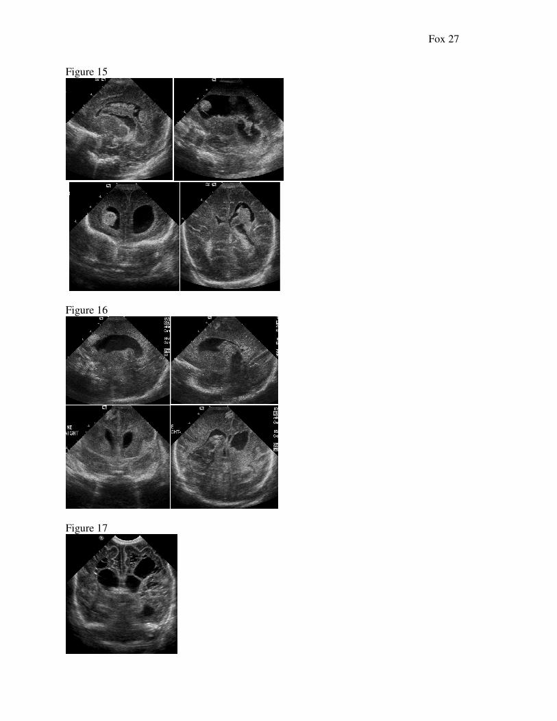
For a bleed to be considered a Grade III, ventricular dilatation will be present in addition
to intraventricular blood (Figure 15). Whereas Grade I and II bleeds are considered to have no
long lasting impact on neurological outcome, Grade III bleeds have a significantly higher
Fox 9
mortality rate and higher neurological impact due to hydrocephalus and increased pressure on the
brain tissue (11).
The so-called Grade IV bleed is an intraparenchymal hemorrhage (IPH) that may or may
not be associated with an intraventricular bleed. It used to be thought that Grade IV bleeds were
a progression from Grade III bleeds, but the current literature recognizes that IPH may occur on
its own and is of a different etiology compared to IVH (10). Grade IV bleeds commonly appear
as an echogenic mass (or masses) in the frontal or parietal lobes, as seen in Figure 16. One of the
chief concerns with the Grade IV bleed is the loss of brain tissue that results from degeneration
of the resulting necrotic area. Regardless of the cause, the neonate with a Grade IV bleed is at
very high risk for adverse neurological outcome (11). As the blood is resorbed, the necrotic brain
matter may connect to the ventricles. The resultant porencephalic cyst is lost brain matter and
carries with it a very poor neurologic outcome (Figure 17) (12).
Hydrocephalus/Ventriculomegaly
Ventriculomegaly, or dilatation of the ventricles, has several etiologies. It may be
associated with congenital anomalies, associated with either increased production or decreased
absorption of CSF, or seen in conjunction with the sequelae of intracranial hemorrhage.
Clinically, hydrocephalus may present with a prominent (“bulging”) anterior fontanel, rapid head
circumference growth and separated cranial sutures (13). In neonates who survive to infancy,
hydrocephalus is the most common congenital malformation. Although the terms
“hydrocephalus” and “ventriculomegaly” are often used interchangeably, the term
“hydrocephalus” is used in by some authors when there is an obstructive cause for the dilatation
of the ventricles (4) or there is increased pressure (14).
Fox 10
Hydrocephalus can be divided into communicating and non-communicating. Non-
communicating hydrocephalus is when the dilatation is the result of blockage from within the
ventricular system, and communicating hydrocephalus is the result of either decreased absorption
or blockage from outside the ventricular system (4). Neonatal infection, intracranial masses and
abnormal vascular processes are other possible causes of hydrocephalus. Hydrocephalus can be
associated with poor outcome, depending on the etiology. Multiple studies have demonstrated
normal mentation occurring anywhere between 15-90% of neonates (15),(16),(17),(18).
After intracranial hemorrhage, atrophy of the brain tissue may cause post-hemorrhagic
ventricular dilatation (hydrocephalus ex vacuo) (14). It is important to note that post-hemorrhage
hydrocephalus does not show clinical symptoms right away, and it can take one to three weeks
after the hemorrhagic event for clinical signs to manifest. For this reason serial ultrasound is
important in the diagnosis of hemorrhage-associated hydrocephalus (19). Ventriculomegaly is
diagnosed by measurement of the frontal horns at the level of the third ventricle. At Thomas
Jefferson University Hospital, a measurement of ≥ 4mm is considered “dilated.” Dilatation of the
third ventricle may reveal a tissue bridge between the two lobes of the thalamus called the massa
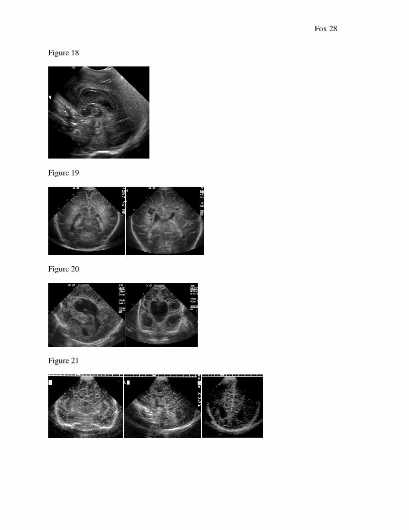
intermedia, not to be confused with intraventricular clot (Figure 18).
Periventricular Leukomalacia
In cases of hypoxia, necrosis of the periventricular white matter may occur, termed
periventricular leukomalacia (PVL). Sonographically, PVL initially appears as an area of highly
echogenic tissue in the parietal lobe adjacent to the lateral ventricles or in the frontal lobes
(Figure 19). Diagnosis relies on the echogenicity of the periventricular brain; if the parenchyma
adjacent to the lateral ventricles appears more echogenic than the choroid plexus, PVL must be
Fox 11
considered (4).The most significant sequela of PVL is cystic change of the affected brain matter
resulting in a “Swiss-cheese” like appearance of the parenchyma. This cystic change may occur
days to weeks after the initial insult. Severe PVL may eventually evolve into cystic
encephalomalacia and porencephaly (Figure 20), which are may lead to cognitive and seizure
disorders (19).
Congenital Anomalies of the Neonatal Brain
A variety of congenital anomalies of varying severity may occur in the brain. These
anomalies may range from mild, with no adverse neurological outcomes (such as a lobulated
choroid plexus), to severe, including hydranencephaly and alobar holoprosencephaly. While
there are many texts on neurosonography detailing all of the potential intracranial anomalies, this
article will highlight the more commonly seen pathologies.
Abnormal brain structure may be seen as the sequela of a spinal condition such as
myelomeningocele and other open neural tube defects. With open spinal defects, the spinal cord
is displaced through a defect, typically located at the base of the spine. This downward
displacement of the spinal cord pulls the brain through the only available opening, the foramen
magnum at the base of the skull. The cerebellum and brainstem are pulled downward,
obliterating the cisterna magna and causing obstruction of the cerebrospinal fluid flow. While the
frontal horns appear small, the posterior horns of the lateral ventricles are typically enlarged and
tear-dropped shaped, an appearance termed colpocephaly (20). There are several midline brain
defects that may visualized in neonatal brain imaging. One type that occurs is called agenesis of
the corpus callosum (ACC). In embryologic development, the anterior corpus callosum develops
first, followed by the posterior components (1). Agenesis of the corpus callosum may be partial
Fox 12
or complete, but if partial typically involves absence of the posterior portion related to the later
development of this portion of the corpus callosum. It is important to note if there are other
associated defects or if the ACC is an isolated event. Postnatal MRI is usually performed to
confirm the type of ACC, whether partial or complete, and to look for other defects as well (1).
In the case of isolated ACC there is usually a good prognosis, although affected children have
been demonstrated to have seizures or developmental delay (21). The sonographic features that
suggest ACC include a high-riding third ventricle and failure to visualize the corpus callosum in
its usual location. As demonstrated in Figure 21, the frontal horns appear to be markedly
separated, and the lateral ventricles run parallel to each other. The occipital horns may appear
tear-drop shaped (colpocephaly), and the sulci/gyri run perpendicular to the third ventricle
instead of parallel to it, causing the so-called “sunburst” sign (20).
One of the most dramatic defects of the midline that can occur is holoprosencephaly.
Holprosencephaly is divided into three types (in decreasing order of severity): alobar, semilobar
and lobar. With alobar holoprosencephaly there are severe intracranial, facial and midline
defects. Alobar holoprosencephaly, which is almost uniformly lethal even without chromosomal
abnormalities, is also often associated with lethal genetic disorders such as trisomy 13, trisomy
18 and triploidy (22). The striking intracranial findings of alobar holoprosencephaly typically
include a dilated single, central monoventricle; a thin, peripherally located cerebral cortex; fused
thalami and absent falx cerebri. Facial features often include a orbit anomalies, varying from a
more mild hypotelorism to a single orbit containing one or two eyeballs; a superiorly-located
proboscis and severe cleft lip/palate defects (23).
Semilobar holoprosencephaly is less severe than the alobar form, but shares some of the
midline characteristics. With semilobar, the thalami are only partially fused and a partial falx
Fox 13
may be seen. There may be mild facial abnormalities or there may be none at all. Lobar
holoprosencephaly, the mildest form, may present as ACC and hypoplasia of the optic nerve.
With lobar holoprosencephaly, the falx cerebri is complete or nearly complete, and there are two
cerebral hemispheres (23). The prognosis is better for the lobar form than with the other forms of
holoprosencephaly, although there may still be severe mental retardation. Facial anomalies, if
present, are usually mild (1).
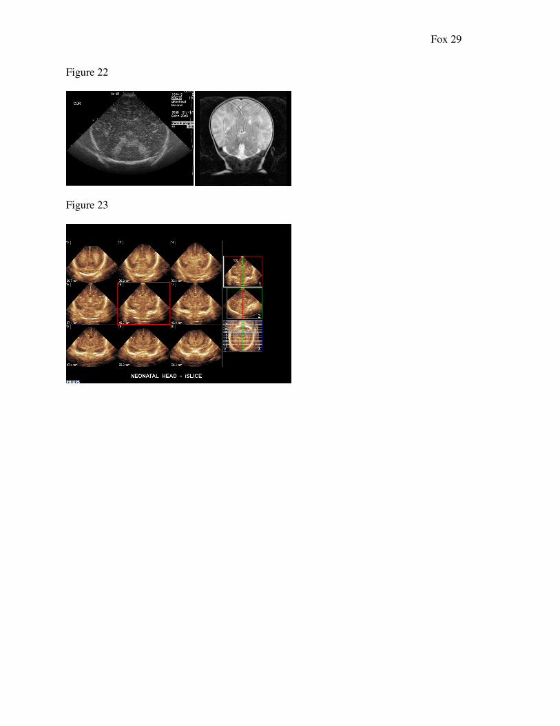
Anomalies of the posterior brain, such as Dandy Walker and Dandy Walker variant are
other brain anomalies that may be seen with neonatal intracranial ultrasound. The Dandy Walker
complex is visualized as a posterior fossa cyst that communicates with the fourth ventricle. The
cerebellar vermis is either hypoplastic or absent, and there may be secondary third or lateral
ventricle dilatation caused by atresia of the foramina of Luschka and Magendie. With Dandy
Walker variant, a normal or hypoplastic cerebellar vermis is present, and instead of a cyst there is
an enlarged cisterna magna that communicates with the fourth ventricle. Either condition may be
associated with other structural defects and/or chromosomal anomalies (Figure 22) (24).
Neonatal Infection
Transmission of maternal infection to the neonate is another condition encountered by the
neonatal sonographer, and a serious concern for the patient. Infectious diseases that may be
transmitted via the placental or birth canal are toxoplasmosis, rubella, cytomegalovirus (CMV),
and herpes, the so-called TORCH infections. The hallmark of intracranial infection is
calcification within the brain of the affected neonate, although cystic encephalomalacia may also
be seen, resulting in neurodevelopmental delay (25).
Fox 14
Three-Dimensional Ultrasound
With advances in three-dimensional US (3DUS), it is now possible to acquire a volume
set of images and visualize the brain in multiple planes via digital reformatting. With 3DUS,
there is a decrease in the sonographer acquisition time compared to 2D imaging, although the
time it takes for the physician to interpret the images increases due to the potential multiple
planes that can be obtained. Figure 23 demonstrates 2D slices of the neonatal brain obtained with
volume imaging. After capturing the volume it is sliced into axial, coronal and/or sagittal planes
as needed. The decrease in acquisition time with 3DUS is important to consider due to the
fragile health of the preterm neonate. Very low-birthweight neonates are less tolerant of being
touched, and there is the added risk that tubes or lines may be displaced with increased scanning
time. Another advantage of 3DUS is that it removes the interoperator variability with obtaining
the images, as well as decreases the chance that pathology may be missed. The chief downside
with modern 3DUS is that the 3D reconstructions are often of less quality then the standard 2D
images. With improvements in computer reconstruction, however, it is possible that 3DUS will
reign superior to 2D imaging for imaging of the neonatal brain (26). As with all 3D imaging,
though, a good 3D study is not possible without a good 2D image. If there is an inadequate
window or much artifact, it will not be likely that an acceptable 3D can be performed.
As the quality of the reconstructions of 3DUS advances, it makes it easier to compare US
images with comparable CT and MRI views. Three-dimensional US also has the added benefit of
educational and training opportunities, as any study can be “virtually rescanned” at a work
station long after the scan is completed (27).
Doppler in Neurosonology
Fox 15
As with most areas of ultrasound, Doppler may be used to evaluate for flow patterns and
velocities. Doppler, although not used routinely in many centers, may be used to evaluate the
perfusion to the brain via the anterior cerebral artery (ACA) and middle cerebral artery (MCA).
Velocity information is used in conjunction with resistive indices (RI), as a change in the RI may
be a predictor for prolonged asphyxia with ICH and/or cerebral edema (28). The resistive index,
which is a marker of the impediment to flow of a vessel bed, is susceptible to extracranial
influences such as a patent ductus arteriosis (PDA) and therefore may vary. At least one author
has developed charts for normal resistive indices of the MCA, ACA and internal carotid arteries
(29), and another author is using RI information along with graded compression of the anterior
fontanel to predict which neonates would benefit from shunting in hydrocephalus (30).
Conclusion
Unlike other areas of sonography (e.g., abdomen and ob/gyn), neurosonography is not a
common study in most non-pediatric institutions. Many sonographers learn the topic in school
and then work in a hospital or outpatient center that doesn’t have a neonatal unit. When the time
does come to perform these exams, it is often learned or re-learned via on the job training. As
technology and technique are constantly changing, all areas of ultrasound require continuous
education and training; it truly is a field in which we are all students regardless of the number of
years behind us. Neurosonography is an important tool in the diagnosis of intracranial problems
in the newborn, and one without the problems of sedation or ionizing radiation. The use of
neurosonography allows for the prediction of neurological development and outcome in this
high-risk population, and it is critical for the sonographer to stay up to date with this ever-
changing field.
Fox 16
Captions
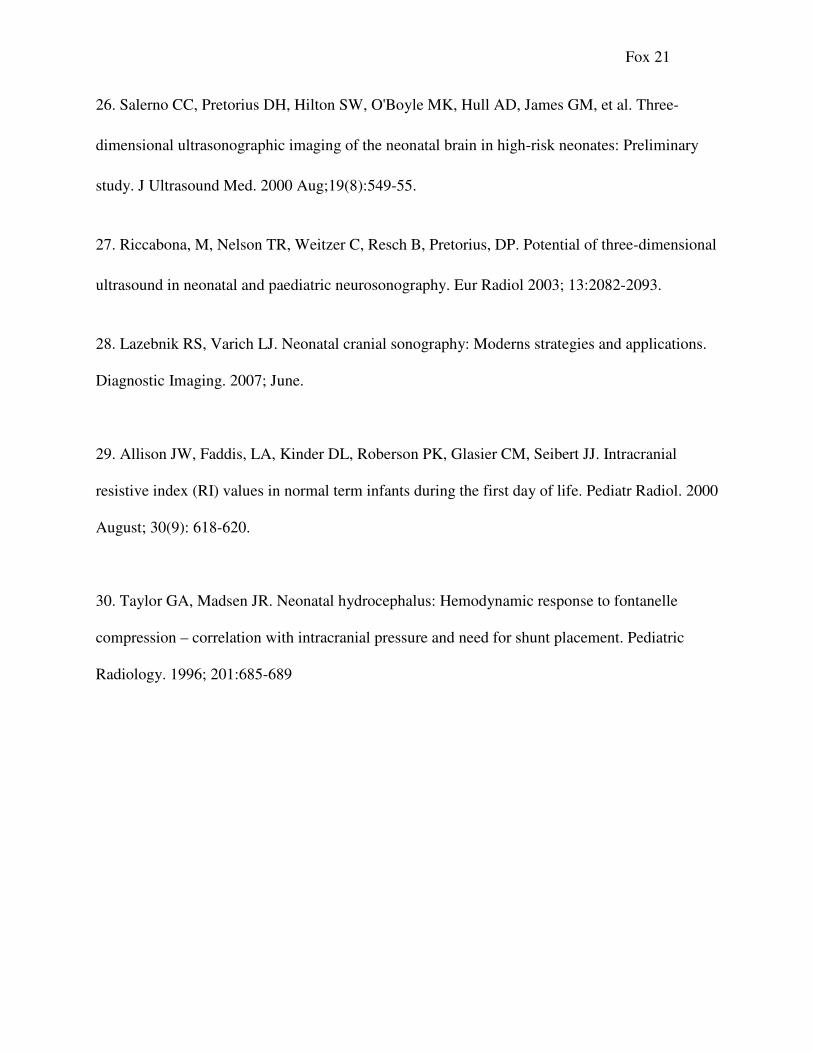
Figure 1 – Long-axis view of the brain showing the four lobes of the cerebrum.
Figure 2 – True sagittal midline.
Figure 3 – Midline structures of the neonatal brain. Fat white arrows: corpus callosum. Thin
white arrow: third ventricle. Black arrow: thalamus. Chevrons: vermis of the cerebellum.
Arrow head: fourth ventricle.
Figure 4 – (a) Midline brain showing the echogenic structures representing the choroid in the
third ventricle and cerebellar vermis. (b) and (c) showing the “lady in the dress” sign.
Demonstration of the corpus callosum and the “lady in the dress” ensures a completely true-
sagittal midline view.
Figure 5 – Para-sagittal view of the caudo-thalamic notch. The choroid plexus tapers in this
region and should not increase in size anteriorly.
Figure 6 – (a) neonatal brain at 26 weeks. Note the lack of defined sulci/gyri. (b) neonatal brain
at 37 weeks gestational age. Note the pronounced sulci/gyri seen with maturity.
Figure 7 – (a) posterior structures are being cut off, as evidenced by lack of visualization of the
inferior cranial bones. In image (b), the cranial bone is seen in its entirety.
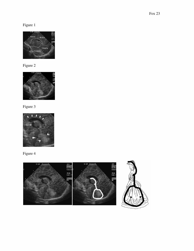
Figure 8 (a - f) – Progressive sagittal sections through the neonatal brain. This is the minimum
that needs to be documented in a normal brain.
Figure 9 (a - f) – Progressive coronal sections through the neonatal brain. This is the minimum
that needs to be documented in a normal brain.
Figure 10 – coronal, magnified view of measurement of the frontal horns of the lateral ventricles.
The frontal horns are measured at the level of the choroid plexus as it resides in the 3rd
ventricle
(arrow).
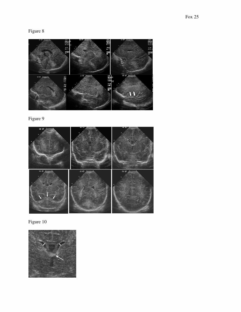
Figure 11 – a) Illustration of the transducer oriented in sagittal plane for obtaining midline
image. b) A slight rotation of the transducer from the midline is required in order to obtain a
caudo-thalamic notch view
Figure 12 – Occipital horn as seen through the posterior fontanel.
Figure 13 (a – c) – Grade I hemorrhage, or sub-ependymal hemorrhage. Notice that the bleed is
confined to the CTN and that the choroid does not taper normally as it travels anteriorly.
Figure 14 (a – c) – Grade II hemorrhage. Note clot extension into occipital horn (arrow).
Fox 17
Figure 15 (a – c) – Grade III hemorrhage. There is ventricular dilatation and clot throughout
ventricular system.
Figure 16 (a – d) – Grade IV, or intraparenchymal hemorrhage. Note the scattered echogenic
areas consistent with parenchymal blood. There is also significant intraventricular blood, as well.
Figure 17 – Grade IV hemorrhage with porencephalic cysts bilaterally. There is an increased
amount of extraaxial fluid, as well, consistent with atrophy of the brain.
Figure 18 – Dilatation of the third ventricle revealing the massa intermedia, the tissue bridge
between the two lobes of the thalamus.
Figure 19 – (a) early PVL. Note the areas of increased echogenicity adjacent to the lateral
ventricles. (b) Several weeks later, cystic change has occurred in the parenchyma.
Figure 20 – severe cystic encephalomalacia, related to early prematurity and PVL.
Figure 21 – Agenesis of the corpus callosum. (a) Widely spaced anterior horns (b) Absence of
normal midline structures. The corpus callosum and cavum septum pellucidum are absent. (c)
Tear-dropped shaped occipital horns (colpocephaly)
Figure 22 – (a) coronal ultrasound of Dandy-Walker variant. Note the splayed cerebellar tonsils
and absence of the echogenic central vermis. (b) Coronal magnetic resonance (MRI) scan of the
same patient.
Figure 23 – Three-dimensional volume imaging of the neonatal brain. (Images courtesy of
Philips Healthcare, Bothell, WA).
Fox 18
References
1. Rumack CM, Drose JA. Neonatal and infant brain imaging. In: Rumack CM, Wilson SR,
Johnson JA, Charboneau JW, editors. Diagnostic Ultrasound. 3rd ed. St. Louis: Elsevier Mosby;
2005. p. 1623.
2. Merton DA, Bega G, Goldberg BB. Multiplanar 3-dimensional neonatal neurosonography:
Initial experiences and potential benefits. Journal of Diagnostic Medical Sonography. 2001 [cited
9/14/2008];17(1):3.
3. Raymond HW, Zwiebel WJ. Neurosonography. Seminars in ultrasound. 1982;3(3).
4. Benson JE, Bishop MR, Cohen HL. Intracranial neonatal neurosonography: An update.
Ultrasound Quarterly. 2002;18(2):89.
5. Timor-Tritsch IE., Monteagudo A, Cohen HL. Ultrasonography of the prenatal and postnatal
brain. 2nd
ed. New York: McGraw-Hill; 2001.
6. Curtis LT. Prevention of hospital-acquired infections: Review of non-pharmacological
interventions. J Hosp Infect. 2008 Jul [cited 9/14/2008];69(3):204-19.
7. Thomson, GD, Teele, RL. High-frequency linear array transducers for neonatal cerebral
sonography. AJR. 2001; 176:995-1001.
8. Di Salvo DN. A new view of the neonatal brain: Clinical utility of supplemental neurologic
US imaging windows. Radiographics. 2001 Jul-Aug [cited 9/14/2008];21(4):943-55.

Fox 19
9. Papile LA, Burstein J, Burstein R, Koffler H. Incidence and evolution of subependymal and
intraventricular hemorrhage: A study of infants with birth weights less than 1,500 gm. J Pediatr.
1978 Apr;92(4):529-34.
10. Volpe JJ. Intraventricular hemorrhage and brain injury in the premature infant.
neuropathology and pathogenesis. Clin Perinatol. 1989 Jun;16(2):361-86.
11. Raab EL. The resuscitation & care of the newborn at risk. In: DeCherney AH, Nathan L,
Goodwin TM, Laufer N, editors. Current diagnosis & treatment: Obstetrics & gynecology - 10th
Ed. (2007). 10th ed. New York: McGraw Hill; 2007.
12. Grant EG, Kerner M, Schellinger D, Borts FT, McCullough DC, Smith Y, et al. Evolution of
porencephalic cysts from intraparenchymal hemorrhage in neonates: Sonographic evidence. Am
J Roentgenol. 1982;138(3):467-70.
13. Panteliadis CP, Darras BT. Encyclopaedia of paediatric neurology: Theory and practice. 2nd
ed. Thessaloniki: Giahoudi - Giapouli; 1999.
14. Siegel MJ. Pediatric sonography. 3rd ed. Philadelphia: Lippincott Williams & Wilkins; 2001.
15. Rosseau GL, McCullough DC, Joseph AL. Current prognosis in fetal ventriculomegaly. J
Neurosurg. 1992 Oct;77(4):551-5.
16. Wyldes M, Watkinson M. Isolated mild fetal ventriculomegaly. Arch Dis Child.
2004;89(1):9-13.
Fox 20
17. Ment LR, Vohr B, Allan W, Westerveld M, Katz KH, Schneider KC, et al. The etiology and
outcome of cerebral ventriculomegaly at term in very low birth weight preterm infants.
Pediatrics. 1999;104(2):243-8.
18. Resch B, Gerdermann A, Maurer U, Ritschl E, Müller W. Neurodevelopmental outcome of
hydrocephalus following intra-/periventricular hemorrhage in preterm infants: Short-and long-
term results. Child's Nervous System. 1996;12(1):27-33.
19. Volpe JJ. Neonatal neurology. 4th ed. Phildelphia: Saunders; 2001.
20. Babcock DS. The normal, absent, and abnormal corpus callosum: Sonographic findings.
Radiology. 1984;151(2):449.
21. Penny SM. Agenesis of the corpus callosum: Neonatal sonographic detection. Radiol
Technol. 2006;78(1):14.
22. Dubourg C, Bendavid C, Pasquier L, Henry C, Odent S, David V. Holoprosencephaly.
Orphanet J Rare Dis. 2007;2(8).
23. Fitz CR. Holoprosencephaly and related entities. Neuroradiology. 1983;25(4):225-38.
24. Ecker JL, Shipp TD, Bromley B, Benacerraf B. The sonographic diagnosis of dandy-walker
and dandy-walker variant: Associated findings and outcomes. Prenat Diagn. 2000;20(4):328-32.
25. Grant EG, Williams AL, Schellinger D, Slovis TL. Intracranial calcification in the infant and
neonate: Evaluation by sonography and CT. Radiology. 1985 Oct;157(1):63-8.
Fox 21
26. Salerno CC, Pretorius DH, Hilton SW, O'Boyle MK, Hull AD, James GM, et al. Three-
dimensional ultrasonographic imaging of the neonatal brain in high-risk neonates: Preliminary
study. J Ultrasound Med. 2000 Aug;19(8):549-55.
27. Riccabona, M, Nelson TR, Weitzer C, Resch B, Pretorius, DP. Potential of three-dimensional
ultrasound in neonatal and paediatric neurosonography. Eur Radiol 2003; 13:2082-2093.
28. Lazebnik RS, Varich LJ. Neonatal cranial sonography: Moderns strategies and applications.
Diagnostic Imaging. 2007; June.
29. Allison JW, Faddis, LA, Kinder DL, Roberson PK, Glasier CM, Seibert JJ. Intracranial
resistive index (RI) values in normal term infants during the first day of life. Pediatr Radiol. 2000
August; 30(9): 618-620.
30. Taylor GA, Madsen JR. Neonatal hydrocephalus: Hemodynamic response to fontanelle
compression – correlation with intracranial pressure and need for shunt placement. Pediatric
Radiology. 1996; 201:685-689

Fox 22
Acknowledgements
I would like to thank Teresa Fonock and Philips Healthcare for image contribution.
Fox 23
Figure 1
Figure 2
Figure 3
Figure 4

Fox 24
Figure 5
Figure 6
Figure 7
Fox 25
Figure 8
Figure 9
Figure 10
Fox 26
Figure 11
Figure 12
Figure 13
Figure 14
Fox 27
Figure 15
Figure 16
Figure 17
Fox 28
Figure 18
Figure 19
Figure 20
Figure 21
Fox 29
Figure 22
Figure 23